Embed Size (px)
Citation preview

Strategic Supporting Partner
Pálmi V. Jónsson, MD, FACP, FRCP LChief of Geriatrics, Landspitali University Hospital,
Professor of Geriatrics, Faculty of Medicine, University of Iceland
Advances in Alzheimer´s Disease and Aging Research
2-4 Jumada II 1441 / 27-29 January 2020Conference Hall – KACST HQ
Riyadh, Saudi Arabia
Organized by
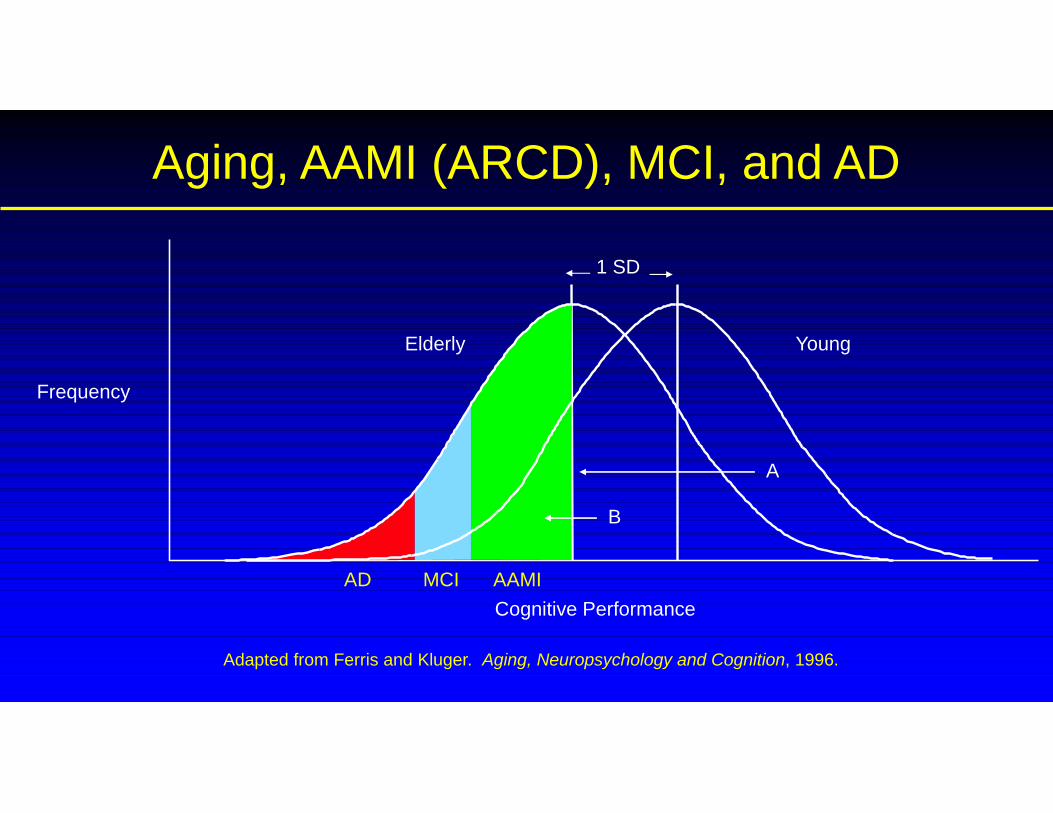
Aging, AAMI (ARCD), MCI, and AD
Cognitive Performance
Frequency
Young
Adapted from Ferris and Kluger. Aging, Neuropsychology and Cognition, 1996.
Elderly
1 SD
A
B
AD MCI AAMI

Brain aging vs. Disease
• How should we understand the fact that three of the major symptoms ofAD observed in vivo
• disruption of episodic memory function,• brain atrophy,• and accumulation of amyloid protein
are also found in many presumably healthy elderly?
• Given these commonalities, it can be argued that AD cannot beunderstood separately from its major risk factor – age.

Fig. 5 Life-span trajectories of volumetric reductions. Cross-sectional estimates of adult life-span trajectories of total cerebralcortex volume and total hippocampal volume. ...
Anders M. Fjell , Linda McEvoy , Dominic Holland , Anders M. Dale , Kristine B. Walhovd Progress in Neurobiology, 2014
Effects of aging, amyloid and Alzheimer's disease on the cerebral cortex and the hippocampusWhat is normal aging?

Fig. 8 Fronto-striatal vs. temporo-parietal networks. While normal aging affects a fronto-striatal network important for cognitivecontrol and executive function (green structures), AD has additional effects on a temporo-parietal network important for episodicmemory
Anders M. Fjell , Linda McEvoy , Dominic Holland , Anders M. Dale , Kristine B. Walhovd Progress in Neurobiology, 2014
Effects of aging, amyloid and Alzheimer's disease on the cerebral cortex and the hippocampusWhat is normal aging?

Fig. 9 Cortical expansion across primates and volume decline in aging. Upper panel shows regions of high vs. lower corticalexpansion from macaque monkeys to humans
Anders M. Fjell , Linda McEvoy , Dominic Holland , Anders M. Dale , Kristine B. Walhovd Progress in Neurobiology, 2014
Effects of aging, amyloid and Alzheimer's disease on the cerebral cortex and the hippocampus
What is normal aging?

DMN; Default Mode Network
•The Default Mode Network consists of a specific set of brain areas that decrease activityduring performance of a wide range of tasks, and that are typically also active duringperiods of rest or introspection.
•The different DMN structures are densely interconnected to each other and limbicstructures by polysynaptic connections, with few connections to sensory and motorareas.
•DMN maps onto a network of core brain areas involved in episodic memory andimagination and is therefore highly relevant for the understanding of episodic memoryproblems in aging.
•Structural and functional aspects of DMN are affected both in normal aging and AD.
•Interestingly, the default mode network (DMN) is almost completely encapsulated in thehigh declining regions.

• Aging itself is the major risk factor for sporadic AD.• Rather than necessarily reflecting early signs of disease, these changes may be
part of normal aging, and may inform on why the aging brain is so much moresusceptible to AD than is the younger brain.
• Regions characterized by a high degree of life-long plasticity are vulnerable todetrimental effects of normal aging, and that this age-vulnerability renders themmore susceptible to additional, pathological AD-related changes.
• It will be difficult to understand AD without understanding why it preferablyaffects older brains.
• A model that accounts for age-related changes in AD-vulnerable regionsindependently of AD-pathology is needed.
The Aging – AD pathology homology is critical to understand

Pálmi V. Jónsson, MD, FACP, FRCP L – co-PI and on the executive committeeChief of Geriatrics, Landspitali University Hospital,
Professor of Geriatrics, Faculty of Medicine, University of Iceland
Insights from the AGES Reykjavik Study on Aging of the Brain

A struggle for independence in Norway brought the first Vikings to Iceland in the ninth century,some 800 farmers, each with 25 persons. Nature's gifts supported a good life for the next 300years, and by the year 1200 the population is estimated to have been 78 000, close to themaximum that the farmland could sustain.
But life in Iceland between 1300 and 1900 was extraordinarily hard with repeated deadlyinfectious epidemics and hunger due to volcanic eruptions and hard weather. The populationhovered around 50 000 during these centuries but eventually crept back up, reaching 78 000 in1901, the same size it had been 700 years earlier.
Ann Intern Med 1998 Jun 1;128(11):941-5. Letter from Reykjavik. Jónsson PV1.
The AGES study
A continuum of the Reykjavik Study, a heart study established in 1967.• A collaborative project with National Institute on Aging• Participants aged between 67 and 95 years• Both sexes
• 5700 participants in AGES I, began in 2002• 3200 participants in AGES II, began in 2007• More than 1200 variables per person• Making use of available data and obtaining extensive phenotypes for
epidemiological and genetic studies of healthy aging• Each study participant contributed 3 half days to the study

COG 1; 45 minutesfor all participants of AGES
MMSE (Mini mental state examination) DSST (Digit symbol substitution test) Memory for a word list + source memory CANTAB (SWM: spatial working memory) computerized Figure comparison Stroop test for interference (colors, words) Digit span Delayed recall for the word list Assessment of time: how long have you been here Post-exam assessment
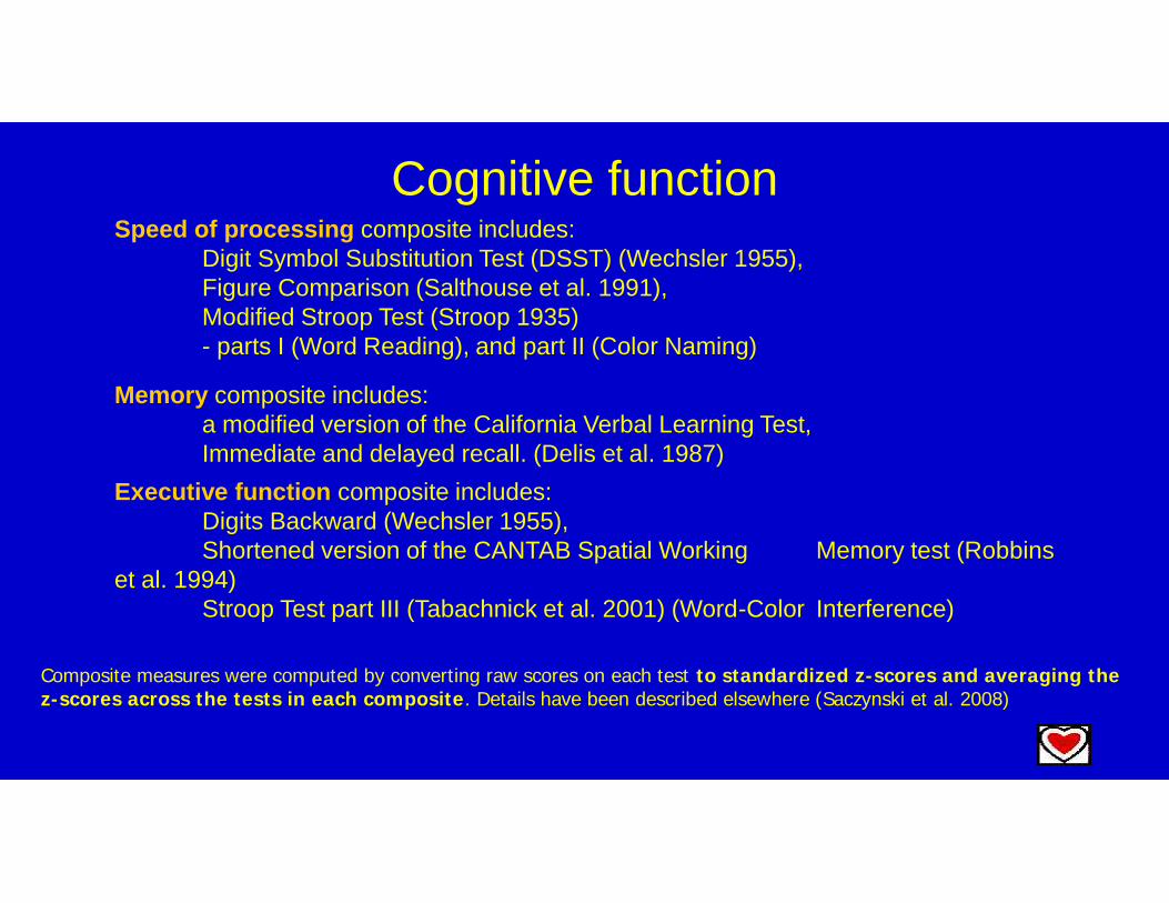
Cognitive function
Memory composite includes:a modified version of the California Verbal Learning Test,Immediate and delayed recall. (Delis et al. 1987)
Executive function composite includes:Digits Backward (Wechsler 1955),Shortened version of the CANTAB Spatial Working Memory test (Robbins
et al. 1994)Stroop Test part III (Tabachnick et al. 2001) (Word-Color Interference)
Speed of processing composite includes:Digit Symbol Substitution Test (DSST) (Wechsler 1955),Figure Comparison (Salthouse et al. 1991),Modified Stroop Test (Stroop 1935)- parts I (Word Reading), and part II (Color Naming)
Composite measures were computed by converting raw scores on each test to standardized z-scores and averaging thez-scores across the tests in each composite. Details have been described elsewhere (Saczynski et al. 2008)

Combined Curve for DSST and MMSE
• The “best” rule:DSST ≤ 17 OR MMSE ≤ 23
Sensitivity=0.92
Specificity=0.70
0.0 0.2 0.4 0.6 0.8 1.0
0.00.2
0.40.6
0.81.0
0.0 0.2 0.4 0.6 0.8 1.0
0.00.2
0.40.6
0.81.0
System 1System 2
False pos itive
True
posit
ive
Optimal Decision Curve
True posOver 90%False posUnder 40%

COG 2; 45 minutesdiagnostic tests for those who fail COG 1
Clock test Nonverbal memory: immediate recognition of faces shown on a computer Verbal memory: immediate memoyr for a word list Copying and immediate memory: Rey-Osterrieth, simplified Wordfluency: S-words and animals Token test: language comprehension Trails, 4 parts Naming of pictures (blcack and white drawings) Go/no-go test Delayed memory( faces, wordlist, picture) Post-exam assessment
ROC analysisFor AVLT andTrails determinesFlow to MC
MRI of the brain
• Semi-quantitative analysis
• Quantitative analysis

Spatial NormalisationSpatial Normalisation
Spatial Normalisation
Original image
Templateimage
Spatially normalised
Determine the spatial transformationthat minimises the sum of squareddifference between an image and alinear combination of one or moretemplates.
Begins with an affine registration tomatch the size and position of theimage.
Followed by a global non-linear warpingto match the overall brain shape.
Uses a Bayesian framework tosimultaneously maximise thesmoothness of the warps.
Deformation field

Automatic Segmentation of Four-Tissue Types
WMGM
WM Lesions
CSF
Raw (DICOM) Images Resultant SegmentationImage Processing
The goal of the image analysis pipeline is to generate automatic brain tissue segmentationof WM, WMH (WM Lesions), GM and CSF following the input of multi-spectral MRimages (from left to right and top to bottom): T1-, PD,- FLAIR and T2-weighted images.
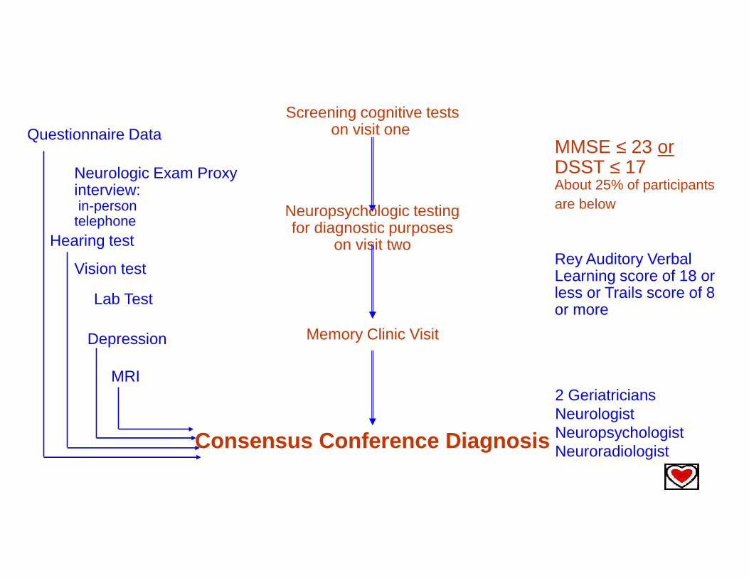
MMSE ≤ 23 orDSST ≤ 17About 25% of participantsare below
Consensus Conference Diagnosis
Rey Auditory VerbalLearning score of 18 orless or Trails score of 8or more
Neurologic Exam Proxyinterview:in-persontelephone
Memory Clinic Visit
Screening cognitive testson visit one
Neuropsychologic testingfor diagnostic purposes
on visit two
Questionnaire Data
Depression
MRI
Hearing test
Vision test
Lab Test
2 GeriatriciansNeurologistNeuropsychologistNeuroradiologist

Mid life to late life associations

High adiposity in midlife might increase risk for late-life brain pathology, including dementia.
In midlife, the prevalence of overweight was 39% and that of obesity was 8%.
After a mean follow-up of 26.2 (standard deviation, 4.9) years, midlife overweight and obesity were not associated with infarct-like brain lesions (relative risk (RR) = 0.82, 95% confidence interval (CI): 0.61, 1.10), cerebral microbleeds (RR = 0.69, 95% CI:0.37, 1.32), total brain volume (β = 0.05, 95% CI: −0.34, 0.45), white matter lesions volume (β = −0.10, 95% CI: −0.20, 0.01), ordementia (RR = 0.91, 95% CI: 0.49, 1.72) compared with normal weight.
These findings do not support the hypothesis that high body mass index inmidlife modulates the risk for dementia.
et. al.

J Gerontol A Biol Sci Med Sci, 2010 Dec;65(12):1369-74. Chang M et. al.
The effect of midlife physical activity on cognitive function among older adults:AGES--Reykjavik Study.
RESULTS:Among the participants, no midlife PA was reported by 68.8%, ≤ 5 hours PA by 26.5%, and >5 hours PA by 4.5%.
Excluding participants with dementia compared with the no PA group, PA groups had significantly faster speed ofprocessing , better memory and executive function after controlling for demographic and cardiovascular factors.
The ≤ 5 hours PA group was significantly less likely to have dementia in late life (odds ratio: 0.6, 95% confidenceinterval: 0.40-0.88) after adjusting for confounders.
CONCLUSION:
Midlife PA may contribute to maintenance of cognitive function and may reduce or delay the riskof late-life dementia.

Imaging Results

?
AGES I
AGES II

AGES I
AGES II

Neuroimage 2012 Feb 15;59(4):3862-70. Sigurdsson S et. Al.
Brain tis sue volumes in the general population of the elderly:the AGE S -R eykjavik s tudy.
A reduction in both normal white matter (NWM)- and gray matter (GM) volumecontributed to the brain shrinkage.
After adjusting for intra-cranial volume, women had larger brain volumescompared to men (3.32%, p < 0.001)
The longitudinal analysis showed a greater rate of annual change in totalbrain volume for men (-0.70%, 95%CI: -0.78% to -0.63%)than women (-0.55%, 95%CI: -0.61% to -0.49%).


We investigated the combined effect of small birth size and mid-life cardiovascular risk on late-lifebrain volumes.
Compared with the reference group (high Ponderal Index/absence of mid-life CVRF), participants with lower Ponderalindex/presence of mid-life CVRF (body mass index >25 kg/m2, hypertension, diabetes, “ever smokers”) had smaller total brainvolume later in life;
The results sugges t that exposure to an unfavorable intrauterine environmentc ontributes to smaller brain volume in old age, but added to that is atrophy whic his as soc iated with mid-life c ardiovasc ular ris k.
L ate-life brain volume: a life-c ourse approac h.The AGE S -R eykjavik s tudy
Neurobiol Aging 2016 May;41:86-92. Muller M et. al.

Diabetic Care: 2009 Sep;32(9):1608-13. Sacynzi JS et. al.
Glyc emic s tatus and brain injury in older individuals :the age gene/environment sus c eptibility-R eykjavik s tudy.
C ONC L US IONS :
•Type 2 diabetic participants have more pronounc ed brain atrophyand are more likely to have cerebral infarc ts .
•Duration of type 2 diabetes is associated with brain changes, suggestingthat type 2 diabetes has a cumulative effec t on the brain.

Ann Neurol. 2009 Oct;66(4):485-93. Palm WM et. al.
Ventric ular dilation: as soc iation with gait and c ognition.
RE S ULT S :Disproportion between ventricular and sulcal CSF volume, defined as the highest quartile of the VV/SV z score, was associated with gait impairment (odds ratio[OR], 1.9; 95% confidence interval [CI], 1.1-3.3) and cognitive impairment (OR , 1.8; 95% CI, 1.1-3.0). We did not find an association between the VV/SV z scoreand bladder dysfunction.
INTE R PR E TAT ION:The prevalenc e and severity of gait impairment and cognitive
impairment inc reases with ventric ular dilationin persons without stroke from the general population,
independent of White Matter Lesion volume.

Stroke. 2009 Mar;40(3):677-82. Saczynski JS et. al.
Cerebral infarc ts and c ognitive performanc e:importanc e of loc ation and number of infarc ts .
CONC L US IONS :
Having infarcts in >1 location is associated with
poor performance in memory, processing speed, and executive function,independent of cardiovascular comorbidities,white matter lesions, and brain atrophy,
suggesting that both the number and the distribution of infarcts jointly contributeto cognitive impairment.

et.al.
Background and Purpose—The differentiation of brain infarcts by region is important because their cause and clinical implicationsmay differ. Information on the incidence of these lesions and association with cognition and dementia from longitudinal populationstudies is scarce.
Conclusions:•Men are at greater risk of developing incident brain infarcts than women.
•Persons with incident brain infarcts decline faster in cognition and have anincreased risk of dementia compared with those free of infarcts.
•Incident subcortical infarcts contribute more than cortical and cerebellarinfarcts to incident dementia which may indicate that infarcts of small vesseldisease origin contribute more to the development of dementia than infarctsof embolic origin in larger vessels. Stroke. 2017;48:2353-2360
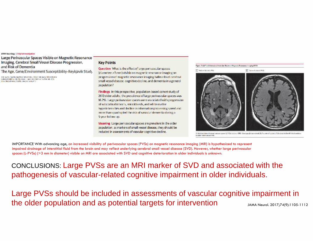
IMPORTANCE With advancing age, an increased visibility of perivascular spaces (PVSs) on magnetic resonance imaging (MRI) is hypothesized to representimpaired drainage of interstitial fluid from the brain and may reflect underlying cerebral small vessel disease (SVD). However, whether large perivascularspaces (L-PVSs) (>3 mm in diameter) visible on MRI are associated with SVD and cognitive deterioration in older individuals is unknown.
CONCLUSIONS: Large PVSs are an MRI marker of SVD and associated with thepathogenesis of vascular-related cognitive impairment in older individuals.
Large PVSs should be included in assessments of vascular cognitive impairment inthe older population and as potential targets for intervention JAMA Neurol. 2017;74(9):1105-1112

Microvasclar disease and aging

J Neurolo Neurosurg Psychiatry. 2008 Sep;79(9):1002-6. Sveinbjörnsdóttir et. al.
Cerebral mic robleeds in the population based AGE S -R eykjavik s tudy: prevalenc eand loc ation.
R E S ULTS :Evidence of CMBs was found in 218 participants (11.1% (95% CI 9.8% to 12.6%));
Men had significantly more CMBs than women (14.4% vs 8.8%; p = 0.0002, age adjusted). The prevalence of CMBsincreased with age (p = 0.0001) in both men (p = 0.006) and women (p = 0.007).
Having a CMB was significantly associated with a homozygote Apo E epsilon4epsilon4 genotype (p = 0.01).C ONC L US ION:
•Cerebral microbleeds are common in older persons.
•The association with homozygote ApoE4 genotype and finding arelative predominance in the parietal lobes might indicate anassociation with amyloid angiopathy.

Cerebral microbleeds (CMBs), as seen on T2* weighted gradient echo type echo planar (GRE-EPI) MRI.
Sveinbjornsdottir S et al. J Neurol Neurosurg Psychiatry2008;79:1002-1006

JAMA Neurol 2015 Jun;72(6):682-8. Ding J et. al.
Risk Fac tors Assoc iated With Inc ident Cerebral Mic robleeds Ac c ording to L oc ation in Older People:The Age, Gene/E nvironment S us c eptibility (AGE S )-R eykjavik S tudy.
The spatial distribution of cerebral microbleeds (CMBs), which are asymptomatic precursors of intracerebralhemorrhage, reflects specific underlying microvascular abnormalities of cerebral amyloid angiopathy (lobar structures)and hypertensive vasculopathy (deep brain structures).
C ONC L US IONS :
Lifestyle and lipid risk profiles for CMBs were similar tothose for symptomatic intracerebral hemorrhage.
Modification of these risk factors could have the potential toprevent new-onset CMBs.
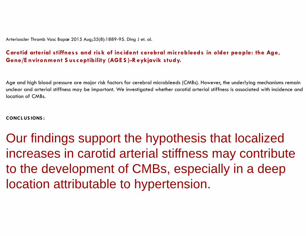
Arterioscler Thromb Vasc Bopæ 2015 Aug;35(8):1889-95. Ding J et. al.
C arotid arterial s tiffness and ris k of inc ident cerebral mic robleeds in older people: the Age,Gene/E nvironment S us c eptibility (AGE S )-R eykjavik study.
Age and high blood pressure are major risk factors for cerebral microbleeds (CMBs). However, the underlying mechanisms remainunclear and arterial stiffness may be important. We investigated whether carotid arterial stiffness is associated with incidence andlocation of CMBs.
CONC L US IONS :
Our findings support the hypothesis that localizedincreases in carotid arterial stiffness may contributeto the development of CMBs, especially in a deeplocation attributable to hypertension.

Aortic stiffness is associated with cognitive decline.
Here, we examined the association between carotid-femoral pulse wave velocity and cognitive function and investigated whethercerebrovascular remodeling and parenchymal small vessel disease damage mediate the relation.
The results suggest that in older people, associations between aorticstiffness and memory are mediated by pathways that include cerebralmicrovascular remodeling and microvascular parenchymal damage.
Hypertension 2016 Jan;67(1):176-82. Cooper LL et. al.
Cerebrovasc ular Damage Mediates R elations Between Aortic S tiffnes s andMemory.

Important papers on genetics from IcelandResearch by the DECODE company

Whole Genome Sequencing by DeCode- A revolution
Purpose: The whole-genome sequencing; WGS); of up to 3700 Icelanders
Allows genetic variance to be detected with >0.1% prevalence
With the use of genealogy of the Icelandic people and so called long-range phasing ofhaplotypes (bit´s of the genome) one can impute the genomic sequence of a largeproportion of the nation
With this method one can find:
Common genetic variance which has not been identified with other methods Rare genetic variance associated with great increase in risk
Has already shown results for ovarian cancer, glioma, sick sinus syndrome and gout Sulem e t al. Nat Genet 2011 Oct 9 Epub, Rafnar e t al. Nat Genet 2011 Oct 2 Epub, Stacey e t al. Nat Genet 2011 Sept 25 Epub, Holm e t al. Nat Genet 43(4):316-20 (2011)

Identification of a protective variant in APP
42
Nature. 2012 Aug 2;488(7409):96-9

A673T ( instead of V ) promotes intact cognition in the elderly without AD
∆ = -1.03P = 0.0021
A673T carriers (N=42; in red)A673T non-carriers (N=3,673; in blue)AD cases not included

• A673T protects significantly against AD
• Promotes healthy cognition in the elderly in the absence of AD
• in vitro studies show that A673T alters APP processing:
• Less cleavage by β-secretase (BACE1)• More cleavage by α-secretase• Less cleavage by recombinant BACE1 in cleavage assays
A673T in APP - Summary
44

Coding variant in TREM2 confers risk of AD

rs75932628-T is also predictive of loss of cognitive functionin the elderly in the absence of AD
rs75932628-T carriers
rs75932628-T non-carriers

rs75932628-T lowers age at onset of AD
• Each copy of rs75932628-T lowers age at onset of AD by 3.18 years in Iceland(P = 0.20)
• Comparable to the effect of ApoE ε4 (3.22 years)
• Similar result obtained in Dutch data (3.65 years/allele of rs75932628-T)
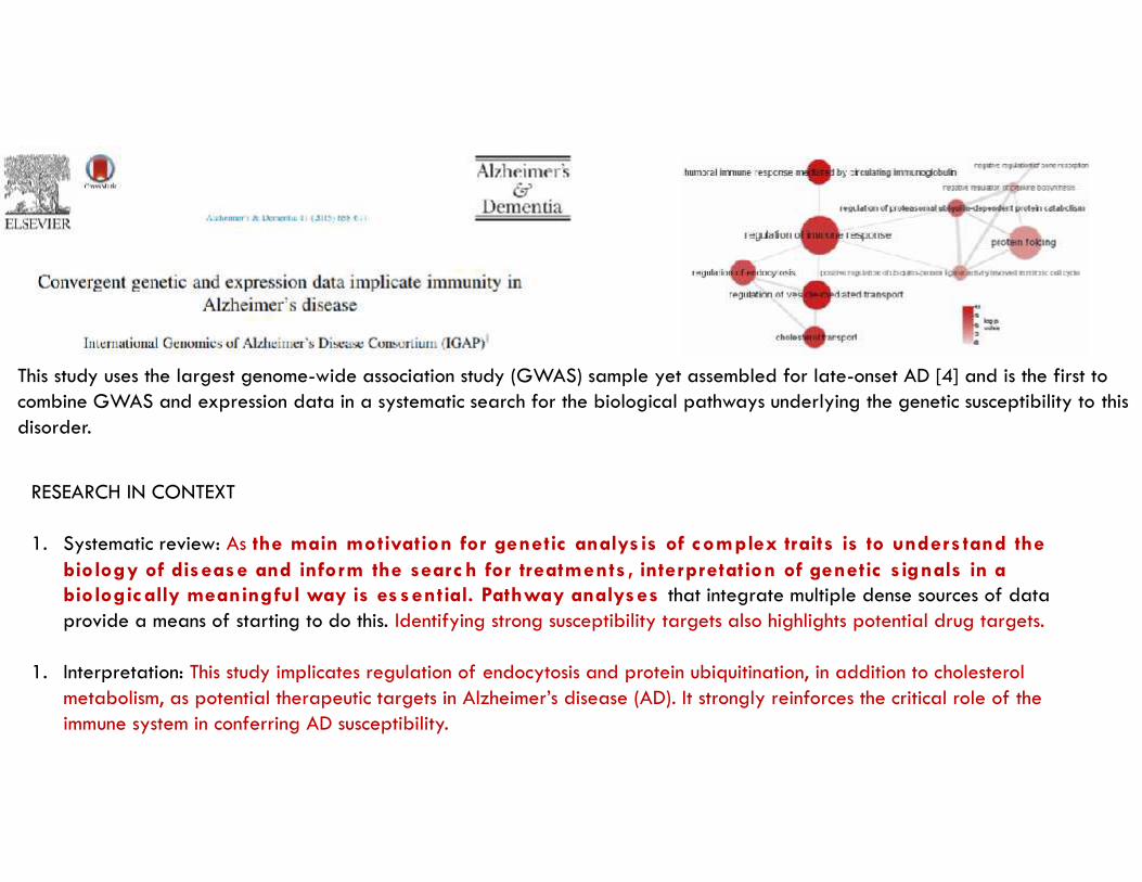
This study uses the largest genome-wide association study (GWAS) sample yet assembled for late-onset AD [4] and is the first tocombine GWAS and expression data in a systematic search for the biological pathways underlying the genetic susceptibility to thisdisorder.
RESEARCH IN CONTEXT
1. Systematic review: As the main motivation for genetic analys is of complex traits is to unders tand thebiology of disease and inform the searc h for treatments , interpretation of genetic signals in abiologic ally meaningful way is es sential. Pathway analyses that integrate multiple dense sources of dataprovide a means of starting to do this. Identifying strong susceptibility targets also highlights potential drug targets.
1. Interpretation: This study implicates regulation of endocytosis and protein ubiquitination, in addition to cholesterolmetabolism, as potential therapeutic targets in Alzheimer’s disease (AD). It strongly reinforces the critical role of theimmune system in conferring AD susceptibility.

Iris E. Jansen et. al. Nature Genetics volume 51, 2019,pages404–413(2019)
Alzheimer’s disease (AD) is highly heritable and recent studies have identified over 20 disease-associated genomic loci. Yet these only explain a small proportion of the genetic variance, indicating thatundiscovered loci remain.
Here, we performed a large genome-wide association study of clinically diagnosed AD and AD-by-proxy(71,880 cases, 383,378 controls). AD-by-proxy, based on parental diagnoses, showed strong geneticcorrelation with AD (rg = 0.81).
Meta-analysis identified 29 risk loci, implicating 215 potential causative genes.
Associated genes are strongly expressed in immune-related tissues and cell types (spleen, liver, andmicroglia).Gene-set analyses indicate biological mechanisms involved in lipid-related processes and degradation ofamyloid precursor proteins.
We show strong genetic correlations with multiple health-related outcomes, and Mendelian randomizationresults suggest a protective effect of cognitive ability on AD risk.
Genome-wide meta-analysis identifies new loci and functional pathways influencing Alzheimer’s disease risk


![Enfermedad de Alzheimer - COC3000 · La enfermedad de Alzheimer (EA), también denominada mal de Alzheimer, o demencia senil de tipo Alzheimer (DSTA) o simplemente alzhéimer[10]](https://img.dokumen.tips/doc/110x75/5f0f53777e708231d4439b0b/enfermedad-de-alzheimer-coc3000-la-enfermedad-de-alzheimer-ea-tambin-denominada.jpg)