Embed Size (px)
Citation preview
Advances and challenges inbiosensor-based diagnosis ofinfectious diseasesExpert Rev. Mol. Diagn. Early online, 1–20 (2014)
Mandy LY Sin1,2,Kathleen E Mach1,2,Pak Kin Wong3 andJoseph C Liao*1,2
1Department of Urology, Stanford
University School of Medicine, Stanford,
CA 94305-5118, USA2Veterans Affairs Palo Alto Health Care
System, Palo Alto, CA 94304, USA3Department of Aerospace and
Mechanical Engineering, University of
Arizona, Tucson, AZ 85721, USA
*Author for correspondence:
Rapid diagnosis of infectious diseases and timely initiation of appropriate treatment are criticaldeterminants that promote optimal clinical outcomes and general public health. Conventionalin vitro diagnostics for infectious diseases are time-consuming and require centralizedlaboratories, experienced personnel and bulky equipment. Recent advances in biosensortechnologies have potential to deliver point-of-care diagnostics that match or surpassconventional standards in regards to time, accuracy and cost. Broadly classified as eitherlabel-free or labeled, modern biosensors exploit micro- and nanofabrication technologies anddiverse sensing strategies including optical, electrical and mechanical transducers. Despiteclinical need, translation of biosensors from research laboratories to clinical applications hasremained limited to a few notable examples, such as the glucose sensor. Challenges to beovercome include sample preparation, matrix effects and system integration. We review theadvances of biosensors for infectious disease diagnostics and discuss the critical challengesthat need to be overcome in order to implement integrated diagnostic biosensors in realworld settings.
KEYWORDS: biosensor • infectious diseases • matrix effects • microfluidics • sample preparation • system integration
Despite significant progress in prevention,diagnosis and treatment in the last century,infectious diseases have remained as significantglobal health problems [1–3]. Major challengesfor management of infectious diseases includeinjudicious use of antimicrobials, proliferationof multidrug-resistant (MDR) pathogens,emergence of new infectious agents and ease ofrapid disease dissemination due to overpopula-tion and globalization. Timely diagnosis andinitiation of targeted antimicrobial treatmentare essential for successful clinical managementof infectious diseases [4].
Current diagnosis of clinically significantinfectious diseases caused by bacterial (e.g.,pneumonia, sepsis, genitourinary tract infec-tions), mycobacterial (e.g., tuberculosis), viral(e.g., HIV, hepatitis), fungal (e.g., candidiasis)and parasitic (e.g., malaria) pathogens relyon a variety of laboratory-based tests includingmicroscopy, culture, immunoassays andnucleic-acid amplification (TABLE 1). While widelyused, these in vitro diagnostics have well-recognized shortcomings. Microscopy lack sen-sitivity in many clinical scenarios and culture
has a significant time delay. Immunoassays suchas ELISA, while highly sensitive, are labor inten-sive and challenging to implement multiplexdetection. Nucleic-acid amplification tests suchas PCR offer molecular specificity but havecomplex sample preparation and potential forfalse positives.
Standard process flow for common infec-tious disease diagnostics includes collectionand transport of biological samples (i.e., blood,urine, sputum, cerebrospinal fluid, tissueswabs) from the point of care to a centralizedlaboratory for sample processing by experi-enced personnel. After the results becomeavailable (usually days), the laboratory notifiesthe clinicians, who in turn contact the patientsand modify the treatment as needed. Thisinherent inefficiency complicates timely deliv-ery of evidence-based care and has contributedto the injudicious use of antimicrobials. Innon-traditional and resource-poor healthcaresettings, the shortcomings of standard diagnos-tics are further highlighted.
A biosensor is an analytical device that con-verts molecular recognition of a target analyte
informahealthcare.com 10.1586/14737159.2014.888313 � 2014 Informa UK Ltd ISSN 1473-7159 1
Review
Exp
ert R
evie
w o
f M
olec
ular
Dia
gnos
tics
Dow
nloa
ded
from
info
rmah
ealth
care
.com
by
24.1
73.1
08.1
16 o
n 02
/13/
14Fo
r pe
rson
al u
se o
nly.

Table
1.Standard
invitro
diagnosticsforrepresentativeinfectiousdiseases.
Disease
Sitesofinfection
Typesofanalytes
Sample
Diagnostic
tests
Challengestoward
biosenso
rdiagnosis
Ref.
HIV/AIDS
Immunesystem
Viruses;antigens;
antibodies;host
cells;
nucleicacids
Blood;saliva;
urine
CD4T-cellcounts;dipstick
immunoassay;
ELISA;
western
blot;NAAT;
viralload
Viral
isolationandviralload
determ
inationarechallenging;multiple
biomarkers
are
requiredfordefinitive
diagnosis
[114–116]
Tuberculosis
Lung
Mycobacteria;antigen
s;
antibodies;nucleicacids
Sputum;urine;
blood
Sputum
smear
microscopy;
IFN-g
release
assay;
tuberculin
skin
test;
culture;NAAT;ELISA
Sample
processingofsputum
challenging;lack
ofestablished
biomarkers
[117–119]
Malaria
Liver;blood
Parasites;antigens;
antibodies;nucleicacids
Blood
Bloodfilm
microscopy;
dipstickim
munoassay;
ELISA;NAAT
Requiresexpertiseto
perform
microscopy(diagnosticstan
dard);lack
ofmoleculartargets
fornon-falciparum
infections
[120–122]
Herpessimplex
virus
Skin
Viruses;antigens;
antibodies;cells;nucleic
acids
Skin
swab
;
blood
Culture;ELISA;western
blot;direct
immunofluorescence
assay;
NAAT
Viral
isolationischallenging;sample
processingofskin
swab
s;need
type-specificserological
assays
[123–125]
Viral
hep
atitis
Liver
Antigens;antibodies;
nucleicacids
Blood;stool
ELISA;recombinant
immunoblotassay;
NAAT
Multiple
serologicalmarkers
are
required
fordifferentstagesof
infectionandconvalescence
[126,127]
Den
guefever
Immunesystem
Viruses;antigens;
antibodies;nucleicacids
Blood
Culture;ELISA;NAAT
Viral
isolationischallenging;presence
ofpre-existingan
tibodiesfrom
aprior
heterologousorflavivirusinfectioncan
affecttheperform
ance
ofman
y
diagnosticassays
[128,129]
Urinary
tract
infections
Bladder;kidney
Bacteria
Urine
Culture;urinedipstick;
urinemicroscopy
Widevariety
ofpotentialpathogens;
sample
concentrationmaybedifficult;
lysisofGram-positive
bacteriaisstill
challenging
[130,131]
Influen
zavirus
infections
Respiratory
tract
Viruses;antigens;
antibodies;nucleicacids
Nasalsw
ab;
sputum;blood
Hemagglutination-
inhibitionassay;
ELISA;
immunofluorescence
assay;
single
radial
hemolysis;culture;NAAT
Sample
processingofsputum
andnasal
swabprocessing;strain-specificassays
are
essential
dueto
thewidediversity
ofthevirus
[132–134]
Gastroenteritis
GItract
Bacteria;viruses,parasites;
leukocytes;toxins;
antigens;antibodies;
nucleicacids
Stool;blood
Culture;stoolGram’s
stain;stoolova
and
parasitesexam;fecal
leukocytes;toxinassay;
antigenassay;
NAAT
Sample
processingofstoolchallenging;
widevariety
ofpotentialpathogens
[135–137]
NAAT:Nucleicacidamplificationtest.
Review Sin, Mach, Wong & Liao
doi: 10.1586/14737159.2014.888313 Expert Rev. Mol. Diagn.
Exp
ert R
evie
w o
f M
olec
ular
Dia
gnos
tics
Dow
nloa
ded
from
info
rmah
ealth
care
.com
by
24.1
73.1
08.1
16 o
n 02
/13/
14Fo
r pe
rson
al u
se o
nly.

into a measurable signal via a transducer. The most well-known example in use today is the glucose sensor, whichhas had a transformative effect on the management of dia-betes since its introduction in the current form 30 yearsago. Other widely used examples include lateral flowassays such as the home pregnancy test [5,6]. For infectiousdiseases, biosensors offer the possibility of an easy-to-use,sensitive and inexpensive technology platform that canidentify pathogens rapidly and predict effectivetreatment [7–9]. Advantages include small fluid volumemanipulation (less reagent and lower cost), short assaytime, low energy consumption, high portability, high-throughput and multiplexing ability [10]. Recent advancesin micro- and nanotechnologies have led to developmentof biosensors capable of performing complex molecularassays required for many of the infectious diseases. Inparallel, significant progress has been made toward theunderstanding of pathogen genomics and proteomics andtheir interplay with the host [11–13]. Biosensor-basedimmunoassays may improve the detection sensitivity ofpathogen-specific antigens, while multiplex detection ofhost immune response antibodies (serology) may improvethe overall specificity. Further system integration mayfacilitate assay developments that can integrate bothpathogen-specific targets as well as biomarkers representa-tive of host immune responses at different stages ofinfection [14].
In this review, we focus on advances in biosensor tech-nologies for infectious diseases, with emphasis on distinc-tion among various signal transducer approaches and theirpotential for clinical translation. Detection strategies aredivided into label-free and labeled assays (FIGURE 1). Label-free assays measure the presence of an analyte directlythrough biochemical reactions on a transducer sur-face [15,16]. For labeled assays, the analyte is sandwichedbetween capture and detector agents, with specific label onthe detector agent such as an enzyme, fluorophore, quan-tum dot or radioisotope, for signal output [17]. Integratedsystems based on nucleic-acid amplification tests is anotherdistinct approach for point-of-care diagnosis [18–21], whichis not the focus of this review. Finally, the challengesposed by sample preparation, which remains as a rate-limiting factor toward point-of-care diagnostics and clini-cal translation, will be discussed.
Label-free biosensorsLabel-free biosensors monitor changes that occur whentarget analytes bind with molecular capturing elementsimmobilized on a solid support, or elicit changes ininterfacial capacities or resistance [15,16]. Label-free biosen-sors require only a single recognition element, leading tosimplified assay design, decreased assay time and reduc-tion in reagent costs. This recognition mode is especiallyappropriate for small molecular targets, which can beburied within the binding pocket of the capturingT
able
1.Standard
invitro
diagnosticsforrepresentativeinfectiousdiseases(cont.).
Disease
Sitesofinfection
Typesofanalytes
Sample
Diagnostic
tests
Challengestoward
biosenso
rdiagnosis
Ref.
Sepsis
Bloodstream
Bacteria;fungi;viruses;
host
immunecells;
nucleicacids
Blood
Culture;whitebloodcell
count;im
munoassays;
FISH;NAAT
Widepan
elofpathogensandhigh
individualized
nature
ofthediseasecan
leadto
more
complicatedsensor
design;lack
ofsuitable
biomarkers
for
immunoassay
[138–1
40]
Sexually
transm
itted
infections
Gen
italtract;
oropharynxan
d
othermucosal
surfaces
Bacteria;fungi;viruses;
parasites;protozoa;host
immunecells;antibodies;
antigens;nucleicacids
Genitalsw
ab;
urine;
blood;
skin
NAAT;culture;ELISA;
immunochromatographic
assay;
Papanicolaoutest;
microscopy;
whiteblood
cellcount
Multiple
biomarkers
are
requiredas
variantstrainscanbedetected;wide
panelofpotentialpathogens
[141,142]
Wound
infections
Skin
Bacteria;fungi;viruses;
parasites;antigens;
antibodies;host
immune
cells
Woundsw
ab;
blood;skin
Bloodtest;culture;ELISA;
NAAT;microscopy;
wood’slampexamination
Widepan
elofpathogenscanlead
to
more
complicatedsensordesign
[143,144]
Fungal
infections
Skin;nails;blood;
respiratory
tract;
urogenitaltract;GI
tract
Fungi;antigens;
antibodies;nucleicacids
Woundsw
ab;
blood;sputum;
urine
Culture;microscopy;
NAAT;ELISA;bloodtest;
FISH
Combinationoftestsmay
beneeded
toim
prove
invasive
fungalinfection
diagnosis
[145,146]
NAAT:Nucleicacidam
plificationtest.
Advances & challenges in biosensor-based diagnosis of infectious diseases Review
informahealthcare.com doi: 10.1586/14737159.2014.888313
Exp
ert R
evie
w o
f M
olec
ular
Dia
gnos
tics
Dow
nloa
ded
from
info
rmah
ealth
care
.com
by
24.1
73.1
08.1
16 o
n 02
/13/
14Fo
r pe
rson
al u
se o
nly.

element, leaving little room for interaction with a detectoragent that would be required in a labeled assay. Anotheradvantage of label-free method is the ability to performquantitative measurement of molecular interaction in real-time, allowing continuous data recording. Also, target analytesare detected in their natural form without labeling and chemi-cal modification, thus can be preserved for further analysis.The label-free sensing strategies for various infectious diseasesdiscussed below operate through a binding-event-generatedperturbation in optical, electrical or mechanical signals(TABLE 2).
Optical transducer
Optical transducers are widely used due to their high sensitiv-ity with several well-established optical phenomena such assurface plasmon changes, scattering and interferometry [22].Surface plasmon resonance (SPR) is the excitation of an elec-tromagnetic wave propagating along the interface of twomedia with dielectric constants of opposite signs, such asmetal and sample buffer, by a specific angle of incident lightbeam [23]. The signal is based on total internal reflection thatresults in a reduced intensity of the reflected light. The angleat which the resonance occurs is sensitive to any change at theinterface, such as changes in refractive index or formation of ananoscale film thickness due to surface molecular interactions.Therefore, these changes can be measured by monitoring thelight intensity minimum shift over time. A bioanalyzer basedon SPR was employed for the detection of Escherichia coliO157:H7 and methicillin-resistant Staphylococcus aureus(MRSA) using T4 and BP14 bacteriophages, respectively as
capturing elements [24]. Without labeling or enrichment, thisSPR bioanalyzer could detect as few as 103 cfu/ml in lessthan 20 min.
Backscattering interferometry (BI) is another optical detec-tion method used for biosensing [25]. BI systems consist of acoherent single wavelength light source (commonly a lowpower He-Ne or red diode laser) focused onto a microfluidicchannel and a detector to analyze the reflected intensity.Upon coherent-laser illumination of the fluid-filled channel, ahighly modulated interference pattern is produced due to sub-wavelength structures in the channel. Analysis of changes inthe profile of fringe patterns by the detector located in thedirect backscatter direction can facilitate measurement ofrefractive index changes and allow quantification of molecularbinding events. BI can detect both free solution or surfaceimmobilized molecular interactions with unprecedented limitsin microfluidic devices (picoliter detection volume) and allowsreal-time determination of binding constants spanning frommicro- to picomole. Kussrow et al. have shown the potentialof utilizing BI for rapid detection of purified total humanIgG from seropositive syphilis patients using a purified recom-binant treponenmal antigen r17, demonstrating the prospectof using this approach for serological diagnosis in clinicalsamples [26].
Most label-free optical biosensors require precise alignmentof light coupling to the sensing area, which is a major draw-back for point-of-care applications. Therefore, optical sensingcan be significantly improved when this approach is used in anintegration scheme. Integrated optics allow several passive andactive optical components on the same substrate, allowing flexi-ble development of minimized compact sensing devices, withthe possibility of fabrication of multiple sensors on one chip.A novel nanoplasmonic biosensor based on light transmissioneffect in plasmonic nanoholes and group-specific antibodies forhighly divergent strains of rapidly evolving viruses has beendeveloped, allowing direct coupling of perpendicularly incidentlight with the sensing platforms and minimizes the alignmentrequirements for light coupling. This system was used to dem-onstrate the recognition of small enveloped RNA viruses (vesic-ular stomatitis virus and pseudotyped Ebola) as well as largeenveloped DNA viruses (vaccinia virus) at clinically relevantconcentrations [27].
Electrical transducer
Electrical analytical methods are common sensing approachesdue to their innate high sensitivity and simplicity that can beeffectively conjugated to miniaturized hardware. Commontypes of electrical biosensors that have been applied to infec-tious disease diagnostics include voltammetric, amperometric,impedance and potentiometric sensors [28]. Voltammetric andamperometric sensors involve current measurement of anelectrolyte with a DC voltage applied across the electrode asa function of voltage and time, respectively. An immunosen-sor based on the amperometric approach has been developedfor the detection of hepatitis B surface antigen, a major
Analyte
Label freeassay
Labeledassay
Captureelement
Signal output
Signaling moiety
Detector element
Transducer Transducer
Figure 1. Schematic representation of label-free andlabeled assays to biosensing using antibodies.
Review Sin, Mach, Wong & Liao
doi: 10.1586/14737159.2014.888313 Expert Rev. Mol. Diagn.
Exp
ert R
evie
w o
f M
olec
ular
Dia
gnos
tics
Dow
nloa
ded
from
info
rmah
ealth
care
.com
by
24.1
73.1
08.1
16 o
n 02
/13/
14Fo
r pe
rson
al u
se o
nly.

index of hepatitis B viruses [29]. This sensor contains aglassy carbon electrode modified with an assembly of pos-itively charged poly(allyamine)-branched ferrocene andnegatively charged gold nanoparticles (Au NPs). Thecombination of the biocompatible and stable poly(ally-amine)-branched ferrocene composite film with redoxactivity and the conducting Au NPs with larger specificinterfacial area were effective in preventing the leakage ofboth mediator and antibodies and provided sensitive andselective adsorption to hepatitis B surface antigen inhuman serum. Impedance-based electrical transducersmeasure the electrical opposition to current flow at aninterface by applying a sinusoidal voltage at a particularfrequency or at a wide range of frequencies with a con-stant direct current bias voltage [30]. The impedance is theratio between applied sinusoidally varying potential andthe derived current response across the interface. Animpedance biosensor using carbohydrate a-mannoside forrecognition was developed for detecting E. coli ORN 178,a surrogate for the pathogenic E. coli O157:H7, with adetection limit of 102 cfu/ml [31]. Another impedancebiosensor has been developed for detection of viral infec-tions during acute phase, which is crucial since replicationand shedding may occur before detectable antibodiesappear [32]. Shafiee et al. have isolated, enrichedHIV-1 and its multiple subtypes with magnetic beadsconjugated with anti-gp120 antibodies, and detected theviral lysates with impedance analysis at the acute state ofinfection (106–108 copies/ml) on an electrode with simplegeometry [33].
Potentiometry is another simple and widely used tech-nique based on measurement of potential or charge accu-mulation using a high impedance voltmeter with negligiblecurrent flow. An immunosensor developed based on thepotentiometric transduction capabilities of single-walled car-bon nanotubes (SWCNTs) in combination with the recog-nition capabilities of protein-specific RNA aptamers wasexploited for determining variable surface glycoproteins(VSGs) from African Trypanosomes [34]. Similar to antibod-ies, apatmers are small synthetic RNA/DNA molecules thatcan form secondary and tertiary structures capable of specif-ically binding to various molecular targets [35]. A potentialshortcoming with RNA-based aptamers is their short half-life due to susceptibility of the phosphodiester backboneand the 5´ and 3´-termini to ribonucleases and exonucleases,respectively. Nuclease-resistant RNA aptamer sensors weresynthesized based on 2´ F-substituted C- and U-nucleotides.The hybrid nanostructured (VSG-specific and nuclease-resistant RNA aptamers hybridized with SWCNTs) potenti-ometer demonstrated VSG protein detection at attomolarconcentrations in blood.
A closely related electrical sensor is the field effect transis-tor (FET). In this technology, the current-carrying capabil-ity of a semiconductor is varied by the application of anelectric field due to nearby charged particles. In most cases, Table
2.Examplesoflabel-freedetectionstrategies.
Label-free
assay
Tech
nology
Advantages
Disadvantages
Ref.
Optical
transducer
Surface
plasm
on
resonance
Real-tim
edetection;possibility
ofhighthroughput
Sensitive
tosample
matrixeffects;sensorsurface
functionalizationchallenging;bulkyopticalequipment
[23,24]
Electrical
transducer
Redoxelectrochemistry
(amperometric)
Sim
ple
sensordesign;detectionplatform
amenable
toinexpensive
andminiaturization
Redoxspeciesrequiredto
increase
currentproduction;no
real-timedetection;sensitive
tosample
matrixeffects
[29,147,148]
Impedance
spectroscopy
Sim
ple
electrodedesign;real-timedetection
Sensitive
tosample
matrixeffects;bulkyequipmen
t;data
analysismaynotbetrivial(theoreticalmodelmaybe
required)
[30,31,33]
Potentiometry
Real-tim
edetection;consecutive
measurements
on
differentsamplesare
possible
Bulkyequipment,sensitive
tosample
matrix;complicated
sample
preparationsteps;carefulcontroloftemperature
is
essen
tial
[34,149]
Field
effect
transistor
Real-tim
edetection;stab
lesensorresponse;
detectionplatform
amenable
toPOCsystem
Sensitive
tosample
matrixeffects;complicatedsensor
fabrication;carefulcontroloftemperature
isessential
[36,37]
Mechanical
transducer
Microcantilever
Real-tim
edetection;multiplexandhighthroughput
are
possible
Sensitive
tosample
matrixeffects;carefulcontrolof
temperature
isessential;bulkyequipment
[38–
41]
Quartzcrystal
microbalance
Sim
ple
electrodedesign;real-timedetection;
detectionplatform
amenable
toPOCsystem
Sensitive
tosample
matrixeffects;carefulcontrolof
temperature
andstress
isessential
[42,43,150,151]
POC:Point-of-care.
Advances & challenges in biosensor-based diagnosis of infectious diseases Review
informahealthcare.com doi: 10.1586/14737159.2014.888313
Exp
ert R
evie
w o
f M
olec
ular
Dia
gnos
tics
Dow
nloa
ded
from
info
rmah
ealth
care
.com
by
24.1
73.1
08.1
16 o
n 02
/13/
14Fo
r pe
rson
al u
se o
nly.

the sensor response is interpreted as a result of a shift of theflat-band or threshold voltage of the field-effect structure,which is due to the binding process at a gate electrode or atthe current carrying element. A biosensor for detecting thepathogenic yeast Candida albicans was developed based on aFET, in which a network of SWCNTs functionalized withmonoclonal anti-Candida antibodies acts as the conductorchannel [36]. These specific binding sites for yeast membraneantigens provided a sensitive limit of detection as low as50 cfu/ml. Another FET-based biosensor involved an In2O3
nanowire functionalized with antibody mimic proteins (AMPs)for detection of nucleocapsid protein, a biomarker of severeacute respiratory syndrome [37]. Similar to aptamers, AMPs areengineered in vitro to target specific analytes. Tailor-madeAMPs are stable over a wide range of pH and electrolyte con-centrations and can be produced in large quantity at relativelylow cost, making them an ideal capturing element for biosensorsurface specification. This FET-based platform has been usedto demonstrate nucleocapsid protein detection in complexmedia at sensitivities comparable with ELISA.
Mechanical transducer
Advances in micro- and nanofabrication technologies have facil-itated the emergence of micro- and nanoscale mechanical trans-ducers capable of detecting changes in force, motion,mechanical properties and mass that come along with molecu-lar recognition events [38,39]. Among different mechanical bio-sensors, cantilever and quartz crystal microbalances (QCMs)are the most established techniques. Mechanical bending of amicro- or nanocantilever is monitored as analytes bind, withoptical readout typically used to detect the deflection or changein stress/strain profile of the cantilever. In one example, a canti-lever array was functionalized with carbohydrate molecules ascapture agents for E. coli [40]. In this work, the gold-coated topsides of the cantilever array functionalized with self-assembledlayers of distinct mannosides allowed the reproducible real-timedetection of different E. coli strains including ORN 208,178 and 206, with sensitivity range over four orders of magni-tude. As the E. coli strains used bind to mannose but not galac-tose, a structurally similar carbohydrate, an internal referencecantilever with galactose was included to assess non-specificbinding and account for non-specific reactions, including smallchanges in pH, refractive index or reactions occurring on theunderside of the cantilever. Liu et al. expanded the applicationsof the cantilever-based sensor from a cell-screening tool to areal-time cell growth monitor to provide new insights intodrug–cell interactions [41]. They demonstrated real-time growthmonitoring of Saccharomyces cerevisiae yeast strains, YN94-1and YN94-19, on the polymer cantilevers. The enhanced sensi-tivity of the static mode microcantilever-based system differen-tiated the effects of both withholding essential nutrients(synthetic complete-uracil) and drug (5´-fluoroorotic acid)interactions with yeast cells. Further, compared with siliconnitride cantilevers, polymer microcantilever sensors can be fab-ricated at lower cost with laser micromachining and offer
higher sensitivity due to the rubbery modulus of thepolyimide used.
Piezoelectric detection, such as a QCM, measures variationsin resonant frequency of an oscillating quartz crystal inresponse to the changes in surface-adsorbed mass due to a bio-recognition event. The application of an external potential to apiezoelectric material, such as quartz, produces internalmechanical stresses that induce an oscillating electric field,which then initiates an acoustic wave throughout the crystal ina direction perpendicular to the plate surfaces. The resonancefrequency shift in a QCM can be influenced by many factors,such as changes in mass, viscosity, dielectric constant of thesolution and the ionic status of the crystal interface with thebuffer solution. Peduru Hewa et al. [42] developed a QCM-based immunosensor for detection of influenza A and Bviruses. By conjugating Au NPs to the anti-influenza A or Bmonoclonal antibodies, a detection limit of 1 � 103 pfu/ml forlaboratory cultured preparations and clinical samples (nasalwashes) was achieved. In 67 clinical samples, the QCM-basedimmunosensor was comparable with standard methods such asshell vial and cell culture and better than ELISA in terms ofsensitivity and specificity. Another strategy for enhancing thesensitivity and specificity of QCM-based biosensors involvesfabrication of molecular imprinted film on a QCM chip.Molecularly imprinted polymers are a powerful tool for fabrica-tion of synthetic recognition elements. For example, Lu et al.developed a biomimetic sensor based on epitope imprintingfor detection of HIV-1 glycoprotein gp41, an important indexof disease progression and therapeutic response [43]. The advan-tages of epitope-mediated imprinting over traditional proteinimprinting approaches include higher affinity, less non-specificbinding and lower cost. For this sensor, dopamine was used asthe functional monomer and polymerized on the surface of aQCM chip in the presence of a synthetic peptide analogous toresidues 579–613 of gp41. The sensor allowed direct quantita-tive detection of gp41 with a detection limit of 2 ng/ml, whichis comparable with ELISA. The sensor also showed satisfactoryperformance of detecting gp41 spiked in human urine samples,demonstrating the potential for point-of-care application.Another example proposed by Tokonami et al. utilized amolecularly imprinted polymer film consisting of overoxidizedpolypyrrole (OPPy) in combination with QCM for direct bac-terial detection at concentrations as low as 103 cfu/ml within3 min [44]. Furthermore, the bacterial cavities created in theOPPy film had high selectivity and were able to distinguishtarget bacteria, Pseudomonas aeruginosa, in a mixture of similarshaped bacteria including Acinetobacter calcoaceticus, E. coli andSerratia marcescens.
As label-free schemes generally do not include signal amplifi-cation, improvement of specificity and sensitivity of a givendevice depends largely on the proper selection and combina-tion of capturing elements and transducers. With continuingadvances in biochemistry and molecular biology, it is antici-pated that the diversity of capturing elements with higheraffinity, specificity and stability will continue to expand.
Review Sin, Mach, Wong & Liao
doi: 10.1586/14737159.2014.888313 Expert Rev. Mol. Diagn.
Exp
ert R
evie
w o
f M
olec
ular
Dia
gnos
tics
Dow
nloa
ded
from
info
rmah
ealth
care
.com
by
24.1
73.1
08.1
16 o
n 02
/13/
14Fo
r pe
rson
al u
se o
nly.
A major challenge for clinical application of label-free biosen-sors remains in translating the technologies from detection inlaboratory solutions to real-world clinical samples, such asblood, serum and urine. The complex sample matrices of clini-cal samples can lead to non-specific binding and aberrant sig-nals. For example, charge-based label-free biosensors are highlysensitive to changes in pH, ionic strength and environmentaltemperature. Nanowires often require sample desalting prior todetection of analyte, and microcantilevers require sensitive tem-perature regulators [38,45]. Also, non-specific binding events maycontribute a measurable signal indistinguishable from the spe-cific target analyte signal. A number of strategies have beendeveloped to mitigate the sample matrix effect. One of themost common approaches is to exploit hydrophilic‘antifouling’ surfaces, such as polyethylene glycol and its deriv-atives [46]. It has been shown that a polyethylene glycol-modified surface was sufficiently robust for biomarkers detec-tion with clinically relevant sensitivity in undiluted bloodserum by electrochemical impedance spectroscopy [47]. Zwitter-ionic polymers, which are highly hydrophilic and electricallyneutral in nature, have also received much attention as anti-fouling interfaces. Several groups have shown that a coating ofpolycarboxybetaine methacrylate, a zwitterionic-based material,on the sensor surface, prevents non-specific adsorption of pro-teins from blood serum and enhances the antibody target-binding affinity, making label-free detection in clinical samplesa possibility [48,49].
Labeled biosensorsLabeled assays are the most common and robust method ofbiosensing. Classically, in labeled assays, the analyte is sand-wiched between the capture and detector agents [50]. Captureagents are typically immobilized on a solid surface such as elec-trodes, glass chips, nano- or microparticles, while detectoragents are typically conjugated to signaling tags, such as fluoro-phores, enzymes or NPs [17]. As with label-free assays, optical,electrical or mechanical transducers can be coupled to the sig-naling tag. Examples of sensor–tag interactions include opticalsensors used to detect fluorescent [51], colorimetric [52] or lumi-nescent tags [53], electrochemical sensors used to detect redoxreactions from enzyme tags [54] and magnetoresistive sensorsused to detect magnetic tags [55]. With these systems, quantita-tive or semi-quantitative detection of analyte is possible byrelating the signal generated to the amount of analyte captured.In general, capture and detector elements have different bind-ing sites, thus the specificity increased and the backgroundreduced. However, the multistep protocol can make the assaymore costly and complicated.
ELISA is the standard sandwich immunoassay for infectiousdisease applications in clinical laboratories [50]. ELISA typicallyuses a capture antibody and a detector antibody modified withan enzyme tag for catalyzing the conversion of chromogenicsubstrate into colored molecules. In quantitative ELISA, theoptical density of the colored product from the sample is com-pared with a standard serial dilution of a known concentration
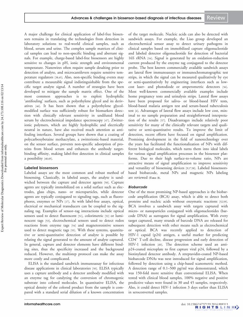
of the target molecule. Nucleic acids can also be detected withsandwich assays. For example, the Liao group developed anelectrochemical sensor assay to detect urinary pathogens inclinical samples based on immobilized capture oligonucleotideand labeled detector oligonucleotide for detection of bacterial16S rRNA [54]. Signal is generated by an oxidation-reductioncurrent produced by the enzyme tag conjugated to the detectorprobe. The best known commercially available sandwich assaysare lateral flow immunoassays or immunochromatographic teststrips, in which the signal can be measured qualitatively by eyeor semi-quantitatively by engineering interfaces such as low-cost laser- and photodiode or amperometric detectors [56].Most well-known commercially available examples includehome pregnancy tests and urinalysis strips. Lateral flow assayshave been proposed for saliva- or blood-based HIV tests,blood-based malaria antigen test and serum-based tuberculosistest [6]. Advantages of lateral flow assays include low cost, min-imal to no sample preparation and straightforward interpreta-tion of the results [57]. Disadvantages include relatively poorsensitivity for many of the clinically relevant targets and quali-tative or semi-quantitative results. To improve the limit ofdetection, recent efforts have focused on signal amplification.Promising development in the field of nanotechnology overthe years has facilitated the functionalization of NPs with dif-ferent biological molecules, which turns them into ideal labelsfor various signal amplification processes in the biosensor plat-forms. Due to their high surface-to-volume ratio, NPs areattractive means of signal amplification to improve sensitivityand versatility of biosensing devices [9,17,58]. Labeled biosensors-based biobarcode, metal NPs and magnetic NPs labelingare reviewed (TABLE 3).
Biobarcode
One of the most promising NP-based approaches is the biobar-code amplification (BCA) assay, which is able to detect bothproteins and nucleic acids without enzymatic reactions [52,59].BCA involves a sandwich assay with targets captured withmicro- or nanoparticles conjugated with oligonucleotides (bar-code DNA) as surrogates for signal amplification. With everytarget captured, many strands of barcode DNA are released forsubsequent detection with other means such as electrochemicalor optical. BCA was recently applied to detection ofHIV-1 capsid (p24) antigen, a useful marker for predictingCD4+ T-cell decline, disease progression and early detection ofHIV-1 infection [60]. The detection scheme used an anti-p24-coated microplate to first capture viral p24, followed by abiotinylated detector antibody. A streptavidin-coated NP-basedbiobarcode DNAs was next introduced for signal amplification,followed by detection using a chip-based scanometric method.A detection range of 0.1–500 pg/ml was demonstrated, whichwas 150-fold more sensitive than conventional ELISA. Whentested with clinical blood samples, 100% negative and positivepredictive values were found in 30 and 45 samples, respectively.Also, it could detect HIV-1 infection 3 days earlier than ELISAin seroconverted samples.
Advances & challenges in biosensor-based diagnosis of infectious diseases Review
informahealthcare.com doi: 10.1586/14737159.2014.888313
Exp
ert R
evie
w o
f M
olec
ular
Dia
gnos
tics
Dow
nloa
ded
from
info
rmah
ealth
care
.com
by
24.1
73.1
08.1
16 o
n 02
/13/
14Fo
r pe
rson
al u
se o
nly.
Metal nanoparticles
Metal NPs can also serve as signal amplification labels for bio-recognition processes based on their unique optical proper-ties [61,62]. Gold and silver NPs exhibit plasmon absorbancebands in the visible light spectrum that are determined by thesize of the respective particles. Therefore, the spectral shifts dueto the aggregation of metal NPs have prompted numerousstudies to develop optical biosensors with biomaterial-metalNPs hybrid systems as the detection amplifiers. One such sen-sor is part of an integrated microfluidic chip using gold-labeledantibodies for simultaneous diagnosis of HIV and syphilis from1 ml of whole blood (FIGURE 2) [63]. The signal amplificationoccurs via the reduction of silver ions onto Au NPs inside amillimeter-sized meandering channel design. The optical den-sity of the silver film is detected and can be quantified with thelow-cost optics or qualitatively by eye. Initial studies indicatethis integrated biosensor is comparable with commercial ELISAkits with near 100% sensitivity and 98–100% specificity forHIV and 82–100% sensitivity and 97–100% specificity forsyphilis.
Magnetic nanoparticles
Magnetic NPs-coupled detectors for biosensing can be used forsignal amplification with the advantage that they are amenableto use in solution phase sandwich assays such as diagnosticmagnetic resonance [55,64]. A major advantage of solution phaseassays is significantly faster assay times compared withdiffusion-dependent surface structure-based assays. With diag-nostic magnetic resonance, both the capture and detectionagents are in solution and linked to magnetic particles. Whenan analyte of interest is present, the magnetic particles cluster asthe antibodies bind the analyte. The clusters of magnetic par-ticles are more efficient at dephasing nuclear spins of the adja-cent water protons, causing a decrease in the spin-spinrelaxation time, resulting in a quantifiable signal. Chung el al.have presented a magneto-DNA platform targeting bacterial16S rRNAs capable of profiling a panel of 13 bacterial speciesfrom clinical samples including urine, pleural fluid, biliary fluid,ascitic fluid and blood [65]. Near single bacterium sensitivity canbe achieved by three signal amplification steps including reverse
transcription-PCR amplification of the 16S rRNA, polymericbead capture and enrichment of target DNA and magneticamplification with magnetically labeled beads conjugated to tar-get DNA (a single magnetic NPs can affect billions of sur-rounding water molecules) [66]. Two drawbacks of the systemare the requirement for manual sample preparation and thePCR experiment is a separate step from the nuclear magneticresonance-based sensor.
Antimicrobial susceptibility testsWhile accurate pathogen identification is the key to diagnosis,assessing pathogen antimicrobial susceptibility is an importantparameter in the management of infection. Rapid antimicrobialsusceptibility test (AST) can expedite appropriate therapy toimpact clinical outcome and may reduce emergence and trans-mission of MDR pathogens. As the rates of MDR pathogensand new infectious diseases rise, the administration of appropri-ate treatment in a timely manner becomes more challengingusing current tests [67]. Hence, a rapid diagnostic system thatcombines pathogen identification and AST would meet a sig-nificant clinical need [68]. Antimicrobial susceptibility can bedetermined phenotypically by measuring bacterial growth/growth inhibition in the presence of a drug, or genotypicallywith PCR-based assays to identify genetic mechanisms thatconfer resistance [69].
Phenotypic ASTs are the mainstay in the clinical microbiol-ogy laboratory. These tests typically require isolation of thepathogen and long incubation time accounting for the lagtime of 24–72 h from sample collection to completion ofanalysis. Recent studies have demonstrated development ofbiosensor and microfluidic devices for rapid AST. Mach et al.demonstrated rapid AST from clinical urine samples by directculture of infected urine in the presence of antibiotic followedby electrochemical detection of 16S rRNA levels as a measureof cell growth [70]. The AST assay was completed in 3.5 hwith 94% agreement with standard AST. Another rapid ASTapproach used an electrochemical biosensor for detection ofprecursor rRNAs (pre-rRNA), an intermediate state in forma-tion of mature rRNA and a marker for cell growth [71]. Thespecificity of the assay was validated with inhibitors of pre-
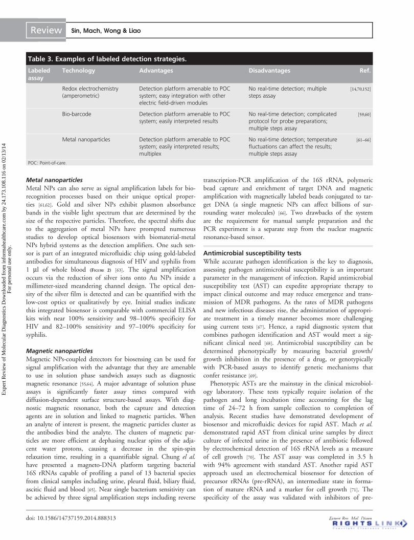
Table 3. Examples of labeled detection strategies.
Labeledassay
Technology Advantages Disadvantages Ref.
Redox electrochemistry
(amperometric)
Detection platform amenable to POC
system; easy integration with other
electric field-driven modules
No real-time detection; multiple
steps assay
[14,70,152]
Bio-barcode Detection platform amenable to POC
system; easily interpreted results
No real-time detection; complicated
protocol for probe preparations;
multiple steps assay
[59,60]
Metal nanoparticles Detection platform amenable to POC
system; easily interpreted results;
multiplex
No real-time detection; temperature
fluctuations can affect the results;
multiple steps assay
[61–66]
POC: Point-of-care.
Review Sin, Mach, Wong & Liao
doi: 10.1586/14737159.2014.888313 Expert Rev. Mol. Diagn.
Exp
ert R
evie
w o
f M
olec
ular
Dia
gnos
tics
Dow
nloa
ded
from
info
rmah
ealth
care
.com
by
24.1
73.1
08.1
16 o
n 02
/13/
14Fo
r pe
rson
al u
se o
nly.

rRNA synthesis and processing (rifampin/rifampicin and chlor-amphenicol) and a DNA gyrase inhibitor (ciprofloxacin).A decline in pre-rRNA was detectable within 15 min in drug-susceptible bacteria but not in resistant strains. Anotherapproach used optical detection for a single cell AST where
bacteria were cultured in with/without antibiotic in microchan-nels. Individual uropathogenic E. coli cells were confined tobacterium-width microchannels with dielectrophoresis (DEP),an electrokinetically driven short-range particle trapping force,applied through an integrated microelectrode [72]. Growth was
00
0.025
0.050
0.075
5 10
Assay time (min)
Ab
sorb
ance
(A
U)
15
BSA
Negativesignal
HIVsignal
Syphilissignal
Positivereference
Waterwash
Waterwash
Gold-labelantibody
BufferwashGold-label
antibody
Leadwash
Bufferwash
Leadwash
Bufferwash
Bufferwash
Silverreagents
Surfacetreatment
Blocked(BSA)
HIV antigen(gp41-gp36)
Au Au Au
Au Au Au
Syphilisantigen(TpN17)
Antibodyto goat IgG
Flow ofsample
Flow ofgold-labeledgoat antibodyto human IgG
Flow ofsilverreagents
Sample
SampleSide view
Flow direction
Inlet
Inlet
Top view Detection zones
Outlet
Outlet
To vacuum (syringe)
Air spacers
Syphilis Ag
HIV AgGoat-specificantibody
Sample: HIV-, syphilis+
silver reagents
20
A B
D
F
E
C
Figure 2. Integrated microfluidic system for multiplexed detection of HIV and syphilis. (A) Photograph of microfluidic chip.(B) Cross-section of microchannels. Scale bar, 500 mm. (C) The design of channel meanders. Scale bar, 1 mm. (D) Schematic diagram ofpassive fluid delivery of preloaded reagents over four detection zones based on vacuum generated by a disposable syringe. (E) Illustrationof reactions at different detection steps. Signal amplification was achieved by the reduction of silver ions on gold nanoparticle-conjugatedantibodies. Signals can be read quantitatively with low-cost optics or qualitatively by eyes. (F) Real-time monitoring of absorbance signalsat the detection zones.Adapted with permission from [63] � Macmillan Publishers Ltd. (2011).
Advances & challenges in biosensor-based diagnosis of infectious diseases Review
informahealthcare.com doi: 10.1586/14737159.2014.888313
Exp
ert R
evie
w o
f M
olec
ular
Dia
gnos
tics
Dow
nloa
ded
from
info
rmah
ealth
care
.com
by
24.1
73.1
08.1
16 o
n 02
/13/
14Fo
r pe
rson
al u
se o
nly.

measured with an epifluorescence microscope and AST profiledetermined within 1 h. Another microfluidic platform forAST was based on stress activation of biosynthetic path-ways [73]. In this assay, S. aureus bound to the bottom of amicrofluidic channel, was subjected to mechanical shear stressand enzymatic stress with subinhibitory concentrations of abactericidal agent resulting in cell wall damage. Subsequenttreatment with the antibiotic oxacillin interfered with therepair process, resulting in rapid cell death of susceptible S.aureus strains, while resistant bacteria remained viable underthe same conditions. Cell viability was monitored using a vitaldye and AST results were established based on normalizedfluorescence values after 60 min. This approach correctly des-ignated oxacillin susceptibility of 16 clinical relevant S. aureusstrains.
In general, microfluidic approaches are promising for theminiaturization and rapid determination of antimicrobialsusceptibility [68,74–77]. These approaches can potentially beintegrated with multiple functionalities into portable chips,which in turn can facilitate AST at the point of care. Addi-tional work is needed to confirm the accuracy of these deviceswith respect to current clinical ASTs.
Sample preparationAdvances in biosensor technology and signal amplificationhave led to highly sensitive detection of pathogen-specificand host immunity biomarkers. However, sample prepara-tion is increasingly recognized as the critical bottleneck intranslating biosensors from the laboratory to clinics [78].Sample preparation involves enrichment of target analyte,removal of matrix inhibitors and sample volume reduction.The strategy of sample preparation depends on the type ofbiological sample, the sample volume and the target analyteconcentration (TABLE 4). Sample preparation begins with speci-men collection: a blood draw to assess serum analytes, a buc-cal swab to collect somatic cells, a lumbar puncture forcerebrospinal fluid or a collection cup for urine, stool orsputum samples. After collection, samples needed to beloaded on the sensing device for preparation and analysis.Whereas specimen loading can be relatively easy for aqueoussamples (i.e., blood, urine, saliva and spinal fluid) [79], addi-tional steps such as digestion and homogenization are neces-sary for viscous or solid samples (i.e., stool andsputum) [64,80]. On-chip sample preparation becomes essen-tial for direct analysis of raw biological samples on detectionplatforms. Unique features of microfluidics such as small fea-tures size (from nanometers to hundreds of micrometers),the laminar nature of fluid flow, fast thermal relaxation,length scale matching with the electric double layer,low fluid volume handling, short assay time and low powerconsumption make these techniques ideal for point-of-caresample preparation [10]. A number of microfluidics-basedsample preparation platforms based on three major samplepreparation steps, separation, concentration and lysis, arereviewed here.T
able
4.Biologicalsamplesandsample
preparationco
nsiderations.
Biological
matrix
Sample
volume(m
l)Advantages
Challenges
Ref.
Blood
0.1–5
Wellestab
lished;baselineconcentrationsof
cellularan
dextracellularconstituents
remain
largelyconstan
t;rapid
changesofan
alyte
concentrationsduringdiseasedstates
Complexmatrix;needforseparationinto
different
bloodcomponents;highbackground;highviscosity;
widedynamicrangeofanalytes
[47–
49,60,63,83–86,98]
Urine
1–100
Ease
ofaccess;non-invasive;abundant;
less
complexmatrixthan
blood
WiderangingpH;highconductivity;
needto
concentrate
analytes;biomarkers
notwellcharacterized
[54,65,70,87,94,111,152]
Saliva
1–5
Ease
ofaccess;non-invasive
Complexmatrix;requires
stim
ulationto
obtain
samples;
variability
ingettingsufficientsample
volume;
biomarkers
notwellcharacterized
[79,100,153]
Sputum
1–5
Ease
ofaccess;less
invasive;localizes
totissueof
interest
Very
highviscosity;complexmatrix;lim
ited
sample
volumeforanalysis
[80,154,155]
Stool
>1g
Ease
ofaccess;non-invasive
Very
challengingmatrix;needdilutionandseparation;
highbackground
[64,156,157]
Tissuesw
ab
(e.g.,buccal,nasal,
wound,vagina)
<1
Ease
ofaccess;less
invasive;localizes
totissueof
interest
Low
andvariab
lesample
volume;complexmatrix;need
todissociate
analyte
from
thesw
ab;low
analytes
concentration
[102,144,158]
Review Sin, Mach, Wong & Liao
doi: 10.1586/14737159.2014.888313 Expert Rev. Mol. Diagn.
Exp
ert R
evie
w o
f M
olec
ular
Dia
gnos
tics
Dow
nloa
ded
from
info
rmah
ealth
care
.com
by
24.1
73.1
08.1
16 o
n 02
/13/
14Fo
r pe
rson
al u
se o
nly.

Separation
Many diagnostic assays are dependent on an initial separationstep. Separation is particularly common with blood samplesthat are commonly fractionated into plasma, white blood cell-rich buffy coat and red blood cells. The conventional means ofblood separation in clinical laboratories are centrifugation andfiltration. Centrifugation is highly efficient but requires a dedi-cated instrumentation that is challenging to integrate with othersteps of sample preparation. While filtration is a cost-effectivealternative to centrifugation, common problems include mem-brane clogging and hemolysis under high pressure.Microfluidic-based alternatives of separation are under activeinvestigation to facilitate integration with advanced biosensors.
One microfluidic technique for rapid separation of plasmafrom a finger prick of whole blood is based on the Zweifach–Fung bifurcation effect [81]. This effect relies on the behavior ofblood cells at a branch point in a microfluidic channel, wherethe blood cells will travel into a channel with a higher flowrate and plasma will end up in the lower flow rate channel.One system integrated a Zweifach–Fung-based microfluidicmodule with a DNA-encoded antibody arrays for rapid on-chip blood separation and measurement of a panel of plasmaproteins using fluorescence detection [82]. The 10-min assaytime from sample collection to detection allows robust detec-tion of proteins that otherwise would rapidly degrade in bloodsamples (FIGURE 3). Another low-cost plasma separation approachutilizes red blood cell agglutination in a paper-based microflui-dic format [83]. Wax hydrophobic barriers printed on paperwere used to filter out agglutinated red blood cells, while theplasma was wicked through the paper substrate onto the testreadout zones where a colorimetric assay was used to detectanalytes of interest. Lab-on-a-disc is another promising
microfluidic technology, which takes advantage of centrifugalforces for various fluid manipulations including plasma separa-tion [84]. With the lab-on-a-disc format, a fully automatedimmunoassay from whole blood has been demonstrated fordetection of hepatitis B virus antigen and antibodies (FIGURE 4).
Although various microfluidic-based approaches can effec-tively separate plasma from whole blood, many of these techni-ques are limited to small volume (up to about 100 ml) due tothe physical restriction of the flow rates. However, clinical sam-ples for bioanalysis are often on the milliliter scale, makinglarge-scale fluid manipulation important especially for low con-centration target analytes. Microfluidic chips that can beadapted for higher flow rates and higher volumes for plasmaseparation are being investigated. One disc-based device is capa-ble of processing 2 ml of whole blood yielding high purityplasma in less than half the time of commercial plasma prepa-ration tubes [85]. Another approach used the temperature effectsto generate high flow rates (between 50 and 200 ml/min) with1 ml of blood sample [86].
Separation is a key step not just for processing whole blood,the sample matrix can impact biosensing from most clinicalsamples. The matrix effect occurs when the interfering com-pounds of a sample, such as, abundant proteins, cells, immuno-globulins or debris, alter the final readout of the biosensor byeither increasing the background or reducing the signal, ulti-mately lowering the sensitivity of the assay [87]. Sample dilutionwith a buffer solution can be sufficient to reduce matrix effectsfor detection of abundant target analytes. However, in mostcases, more complicated sample preparation procedures includ-ing concentration and separation are required. Thus, separatingand concentrating the targets from a larger volume of samplesis a critical sample preparation strategy that can both improve
A B
Who
le b
lood
Plasma
Deal barcodes
Blood barcodes
Plasmaproteins
A B
(5)(3)
(2)
(1)
(4)
C
20 µm
• • •
RBC
WBC
• • •
• • •
• • •
• • •
• • •
• • •
• • •
• • •
A B
Figure 3. Integrated blood barcode chip for multiplexed detection of protein. (A) Schematic of plasma separation based onZweifach–Fung effect from a finger prick of blood. Plasma separation channels are integrated with multiple DNA-encoded antibodybarcode arrays for protein detection. (B) A, B, C represent different DNA codes. (1) is the DNA-antibody conjugate, (2) is plasma protein,(3) is biotin-labeled detection antibody, (4) is streptavidin-Cy5 fluorescence probe and (5) is complementary DNA-Cy3 reference probe.The inset is a barcode of protein biomarkers with the signal measured by fluorescence detection. The green bar denotes as analignment marker.RBC: Red blood cells; WBC: White blood cells.Adapted with permission from [82] � Macmillan Publishers Ltd. (2008).
Advances & challenges in biosensor-based diagnosis of infectious diseases Review
informahealthcare.com doi: 10.1586/14737159.2014.888313
Exp
ert R
evie
w o
f M
olec
ular
Dia
gnos
tics
Dow
nloa
ded
from
info
rmah
ealth
care
.com
by
24.1
73.1
08.1
16 o
n 02
/13/
14Fo
r pe
rson
al u
se o
nly.

the detection limit by increasing the signal (target concentra-tion) and reducing the noise (matrix effect).
Concentration
With real clinical samples in the milliliter scale and relativelylow concentrations of target analyte, both separation and con-centration are frequently needed for biosensor detection. Bead-based analyte capture integrated with microfluidic systems havebeen demonstrated for efficient sample concentration, due tofast diffusion and high surface-to-volume ratio of beads in solu-tion, which provides more binding sites for target analytes orpathogen [88,89]. However, bead manipulation is usually limitedby the applied flow condition. To add another degree of free-dom for particle manipulation, magnetic beads have been used.Magnetic bead capture relies on mixing and capturing of thetargets with the capture agent functionalized beads, followed by
application of a magnetic field to capture and wash the bead-target hybrids. Lien et al. used an immunomagnetic bead(IMB)-based system for rapid detection of influenza A virus [90].Monoclonal antibody-conjugated IMBs were used to target dif-ferent strains of influenza A virus such as A/H1N1 and A/H3N2 in serum specimens. The limit of detection is about 5 �10-4 hemagglutin units (HAU), which is three orders of magni-tude better than bench top systems using flow cytometry. Moreimportantly, this automated microfluidic assay could be com-pleted within 15 min, which is about 1/10 the time requiredfor the comparable manual assay. Similarly, Soelberg et al. haveemployed IMBs for surface plasmon detection of staphylococcalenterotoxin B (SEB) from patient stool samples [64]. With thisapproach, 100 pg/ml SEB in stool samples was easily detected,which is an order of magnitude more sensitive than other com-mercial assay kits.
A
B DC
E GF
Figure 4. Integrated lab-on-a-disc platform for detection of hepatitis B virus. (A) Schematic of the disc showing the microfluidiclayout and function of different compartments. The number indicates the order of operation. (B–G) Illustration of the reactions onthe disc.Adapted with permission from [84] � The Royal Society of Chemistry (2009).
Review Sin, Mach, Wong & Liao
doi: 10.1586/14737159.2014.888313 Expert Rev. Mol. Diagn.
Exp
ert R
evie
w o
f M
olec
ular
Dia
gnos
tics
Dow
nloa
ded
from
info
rmah
ealth
care
.com
by
24.1
73.1
08.1
16 o
n 02
/13/
14Fo
r pe
rson
al u
se o
nly.

Alternative microfluidic devices utilize electrokinetics forsample manipulation. Electrokinetics involve the study of themovement and behavior of particles in suspension when theyare under the influence of electric fields. Among different elec-trokinetics techniques, DEP is one of the most promisingapproaches for separating and concentrating bacteria and cellsas it is a short-range particle force that can directly act on aparticle [91]. When the particle is subjected to an electric field,a dipole is induced in a polarizable particle. If the electric fieldis non-uniform, the particles will experience a net force toward(positive DEP force) or away (negative DEP force) from theelectrode surface depending on the conductivity and permittiv-ity of the particles, the surrounding medium and the appliedelectrical frequency. The magnitude of the DEP force is alsoproportional to the particle volume, thus allowing efficient sep-aration of different size particles or cells. However, the mainchallenge of positive DEP trapping is that it is not effective inbiological fluids that have high conductivity (‡1 S/m) [92]. Toovercome this limitation, Park et al. combined a negative DEP-based separation channel with positive DEP traps that can con-tinuously separate and trap E. coli from either human cerebro-spinal fluid or whole blood samples [93]. The proposedplatform can take 1 ml volumes of crude biological sample andconcentrate target cells into a submicroliter volume withapproximately 104-fold of concentration. In another effort,Gao et al. have designed a hybrid electrokinetics device [91]
combining short range electrophoresis and DEP particle force,with long range AC electrothermal fluid flow for continuouslabel-free isolation of bacteria, such as E. coli, Acinetobacter bau-mannii and Bacillus globigii from biological samples, such asurine and buffy coat with a concentration efficiency of overthree orders of magnitude [94].
In addition to bead-based and electrokinetic assays, othersimple, low-cost microfluidics target concentration platformsare being developed. Zhang et al. have reported a disposablepolymer microfluidic device employing evaporation-induceddragging effect to perform rapid concentration of fluorescentlytagged E. coli [95]. The recovery concentration was above 85%for initial bacterial concentrations lower than 1 � 104 cfu/ml.At the lowest initial concentration, 100 cfu/ml, 100 ml of bac-teria in solution was concentrated into 500 nl droplets withgreater than 90% efficiency in 15 min. However, evaporation-induced concentration will concentrate all components of thesample and may exacerbate the matrix effect. Therefore, thisapproach may only be feasible for clinical samples in conjunc-tion pre-cleaning steps to remove interfering matrix compo-nents of the sample. Another microfluidic approach imitatesthe functions of centrifuge yet operates without moving partsor external forces [96]. The ‘centrifuge-on-a-chip’ employs fluidvortices to passively trap cells using purely hydrodynamicforces. This approach has been used for high-throughput selec-tive enrichment of cells from 10 ml blood samples into smallerml volumes at the high flow rate (ml/min scale), followed byan automated fluorescent labeling detection assay on thetrapped cells.
Lysis
For many assays, cell lysis to release intracellular componentsincluding nucleic acids, proteins and organelle is an essentialstep in sample preparation. Mechanical, electrical, chemical andthermal lysis methods have been demonstrated in microfluidicplatforms [97]. Mechanical lysis involves the generation of shearforce through the application of high pressure, rapid agitationor sonication to crush cells. One system for cell lysis and DNAanalysis uses phononic lattices to generate surface acousticwave-induced rotational vortices to mechanically lyse red bloodcells and malarial parasitic cells present in a drop of blood [98].Subsequent real-time PCR analysis also used surface acousticwave as the heating element and showed that the integrity ofthe genomic DNA was maintained for efficient analysis. Manysystems use friction and collisions between cells and beads insolution for mechanical lysis. One such platform uses a mag-netically actuated bead-beating system on a compact disc(CD)-based centrifugal microfluidic platform [99]. This systemincludes a stationary stand with permanent magnets beneaththe CD and magnetic lysis disks inside the CD. As the CDspins over the stationary magnets, the magnetic lysis disks oscil-late inside the chambers, resulting in mechanical impact andsample shearing. Biological validation of this platform wastested using Bacillus subtilis spores and clinical nasopharyngealaspirates for respiratory virus detection. Although mechanicallysis can be adapted to different cell types, it often requirescooling to remove heat produced by the dissipation of themechanical energy.
Thermal lysis makes use of high temperature to denature cellmembrane proteins and damage the cell to promote release ofcytoplasmic contents. A short pulse of approximately 100˚C issufficient to break the cell membrane without damaging nucleicacid, yet prolonged heat treatment may cause irreversible dena-turation of DNA. This lysis approach is most commonly inte-grated in microfluidic systems with PCR-based genetic assays asa single embedded resistive heater can provide heat for thermallysis and PCR [100,101]. This approach has been verified for lysisand detection of influenza viral particles (infA/H1, infA/H3 and infB) in nasopharyngeal swab samples [102]. In thisassay, amplification and detection were done sequentially byone-step RT-PCR and an optical detection module with a limitof detection of 100 copies for all influenza viruses. Althoughthermal lysis requires no chemical reagents, consistent high heatwill lead to the denaturation of proteins and may interfere withsubsequent assays. Also, large sample volumes require muchmore energy to heat. Thermal lysis may be improved byincreasing the pressure inside the chamber to speed up lysisand use lower temperatures.
Chemical lysis makes use of buffers or other lytic agents tobreak down cell walls and membranes [103]. The chemical agentused for lysis depends on the target cell type and target mole-cule, the type of clinical samples and the detection mecha-nisms. For example, ammonium chloride is effective at lysingerythrocytes but no other mammalian cell types, which can beuseful in clinical samples such as urine and blood when specific
Advances & challenges in biosensor-based diagnosis of infectious diseases Review
informahealthcare.com doi: 10.1586/14737159.2014.888313
Exp
ert R
evie
w o
f M
olec
ular
Dia
gnos
tics
Dow
nloa
ded
from
info
rmah
ealth
care
.com
by
24.1
73.1
08.1
16 o
n 02
/13/
14Fo
r pe
rson
al u
se o
nly.

lysis of erythrocytes is desired. In the electrochemical detectionof 16S rRNA of uropathogens, lysozyme followed by sodiumhydroxide has been shown to be efficient for lysis of bothGram-positive and Gram-negative bacteria from clinical urine
samples [54]. While chemical lysis is simple and effective forwide range of samples, it requires wet chemical storage andmixing, which adds complexity in a microfluidic setting.
Exposure of cells to high-intensity pulsed electric fields canlyse cells due to the dielectric breakdown of the cell membrane.The electric field strength required to reach the threshold toinitiate cell lysis depends on cell shape and size, as well asmembrane composition. Lam et al. demonstrated bacterial lysiswith nanostructured microelectrodes using 100 V, 10 ms DCpulses at a frequency of 1 Hz for 20 s [104]. This lysis approachhas been validated with different bacteria, such as, E. coli,Staphylococcus saprophyticus, S. aureus and MRSA using RT-PCR to measure lysis. While electrical lysis is reagentless andquick, the high voltage in high conductivity physiological buf-fers can lead to chemical electrolysis, undesirable localized heat-ing and denaturation of proteins.
The four main lysis methods described above have theiradvantages and challenges in terms of time, adaptability to dif-ferent cell types, heat generation to samples and interferencewith the subsequent assays. In order to develop an efficient lysisapproach, combinations of the aforementioned methods havebeen designed. For instance, a hybrid chemical and mechanicallysis approach involves directing the bacterial cell through aporous polymer monolith assisted with detergent lytic condi-tions [105]. With this method, both Gram-negative and Gram-positive bacteria were successfully lysed in human bloodsamples.
System integrationThe ideal standalone platform would allow the user to simplyadd sample, click start then view the results. However, fullyintegrated systems that bring together the components of sam-ple preparation and analyte detection remain a critical challengefor technology transfer from laboratories to the clinical mar-ket [56,106]. Recent system-oriented microfluidic strategies thatfacilitate system integration include multilayer soft lithography,multiphase microfluidics, electrowetting-on-dielectric, electroki-netics and centrifugal microfluidics [107]. Microfluidic samplepreparation steps such as concentration, mixing, pumping andseparation can be achieved with these strategies, which makesystem integration a straightforward task with consistent andmonolithic fabrication technologies. Another crucial elementfor system integration is the detection module. For example,many of the optical detection strategies require a bulky micro-scope and laser source, which are not practical in clinical set-tings. Recently, research has focused on portable detectionsystem based on optical, electrical and magnetic sensing. Foroptical detection, lens-free digital microscopy has been imple-mented with miniaturized and cost-effective optical compo-nents mechanically attached to a camera unit of a cellphone [108]. Images of micro-sized objects, for example, redblood cell and white blood cells can be observed with theseportable systems and they will be useful in delivering healthinformation through telecommunication, especially in remotesettings. For electrical detection, electrochemistry is a promising
A B
DC
E F
HG
I
Figure 5. Demonstration of fluid manipulation with foodcolor dyes in an integrated electrode platform for detec-tion of bacterial 16S rRNA. (A & B) Electrolytic pumping oftwo color food dyes into the mixing and sensing chamber in thecenter. (C & D) Electrokinetic mixing of the color food dyes ontop of the electrochemical sensing electrode. (E–H) Electrolyticpumping of washing buffer into the sensing chamber and deliv-ered to the waste reservoirs. (I) Photograph of the universalelectrode array for implementing the electrochemical assay forbacterial 16S rRNA.Reprinted with permission from [112] � IEEE (2013).
Review Sin, Mach, Wong & Liao
doi: 10.1586/14737159.2014.888313 Expert Rev. Mol. Diagn.
Exp
ert R
evie
w o
f M
olec
ular
Dia
gnos
tics
Dow
nloa
ded
from
info
rmah
ealth
care
.com
by
24.1
73.1
08.1
16 o
n 02
/13/
14Fo
r pe
rson
al u
se o
nly.

candidate for lab-on-a-chip [109]. Not only can the electrical sig-nal be processed by conventional electronics, the miniaturiza-tion and integration of the electrochemical transducer into amicrofluidic platform is viable. Moreover, as the electrochemi-cal sensor share a similar electrode interface with other electricfield-driven microfluidic platforms, such as, electrowetting-on-dielectric [110] and electrokinetics [111], integrating sample prepa-ration components with the detection module is simplified.A multifunctional electrode approach has been demonstratedrecently showing the implementation of electrokinetic-inducedmixing directly on an electrochemical sensor, resulting in signalenhancement for detecting urinary tract infections (FIGURE 5) [112].With the lab-on-a-disc format, a fully automated immunoassayfrom whole blood has been demonstrated for detection of hep-atitis B virus antigen and antibodies (FIGURE 4) [84]. Comparedwith over 2 h for conventional ELISA, the lab-on-a-disc assaywas complete in less than 30 min with a similar limit of detec-tion. Indeed, as there is no ‘one size fits all’, system integrationsolutions must be tailored for the intended application withdesign inputs from all stakeholders.
Expert commentaryInfectious diseases are ideal applications for the emerging bio-sensor technologies. For many infectious diseases, rapid diagno-sis and timely initiation of effective treatment can be criticalfor patient outcome and public health. When integrated withadvanced microfluidic systems, biosensor can form the founda-tion of rapid point-of-care devices with the potential to posi-tively impact patient care. As the rate of emergence of MDRpathogens and new infectious diseases continues to increase, anideal diagnostic system will include pathogen identification,AST and host immune response.
While significant improvements in sensitivity and specificityhave been achieved in recent years, the commercialization ofbiosensors for infectious diseases is still in its infancy [56]. Forassay development, both label-free and labeled assays have theiradvantages and limitations. Label-free assays allow ease of sam-ple preparation and quantitative real-time measurement, butsuffer from matrix effects and potential for non-specific bind-ings. While the multistep protocols for labeled assays makethem modestly more complicated, the incorporation of multi-ple binding events increases specificity and amplification tagsimprove sensitivity. Taken together, labeled assays appear tohave a greater potential for clinical translation, particularly indealing with real-world samples.
Translating sample preparation techniques imposes a chal-lenging bottleneck for point-of-care device development. Thematrix effect of clinical samples presents an important problemfor many biosensor devices and needs to be addressed witheach device/matrix/analyte scenario. Most biosensors demon-strate excellent performance with the pristine samples such aspure bacterial/viral cultures or purified biomolecules isolatedfrom clinical samples. Promising performance is also commonlyobserved in spiked samples. Not uncommonly, the matrixbecomes more complex in the setting of active infection. For
example, healthy urine contains few cells, yet upon infection,the urine can go from a clear salt solution to a cloudy mixtureof bacteria, white blood cells, red blood cells and epithelialcells. This change can lead to clogging of microchannels andreduction of the signal. Therefore, even the most promisingsensors need to be critically evaluated with the clinical samplesspanning the anticipated range of the analyte. A number ofrecent studies have demonstrated chip-compatible sample prep-aration strategies that can start with original clinical samples inmicrofluidic systems [78], yet the complexity in reducing millili-ter sample volume down to microliter volume has not beenfully addressed.
Finally, system integration remains the most criticalchallenge for the technology transfer from laboratories intothe clinics. Although successful detection mechanisms,microfluidics-based sample preparation strategies and detectormodules have been demonstrated separately, hurdles remain inthe integration of these modules into a fully automated, stand-alone platform that is easily operated by a non-technical enduser. If these issues can be adequately addressed, it will signifi-cantly increase the likelihood of translating research gradebiosensors from the research laboratories into the fieldsand clinics.
Five-year viewAlthough the potential of microfluidics technology to benefitpoint-of-care diagnostics has been demonstrated for decades, itis unlikely that integrated lab-on-a-chip system that can directlydeal with raw samples will be on market in the next 5 years.The driving force for commercialization fully relies on thecost–effectiveness delivered by the technology, which involvesnot only the cost, but also the real clinical benefits of the testmeasured based on disability-adjusted life-years [113]. From thetechnological point of view, one way to maximize the benefitsdelivered is to develop a universal integrated system that canefficiently handle a wider range of clinical samples such asurine, blood, saliva for different infectious viruses or bacteria.Another challenge for moving toward the practical goal is thegap between the innovative concepts at the academic level andthe clinical validation, which is mainly due to the inaccessibilityof the raw samples for most of the researchers in the fields ofmicrofluidics and their limited experiences on the marketabledevices. Moving forward, more comprehensive collaborationamong academies, healthcare units and industries is the key forthe realization of the real lab-on-a-chip devices.
Financial & competing interests disclosure
JC Liao and PK Wong were supported by NIH/NIAID grant
U01 AI082457. The authors have no other relevant affiliations or finan-
cial involvement with any organization or entity with a financial interest
in or financial conflict with the subject matter or materials discussed in
the manuscript apart from those disclosed. This includes employment, con-
sultancies, honoraria, stock ownership or options, expert testimony, grants
or patents received or pending or royalties.
No writing assistance was utilized in the production of this manuscript.
Advances & challenges in biosensor-based diagnosis of infectious diseases Review
informahealthcare.com doi: 10.1586/14737159.2014.888313
Exp
ert R
evie
w o
f M
olec
ular
Dia
gnos
tics
Dow
nloa
ded
from
info
rmah
ealth
care
.com
by
24.1
73.1
08.1
16 o
n 02
/13/
14Fo
r pe
rson
al u
se o
nly.

Key issues
• Current in vitro diagnostics require centralized laboratory, experienced personnel and large expensive equipment.
• Timely diagnosis and initiation of targeted antimicrobial treatment with portable, sensitive, specific and cost-effective biosensor
technologies are keys to clinical management in decentralized and resource-poor settings.
• Label-free assays may allow quantitative real-time measurement, yet suffer from a significant degree of non-specific bindings and
aberrant signals with analytes in complex matrices.
• In labeled assays, incorporation of multiple binding events and amplification tags increase specificity and sensitivity, yet multistep
protocols increase the assay complexity.
• Matrix effects of clinical samples present a common problem for biosensor technologies and biosensors demonstrating promising
performance with original clinical samples are rare.
• Developing chip-based sample preparation strategies in microfluidic systems for enrichment of target analytes, removal of matrix
inhibitors and sample volume reduction is essential for translating biosensors from laboratory to clinic.
• System integration of three major modules of a biosensor, which are detection mechanism, microfluidics-based sample preparation
strategies and a transducer, into a fully automated and standalone platform remains the most critical challenge for point-of-care
device commercialization.
References
Papers of special note have been highlighted as:
• of interest
•• of considerable interest
1. Franca RF, Da Silva CC, De Paula SO.
Recent advances in molecular medicine
techniques for the diagnosis, prevention,
and control of infectious diseases. Eur J
Clin Microbiol Infect Dis 2013;32(6):723-8
2. Fauci AS, Morens DM. 200 NEJM
ANNIVERSARY ARTICLE the perpetual
challenge of infectious diseases. N Engl J
Med 2012;366(5):454-61
3. Yager P, Domingo GJ, Gerdes J.
Point-of-care diagnostics for global health.
Annu Rev Biomed Eng 2008;10:107-44
• Overview of point-of-care diagnostic
technologies for global health issues.
4. Mabey D, Peeling RW, Ustianowski A,
Perkins MD. Diagnostics for the developing
world. Nat Rev Microbiol 2004;2(3):231-40
5. Luong JH, Male KB, Glennon JD.
Biosensor technology: technology push
versus market pull. Biotechnol Adv 2008;
26(5):492-500
6. Ngom B, Guo Y, Wang X, Bi D.
Development and application of lateral flow
test strip technology for detection of
infectious agents and chemical
contaminants: a review. Anal Bioanal Chem
2010;397(3):1113-35
7. Foudeh AM, Fatanat Didar T, Veres T,
Tabrizian M. Microfluidic designs and
techniques using lab-on-a-chip devices for
pathogen detection for point-of-care
diagnostics. Lab Chip 2012;12(18):3249-66
8. Pejcic B, De Marco R, Parkinson G. The
role of biosensors in the detection of
emerging infectious diseases. Analyst 2006;
131(10):1079-90
9. D’orazio P. Biosensors in clinical chemistry -
2011 update. Clin Chim Acta 2011;
412(19–20):1749-61
10. Whitesides GM. The origins and the future
of microfluidics. Nature 2006;442(7101):
368-73
11. Fournier PE, Raoult D. Prospects for the
Future Using Genomics and Proteomics in
Clinical Microbiology. Annu Rev Microbiol
2011;65:169-88
12. Hodges EN, Connor JH. Translational
control by negative-strand RNA viruses:
methods for the study of a crucial virus/host
interaction. Methods 2013;59(2):180-7
13. Mairiang D, Zhang HM, Sodja A, et al.
Identification of new protein interactions
between dengue fever virus and its hosts,
human and mosquito. PLoS One 2013;
8(1):e53535
14. Mohan R, Mach KE, Bercovici M, et al.
Clinical validation of integrated nucleic acid
and protein detection on an electrochemical
biosensor array for urinary tract infection
diagnosis. PLoS One 2011;6(10):e26846
15. Hunt HK, Armani AM. Label-free
biological and chemical sensors. Nanoscale
2010;2(9):1544-59
16. Rapp BE, Gruhl FJ, Lange K. Biosensors
with label-free detection designed for
diagnostic applications. Anal Bioanal Chem
2010;398(6):2403-12
17. Ju H, Zhang X, Wang J. Signal
amplification for nanobiosensing. In:
NanoBiosensing. Springer, NY, USA; 2011;
p. 39-84
18. Scanvic A, Courdavault L, Sollet JP,
Le Turdu F. Interest of real-time PCR
Xpert MRSA/SA on GeneXpert (R) DX
System in the investigation of staphylococcal
bacteremia. Pathol Biol 2011;59(2):67-72
19. Ioannidis P, Papaventsis D, Karabela S,
et al. Cepheid GeneXpert MTB/RIF Assay
for Mycobacterium tuberculosis Detection
and Rifampin Resistance Identification in
Patients with Substantial Clinical
Indications of Tuberculosis and
Smear-Negative Microscopy Results. J Clin
Microbiol 2011;49(8):3068-70
20. Park S, Zhang Y, Lin S, et al. Advances in
microfluidic PCR for point-of-care
infectious disease diagnostics. Biotechnol
Adv 2011;29(6):830-9
21. Ahmad F, Hashsham SA. Miniaturized
nucleic acid amplification systems for rapid
and point-of-care diagnostics: a review. Anal
Chim Acta 2012;733:1-15
22. Citartan M, Gopinath SC, Tominaga J,
Tang TH. Label-free methods of reporting
biomolecular interactions by optical
biosensors. Analyst 2013;138(13):3576-92
23. Guo X. Surface plasmon resonance based
biosensor technique: a review. J Biophoton
2012;5(7):483-501
24. Tawil N, Sacher E, Mandeville R,
Meunier M. Surface plasmon resonance
detection of E. coli and methicillin-resistant
S. aureus using bacteriophages. Biosens
Bioelectron 2012;37(1):24-9
25. Kussrow A, Baksh MM, Bornhop DJ,
Finn MG. Universal sensing by transduction
of antibody binding with backscattering
interferometry. Chembiochem 2011;12(3):
367-70
Review Sin, Mach, Wong & Liao
doi: 10.1586/14737159.2014.888313 Expert Rev. Mol. Diagn.
Exp
ert R
evie
w o
f M
olec
ular
Dia
gnos
tics
Dow
nloa
ded
from
info
rmah
ealth
care
.com
by
24.1
73.1
08.1
16 o
n 02
/13/
14Fo
r pe
rson
al u
se o
nly.

26. Kussrow A, Enders CS, Castro AR, et al.
The potential of backscattering
interferometry as an in vitro clinical
diagnostic tool for the serological diagnosis
of infectious disease. Analyst 2010;135(7):
1535-7
27. Yanik AA, Huang M, Kamohara O, et al.
An optofluidic nanoplasmonic biosensor for
direct detection of live viruses from
biological media. Nano Lett 2010;10:
4962-9
28. Luo X, Davis JJ. Electrical biosensors and
the label free detection of protein disease
biomarkers. Chem Soc Rev 2013;42(13):
5944-62
29. Qiu J-D, Huang H, Liang R-P.
Biocompatible and label-free amperometric
immunosensor for hepatitis B surface
antigen using a sensing film composed of
poly(allylamine)-branched ferrocene and
gold nanoparticles. Microchim Acta 2011;
174(1-2):97-105
30. Daniels JS, Pourmand N. Label-free
impedance biosensors: opportunities and
challenges. Electroanalysis 2007;19(12):
1239-57
31. Guo X, Kulkarni A, Doepke A, et al.
Carbohydrate-based label-free detection of
Escherichia coli ORN 178 using
electrochemical impedance spectroscopy.
Anal Chem 2012;84(1):241-6
32. Patel P, Klausner JD, Bacon OM, et al.
Detection of acute HIV infections in
high-risk patients in California. J Acquir
Immune Defic Syndr 2006;42(1):75-9
33. Shafiee H, Jahangir M, Inci F, et al. Acute
on-chip HIV detection through label-free
electrical sensing of viral nano-lysate. Small
2013;9(15):2553-63
34. Zelada-Guillen GA, Tweed-Kent A,
Niemann M, et al. Ultrasensitive and
real-time detection of proteins in blood
using a potentiometric carbon-nanotube
aptasensor. Biosens Bioelectron 2013;41:
366-71
35. Song S, Wang L, Li J, et al. Aptamer-based
biosensors. TrAC Trends Anal Chem 2008;
27(2):108-17
36. Villamizar RA, Maroto A, Rius FX.
Improved detection of Candida albicans
with carbon nanotube field-effect transistors.
Sens Actuators B Chem 2009;136(2):451-7
37. Ishikawa FN, Chang HK, Curreli M, et al.
Label-Free, Electrical Detection of the SARS
Virus N-Protein with Nanowire Biosensors
Utilizing Antibody Mimics as Capture
Probes. ACS Nano 2009;3(5):1219-24
38. Tamayo J, Kosaka PM, Ruz JJ, et al.
Biosensors based on nanomechanical
systems. Chem Soc Rev 2013;42(3):
1287-311
39. Arlett JL, Myers EB, Roukes ML.
Comparative advantages of mechanical
biosensors. Nat Nanotechnol 2011;6(4):
203-15
40. Mader A, Gruber K, Castelli R, et al.
Discrimination of Escherichia coli strains
using glycan cantilever array sensors. Nano
Lett 2012;12(1):420-3
41. Liu Y, Schweizer LM, Wang W, et al.
Label-free and real-time monitoring of yeast
cell growth by the bending of polymer
microcantilever biosensors. Sens Actuators B
Chem 2013;178:621-6
42. Peduru Hewa TM, Tannock GA,
Mainwaring DE, et al. The detection of
influenza A and B viruses in clinical
specimens using a quartz crystal
microbalance. J Virol Methods 2009;
162(1-2):14-21
43. Lu CH, Zhang Y, Tang SF, et al. Sensing
HIV related protein using epitope imprinted
hydrophilic polymer coated quartz crystal
microbalance. Biosens Bioelectron 2012;
31(1):439-44
44. Tokonami S, Nakadoi Y, Takahashi M,
et al. Label-free and selective bacteria
detection using a film with transferred
bacterial configuration. Anal Chem 2013;
85(10):4925-9
45. Zhang GJ, Ning Y. Silicon nanowire
biosensor and its applications in disease
diagnostics: a review. Anal Chim Acta
2012;749:1-15
46. Statz AR, Meagher RJ, Barron AE,
Messersmith PB. New peptidomimetic
polymers for antifouling surfaces. J Am
Chem Soc 2005;127(22):7972-3
47. Bryan T, Luo X, Bueno PR, Davis JJ. An
optimised electrochemical biosensor for the
label-free detection of C-reactive protein in
blood. Biosens Bioelectron 2013;39(1):94-8
48. Brault ND, Gao C, Xue H, et al. Ultra-low
fouling and functionalizable zwitterionic
coatings grafted onto SiO2 via a biomimetic
adhesive group for sensing and detection in
complex media. Biosens Bioelectron 2010;
25(10):2276-82
49. Kirk JT, Brault ND, Baehr-Jones T, et al.
Zwitterionic polymer-modified silicon
microring resonators for label-free
biosensing in undiluted human plasma.
Biosens Bioelectron 2013;42:100-5
50. Peruski AH, Peruski LF. Immunological
Methods for Detection and Identification of
Infectious Disease and Biological Warfare
Agents. Clin Vaccine Immunol 2003;10(4):
506-13
51. Li B, Yu Q, Duan Y. Fluorescent labels in
biosensors for pathogen detection. Crit Rev
Biotechnol 2013. [Epub ahead of print]
52. Nam JM, Jang KJ, Groves JT. Detection of
proteins using a colorimetric bio-barcode
assay. Nat protocols 2007;2(6):1438-44
53. Sapsford KE, Pons T, Medintz IL,
Mattoussi H. Biosensing with luminescent
semiconductor quantum dots. Sensors 2006;
6(8):925-53
54. Mach KE, Du CB, Phull H, et al. Rapid
molecular diagnosis of urinary tract
infections. J Urol 2009;181(4):138-9
55. Haun JB, Yoon TJ, Lee H, Weissleder R.
Magnetic nanoparticle biosensors. Wiley
Interdiscip Rev Nanomed Nanobiotechnol
2010;2(3):291-304
56. Chin CD, Linder V, Sia SK.
Commercialization of microfluidic
point-of-care diagnostic devices. Lab Chip
2012;12(12):2118-34
•• Discussion of challenges on
commercialization of microfluidic
point-of-care devices.
57. Martinez AW, Phillips ST, Whitesides GM,
Carrilho E. Diagnostics for the developing
world: microfluidic paper-based analytical
devices. Anal Chem 2010;82(1):3-10
58. Wang EC, Wang AZ. Targeted
Nanoparticles and their Applications in
Biology. Integr Biol 2014;6:9-26
59. Nam JM, Thaxton CS, Mirkin CA.
Nanoparticle-based bio-bar codes for the
ultrasensitive detection of proteins. Science
2003;301(5641):1884-6
60. Tang SX, Zhao JQ, Storhoff JJ, et al.
Nanoparticle-based biobarcode amplification
assay (BCA) for sensitive and early detection
of human immunodeficiency type 1 capsid
(p24) antigen. J Acquir Immune Defic
Syndr 2007;46(2):231-7
61. Cao X, Ye Y, Liu S. Gold
nanoparticle-based signal amplification for
biosensing. Anal Biochem 2011;417(1):1-16
62. Lin M, Pei H, Yang F, et al. Applications
of Gold Nanoparticles in the Detection and
Identification of Infectious Diseases and
Biothreats. Adv Materials 2013;25(25):
3490-6
63. Chin CD, Laksanasopin T, Cheung YK,
et al. Microfluidics-based diagnostics of
infectious diseases in the developing world.
Nat Med 2011;17(8):1015-19
• An integrated microfluidic device for
multiplex detection of infectious diseases
directly from whole blood.
64. Soelberg SD, Stevens RC, Limaye AP,
Furlong CE. Surface Plasmon Resonance
Advances & challenges in biosensor-based diagnosis of infectious diseases Review
informahealthcare.com doi: 10.1586/14737159.2014.888313
Exp
ert R
evie
w o
f M
olec
ular
Dia
gnos
tics
Dow
nloa
ded
from
info
rmah
ealth
care
.com
by
24.1
73.1
08.1
16 o
n 02
/13/
14Fo
r pe
rson
al u
se o
nly.

Detection Using Antibody-Linked Magnetic
Nanoparticles for Analyte Capture,
Purification, Concentration, and Signal
Amplification. Anal Chem 2009;81(6):
2357-63
65. Chung HJ, Castro CM, Im H, et al.
A magneto-DNA nanoparticle system for
rapid detection and phenotyping of bacteria.
Nat Nanotechnol 2013;8(5):369-75
66. Lee H, Sun E, Ham D, Weissleder R.
Chip-NMR biosensor for detection and
molecular analysis of cells. Nat Med 2008;
14(8):869-74
67. Moore DA, Shah NS. Alternative methods
of diagnosing drug resistance–what can they
do for me? J Infect Dis 2011;204(Suppl 4):
S1110-19
68. Pulido MR, Garcia-Quintanilla M,
Martin-Pena R, et al. Progress on the
development of rapid methods for
antimicrobial susceptibility testing. J
Antimicrob Chemother 2013;68(12):
2710-17
69. Bergeron MG, Ouellette M. Preventing
antibiotic resistance through rapid genotypic
identification of bacteria and of their
antibiotic resistance genes in the clinical
microbiology laboratory. J Clin Microbiol
1998;36(8):2169-72
70. Mach KE, Mohan R, Baron EJ, et al.
A biosensor platform for rapid antimicrobial
susceptibility testing directly from clinical
samples. J Urol 2011;185(1):148-53
71. Halford C, Gonzalez R, Campuzano S,
et al. Rapid antimicrobial susceptibility
testing by sensitive detection of precursor
rRNA using a novel electrochemical
biosensing platform. Antimicrob Agents
Chemother 2013;57(2):936-43
72. Lu Y, Gao J, Zhang DD, et al. Single cell
antimicrobial susceptibility testing by
confined microchannels and electrokinetic
loading. Anal Chem 2013;85(8):3971-6
73. Kalashnikov M, Lee JC, Campbell J, et al.
A microfluidic platform for rapid,
stress-induced antibiotic susceptibility testing
of Staphylococcus aureus. Lab Chip 2012;
12(21):4523-32
74. Chen CH, Lu Y, Sin ML, et al.
Antimicrobial susceptibility testing using
high surface-to-volume ratio microchannels.
Anal Chem 2010;82(3):1012-19
75. Kim KP, Kim YG, Choi CH, et al. In situ
monitoring of antibiotic susceptibility of
bacterial biofilms in a microfluidic device.
Lab Chip 2010;10(23):3296-9
76. Tang Y, Zhen L, Liu J, Wu J. Rapid
antibiotic susceptibility testing in a
microfluidic pH sensor. Anal Chem 2013;
85(5):2787-94
77. Tawil N, Mouawad F, Levesque S, et al.
The differential detection of
methicillin-resistant, methicillin-susceptible
and borderline oxacillin-resistant
Staphylococcus aureus by surface plasmon
resonance. Biosens Bioelectron 2013;49:
334-40
78. Ritzi-Lehnert M. Development of
chip-compatible sample preparation for
diagnosis of infectious diseases. Expert Rev
Mol Diagn 2012;12(2):189-206
•• Review of most recent development in
on-chip sample preparation for
microfluidic-based diagnosis of infectious
diseases.
79. Fu E, Chinowsky T, Nelson K, et al. SPR
imaging-based salivary diagnostics system for
the detection of small molecule analytes.
Ann NY Acad Sci 2007;1098:335-44
80. Cattamanchi A, Davis JL, Pai M, et al.
Does bleach processing increase the accuracy
of sputum smear microscopy for diagnosing
pulmonary tuberculosis? J Clin Microbiol
2010;48(7):2433-9
81. Yang S, Undar A, Zahn JD. A microfluidic
device for continuous, real time blood
plasma separation. Lab Chip 2006;6(7):
871-80
82. Fan R, Vermesh O, Srivastava A, et al.
Integrated barcode chips for rapid,
multiplexed analysis of proteins in microliter
quantities of blood. Nat Biotechnol 2008;
26(12):1373-8
83. Yang X, Forouzan O, Brown TP,
Shevkoplyas SS. Integrated separation of
blood plasma from whole blood for
microfluidic paper-based analytical devices.
Lab Chip 2012;12(2):274-80
84. Lee BS, Lee JN, Park JM, et al. A fully
automated immunoassay from whole blood
on a disc. Lab Chip 2009;9(11):1548-55
• A fully automated system integrated with
sample preparation modules for diagnosis
of hepatitis B infection from whole
blood.
85. Amasia M, Madou M. Large-volume
centrifugal microfluidic device for blood
plasma separation. Bioanalysis 2010;2(10):
1701-10
86. Rodriguez-Villarreal AI, Arundell M,
Carmona M, Samitier J. High flow rate
microfluidic device for blood plasma
separation using a range of temperatures.
Lab Chip 2010;10(2):211-19
87. Chiu ML, Lawi W, Snyder ST, et al.
Matrix effects – a challenge toward
automation of molecular analysis. JALA
2010;15(3):233-42
88. Mulvaney SP, Cole CL, Kniller MD, et al.
Rapid, femtomolar bioassays in complex
matrices combining microfluidics and
magnetoelectronics. Biosens Bioelectron
2007;23(2):191-200
89. Dimov IK, Garcia-Cordero JL, O’grady J,
et al. Integrated microfluidic
tmRNA purification and real-time
NASBA device for molecular diagnostics.
Lab Chip 2008;8(12):2071-8
90. Lien KY, Hung LY, Huang TB, et al.
Rapid detection of influenza A virus
infection utilizing an immunomagnetic
bead-based microfluidic system. Biosens
Bioelectron 2011; Epub ahead of print
91. Gao J, Sin ML, Liu T, et al. Hybrid
electrokinetic manipulation in
high-conductivity media. Lab Chip 2011;
11(10):1770-5
92. Krishnan R, Sullivan BD, Mifflin RL, et al.
Alternating current electrokinetic separation
and detection of DNA nanoparticles in
high-conductance solutions. Electrophoresis
2008;29(9):1765-74
93. Park S, Zhang Y, Wang TH, Yang S.
Continuous dielectrophoretic bacterial
separation and concentration from
physiological media of high conductivity.
Lab Chip 2011;11(17):2893-900
94. Gao J, Riahi R, Sin ML, et al.
Electrokinetic focusing and separation of
mammalian cells in conductive biological
fluids. Analyst 2012;137(22):5215-21
95. Zhang JY, Do J, Premasiri WR, et al. Rapid
point-of-care concentration of bacteria in a
disposable microfluidic device using
meniscus dragging effect. Lab Chip 2010;
10(23):3265-70
96. Mach AJ, Kim JH, Arshi A, et al.
Automated cellular sample preparation using
a Centrifuge-on-a-Chip. Lab Chip 2011;
11(17):2827-34
97. Kim J, Johnson M, Hill P, Gale BK.
Microfluidic sample preparation: cell lysis
and nucleic acid purification. Integr Biol
2009;1(10):574-86
98. Reboud J, Bourquin Y, Wilson R, et al.
Shaping acoustic fields as a toolset for
microfluidic manipulations in diagnostic
technologies. Proc Natl Acad Sci USA
2012;109(38):15162-7
99. Siegrist J, Gorkin R, Bastien M, et al.
Validation of a centrifugal microfluidic
sample lysis and homogenization platform
for nucleic acid extraction with clinical
samples. Lab Chip 2010;10(3):363-71
Review Sin, Mach, Wong & Liao
doi: 10.1586/14737159.2014.888313 Expert Rev. Mol. Diagn.
Exp
ert R
evie
w o
f M
olec
ular
Dia
gnos
tics
Dow
nloa
ded
from
info
rmah
ealth
care
.com
by
24.1
73.1
08.1
16 o
n 02
/13/
14Fo
r pe
rson
al u
se o
nly.

100. Oblath EA, Henley WH, Alarie JP,
Ramsey JM. A microfluidic chip integrating
DNA extraction and real-time PCR for the
detection of bacteria in saliva. Lab Chip
2013;13(7):1325-32
101. Marshall LA, Wu LL, Babikian S, et al.
Integrated printed circuit board device for
cell lysis and nucleic acid extraction. Anal
Chem 2012;84(21):9640-5
102. Wang C-H, Lien K-Y, Hung L-Y, et al.
Integrated microfluidic system for the
identification and multiple subtyping of
influenza viruses by using a molecular
diagnostic approach. Microfluid Nanofluid
2012;13(1):113-23
103. Omiatek DM, Santillo MF, Heien ML,
Ewing AG. Hybrid capillary-microfluidic
device for the separation, lysis, and
electrochemical detection of vesicles. Anal
Chem 2009;81(6):2294-302
104. Lam B, Fang Z, Sargent EH, Kelley SO.
Polymerase chain reaction-free,
sample-to-answer bacterial detection in
30 minutes with integrated cell lysis. Anal
Chem 2012;84(1):21-5
105. Mahalanabis M, Al-Muayad H,
Kulinski MD, et al. Cell lysis and
DNA extraction of gram-positive and
gram-negative bacteria from whole blood in
a disposable microfluidic chip. Lab Chip
2009;9(19):2811-17
106. Sorger PK. Microfluidics closes in on
point-of-care assays. Nat Biotechnol 2008;
26(12):1345-6
107. Sin ML, Gao J, Liao JC, Wong PK. System
integration – a major step toward lab on a
chip. J Biol Eng 2011;5:6
• Review of system integration approaches
for point-of-care devices.
108. Gurkan UA, Moon S, Geckil H, et al.
Miniaturized lensless imaging systems for
cell and microorganism visualization in
point-of-care testing. Biotechnol J 2011;
6(2):138-49
109. Kimmel DW, Leblanc G, Meschievitz ME,
Cliffel DE. Electrochemical sensors and
biosensors. Anal Chem 2012;84(2):685-707
110. Karuwan C, Sukthang K, Wisitsoraat A,
et al. Electrochemical detection on
electrowetting-on-dielectric digital
microfluidic chip. Talanta 2011;84(5):
1384-9
111. Sin ML, Liu T, Pyne JD, et al. In situ
electrokinetic enhancement for self-
assembled-monolayer-based electrochemical
biosensing. Anal Chem 2012;84(6):2702-7
112. Sin ML, Gau V, Liao JC, Wong PK.
A universal electrode approach for
automated electrochemical molecular
analyses. J Microelectromech Syst 2013;
22(5):1126-32
113. Laxminarayan R, Mills AJ, Breman JG,
et al. Advancement of global health: key
messages from the Disease Control Priorities
Project. Lancet 2006;367(9517):1193-208
114. Clarke JR. Molecular diagnosis of HIV.
Expert Rev Mol Diagn 2002;2(3):233-9
115. Bourlet T, Memmi M, Saoudin H,
Pozzetto B. Molecular HIV screening.
Expert Rev Mol Diagn 2013;13(7):693-705
116. Branson BM. State of the art for diagnosis
of HIV infection. Clin Infect Dis 2007;45:
S221-5
117. Al-Zamel FA. Detection and diagnosis of
Mycobacterium tuberculosis. Expert Rev
Anti Infect Ther 2009;7(9):1099-108
118. Helb D, Jones M, Story E, et al. Rapid
detection of Mycobacterium tuberculosis
and rifampin resistance by use of
on-demand, near-patient technology. J Clin
Microbiol 2010;48(1):229-37
119. Van Rie A, Page-Shipp L, Scott L, et al.
Xpert (R) MTB/RIF for point-of-care
diagnosis of TB in high-HIV burden,
resource-limited countries: hype or hope?
Expert Rev Mol Diagn 2010;10(7):937-46
120. Hawkes M, Kain KC. Advances in malaria
diagnosis. Expert Rev Anti Infect Ther
2007;5(3):485-95
121. Moody A. Rapid Diagnostic Tests for
Malaria Parasites. Clin Microbiol Rev 2002;
15(1):66-78
122. Wongsrichanalai C, Barcus MJ, Muth S,
et al. A review of malaria diagnostic tools:
microscopy and rapid diagnostic test
(RDT). Am J Trop Med Hyg 2007;77(6):
119-27
123. Strick L, Wald A. Type-specific testing for
herpes simplex virus. Expert Rev Mol Diagn
2004;4(4):443-53
124. Corey L. Laboratory diagnosis of
herpes-simplex virus-infections - principles
guiding the development of rapid
diagnostic-tests. Diagn Micrbiol Infect Dis
1986;4(3):S111-19
125. Ryan C, Kinghorn G. Clinical assessment of
assays for diagnosis of herpes simplex
infection. Expert Rev Mol Diagn 2006;6(5):
767-75
126. Bayliss J, Nguyen T, Lesmana CR, et al.
Advances in the molecular diagnosis of
hepatitis B infection: providing insight into
the next generation of disease. Semin Liver
Dis 2013;33(2):113-21
127. Nainan OV, Xia G, Vaughan G,
Margolis HS. Diagnosis of hepatitis a virus
infection: a molecular approach. Clin
Microbiol Rev 2006;19(1):63-79
128. Wiwanitkit V. Dengue fever: diagnosis and
treatment. Expert Rev Anti Infect Ther
2010;8(7):841-5
129. Tang KF, Ooi EE. Diagnosis of dengue:
an update. Expert Rev Anti Infect Ther
2012;10(8):895-907
130. Hooton TM, Bradley SF, Cardenas DD,
et al. Diagnosis, prevention, and treatment
of catheter-associated urinary tract infection
in adults: 2009 International Clinical
Practice Guidelines from the Infectious
Diseases Society of America. Clin Infect Dis
2010;50(5):625-63
131. Schmiemann G, Kniehl E, Gebhardt K,
et al. The diagnosis of urinary tract
infection: a systematic review. Deutsch
Arztebl Int 2010;107(21):361-7
132. Katz JM, Hancock K, Xu XY. Serologic
assays for influenza surveillance, diagnosis
and vaccine evaluation. Expert Rev Anti
Infect Ther 2011;9(6):669-83
133. Dimaio MA, Sahoo MK, Waggoner J,
Pinsky BA. Comparison of Xpert Flu rapid
nucleic acid testing with rapid antigen
testing for the diagnosis of influenza A and
B. J Virol Methods 2012;186(1-2):137-40
134. Chauhan N, Narang J, Pundir S, et al.
Laboratory diagnosis of swine flu: a review.
Artif Cell Nanomed B 2013;41(3):189-95
135. Guerrant RL, Van Gilder T, Steiner TS,
et al. Practice guidelines for the
management of infectious diarrhea. Clin
Infect Dis 2001;32(3):331-51
136. O’Ryan M, Lucero Y, O’Ryan-Soriano MA,
Ashkenazi S. An update on management of
severe acute infectious gastroenteritis in
children. Expert Rev Anti Infect Ther 2010;
8(6):671-82
137. Feeney SA, Armstrong VJ, Mitchell SJ,
et al. Development and clinical validation of
multiplex TaqMan(R) assays for rapid
diagnosis of viral gastroenteritis. J Med
Virol 2011;83(9):1650-6
138. Bauer M, Reinhart K. Molecular diagnostics
of sepsis – where are we today? Int J Med
Microbiol 2010;300(6):411-13
139. Mancini N, Carletti S, Ghidoli N, et al.
The era of molecular and other
non-culture-based methods in diagnosis of
sepsis. Clin Microbiol Rev 2010;23(1):
235-51
140. Kotsaki A, Giamarellos-Bourboulis EJ.
Molecular diagnosis of sepsis. Expert Opin
Med Diagn 2012;6(3):209-19
141. Graseck AS, Shih SL, Peipert JF. Home
versus clinic-based specimen collection for
Advances & challenges in biosensor-based diagnosis of infectious diseases Review
informahealthcare.com doi: 10.1586/14737159.2014.888313
Exp
ert R
evie
w o
f M
olec
ular
Dia
gnos
tics
Dow
nloa
ded
from
info
rmah
ealth
care
.com
by
24.1
73.1
08.1
16 o
n 02
/13/
14Fo
r pe
rson
al u
se o
nly.

Chlamydia trachomatis and Neisseria
gonorrhoeae. Expert Rev Anti Infect Ther
2011;9(2):183-94
142. Munson E, Napierala M, Schell RF.
Insights into trichomoniasis as a result of
highly sensitive molecular diagnostics
screening in a high-prevalence sexually
transmitted infection community. Expert
Rev Anti Infect Ther 2013;11(8):845-63
143. Stevens DL, Bisno AL, Chambers HF, et al.
Practice guidelines for the diagnosis and
management of skin and soft-tissue
infections. Clin Infect Dis 2005;41(10):
1373-406
144. Bonham PA. Swab cultures for diagnosing
wound infections: a literature review and
clinical guideline. J Wound Ostomy
Continence Nurs 2009;36(4):389-95
145. Oz Y, Kiraz N. Diagnostic methods for
fungal infections in pediatric patients:
microbiological, serological and molecular
methods. Expert Rev Anti Infect Ther 2011;
9(3):289-98
146. Peman J, Zaragoza R. Combined use of
nonculture-based lab techniques in the
diagnosis and management of critically ill
patients with invasive fungal infections.
Expert Rev Anti Infect Ther 2012;10(11):
1321-30
147. Liang RP, Fan LX, Huang DM, Qiu JD.
A label-free amperometric immunosensor
based on redox-active ferrocene-branched
chitosan/multiwalled carbon nanotubes
conductive composite and gold
nanoparticles. Electroanalysis 2011;23(3):
719-27
148. Shi WT, Ma ZF. A novel label-free
amperometric immunosensor for
carcinoembryonic antigen based on redox
membrane. Biosens Bioelectron 2011;26(6):
3068-71
149. Koncki R. Recent developments in
potentiometric biosensors for biomedical
analysis. Anal Chim Acta 2007;599(1):7-15
150. Hiatt LA, Cliffel DE. Real-time
Recognition of Mycobacterium tuberculosis
and Lipoarabinomannan using the Quartz
Crystal Microbalance. Sens Actuators B
Chem 2012;174:245-52
151. Cheng CI, Chang YP, Chu YH.
Biomolecular interactions and tools for their
recognition: focus on the quartz crystal
microbalance and its diverse surface
chemistries and applications. Chem Soc Rev
2012;41(5):1947-71
152. Pan Y, Sonn GA, Sin MLY, et al.
Electrochemical immunosensor detection of
urinary lactoferrin in clinical samples for
urinary tract infection diagnosis. Biosens
Bioelectron 2010;26(2):649-54
153. Kaufman E, Lamster IB. The diagnostic
applications of saliva – a review. Crit Rev
Oral Biol Med 2002;13(2):197-212
154. Thanyani ST, Roberts V, Siko DGR, et al.
A novel application of affinity biosensor
technology to detect antibodies to mycolic
acid in tuberculosis patients. J Immunol
Methods 2008;332(1-2):61-72
155. Zuo BL, Li SM, Guo Z, et al. Piezoelectric
immunosensor for SARS-associated
coronavirus in sputum. Anal Chem 2004;
76(13):3536-40
156. Chua AL, Yean CY, Ravichandran M, et al.
A rapid DNA biosensor for the molecular
diagnosis of infectious disease. Biosens
Bioelectron 2011;26(9):3825-31
157. Huang JL, Yang GJ, Meng WJ, et al. An
electrochemical impedimetric immunosensor
for label-free detection of Campylobacter
jejuni in diarrhea patients’ stool based on
O-carboxymethylchitosan surface modified
Fe3O4 nanoparticles. Biosens Bioelectron
2010;25(5):1204-11
158. Mahilum-Tapay L, Laitila V,
Wawrzyniak JJ, et al. New point of care
Chlamydia Rapid Test – bridging the gap
between diagnosis and treatment:
performance evaluation study. BMJ 2007;
335(7631):1190-4
Review Sin, Mach, Wong & Liao
doi: 10.1586/14737159.2014.888313 Expert Rev. Mol. Diagn.
Exp
ert R
evie
w o
f M
olec
ular
Dia
gnos
tics
Dow
nloa
ded
from
info
rmah
ealth
care
.com
by
24.1
73.1
08.1
16 o
n 02
/13/
14Fo
r pe
rson
al u
se o
nly.