Embed Size (px)
Citation preview

Advanced Topics and Diffusion MRI
Modified by Mark Chiew ([email protected])
Slides available at:http://users.fmrib.ox.ac.uk/~mchiew/teaching/
Slides originally by Karla Miller, FMRIB Centre

MRI Physics
★ Spin vs. gradient echo (T2 & T2*)★ Fast imaging & artefacts★ Diffusion MRI
✦ Diffusion weighting✦ Acquisition techniques✦ Tradeoffs & complications

Phase
Magnitude (length or st
rength)
Phase (orientation or direction)

Voxel Volume Net Magnetization (sum)
Net Magnetization (in-phase)

B0
Precession (in-phase)
courtesy of William Overall
ω0 = γ(B0+ΔB)main field offset (gradients or errors)
rotational frequency
∆B+

Magnetic field imperfections: T2* decay
Always some local imperfections in magnetic field = range of precession frequencies in a voxel
Over time, spins lose alignment (“dephase”)

Voxel Volume Net Magnetization (sum)
Net Magnetization (in-phase)

Net Magnetization (dephasing)Voxel Volume Net Magnetization (sum)

Voxel Volume Net Magnetization (sum)
Net Magnetization (dephased)

Simple Voxel Model Voxel Signal
Dephasing

Refocusing (180o RF pulse) with no dephasing
90°180°
The 180º RF pulse complete “flips” the magnetisation around an axis

Spin echo: The time at which the spins are re-aligned Refocusing pulse: 180o pulse that creates a spin echo
90°180°
Refocusing (180o RF pulse) with dephasing

Simple Voxel Model Voxel Signal
Dephasing

180º Refocusing Pulse!
Rephasing After Spin Echo
Simple Voxel Model Voxel Signal

Refocusing (180° pulse) spin echo
Spin echo: The time at which the spins are re-aligned Refocusing pulse: 180o pulse that creates a spin echo
time
sign
al
90°180°

Gradient echo vs. spin echo
GRE signal(sometimes called a FreeInduction Decay = FID)
SE signal(signal decays, then
comes back as “echo”)
time
sign
al
90°
time
sign
al
90°180°
pure signal decay decay with partial recovery
(i.e. no spin-echo)
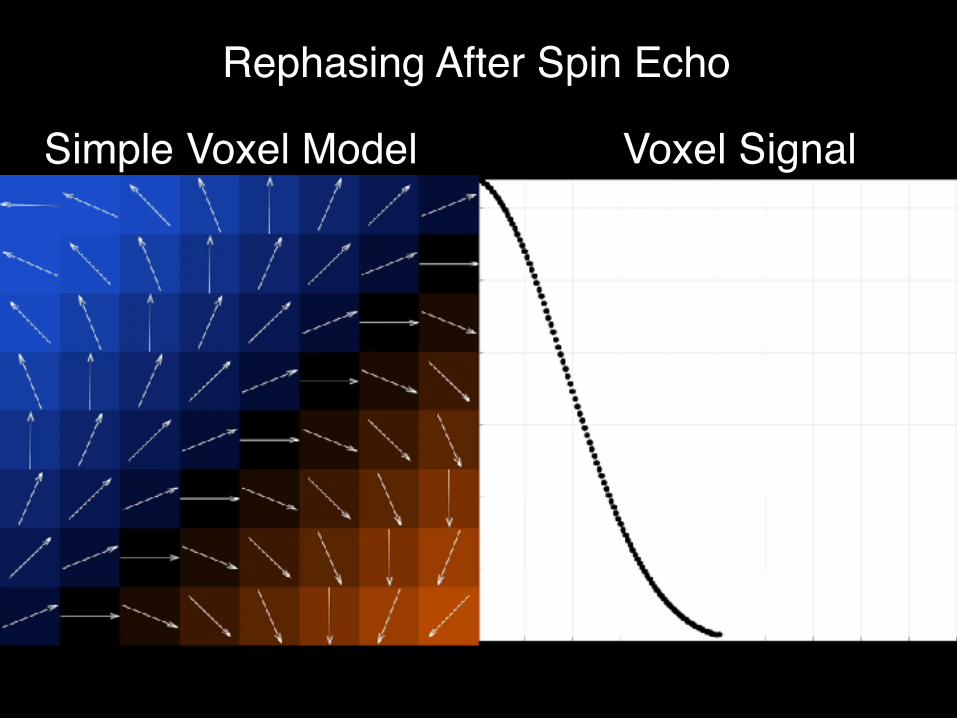
T2 vs T2* Relaxation
Spin echo refocuses part of the signal decay– T2* includes parts that can be refocused– Without refocusing, signal will have T2* contrast
time
sign
al
SE signalT2* decay (imperfect field)T2 decay (perfect field)
Even spin echo signal experiences some decay– T2 refers to signal decay that cannot be refocused– With refocusing, signal will have T2 contrast
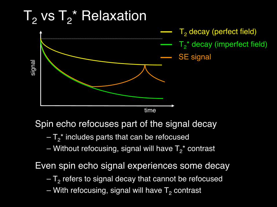
GRE refers to a sequence with: excitation-delay-readoutAny kind of readout can be used (linescan, EPI, spiral…)Image signal depends on TE, but not on readout method!
time
sign
al
TE
ACQ
time
sign
al
TE
ACQ
What defines a gradient echo sequence?

What defines a spin echo sequence?
SE refers to a sequence with: excitation-refocus-readoutThe key is the formation of an echo (signal peak)!Like GRE, any kind of readout can be used
time
sign
al
timesi
gnal
TE TE
`
ACQACQ

MRI Physics
★ Spin vs. gradient echo★ Fast imaging & artefacts★ Diffusion MRI
✦ Diffusion weighting✦ Acquisition techniques✦ Tradeoffs & complications

Echo-planar Imaging (EPI) Acquisition
Acquire all of k-space in a “single shot”Used for FMRI, diffusion imaging

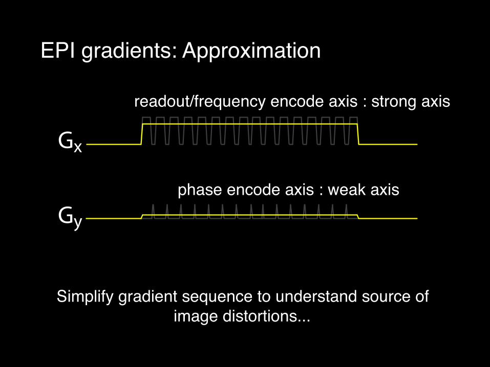
EPI gradients: Approximation
Gy
Gx
Gy
Gx
Gy
Gx
Gy
Gx
Simplify gradient sequence to understand source of image distortions...
readout/frequency encode axis
phase encode axis
EPI gradients: Approximation
Gy
Gx
Gy
Gx
Gy
Gx
Gy
Gx
Simplify gradient sequence to understand source of image distortions...
readout/frequency encode axis : strong axis
phase encode axis : weak axis

EPI gradients: Approximation
Gy
Gx
Gy
Gx
Gy
Gx
Gy
Gx
Simplify gradient sequence to understand source of image distortions...
readout/frequency encode axis : fast axis
phase encode axis : slow axis

kslow
kfast
slowfast
field map fast gradient net (fast)
EPI undistorted along “fast” direction (frequency encode)
+ =
Field map errors have negligible effect on fast gradient

kslow
kfast
slowfast
field map slow gradient net (fast)
EPI distorted along “slow” direction (phase encode)
+ =
Field map errors dominate slow gradient, signal is misplaced

EPI distortion
Echo spacing: time between acquisition of adjacent lines (“speed” along slow axis”)Long echo spacing = worse distortion
EPI trajectoryLong inter-echospacing
Short inter-echospacing
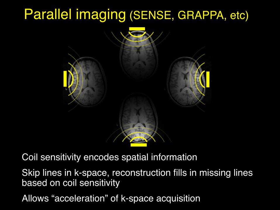
Coil sensitivity encodes spatial information Skip lines in k-space, reconstruction fills in missing lines based on coil sensitivityAllows “acceleration” of k-space acquisition
Parallel imaging (SENSE, GRAPPA, etc)

Parallel imaging
EPI trajectory
Parallel imaging (SENSE, GRAPPA, etc) can reconstruct complete image from subset of k-space lines
No ParallelImaging
ParallelImaging (2x)
Current vendor coils enable 2-4x acceleration
Enables “accelerated” acquisition with lower distortion

Many possible trajectories through k-space…

EPIdistortion
& ghosting
Spiralblurring
& streaking
Cartesian vs Non-Cartesian: Image artifacts

time
tissue 1
Single-shot acquisition takes 30-40 ms, so T2/T2* contrast varies during acquisition... what is contrast of image?
Rule of thumb: contrast of image reflects time at which central k-space was acquired (“effective” TE)
tissue 2
Cartesian vs Non-Cartesian: Contrast

MRI Physics
★ Spin vs. gradient echo★ Fast imaging & artefacts★ Diffusion MRI
✦ Diffusion weighting✦ Acquisition techniques✦ Tradeoffs & complications

What is diffusion?

What is diffusion?
Random motion of particles due to thermal energyWater molecules collide and experience net displacement
Displacement described by diffusion coefficient (D)Normally, diffusion is isotropic (equal in all directions)
√2DT
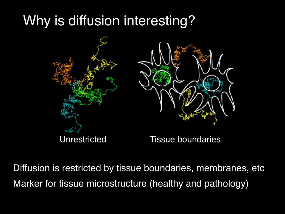
Why is diffusion interesting?
Diffusion is restricted by tissue boundaries, membranes, etcMarker for tissue microstructure (healthy and pathology)
Unrestricted Tissue boundaries

Diffusion anisotropy in white matter
Water can diffuse more freely along white matter fibres than across them

Diffusion anisotropy in white matter
Directionality of diffusion tells us about fibre integrity/structure and orientation
Diffusion in white matter fibres is “anisotropic”

The diffusion tensor
Displacement due to diffusion is approximately ellipsoidalEigenvectors = axes of ellipsoid (direction of fibres)Eigenvalues = size of axes (strength of diffusion)

The diffusion tensor: Useful quantities
Principal diffusion direction (PDD): what direction is greatest diffusion along? Info about fibre orientationFractional anisotropy (FA): how elongated is the ellipsoid? Info about fibre integrityMean diffusivity (MD): Info about tissue integrity

Diffusion tensor imaging
Principal diffusion
direction (PDD)Fractional
anisotropy (FA)Mean
diffusivity (MD)
At each voxel, fit the diffusion tensor modelCan then calculate MD, FA, PDD from fitted parameters
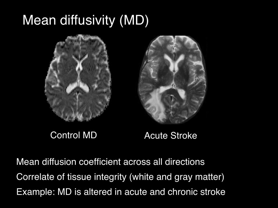
Mean diffusivity (MD)
Mean diffusion coefficient across all directionsCorrelate of tissue integrity (white and gray matter)Example: MD is altered in acute and chronic stroke
Control MD Acute Stroke

Fractional Anisotropy (FA)
Inequality of diffusion coefficient across different directionsHigh in regions where diffusion is most directionalRelates to integrity of white matter fibre bundles
FA=0.8
FA=0

Principal diffusion direction (PDD)
Direction along which greatest diffusion occursRelates to direction of fibre orientationsTypically, will use this as starting point for fibre tracking

Diffusion tractography
Jones et al
Follow PDD to trace white matter fibers (“tractography”)

MRI Physics
★ Spin vs. gradient echo★ Fast imaging & artefacts★ Diffusion MRI
✦ Diffusion weighting✦ Acquisition techniques✦ Tradeoffs & complications

Spin Echo
90°180°

Diffusion-weighted spin echo

Case 1:No diffusion

Case 2:Diffusion

+5
-3
No motion (diffusion)No net dephasingNo contrast!
0
0
contrast
-5
+3
Posi
tion

+5
-3
+ motion (diffusion)+ net dephasing+ diffusion contrast!
-2
-1
contrastPo
sitio
n
-3
+4

No diffusion
Diffusion
If diffusion is present, gradients cause a drop in signal.
Greater Diffusion = Less Signal
Diffusion contrast

Δ
+A+A
If diffusion is present, gradients cause a drop in signal.
Greater Diffusion = Less Signal
Diffusion contrast
b ∝ A2∆
S = S0e−bD
diffusioncoefficient
diffusionweighting

If diffusion is present, gradients cause a drop in signal.
Greater Diffusion = Less Signal
Diffusion contrast

Diffusion-weighted imaging
Directional encodingb=0
⊕
Fittedparameter
maps

Spin Echo
Most commonly used sequence in diffusion imaging• Spin echo reduces image artefacts• Efficient diffusion preparation• Long TE ⇒ strong T2 decay
TE

Gradient Echo
Spin Echo
Stimulated Echo
Hyper Echo
Steady State

MRI Physics
★ Spin vs. gradient echo★ Fast imaging & artefacts★ Diffusion MRI
✦ Diffusion weighting✦ Acquisition techniques✦ Tradeoffs & complications

Acquiring the image
Theoretically, any acquisition can be used• linescan • rapid scan (EPI)• etc...
In practice, motion sensitivity dictates what is possible

Motion in DWI
Diffusion gradients encode tiny displacementSubject motion is also accidentally encodedImage artefacts if we try to combine data from multiple excitations (different motion)
Linescan diffusion image

Can motion be avoided?
Subject restraints can reduce bulk motion, but......in the brain, there is significant non-rigid motion from cardiac pulsatilitycardiac gating helps, but brain is never very still!
blood
tissue

b=1000 s/mm2
Single-shot echo-planar imaging (EPI)
Single-shot imaging freezes motion
magnetization k-spaceEPI acquisition
Most common method is echo-planar imaging (EPI)
Images have serious distortion and limited resolution

Typical* Diffusion Imaging Parameters
Parameter Value Relevant points
TE(echo time) 100 ms Limited by b-value
Matrix size / Resolution
128x128 / 2 mm
Limited by distortion, SNR
Number of directions 6-60 Lower limit: tensor model
Upper limit: scan time
b-value 1000 s/mm2 Larger b = more contrast Smaller b = more signal
* Typical, not fixed!!

MRI Physics
★ Spin vs. gradient echo★ Fast imaging & artefacts★ Diffusion MRI
✦ Diffusion weighting✦ Acquisition techniques✦ Tradeoffs & complications

signal
Tradeoff: diffusion weighting vs TE
time
long TE = low signal
TE

Eddy Currents
Diffusion gradients create large eddy currents, which persist into acquisition windowDistort the k-space trajectory, casing shears/scaling of images
effective gradient fields
Eddy currents “resist” gradient field changes

Eddy Currents
Fractional Anisotropy(“variance”)
Diffusion-weighteddirections

Twice Refocused Spin Echo
90o
length TE
180o 180o
EPI Readout
time
+ve
-ve
Eddy Currents
Reese MRM 2003

Signal dependence: b-value
Signal is not a mono-exponential decay with b-value!“Apparent diffusion coefficient” (ADC)
Theory Measurement
S = S0e−bD
Beaulieu 2002

Signal dependence: Orientation
b=100b=2500
Signal DisplacementEllipsoid

More complicated models: Crossing fiber populations

More complicated models: Crossing fiber populations