Embed Size (px)
Citation preview

Advanced Concepts in Neuro-Optometric Rehabilitation
Jason Clopton, OD, FCOVD

Who am I?
Jason Clopton, OD, FCOVD
• Fellowship trained in Vision Development and Neuro-
Rehabilitation
• Diplomate, American Board of Optometry
• Center Director, Center of Vision Development
– Centers of Development, PLLC
• OT, PT, SLP, MT, Nutrition, ABA, MD, OD
• Past Board of Directors, College of Optometry in Vision
Development
• Past Board of Directors, Tn Brain Injury Association
• Current Chair Vision Rehabilitation Committee - AOA

Who am I?
Husband to Heidi
Father to Casey, Ella, Leah, Jason Jr.
Great cyclist
Master/Rescue scuba diver
Eye doctor

Neuro-optometric rehabilitation
‘‘Myopia is not just a problem of vision. It is a problem of the whole body and mind. For the
practitioner, it is sometimes the point at which a conclusion is reached, where one has stopped
thinking.’’ Dr. Albert A. Sutton, 1968
“Watch a patient from the minute they enter your office and they will tell you the problem before the examination”. Ret rADM David
Sullins, OD, FAAO

Today’s Outline – 2 1hr lectures
Introduction – The eyes are just for seeing, right?
Definitions and Statistics
Vision Problems Encountered
Evaluation & Management
Case Studies
Screening Techniques
Rehabilitation Techniques

The eyes are for seeing…
Visual processing problems affect a majority of patients following traumatic brain injury (TBI) [1,2]. Sensory, motor, emotional and cognitive systems interact and process stimuli transmitted via retinal fiber pathways [3]; therefore, these systems are susceptible to TBI-related visual processing dysfunctions. Retinal processing problems can be visual, non visual or both [4]. Thousands of retinal fibers are part of the visual system but not necessarily involved with eyesight [5].

The eyes are for seeing…
More than 30% of the human cortex is devoted to vision and visual processing connections with non-visual systems.

The eyes are for seeing…
Of approximately 1 million nerve cells per eye that
are involved in processing light, more than 80%
travel to the visual cortex to be used in central
eyesight. The signals are specifically bundled or
grouped. Signals representing details and color
travel from the visual cortex to the inferior temporal
lobes, whereas others, signals of position, speed
and size, travel from the visual cortex to the
superior and middle temporal lobes.

The eyes are for seeing…
The retinal signals from the remaining 20%
(approximately 200,000 fibers) of the 1 million eye
nerve fibers branch off to non-visual structures …
where a majority of visually responsive neurons
receive non-visual sensory signals.
What do these non-visual pathways connect to?

The eyes are for seeing…
For example, the retino-hypothalamic tract is a
non-visual pathway, and there is a specific
mammalian non-visual irradiance detection
pathway, a complex non visual photoreceptive
system in the inner retina and visual functions that
do not require image formation on the retina [6–9].
Signals transmitted through these fibers affect
balance, posture, motor function, sensory
integration, visualization, sleep and emotion
centers in the brain and can function even with the
eyelids closed [10].

The eyes are for seeing…
These multi-sensory neurons are cross-modal and their
non-visual inputs can have a significant impact on visual as
well as non-visual responses (conscious cortical level),
reactions (subconscious cortical level) or reflexes
(unconscious subcortical level) [22]. The superior colliculus
processes retinal signals at reflexive sub-cortical,
subconscious, and conscious cortical levels [23]. It
functions independent of and parallel with the visual
cortex. The superior colliculus links incoming sensory
information with motor output. [11]
What happens when this system doesn’t function well?

Vision Rehabilitation:Neuro-Optometric (NOR)
Identify visual-motor, visual-spatial processing,
and visual-information-processing dysfunctions
in the neurologically affected person.
Correlate these visual dysfunctions with ADL
dysfunctions
Develop a plan of visual rehabilitation to
maximize ADL’s

NOR Therapy / Management
Lenses – non-compensatory (near, far, astigmatism)
Prisms – non-compensatory
Selective occlusion
Filters
Vision therapy
VEP visually evoked potential
Syntonic Light Therapy
Combinations of the above

NOR is Part of the Rehab Team:
Medicine
Occupational Therapy
Physical Therapy
Speech Therapy
Cognitive Therapy
Driving Rehab
Nutrition
Chiropractic
Cranio-Sacral
Neuro-Optometric Rehab
Case Manager

Who benefits from NOR?
Patients of all ages who have
experienced neurological insults
require Neuro-Optometric
Evaluation and may benefit from
Neuro-Optometric Rehabilitation

I Love to ride my bicycle
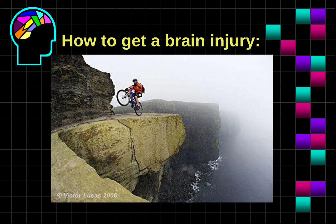
How to get a brain injury:
Picture 006.mpg

Why is NOR required?
80% of the brain is connected to visual processing

Why is NOR required?
Until the visual system is
rehabilitated, the patient is not
capable of reaching his/her highest
functional potential in post-injury
Activities of Daily Living

Why is NOR required?
Without accurate perception of the environment,
the individual is unable to make clear and effective
sense of what is happening
Visual processing problems have been shown to
be associated with poor performance in reading,
accident proneness and dependence in self-care
activities after ABI’s
Cognitive deficits are worse when visual
processing problems are not identified or treated
Cost effective rehabilitation

When is NOR required?
As soon as (ASAP) visual
dysfunction is identified or is
suspected in interfering with the
progress of rehabilitation.

NOR Serves Patients With:
Traumatic Brain Injury
Cerebrovascular accident
Cerebral palsy
Multiple sclerosis, Alzheimer’s, Parkinson’s,
other degenerative brain diseases /
syndromes
Anoxia (drowning, shock, anaphylaxis,
choking)

NOR Serves Patients With:
Chemical
Penetrating
Trauma patients with Ocular Involvement
Complicated Surgical Cases
• Prolonged Heart Surgery (anoxia)
Post-Op Brain Surgery
Congenital Brain Anomalies

Cerebrovascular Accidents(CVA) Statistics
3rd leading cause of death in developed
countries
794 per 100,000 prevalence
5% of the population over age 65 is affected by
stroke
80% survival rate after stroke

Traumatic Brain Injury (TBI)Statistics
400,000 people / year in the US acquire a TBI
and over 50% of these suffer from visual
impairment
1 out of 500 school children hospitalized each
year for a TBI
TBI is the leading cause of death and disability
of children and adolescents in US
“Mild” TBI often never see a health care
professional

Visual Processing Following Brain Injury
50-75% of severely brain injured individuals
had visual processing problems which
required further professional exploration by
an ophthalmologist or optometrist
(Gianutsos et al)

Visual Processing Following Brain Injury
38% of acute traumatic brain injured individuals had vergence difficulties
42% had vergence insufficiencies at a 3 year follow-up
(Cohen et al)

Visual Processing Following Brain Injury
79% of brain injured individuals had
strabismus with diplopia
(Mitchell, MacFarlane, and Cornell)

Visual Processing Following Brain Injury
20% of all stroke patients suffered from
some sort of visual problem
(Samo and Samo)

Visual Processing Following Brain Injury
20% of all individuals admitted to a
rehabilitation center had perceptual problems
and that these were clearly associated with
increased length of stay in the hospital and an
adverse affect on discharge and subsequent
placement
(Ferguson, McCarthy, Greenberg, and Feingenson)
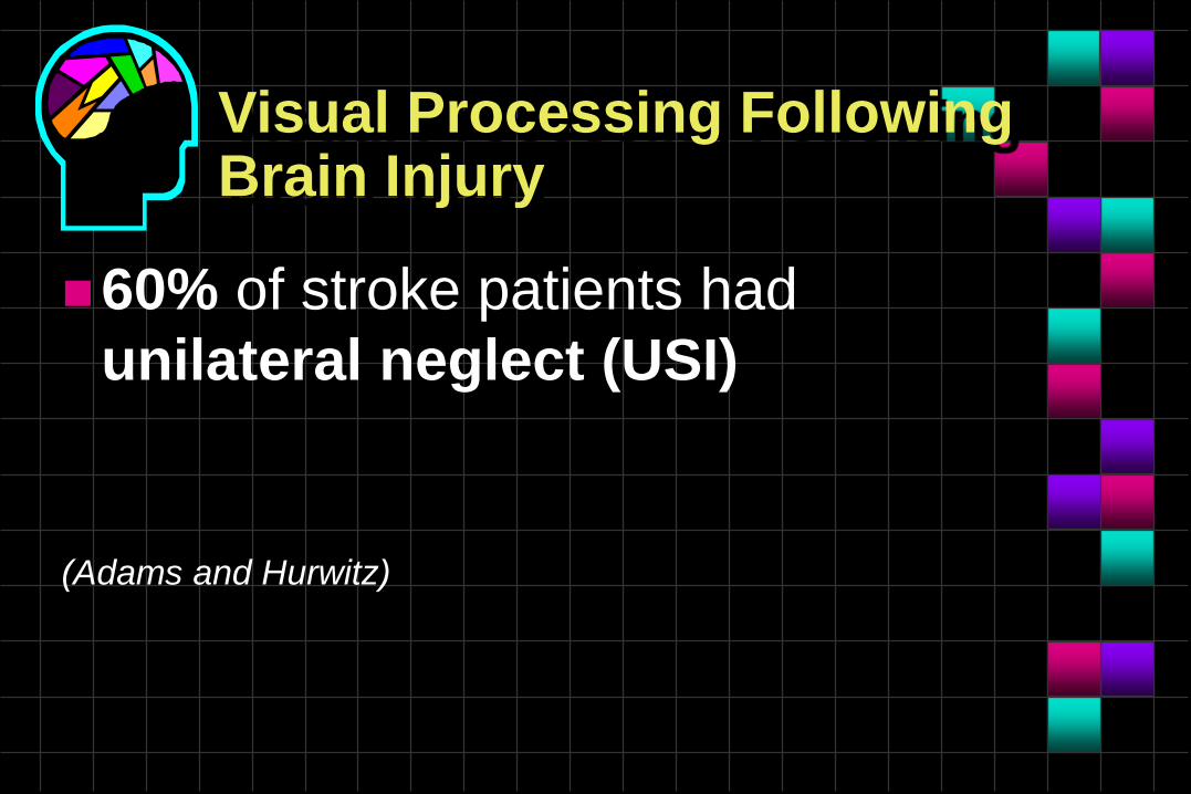
Visual Processing Following Brain Injury
60% of stroke patients had
unilateral neglect (USI)
(Adams and Hurwitz)

Visual Processing Following Brain Injury
59% of brain injured patients in
their study displayed visual
problems
(Schlageter, Gray, Hall, Shay, and Sammet)

How Big is Vision?
Vision is the dominant sense
Vision plays a critical role in all activities of daily
living (ADL’s)
Over 1 million nerve fibers exit each eye--this
represents about 70% of the sensory
nerve fibers in the entire body
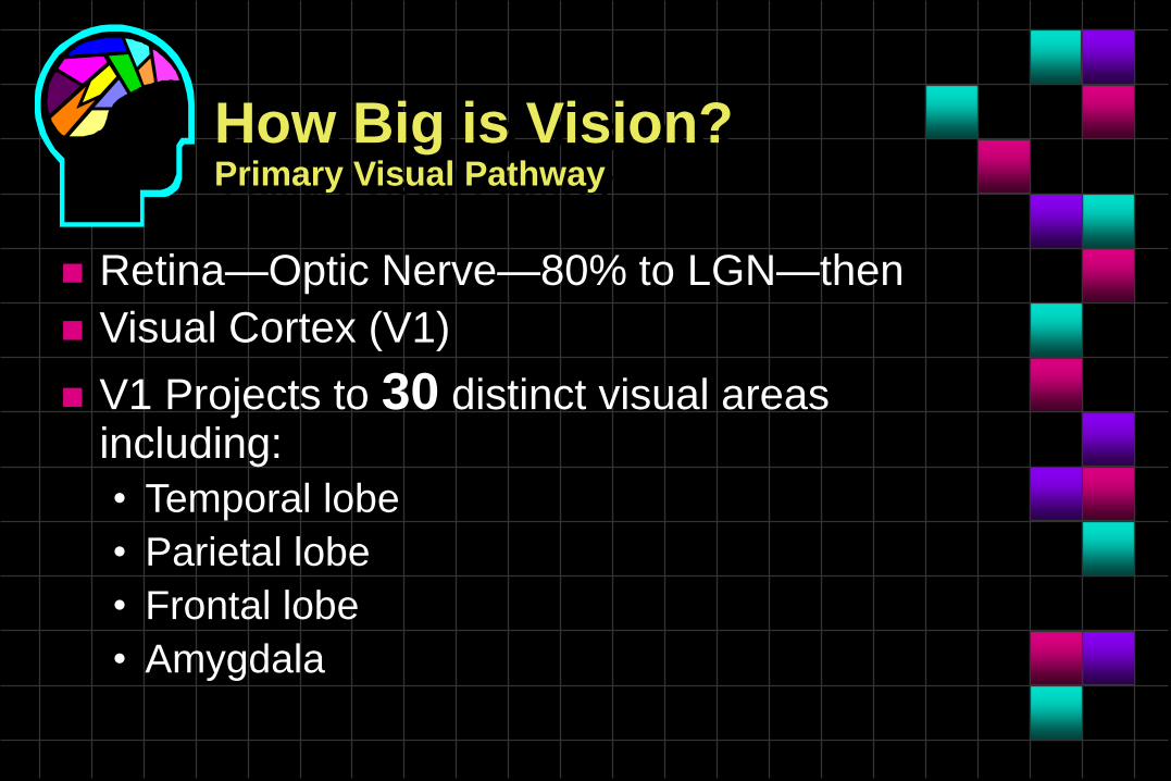
How Big is Vision?Primary Visual Pathway
Retina—Optic Nerve—80% to LGN—then
Visual Cortex (V1)
V1 Projects to 30 distinct visual areas including:
• Temporal lobe
• Parietal lobe
• Frontal lobe
• Amygdala

How Big is Vision?Primary Visual Pathway
These projections integrate with:
• Memory
• Audition
• Language
• Cognition
• Emotions

How Big is Vision?Collicular Visual Pathway
Retina—Optic Nerve—20% to Superior Colliculus
These fibers integrate with other sensory input
from:
• Vestibular
• Proprioception
• Kinesthetic
• Tactile
for balance and orientation.

Vision Problems Encountered

Medical Problems Encountered:
Dry eyes
Glaucoma
Cataracts
Diabetic Retinopathy
Macular Degeneration
Venous Occlusive Disease
Other Retinal Degenerative Diseases
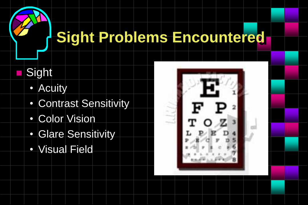
Sight Problems Encountered
Sight
• Acuity
• Contrast Sensitivity
• Color Vision
• Glare Sensitivity
• Visual Field

Functional Vision Problems Encountered
Ocular-Motor Problems
• Fixation
• Pursuits
• Saccades
• Nystagmus
• Vestibular-Ocular Reflex
• Pupils
• Accommodation

Functional Vision Problems Encountered
Visual Processing Problems
• Visual-Motor-Integration Skills
–Handwriting
–Reach & Grab
–Ambulating
–Driving

Functional Vision Problems Encountered
Visual Processing Problems
• Visual-Analysis Skills
– Visual Discrimination
– Visual Closure
– Visual Form Constancy
– Visual Figure Ground
– Visual-Spatial Relations
– Visual Memory
– Examples….

Visual Discrimination

Visual Closure

Visual Form Constancy

Visual Figure Ground
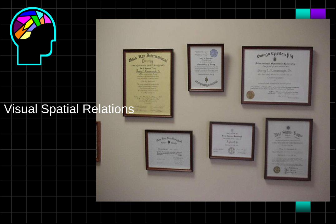
Visual Spatial Relations

Visual Memory

Visual Memory

Functional Vision Problems Encountered
Visual Processing Problems
• Visual–Motor Integration Skills
• Visual Analysis Skills
• Visual-Spatial Skills
–Visual Midline
–Unilateral Spatial Inattention (Neglect)
–Visual Multitasking

Visual Spatial ProblemsTHE MAJORS
“The Dangerous Ones”
• All 6 are Visual-Spatial Problems
1. Diplopia
2. Visual Field Defects
3. Unilateral Spatial Inattention (Neglect)
4. Visual Midline Shift Syndrome
5. Post-Traumatic Vision Syndrome
6. Visual Multitasking Dysfunction

THE MAJORS“The Dangerous Ones”“The Disabling Ones”
All 6 can slow the rehab process
All 6 can lead to greater risk of subsequent
injury and illness
All 6 can lead to adverse psychological
sequellae
All 6 can lead to more medications
All 6 can lead to greater dependence on others

Diplopia (double vision)
Occurs when the eyeballs do not point in the
same direction at the same time.
• Ocular Trauma
• Cranial Nerve Palsy (3rd, 4th, 6th)
• Supranuclear Palsy
• Decompensation
• Visual-Spatial Dysfunction

Diplopia
Not always reported by patient.
• May claim “blurry vision”
Patient may exhibit postural shifts
Can cause dizziness / vertigo
Can cause visual-motor-integration problems
Eliminated by covering one eye but, patching is
not the best treatment
NOW - EBO says do not patch!!!

Diplopia Treatments
compensatory prisms
partial occlusion
vision therapy
total occlusion
patching
surgery


Visual Field Defects
Unilateral
• Central scotoma
• Paracentral scotoma
Superior and Inferior
Concentric
Homonymous Hemianopsia
Unilateral Spatial Inattention

Homonymous Hemianopsia
Bump into objects on one side
Can’t find objects on one side of the room
Says they can’t see out of one eye
Is surprised by objects or people that seem to
pop into view
Is aware of their deficit
High accident risk for elderly

Unilateral Spatial Inattention
“Neglect”: term connotes a purposeful act
Damage to the right posterior parietal lobe
Mostly seen on left side (due to time of exam?)
Can be with or without field defect
Generally causes visual midline shift
Patient at high risk of injury
Patient has higher dependency
Patient is unaware of their deficit

Unilateral Spatial Inattention
Bumps into things on one side
Can’t find things on one side
May not shave or dress one side
May not use one side of body (even non-paralytic)
Orients and postures away from USI side
Doesn’t know to look in direction of USI
Is unaware that there is space that they can look into
May not be able to turn eyes in USI direction
Can be positional

Unilateral Spatial Inattention3 Key considerations:
Pt is prone to Extinction Phenomenon, in
terms of body and in the two visual spaces
Can occur in the presence or absence of a
basic motor or basic sensory dysfunction
Key is an unawareness or denial of the USI


THE MAJORS“The Dangerous Ones”
1. Diplopia
2. Visual Field Defects
3. Unilateral Spatial Inattention (Neglect)
4. Visual Midline Shift Syndrome
5. Post-Traumatic Vision Syndrome
6. Visual Multitasking Dysfunction

Visual Information Processing
Two separate processes
• Focal process (mostly the 80% of visual processing)
• Ambient process (mostly the 20% of visual processing)

Focal Visual Process
Through the macula (detailed vision)
Through the peripheral vision (detailed
attention process)
80% of the fibers leaving the eyes go to the
Occipital Cortex

Ambient Visual Process
20% of the fibers leaving the eyes go to the
Midbrain
Provides ambient information used for
• balance
• movement
• coordination
• posture
Lets you know where you are in space

Ambient Visual Process
Part of the sensory motor feedback loop in the midbrain
Matches up with information from the following systems for the purpose of orientation and acting as a master organizer of these other systems:
• kinesthetic
• proprioceptive
• vestibular
• Tactile
CAUSES A SUPPRESIVE FEEDBACK LOOP TO THE FOCAL PROCESSING SYSTEM (processing deficit basis in USI)

Major Ambient Visual Process Syndromes
Visual Midline Shift Syndrome (VMSS)
Post-traumatic Vision Syndrome (PTVS)

Visual Midline Shift Syndrome(VMSS)
Patient has mismatch in perception of space
related to self
Common Characteristics
• Dizziness or nausea
– (balance, coordination, and posture)
• Spatial disorientation
• Leaning forward / backward / one side
• Other associated Neuro-motor difficulties

Visual Midline Shift Syndrome(VMSS)
Common Characteristics
• Seeing the floor tilted
• Illusions of movement of environment
• Constantly walking to one side of hall or room
• Bumping into objects when walking
• Leaning to one side, back or forward or posture
changes


Post Trauma Vision Syndrome(PTVS)
Common Characteristics
• exotropia / exophoria (eyes turned out)
• convergence insufficiency
• accommodative insufficiency
• ocular motor dysfunction

Post Trauma Vision Syndrome(PTVS)
Common Characteristics
• Increased myopia or nearsightedness (almost
every ABI pt. over time)
• Low blink rate
• Spatial disorientation
• Poor fixation
• Unstable ambient vision
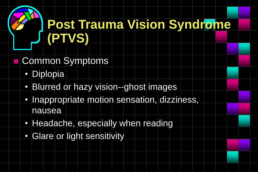
Post Trauma Vision Syndrome(PTVS)
Common Symptoms
• Diplopia
• Blurred or hazy vision--ghost images
• Inappropriate motion sensation, dizziness,
nausea
• Headache, especially when reading
• Glare or light sensitivity

Post Trauma Vision Syndrome(PTVS)
Common Symptoms
• Headache with light exposure (photophobia)
• Dry eye syndrome
• Reading problems
– including unstable focusing, poor line tracking,
losing place in text, poor comprehension and
memory of text

Visual Multitasking
Simultaneous Central / Peripheral Visual
Processing
• The ability to attend to a central visual target
and simultaneously respond to peripheral visual
stimuli.

Visual Multitasking Dysfunction
Virtually ALL brain-injured patients have some
dysfunction of this ability
They approach the world with a “tubular-vision”
approach, sequentially looking at one thing at a
time.
Significantly slows “Speed of Processing” down

Visual Multitasking Dysfunction
Anxious in crowds
Difficulty shopping
• Overwhelmed by too much stuff
• Gets lost easily in store
Driving is dangerous
• Poor lane maintenance
• Anxious with increased traffic and speed, or night driving
Words to live by
“Watch a patient from the minute they
enter your office and they will tell you
the problem before the examination”.
(Ret) rADM David Sullins, OD, DOS, FAAO

Sight Testing
Visual Acuity
Contrast Sensitivity
Color Vision
Glare Sensitivity
VEP visually evoked potential (only objective
measure)

Acuity

Contrast Sensitivity

Color Vision

Sight Testing
Visual Fields
• Confrontation
• Amsler Grid
• Static Threshold
• Campimetry (functional visual fields)
• Frequency Doubling
• Functional Binocular Testing—Accuvision, Dynavision, or Saccadic Fixator


Visual Motor Testing
Ocular Alignment
Ocular Motility
• Pursuits
• Saccades
• VOR
• Visagraph Recording
Accommodation
Pupils


Fusion Testing
Stereopsis
Suppression
Vergence Compensatory Ranges
Refractive Testing
Objective
Subjective
Distance and Near
Visual Spatial Testing
USI
Walking / ambulating through the office
Visual Midline Shift
Loop Poking/Capping Pen
Visual Scanning
Visual Multitasking
Visual Analysis Testing
MVPT, Line bisection, VMI, etc
Computer testing (visagraph)
VEP – monocular and binocular
Tachistoscope
VTS3 for vergence, motor field, etc.

Neuro-optometric management of the brain injured patient

NOR Therapy / Management
Lenses--noncompensatory
Prisms--noncompensatory
Selective occlusion
Filters
Vision therapy
(new) NOVA vision restoration therapy
Syntonic Light Therapy (recent article in USA WEEKEND Magazine)
Combinations of the above

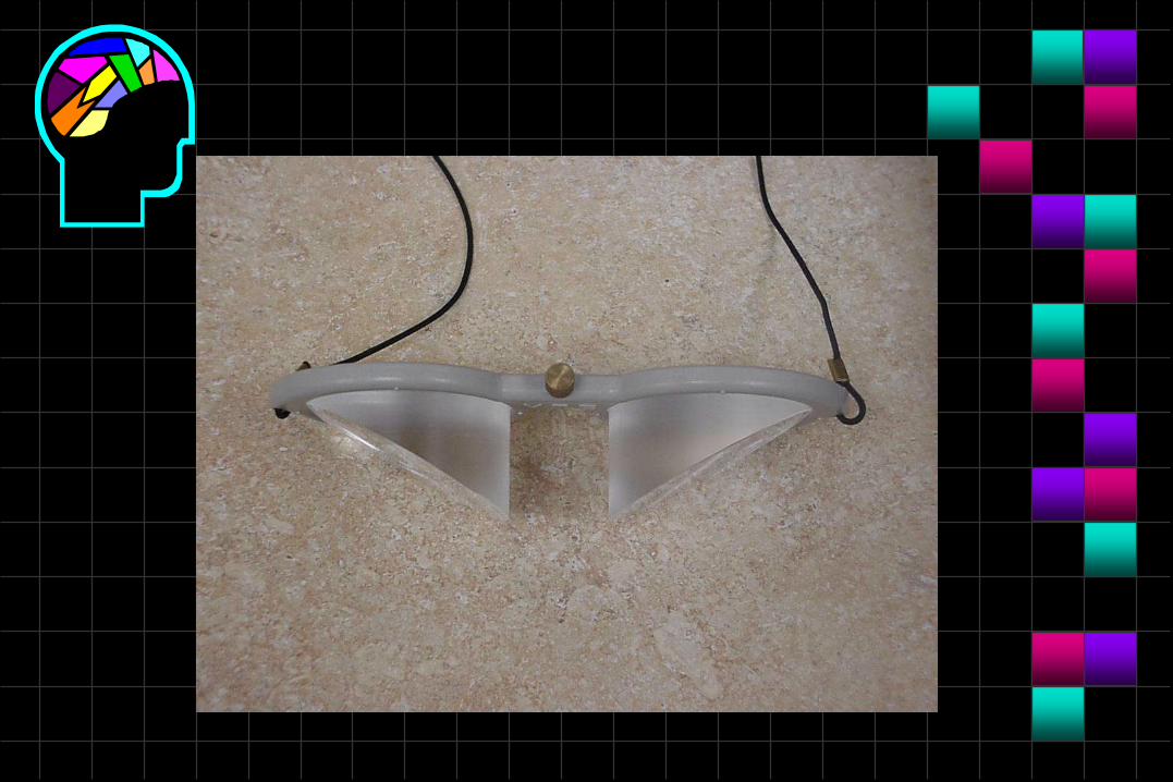
Peli
Prism


Treatment
Selective occlusion
• Binasal – Decreases peripheral processing
load to enable hypo-thalamic tract function
• Bitemporal – works with long standing
exofusional difficulties
• Sectoral – as needed
Treatment
Filters
• Glare
–Polarization – organizes light in straight lines to
reduce glare
–Tints
–Gradient
–Transition
–Combinations
Treatment
Vision therapy
• Eye movement – must be SLOW and in
developmentally correct order – vertical, horizontal,
rotational, then diagonal
• Vestibular (root of eye movements)
• Vergence ranges (range of motion for VOR)
• Awareness – peripheral awareness stationary/mobile
• Ambulation

Treatment
Syntonic Light Therapy
• Applied use of specific wavelengths to the eyes
for specific changes to blood chemistry
(affecting endocrine)
• Started in the 1900’s by Spitler, MD. Used in
many other countries
www.syntonicphototherapy.com

Summary of NOR Therapy / Management
Must Have Coordination of Care• OT
• PT
• ST
• Physiatrist
• Neurology
• Primary Care
• Voc Rehab
• VT
• Others

The Efficacy of Vision Rehabilitation Therapy
Training of visual scanning reduced neglect in
right damaged stroke patients
(Diller and Weinberg)
4/5 studies reviewed (150 patients) found
consistent beneficial effects of visual processing
skills training for the patients which were over
and above improvements resulting from
conventional therapy
(Gianutsos and Matheson)

The Efficacy of Vision Rehabilitation Therapy
Research indicates that visual processing
therapy has a possibility of reducing the size of
the visual field problem
(Warren)
Visual processing gains through therapy are
maintained a year after discharge
(Weinberg et al)

What is Vision Therapy?
“…the art and science of developing visual
abilities to achieve optimal visual performance
and comfort. It provides the patient with an
opportunity for both development and learning
experience.”
Journal of Behavioral Optometry, Vol. 1, 1990,
Number 3, p. 66-67.
What is Vision Therapy?
“…a clinical approach for correcting and ameliorating the
effects of eye movement disorders, non-strabismic
binocular dysfunctions, focusing disorders, strabismus,
amyblyopia, nystagmus, and certain visual perceptual
(information processing) disorders. The practice of
vision therapy entails a variety of non-surgical
therapeutic procedures designed to modify different
aspects of visual function.”
Advanced Therapist Vol. 34, Number 1, p. 131

What is Vision Therapy?
“…creates a visual environment in which the
patient may modify their visual behavior and
alter their visual performance to meet the visual
needs of their specific environment.”
Rehabilitation Techniques for Binocular
Dysfunctions by Richman and Cohen, p. 1.

What is Vision Therapy?
“…vision therapy arranges conditions….so that the patient can learn many visual abilities which will allow them to take in, use and understand more information over a large area in less time. It allows one to use his vision to learn more and succeed better at his task or pastime, whatever it may be. Rather than exercises for the eyes, it involves an awareness of movement, control of movement and, ultimately, the automatic movement of eyes integrated with movement in many of the body systems.”
Learning to See and Seeing to Learn—Orem, R.C., p. 84
What is Vision Therapy?
“…is the art and science of developing visual
abilities to achieve optimal visual performance
and comfort.”
R M Greenburg, J. American Optometric
Association, Jan 1985, 56 (1): 57-58

What is Vision Therapy?
“…is based on a medically necessary plan of
treatment which is designed to improve specific
vision dysfunctions determined by standardized
diagnostic criteria. Treatment plans encompass
lenses, prisms, occlusion and other appropriate
materials, modalities and equipment.”
AOA Definition of Vision Therapy

Locating a Doctor
To locate a Neuro-Optometric Rehabilitation
Specialist
• www.covd.org vision development
• www.nora.cc neuro rehabilitation
• www.drclopton.com

Jason Clopton, OD, FCOVDCenter of Vision Development
(931) 372-2020
1445 East 10th street
Cookeville, TN 38501 USA

References
[1] Padula WV, Shapiro JB, Jason P. Head injury causing post trauma vision syndrome. New England Journal of Optometry 1988;41(2):16–21.
[2] Schlageter K, Gray B, Hall K, et al. Incidence and treatment of visual dysfunction in traumatic brain injuries. Brain Inj 1993;7:439–48.
[3] Portas CM, Rees G, Howseman AM, et al. A specific role for the thalamus in mediating the interaction of attention and arousal in humans. J Neurosci 1998;18(21):8979–89.
[4] Klemm WR. Understanding neuroscience. St. Louis (MO): Mosby; 1996. p. 101–52.
[5] Casagrande VA, Royal D. Parallel visual pathways in a dynamic system. In: Kaas JH, Collins CE, editors. The primate visual system. Philadelphia: CRC Press; 2003. p. 1–28.
[6] Van Gelder RN, Wee R, Lee JA, et al. Reduced pupillary light responses in mice lacking cryptochromes. Science 2003;299(5604):222

References
[6] Van Gelder RN, Wee R, Lee JA, et al. Reduced pupillary light responses in mice lacking cryptochromes. Science 2003;299(5604):222.
[7] Brainard G, Hannifin J, Grison J, et al. Action spectrum for melatonin regulation in humans: evidence for a novel circadian photoreceptor. Neuroscience 2001;21(16):6405–12.
[8] Pickard G. Studies of circadian rhythms. As reported in Insight: The College of Veterinary Medicine and Biomedical Sciences, Colorado State University, Fort Collins, Colorado 2002;29(2):4–5.
[9] Thapan K, Arendt J, Skene DJ. An action spectrum for melatonin suppression: evidence for a novel non-rod, non-cone photoreceptor system in humans. J Physiol 2001;535(1):261–7.
[10] Moseley MJ, Bayliss SC, Fielder AR. Light transmission through the human eyelid: in vivo measurement. Ophthalmic Physiol Opt 1988;8(2):229–30.
[11] Zelinsky. Rehabilitation. Phys Med Rehabil Clin N Am. 18 (2007) 87–107
![Brain Computer Interface for Neuro-rehabilitation With Deep … · neuro-rehabilitation,measuredontheFugl-MeyerAssessmentscale [30]. Besides, it can boost the interest, motivation](https://img.dokumen.tips/doc/110x75/5edf38a0ad6a402d666a91f7/brain-computer-interface-for-neuro-rehabilitation-with-deep-neuro-rehabilitationmeasuredonthefugl-meyerassessmentscale.jpg)