Embed Size (px)
Citation preview

Adeel Husain PGY 3Loma Linda UniversityDept of Orthopaedic
Surgery
Open Fracture
s

Definition• Break in the skin and underlying soft tissue
leading directly into or communicating with the fracture and its hematoma

• Last century, high mortality with open fractures of long bones
• Early amputation in order to prevent death• WWI, mortality of open femur fractures > 70%• 1939 Trueta “closed treatment of war fractures”– Included open wound treatment and then enclosure
of the extremity in a cast– “Greatest danger of infection lay in muscle, not bone”
Trueta J: "Closed" treatment of war fractures, Lacet 1939;1:1452-1455
History

• 1943 PCN on the battlefield quickly reduced rate of wound sepsis
• Delayed closure of wounds• Hampton: closure btwn 4th and 7th day• Larger defects continued to be left open to
heal by secondary intention
Hampton OP Jr: Basic principles in management of open fractures; JAMA 1955; 159:417-419
History

• Advances shifted the focus– Preservation of life and limb preservation of
function and prevention of complications
• However, amputation rates still exceed 50% in the most severe open tibial fractures assoc with vascular injury*
Lange RH, Bach AW, Hansen ST et al: Open tibial fractures with associated vascular injuries: prognosis for limb salvage. J Trauma; 25(3):203-208
History

Epidemiology
• 3% of all limb fractures• 21.3 per 100,000 per year
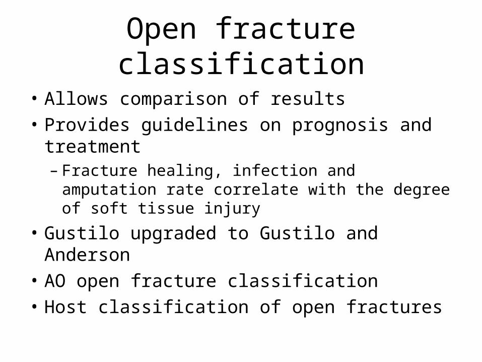
Open fracture classification
• Allows comparison of results• Provides guidelines on prognosis and
treatment– Fracture healing, infection and amputation rate
correlate with the degree of soft tissue injury
• Gustilo upgraded to Gustilo and Anderson• AO open fracture classification• Host classification of open fractures
Gustilo and Anderson Classification
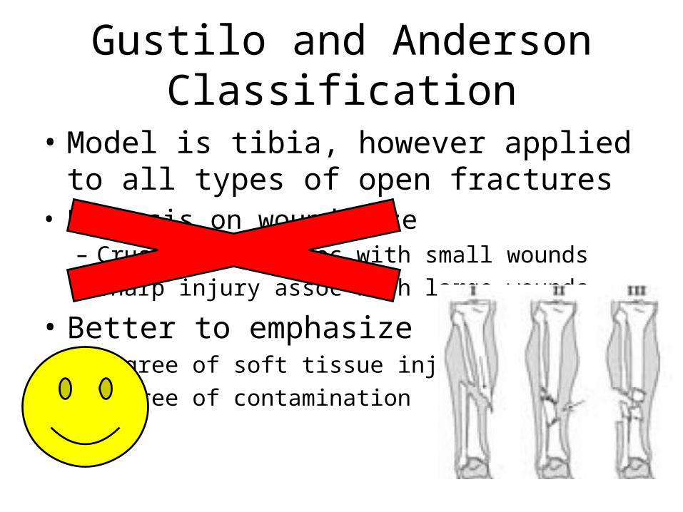
• Model is tibia, however applied to all types of open fractures
• Emphasis on wound size– Crush injury assoc with small wounds– Sharp injury assoc with large wounds
• Better to emphasize– Degree of soft tissue injury– Degree of contamination

Type 1 Open Fractures
• Inside-out injury• Clean wound• Minimal soft tissue damage• No significant periosteal
stripping

Type 2 Open Fractures• Moderate soft tissue
damage• Outside-in• Higher energy• Some necrotic muscle• Some periosteal
stripping

Type 3a Open Fractures• High energy• Outside-in• Extensive muscle
devitalization• Bone coverage with
existing soft tissue

Type 3b Open Fractures
• High energy• Outside in• Extensive muscle
devitalization • Requires a flap for
bone coverage and soft tissue closure
• Periosteal stripping

Type 3c Open Fractures
• High energy• Increased risk of
amputation and infection
• Any grade 3 with major vascular injury
requiring repair

Why use this classification?
• Grades of soft tissue injury correlates with infection and fracture healing
Grade 1 2 3A 3B 3C
InfectionRates 0-2% 2-7% 10-25% 10-50% 25-50%
Fracture Healing (weeks)
21-28 28-28 30-35 30-35
Amputation Rate 50%

Gustilo and AndersonBowen and Widmaier*
• 2005 Host classification predicts infection after open fracture– Gustilo and Anderson classification and the
number of comorbidities predict infection risk– 174 patients with open fractures of long bones– Sorted into three classes based on 14
immunocompromising factors• Age>80, current nicotine use, DM, malignancy,
pulmonary insufficiency, systemic immunodeficiency, etc
Bowen TR, Widmaier JC. Host classification predicts infection after open fracture. Clin Orthop Relat Res. 2005;433:205-11.

What they found…
• Patients with any compromising risk factor has increased risk of infection
• May benefit from additional therapies that decrease the risk of infection.
Bowen TR, Widmaier JC. Host classification predicts infection after open fracture. Clin Orthop Relat Res. 2005;433:205-11.
Class Compromising factors Infection rates
A 0 4%
B 1-2 15%
C 3 or more 31%

Gustilo Classification: a simple and useful tool, but is it accurate?• 1994 Brumback et al. • 125 randomized open fractures• 245 surgeons of various levels of training• 12 cases of open tibia fractures, videos used
• Interobserver agreement poor– Range 42-94% for each fracture
• Ortho attendings - 59% agreement• Ortho Trauma Fellowship trained attendings - 66% agreement
Brumback RJ, Jones AL (1994) Interobserver agreement in the classification of open fractures of the tibia. The results of a survey of two hundred and forty-five orthopaedic surgeons. J Bone and Joint Am; 76(8):1162–1166.

So……….
• Fracture type should not be classified in the ER
• Most reliably done in the OR at the completion of primary wound care and debridement

Microbiology• Most acute infections are caused by pathogens
acquired in the hospital• 1976 Gustilo and Anderson– most infections in their study of 326 open fxs
developed secondarily• When left open for >2wks, wounds were prone to
nocosomial contaminants such as Pseudomonas and other GN bacteria
• Currently most open fracture infections are caused by GNR and GP staph
Gustilo RB, Anderson JT: Prevention of Infection in the Treatment of One Thousand and Twenty-five Open Fractures of Long Bones; JBJS, 58(4):453-458, June 1976

Nocosomial infection?!!!! • Only 18% of infections were caused by the
same organism initially isolated in the perioperative cultures*
• Carsenti-Etesse et al. 1999– 92% of open fracture infections were caused by
bacteria acquired while the patient was in the hospital**
*Patzakis MJ, Wilkins J, Moore TM: Considerations in reducing the infection rate in open tibial fractures. Clin Orthop Relat Res. 1983 Sep;(178):36-41.*Patzakis MJ, Bains RS, Lee J, Shepherd L, Singer G, Ressler R, Harvey F, Holtom P: Prospective, randomized, double-blind study comparing single antibiotic therapy, ciprofloxacin, to combo antibiotic therapy in open fracture wounds. J Orthop Trauma. 2000 Nov;14(8):529-33. **Carsenti-Etesse H, Doyon F, Desplaces N, Gagey O, Tancrede C, Pradier C, Dunais B, Dellamonica P. Epidemiology of bacterial infection during management of open leg fractures. Eur J Clin Microbiol Infect Dis. 1999;18:315-23.
Cover the
wounds quickly

Common bacteria encountered with open fractures
Blunt Trauma, Low Energy GSW
Staph, Strept
Farm Wounds Clostridia
Fresh Water Pseudomonas, Aeromonas
Sea Water Aeromonas, Vibrios
War Wounds, High Energy GSW Gram Negative

What systemic antibiotic?
1st Gen Ceph
Gent PCN
Grade 1
Grade 2 +/-
Grade 3 +/-
Farm/War Wounds
(Gustilo, et al; JBJS 72A 1990)

Antibiotic comparisons• No difference btwn clindamycin and cefazolin*• Patzakis et al. **– For type 1&2, cipro = cefamandole+gentamicin– For type 3, cipro worse (31% vs 7.7% infection)
• Cipro and other fluoroquinolones inhibit osteoblast activity and fracture healing***
*Benson DR, Riggins RS, Lawrence RM, Hoeprich PD, Huston AC, Harrison JA. Treatment of open fractures: a prospective study. J Trauma. 1983;23:25-30.**Patzakis MJ, Bains RS, Lee J, Shepherd L, Singer G, Ressler R, Harvey F, Holtom P. Prospective, randomized, double-blind study comparing single-agent antibiotic therapy, ciprofloxacin, to combination antibiotic therapy in open fracture wounds . J Orthop Trauma. 2000;14:529-33.***Holtom PD, Pavkovic SA, Bravos PD, Patzakis MJ, Shepherd LE, Frenkel B. Inhibitory effects of the quinolone antibiotics trovafloxacin, ciprofloxacin, and levofloxacin on osteoblastic cells in vitro. J Orthop Res. 2000;18:721-7.***Huddleston PM, Steckelberg JM, Hanssen AD, Rouse MS, Bolander ME, Patel R. Ciprofloxacin inhibition of experimental fracture healing. J Bone Joint Surg Am. 2000;82:161-73.

When and for how long?• Start abx as soon as possible*– Less than 3 hours 4.7 % infection rate– Greater than 3 hours 7.4%
• No difference btwn 1 and 5 days of post op abx treatment**
• Mass Gen recommended treatment:***– Cefazolin Q 8 until 24 hours after wound closed– Gentamicin or levofloxacin added for type 3
*Patzakis MJ, Wilkins J. Factors influencing infection rate in open fracture wounds. Clin Orthop Relat Res. 1989;243:36-40.**Dellinger EP, Caplan ES, Weaver LD, Wertz MJ, Brumback R, Burgess A, Poka A, Benirschke SK, Lennard S, Lou MA. Duration of preventive antibiotic administration for open extremity fractures. Arch Surg. 1988;123:333-9.***Okike K, Bhattacharyya T: Trends in the management of open fractures. A critical analysis. J Bone Joint Surg. 2006 Dec;88(12):2739-48.

Local antibiotic therapy
• High abx conc within the wound and low systemic conc– Reduces risk of systemic side effect
• Vancomycin or aminoglycosides– Heat stable– Available in powder form– Active against suspected pathogens
Eckman JB Jr, Henry SL, Mangino PD, Seligson D. Wound and serum levels of tobramycin with the prophylactic use of tobramycin-impregnated polymethylmethacrylate beads in compound fractures. Clin Orthop Relat Res. 1988; 237:213-5.

Antibiotics - locally
• Prevents secondary contamination by nocosomial pathogens
• Useful adjunct to systemic abx• Potential for abx impregnated bone graft,
bone graft substitute, and abx coated IMN
Ostermann PA, Seligson D, Henry SL: Local antibiotic therapy for severe open fractures. A review of 1085 consecutive cases; J Bone Joint Surg Br. 1995 Jan;77(1):93-7.
Antibiotic Infection Rate
IV Abx 12%
IV Abx + local aminoglycoside impregnated PMMA beads
3.7%

Antibiotic Beads• Pros– Very high levels of
antibiotics locally– Dead space
management
• Cons– Requires removal– Limited to heat
stable antibiotics– Increased drainage
from wound

Goals of treatment
• 1. preserve life• 2. preserve limb• 3. preserve function
• Also….– Prevent infection– Fracture stabilization– Soft tissue coverage

Stages of care for open fractures

Initial assessment & management• ABC’s• Assess entire patient• Careful PE, neurovasc• Abx and tetanus• Local irrigation 1-2 liters• Sterile compressive dressings• Realign fracture and splint• Do not culture wound in the ED*
– 8% of bugs grown caused deep infection
– cultures were of no value and not to be done
• Recheck pulse, motor and sensation
Lee J. Efficacy of cultures in the management of open fractures. Clin Orthop Relat Res. 1997;339:71-5.
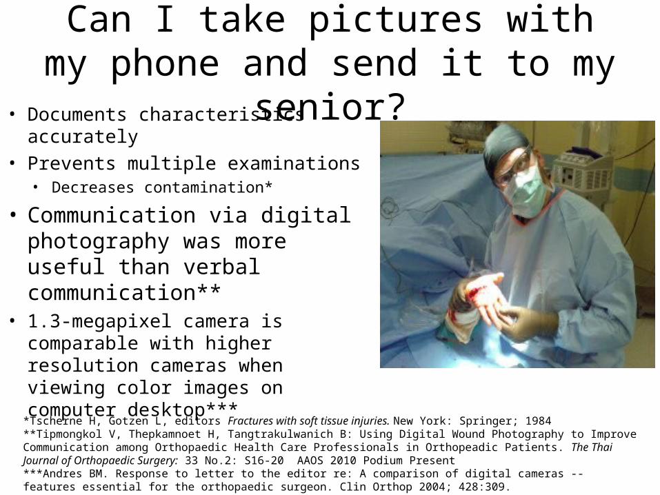
Can I take pictures with my phone and send it to my senior?
• Documents characteristics accurately• Prevents multiple examinations
• Decreases contamination*
• Communication via digital photography was more useful than verbal communication**
• 1.3-megapixel camera is comparable with higher resolution cameras when viewing color images on computer desktop***
*Tscherne H, Gotzen L, editors Fractures with soft tissue injuries. New York: Springer; 1984**Tipmongkol V, Thepkamnoet H, Tangtrakulwanich B: Using Digital Wound Photography to Improve Communication among Orthopaedic Health Care Professionals in Orthopeadic Patients. The Thai Journal of Orthopaedic Surgery: 33 No.2: S16-20 AAOS 2010 Podium Present***Andres BM. Response to letter to the editor re: A comparison of digital cameras -- features essential for the orthopaedic surgeon. Clin Orthop 2004; 428:309.

Primary surgery
• Objectives of initial surgical management– Preservation of life and limb– Wound debridement– Definitive injury assessment– Fracture stabilization
Stages of open fracture management in the OR

Surgical emergency! • 1898 Friedrich guinea pigs
– Take to the OR within 6-8 hours*• 1973 Robson:
bacteria multiply in contaminated wounds **– 105 organisms/gram of tissue is the
infection threshold– Reached at 5.17 hours
• 1995 Kindsfater et al: – 47 G2/3 fxs at 4.8 months out….
• Less than 5 hrs 7% infection• Greater than 5 hrs 38% infection
– However G3 fxs were treated later
*Friedrich PL. Die aseptische Versorgung frischer Wundern. Arch Klin Chir. 1898;57:288-310.**Robson MC, Duke WF, Krizek TJ. Rapid bacterial screening in the treatment of civilian wounds . J Surg Res. 1973;14:426-30.

Or not?.... Calling the “6 hour rule” into question• 1993 Bednar and Parikh…. No significant difference *
– 3.4% vs 9%; 82 open femoral/tibial fxs• 2004 Ashford et al…. No significant difference **
– 11% vs 17%; pts from the austrailian outback• 2004 Spencer et al.... No significant difference ***
– 10.1% vs 10.9%; 142 open long bone fxs from UK• 2003 Pollack and the LEAP investigators…. No correlation****
– 315 open long bone fxs• 2005 Skaggs et al….No significant difference *****
– children with all types of open fractures; 554 open fractures
*Bednar DA, Parikh J. Effect of time delay from injury to primary management on the incidence of deep infection after open fractures of the lower extremities caused by blunt trauma in adults. J Orthop Trauma. 1993;7:532-5.**Ashford RU, Mehta JA, Cripps R. Delayed presentation is no barrier to satisfactory outcome in the management of open tibial fractures. Injury. 2004;35:411-6.***Spencer J, Smith A, Woods D. The effect of time delay on infection in open long-bone fractures: a 5-year prospective audit from a district general hospital. Ann R Coll Surg Engl. 2004;86:108-12.****Pollack AN, Castillo RC, Jones AL, Bosse MJ, MacKenzie EJ, and the LEAP Study Group. Time to definitive treatment significantly influences incidence of infection after open high-energy lower-extremity trauma. Read at the Annual Meeting of the Orthopaedic Trauma Association; 2003 Oct 9-11; Salt Lake City, UT.*****Skaggs DL, Friend L, Alman B, Chambers HG, Schmitz M, Leake B, Kay RM, Flynn JM. “The Effect of Surgical Delay on Acute Infection Following 554 Open Fractures in Children.” JBJS-A 2005. 87:8-12
No significant difference
before or after 6 hours!!!

Do we even need to do operative debridement?
• Orcutt et al... No significant difference, BUT…*– 50 type 1 &2 open fractures– less infection in nonoperative group (3% vs 6%)– Less delayed union in nonop group (10% vs 16%)
• Yang et al….0% infections **– 91 type 1 open fractures treated without I&D
*Orcutt S, Kilgus D, Ziner D. The treatment of low-grade open fractures without operative debridement. Read at the Annual Meeting of the Orthopaedic Trauma Association; 1988 Oct 28; Dallas, TX.**Yang EC, Eisler J. “Treatment of Isolated Type 1 Open Fractures: Is Emergent Operative Debridement Necessary?” Clin Orthop Relat Res 2003. 410: 289-294.
Do we even need to
debride low grade open fractures?

However, after review of all literature….….
• Okike et al. states….• “Thorough operative debridement is the
standard of care for all open fractures.”• “Even if the benefits of formal I&D were
insignificant for low grade fractures, operative debridement is still required for proper wound classification.”
• “Open fractures graded on the basis of superficial characteristics are often misclassified.”
• Huge risk not to explore and debride!
Okike K, Bhattacharyya T: Trends in the management of open fractures. A critical analysis. J Bone Joint Surg Am. 2006 Dec;88(12):2739-48.

URGENTLY debride, not EMERGENTLY• Time to OR is probably less important than:*
– Adequacy of debridement– Time to soft tissue coverage
• Timing depends on….**– Is patient stable?– Is the OR prepared?– Is appropriate assistance available?
• Ortho trained scrub techs, assistant surgeons, xray techs, and other OR staff
• 2005 Skaggs et al:***– If after 10pm, keep until the morning! Or at least
within 24 hours.– Unless….
• neurovasc compromise• horrible soft tissue contamination• compartment syndrome
*Okike K, Bhattacharyya T: Trends in the management of open fractures. A critical analysis. J Bone Joint Surg. 2006 Dec;88(12):2739-48.**Werner CM, Pierpont Y, Pollak AN: The urgency of surgical débridement in the management of open fractures. J Am Acad Orthop Surg. 2008 Jul;16(7):369-75.***Stewart DJ, Kay RM, Skaggs DL: Open Fractures in Children. Principles of Evaluation and Management. JBJS-A. 2005;87:2784-2798.
Within 24
hours
Within6
hours

I&D in the OR• Trauma scrub– Soap and saline to remove gross debris
• “Zone of injury”– Skin wound is the window through which
the true wound communicates with the exterior
• Extend the traumatic wound– Excise margins– Resect muscle and skin to healthy tissue
• color, consistency, capacity to bleed and contractility
• Bone ends are exposed and debrided• Irrigate• Serial debridements?– If needed, 2nd or 3rd debridement after 24-
48 hours should be planned

The Irrigation• Amount– No good data, copious is better– Animal studies show improved
removal of particulate matter and bacteria but effect plateaus
– Irrigation bags typically contain 3 L of fluid
– Anglen recommends:*• 3L (one bag) for type 1 • 6L (two bags) for type 2 • 9L (three bags) for type 3
*Anglen JO. “Wound Irrigation in Musculoskeletal Injury.” JAAOS 2001. 9: 219-226.

How to deliver the irrigation?(what animal studies show)
• Bulb Syringe vs Pulsatile Lavage– Pulsatile lavage
• Detrimental for early bone healing – this is no longer present at 2 wks*
• More soft tissue destruction**
• More effective in removing particulate matter and bacteria***
• High or low pressure?– Higher pressure
• Better bone cleaning• Worse soft tissue cleaning• Slows bone healing
*Dirschl DR, Duff GP, Dahners LE, Edin M, Rahn BA, Miclau T. “High Pressure Pulsatile Lavage Irrigation of Intraarticular Fractures: Effects on Fracture Healing.” JOT 1998. 12(7): 460-463.**Boyd JI, Wongworawat MD. “High-Pressure Pulsatile Lavage Causes Soft Tissue Damage.” CORR 2004. 427: 13-17***Bhandari M, Schemitsch EH, Adili A, Lachowski RJ, Shaughnessy SG. “High and Low Pressure Pulsatile Lavage of Contaminated Tibial Fractures: An in vitro Study of Bacterial Adherence and Bone Damage.” JOT 1999. 13: 526-533.

Antibiotics in the irrigation?
• Antibiotics (bacitracin and/or neomycin)– Mixed results, controversial– Costly • bacitracin alone around $500/washout
– ?? Causing resistance– Wound healing problems?– Few reported cases of anaphylaxis– Anglen: “No proven value in the care of open
fracture wounds…some risk, albeit small.”
No proven benefit!
*Anglen JO. “Wound Irrigation in Musculoskeletal Injury.” JAAOS 2001. 9: 219-226.
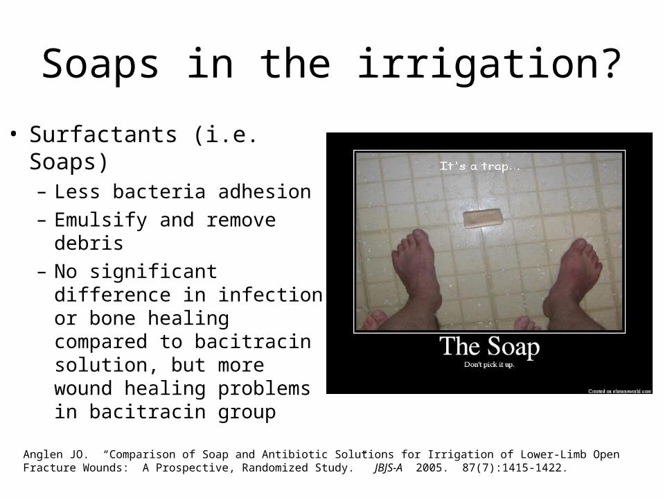
Soaps in the irrigation?
• Surfactants (i.e. Soaps)– Less bacteria adhesion– Emulsify and remove debris– No significant difference in
infection or bone healing compared to bacitracin solution, but more wound healing problems in bacitracin group
Anglen JO. “Comparison of Soap and Antibiotic Solutions for Irrigation of Lower-Limb Open Fracture Wounds: A Prospective, Randomized Study.” JBJS-A 2005. 87(7):1415-1422.

Level 4 evidence based recommendations
• 1st washout, highly contaminated
Soap solution• Repeat washout of clean wounds
Saline• Infected wounds
Soap, then antibiotic
*Anglen JO. “Wound Irrigation in Musculoskeletal Injury.” JAAOS 2001. 9: 219-226.

Wound closure after contaminated fracture• Timing and technique is
controversialOPEN WOUND should be left OPEN!
– Prevents anaerobic conditions in wound: Clostridium
– Facilitates drainage– Allows repeat debridement
Zalavras CG, Patzakis MJ:Open fractures: evaluation and management. J Am Acad Orthop Surg. 2003 May-Jun;11(3):212-9.
Dubunked!

To close or not to close?• Recently, renewed interest
in primary closure• Collinge, OTA 2004• Moola, OTA 2005• Russell, OTA 2005• DeLong, J Trauma 2004/• Bosse, JAAOS 2002
– Improved abx management– Better stabilization– Less morbidity– Shorter hospital stay, lower
cost– NO increase in wound
infection• These wounds are at
higher risk of clostridia perfringens if they do get infected.
• 1999 Delong et al: 119 open fxs– No significant difference
• delayed/nonunion and infection rates btwn immediate and delayed closure
– Immediate closure is a “viable option”
DeLong WG Jr, Born CT, Wei SY, Petrik ME, Ponzio R, Schwab CW: Aggressive treatment of 119 open fracture wounds. J Trauma. 1999 Jun;46(6):1049-54.
infection rate 7%
Overall delayed/nonunion rate 16%
Grade Percent of primary closures
1 88%
2 86%
3a 75%
3b 33%
3c 0%

Contraindications to primary closure
• Inadequate debridement• Gross contamination• Farm related or freshwater immersion injuries• Delay in treatment >12 hours• Delay in giving abx• Compromised host or tissue viability
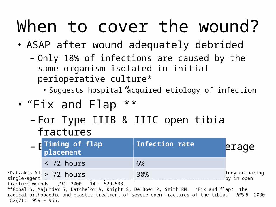
When to cover the wound?• ASAP after wound adequately debrided– Only 18% of infections are caused by the same organism
isolated in initial perioperative culture*• Suggests hospital acquired etiology of infection
• “Fix and Flap”**– For Type IIIB & IIIC open tibia fractures– Early if not immediate flap coverage
•Patzakis MJ, Bains RS, Lee J, et al. “Prospective, randomized, double-blind study comparing single-agent antibiotic therapy, ciprofloxacin, to combination antibiotic therapy in open fracture wounds.” JOT 2000. 14: 529-533.**Gopal S, Majumder S, Batchelor A, Knight S, De Boer P, Smith RM. “Fix and flap: the radical orthopaedic and plastic treatment of severe open fractures of the tibia.” JBJS-B 2000. 82(7): 959 – 966.
Timing of flap placement Infection rate
< 72 hours 6%
> 72 hours 30%

Dressings• Temporary closures – rubber bands
• Wet to dry dressings
• Semi-permeable membranes
• Antibiotic bead pouch
• VAC

VAC
• Vacuum assisted wound closure– Recommended for temporary management– Mechanically induced negative pressure in a closed
system– Removes fluid from extravascular space– Reduced edema– Improves microcirculation– Enhances proliferation of reparative granulation tissue
• Open cell polyurethane foam dressing ensures an even distribution of negative pressure
-Webb LX: New techniques in wound management: vacuum-assisted wound closure. J Am Acad Orthop Surg. 2002 Sep-Oct;10(5):303-11.-Dedmond BT, Kortesis B, Punger K, Simpson J, Argenta A, Kulp B, Morykwas M, Webb L. “The use of Negative Pressure Wound Therapy in the Temporary Treatment of Soft Tissue Injuries associated with High Energy Open Tibial Shaft Fractures.” JOT. 2007

Types of fracture stabilization• Splint– Good option if operative
fixation not required
• Internal fixation– Wound is clean and soft tissue
coverage available
• External fixation– Dirty wounds or extensive soft
tissue injury

Fracture stabilization
• Gustilo type 1 injury can be treated the same way as a comparable closed fracture
• Most cases involve surgical fixation• Outcome is similar to closed counterparts
Fracture stabilization
• Gustilo type 2&3 usually displaced and unstable– dictate surgical fixation
• Restore length, alignment, rotation and provide stability – ideal environment for soft tissue healing and reduces
wound infection– reduces dead space and hematoma volume
• Inflammatory response dampened• Exudates and edema is reduced• Tissue revascularization is encouraged
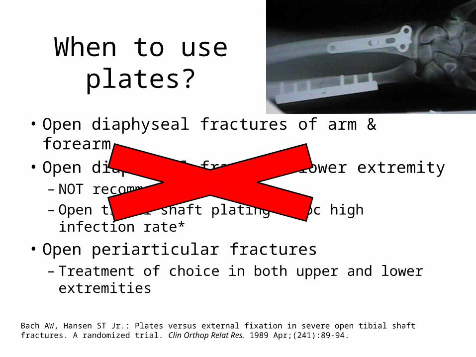
When to use plates?
• Open diaphyseal fractures of arm & forearm• Open diaphyseal fractures lower extremity – NOT recommended– Open tibial shaft plating assoc high infection rate*
• Open periarticular fractures – Treatment of choice in both upper and lower
extremities
Bach AW, Hansen ST Jr.: Plates versus external fixation in severe open tibial shaft fractures. A randomized trial. Clin Orthop Relat Res. 1989 Apr;(241):89-94.

When to use IM nails?• Treatment of choice for most
diaphyseal fractures of the lower extremity
• Inserted without disrupting the already injured soft tissue envelope
• Preserves the remaining extra osseous blood supply to cortical bone
• Malunion is uncommon

To ream or not to ream?• Does reaming cause additional damage to the
endosteal blood supply?• Solid IM nails without reaming has a lower risk of
infection that tubular nails with a large dead space*• However reamed IM nails are biomechanically
stronger and can reliably maintain fracture reduction if statically locked
• 2000 Finkemeier et al. – reamed vs unreamed interlocked nails of open tibias– NO statistical difference in outcome and risk of
complication***Melcher GA, Claudi B, Schlegel U, Perren SM, Printzen G, Munzinger J.Influence of type of medullary nail on the development of local infection. An experimental study of solid and slotted nails in rabbits; .J Bone Joint Surg Br. 1994 Nov;76(6):955-9.**Keating JF, O'Brien PJ, Blachut PA, Meek RN, Broekhuyse HM: Locking intramedullary nailing with and without reaming for open fractures of the tibial shaft. A prospective, randomized study. J Bone Joint Surg Am. 1997 Mar;79(3):334-41.**Finkemeier CG, Schmidt AH, Kyle RF, Templeman DC, Varecka TF: A prospective, randomized study of intramedullary nails inserted with and without reaming for the treatment of open and closed fractures of the tibial shaft. J Orthop Trauma. 2000 Mar-Apr;14(3):187-93.

When to use external fixation?
• Diaphyseal fractures not amenable to IM nails
• Ring fixators for periarticular fractures
• Temporary joint spanning ex fix is popular for knee, ankle, elbow and wrist
• If temporary, plan for conversion to IM nail within 3 weeks

Ex-fix: Weigh the pros and cons!• Historically was definitive treatment• Now, more commonly as temporary fixation• Can be applied almost always and everywhere• Severe soft tissue damage and contamination
• Advantages:– Easy and quick– Relatively stable fixation– No further damage done– Avoids hardware in the
open wound
• Disadvantages:– Pin track infections– Malalignment– Delayed union– Poor patient
compliance
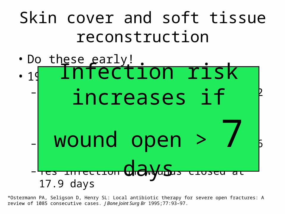
Skin cover and soft tissue reconstruction
• Do these early!• 1994 Osterman et al.*– Retrospective 1085 fractures, 115 G2 and 239 G3• All treated with appropriate IV Abx and I&D
– No infection if wounds closed at 7.6 days– Yes infection if wounds closed at 17.9 days
*Ostermann PA, Seligson D, Henry SL: Local antibiotic therapy for severe open fractures: A review of 1085 consecutive cases. J Bone Joint Surg Br 1995;77:93–97.
Infection risk increases if wound
open > 7 days

Reconstructive ladder: options for wound coverage
Type 1 open fx
Type 3B open fx
Type 2/3A open fx

Flap coverage for type 3b

Type 3c, a bad injury!• Devastating damage to
bone and soft tissue• Major arterial injuries
that require repair• Poor functional outcome• Consensus btwn ortho,
vascular and plastics• Salvage is technically
possible in most cases• However it is not always
the correct choice esp type 3c tibia fractures

We can do both, salvage & amputate.• Vascular surgery can revascularize
with bypass graft– Generally before fracture stabilization
• Plastics can provide soft tissue coverage
• However, in the tibia, the severity to soft tissue envelope and bone may result in infected nonunion
• If salvage…. long course of repeated surgical procedures– Painful and psychologically distressing– Functional outcome may be poor and
no better than amputation

How to decide, salvage or amputate?
• Important factors in decision making:*– General condition of the patient (shock)– Warm ischemia time (>6hours)– Age (>30 years)– Cut to crush ratio (blunt injuries has a large zone
of crush)
Howe HR Jr, Poole GV Jr, Hansen KJ, Clark T, Plonk GW, Koman LA, Pennell TC: Salvage of lower extremities following combined orthopedic and vascular trauma. A predictive salvage index. Am Surg. 1987 Apr;53(4):205-8.

Gunshot injuries• Energy dissipated at impact = damage
severity• High velocity rifles and close range
shotguns– Worst, high energy of impact– Huge secondary cavitation– Secondary effects of shattered bone
fragments• Bullets lodged in joints should be removed– avoid lead arthropathy and systemic lead
poisoning

Low velocity GSW <2000 ft/sec
• Low velocity handguns– Less severe, not treated like open fractures– Cavitation is not significant– Secondary missile effects are minimal – Bone fragments rarely stripped of soft tissue
attachments and blood supply– Soft tissue injuries not severe and skin wounds are
small

Low velocity GSW open fractures
• Geisslar et al. *• If neurovascular status
normal, do local debridement
• NO formal I&D needed• IV Abx• Approach fx fixation as
if closed
• Dickey et al.**– No abx vs IV Ancef x 3d– 67 low velocity GSW fxs– Not requiring operative
fixation– No difference in
infection rates
*Geisslar ett al, J Ortho Trauma, 4;39-41,1990
**Dickey et al, J Ortho Trauma, 3;6-10,1989
Treat open fractures from
low velocity GSW as closed
fractures without Abx
Pitfalls and complications• Infection delayed union, nonunion, malunion
and loss of function• Plan ahead to avoid delayed union and nonunion• Predict nonunion in severe injuries with bone loss– Bone grafting usually delayed 6 weeks when soft
tissues have soundly healed– Autogenous bone grafting is usual strategy– Fibular transfer, free composite graft or distraction
osteogenesis for complex defects– Recombinant human BMP in open tibia fracture
reduces risk of delayed union

Advances…
• BMPs– 40% decreased infection rate with BMP in type 3
open tibia fractures*
• Antibiotic Laden Bone Graft**– Tobramycin-impregnated calcium sulfate pellets
with demineralized bone matrix– Animal study: successful in preventing infection
*BESTT Study Group, Govender S, Csimma C, Genant H, Valentin-Opran A. “Recombinant Human Bone Morphogenetic Protein-2 for Treatment of Open Tibial Fractures: A prospective, controlled, randomized study of four hundred and fifty patients.” JBJS-A 2002. 84(12): 2123-2134.**Beardmore AA, Brooks DE, Wenke JC, Thomas DB. “Effectiveness of local antibiotic delivery with an osteoinductive and osteoconductive bone-graft substitute.” JBJS-A 2005. 87(1): 107-112.
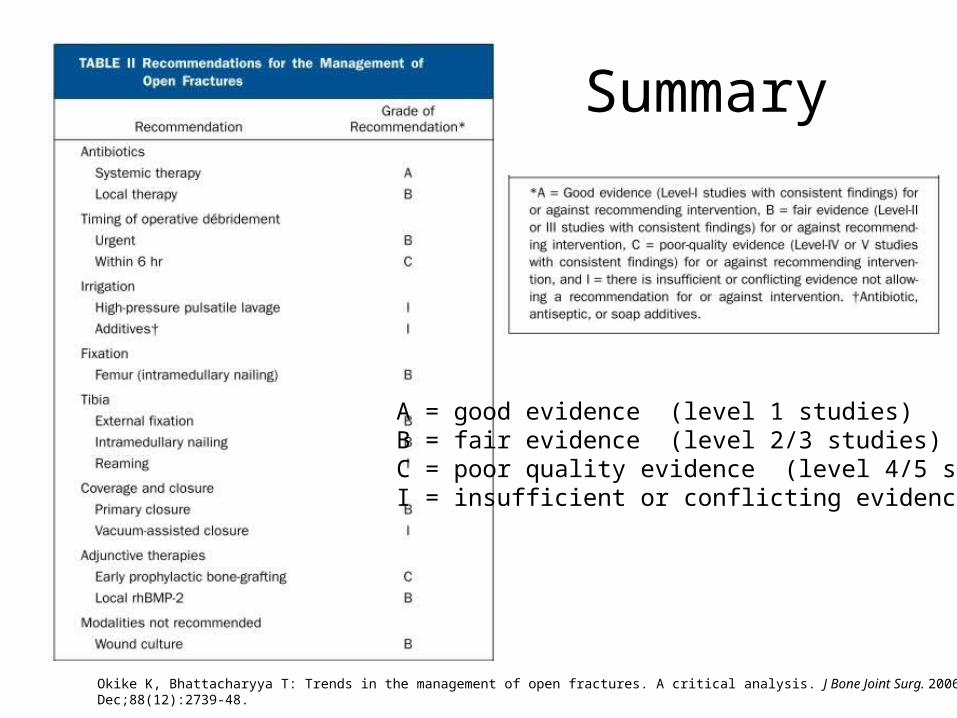
Summary
A = good evidence (level 1 studies)B = fair evidence (level 2/3 studies)C = poor quality evidence (level 4/5 studies)I = insufficient or conflicting evidence
Okike K, Bhattacharyya T: Trends in the management of open fractures. A critical analysis. J Bone Joint Surg. 2006 Dec;88(12):2739-48.

Thank you