Embed Size (px)
Citation preview

Adding Diverse Noncanonical Backbones to Rosetta:Enabling Peptidomimetic DesignKevin Drew1., P. Douglas Renfrew1., Timothy W. Craven1, Glenn L. Butterfoss1, Fang-Chieh Chou2,
Sergey Lyskov3, Brooke N. Bullock4, Andrew Watkins4, Jason W. Labonte3, Michael Pacella5, Krishna
Praneeth Kilambi3, Andrew Leaver-Fay6, Brian Kuhlman6, Jeffrey J. Gray3,7, Philip Bradley8,
Kent Kirshenbaum4, Paramjit S. Arora4, Rhiju Das2, Richard Bonneau1,9*
1Department of Biology, Center for Genomics and Systems Biology, New York University, New York, New York, United States of America, 2Department of Biochemistry,
Stanford University, Stanford, California, United States of America, 3Department of Chemical and Biomolecular Engineering, The Johns Hopkins University, Baltimore,
Maryland, United States of America, 4Department of Chemistry, New York University, New York, New York, United States of America, 5Department of Biomedical
Engineering, The Johns Hopkins University, Baltimore, Maryland, United States of America, 6Department of Biochemistry, University of North Carolina, Chapel Hill, North
Carolina, United States of America, 7 Program in Molecular Biophysics, The Johns Hopkins University, Baltimore, Maryland, United States of America, 8 Fred Hutchinson
Cancer Research Center, Seattle, Washington, United States of America, 9Computer Science Department, Courant Institute of Mathematical Sciences, New York University,
New York, United States of America
Abstract
Peptidomimetics are classes of molecules that mimic structural and functional attributes of polypeptides. Peptidomimeticoligomers can frequently be synthesized using efficient solid phase synthesis procedures similar to peptide synthesis.Conformationally ordered peptidomimetic oligomers are finding broad applications for molecular recognition and forinhibiting protein-protein interactions. One critical limitation is the limited set of design tools for identifying oligomersequences that can adopt desired conformations. Here, we present expansions to the ROSETTA platform that enablestructure prediction and design of five non-peptidic oligomer scaffolds (noncanonical backbones), oligooxopiperazines,oligo-peptoids, b-peptides, hydrogen bond surrogate helices and oligosaccharides. This work is complementary to prioradditions to model noncanonical protein side chains in ROSETTA. The main purpose of our manuscript is to give a detaileddescription to current and future developers of how each of these noncanonical backbones was implemented. Furthermore,we provide a general outline for implementation of new backbone types not discussed here. To illustrate the utility of thisapproach, we describe the first tests of the ROSETTA molecular mechanics energy function in the context ofoligooxopiperazines, using quantum mechanical calculations as comparison points, scanning through backbone and sidechain torsion angles for a model peptidomimetic. Finally, as an example of a novel design application, we describe theautomated design of an oligooxopiperazine that inhibits the p53-MDM2 protein-protein interaction. For the generalbiological and bioengineering community, several noncanonical backbones have been incorporated into web applicationsthat allow users to freely and rapidly test the presented protocols (http://rosie.rosettacommons.org). This work helpsaddress the peptidomimetic community’s need for an automated and expandable modeling tool for noncanonicalbackbones.
Citation: Drew K, Renfrew PD, Craven TW, Butterfoss GL, Chou F-C, et al. (2013) Adding Diverse Noncanonical Backbones to Rosetta: Enabling PeptidomimeticDesign. PLoS ONE 8(7): e67051. doi:10.1371/journal.pone.0067051
Editor: Vladimir N. Uversky, University of South Florida College of Medicine, United States of America
Received February 4, 2013; Accepted May 13, 2013; Published July 15, 2013
Copyright: � 2013 Drew et al. This is an open-access article distributed under the terms of the Creative Commons Attribution License, which permitsunrestricted use, distribution, and reproduction in any medium, provided the original author and source are credited.
Funding: The authors acknowledge support from NSF CHE-1151554, NSF IOS-1126971, NSF CHE-1152317, NSF CHE-0848410, NIH U54CA143907-01, NIH PN2EY016586-06, NIH T32 GM 88118-2, NIH R01GM073943, NIH RO1GM073151 (SL, JG, BK and AL), NIH RO1GM073960 (BK and AL), the Burroughs WellcomeFoundation (Career Award to RD), and an HHMI International Student Fellowship (FC). The funders had no role in study design, data collection and analysis,decision to publish, or preparation of the manuscript.
Competing Interests: The authors have declared that no competing interests exist.
* E-mail: [email protected]
. These authors contributed equally to this work.
Introduction
A variety of peptidomimetic oligomers have been identified that
can mimic protein secondary structure features and can exhibit
many of the physiochemical properties of polypeptides, including
the spacing and geometry of side chains [1–5]. Here we focus on
several peptidomimetic scaffolds that enable the use of large
libraries of potential side chains and are compatible with facile
monomer or sub-monomer synthesis. Of particular importance is
the fact that functional groups on peptidomimetics often have
substantial spatial and geometrical congruence with side chains
presented on protein secondary structure [6]. For example,
peptidomimetics can be used to mimic large binding interfaces
mediated by protein helices or strands [7]. The chemical identity
of peptidomimetic side chains and termini can be tailored to
establish proteolytic stability, membrane permeability, and addi-
tional desirable pharmacological properties. These characteristics
may endow peptidomimetics with improved therapeutic potential
relative to canonical peptide analogues. Specific examples relevant
to this work include the development of protein interaction
PLOS ONE | www.plosone.org 1 July 2013 | Volume 8 | Issue 7 | e67051

inhibitors and the antagonism of interfaces larger than those
targeted by small molecules [8,9]. Additional conformational
diversity provided by some classes of peptidomimetics make them
an attractive system for ‘‘foldamer’’ research with the goal of
developing new secondary or tertiary structural motifs. Here we
discuss five non-peptidic oligomer systems capable of addressing
these goals: oligooxopiperazines, oligo-peptoids, b-peptides, pep-
tide hydrogen bond surrogate helices and oligosaccharides.
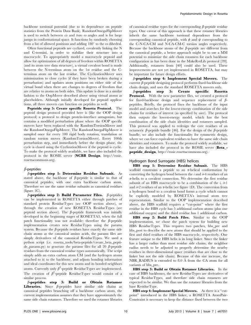
The oligooxopiperazine scaffold (OOP) (figure 1A) is a
peptidomimetic with side chains that can mimic the i, i+4 and
i+7 positions of an a-helix [10]. An OOP monomer is synthesized
from amino acids with ethylene bridges linking neighboring pairs
of backbone amides, resulting in a backbone composed of linked
six-membered rings. Because 62% of protein complexes in the
PDB include an a-helix at an interface [7], helical mimetics such as
OOPs serve as useful inhibitor scaffolds for many large protein
interfaces [4,11].
Peptoids (figure 1B) are oligomers of N-substituted glycine units
in which the side chains are positioned on the backbone nitrogen
(as distinct from the a-carbon) [12–14]. This results in an oligomer
backbone that is achiral, proteolytically stable, and displays
increased flexibility at its amide (v) dihedral angle, exhibiting
both cis and trans conformations [15]. The ability of the backbone
amide to populate both cis and trans conformations allows peptoids
to mimic diverse protein secondary structure features including
both poly-proline type I and type II helices [16,17]. Peptoid
structures are being explored in the context of material science and
biomedical applications [18–22].
b-peptides (figure 1C) are peptides with an additional backbone
carbon resulting in an extra dihedral angle along the backbone
and extended length between adjacent side chains. Several groups
have used b-peptides as a system for foldamer research where the
goal is to create protein-like or DNA-like structure and function
with oligomers other than a-peptides and nucleic acids [23–25].
The Gellman lab, for example, has pioneered the creation of
heterogeneous backbones, combining both a and b peptides in an
alternating fashion to form helices. These molecules have been
used as high affinity binders to the Bcl-xL family of prosurvival
proteins [26] as well as gp41 inhibitors [27].
Hydrogen bond surrogate (HBS) helices (figure 1D) contain a
carbon-carbon bond in place of the canonical i and i+4 backbone
hydrogen bond in alpha-helices [28]. This covalent bond allows
much shorter peptides to stably form an a-helix conformation.
HBS helices have been shown to mimic protein interaction
interfaces including those of p53 [29], HIF-1alpha [9], SOS [30]
and MCL-1 [31].
Oligosaccharides (figure 1E) are chains of glycosidically linked
monosaccharides, which preform diverse functions in the cell
including cell-cell recognition [32]. Additionally, glyco-proteins
(i.e. proteins that are covalently linked to saccharides) are involved
in protein folding [33], cell signaling [34,35] and when mis-
regulated, prion diseases [36,37]. Although oligosaccharides are
not traditional peptidomimetics, their implementation into the
ROSETTA framework provides an excellent example of the type
of diverse backbones that can be implemented and modeled using
this approach.
Currently many peptidomimetics are designed manually with
no computational framework to efficiently search their available
conformational and design spaces. Database mining tools are
available to match potential inhibitor scaffolds to specific protein
interaction interfaces but these tools lack the ability to explicitly
model and redesign the scaffold [6]. This scarcity of modeling tools
limits the progress of applying peptidomimetics to many applica-
tions for which they are attractive. Recent advances to the
Figure 1. Chemical structures of noncanonical backbones inRosetta. A) Oligooxopiperazines (OOP), B) Peptoids, C) b3-peptides, D)Hydrogen Bond Surrogate (HBS) helices, E) Oligosaccharides. In A–D,bonds highlighted in red deviate from the traditional peptidebackbone. Arrows point to ‘‘cut-points’’ described in text. Bracketsdelineate a single residue subunit.doi:10.1371/journal.pone.0067051.g001
Peptidomimetic Design in Rosetta
PLOS ONE | www.plosone.org 2 July 2013 | Volume 8 | Issue 7 | e67051
molecular modeling suite ROSETTA create a framework that
allows the modeling and design of noncanonical backbones
including the ones just described. The framework allows access
to a variety of energy functions and algorithms provided by
ROSETTA such as minimization, side chain packing/design and
Monte Carlo conformational search. Important precedents for this
work include prior efforts of ligand docking in ROSETTA [38],
the incorporation of noncanonical side chains into ROSETTA
[39] and work to derive methods for creating rotamer libraries for
noncanonical amino acids (NCAAs) [39,40].
To take advantage of the algorithms and scoring functions
available in ROSETTA, noncanonical backbones must have their
chemical descriptions (the start and stop of each monomer) and
kinematics (methods for altering conformations) instantiated
properly within the framework. Here we describe the implemen-
tation of five noncanonical backbones (OOP, oligo-peptoids, b-
peptides, HBS helices and oligosaccharides) as working illustra-
tions of a general approach for implementing noncanonical
backbones in ROSETTA. We outline five steps (figure 2) to
achieve this goal: 1) determination of the boundaries of each
residue subunit from its backbone chemical structure, 2) descrip-
tion of the chemical connectivity within the ROSETTA frame-
work, 3) building and parameterizing rotamer libraries, 4)
implementing movers (kinematics) to sample conformations and
5) creation of overall modeling/design protocols (this last step will
be variable depending on project goals; in this work we focus on
design protocols for antagonists of large protein-protein interfaces
as well as the redesign of b-peptide helix bundles).
We also present new computational results, including compar-
isons of a ROSETTA score function to quantum mechanical
calculations on the OOP backbone. Our target audience is
primarily current and future developers interested in expanding
ROSETTA to additional classes of polymers; casual developers
interested in creating new ROSETTA protocols incorporating one
or more noncanonical backbone positions or side chains; and non-
experts interested in using these tools through the ROSIE (Rosetta
Online Server that Includes Everyone) server (http://rosie.
rosettacommons.org). All associated code is freely accessible to
academic users via the ROSETTACOMMONS website (http://
www.rosettacommons.org).
Methods
The recent reorganization of the ROSETTA code (i.e.
ROSETTA3) to comply with standard object oriented software
practices has provided a flexible framework to model oligomers
other than the traditional peptide and nucleic acid backbones [41]
and also to model large heteromeric complexes involving mixed
types of polymers (like protein/peptidomimetic complexes, see
below). In this framework the residue object is the central object;
all algorithms and scoring functions within ROSETTA act upon
residues (whether amino acids, bases, peptoid monomers, etc.). A
first step is to define the chemical structure of the repeating subunit
in the noncanonical backbone, as a residue (described as a
ResidueType object within ROSETTA). Generally, residues in
ROSETTA are defined such that a subunit contains a single side
chain with a corresponding rotamer set.
The next step after determining a residue subunit is to describe
the subunit in terms of its chemical configuration in a format
readable by ROSETTA (ROSETTA ResidueType). ROSETTA
provides two ways to describe new ResidueTypes, the residue
params system and the residue patch system, both based on easily
edited text files placed in the ROSETTA database directory. The
residue params system describes a ResidueType completely with
all atoms and bonds as if it were fully connected to its neighboring
residues. The fully connected ResidueType is called a mid or base
variant and includes a unique residue name, atom descriptions (i.e.
name, type, charge), bond connectivity (including to neighboring
residues), and idealized internal coordinates. The residue params
system is useful when defining polymer subunits that have limited
similarity to existing ResidueTypes (see peptoid section).
Another way ROSETTA allows ResidueType declarations is
using the patch system. This system defines new ResidueTypes
based on previously declared ResidueTypes. For example, the
CtermProteinFull patch is applied to all existing ResidueTypes
(e.g. alanine) to define the C-terminal variants of standard residues
(e.g. C-terminal alanine). Additionally, ResidueTypes may have
multiple patches applied. For instance a C-terminal phospho-
tyrosine is created by applying both a C-terminal patch and a
phosphorylation patch. The patch description includes a unique
patch name, a section defining which ResidueTypes to apply the
patch to, and atoms to be added (or deleted) to applicable
ResidueTypes. The residue patch system is useful when defining
polymer subunits that are modifications of existing ResidueTypes
and can be implemented with a single patch declaration (see OOP
section).
After the new ResidueTypes are defined and readable by
ROSETTA, it is necessary to create rotamer libraries. Backbone
dependent rotamer libraries can be generated using the MakeR-
otLib protocol that samples side chain x angles of an amino acid
for w=y combinations of backbone angles. Side chain conforma-
tion samples are clustered and an energy score is calculated for
each cluster. The energy is then converted into a probability that
serves as an entry in the ResidueType’s rotamer library. The full
protocol is discussed by Renfrew et al. 2012, along with a protocol
capture describing how to run the code [39]. It should be noted
that it is unnecessary to create new rotamer libraries if it is believed
there is a suitable available rotamer library. For example in the
case of HBS helices the backbone is similar to the peptide
backbone (identical at most positions) and thus protein/peptide
rotamer libraries are used. This is often the case when making new
ResidueTypes using the patch system. If the modifications made to
the base ResidueType do not affect side chain degrees of freedom
(or do not make other significant changes to the chemistry), the
base rotamer libraries may be used for patched variants.
Once the noncanonical backbone chemical structure is fully
described, kinematic functionality (specialized movers) can be
developed to properly sample the conformational space specific to
the backbone. Backbones with additional degrees of freedom, such
as a flexible v angle (e.g. oligo-peptoids) or an added backbone
dihedral (e.g. b-peptides), require the implementation of one or
more new movers. It should be noted that the degrees of freedom
sampled are dependent on the modeling goal. For instance, if
appropriate modeling can be achieved using fixed backbone
design, specialized backbone moves may not be necessary.
Finally, with both the chemical structure and kinematics
defined, a full protocol can be developed that focuses on the
specific modeling goals of the application. A full protocol requires
a combination of the specialized movers and traditional RO-
SETTA movers to properly sample the conformational space of
the noncanonical molecule and other interacting molecules. Here
we describe protocols for designing peptidomimetics that bind
target proteins (with specific examples including the design of an
oligooxopiperazine inhibitor to the p53-MDM2 protein interac-
tion).
In the following sections, we discuss examples of each of the five
noncanonical backbones (OOPs, peptoids, b-peptides, HBS
helices and oligosaccharides) that have recently been added into
Peptidomimetic Design in Rosetta
PLOS ONE | www.plosone.org 3 July 2013 | Volume 8 | Issue 7 | e67051

ROSETTA. The aim of this work is to provide a complete, and
thus reproducible, description of all required modifications.
OligooxopiperazinesOOPs step 1: Determine Residue Subunit. OOPs are
helical mimetics with a backbone very similar to that of a peptide
but with the addition of an ethylene bridge forming a cycle
between adjacent residues (R1 and R2 in figure 1A). To
implement a new backbone in ROSETTA, the bonds between
atoms must be described in such a way to be compatible with
ROSETTA’s atom tree representation where explicit cycles are
not allowed between residues. We therefore chose a ‘‘cut-point’’
through the ethylene bridge making the OOP residue subunit a
single amino acid plus the additional carbon atom. The additional
atoms are named either CYP for the first residue of the ring (R1)
or CZP for the second residue (R2). The covalent bond between
the carbon atoms of neighboring residues, CYP and CZP, is not
represented explicitly within the ROSETTA atom tree but rather
with a inter-residue connection (described next in step 2,
ADD_CONNECT). The inter-residue connection notifies the
score function that these two atoms are covalently bonded; this
prevents, for example, the close distances between CYP and CZP
from incurring a clash penalty.
Figure 2. Noncanonical backbone implementation flow chart. Outline of the five steps needed for implementation of a noncanonicalbackbone into ROSETTA.doi:10.1371/journal.pone.0067051.g002
Peptidomimetic Design in Rosetta
PLOS ONE | www.plosone.org 4 July 2013 | Volume 8 | Issue 7 | e67051
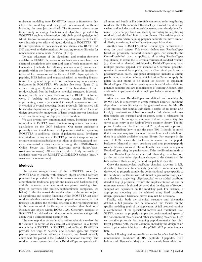
OOPs step 2: Build Patch Files. OOP scaffolds can be
synthesized from most amino acids given that they have a
backbone primary amine. We chose ROSETTA’s patch system to
define ResidueTypes because we do not have to define separate
OOP ResidueTypes for all residues defined in ROSETTA (20
canonical in addition to &100 noncanonical amino acids).
Additionally, it is extensible as all new ResidueTypes added in
the future will also have OOP ResidueType variants automati-
cally. We created two OOP patches describing the first (R1) and
second (R2) residues of the OOP ring, named the oop_pre patch
and oop_post patch respectively. The oop_pre patch file as shown
in figure 3, figure 4 and completely in file S1, begins with the
declaration of a unique name (NAME) and a variant type (TYPES)
(see figure 3A). The VariantType is a class of variants that can be
referred to later within ROSETTA to apply specific functionality
only to ResidueTypes of that variant class. An example of a more
general VariantType is the LOWER_TERMINUS that applies to
the collection of all N-terminal residues.
The chemical synthesis of OOPs is incompatible with certain
ResidueTypes and therefore we must specify eligible and ineligible
residues. The selector section (BEGIN_SELECTOR, END_-
SELECTOR, figure 3B), declares what ResidueTypes will have
the OOP patches applied. The first requirement for an OOP
patch is that a ResidueType must have the property of a
PROTEIN which restricts the patch to only amino acids (line 10
figure 3B). The next part of the selector section requires
ResidueTypes to be synthetically compatible with being an
OOP residue (lines 13–22 figure 3B). OOP chemical synthesis
requires precursor amino acids with a free primary amine. The
restriction is done as a negation (e.g. NOT VARIANT_TYPE)
where only ResidueTypes that, for example, do not have
acetylated N-termini are applicable. It should also be noted that
a ResidueType with two OOP_PRE variants or an OOP_PRE
and OOP_POST should not exist and therefore is not allowed.
The other entries in this subsection do not allow proline or proline-
like amino acids. Finally, OOP_PRE residues with C-terminal
variants are not allowed because a residue with an OOP_PRE
variant must be immediately followed by an OOP_POST variant
residue.
It is often the case that different ResidueTypes may need to
have the patch applied differently. The remaining sections in the
oop_pre patch file, between the BEGIN_CASE and END_CASE
keywords, address different cases of the patch application
beginning with the most specific. These sections are where the
modifications to the base ResidueTypes are described in order to
create a new patched ResidueType. The oop_pre patch has two
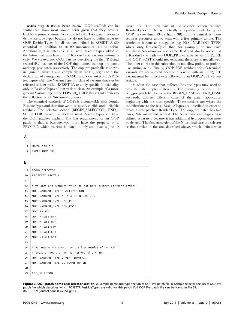
cases, N-terminal and general. The N-terminal case (figure 4) is
defined separately because it has additional hydrogens that must
be deleted. The first subsection of the N-terminal case is a selector
section similar to the one described above, which defines what
Figure 3. OOP patch name and selector section. A. Sample name and type section of OOP Pre patch file. B. Sample selector section of OOP Prepatch file which describes which ROSETTA ResidueTypes are valid for this patch. Full OOP Pre patch file can be found in file S1.doi:10.1371/journal.pone.0067051.g003
Peptidomimetic Design in Rosetta
PLOS ONE | www.plosone.org 5 July 2013 | Volume 8 | Issue 7 | e67051
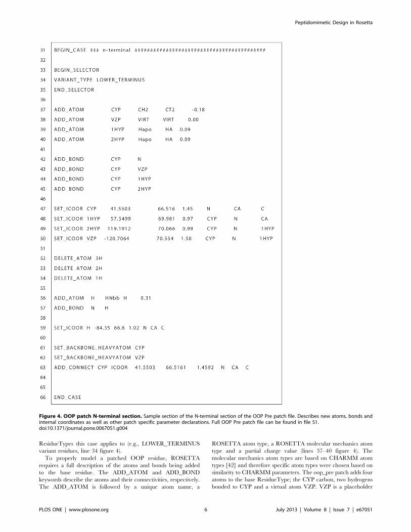
ResidueTypes this case applies to (e.g., LOWER_TERMINUS
variant residues, line 34 figure 4).
To properly model a patched OOP residue, ROSETTA
requires a full description of the atoms and bonds being added
to the base residue. The ADD_ATOM and ADD_BOND
keywords describe the atoms and their connectivities, respectively.
The ADD_ATOM is followed by a unique atom name, a
ROSETTA atom type, a ROSETTA molecular mechanics atom
type and a partial charge value (lines 37–40 figure 4). The
molecular mechanics atom types are based on CHARMM atom
types [42] and therefore specific atom types were chosen based on
similarity to CHARMM parameters. The oop_pre patch adds four
atoms to the base ResidueType; the CYP carbon, two hydrogens
bonded to CYP and a virtual atom VZP. VZP is a placeholder
Figure 4. OOP patch N-terminal section. Sample section of the N-terminal section of the OOP Pre patch file. Describes new atoms, bonds andinternal coordinates as well as other patch specific parameter declarations. Full OOP Pre patch file can be found in file S1.doi:10.1371/journal.pone.0067051.g004
Peptidomimetic Design in Rosetta
PLOS ONE | www.plosone.org 6 July 2013 | Volume 8 | Issue 7 | e67051

atom that optimally has the same X,Y,Z coordinates of the CZP
atom in the connecting oop_post residue. The presence of VZP
allows us to later calculate torsion dimensions with respect to the
CZP. Additionally, it helps keep a physically realistic OOP ring
structure by allowing the minimization of the difference between
the X,Y,Z coordinates of VZP and CZP. The ADD_BOND
keyword is followed by the unique atom names that are covalently
bonded in the patched ResidueType (lines 42–45 figure 4).
The next subsection (lines 47–50 figure 4) declares the internal
coordinates of each added atom with respect to other previously
declared atoms. The SET_ICOOR keyword is followed by a
unique atom name, dihedral value, three-atom angle value, bond
length and the unique atom names of the relative atoms. For
example, the CYP internal coordinates are described relative to
the atoms N, CA and C. The dihedral value describes the dihedral
between CYP-N-CA-C atoms (41.5u in the example). The three-
atom angle value is the degree angle change of CA from the CYP-
N vector (i.e. 180u minus the CYP-N-CA angle value). And the
bond length describes the length of the CYP-N bond. Atoms that
have internal coordinates defined by LOWER (or UPPER) are
defined relative to the preceding residue’s C-terminal atom (or
succeeding residue’s N-terminal atom). This allows the ability to
make changes to degrees of freedom that span multiple residues
(e.g. w backbone dihedral). It should be noted that these internal
coordinate values describe the idealized conformation of the atoms
added to the oop_pre residue and the values may change during
the course of a protocol. We chose the half-chair conformation
coordinates as the oop_pre idealized coordinates because they are
the lowest energy according to quantum energy calculations. The
actual values are averages of three OOP ring instances in
crystallographic data from the Cambridge Structural Database
[43] (CSD codes: ZOZTUD, ZARZOH, FOBFEH) and one
quantum optimized OOP ring structure.
Lines 52–59 (figure 4) in the example OOP patch file handle the
special case of the N-terminus where the three hydrogens bound to
the terminal nitrogen are deleted with the DELETE_ATOM
keyword and replaced with a single hydrogen. The new hydrogen
atom is declared similarly as above.
The final section for the N-terminal case declares the CYP and
VZP atoms as backbone heavy atoms (lines 61–62 figure 4) as the
ROSETTA movers may distinguish between backbone and side
chain atoms. For instance, it is possible to minimize only the side
chain atoms of a residue and therefore the CYP and VZP atoms
would not be included in the minimization. The last declaration is
using the ADD_CONNECT keyword (line 63) that defines the
CYP atom as having a covalent bond with an atom in another
residue, which will be the CZP atom in the succeeding oop_post
residue.
In the oop_pre patch file, the general case is very similar to the
N-terminal case just described. Additionally, the oop_post patch
file is similar in defining the CZP atom, the virtual atom VYP, and
corresponding hydrogens, which make up the oop_post variant
ResidueType.
OOPs step 3: Build or Obtain Rotamer Libraries. At this
stage of implementation it is important to decide whether rotamer
libraries need to be built for the new ResidueTypes. The chemistry
of OOP side chains is similar to the side chain on their respective
amino acids; therefore, we decided that rotamer libraries of the
standard ResidueType were sufficient. For instance, a phenylal-
anine side chain branching off an OOP ring has similar rotamers
to that of a typical phenylalanine. Rotamer libraries of the
standard ResidueType are the default and no additional declara-
tions are necessary.
OOPs step 4: Implement Special Movers. The OOP
backbone differs from canonical peptides by the addition of several
atoms that form a ring between two residues. We therefore
developed additional movers to properly sample the conforma-
tional space of the OOP ring, which ROSETTA’s peptide-centric
movers do not sample properly. The OOP ring has two low energy
conformations: a half-chair pucker and a boat pucker, as well as
small pucker changes dependent on side chain x angles. Therefore
we created the OopMover class within ROSETTA to change the
pucker of an OOP ring. OOP ring movement is determined by the
w and y dihedrals of an oop_pre residue (R1). The OopMover
class implements functions that set w and y dihedral values of a
oop_pre residue and updates the OOP specific hydrogen positions.
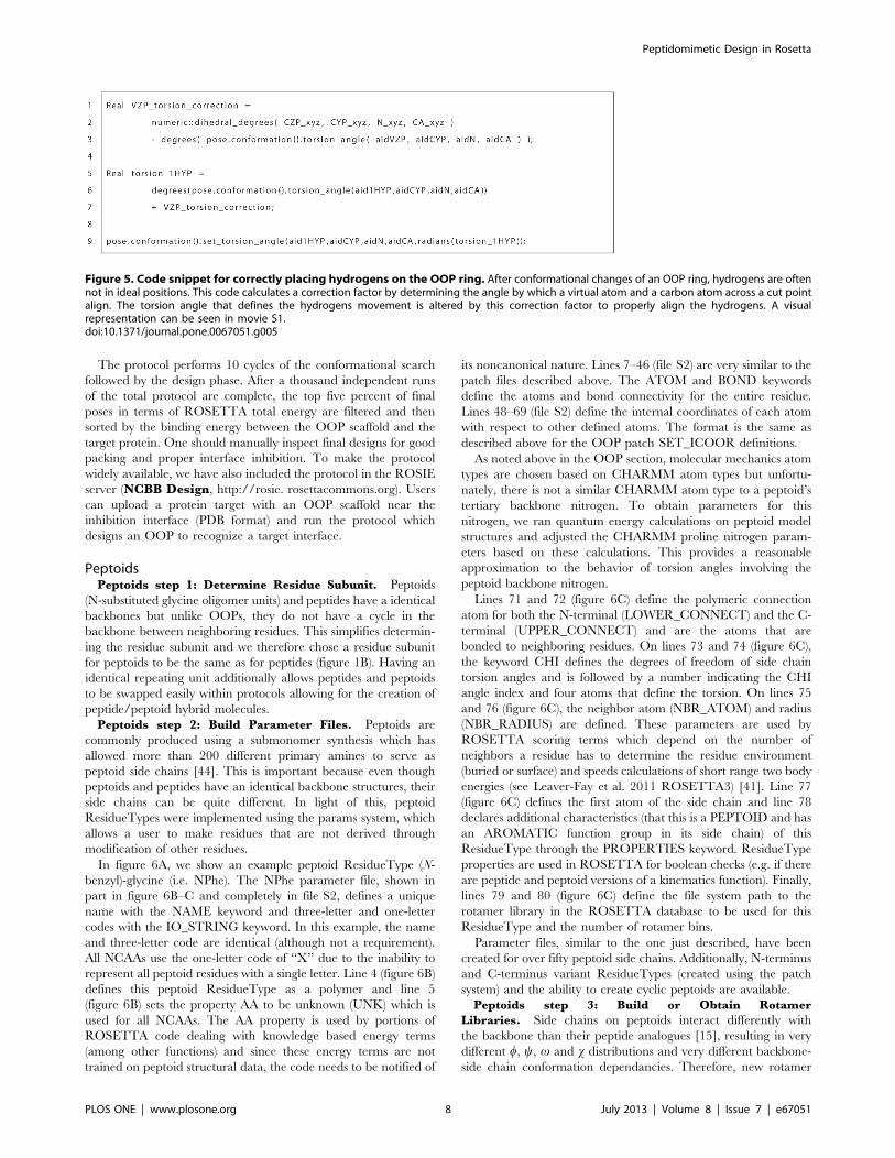
As seen in movie S1, when the w of the oop_pre residue is
changed, the VZP virtual atom of the oop_pre residue no longer
overlaps the CZP atom on the oop_post residue (again, the VZP
atom is a placeholder atom that is meant to track the CZP atom).
The difference between the torsion angle of the CA-N-CYP-CZP
and CA-N-CYP-VZP is calculated (line 1–3 in figure 5) and the
hydrogens (i.e. CA-N-CYP-1HYP torsion) are corrected by this
value (line 5–7, 9 in figure 5). The VZP and 2HYP atom positions
are corrected automatically because they were defined relative to
the 1HYP atom in the oop_pre patch file. A similar method is used
for correcting the hydrogens bound to the CZP atom in the
oop_post residue, also shown in movie S1.
The OopMover class provides basic functionality to alter the
conformation of an OOP ring. More sophisticated OOP classes
that rely and extend the OopMover class include OopPuckMover
and OopRandomSmallMover that extend the OopMover class.
The OopPuckMover makes changes to w and y angles in order to
change the OOP ring pucker from half-chair (w~{130 ,
y~{10 ) to boat (w~{147 , y~{36 ) (or vice versa).
OopRandomSmallMover randomly changes the w and y angles
of the oop_pre residue by small degree changes to optimize the
energy of the ring.
In a final piece of code, we address the need for a constraint on
the atoms at the OOP ‘‘cut-point’’. These atoms, CYP and CZP,
are connected insofar that the energy functions treat them as
covalently bound. However, a ROSETTA protocol may sample a
low energy conformation where CYP and CZP are at a distance
outside the range of a covalent carbon carbon bond. Therefore, we
implemented the add_oop_constraint function (OopPatcher.cc),
where a ROSETTA AtomPairConstraint is placed on the CYP
and CZP atoms to ensure they stay within 1.5 A.
OOPs step 5: Create OOPs specific Rosetta
Protocol. Once the special movers for the noncanonical
backbone have been implemented, the movers can be combined
to create a protocol that samples the conformational space of the
molecular system of interest. Here we describe a protocol to design
an OOP to bind a target protein. Briefly, the protocol iterates
between finding low energy conformations and designing low
energy sequences on the OOP scaffold while in contact with a
target protein interaction interface. The conformational search
involves random selection of four movers, 1) rigid body rotation
and translation of the OOP scaffold with respect to the target
protein, 2) OopPuckMover applied to any OOP rings, 3)
OopRandomSmallMover applied to any OOP rings and 4) small
angle perturbations to the w and y torsion angles between OOP
rings. This conformational search via randomly selected movers
iterates 100 times. The protocol then applies a design phase
(repack) where side chains on the OOP scaffold are substituted for
lower energy ones. Side chains on both sides of the interface are
repacked to find low energy combinations of rotamers.
Peptidomimetic Design in Rosetta
PLOS ONE | www.plosone.org 7 July 2013 | Volume 8 | Issue 7 | e67051
The protocol performs 10 cycles of the conformational search
followed by the design phase. After a thousand independent runs
of the total protocol are complete, the top five percent of final
poses in terms of ROSETTA total energy are filtered and then
sorted by the binding energy between the OOP scaffold and the
target protein. One should manually inspect final designs for good
packing and proper interface inhibition. To make the protocol
widely available, we have also included the protocol in the ROSIE
server (NCBB Design, http://rosie. rosettacommons.org). Users
can upload a protein target with an OOP scaffold near the
inhibition interface (PDB format) and run the protocol which
designs an OOP to recognize a target interface.
PeptoidsPeptoids step 1: Determine Residue Subunit. Peptoids
(N-substituted glycine oligomer units) and peptides have a identical
backbones but unlike OOPs, they do not have a cycle in the
backbone between neighboring residues. This simplifies determin-
ing the residue subunit and we therefore chose a residue subunit
for peptoids to be the same as for peptides (figure 1B). Having an
identical repeating unit additionally allows peptides and peptoids
to be swapped easily within protocols allowing for the creation of
peptide/peptoid hybrid molecules.
Peptoids step 2: Build Parameter Files. Peptoids are
commonly produced using a submonomer synthesis which has
allowed more than 200 different primary amines to serve as
peptoid side chains [44]. This is important because even though
peptoids and peptides have an identical backbone structures, their
side chains can be quite different. In light of this, peptoid
ResidueTypes were implemented using the params system, which
allows a user to make residues that are not derived through
modification of other residues.
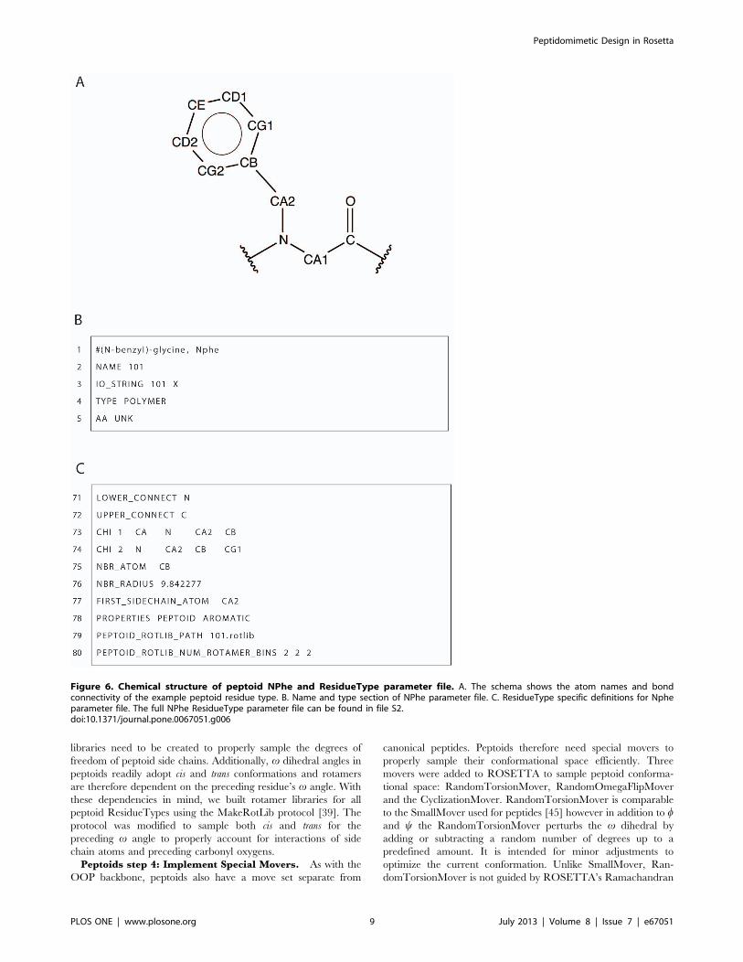
In figure 6A, we show an example peptoid ResidueType (N-
benzyl)-glycine (i.e. NPhe). The NPhe parameter file, shown in
part in figure 6B–C and completely in file S2, defines a unique
name with the NAME keyword and three-letter and one-letter
codes with the IO_STRING keyword. In this example, the name
and three-letter code are identical (although not a requirement).
All NCAAs use the one-letter code of ‘‘X’’ due to the inability to
represent all peptoid residues with a single letter. Line 4 (figure 6B)
defines this peptoid ResidueType as a polymer and line 5
(figure 6B) sets the property AA to be unknown (UNK) which is
used for all NCAAs. The AA property is used by portions of
ROSETTA code dealing with knowledge based energy terms
(among other functions) and since these energy terms are not
trained on peptoid structural data, the code needs to be notified of
its noncanonical nature. Lines 7–46 (file S2) are very similar to the
patch files described above. The ATOM and BOND keywords
define the atoms and bond connectivity for the entire residue.
Lines 48–69 (file S2) define the internal coordinates of each atom
with respect to other defined atoms. The format is the same as
described above for the OOP patch SET_ICOOR definitions.
As noted above in the OOP section, molecular mechanics atom
types are chosen based on CHARMM atom types but unfortu-
nately, there is not a similar CHARMM atom type to a peptoid’s
tertiary backbone nitrogen. To obtain parameters for this
nitrogen, we ran quantum energy calculations on peptoid model
structures and adjusted the CHARMM proline nitrogen param-
eters based on these calculations. This provides a reasonable
approximation to the behavior of torsion angles involving the
peptoid backbone nitrogen.
Lines 71 and 72 (figure 6C) define the polymeric connection
atom for both the N-terminal (LOWER_CONNECT) and the C-
terminal (UPPER_CONNECT) and are the atoms that are
bonded to neighboring residues. On lines 73 and 74 (figure 6C),
the keyword CHI defines the degrees of freedom of side chain
torsion angles and is followed by a number indicating the CHI
angle index and four atoms that define the torsion. On lines 75
and 76 (figure 6C), the neighbor atom (NBR_ATOM) and radius
(NBR_RADIUS) are defined. These parameters are used by
ROSETTA scoring terms which depend on the number of
neighbors a residue has to determine the residue environment
(buried or surface) and speeds calculations of short range two body
energies (see Leaver-Fay et al. 2011 ROSETTA3) [41]. Line 77
(figure 6C) defines the first atom of the side chain and line 78
declares additional characteristics (that this is a PEPTOID and has
an AROMATIC function group in its side chain) of this
ResidueType through the PROPERTIES keyword. ResidueType
properties are used in ROSETTA for boolean checks (e.g. if there
are peptide and peptoid versions of a kinematics function). Finally,
lines 79 and 80 (figure 6C) define the file system path to the
rotamer library in the ROSETTA database to be used for this
ResidueType and the number of rotamer bins.
Parameter files, similar to the one just described, have been
created for over fifty peptoid side chains. Additionally, N-terminus
and C-terminus variant ResidueTypes (created using the patch
system) and the ability to create cyclic peptoids are available.
Peptoids step 3: Build or Obtain Rotamer
Libraries. Side chains on peptoids interact differently with
the backbone than their peptide analogues [15], resulting in very
different w, y, v and x distributions and very different backbone-
side chain conformation dependancies. Therefore, new rotamer
Figure 5. Code snippet for correctly placing hydrogens on the OOP ring. After conformational changes of an OOP ring, hydrogens are oftennot in ideal positions. This code calculates a correction factor by determining the angle by which a virtual atom and a carbon atom across a cut pointalign. The torsion angle that defines the hydrogens movement is altered by this correction factor to properly align the hydrogens. A visualrepresentation can be seen in movie S1.doi:10.1371/journal.pone.0067051.g005
Peptidomimetic Design in Rosetta
PLOS ONE | www.plosone.org 8 July 2013 | Volume 8 | Issue 7 | e67051
libraries need to be created to properly sample the degrees of
freedom of peptoid side chains. Additionally, v dihedral angles in
peptoids readily adopt cis and trans conformations and rotamers
are therefore dependent on the preceding residue’s v angle. With
these dependencies in mind, we built rotamer libraries for all
peptoid ResidueTypes using the MakeRotLib protocol [39]. The
protocol was modified to sample both cis and trans for the
preceding v angle to properly account for interactions of side
chain atoms and preceding carbonyl oxygens.
Peptoids step 4: Implement Special Movers. As with the
OOP backbone, peptoids also have a move set separate from
canonical peptides. Peptoids therefore need special movers to
properly sample their conformational space efficiently. Three
movers were added to ROSETTA to sample peptoid conforma-
tional space: RandomTorsionMover, RandomOmegaFlipMover
and the CyclizationMover. RandomTorsionMover is comparable
to the SmallMover used for peptides [45] however in addition to wand y the RandomTorsionMover perturbs the v dihedral by
adding or subtracting a random number of degrees up to a
predefined amount. It is intended for minor adjustments to
optimize the current conformation. Unlike SmallMover, Ran-
domTorsionMover is not guided by ROSETTA’s Ramachandran
Figure 6. Chemical structure of peptoid NPhe and ResidueType parameter file. A. The schema shows the atom names and bondconnectivity of the example peptoid residue type. B. Name and type section of NPhe parameter file. C. ResidueType specific definitions for Npheparameter file. The full NPhe ResidueType parameter file can be found in file S2.doi:10.1371/journal.pone.0067051.g006
Peptidomimetic Design in Rosetta
PLOS ONE | www.plosone.org 9 July 2013 | Volume 8 | Issue 7 | e67051
backbone torsional potential (due to its dependence on peptide
statistics from the Protein Data Bank). RandomOmegaFlipMover
is used to switch between cis and trans v angles and is for large
jumps in conformational space. It functions by randomly choosing
from a list of allowed positions and adding 180u to the v dihedral.
Often functional peptoids are cyclized, covalently linking the N
and C-termini, in order to stabilize their structure into a
macrocycle. To appropriately model a macrocycle peptoid and
allow for optimization of all degrees of freedom within ROSETTA
(and its atom tree data structure), a virtual covalent bond is made
between the N-terminus atom on the 1st residue and the C-
terminus atom on the last residue. The CyclizationMover uses
minimization to close cycles (if they have been broken during a
perturbation) and updates atom positions on either side of the
virtual bond when there are changes to degrees of freedom that
are relative to atoms on both sides. This update is done in a similar
fashion to the OopMover described above using virtual atoms as
placeholders. Although initially developed for peptoid applica-
tions, all three movers can function on peptides as well.
Peptoids step 5: Create specific Rosetta Protocol. The
peptoid design protocol provided here is (like the OOP design
protocol) a protocol to design protein-interface antagonists, but
contains a modified perturbation phase where the OOP specific
movers have been replaced with the RandomTorsionMover and
the RandomOmegaFlipMover. The RandomOmegaFlipMover is
sampled once for every 100 rigid body rotation, translation or
random torsion moves (RandomTorsionMover). Every 100th
perturbation step, and immediately before the design phase, the
cycle is closed using the CyclizationMover if the peptoid is cyclic.
To make the protocol widely available, we have also included the
protocol in the ROSIE server (NCBB Design, http://rosie.
rosettacommons.org).
b-peptidesb-peptides step 1: Determine Residue Subunit. As
stated above, the backbone of b-peptide is similar to that of
canonical peptides, with one extra backbone carbon atom.
Therefore we use the same residue subunits as canonical residues
(figure 1C).
b-peptides step 2: Build Parameter Files. b-peptides
can be implemented in ROSETTA either through patches of
standard protein ResidueTypes (see OOP section above), or
through the creation of new ROSETTA ResidueTypes (see
peptoid section above). The b-peptide framework was initially
developed in the beginning stages of ROSETTA3, when the full
patch functionality was not available; therefore the b-peptide
implementation creates new ResidueTypes using the params
system. Because the b-peptide residues have exactly the same side
chain atoms as the canonical amino acids, the params files are
simply derivatives of the canonical ResidueTypes. We used a
python script (i.e. rosetta_tools/beta-peptide/create_beta_pepti-
de_params.py) to generate the params files for all 20 b-peptide
residues from the canonical residue types automatically. The script
simply adds an extra carbon atom CM (and the hydrogen atoms
attached to it) to the backbone, and adjusts bonding information
and ideal coordinates for this new atoms and its bonded neighbors
atoms. Currently only b3-peptide ResidueTypes are implemented.
The creation of b2-peptide ResidueTypes would consist of a
similar process.
b-peptides step 3: Build or Obtain RotamerLibraries. Since b-peptides have similar side chains as
canonical peptides branching off a backbone carbon atom, the
current implementation assumes that they have approximately the
same side chain rotamers. Therefore we used the rotamer libraries
of canonical residue types for the corresponding b-peptide residue
types. One caveat of this approach is that these rotamer libraries
inherit the same backbone torsional dependence from the
corresponding canonical peptide, with w and y corresponding to
the C-N-CA-CM and N-CA-CM-C torsion angles respectively.
Because the backbone atoms of the b-peptide are different from
the canonical peptide, a better approach might be to use a MM
potential to minimize the side chain rotamers for each backbone
configuration as has been done in the MakeRotLib protocol [39].
Additionally, rotamers from [40] could also be used. These
improvements are not yet implemented in ROSETTA, but may
be important for future design efforts.
b-peptides step 4: Implement Special Movers. The
current b-peptide designing protocol performs fixed-backbone side
chain design, and uses the standard ROSETTA movers only.
b-peptides step 5: Create specific RosettaProtocol. With the new parameter files, we created a protocol
for fixed-backbone design and sequence replacement of b-
peptides. Briefly, the protocol fixes the backbone of the input
model and searches for the lowest-energy combination of the side
chains for residues of interest (as specified by user). The protocol
then outputs the lowest-energy model, which has the best
combination of the side chain identities and rotamers sampled.
This protocol was applied to redesign the core residues of an
octameric b-peptide bundle [46]. For the design of the b-peptide
bundle, we also include the functionality for symmetric design,
where we can force equivalent residues to have the same side chain
identities and rotamers. To make the protocol widely available, we
have also included the protocol in the ROSIE server (Beta_-peptide_design, http://rosie.rosettacommons.org).
Hydrogen Bond Surrogate (HBS) helicesHBS step 1: Determine Residue Subunit. The HBS
scaffold constrains a peptide to an a-helical conformation by
converting the hydrogen bond between the i and i+4 residues of an
a-helix to a covalent connection. We determine the first residue
subunit of an HBS macrocycle to be a combination of the i, i+1
and i+2 residues of an a-helix (see figure 1D). The conversion from
a hydrogen bond to a covalent bond forms a cycle which cannot
be explicitly modeled by ROSETTA’s internal atom tree
representation. Similar to the OOP implementation described
above, the HBS scaffold requires a ‘‘cut-point’’ where the first
residue in the HBS cycle has 5 additional carbon atoms (plus one
additional oxygen) and the third residue has 1 additional carbon.
HBS step 2: Build Patch Files. Similar to the OOP
implementation, we chose the patch system for building new
HBS ResidueTypes. This requires two patches, hbs_pre and
hbs_post to describe the new atoms that should be applied to the
first and third residues of the HBS macrocycle, respectively. One
feature unique to the HBS helix is its long linker. Since the linker
has a larger radius than most residue side chains, the neighbor
radius needs to be adjusted to properly determine the nearby
residues in three-dimensional space (residues that are close to the
linker but not the side chain). Because of this size increase, the
NBR_RADIUS is extended to 8.0 A from the CA atom for all
variants of hbs_pre.
HBS step 3: Build or Obtain Rotamer Libraries. In the
case of HBS backbones, the new ResidueTypes are derivatives of
typical ResidueTypes, and therefore side chain rotamers are
expected to be similar. We thus use the rotamer libraries from the
base ResidueType.
HBS step 4: Implement Special Movers. As there is a ‘‘cut-
point’’ introduced in the HBS linker, a ROSETTA AtomPair-
Constraint is necessary to keep the distance fixed between the two
Peptidomimetic Design in Rosetta
PLOS ONE | www.plosone.org 10 July 2013 | Volume 8 | Issue 7 | e67051
atoms on either side. We implemented functionality similar to the
add_oop_constraint to automatically detect HBS patched residues
and apply this constraint. Additionally, the HBS linker is expected
to have a fairly stable conformation according to NMR studies
[28]. Because of this conformational stability, no special movers
are currently implemented to sample linker conformations. If one
chooses, however, a mover similar to the OopMover described
above may be useful to get more fine grained moves if further
optimization of the linker is necessary.
HBS step 5: Create Rosetta Protocol. We have created a
protocol to design potential high affinity HBS binders to a given
target protein. The protocol iterates between rigid body pertur-
bations and design of user specified residues (unspecified residues
are repacked) to find a low energy conformation. To make the
protocol widely available, we have also included the protocol in
the ROSIE server (NCBB Design, http://rosie.rosettacommons.
org).
OligosaccharidesOligosaccharides step 1: Determine Residue
Subunit. The ‘‘backbone’’ of a oligosaccharide is a chain of
rings – composed of carbons closed by an oxygen atom – in
addition to exocyclic atoms connecting those rings. Since the
oligosaccharide backbone differs significantly from a peptide
backbone, we could not modify the standard peptide subunit to
create the saccharide subunit, as was done in prior examples.
Instead we defined each ring as a separate subunit where the start
atom of the residue is the anomeric carbon (labeled 1 in figure 1E)
and the last atom is the oxygen in the glycosidic bond. (The full
subunit is shown in brackets in figure 1E). As mentioned above for
the case of OOPs, explicit cycles are not compatible with
ROSETTA’s atom tree. We chose to add a cut-point between
the cyclic oxygen atom and the anomeric carbon, because this is
the bond formed when a linear saccharide isomerizes to its
common cyclic form.
Oligosaccharides step 2: Build Parameter and Patch
Files. The number of possible saccharide ResidueTypes is quite
large due to the variability of ring size (five- or six-membered
rings), number of carbons (between three and nine), position of
glycosidic bond connections, stereoisomerism of the carbons, and
the diverse set of side chain groups that can be attached to most of
the carbons in the ring. To deal with this complexity, our strategy
has been to use a combination of the params system and the patch
system. The ‘‘base’’ saccharides – single rings with only hydroxyl
groups attached to the ring carbons – each have their own
parameter file. This requires a separate parameter file for each
ring size, glycosidic bond connection, and stereoisomer combina-
tion. Patch files are then used for applying any conceivable side
chain to the base saccharide ResidueTypes.
Oligosaccharides step 3: Build or Obtain Rotamer
Libraries. The ‘‘base’’ saccharides, which only have hydroxyl
groups as side chains, do not require rotamer libraries. To
properly orient the hydroxyl hydrogen atom, a set of PROTO-
N_CHI angles (Cn{1–Cn–On–Hn) is defined in the parameter file
for each hydroxyl group. The PROTON_CHI keyword is
followed by the torsion ID and a listing of three torsion angle
samples of 600, 260u, and 180u.For modified saccharides, those with non-hydroxyl side chains,
we define rotamers by identifying low-energy side chain confor-
mations. In the absence of experimental structures from which to
derive statistical potentials, we scanned the side chain chi angles
and ran quantum mechanical calculations on the resulting
conformations (Gaussian software package [47] at a HF/6-
31G(d)//MP2/6-31G(d) level of theory). Energy plots were
created and energy wells within 1 kcal/mol of the lowest energy
were selected by visual inspection as rotamer bins. Once rotamer
bins were selected, the ADD_CHI_ROTAMER keyword was
used in the patch file to specify backbone independent rotamers.
Oligosaccharides step 4: Implement Special
Movers. Oligosaccharide backbones are not compatible with
the peptide centric movers in ROSETTA and therefore require a
unique set of movers to alter backbone torsion angles and ring
conformations. Oligosaccharides have rotatable torsion angles
about their glycosidic bonds defined as w and y as shown in
figure 1E. Additionally, ?6 linked oligosaccharides also contain a
torsion angle defined as v. Since this nomenclature overlaps that
used for peptides, we modified the ROSETTA code for getting
and setting w, y and v (Pose.cc). The code recognizes whether a
peptide or saccharide residue is being modified and accesses the
proper torsion angles in the AtomTree. This allows the use of
standard ROSETTA movers such as small and shear moves. A
RingConformationMover has also been developed, similar to the
OopPuckMover described above, to use internal ring torsion
angles to sample various ring conformations.
Oligosaccharides step 5: Create Rosetta Protocol. We
developed a docking protocol for the prediction of the structure of
oligosaccharide–protein complexes. The protocol starts with a
pseudo-random perturbation of the oligosaccharide backbone w/yangles biased towards energy minima obtained from pre-comput-
ed w/y energy maps from quantum calculations for specific
saccharide residue pairs (the same quantum parameters were used
for rotamer libraries described above). The standard ROSETTA
small and shear movers are then used in conjunction with the
RingConformationMover to account for sugar backbone flexibil-
ity. Additionally, the standard RotamerTrialsMover is used for
sampling saccharide ‘‘side chains’’. To maximize the conforma-
tional search space, the sequence of moves is repeated multiple
times while simultaneously ramping the van der Waals repulsive
and attractive terms in the score-function up and down
respectively. In the final step, the rigid body orientation of the
oligosaccharide–protein complex and the residue side chains are
simultaneously minimized. We successfully tested the protocol by
docking a heparin-like six-residue oligosaccharide to protein
BT4661 as a part of the 2012 Critical Assessment of the
PRediction of Interactions (CAPRI) challenge, resulting in one
acceptable prediction.
Noncanonical Backbones and compatibility with peptide-centric Rosetta movers and protocols
Due to the peptide-centric nature of ROSETTA, many of the
movers and protocols were developed with only peptides and
proteins in mind. Many general movers in ROSETTA (such as
packing, rigid body perturbations and minimization) however still
apply to noncanonical backbones and can be used seamlessly.
Additionally, protocols built upon these general movers can be
used with noncanonical backbones with minor adjustments to the
commandline. For instance, to use the FastRelax protocol, which
is based on packing and minimization, one needs to load any
required ResidueTypes not on by default with a commandline
option (e.g. -include_patches patches/oop_pre.txt patches/oop_-
post.txt) or uncomment appropriate lines in the residue_types.txt
or patches.txt found in the ROSETTA database. Additionally,
since compiled protocols may not call specific constraint setup
functions such as the AtomPairConstraint function described
above, constraints should be defined on the commandline using
the ROSETTA constraint file interface.
Other ROSETTA movers, however, assume canonical peptide
atom names and connectivity or were created specifically for
Peptidomimetic Design in Rosetta
PLOS ONE | www.plosone.org 11 July 2013 | Volume 8 | Issue 7 | e67051
peptide movement. These may produce undefined behavior when
applied to noncanonical backbones. For example, the commonly
used SmallMover is designed to alter a peptide’s w and y backbone
torsion angles. In ROSETTA, w and y are defined as the first and
second torsions respectively found along the backbone starting at
the LOWER_CONNECT atom and ending at the UPPER_-
CONNECT atom. If a specific noncanonical backbone has
redefined or has additional backbone torsion angles, the Small-
Mover may not be appropriate for use because it may alter
unexpected torsions or will not sample all torsions necessary for
complete sampling. An illustrative example of how to make
noncanonical backbones compatible with existing movers can be
found in the oligosaccharide section where it was made compatible
with the SmallMover. In all cases, however, we advise careful
consideration of all movers used in noncanonical backbone
protocols.
Results
Implementation of noncanonical backbones is enabling a
number of novel applications in peptidomimetic structure
modeling and design. Examples, including detailed experimental
tests, have been published recently or are in preparation for
publication in application-specific manuscripts. As the primary
purpose of this manuscript is to explain the conceptual
implementation of noncanonical backbones in ROSETTA, we
concentrate below on results that illustrate the steps described in
the Methods section, with specific examples from score function
comparisons and design of a protein-protein interaction inhibitor
with oligooxopiperazines.
Score function validation using quantum calculationsThe original ROSETTA score function was trained on protein
structures from the Protein Data Bank (PDB). This knowledge-
based energy function is useful for certain applications of
noncanonical backbones (i.e. fixed backbone) but does not apply
to applications with large backbone sampling. One reason why is
that statistics from the PDB may not be readily transferred to the
behavior of noncanonical backbones. For example, relationships
between w, y and x1 angles do not have any correspondence
between peptoids and peptides. A second reason why a knowledge
based score function is insufficient is that there are not enough
samples of experimentally solved structures of the specific
noncanonical backbones to properly retrain a new statistics-based
scoring function. Fortunately, ROSETTA has recently been
extended to include a physics-based molecular mechanics score
function, mm_std, developed by Renfrew et al. [39] that does not
rely on knowledge-based terms but incorporates terms from the
CHARMM force field [42] in combination with ROSETTA full-
atom scoring function.
In this section, we compare the ROSETTA mm_std score
function to a more accurate, albeit more computationally intense,
quantum (QM) energy calculation. The comparison analysis of
different OOP conformations provides a performance measure to
determine the accuracy of the ROSETTA energy function using
noncanonical backbones as well as a roadmap for future
noncanonical backbone implementations to follow in order to
ensure the accuracy of energy calculations. The QM calculation
was carried out using the Gaussian software package [47] with a
Hartree-Fock optimization followed by a B3LYP energy calcula-
tion and a 6–31G(d) basis set. Energy comparisons were made for
1) backbone w and y dihedral angles between two OOP rings, and
2) the side chain x1 angle for both half-chair and boat OOP ring
conformations.
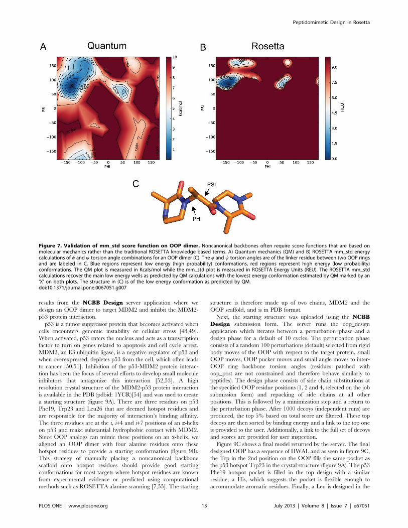
Figure 7 shows a Ramachandran plot of the w (x-axis) and y (y-
axis) dihedral angles between two OOP rings where regions
colored in red are high energy and those colored in blue are low
energy. Figure 7A, shows a plot based on the quantum
calculations. The low energy wells are fewer and smaller than a
peptide Ramachandran plot and therefore suggests the OOP is
more stable. This is expected due to the steric hindrance between
the additional atoms in the OOP rings.
Figure 7B shows the Ramachandran plot based on the
ROSETTA mm_std score function. The ROSETTA mm_std
score function captures the low energy wells of the quantum
results. Additionally, the lowest energy conformation from the
quantum optimizations, marked by an ‘X’, is in a low energy well
in both plots. There are additional regions of the QM plot that are
not captured by the mm_std score function. This is most likely due
to the ability of QM optimization to alter bond lengths and bond
angles which is not done in ROSETTA optimizations. This result
does, however, suggest that the ROSETTA mm_std score
function is a good approximation to the likely energy landscape
and can be used to accurately estimate energies of conformations
with varying backbone dihedral angles.
It is also important to test the performance of side chain
dihedrals when implementing a noncanonical backbone. Side
chain groups branching off OOP rings may encounter steric effects
from the additional atoms in the OOP ring. The OOP ring can be
in either a half-chair (figure 8B) or boat conformation (figure 8C)
(overlay is shown in figure 8A) and the energy of the side chain x1angle is affected by this ring puckering. Figure 8D shows an angle
vs energy plot of the quantum energy calculations similar to the
parameters described above. The half-chair (blue) is consistently
lower in energy across all x angles sampled than the boat
conformation (green) but the rotamer at 180u is nearly isoenergetic
between the half-chair and boat conformations. This reinforces the
importance of the OopPuckMover conformational sampling class
described above, as an OOP residue may occupy both conforma-
tions with similar probability.
ROSETTA mm_std score function calculations were also
computed (dashed lines) and overlay the quantum results (solid
lines) for both the half-chair (figure 8E) and boat (figure 8F)
conformations. These comparisons show the ROSETTA mm_std
score function accurately reflects the energy behavior of the xangle by properly aligning minima in each of the quantum low
energy rotamer wells. Additionally, there are two crystal structures
of oxopiperazine half-chair rings with side chains in the Cam-
bridge Structural Database [43], one with a tyrosine side chain
(code: FOBFEH) and another with two phenylalanine side chains
(code: ZOZTUD). The x1 values of these side chains are marked
as red X’s in figure 8E and show correspondence with the minima
of the energy landscape. This side chain-scanning result and the
backbone-scanning result above suggest ROSETTA is capable of
accurately calculating energies of a general class of noncanonical
backbone conformations.
Noncanonical backbone applications on the ROSIEserver: a design example
In an effort to increase the usability of ROSETTA molecular
modeling suite, ROSETTA applications can now be installed in a
unified webserver framework called ROSIE (http://rosie.
rosettacommons.org), which provides a user friendly interface to
ROSETTA without the traditional need of Unix development
skills. Server applications using several of the noncanonical
backbones described above have been implemented into the
ROSIE (ROSETTA Online Server that Includes Everyone)
framework and are available for public use. Here we describe
Peptidomimetic Design in Rosetta
PLOS ONE | www.plosone.org 12 July 2013 | Volume 8 | Issue 7 | e67051
results from the NCBB Design server application where we
design an OOP dimer to target MDM2 and inhibit the MDM2-
p53 protein interaction.
p53 is a tumor suppressor protein that becomes activated when
cells encounters genomic instability or cellular stress [48,49].
When activated, p53 enters the nucleus and acts as a transcription
factor to turn on genes related to apoptosis and cell cycle arrest.
MDM2, an E3 ubiquitin ligase, is a negative regulator of p53 and
when overexpressed, depletes p53 from the cell, which often leads
to cancer [50,51]. Inhibition of the p53-MDM2 protein interac-
tion has been the focus of several efforts to develop small molecule
inhibitors that antagonize this interaction [52,53]. A high
resolution crystal structure of the MDM2-p53 protein interaction
is available in the PDB (pdbid: 1YCR)[54] and was used to create
a starting structure (figure 9A). There are three residues on p53
Phe19, Trp23 and Leu26 that are deemed hotspot residues and
are responsible for the majority of interaction’s binding affinity.
The three residues are at the i, i+4 and i+7 positions of an a-helix
on p53 and make substantial hydrophobic contact with MDM2.
Since OOP analogs can mimic these positions on an a-helix, we
aligned an OOP dimer with four alanine residues onto these
hotspot residues to provide a starting conformation (figure 9B).
This strategy of manually placing a noncanonical backbone
scaffold onto hotspot residues should provide good starting
conformations for most targets where hotspot residues are known
from experimental evidence or predicted using computational
methods such as ROSETTA alanine scanning [7,55]. The starting
structure is therefore made up of two chains, MDM2 and the
OOP scaffold, and is in PDB format.
Next, the starting structure was uploaded using the NCBBDesign submission form. The server runs the oop_design
application which iterates between a perturbation phase and a
design phase for a default of 10 cycles. The perturbation phase
consists of a random 100 perturbations (default) selected from rigid
body moves of the OOP with respect to the target protein, small
OOP moves, OOP pucker moves and small angle moves to inter-
OOP ring backbone torsion angles (residues patched with
oop_post are not constrained and therefore behave similarly to
peptides). The design phase consists of side chain substitutions at
the specified OOP residue positions (1, 2 and 4, selected on the job
submission form) and repacking of side chains at all other
positions. This is followed by a minimization step and a return to
the perturbation phase. After 1000 decoys (independent runs) are
produced, the top 5% based on total score are filtered. These top
decoys are then sorted by binding energy and a link to the top one
is provided to the user. Additionally, a link to the full set of decoys
and scores are provided for user inspection.
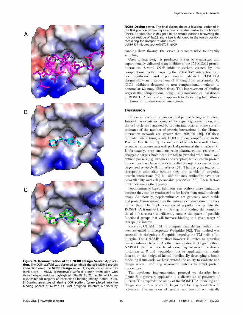
Figure 9C shows a final model returned by the server. The final
designed OOP has a sequence of HWAL and as seen in figure 9C,
the Trp in the 2nd position on the OOP fills the same pocket as
the p53 hotspot Trp23 in the crystal structure (figure 9A). The p53
Phe19 hotspot pocket is filled in the top design with a similar
residue, a His, which suggests the pocket is flexible enough to
accommodate aromatic residues. Finally, a Leu is designed in the
Figure 7. Validation of mm_std score function on OOP dimer. Noncanonical backbones often require score functions that are based onmolecular mechanics rather than the traditional ROSETTA knowledge based terms. A) Quantum mechanics (QM) and B) ROSETTA mm_std energycalculations of w and y torsion angle combinations for an OOP dimer (C). The w and y torsion angles are of the linker residue between two OOP ringsand are labeled in C. Blue regions represent low energy (high probability) conformations, red regions represent high energy (low probability)conformations. The QM plot is measured in Kcals/mol while the mm_std plot is measured in ROSETTA Energy Units (REU). The ROSETTA mm_stdcalculations recover the main low energy wells as predicted by QM calculations with the lowest energy conformation estimated by QM marked by an‘X’ on both plots. The structure in (C) is of the low energy conformation as predicted by QM.doi:10.1371/journal.pone.0067051.g007
Peptidomimetic Design in Rosetta
PLOS ONE | www.plosone.org 13 July 2013 | Volume 8 | Issue 7 | e67051

fourth position of the OOP scaffold but in a different orientation
than the hotspot Leu26 on p53. A possible reason for this maybe
the starting conformation of the OOP scaffold with respect to the
target protein. Creating several starting conformations and
Figure 8. OOP ring conformations and validation of mm_std score function on side chain dihedrals in different ring conformations.A) Overlay of half-chair (blue) and boat (green) OOP ring conformations. B) Structure of half-chair OOP ring conformation. C) Structure of boat OOPring conformation. D) QM energy calculations (Kcal/mol) of different side chain x1 dihedral angles. Blue represents half-chair conformation. Greenrepresents boat conformation. The half-chair conformation is lower in energy in two out of the three x1 dihedral energy wells (x1~600 and x1~3000)but nearly isoenergetic with the boat conformation in the third (x1~1800). E) QM energy (solid) and ROSETTA mm_std (dash, ROSETTA Energy Units)energy calculations for x1 dihedral angles with OOP ring in half-chair conformation. Red X’s show x1 values of oxopiperazine side chains (labeled withCambridge Structural Database code). F) QM energy (solid) and ROSETTA mm_std (dash, ROSETTA Energy Units) energy calculations for x1 dihedralangles with OOP ring in boat conformation. For both (E) and (F), ROSETTA mm_std score function recapitulates the low energy minima.doi:10.1371/journal.pone.0067051.g008
Peptidomimetic Design in Rosetta
PLOS ONE | www.plosone.org 14 July 2013 | Volume 8 | Issue 7 | e67051
running them through the server is recommended to diversify
sampling.
Once a final design is produced, it can be synthesized and
experimentally validated as an inhibitor of the p53-MDM2 protein
interaction. Several OOP inhibitor designs created by this
computational method targeting the p53-MDM2 interaction have
been synthesized and experimentally validated. ROSETTA
designs show an improvement of binding from micromolar Kd
(OOP inhibitors designed by non computational methods) to
nanomolar Kd (unpublished data). This improvement of binding
suggests that computational design using noncanonical backbones
in ROSETTA is a powerful approach to discovering high affinity
inhibitors to protein-protein interactions.
Discussion
Protein interactions are an essential part of biological function.
Intracellular events including cellular signaling, transcription, and
the cell cycle are regulated by protein interactions. Some current
estimates of the number of protein interactions in the Human
interaction network are greater than 300,000 [56]. Of these
estimated interactions, nearly 15,000 protein complexes are in the
Protein Data Bank [57], the majority of which have well defined
secondary structure at a well packed portion of the interface [7].
Unfortunately, most small molecule pharmaceutical searches of
druggable targets have been limited to proteins with small, well
defined pockets (e.g. enzymes and receptors) while protein-protein
interactions have been considered difficult targets because of their
larger and relatively flat interfaces [58]. There is great interest in
therapeutic antibodies because they are capable of targeting
protein interactions [59] but unfortunately antibodies have poor
bioavailability and cell permeable properties [58]. These factors
limit their use as therapeutics.
Peptidomimetic based inhibitors can address these limitations
because they can be synthesized to be larger than small molecule
drugs. Additionally, peptidomimetics are generally more stable
and proteolysis-resistant than the natural secondary structures they
mimic [60]. The implementation of peptidomimetics into the
ROSETTA framework is a first step in providing the computa-
tional infrastructure to efficiently sample the space of possible
functional groups that will increase binding to a given target of
therapeutic interest.
Recently, CHAMP [61], a computational design method, has
been extended to incorporate b-peptides [62]. The method was
successful in designing a b-peptide targeting the TM helix of an
integrin. The CHAMP method however is limited to targeting
transmembrane helices. Another computational design method,
NAPOLI [63], is capable of designing arbitrary backbones
(including a, b and c-peptides), but its application is mainly
focused on the design of helical bundles. By developing a broad
modeling framework, we have created the ability to evaluate and
design several promising oligomeric systems to target protein
interactions.
The backbone implementation protocol we describe here
(figure 2) is generally applicable to a diverse set of polymers of
interest. This expands the utility of the ROSETTA modeling and
design suite into a powerful design tool for a general class of
polymers. The inclusion of greater numbers of synthetically
Figure 9. Demonstration of the NCBB Design Server Applica-tion. The OOP scaffold was designed to inhibit the p53-MDM2 proteininteraction using the NCBB Design server. A) Crystal structure of p53(pink sticks) - MDM2 (electrostatic surface) protein interaction withthree hotspot residues highlighted (Phe19, Trp23, Leu26) which areresponsible for majority of interaction’s binding affinity (pdbid: 1YCR).B) Starting structure of alanine OOP scaffold (cyan) placed into thebinding pocket of MDM2. C) Final designed structure reported by
NCBB Design server. The final design shows a histidine designed inthe first position recovering an aromatic residue similar to the hotspotPhe19. A tryptophan is designed in the second position recovering thehotspot residue of Trp23 and a Leu is designed in the fourth positionrecovering the hotspot residue Leu26.doi:10.1371/journal.pone.0067051.g009
Peptidomimetic Design in Rosetta
PLOS ONE | www.plosone.org 15 July 2013 | Volume 8 | Issue 7 | e67051
compatible oligomer scaffolds would allow the design of combi-
natorial molecules that functionally exceed what is capable by the
biopolymers of peptides and nucleic acids. The current imple-
mentation as described above is fully compatible with combining
various backbones into one molecule such as a-b-peptide hybrids
or peptoid–OOP hybrids. We therefore anticipate the ROSETTA
framework to be a useful tool in creating novel polymer designs for
a variety of applications.
Supporting Information
File S1 OOP Pre ResidueType patch file. Complete
example of OOP patch file.
(TXT)
File S2 Peptoid NPhe ResidueType parameter fileComplete example of Peptoid parameter file.
(TXT)
Movie S1 Movie demonstration of OopPuckMover func-tionality. Special movers are often necessary to implement which
allow proper sampling of noncanonical backbone conformations.
The movie shows the steps necessary to change the conformation
of an OOP ring from the half-chair to the boat conformation. The
OopPuckMover first alters the w and y angles and then, second,
updates the hydrogens into proper alignment. Gray spheres
represent virtual atoms.
(MOV)
Acknowledgments
We thank Dennis Shasha, the ROSETTA Developers Community, Karl
Ward and NYU IT Services for helpful discussions and high performance
computing support. We would also like to thank Ingemar Andre and
Wojtek Potrzebowski for helpful comments on the project.
Author Contributions
Conceived and designed the experiments: KD PDR FCC JWL JJG KK
PSA RD RB. Performed the experiments: KD PDR TWC FCC JWL BNB
AW MP KPK. Analyzed the data: KD PDR TWC GLB FCC JWL MP
KPK. Contributed reagents/materials/analysis tools: ALF BK PB KK
PSA. Wrote the paper: KD PDR FCC JWL KK PSA RD RB. Contributed
software for server: SL.
References
1. Gellman SH (1998) Foldamers: a manifesto. Accounts of Chemical Research 31:
173–180.
2. Stigers KD, Soth MJ, Nowick JS (1999) Designed molecules that fold to mimic
protein secondary structures. Curr Opin Chem Biol 3: 714–23.
3. Goodman CM, Choi S, Shandler S, DeGrado WF (2007) Foldamers as versatile
frameworks for the design and evolution of function. Nat Chem Biol 3: 252–62.
4. Edwards TA, Wilson AJ (2011) Helix-mediated protein–protein interactions as
targets for intervention using foldamers. Amino Acids 41: 743–54.
5. Henchey LK, Jochim AL, Arora PS (2008) Contemporary strategies for the
stabilization of peptides in the alpha-helical conformation. Curr Opin Chem
Biol 12: 692–7.
6. Ko E, Raghuraman A, Perez LM, Ioerger TR, Burgess K (2013) Exploring keyorientations at protein-protein interfaces with small molecule probes. J Am
Chem Soc 135: 167–73.
7. Bullock BN, Jochim AL, Arora PS (2011) Assessing helical protein interfaces for
inhibitor design. J Am Chem Soc 133: 14220–3.
8. Horne WS, Boersma MD, Windsor MA, Gellman SH (2008) Sequence-baseddesign of alpha/betapeptide foldamers that mimic bh3 domains. Angew Chem
Int Ed Engl 47: 2853–6.
9. Henchey LK, Kushal S, Dubey R, Chapman RN, Olenyuk BZ, et al. (2010)
Inhibition of hypoxia inducible factor 1-transcription coactivator interaction by
a hydrogen bond surrogate alpha-helix. J Am Chem Soc 132: 941–3.
10. Tosovska P, Arora PS (2010) Oligooxopiperazines as nonpeptidic alpha-helix
mimetics. Org Lett 12: 1588–91.
11. Cummings CG, Hamilton AD (2010) Disrupting protein-protein interactions
with non-peptidic, small molecule alpha-helix mimetics. Curr Opin Chem Biol
14: 341–6.
12. Simon RJ, Kania RS, Zuckermann RN, Huebner VD, Jewell DA, et al. (1992)
Peptoids: a modular approach to drug discovery. Proc Natl Acad Sci U S A 89:
9367–71.
13. Armand P, Kirshenbaum K, Goldsmith RA, Farr-Jones S, Barron AE, et al.
(1998) Nmr determination of the major solution conformation of a peptoidpentamer with chiral side chains. Proc Natl Acad Sci U S A 95: 4309–14.
14. Kirshenbaum K, Barron AE, Goldsmith RA, Armand P, Bradley EK, et al.
(1998) Sequence-specific polypeptoids: a diverse family of heteropolymers with
stable secondary structure. Proc Natl Acad Sci U S A 95: 4303–8.
15. Butterfoss GL, Renfrew PD, Kuhlman B, Kirshenbaum K, Bonneau R (2009) Apreliminary survey of the peptoid folding landscape. J Am Chem Soc 131:
16798–807.
16. Stringer JR, Crapster JA, Guzei IA, Blackwell HE (2011) Extraordinarily robust
polyproline type i peptoid helices generated via the incorporation of -chiral
aromatic n-1-naphthylethyl side chains. J Am Chem Soc 133: 15559–67.
17. Shah NH, Butterfoss GL, Nguyen K, Yoo B, Bonneau R, et al. (2008) Oligo(n-
aryl glycines): a new twist on structured peptoids. J Am Chem Soc 130: 16622–
32.
18. Dohm MT, Kapoor R, Barron AE (2011) Peptoids: bio-inspired polymers as
potential pharmaceuticals. Curr Pharm Des 17: 2732–47.
19. Tran H, Gael SL, Connolly MD, Zuckermann RN (2011) Solid-phase
submonomer synthesis of peptoid polymers and their self-assembly into highly-
ordered nanosheets. J Vis Exp: e3373.
20. Fowler SA, Blackwell HE (2009) Structure-function relationships in peptoids:
recent advances toward deciphering the structural requirements for biologicalfunction. Org Biomol Chem 7: 1508–24.
21. Levine PM, Lee E, Greenfield A, Bonneau R, Logan SK, et al. (2012) Androgen
receptor antagonism by divalent ethisterone conjugates in castrate-resistantprostate cancer cells. ACS Chem Biol 7: 1693–701.
22. Yoo B, Kirshenbaum K (2005) Protease-mediated ligation of abiotic oligomers.
J Am Chem Soc 127: 17132–3.
23. Horne WS, Gellman SH (2008) Foldamers with heterogeneous backbones. Acc
Chem Res 41: 1399–408.
24. Seebach D, Gardiner J (2008) Beta-peptidic peptidomimetics. Acc Chem Res 41:1366–75.
25. Daniels DS, Petersson EJ, Qiu JX, Schepartz A (2007) High-resolution structure
of a beta-peptide bundle. J Am Chem Soc 129: 1532–3.
26. Boersma MD, Haase HS, Peterson-Kaufman KJ, Lee EF, Clarke OB, et al.
(2012) Evaluation of diverse /-backbone patterns for functional -helix mimicry:
analogues of the bim bh3 domain. J Am Chem Soc 134: 315–23.
27. HorneWS, Johnson LM, Ketas TJ, Klasse PJ, Lu M, et al. (2009) Structural and
biological mimicry of protein surface recognition by alpha/beta-peptide
foldamers. Proc Natl Acad Sci U S A 106: 14751–6.
28. Patgiri A, Jochim AL, Arora PS (2008) A hydrogen bond surrogate approach for
stabilization of short peptide sequences in alpha-helical conformation. Acc
Chem Res 41: 1289–300.
29. Henchey LK, Porter JR, Ghosh I, Arora PS (2010) High specificity in protein
recognition by hydrogen-bond-surrogate ı6-helices: selective inhibition of the
p53/mdm2 complex. Chembiochem 11: 2104–7.
30. Patgiri A, Yadav KK, Arora PS, Bar-Sagi D (2011) An orthosteric inhibitor of
the ras-sos interaction. Nat Chem Biol 7: 585–7.
31. Stewart ML, Fire E, Keating AE, Walensky LD (2010) The mcl-1 bh3 helix isanexclusive mcl-1 inhibitor and apoptosis sensitizer. Nat Chem Biol 6: 595–601.
32. Pang PC, Chiu PCN, Lee CL, Chang LY, Panico M, et al. (2011) Human sperm
binding is mediated by the sialyl-lewis(x) oligosaccharide on the zona pellucida.Science 333: 1761–4.
33. Helenius A (1994) How n-linked oligosaccharides affect glycoprotein folding in
the endoplasmic reticulum. Mol Biol Cell 5: 253–65.
34. Kornfeld S (1990) Lysosomal enzyme targeting. Biochem Soc Trans 18: 367–74.
35. Hauri H, Appenzeller C, Kuhn F, Nufer O (2000) Lectins and traffic in the
secretory pathway. FEBS Lett 476: 32–7.
36. Baral PK, Wieland B, Swayampakula M, Polymenidou M, Rahman MH, et al.
(2012) Structural studies on the folded domain of the human prion protein
bound to the fab fragment of the antibody pom1. Acta Crystallogr D BiolCrystallogr 68: 1501–12.
37. Ermonval M, Mouillet-Richard S, Codogno P, Kellermann O, Botti J (2003)Evolving views in prion glycosylation: functional and pathological implications.
Biochimie 85: 33–45.
38. Lemmon G, Meiler J (2012) Rosetta ligand docking with flexible xml protocols.Methods Mol Biol 819: 143–55.
39. Renfrew PD, Choi EJ, Bonneau R, Kuhlman B (2012) Incorporation of
noncanonical amino acids into rosetta and use in computational protein-peptideinterface design. PLoS One 7: e32637.
40. Shandler SJ, Shapovalov MV, Dunbrack RL Jr, DeGrado WF (2010)
Development of a rotamer library for use in beta-peptide foldamer computa-tional design. J Am Chem Soc 132: 7312–20.
41. Leaver-Fay A, Tyka M, Lewis SM, Lange OF, Thompson J, et al. (2011)
Rosetta3: an objectoriented software suite for the simulation and design ofmacromolecules. Methods Enzymol 487: 545–74.
Peptidomimetic Design in Rosetta
PLOS ONE | www.plosone.org 16 July 2013 | Volume 8 | Issue 7 | e67051

42. Brooks BR, Brooks CL 3rd, Mackerell AD Jr, Nilsson L, Petrella RJ, et al. (2009)
Charmm: thebiomolecular simulation program. J Comput Chem 30: 1545–614.43. Allen FH (2002) The cambridge structural database: a quarter of a million
crystal structures and rising. Acta Crystallogr B 58: 380–8.
44. Culf AS, Ouellette RJ (2010) Solid-phase synthesis of n-substituted glycineoligomers (Peptoids) and derivatives. Molecules 15: 5282–5335.
45. Rohl CA, Strauss CEM, Misura KMS, Baker D (2004) Protein structureprediction using rosetta. Methods Enzymol 383: 66–93.
46. Molski MA, Goodman JL, Chou FC, Baker D, Das R, et al. (2013) Remodeling
a beta-peptide bundle. Chemical Science 4: 319–324.47. Frisch MJ, Trucks GW, Schlegel HB, Scuseria GE, Robb MA, et al. Gaussian 09
Revision A.1. Gaussian Inc. Wallingford CT 2009.48. Vousden KH, Lu X (2002) Live or let die: the cell’s response to p53. Nat Rev
Cancer 2: 594–604.49. Vogelstein B, Lane D, Levine AJ (2000) Surfing the p53 network. Nature 408:
307–10.
50. Vassilev LT (2007) Mdm2 inhibitors for cancer therapy. Trends Mol Med 13:23–31.
51. Vazquez A, Bond EE, Levine AJ, Bond GL (2008) The genetics of the p53pathway, apoptosis and cancer therapy. Nat Rev Drug Discov 7: 979–87.
52. Michelsen K, Jordan JB, Lewis J, Long AM, Yang E, et al. (2012) Ordering of
the n-terminus of human mdm2 by small molecule inhibitors. J Am Chem Soc134: 17059–67.
53. Vassilev LT, Vu BT, Graves B, Carvajal D, Podlaski F, et al. (2004) In vivoactivation of the p53 pathway by small-molecule antagonists of mdm2. Science
303: 844–8.
54. Kussie PH, Gorina S, Marechal V, Elenbaas B, Moreau J, et al. (1996) Structure
of the mdm2 oncoprotein bound to the p53 tumor suppressor transactivationdomain. Science 274: 948–53.
55. Jochim AL, Arora PS (2010) Systematic analysis of helical protein interfaces
reveals targets for synthetic inhibitors. ACS Chem Biol 5: 919–23.56. Hart GT, Ramani AK, Marcotte EM (2006) How complete are current yeast
and human proteininteraction networks? Genome Biol 7: 120.57. Berman HM, Westbrook J, Feng Z, Gilliland G, Bhat TN, et al. (2000) The
protein data bank. Nucleic Acids Res 28: 235–42.
58. Arkin MR, Wells JA (2004) Small-molecule inhibitors of protein-proteininteractions: progressing towards the dream. Nat Rev Drug Discov 3: 301–17.
59. Stockwin LH, Holmes S (2003) Antibodies as therapeutic agents: vive larenaissance! Expert Opin Biol Ther 3: 1133–52.
60. Miller SM, Simon RJ, Ng S, Zuckermann RN, Kerr JM, et al. (1995)Comparison of the proteolytic susceptibilities of homologous l-amino acid, d-
amino acid, and n-substituted glycine peptide and peptoid oligomers. Drug
Development Research 35: 20–32.61. Yin H, Slusky JS, Berger BW, Walters RS, Vilaire G, et al. (2007)
Computational design of peptides that target transmembrane helices. Science315: 1817–22.
62. Shandler SJ, Korendovych IV, Moore DT, Smith-Dupont KB, Streu CN, et al.
(2011) Computational design of a ı-peptide that targets transmembrane helices.J Am Chem Soc 133: 12378–81.
63. Korendovych IV, Kim YH, Ryan AH, Lear JD, Degrado WF, et al. (2010)Computational design of a self-assembling -peptide oligomer. Org Lett 12:
5142–5.
Peptidomimetic Design in Rosetta
PLOS ONE | www.plosone.org 17 July 2013 | Volume 8 | Issue 7 | e67051