Embed Size (px)
Citation preview

ADAR1 Modulation of p21WAF1 ModulationLuis Espinoza-DelgadoCentral Catholic High SchoolGrade 12

Introduction
• ADAR1 (Adenosine Deaminase Acting on RNA 1) is a mRNA editing protein that post-transcriptionally modifies nucleic acid by catalyzing the conversion of adenosine to inosine, allowing for a greater variety of proteins.
• ADAR1 contains both a catalytic (editing) domain and a RNA/DNA binding domain. It exists as a long (p150) and short (p110) isoform.• We are using constructs that express either wild type ADAR1
p150 or a mutant that cannot edit RNA (MUT).

Introduction
• p21WAF1 is a cyclin-dependent kinase inhibitor has been linked to senescence and to either increased or decreased growth arrest and to increased or decreased cell death.
• Preliminary studies indicate a negative correlation between levels of p21WAF1 expression and those of ADAR1 (Sharma et. al. in preparation).
• Loss of ADAR1 has been linked to embryonic lethality and can result in death of liver cells and of leukemia cells.

Introduction• Chronic myelogenous leukemia (CML) is a cancer of the
white blood cells caused by a genetic translocation (the Philadelphia chromosome).• Rearrangement makes a new gene Bcr Abl—a tyrosine kinase that is always
on.
• CML patients respond well to tyrosine kinase inhibitors (TKIs) of the Bcr-Abl oncoprotein.• TKIs fail to eradicate immature leukemia-initiating cells that arise
from myeloid progenitor cells—sensitive to the deletion of mRNA editing protein ADAR1.
• Chronic myelogenous leukemia cells die without ADAR1 and leukemic cells depend on ADAR1 more than normal white blood cells (Steinman et. al. submitted).
• Targets of ADAR1 are largely unknown.

Project Goals
1. Validate inverse relation between ADAR1 and p21WAF1
2. Map ADAR1 effect as a transcriptional, post-transcriptional, or translational mechanism
3. Establish whether ADAR1 suppression of p21WAF1 involves RNA editing domain

Hypothesis
• Null: ADAR1 will not have a significant effect on p21 transcription.

Strategic Plan
Significant Variation
Insignificant Variation
Preliminary Procedure• WT and -/- ADAR1 mouse
embryonic fibroblasts were obtained from Dr. Qingde Wang.
• A Western Blot using Ponceau Staining was performed to validate intended presence/absence of ADAR1.
Wt -/- MEFs
p110
p150
Ponceau stain

Preliminary Procedure• Plasmid constructs were obtained and
digested with restriction enzymes to alter the promoter length of p21 (FL 2.4kB).
luc
luc
luc
luc
luc
p21 WT
p21 Hinf1▽
p21 1.1 ▽
p21 2.2▽
pgl2basic

Materials• Western Blot Kit• Lipofectamine 2000 Transfection Kit• Luciferase Assay Kit• ß-galactosidase Assay Kit• ADAR-modified Mouse Embryonic Fibroblasts• p21 promoter and ‘UTR constructs• ADAR plasmid vectors• Clontech X Transfection Kit

ProcedureLipofectamine 2000 Transfection1. Adherent cells: One day before transfection, plate 0.5–2 × 10⁵cells in 500 μL of
growth medium without antibiotics so that cells will be 90–95% confluent at the time of transfection.
2. For each transfection sample, prepare complexes as follows: a. Dilute DNA in 50 μL of Opti-MEM®I Reduced Serum Medium without serum. Mix gently. b. Dilute the appropriate amount in 50 μL of Opti-MEM®I Reduced Serum Medium without serum. Incubate for 5 minutes at room temperature. c. After the 5 minute incubation, combine the diluted DNA with diluted Lipofectamine®2000 Transfection Reagent (total volume = 100 μL). Mix gently and incubate for 20 minutes at room temperature.
3. Add the 100 μL of complexes to each well containing cells and medium. 4. Incubate cells at 37°C in a CO2 incubator for 18–48 hours prior to testing for
transgene expression. The medium may be changed after 4–6 hours. 5. For stable cell lines: Passage cells at a 1:10 (or higher dilution) into fresh growth
medium 24 hours after transfection. Add selective medium (if desired) the following day
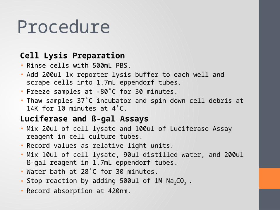
ProcedureCell Lysis Preparation• Rinse cells with 500mL PBS.• Add 200ul 1x reporter lysis buffer to each well and scrape cells into 1.7mL
eppendorf tubes.• Freeze samples at -80˚C for 30 minutes.• Thaw samples 37˚C incubator and spin down cell debris at 14K for 10 minutes
at 4˚C.
Luciferase and ß-gal Assays• Mix 20ul of cell lysate and 100ul of Luciferase Assay reagent in cell culture
tubes.• Record values as relative light units.• Mix 10ul of cell lysate, 90ul distilled water, and 200ul ß-gal reagent in 1.7mL
eppendorf tubes. • Water bath at 28˚C for 30 minutes.
• Stop reaction by adding 500ul of 1M Na2CO3 .
• Record absorption at 420nm.
pgl2basic pcDNA p21luc pcDNA pgl2basic pADARMUT
p21luc pADARMUT
0
20
40
60
80
100
120
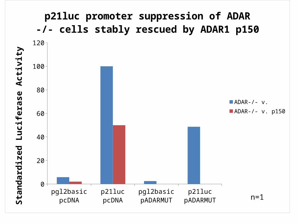
p21luc promoter suppression of ADAR -/- cells stably rescued by ADAR1 p150
ADAR-/- v.ADAR-/- v. p150
Stan
dard
ized
Luc
ifera
se A
ctivi
ty
n=1
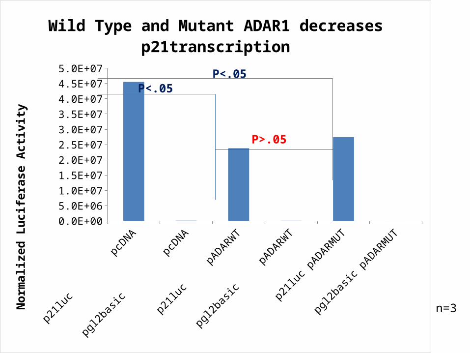
n=3
p21luc pcDNA
pgl2basic pcDNA
p21luc pADARWT
pgl2basic pADARWT
p21luc pADARMUT
pgl2basic pADARMUT
0.0E+00
5.0E+06
1.0E+07
1.5E+07
2.0E+07
2.5E+07
3.0E+07
3.5E+07
4.0E+07
4.5E+07
5.0E+07
Wild Type and Mutant ADAR1 decreases p21-transcription
Nor
mal
ized
Luc
ifera
se A
ctivi
ty
P<.05
P<.05
P>.05

p21luc pcD
NA
pgl2basi
c pcD
NA
p21luc Hinf1 pcD
NA
▽p21
2.2 pcDNA
▽p21
1.1 pcDNA
▽
p21luc pADARW
T
pgl2basi
c pADARW
T
p21luc Hinf1 pADARW
T
▽p21
2.2 pADARWT
▽p21
1.1 pADARWT
▽p21luc p
ADARMUT
pgl2basi
c pADARMUT
0
20
40
60
80
100
120
ADAR suppression of p21 maps to the distal promoter and does not involve p53 site #1 in the promoterSt
anda
rdiz
ed L
ucife
rase
Acti
vity
P<.05
n=3
n=1

p21luc pcDNA
p21luc Hinf ▽
pcDNA
p21 2.2 ▽pcDNA
p21 1.1 ▽pcDNA
p21luc pADARWT
p21luc Hinf ▽
pADARWT
p21 2.2 ▽pADARWT
p21 1.1 ▽pADARWT
0
100
200
300
400
500
600
ADAR1 Effects Differ with Clontech X ReagentSt
anda
rdiz
ed L
ucife
rase
Acti
vity
n=2
p21 3' UTR Cu pcDNA▽
p21 3' UTR Cu ADARWT▽
p21 3' UTR Cu ▽
ADARMUT
p21 FL 3' UTR pcDNA
p21 FL 3' UTR ADARWT
p21 FL 3' UTR ADARMUT
0
20
40
60
80
100
120
140
Stan
dard
ized
Luc
ifera
se A
ctivi
ty
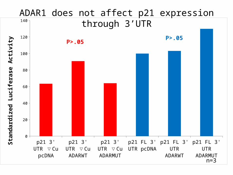
n=3
ADAR1 does not affect p21 expression through 3’UTR
P>.05P>.05

Conclusions• Lipofectamine 2000 transfections suggested ADAR1
involvement in p21 transcription.• Observation: Lipofectamine 2000 transfected cells looked sick. • Toxic effect that possibly increases p21 levels through p53.
• Less toxic transfection reagent (Clontech X-fect) indicated that ADAR1 did not affect p21 transcription.
• p53 levels are possibly elevated in response to the toxicity brought about by Lipofectamine 2000.• ADAR1 somehow opposes this action.

Future Directions• To test this opposition against p53, ADAR1 must be added to
p21 promoter constructs without both of p53 binding sites.
• It is possible that ADAR1 suppresses the p21 promoter directly, but future work is needed.
• ADAR1 did not significantly affect p21 expression at the level of the 3’-UTR. It is possible that ADAR1 suppresses p21 protein translation or protein stability.

Acknowledgements• Richard A Steinman MD, PhD• Qingde Wang MD, PhD• Christine Stehle• Mark Krotec

References• "Chronic Myelogenous Leukemia." National Cancer Institute.
03 July 2011. http://www.cancer.gov/cancertopics/pdq/treatment/CML/Patient/page1
• Richard A Steinman et. al. “Deletion of the RNA-editing enzyme ADAR1 causes regression of established chronic myelogenous leukemia in mice” (Under review, 2011)
• Rohit Sharma, Qingde Wang "RNA-editing enzyme ADAR1 supports cell division by inhibiting p21" (In preparation, 2011)
• Weinberg, Robert A. The Biology of Cancer. New York: Garland Science, 2007
Procedure
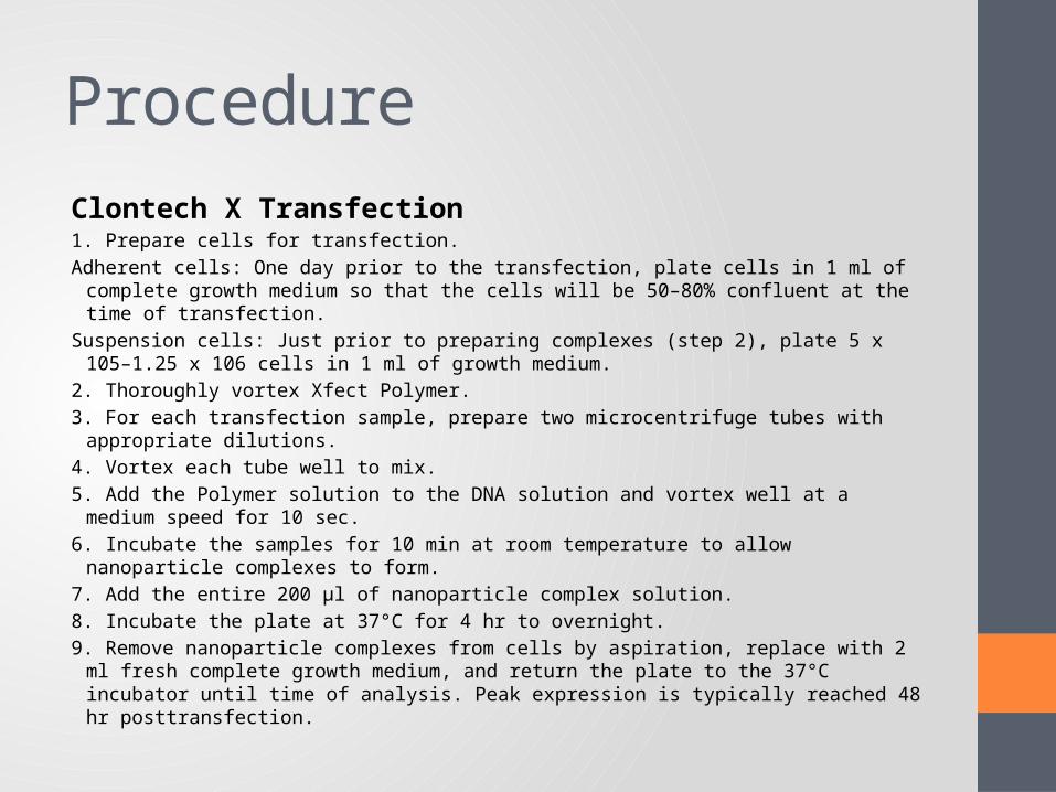
Clontech X Transfection1. Prepare cells for transfection.Adherent cells: One day prior to the transfection, plate cells in 1 ml of complete growth
medium so that the cells will be 50–80% confluent at the time of transfection.Suspension cells: Just prior to preparing complexes (step 2), plate 5 x 10⁵–1.25 x 10⁶ cells in 1
ml of growth medium.2. Thoroughly vortex Xfect Polymer.3. For each transfection sample, prepare two microcentrifuge tubes with appropriate dilutions.4. Vortex each tube well to mix.5. Add the Polymer solution to the DNA solution and vortex well at a medium speed for 10 sec.6. Incubate the samples for 10 min at room temperature to allow nanoparticle complexes to
form.7. Add the entire 200 μl of nanoparticle complex solution.8. Incubate the plate at 37°C for 4 hr to overnight.9. Remove nanoparticle complexes from cells by aspiration, replace with 2 ml fresh complete
growth medium, and return the plate to the 37°C incubator until time of analysis. Peak expression is typically reached 48 hr posttransfection.