Embed Size (px)
Citation preview

Adaptive Signal Recovery on Graphs viaHarmonic Analysis for
Experimental Design in Neuroimaging
Won Hwa Kim†, Seong Jae Hwang†, Nagesh Adluru‡,Sterling C. Johnson∗, and Vikas Singh§†
†Dept. of Computer Sciences, Univ. of Wisconsin, Madison, WI, U.S.A.§Dept. of Biostatistics & Med. Informatics, Univ. of Wisconsin, Madison, WI, U.S.A.
∗GRECC, William S. Middleton VA Hospital, Madison, WI, U.S.A.‡Waisman Center, Madison, WI, U.S.A.http://pages.cs.wisc.edu/∼wonhwa
Abstract. Consider an experimental design of a neuroimaging study,where we need to obtain p measurements for each participant in a set-ting where p′(< p) are cheaper and easier to acquire while the remaining(p − p′) are expensive. For example, the p′ measurements may includedemographics, cognitive scores or routinely offered imaging scans whilethe (p − p′) measurements may correspond to more expensive types ofbrain image scans with a higher participant burden. In this scenario, itseems reasonable to seek an “adaptive” design for data acquisition soas to minimize the cost of the study without compromising statisticalpower. We show how this problem can be solved via harmonic analysisof a band-limited graph whose vertices correspond to participants andour goal is to fully recover a multi-variate signal on the nodes, given thefull set of cheaper features and a partial set of more expensive measure-ments. This is accomplished using an adaptive query strategy derivedfrom probing the properties of the graph in the frequency space. Todemonstrate the benefits that this framework can provide, we presentexperimental evaluations on two independent neuroimaging studies andshow that our proposed method can reliably recover the true signal withonly partial observations directly yielding substantial financial savings.
1 Introduction
Consider an experimental design setting which involves a cohort S comprised ofN individuals (or examples) in total. We are allowed to obtain a maximum of pmeasurements (or features) for each participant (or example) in S. Dependingon the application, these p measurements may be variously interpreted — forexample, in a machine learning experiment, we may have p distinct numerical
This research was supported by NIH grants AG040396, and NSF CAREER award 1252725, UWADRC AG033514, UW ICTR 1UL1RR025011, UW CPCP AI117924, UW CIBM 5T15LM007359-14 and Waisman Core Grant P30 HD003352-45.

2 W. H. Kim et al.
preferences a user assigns to each item whereas in computer vision, the measure-ments may reflect p specific requests for supervision or indication on each imagein S [1–4]. In a neuroscience experiment, the cohort corresponds to individualsubjects — the p measurements will denote various types of imaging and clinicalmeasures we can acquire. Of course, independent of the application, the “cost”of measurements is quite variable: while features such as gender and age of aparticipant have negligible cost, requesting a user to rate an image in abstractterms, “How natural is this image on a scale of 1 to 5?”, may be more expen-sive. In neuroimaging, acquiring some clinical and cognitive measures is cheap,whereas certain image scans can cost several thousands of dollars [5, 6].
In the past, when datasets were smaller, these issues were understandablynot very important. But as we move towards acquiring and annotating largescale datasets in machine learning and vision [7–9], the cost implications can besubstantial. For instance, if the budget for a multi-modal brain imaging study in-volving several different types of image scans for ∼200 subjects is $3M+ and weknow a priori which type of inference models will finally be estimated using thisdata, it seems reasonable to ask if “adaptive” data acquisition can bring downcosts by 25% with negligible deterioration in statistical power. While experimentdesign concepts in classical statistics provide an excellent starting point, theyprovide little guidance in terms of practical technical issues one faces in address-ing the question above. Outside of a few recent works [10–12], this topic is stillnot extensively studied within mainstream machine learning and vision.
In this paper, we study a natural form of the experimental design problemin the context of an important brain imaging application. Assume that we haveaccess to a cohort S of n subjects. In principle, we can acquire p measurementsfor each participant. But all p measures are not easily available — say, we startonly with a default set of p′ measures for each subject which may be consideredas “inexpensive”. This yields a matrix of size N × p′. We are also provided theremaining set of (p− p′) measurements but only for a small subset S ′ of n′ sub-jects — possibly due to the associated expense of the measurement. We can, ifdesired, acquire these additional (p−p′) measures for each individual participantin S \ S ′, but at a high per-individual cost. Our goal is to eventually estimate astatistical model that has high fidelity to the “true” model estimated using thefull set of p measures/features for the full cohort S. The key question is whetherwe can design an adaptive query strategy that minimizes the overall cost we in-cur and yet provides high confidence in the parameter estimates we obtain. Theproblem statement is quite general and models experimental design considera-tions in numerous scientific disciplines including systems biology and statisticalgenomics where an effective solution can drive improvements in efficiency.
1.1 Related Work
There are three distinct areas of the literature that are loosely related to thedevelopment described in this paper. At the high level, perhaps the most closelyrelated to our work is active learning which is motivated by similar cost-benefitconsiderations, but in terms of minimizing the number of queries (seeking thelabel of an example) [13]. Here, one starts with a pool of unlabeled data and

Adaptive Signal Recovery on Graphs via Harmonic Analysis 3
picks a few examples at random to obtain their labels. Then, we repeatedly fita classifier to the labeled examples seen so far and query the unlabeled examplethat is most uncertain or likely to decrease overall uncertainty. This strategy isgenerally successful though may asymptotically converge to a sub-optimal clas-sifier [14]. Adaptive query strategies have been presented to guarantee that thehypothesis space is fully explored and to obtain theoretically consistent results[15, 16]. Much of active learning focuses on learning discriminative classifiers;while the Bayesian versions of active learning can, in principle, be applied to farmore general settings, it is not clear whether such formulations can be adaptedfor the stratified cost structure we encounter in the motivating example aboveand for general parameter estimation problems where the likelihood expressionsare not computationally ‘nice’.
Within the statistics literature, the problem of experiment design has a richhistory going back at least four decades [17–19], and seeks to formalize howone deals with the non-deterministic nature of physical experiments. In con-trast to the basic setting here and even data-driven measures of merit such asD-optimality [20, 21], experiment design concepts such as the Latin hypercubedesign [22] intentionally assume very little about the relationship between inputfeatures and the output labels. Instead, with d features, such procedures will gen-erate a space-filling design so that each of the dimensions is divided into equallevels — the calculated configuration merely provides a selection of inputs atwhich to compute the output of an experiment to achieve specific goals. Despitea similar name, the goals of these ideas are quite different from ours.
Within machine learning and vision, papers related to collaborative filtering(and matrix completion) [23–26] share a number of technical similarities to thedevelopment in our work. For instance, one may assume that in a matrix of sizeN×p (subjects × measurements), the first p′ columns are fully observed whereasmultiple rows in the remaining (p− p′) columns are missing. This clearly yieldsa matrix completion problem; unfortunately, the setup lies far from incoherentsampling and the matrix versions of restricted isometry property (RIP) thatmake the low-rank completion argument work in practice [27, 28]. This observa-tion has been made in recent works where collaborative filtering was generalizedto the graph domain [29] and where random sampling was introduced for graphsin [30]. However, these approaches, which will serve as excellent baselines, do notexploit the band-limited nature of measurements in frequency space. Separately,matrix completion within an adaptive query setting [31, 32] yields importanttheoretical benefits but so far, no analogs for the graph setting exist.
The contribution of this paper is to provide a harmonic analysis inspiredalgorithm to estimate band-limited signals that are defined on graphs. It turnsout that such solutions directly yield an efficient procedure to conduct adaptivequeries for designing experiments involving stratified costs of measurements, i.e.,where the first subset of measures is free whereas the second set of (p− p′) mea-sures is expensive and must be requested for a small fraction of participants. Ourframework relies on the design of an efficient decoder to recover the band-limited

4 W. H. Kim et al.
original signal involving multiple channels which was only partially observed. Inorder to accomplish these goals, the paper makes the following contributions.(i) We propose a novel sampling and signal recovery strategy on a graph that
is derived via harmonic analysis of the graph.(ii) We show how a band-limited multi-variate signal on a graph can be recon-
structed with only a few observations via a simple optimization scheme.(iii) We provide an extensive set of experiments on two independent datasets
which demonstrate that our framework works well in estimating expensiveimage-derived measurements based on (a) a partial set of observations (in-volving less expensive image-scan data) and (b) a full set of measurementson only a small fraction of the cohort.
2 Preliminaries: Linear Transforms in Euclidean andNon-Euclidean Spaces
Well known signal transforms in the forward/inverse directions such as thewavelet and Fourier transforms (in non-Euclidean space) are fundamental toour proposed framework. These transforms are well understood in the Euclideansetting, however, their analogues in non-Euclidean spaces have not been stud-ied until recently [33]. We provide a brief overview of these transforms in bothEuclidean and non-Euclidean spaces.
2.1 Continuous (Forward) Wavelet Transform
The Fourier transform is a fundamental tool for frequency analyses of a signalby transforming the signal f(x) into the frequency domain as
f(ω) = 〈f, ejωx〉 =
∫f(x)e−jωxdx (1)
where f(ω) is the resultant Fourier coefficient. Wavelet transform is similar tothe Fourier transform, but it uses a different type of oscillating basis function(i.e., mother wavelet). Unlike Fourier basis (i.e., sin()) with infinite support, awavelet ψ is a localized function with finite support. One can define a motherwavelet ψs,a(x) = 1
sψ(x−as ) with scale and translation properties, controlled by sand a respectively. Here, changing s controls the dilation and varying a controlsthe location of ψ. Using ψs,a as bases, a wavelet transform of a function f(x)results in wavelet coefficients Wf (s, a) at scale s and at location a as
Wf (s, a) = 〈f, ψ〉 =1
s
∫f(x)ψ∗(
x− as
)dx (2)
where ψ∗ is the complex conjugate of ψ [34].Interestingly, ψs is localized not only in the original domain but also in the
frequency domain. It behaves as a band-pass filter covering different bandwidthscorresponding to scales s. These band-pass filters do not cover the low-frequencycomponents, therefore an additional low-pass filter φ, a scaling function, is typ-ically introduced. A transform with the scaling function φ results in a low-passfiltered representation of the original function f . In the end, filtering at multiplescales s of the wavelet offers a multi-resolution view of the given signal.

Adaptive Signal Recovery on Graphs via Harmonic Analysis 5
2.2 Wavelet Transform in Non-Euclidean Spaces
Defining a wavelet transform in the Euclidean space is convenient because of theregularity of the domain (i.e., a regular lattice). In this case, one can easily definethe shape of a mother wavelet in the context of an application. However, in non-Euclidean spaces (e.g., graphs that consists of a set of vertices and edges witharbitrary connections), an implementation of a mother wavelet becomes difficultdue to the ambiguity of dilation and translation. Due to these issues, the classicaldefinition of the wavelet transform has not been suitable for analyses of data innon-Euclidean spaces until recently when [33, 35] proposed wavelet and Fouriertransforms in non-Euclidean spaces.
The key idea in [33] for constructing a mother wavelet ψ on the nodes of agraph is simple. Instead of defining it in the original domain where the propertiesof ψ are ambiguous, we define a mother wavelet in a dual domain where itsrepresentation is clear and then transform it back to the original domain. Thecore ingredients for such a construction are 1) a set of “orthonormal” basesthat provide the means to transform a signal between a graph and its dualdomain (i.e., an analogue of the frequency domain) and 2) a kernel function h()that behaves as a band-pass filter determining the shape of ψ. Utilizing theseingredients, a mother wavelet is first constructed as a kernel function in thefrequency domain and then localized in the original domain using a δ functionand the orthonormal bases. Such an operation will implement a mother waveletψ on the original graph. Defining a kernel function in the 1-D frequency domainis simple, and one can rely on spectral graph theory to obtain the orthonormalbases of a graph [33] which can be used for graph Fourier transform.
A graph G = V, E is formally defined by a vertex set V with N numberof vertices and a edge set E with edges that connect the vertices. Such a graphis generally represented by an adjacency matrix AN×N where each element aijdenotes the connection between ith and jth vertices by a corresponding edgeweight. Another matrix that summarizes the graph, a degree matrix DN×N , isa diagonal matrix where the ith diagonal is the sum of edge weights connectedto the ith vertex. A graph Laplacian is then defined from these two matricesas L = D − A, which is a self-adjoint and positive semi-definite operator. Thematrix L can be decomposed into pairs of eigenvalues λl ≥ 0 and correspondingeigenvectors χl where l = 0, 1, · · · , N − 1. The orthonormal bases χ can be usedas analogues of Fourier bases in the Euclidean space to define the graph Fouriertransform of a function f(n) defined on the vertices n as
f(l) =
N∑n=1
χ∗l (n)f(n) and f(n) =
N−1∑l=0
f(l)χl(n) (3)
where the forward transform yields the graph Fourier coefficient f(l) and theinverse transform reconstructs the original function f(n). If the signal f(n) liesin the spectrum of the first k number of χl in the dual space, we say that f(n)is k band-limited. Just like in the conventional Fourier transform, this graphFourier transform offers a mechanism to transform a signal on graph verticesback and forth between the original and the frequency domain.

6 W. H. Kim et al.
(a) (b) (c) (d)Fig. 1: Examples of bases functions on a graph. a) Cat shaped graph, b) A graph Fourier basisχ2, c) Graph wavelet bases ψ1 at two different locations (ear and paw), d) Graph wavelet basis ψ4
as in c). Notice that wavelet bases in c) and d) are localized while χ2 is spread all over the mesh.
Using the graph Fourier transform, a mother wavelet ψ is implemented byfirst defining a kernel function h() and then localizing it by a Dirac delta functionδn in the original graph through the inverse graph Fourier transform. Since〈δn, χl〉 = χ∗l (n), the mother wavelet ψs,n at vertex n at scale s is defined as
ψs,n(m) =
N−1∑l=0
h(sλl)χ∗l (n)χl(m). (4)
Here, using the scaling property of Fourier transform [36], the scale s can bedefined as a parameter in the kernel function h() independent from the bases χ.Representative examples of a graph Fourier basis and graph wavelet bases areshown in Fig. 1. A cat shaped graph is given in Fig. 1 a), and one of its graphFourier basis χ2 is shown in b). Also, graph wavelets at two different scales (i.e.,dilation) at two different locations (ear and paw) are shown in Fig. 1 c) and d).Notice that χ in Fig. 1 b) is diffused all over the graph, while the wavelet basesin c) and d) are localized with finite support.
Once the bases ψ are defined, the wavelet transform of a function f on graphvertices at scale s follows the classical definition of the wavelet transform:
Wf (s, n) = 〈f, ψs,n〉 =
N−1∑l=0
h(sλl)f(l)χl(n) (5)
resulting in wavelet coefficients Wf (s, n) at scale s and location n. This trans-form offers a multi-resolution view of signals defined on graph vertices by multi-resolution filtering. Our framework, to be described shortly, will utilize the defi-nition of the mother wavelet in (4) for data sampling strategy on graphs as wellas the graph Fourier transform for signal recovery.
3 Adaptive Sampling and Signal Recovery on Graphs
Suppose there exists a band-limited signal (of p channels/features) defined ongraph vertices, and we have limited access to the observation on only a few ofthe vertices in the graph. Our goal is to estimate the entire signal using only thepartial observations. Since the signal is band-limited, we do not need to sampleevery location in the native domain (i.e., Nyquist rate). Unfortunately, we do nothave powerful sampling theorems for graphs. In this regime, in order to recoverthe original signal, we need an efficient sampling strategy for the data. In the

Adaptive Signal Recovery on Graphs via Harmonic Analysis 7
following, we describe how the vertices should be selected for accurate recoveryof the band-limited signal and propose a novel decoder working in a dual spacethat is more efficient than alternative techniques.
3.1 Graph Adaptive Sampling Strategy
In order to derive a random sampling of the data measurement on a graph (i.e.,signal measurement on vertices), we first need to assign a probability distributionp on the graph nodes. This probability tells us which vertices are more likely tobe sampled for measurements, and needs to satisfy the definition of a probabilitydistribution as
∑Nn=1 p(n) = 1 where p > 0. The construction of p is based on
how the energy spreads over the graph vertices, given the graph structure. Itmeans that it is easier to reconstruct a given signal with limited number ofbases at some vertices than other vertices, and prioritizing those vertices forsampling will yield better estimation of the original signal.
In order to define the probability distribution p over the vertices, we makeuse of the eigenvalues and eigenvectors from spectral graph theory to describethe energy propagation on the graph. In [30], the authors show how well a δncan be reconstructed at a vertex n with k number of eigenvectors and normalizethem to construct a probability distribution as
p(n) =1
k||V Tk δn||22 =
1
k
k−1∑l=0
χl(n)2 (6)
where Vk is a matrix with column vectors as Vk = [χ0 · · ·χk−1]. Their solutionputs the same weight on each eigenvector to compute the distribution, assumingthat the signal is uniformly distributed in the k-band (i.e., the spectrum of thefirst k eigenvectors). Such a strategy uses the graph Fourier bases to reconstruct adelta function, which typically is not desirable in many applications since Fourierbases suffers from ringing artifacts. Moreover, in many cases, the signal may belocalized even within the k-band, and it necessitates a scaling (i.e., filtering) ofthe signal at multiple scales in the frequency domain.
Interestingly, it turns out that the definition of p above can be viewed entirelyvia a non-Euclidean wavelet expansion described in Section 2. Recall that amother wavelet ψs,n is implemented by localizing a wavelet operation at scale sas in (4). It constructs a mother wavelet at scale s localized at n as a unit energypropagating from n to neighboring vertices as a diminishing wave function. Whenwe look at ψs,n(n), the self-effect of a mother wavelet at vertex n is written as
ψn(s, n) =
N−1∑l=0
h(sλl)χl(n)2. (7)
At the high level, (7) tells us how much of the unit energy is maintained at nitself at scale s. Notice that (7) is a kernelized version of (6) using a kernel func-tion h(). Depending on the design of the kernel function h(), we may interpret itas robust graph-based signatures such as heat-kernel signature (HKS) [37], wave

8 W. H. Kim et al.
Fig. 2: Sampling probability distribution ps in different scales derived from “Meyer” wavelet onMinnesota graph. Left: at scale s = 1, Middle: at scale s = 2, Right: at scale s = 3.
kernel signature (WKS) [38], global point signature (GPS) [39] and wavelet ker-nel descriptor (WKD) [40], which were introduced in computer vision literaturefor detecting interest points on graphs and mesh segmentation.
Our idea is to make use of the wavelet expansion to define a probabilitydistribution at scale s as
ps(n) =1
Zsψn(s, n) =
1
Zs
N−1∑l=0
h(sλl)χl(n)2 (8)
where Zs =∑Nn=1 ψn(s, n). Then ps is used as a sampling probability distribu-
tion which drives how we adaptively query the measurements at the unobservedvertices. Depending on application purposes, h() can be designed as any knownfilters for wavelets such as Morlet, Meyer, difference of Gaussians (DOG) and soon. Examples of ps using Meyer wavelet are shown in Fig. 2.
Our formulation in (8) is especially useful when we know the distribution ofλ prior to the analysis by imposing higher weights on the band where signal isconcentrated. We also work with only k eigenvectors when a full diagonalizationof L is expensive. We will see that this observation is important in the nextSection, where we utilize a low dimensional space spanned by the k eigenvectorsfor an efficient solver, while other methods require the full eigenspectrum.
3.2 Recovery of a Band-limited Signal in a Dual Space
Consider a setting where we observe only a partial signal y ∈ Rm×p of a full signalf ∈ RN×p where m N , and our goal is to recover the original signal f given y.Suppose that our budget allows querying m vertices (to acquire measurements)in the setting phase. Let the locations where we observe the signal be denoted asΩ = ω1, · · · , ωm yielding y(i) = f(ωi), ∀i ∈ 1, 2, · · · ,m. Now the questionis how Ω should be selected for optimal (or high fidelity) recovery of f . Ourframework uses the strategy described in section 3.1 to sample data accordingto a sampling probability. Based on the m samples (observations), we can builda projection operator Mm×N (i.e., a sampling matrix) yielding Mf = y as
Mi,j =
1 if j = ωi
0 o.w.(9)
Using the ideas described above, a typical decoder would solve for an esti-mation g of the original signal f using a convex problem as
g∗ = arg ming∈Rn
||P−12
Ω (Mg − y)||22 + γgTh(L)g (10)
Adaptive Signal Recovery on Graphs via Harmonic Analysis 9
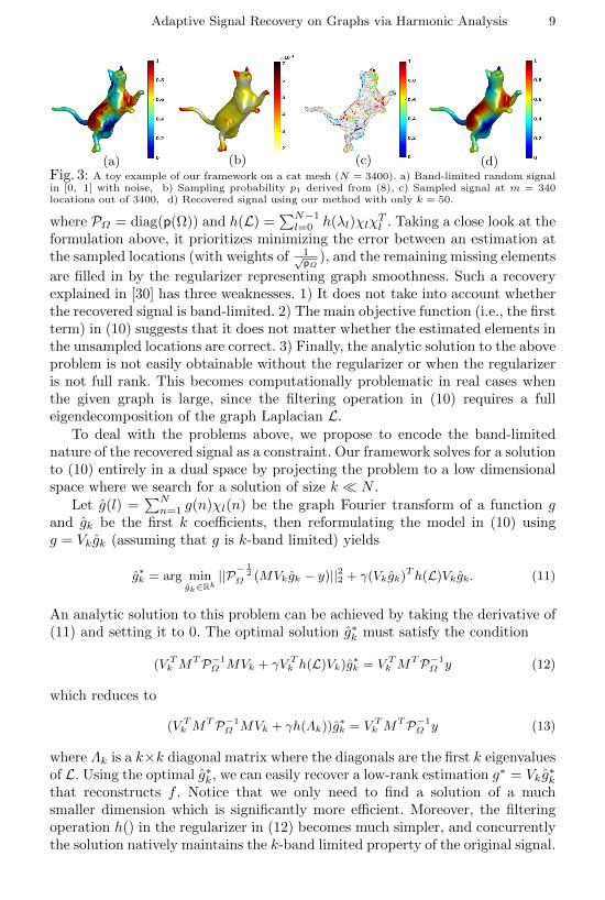
(a) (b) (c) (d)Fig. 3: A toy example of our framework on a cat mesh (N = 3400). a) Band-limited random signalin [0, 1] with noise, b) Sampling probability p1 derived from (8), c) Sampled signal at m = 340locations out of 3400, d) Recovered signal using our method with only k = 50.
where PΩ = diag(p(Ω)) and h(L) =∑N−1l=0 h(λl)χlχ
Tl . Taking a close look at the
formulation above, it prioritizes minimizing the error between an estimation atthe sampled locations (with weights of 1√
pΩ), and the remaining missing elements
are filled in by the regularizer representing graph smoothness. Such a recoveryexplained in [30] has three weaknesses. 1) It does not take into account whetherthe recovered signal is band-limited. 2) The main objective function (i.e., the firstterm) in (10) suggests that it does not matter whether the estimated elements inthe unsampled locations are correct. 3) Finally, the analytic solution to the aboveproblem is not easily obtainable without the regularizer or when the regularizeris not full rank. This becomes computationally problematic in real cases whenthe given graph is large, since the filtering operation in (10) requires a fulleigendecomposition of the graph Laplacian L.
To deal with the problems above, we propose to encode the band-limitednature of the recovered signal as a constraint. Our framework solves for a solutionto (10) entirely in a dual space by projecting the problem to a low dimensionalspace where we search for a solution of size k N .
Let g(l) =∑Nn=1 g(n)χl(n) be the graph Fourier transform of a function g
and gk be the first k coefficients, then reformulating the model in (10) usingg = Vkgk (assuming that g is k-band limited) yields
g∗k = arg mingk∈Rk
||P−12
Ω (MVkgk − y)||22 + γ(Vkgk)Th(L)Vkgk. (11)
An analytic solution to this problem can be achieved by taking the derivative of(11) and setting it to 0. The optimal solution g∗k must satisfy the condition
(V Tk MTP−1
Ω MVk + γV Tk h(L)Vk)g∗k = V Tk MTP−1
Ω y (12)
which reduces to
(V Tk MTP−1
Ω MVk + γh(Λk))g∗k = V Tk MTP−1
Ω y (13)
where Λk is a k×k diagonal matrix where the diagonals are the first k eigenvaluesof L. Using the optimal g∗k, we can easily recover a low-rank estimation g∗ = Vkg
∗k
that reconstructs f . Notice that we only need to find a solution of a muchsmaller dimension which is significantly more efficient. Moreover, the filteringoperation h() in the regularizer in (12) becomes much simpler, and concurrentlythe solution natively maintains the k-band limited property of the original signal.

10 W. H. Kim et al.
Connection name Description(count)
Forceps Major (FMajor) inter-hemispheric (1)Forceps Minor (FMinor) inter-hemispheric (1)Fornix inter-hemispheric (1)Cingulum Bundle Frontal (CingAnt) bi-lateral (2)Cingulum Bundle Hippocampal (CingHipp) bi-lateral (2)Cortico-spinal Tracts (CST) bi-lateral (2)Inferior Fronto-occipital (IFO) bi-lateral (2)Inferior Longitudinal Fasciculus (ILF) bi-lateral (2)Superior Longitudinal Fasciculus (SLF) bi-lateral (2)Uncinate Fasciculus (UF) bi-lateral (2)
Fig. 4: Top: The 17 major white matter pathways analyzed in the HCP study [41], Bottom: ROIsand measures analyzed in the WRAP study (Left: A sample FA map and the 162 gray matter ROIsfor DTI, Right: Sample 11C PiB DVR map and the 16 gray matter ROIs).
A toy example demonstrating this idea is shown in Fig. 3. Given a cat meshwith N = 3400 vertices, we first define a random signal f ∈ [0, 1] that is band-limited in the spectrum of L with Gaussian noise of N(0, 0.1). We take p1 for thesampling distribution and sample m = 340 (10% of the total) vertices withoutreplacement. Our estimation g using only k = 50 bases is shown in Fig. 3 d),where the error between the true f and g is extremely small despite using suchlittle data to begin with. We also can see that our method is robust to noise.
4 Experiment Design in Neuroimaging
In this section, we present proof of principle experimental results on two differentneuroimaging studies: 1) the Human Connectome Project (HCP) dataset and2) Wisconsin Registry for Alzheimer’s Prevention (WRAP) dataset. In bothstudies, we demonstrate the performance of our method in estimating expensiveneuroimage-derived measurements at regions of interests (ROI) in the brain using1) a set of p′ less expensive measures of all p measures available to the full cohortS of N subjects and 2) a set of (p− p′) expensive measures available to a smallcohort subset S ′ which includes m subjects. Given these datasets, the goal ofthese experiments is to see if we can get accurate estimates of the (p − p′)expensive measures of the full cohort S of N subjects in a way that statisticalpower for the follow-up analysis is not greatly compromised.
4.1 Experimental Setup
We compare the performance of our method with two other state-of-the-artmethods, 1) Collaborative filtering by Rao et al. [29] and 2) Random samplingof band-limited signals by Puy et al. [30]. For all three methods: (a) We derivedadjacency matrices A using data from the full set S of N samples and p′ econom-ical measures (i.e., more widely available and/or less expensive modalities) andthe radial basis function exp(−||x − y||2/σ2). We then constructed normalizedgraph Laplacians L = D−1/2(D − A)D−1/2 used in our framework. (b) We seth(λl) = λ4
l for h(L) for the filtering operation in the regularizer and set γ = 0.01

Adaptive Signal Recovery on Graphs via Harmonic Analysis 11
Dataset Method 20% 40% 60%
HCP Ours 2.83 2.22 1.79
HCP Puy et al. 3.46 2.70 2.08
HCP Rao et al. 3.00 2.4 1.97
WRAP Ours 1.12 0.79 0.65
WRAP Puy et al. 1.82 1.30 1.06
WRAP Rao et al. 1.61 1.36 1.18
Fig. 5: Sampling ratio versus error plot (left) on the HCP dataset (dashed lines) and the WRAPdataset (straight lines). The corresponding values are in the table on the right.
in (11). (c) We show estimation results of the (p− p′) expensive measures usingR ∈ 20, 40, 60% of total N samples for both studies and assess the `2-normerror of the difference between the estimated and observed measures. Because ofthe stochastic nature of the sampling step, we ran the estimation 100 times anduse the average of the corresponding errors for comparisons. In addition, we alsocompare the predicted values of the (p−p′) neuroimaging measures at each ROI(averaged across subjects) against true values and the estimates of the other twobaseline methods. For example, given a cohort of N = 100 subjects, suppose wehave full data for p′ = 10 low-cost measurements. Then, the goal is to acquirethe p − p′ = 5 measurements on only m = 20 subjects (i.e., 20% of the cohort)and estimate the (p− p′) measurements on the remaining N −m subjects.
4.2 Prediction on the Human Connectome Project
Dataset. The diffusion weighted MR images (DW-MRI) from HCP ([42]) wereacquired on custom built hardware using advanced pulse sequences [43] and for alengthy scan time (∼ 1 hour). It allows estimating microstructural properties ofthe brain, accurate reconstruction of the white matter pathways ([44]) (e.g., seeFig. 4) which form a crucial component in mapping the structural connectomeof the human brain [45–48]. Typically, such an acquisition of DW-MRI is notfeasible in many research sites due to limitations of hardware and software. Onthe other hand, the set of non-imaging measurements are cheaper and easierto acquire. Hence the ability to predict such high quality diffusion metrics (e.g.fractional anisotropy (FA)) from only a small sample of the DW-MRI scans andthe non-imaging measurements has value. HCP provides several categories ofnon-imaging covariates for the subjects [49] covering factors spanning severaldifferent categories. (The full list of covariates is given in the appendix.) Wedemonstrate the performance of our model on the task of FA prediction in 17widely studied fiber bundles (shown in Fig. 4) [41, 50] using 27 variables relatedto cognition, demographics, education and so on.Results. Given the full cohort S of N = 487 subjects from the HCP dataset withthe selected p′ = 27 low-cost covariates, we recovered high-cost FA measures inp − p′ = 17 ROIs (i.e. pathways) using p′ covariates and the FA values fromm N participants. The p′ measures were used to construct L with σ = 5and k = 100 for generating the sampling distribution p for our framework.We analyzed three cases by sampling 20%, 40%, 60% of the total populationaccording to p for m observations to predict FA on N subjects.

12 W. H. Kim et al.
Fig. 6: Distribution of mean errors over the ROIs from 100 runs using 20% (left column), 40%(middle column) and 60% (right column) samples on the HCP (top row) and the WRAP dataset(bottom row). Ours (red) show the lower errors than Puy et al. (green) and Rao et al. (blue).
Fig. 5 (dashed lines) summarizes the overall estimation errors using R =20, 40, 60% samples of the total population. For all three methods, the errorsdecreased with an increase in sample size, and our method (red) consistentlyoutperformed the other two methods (blue and green). When we look at thedistribution of errors, shown in the top row of Fig. 6, the center of the error dis-tribution using our framework (red) is far lower than the other methods (blue andgreen). Anatomical specificity of the estimation measures (using 40% samples) isillustrated on the top panel of Fig. 7 where the location of spheres represents theposition of the ROIs and their sizes and colors correspond to the mean errors.As seen in Fig. 7, our method (top-left) clearly has smaller and blue spherescompared to the other methods (middle and right). The quantitative error forindividual ROIs used for the spheres are provided in left table of Tab. 1, and thepredicted FA for all ROIs (averaged across subjects) are presented in Fig. 8. Forall 17 FA measures, with 40% sampling, we see that our results (blue) are closestto the ground truth (red) while other methods under/over estimate. (Additionalresults shown in supplement.) When the `2−norm error is small, we expect re-sults from downstream statistical analysis (e.g., p-values) will be accurate sincethe distributions of measurements are closer to the true sample distribution.
4.3 Prediction on a Preclinical Alzheimer’s Disease Project
Dataset. Alzheimer’s disease (AD) is known as a disconnection syndrome [51,52] because connectivity disruption can impede functional communication be-tween brain regions, resulting in reduced cognitive performance [53, 54]. Cur-rently, positron emission tomography (PET) using radio-chemicals such as 11CPittsburgh compound B (PiB) is important in mapping functional AD pathol-ogy. Distribution volume ratios (DVR) of PiB in the brain offer a good measureof the plaque pathology which is considered specific to AD. Unfortunately thesePET scans are costly and involve lengthy procedures. WRAP dataset consistsof partipants in preclinical stages of AD [55, 54] and contains 140 samples withboth low-cost FA measures and high-cost PiB DVR (examples shown in Fig. 4).Utilizing the FA values over the entire set of subjects and a partial observa-tion of the PiB measures from a fraction of the population, we investigate theperformance of our model for the recovery of PiB measures.

Adaptive Signal Recovery on Graphs via Harmonic Analysis 13
Fig. 7: Spherical representations of the prediction errors (`2-norm) in the HCP study (top) andin the WRAP study (bottom). Left: errors using Ours, Middle: errors using Puy et al, Right:errors using Rao et al. The spheres are centered at the center-of-mass of the specific bundle/regionalvolumes, and the radius of the spheres are proportional to the prediction error.
Remark. From a neuroimaging perspective, predicting PiB measures accu-rately enough for actual scientific analysis is problematic. Utilizing a modality(e.g., cerebrospinal fluid) will be more appropriate for predicting PiB measures,and such results are available on the project homepage. The results below demon-strate that such a prediction task yields results numerically feasible compared tobaseline strategies although not directly deployable for neuroscientific studies.Results. For this set of experiments, we selected p′ = 17 pathways with mostreliable FA measures to construct a graph with N = 140 vertices (i.e., subjects).Utilizing the graph and a partial set of PiB DVR measurements from m N participants (20%, 40% and 60% of the total population), we predicted theexpensive PiB DVR values on 16 ROIs over the whole subjects. To define L andp, we used σ = 3 and k = 50. As shown in Fig. 5 in straight lines, our estimation(red) yields the smallest error compared to [30] (green) and [29] (blue) for allthree sampling cases. The bottom row in Fig. 6 shows that the centers of errordistribution using our algorithm (red) have lower errors than those of othermethods (green and blue). As seen in the bottom panel of Fig. 7, similar to theHCP results in section 4.2, we observe smaller errors in every ROI, where theactual region-wise errors are given in the right table of Tab. 1. Fig. 8 presentsthe predicted regional PiB DVR values against the ground truth where ourprediction in blue are consistently closer to the ground truth in red. Additionalresults using 20% and 60% of the subjects are presented in the appendix.
HCP ROIs Ours Puy et al. Rao et al.
FMajor 1.15 1.93 1.70FMinor 1.21 1.99 1.75Fornix 1.20 1.84 1.65LCingAnt 0.95 1.49 1.33LcingHipp 1.17 1.87 1.65LCST 1.25 2.06 1.82LIFO 1.14 1.87 1.65LILF 1.16 1.90 1.68LSLF 1.08 1.77 1.56LUnc 0.99 1.61 1.42RCingAnt 0.93 1.48 1.32RcingHipp 1.20 1.92 1.71RCST 1.25 2.07 1.83RIFO 1.11 1.82 1.61RIFL 1.12 1.83 1.62RSLF 1.04 1.69 1.50RUnc 1.05 1.70 1.50
PiB ROIs Ours Puy et al. Rao et al.
Angular L 2.89 3.42 2.98Angular R 2.73 3.20 2.82Cingulum Ant L 3.19 3.73 3.30Cingulum Ant R 3.18 3.78 3.32Cingulum Post L 3.29 4.10 3.49Cingulum Post R 3.20 4.03 3.43Frontal Med Orb L 2.90 3.44 3.05Frontal Med Orb R 3.08 3.66 3.24Precuneus L 2.88 3.45 3.03Precuneus R 3.03 3.61 3.15SupraMarginal L 2.43 3.09 2.67SupraMarginal R 2.51 3.13 2.70Temporal Mid L 2.47 3.13 2.68Temporal Mid R 2.59 3.22 2.78Temporal Sup L 2.42 3.14 2.68Temporal Sup R 2.52 3.21 2.75
Table 1: Region-wise mean `2-norm of 100 runs of HCP-FA (left) and PiB DVR (right) with 40%samples. Errors from our method are the lowest shown in bold.

14 W. H. Kim et al.
Fig. 8: Average estimations of the HCP-FA in the fiber bundle (top) and the WRAP PiB DVRs(bottom) using 40% samples. For each measurement, the bars from the left to right are the mea-surements of the ground truth, ours, Puy et al. and Rao et al. Ours most closely estimate the actualground truth values of all the measurements.
5 ConclusionIn this paper, we presented an adaptive sampling scheme for signals definedon a graph. Using a dual space of these measurements obtained via a non-Euclidean wavelet transform, we show how signals can be recovered with highfidelity based on a stratified set of partial observations on the nodes of a graph.We demonstrated the application of this core technical development on accu-rately estimating diffusion imaging and PET imaging measures from two inde-pendent neuroimaging studies, so that one can perform standard analysis justas if the measurements were acquired directly. We presented experimental re-sults demonstrating that our framework can provide accurate recovery usingobservations from only a small fraction of the full samples. We believe that thisability to estimate unobserved data based on a partial set of measurements canhave impact in numerous computer vision and machine learning applicationswhere acquisitions of large datasets often involve varying degrees of stratifiedhuman interaction. Many real experiments involve entities that have intrinsicrelationships best captured as a graph. Mechanisms to exploit the properties ofthese graphs using similar formulations as those presented in this work may haveimportant practical and immediate ramifications for many experimental designconsiderations in numerous scientific domains.
References
1. Blum, A.L., Langley, P.: Selection of relevant features and examples in machinelearning. Artificial intelligence 97(1) (1997) 245–271

Adaptive Signal Recovery on Graphs via Harmonic Analysis 15
2. Biswas, A., Parikh, D.: Simultaneous active learning of classifiers & attributes viarelative feedback. In: CVPR. (2013) 644–651
3. Jayaraman, D., Grauman, K.: Zero-shot recognition with unreliable attributes. In:NIPS. (2014) 3464–3472
4. Lughofer, E.: Hybrid active learning for reducing the annotation effort of operatorsin classification systems. Pattern Recognition 45(2) (2012) 884–896
5. Hancock, C., Bernal, B., Medina, C., et al.: Cost analysis of Diffusion TensorImaging and MR tractography of the brain. Open Journal of Radiology 2014(2014)
6. Saif, M.W., Tzannou, I., Makrilia, N., et al.: Role and cost effectiveness of PET/CTin management of patients with cancer. Yale J Biol Med 83(2) (2010) 53–65
7. Prasad, A., Jegelka, S., Batra, D.: Submodular meets structured: Finding diversesubsets in exponentially-large structured item sets. In: NIPS. (2014) 2645–2653
8. Deng, J., Dong, W., Socher, R., et al.: Imagenet: A large-scale hierarchical imagedatabase. In: CVPR. (2009) 248–255
9. Vijayanarasimhan, S., Grauman, K.: Large-scale live active learning: Trainingobject detectors with crawled data and crowds. IJCV 108(1-2) (2014) 97–114
10. Deng, J., Russakovsky, O., Krause, J., et al.: Scalable multi-label annotation. In:SIGCHI, ACM (2014) 3099–3102
11. Bragg, J., Weld, D.S., et al.: Crowdsourcing multi-label classification for taxonomycreation. In: AAAI. (2013)
12. Read, J., Bifet, A., Holmes, G., et al.: Scalable and efficient multi-label classifica-tion for evolving data streams. Machine Learning 88(1-2) (2012) 243–272
13. Settles, B.: Active learning literature survey. University of Wisconsin, Madison52(55-66) (2010) 11
14. Dasgupta, S.: Analysis of a greedy active learning strategy. In: NIPS. (2004)337–344
15. Beygelzimer, A., Dasgupta, S., Langford, J.: Importance weighted active learning.In: ICML, ACM (2009) 49–56
16. Dasgupta, S., Hsu, D.: Hierarchical sampling for active learning. In: ICML, ACM(2008) 208–215
17. Winer, B.J., Brown, D.R., Michels, K.M.: Statistical principles in experimentaldesign. Volume 2. McGraw-Hill New York (1971)
18. Lentner, M.: Generalized least-squares estimation of a subvector of parameters inrandomized fractional factorial experiments. The Annals of Mathematical Statis-tics (1969) 1344–1352
19. Myers, J.L.: Fundamentals of experimental design. Allyn & Bacon (1972)20. Mitchell, T.J.: An algorithm for the construction of D-optimal experimental de-
signs. Technometrics 16(2) (1974) 203–21021. De Aguiar, P.F., Bourguignon, B., Khots, M., et al.: D-optimal designs. Chemo-
metrics and Intelligent Laboratory Systems 30(2) (1995) 199–21022. Park, J.S.: Optimal Latin-hypercube designs for computer experiments. Journal
of statistical planning and inference 39(1) (1994) 95–11123. Su, X., Khoshgoftaar, T.M.: A survey of collaborative filtering techniques. Ad-
vances in Artificial Intelligence 2009 (2009) 4:2–4:224. Dabov, K., Foi, A., Katkovnik, V., et al.: Image denoising by sparse 3-D transform-
domain collaborative filtering. Image Processing 16(8) (2007) 2080–209525. Yu, K., Zhu, S., Lafferty, J., et al.: Fast nonparametric matrix factorization for
large-scale collaborative filtering. In: SIGIR, ACM (2009) 211–21826. Srebro, N., Salakhutdinov, R.R.: Collaborative filtering in a non-uniform world:
Learning with the weighted trace norm. In: NIPS. (2010) 2056–2064
16 W. H. Kim et al.
27. Juditsky, A., Nemirovski, A.: On verifiable sufficient conditions for sparse signalrecovery via `1 minimization. Mathematical programming 127(1) (2011) 57–88
28. Krahmer, F., Ward, R.: Stable and robust sampling strategies for compressiveimaging. Image Processing 23(2) (2014) 612–622
29. Rao, N., Yu, H.F., Ravikumar, P.K., et al.: Collaborative Filtering with GraphInformation: Consistency and Scalable Methods. In: NIPS. (2015)
30. Puy, G., Tremblay, N., Gribonval, R., et al.: Random sampling of bandlimitedsignals on graphs. Applied and Computational Harmonic Analysis (2016)
31. Kumar, S., Mohri, M., Talwalkar, A.: Sampling methods for the Nystrom method.JMLR 13(1) (2012) 981–1006
32. Krishnamurthy, A., Singh, A.: Low-rank matrix and tensor completion via adaptivesampling. In: NIPS. (2013) 836–844
33. Hammond, D., Vandergheynst, P., Gribonval, R.: Wavelets on graphs via spectralgraph theory. Applied and Computational Harmonic Analysis 30(2) (2011) 129 –150
34. Mallat, S.: A wavelet tour of signal processing. Academic press (1999)35. Coifman, R., Maggioni, M.: Diffusion wavelets. Applied and Computational Har-
monic Analysis 21(1) (2006) 53 – 9436. S.Haykin, Veen, B.V.: Signals and Systems. Wiley (2005)37. Bronstein, M.M., Kokkinos, I.: Scale-invariant heat kernel signatures for non-rigid
shape recognition. In: CVPR, IEEE (2010) 1704–171138. Aubry, M., Schlickewei, U., Cremers, D.: The wave kernel signature: A quantum
mechanical approach to shape analysis. In: ICCV Workshops, IEEE (2011) 1626–1633
39. Rustamov, R.M.: Laplace-Beltrami eigenfunctions for deformation invariant shaperepresentation. In: Eurographics Symposium on Geometry Processing, Eurograph-ics Association (2007) 225–233
40. Kim, W.H., Chung, M.K., Singh, V.: Multi-resolution shape analysis via Non-euclidean wavelets: Applications to mesh segmentation and surface alignment prob-lems. In: CVPR, IEEE (2013) 2139–2146
41. Varentsova, A., Zhang, S., Arfanakis, K.: Development of a high angular resolutiondiffusion imaging human brain template. NeuroImage 91 (2014) 177–186
42. Van Essen, D.C., Smith, S.M., Barch, D.M., et al.: The WU-Minn human connec-tome project: an overview. NeuroImage 80 (2013) 62–79
43. Setsompop, K., Cohen-Adad, J., Gagoski, B., et al.: Improving diffusion MRI usingsimultaneous multi-slice echo planar imaging. NeuroImage 63(1) (2012) 569–580
44. Jbabdi, S., Sotiropoulos, S.N., Haber, S.N., et al.: Measuring macroscopic brainconnections in vivo. Nature neuroscience 18(11) (2015) 1546–1555
45. Sporns, O., Tononi, G., Kotter, R.: The human connectome: a structural descrip-tion of the human brain. PLoS Comput Biol 1(4) (2005) e42
46. Van Essen, D.C., Ugurbil, K.: The future of the human connectome. Neuroimage62(2) (2012) 1299–1310
47. Toga, A.W., Clark, K.A., Thompson, P.M., et al.: Mapping the human connectome.Neurosurgery 71(1) (2012) 1
48. Sporns, O.: The human connectome: origins and challenges. NeuroImage 80 (2013)53–61
49. Herrick, R., McKay, M., Olsen, T., et al.: Data dictionary services in XNAT andthe Human Connectome Project. Frontiers in neuroinformatics 8 (2014)
50. Kim, W.H., Kim, H.J., Adluru, N., et al.: Latent variable graphical model selectionusing harmonic analysis: Applications to the human connectome project (hcp). In:CVPR, IEEE (2016)

Adaptive Signal Recovery on Graphs via Harmonic Analysis 17
51. Brier, M.R., Thomas, J.B., Ances, B.M.: Network dysfunction in Alzheimer’sdisease: refining the disconnection hypothesis. Brain connectivity 4(5) (2014) 299–311
52. Delbeuck, X., Van der Linden, M., Collette, F.: Alzheimer’s disease as a discon-nection syndrome? Neuropsychology review 13(2) (2003) 79–92
53. Geschwind, N.: Disconnexion syndromes in animals and man. Springer (1974)54. Kim, W.H., Adluru, N., Chung, M.K., et al.: Multi-resolution statistical analysis
of brain connectivity graphs in preclinical Alzheimer’s disease. NeuroImage 118(2015) 103–117
55. Kim, W.H., Singh, V., Chung, M.K., et al.: Multi-resolutional shape features vianon-Euclidean wavelets: Applications to statistical analysis of cortical thickness.NeuroImage 93 (2014) 107–123


![Closeness Centrality Extended To Unconnected Graphs : The ...EN]ASNA09.pdf · Closeness Centrality Extended To Unconnected Graphs : The Harmonic Centrality Index Yannick Rochat1 Institute](https://img.dokumen.tips/doc/110x75/5e68c4d8d85073536033bf7b/closeness-centrality-extended-to-unconnected-graphs-the-enasna09pdf-closeness.jpg)


![i .] APPROXIMATING HARMONIC FUNCTIONS 499€¦ · APPROXIMATING HARMONIC FUNCTIONS 499 THE APPROXIMATION OF HARMONIC FUNCTIONS BY HARMONIC POLYNOMIALS AND BY HARMONIC RATIONAL FUNCTIONS*](https://img.dokumen.tips/doc/110x75/5f0873ba7e708231d42214c2/i-approximating-harmonic-functions-499-approximating-harmonic-functions-499-the.jpg)