Embed Size (px)
Citation preview

The Pennsylvania State University
The Graduate School
College of Engineering
ADAPTIVE NANOPARTICLE AMPHIPHILES
AS MULTIFUNCTIONAL PARTICLE SURFACTANTS
A Dissertation in
Chemical Engineering
by
Sun Hae Ra Shin
copy 2015 Sun Hae Ra Shin
Submitted in Partial Fulfillment
of the Requirements
for the Degree of
Doctor of Philosophy
December 2015
ii
The dissertation of Sun Hae Ra Shin was reviewed and approved by the following
Kyle JM Bishop
Assistant Professor of Chemical Engineering
Dissertation Adviser
Chair of Committee
Robert Rioux
Friedrich G Helfferich Associate Professor of Chemical Engineering
Darrell Velegol
Distinguished Professor of Chemical Engineering
Raymond E Schaak
Professor of Chemistry
Phillip E Savage
Professor of Chemical Engineering
Department Head and Walter L Robb Family Endowed Chair
Signatures are on file in the Graduate School
iii
ABSTRACT
A wide range of molecules polymers and particles which possess both hydrophilic and
hydrophobic domains are desirable as surface active agents Unlike their molecular analogs
inorganic nanoparticle (NP) amphiphiles have the ability both to adapt their surface chemistry in
response to environmental changes and to respond to external stimuli acting on the particle cores
These unique capabilities enable new strategies for a variety of applications in drug delivery
emulsification assembly of hierarchical structures surface coatings and optoelectronic devices
This dissertation describes experimental methods for preparing and characterizing NP
amphiphiles and also discusses their applications in assembly and biomedical engineering
The synthesis of gold NPs functionalized asymmetrically with domains of hydrophobic
and hydrophilic ligands on their respective hemispheres is described in Chapter 2 These particle
amphiphiles form spontaneously by a thermodynamically controlled process in which the particle
cores and two competing ligands assemble at the interface between two immiscible liquids to
reduce the interfacial energy The asymmetric surface chemistry resulting from this process is
confirmed using contact angle measurements of water droplets on NP monolayers deposited onto
hydrophobic and hydrophilic substrates The spontaneous assembly process is rationalized by a
thermodynamic model which accounts both for the energetic contributions driving the assembly
and for the entropic penalties that must be overcome In addition to gold NPs and thiolate ligands
generality of this approach is demonstrated by extending it to the preparation of amphiphilic iron
oxide NPs using two types of diol-terminated ligands The synthetic strategy described here
provides a comprehensive solution for designing NP amphiphiles with controlled surface
composition and ligand distribution
iv
The self-assembly of gold NP amphiphiles in water is discussed in Chapter 3 Unlike
typical amphiphilic particles with ldquofixedrdquo surface chemistries the ligands used here are not bound
irreversibly but can rearrange dynamically on the particles surface during their assembly from
solution Depending on the assembly conditions these adaptive amphiphiles form compact
micellar clusters or extended chain-like assemblies in aqueous solution By controlling the
amount of hydrophobic ligands on the particles surface the average number of nearest neighbors
ndash that is the preferred coordination number ndash can be varied systematically from ~1 (dimers) to ~2
(linear chains) to ~3 (extended clusters) To explain these experimental findings an assembly
mechanism is presented in which hydrophobic ligands organize dynamically to form discrete
patches between proximal NPs to minimize contact with their aqueous surroundings Monte Carlo
simulations incorporating these adaptive hydrophobic interactions reproduce the three-
dimensional assemblies observed in experiment
The spontaneous incorporation of amphiphilic gold NPs into the walls of surfactant
vesicles is reported in Chapter 4 When the NP amphiphiles are mixed with preformed surfactant
vesicles the hydrophobic ligands on the NP surface interact favorably with the hydrophobic core
of the bilayer structure and guide the incorporation of NPs into the vesicle walls Unlike previous
strategies based on small hydrophobic NPs the approach described here allows for the
incorporation of water soluble particles even when the size of the particles greatly exceeds the
bilayer thickness
Finally a strategy to stabilize open bilayer structures using NP amphiphiles is described
in Chapter 5 Molecular amphiphiles self-assemble in polar media to form ordered structures such
as micelles and vesicles essential to a broad range of industrial and biological processes Some of
these architectures such as bilayer sheets helical ribbons and hollow tubules are potentially
useful but inherently unstable owing to the presence of open edges that expose the hydrophobic
bilayer core The particle amphiphiles described here bind selectively to the open edge of bilayer
v
membranes to stabilize otherwise transient amphiphile assemblies It is demonstrated how such
particles can precisely control the size of lipid tubules how they can inhibit the formation of
undesirable assemblies such as gallstone precursors and how they can stabilize free-floating lipid
microdiscs
vi
TABLE OF CONTENTS
LIST OF FIGURES middotmiddotmiddotmiddotmiddotmiddotmiddotmiddotmiddotmiddotmiddotmiddotmiddotmiddotmiddotmiddotmiddotmiddotmiddotmiddotmiddotmiddotmiddotmiddotmiddotmiddotmiddotmiddotmiddotmiddotmiddotmiddotmiddotmiddotmiddotmiddotmiddotmiddotmiddotmiddotmiddotmiddotmiddotmiddotmiddotmiddotmiddotmiddotmiddotmiddotmiddotmiddotmiddotmiddotmiddotmiddotmiddotmiddotmiddotmiddotmiddotmiddotmiddotmiddotmiddotmiddotmiddotmiddotmiddotmiddotmiddotmiddotmiddotmiddotmiddotmiddotmiddotmiddotmiddotmiddotmiddotmiddotmiddotmiddotmiddotmiddotmiddotmiddot viii
ACKNOWLEDGEMENTS middotmiddotmiddotmiddotmiddotmiddotmiddotmiddotmiddotmiddotmiddotmiddotmiddotmiddotmiddotmiddotmiddotmiddotmiddotmiddotmiddotmiddotmiddotmiddotmiddotmiddotmiddotmiddotmiddotmiddotmiddotmiddotmiddotmiddotmiddotmiddotmiddotmiddotmiddotmiddotmiddotmiddotmiddotmiddotmiddotmiddotmiddotmiddotmiddotmiddotmiddotmiddotmiddotmiddotmiddotmiddotmiddotmiddotmiddotmiddotmiddotmiddotmiddotmiddotmiddotmiddotmiddotmiddotmiddotmiddotmiddotmiddotmiddotmiddotmiddotmiddotmiddotmiddot xiv
Chapter 1 Inorganic Nanoparticle Amphiphiles Preparation and Application middotmiddotmiddotmiddotmiddotmiddotmiddotmiddotmiddotmiddotmiddotmiddotmiddotmiddotmiddotmiddotmiddotmiddotmiddotmiddotmiddotmiddotmiddot 1
11 Introductionmiddotmiddotmiddotmiddotmiddotmiddotmiddotmiddotmiddotmiddotmiddotmiddotmiddotmiddotmiddotmiddotmiddotmiddotmiddotmiddotmiddotmiddotmiddotmiddotmiddotmiddotmiddotmiddotmiddotmiddotmiddotmiddotmiddotmiddotmiddotmiddotmiddotmiddotmiddotmiddotmiddotmiddotmiddotmiddotmiddotmiddotmiddotmiddotmiddotmiddotmiddotmiddotmiddotmiddotmiddotmiddotmiddotmiddotmiddotmiddotmiddotmiddotmiddotmiddotmiddotmiddotmiddotmiddotmiddotmiddotmiddotmiddotmiddotmiddotmiddotmiddotmiddotmiddotmiddotmiddotmiddotmiddotmiddotmiddotmiddotmiddotmiddotmiddotmiddotmiddotmiddotmiddot 1
12 Preparation of Inorganic Nanoparticle Amphiphiles middotmiddotmiddotmiddotmiddotmiddotmiddotmiddotmiddotmiddotmiddotmiddotmiddotmiddotmiddotmiddotmiddotmiddotmiddotmiddotmiddotmiddotmiddotmiddotmiddotmiddotmiddotmiddotmiddotmiddotmiddotmiddotmiddotmiddotmiddotmiddotmiddotmiddotmiddotmiddotmiddotmiddotmiddotmiddotmiddot 2
13 Applications of Nanoparticle Amphiphiles middotmiddotmiddotmiddotmiddotmiddotmiddotmiddotmiddotmiddotmiddotmiddotmiddotmiddotmiddotmiddotmiddotmiddotmiddotmiddotmiddotmiddotmiddotmiddotmiddotmiddotmiddotmiddotmiddotmiddotmiddotmiddotmiddotmiddotmiddotmiddotmiddotmiddotmiddotmiddotmiddotmiddotmiddotmiddotmiddotmiddotmiddotmiddotmiddotmiddotmiddotmiddotmiddotmiddotmiddotmiddot 7
131 Building Blocks for Self-Assembly middotmiddotmiddotmiddotmiddotmiddotmiddotmiddotmiddotmiddotmiddotmiddotmiddotmiddotmiddotmiddotmiddotmiddotmiddotmiddotmiddotmiddotmiddotmiddotmiddotmiddotmiddotmiddotmiddotmiddotmiddotmiddotmiddotmiddotmiddotmiddotmiddotmiddotmiddotmiddotmiddotmiddotmiddotmiddotmiddotmiddotmiddotmiddotmiddotmiddotmiddotmiddotmiddotmiddotmiddotmiddotmiddotmiddotmiddot 7
132 Interfacial Engineering middotmiddotmiddotmiddotmiddotmiddotmiddotmiddotmiddotmiddotmiddotmiddotmiddotmiddotmiddotmiddotmiddotmiddotmiddotmiddotmiddotmiddotmiddotmiddotmiddotmiddotmiddotmiddotmiddotmiddotmiddotmiddotmiddotmiddotmiddotmiddotmiddotmiddotmiddotmiddotmiddotmiddotmiddotmiddotmiddotmiddotmiddotmiddotmiddotmiddotmiddotmiddotmiddotmiddotmiddotmiddotmiddotmiddotmiddotmiddotmiddotmiddotmiddotmiddotmiddotmiddotmiddotmiddotmiddotmiddotmiddotmiddot 11
133 Biomedicial Engineering middotmiddotmiddotmiddotmiddotmiddotmiddotmiddotmiddotmiddotmiddotmiddotmiddotmiddotmiddotmiddotmiddotmiddotmiddotmiddotmiddotmiddotmiddotmiddotmiddotmiddotmiddotmiddotmiddotmiddotmiddotmiddotmiddotmiddotmiddotmiddotmiddotmiddotmiddotmiddotmiddotmiddotmiddotmiddotmiddotmiddotmiddotmiddotmiddotmiddotmiddotmiddotmiddotmiddotmiddotmiddotmiddotmiddotmiddotmiddotmiddotmiddotmiddotmiddotmiddotmiddotmiddotmiddotmiddotmiddot 13
134 Stimuli-Responsive Systems middotmiddotmiddotmiddotmiddotmiddotmiddotmiddotmiddotmiddotmiddotmiddotmiddotmiddotmiddotmiddotmiddotmiddotmiddotmiddotmiddotmiddotmiddotmiddotmiddotmiddotmiddotmiddotmiddotmiddotmiddotmiddotmiddotmiddotmiddotmiddotmiddotmiddotmiddotmiddotmiddotmiddotmiddotmiddotmiddotmiddotmiddotmiddotmiddotmiddotmiddotmiddotmiddotmiddotmiddotmiddotmiddotmiddotmiddotmiddotmiddotmiddotmiddotmiddotmiddotmiddot 16
14 Conclusionsmiddotmiddotmiddotmiddotmiddotmiddotmiddotmiddotmiddotmiddotmiddotmiddotmiddotmiddotmiddotmiddotmiddotmiddotmiddotmiddotmiddotmiddotmiddotmiddotmiddotmiddotmiddotmiddotmiddotmiddotmiddotmiddotmiddotmiddotmiddotmiddotmiddotmiddotmiddotmiddotmiddotmiddotmiddotmiddotmiddotmiddotmiddotmiddotmiddotmiddotmiddotmiddotmiddotmiddotmiddotmiddotmiddotmiddotmiddotmiddotmiddotmiddotmiddotmiddotmiddotmiddotmiddotmiddotmiddotmiddotmiddotmiddotmiddotmiddotmiddotmiddotmiddotmiddotmiddotmiddotmiddotmiddotmiddotmiddotmiddotmiddotmiddotmiddotmiddotmiddotmiddot 18 15 References middotmiddotmiddotmiddotmiddotmiddotmiddotmiddotmiddotmiddotmiddotmiddotmiddotmiddotmiddotmiddotmiddotmiddotmiddotmiddotmiddotmiddotmiddotmiddotmiddotmiddotmiddotmiddotmiddotmiddotmiddotmiddotmiddotmiddotmiddotmiddotmiddotmiddotmiddotmiddotmiddotmiddotmiddotmiddotmiddotmiddotmiddotmiddotmiddotmiddotmiddotmiddotmiddotmiddotmiddotmiddotmiddotmiddotmiddotmiddotmiddotmiddotmiddotmiddotmiddotmiddotmiddotmiddotmiddotmiddotmiddotmiddotmiddotmiddotmiddotmiddotmiddotmiddotmiddotmiddotmiddotmiddotmiddotmiddotmiddotmiddotmiddotmiddotmiddotmiddotmiddotmiddot 18
Chapter 2 Templated Synthesis of Amphiphilic Nanoparticles at the Liquid ndash Liquid Interface middot 30
21 Introductionmiddotmiddotmiddotmiddotmiddotmiddotmiddotmiddotmiddotmiddotmiddotmiddotmiddotmiddotmiddotmiddotmiddotmiddotmiddotmiddotmiddotmiddotmiddotmiddotmiddotmiddotmiddotmiddotmiddotmiddotmiddotmiddotmiddotmiddotmiddotmiddotmiddotmiddotmiddotmiddotmiddotmiddotmiddotmiddotmiddotmiddotmiddotmiddotmiddotmiddotmiddotmiddotmiddotmiddotmiddotmiddotmiddotmiddotmiddotmiddotmiddotmiddotmiddotmiddotmiddotmiddotmiddotmiddotmiddotmiddotmiddotmiddotmiddotmiddotmiddotmiddotmiddotmiddotmiddotmiddotmiddotmiddotmiddotmiddotmiddotmiddotmiddotmiddotmiddotmiddotmiddot 30
22 Experimental Section middotmiddotmiddotmiddotmiddotmiddotmiddotmiddotmiddotmiddotmiddotmiddotmiddotmiddotmiddotmiddotmiddotmiddotmiddotmiddotmiddotmiddotmiddotmiddotmiddotmiddotmiddotmiddotmiddotmiddotmiddotmiddotmiddotmiddotmiddotmiddotmiddotmiddotmiddotmiddotmiddotmiddotmiddotmiddotmiddotmiddotmiddotmiddotmiddotmiddotmiddotmiddotmiddotmiddotmiddotmiddotmiddotmiddotmiddotmiddotmiddotmiddotmiddotmiddotmiddotmiddotmiddotmiddotmiddotmiddotmiddotmiddotmiddotmiddotmiddotmiddotmiddotmiddotmiddotmiddot 31
221 Materials middotmiddotmiddotmiddotmiddotmiddotmiddotmiddotmiddotmiddotmiddotmiddotmiddotmiddotmiddotmiddotmiddotmiddotmiddotmiddotmiddotmiddotmiddotmiddotmiddotmiddotmiddotmiddotmiddotmiddotmiddotmiddotmiddotmiddotmiddotmiddotmiddotmiddotmiddotmiddotmiddotmiddotmiddotmiddotmiddotmiddotmiddotmiddotmiddotmiddotmiddotmiddotmiddotmiddotmiddotmiddotmiddotmiddotmiddotmiddotmiddotmiddotmiddotmiddotmiddotmiddotmiddotmiddotmiddotmiddotmiddotmiddotmiddotmiddotmiddotmiddotmiddotmiddotmiddotmiddotmiddotmiddotmiddotmiddotmiddotmiddotmiddotmiddotmiddot 31
222 Nanoparticle Synthesis and Functionalization middotmiddotmiddotmiddotmiddotmiddotmiddotmiddotmiddotmiddotmiddotmiddotmiddotmiddotmiddotmiddotmiddotmiddotmiddotmiddotmiddotmiddotmiddotmiddotmiddotmiddotmiddotmiddotmiddotmiddotmiddotmiddotmiddotmiddotmiddotmiddotmiddotmiddotmiddotmiddotmiddotmiddotmiddotmiddotmiddotmiddot 32
223 Characterization middotmiddotmiddotmiddotmiddotmiddotmiddotmiddotmiddotmiddotmiddotmiddotmiddotmiddotmiddotmiddotmiddotmiddotmiddotmiddotmiddotmiddotmiddotmiddotmiddotmiddotmiddotmiddotmiddotmiddotmiddotmiddotmiddotmiddotmiddotmiddotmiddotmiddotmiddotmiddotmiddotmiddotmiddotmiddotmiddotmiddotmiddotmiddotmiddotmiddotmiddotmiddotmiddotmiddotmiddotmiddotmiddotmiddotmiddotmiddotmiddotmiddotmiddotmiddotmiddotmiddotmiddotmiddotmiddotmiddotmiddotmiddotmiddotmiddotmiddotmiddotmiddotmiddotmiddotmiddot 34
23 Results and Discussions middotmiddotmiddotmiddotmiddotmiddotmiddotmiddotmiddotmiddotmiddotmiddotmiddotmiddotmiddotmiddotmiddotmiddotmiddotmiddotmiddotmiddotmiddotmiddotmiddotmiddotmiddotmiddotmiddotmiddotmiddotmiddotmiddotmiddotmiddotmiddotmiddotmiddotmiddotmiddotmiddotmiddotmiddotmiddotmiddotmiddotmiddotmiddotmiddotmiddotmiddotmiddotmiddotmiddotmiddotmiddotmiddotmiddotmiddotmiddotmiddotmiddotmiddotmiddotmiddotmiddotmiddotmiddotmiddotmiddotmiddotmiddotmiddotmiddotmiddotmiddotmiddot 34
24 Conclusionsmiddotmiddotmiddotmiddotmiddotmiddotmiddotmiddotmiddotmiddotmiddotmiddotmiddotmiddotmiddotmiddotmiddotmiddotmiddotmiddotmiddotmiddotmiddotmiddotmiddotmiddotmiddotmiddotmiddotmiddotmiddotmiddotmiddotmiddotmiddotmiddotmiddotmiddotmiddotmiddotmiddotmiddotmiddotmiddotmiddotmiddotmiddotmiddotmiddotmiddotmiddotmiddotmiddotmiddotmiddotmiddotmiddotmiddotmiddotmiddotmiddotmiddotmiddotmiddotmiddotmiddotmiddotmiddotmiddotmiddotmiddotmiddotmiddotmiddotmiddotmiddotmiddotmiddotmiddotmiddotmiddotmiddotmiddotmiddotmiddotmiddotmiddotmiddotmiddotmiddotmiddot 47
25 References middotmiddotmiddotmiddotmiddotmiddotmiddotmiddotmiddotmiddotmiddotmiddotmiddotmiddotmiddotmiddotmiddotmiddotmiddotmiddotmiddotmiddotmiddotmiddotmiddotmiddotmiddotmiddotmiddotmiddotmiddotmiddotmiddotmiddotmiddotmiddotmiddotmiddotmiddotmiddotmiddotmiddotmiddotmiddotmiddotmiddotmiddotmiddotmiddotmiddotmiddotmiddotmiddotmiddotmiddotmiddotmiddotmiddotmiddotmiddotmiddotmiddotmiddotmiddotmiddotmiddotmiddotmiddotmiddotmiddotmiddotmiddotmiddotmiddotmiddotmiddotmiddotmiddotmiddotmiddotmiddotmiddotmiddotmiddotmiddotmiddotmiddotmiddotmiddotmiddotmiddotmiddot 49
Chapter 3 Self-Assembly of Nanoparticle Amphiphiles with Adaptive Surface Chemistry middotmiddotmiddotmiddotmiddotmiddotmiddot 53
31 Introductionmiddotmiddotmiddotmiddotmiddotmiddotmiddotmiddotmiddotmiddotmiddotmiddotmiddotmiddotmiddotmiddotmiddotmiddotmiddotmiddotmiddotmiddotmiddotmiddotmiddotmiddotmiddotmiddotmiddotmiddotmiddotmiddotmiddotmiddotmiddotmiddotmiddotmiddotmiddotmiddotmiddotmiddotmiddotmiddotmiddotmiddotmiddotmiddotmiddotmiddotmiddotmiddotmiddotmiddotmiddotmiddotmiddotmiddotmiddotmiddotmiddotmiddotmiddotmiddotmiddotmiddotmiddotmiddotmiddotmiddotmiddotmiddotmiddotmiddotmiddotmiddotmiddotmiddotmiddotmiddotmiddotmiddotmiddotmiddotmiddotmiddotmiddotmiddotmiddotmiddotmiddot 53
32 Experimental Section middotmiddotmiddotmiddotmiddotmiddotmiddotmiddotmiddotmiddotmiddotmiddotmiddotmiddotmiddotmiddotmiddotmiddotmiddotmiddotmiddotmiddotmiddotmiddotmiddotmiddotmiddotmiddotmiddotmiddotmiddotmiddotmiddotmiddotmiddotmiddotmiddotmiddotmiddotmiddotmiddotmiddotmiddotmiddotmiddotmiddotmiddotmiddotmiddotmiddotmiddotmiddotmiddotmiddotmiddotmiddotmiddotmiddotmiddotmiddotmiddotmiddotmiddotmiddotmiddotmiddotmiddotmiddotmiddotmiddotmiddotmiddotmiddotmiddotmiddotmiddotmiddotmiddotmiddotmiddot 54
321 Materials middotmiddotmiddotmiddotmiddotmiddotmiddotmiddotmiddotmiddotmiddotmiddotmiddotmiddotmiddotmiddotmiddotmiddotmiddotmiddotmiddotmiddotmiddotmiddotmiddotmiddotmiddotmiddotmiddotmiddotmiddotmiddotmiddotmiddotmiddotmiddotmiddotmiddotmiddotmiddotmiddotmiddotmiddotmiddotmiddotmiddotmiddotmiddotmiddotmiddotmiddotmiddotmiddotmiddotmiddotmiddotmiddotmiddotmiddotmiddotmiddotmiddotmiddotmiddotmiddotmiddotmiddotmiddotmiddotmiddotmiddotmiddotmiddotmiddotmiddotmiddotmiddotmiddotmiddotmiddotmiddotmiddotmiddotmiddotmiddotmiddotmiddotmiddotmiddot 54
322 Nanoparticle Synthesis and Surface Functionalization middotmiddotmiddotmiddotmiddotmiddotmiddotmiddotmiddotmiddotmiddotmiddotmiddotmiddotmiddotmiddotmiddotmiddotmiddotmiddotmiddotmiddotmiddotmiddotmiddotmiddotmiddotmiddotmiddotmiddotmiddotmiddotmiddotmiddotmiddotmiddot 55
323 Ligand Composition by 1H NMR middotmiddotmiddotmiddotmiddotmiddotmiddotmiddotmiddotmiddotmiddotmiddotmiddotmiddotmiddotmiddotmiddotmiddotmiddotmiddotmiddotmiddotmiddotmiddotmiddotmiddotmiddotmiddotmiddotmiddotmiddotmiddotmiddotmiddotmiddotmiddotmiddotmiddotmiddotmiddotmiddotmiddotmiddotmiddotmiddotmiddotmiddotmiddotmiddotmiddotmiddotmiddotmiddotmiddotmiddotmiddotmiddotmiddotmiddotmiddot 56
324 Investigation of Self-Assembly by Cryo-TEM middotmiddotmiddotmiddotmiddotmiddotmiddotmiddotmiddotmiddotmiddotmiddotmiddotmiddotmiddotmiddotmiddotmiddotmiddotmiddotmiddotmiddotmiddotmiddotmiddotmiddotmiddotmiddotmiddotmiddotmiddotmiddotmiddotmiddotmiddotmiddotmiddotmiddotmiddotmiddotmiddotmiddotmiddotmiddotmiddot 59
325 Characterization middotmiddotmiddotmiddotmiddotmiddotmiddotmiddotmiddotmiddotmiddotmiddotmiddotmiddotmiddotmiddotmiddotmiddotmiddotmiddotmiddotmiddotmiddotmiddotmiddotmiddotmiddotmiddotmiddotmiddotmiddotmiddotmiddotmiddotmiddotmiddotmiddotmiddotmiddotmiddotmiddotmiddotmiddotmiddotmiddotmiddotmiddotmiddotmiddotmiddotmiddotmiddotmiddotmiddotmiddotmiddotmiddotmiddotmiddotmiddotmiddotmiddotmiddotmiddotmiddotmiddotmiddotmiddotmiddotmiddotmiddotmiddotmiddotmiddotmiddotmiddotmiddotmiddotmiddotmiddot 60
326 Monte Carlo Simulations middotmiddotmiddotmiddotmiddotmiddotmiddotmiddotmiddotmiddotmiddotmiddotmiddotmiddotmiddotmiddotmiddotmiddotmiddotmiddotmiddotmiddotmiddotmiddotmiddotmiddotmiddotmiddotmiddotmiddotmiddotmiddotmiddotmiddotmiddotmiddotmiddotmiddotmiddotmiddotmiddotmiddotmiddotmiddotmiddotmiddotmiddotmiddotmiddotmiddotmiddotmiddotmiddotmiddotmiddotmiddotmiddotmiddotmiddotmiddotmiddotmiddotmiddotmiddotmiddotmiddotmiddotmiddotmiddotmiddot 60
33 Results and Discussions middotmiddotmiddotmiddotmiddotmiddotmiddotmiddotmiddotmiddotmiddotmiddotmiddotmiddotmiddotmiddotmiddotmiddotmiddotmiddotmiddotmiddotmiddotmiddotmiddotmiddotmiddotmiddotmiddotmiddotmiddotmiddotmiddotmiddotmiddotmiddotmiddotmiddotmiddotmiddotmiddotmiddotmiddotmiddotmiddotmiddotmiddotmiddotmiddotmiddotmiddotmiddotmiddotmiddotmiddotmiddotmiddotmiddotmiddotmiddotmiddotmiddotmiddotmiddotmiddotmiddotmiddotmiddotmiddotmiddotmiddotmiddotmiddotmiddotmiddotmiddotmiddot 60
34 Conclusionsmiddotmiddotmiddotmiddotmiddotmiddotmiddotmiddotmiddotmiddotmiddotmiddotmiddotmiddotmiddotmiddotmiddotmiddotmiddotmiddotmiddotmiddotmiddotmiddotmiddotmiddotmiddotmiddotmiddotmiddotmiddotmiddotmiddotmiddotmiddotmiddotmiddotmiddotmiddotmiddotmiddotmiddotmiddotmiddotmiddotmiddotmiddotmiddotmiddotmiddotmiddotmiddotmiddotmiddotmiddotmiddotmiddotmiddotmiddotmiddotmiddotmiddotmiddotmiddotmiddotmiddotmiddotmiddotmiddotmiddotmiddotmiddotmiddotmiddotmiddotmiddotmiddotmiddotmiddotmiddotmiddotmiddotmiddotmiddotmiddotmiddotmiddotmiddotmiddotmiddotmiddot 77 35 References middotmiddotmiddotmiddotmiddotmiddotmiddotmiddotmiddotmiddotmiddotmiddotmiddotmiddotmiddotmiddotmiddotmiddotmiddotmiddotmiddotmiddotmiddotmiddotmiddotmiddotmiddotmiddotmiddotmiddotmiddotmiddotmiddotmiddotmiddotmiddotmiddotmiddotmiddotmiddotmiddotmiddotmiddotmiddotmiddotmiddotmiddotmiddotmiddotmiddotmiddotmiddotmiddotmiddotmiddotmiddotmiddotmiddotmiddotmiddotmiddotmiddotmiddotmiddotmiddotmiddotmiddotmiddotmiddotmiddotmiddotmiddotmiddotmiddotmiddotmiddotmiddotmiddotmiddotmiddotmiddotmiddotmiddotmiddotmiddotmiddotmiddotmiddotmiddotmiddotmiddotmiddot 79
Chapter 4 Integration of Gold Nanoparticles into Bilayer Structures via Adaptive Surface
Chemistry middotmiddotmiddotmiddotmiddotmiddotmiddotmiddotmiddotmiddotmiddotmiddotmiddotmiddotmiddotmiddotmiddotmiddotmiddotmiddotmiddotmiddotmiddotmiddotmiddotmiddotmiddotmiddotmiddotmiddotmiddotmiddotmiddotmiddotmiddotmiddotmiddotmiddotmiddotmiddotmiddotmiddotmiddotmiddotmiddotmiddotmiddotmiddotmiddotmiddotmiddotmiddotmiddotmiddotmiddotmiddotmiddotmiddotmiddotmiddotmiddotmiddotmiddotmiddotmiddotmiddotmiddotmiddotmiddotmiddotmiddotmiddotmiddotmiddotmiddotmiddotmiddotmiddotmiddotmiddotmiddotmiddotmiddotmiddotmiddotmiddotmiddotmiddotmiddotmiddotmiddotmiddotmiddotmiddotmiddotmiddotmiddotmiddotmiddotmiddot 85
41 Introductionmiddotmiddotmiddotmiddotmiddotmiddotmiddotmiddotmiddotmiddotmiddotmiddotmiddotmiddotmiddotmiddotmiddotmiddotmiddotmiddotmiddotmiddotmiddotmiddotmiddotmiddotmiddotmiddotmiddotmiddotmiddotmiddotmiddotmiddotmiddotmiddotmiddotmiddotmiddotmiddotmiddotmiddotmiddotmiddotmiddotmiddotmiddotmiddotmiddotmiddotmiddotmiddotmiddotmiddotmiddotmiddotmiddotmiddotmiddotmiddotmiddotmiddotmiddotmiddotmiddotmiddotmiddotmiddotmiddotmiddotmiddotmiddotmiddotmiddotmiddotmiddotmiddotmiddotmiddotmiddotmiddotmiddotmiddotmiddotmiddotmiddotmiddotmiddotmiddotmiddotmiddot 85
42 Experimental Section middotmiddotmiddotmiddotmiddotmiddotmiddotmiddotmiddotmiddotmiddotmiddotmiddotmiddotmiddotmiddotmiddotmiddotmiddotmiddotmiddotmiddotmiddotmiddotmiddotmiddotmiddotmiddotmiddotmiddotmiddotmiddotmiddotmiddotmiddotmiddotmiddotmiddotmiddotmiddotmiddotmiddotmiddotmiddotmiddotmiddotmiddotmiddotmiddotmiddotmiddotmiddotmiddotmiddotmiddotmiddotmiddotmiddotmiddotmiddotmiddotmiddotmiddotmiddotmiddotmiddotmiddotmiddotmiddotmiddotmiddotmiddotmiddotmiddotmiddotmiddotmiddotmiddotmiddotmiddot 86
421 Surface Functionalization of AuMUAODT Nanoparticles middotmiddotmiddotmiddotmiddotmiddotmiddotmiddotmiddotmiddotmiddotmiddotmiddotmiddotmiddotmiddotmiddotmiddotmiddotmiddotmiddotmiddotmiddotmiddotmiddotmiddotmiddotmiddotmiddotmiddotmiddot 86
422 Ligand Composition by 1H NMR middotmiddotmiddotmiddotmiddotmiddotmiddotmiddotmiddotmiddotmiddotmiddotmiddotmiddotmiddotmiddotmiddotmiddotmiddotmiddotmiddotmiddotmiddotmiddotmiddotmiddotmiddotmiddotmiddotmiddotmiddotmiddotmiddotmiddotmiddotmiddotmiddotmiddotmiddotmiddotmiddotmiddotmiddotmiddotmiddotmiddotmiddotmiddotmiddotmiddotmiddotmiddotmiddotmiddotmiddotmiddotmiddotmiddotmiddotmiddot 86
43 Results and Discussions middotmiddotmiddotmiddotmiddotmiddotmiddotmiddotmiddotmiddotmiddotmiddotmiddotmiddotmiddotmiddotmiddotmiddotmiddotmiddotmiddotmiddotmiddotmiddotmiddotmiddotmiddotmiddotmiddotmiddotmiddotmiddotmiddotmiddotmiddotmiddotmiddotmiddotmiddotmiddotmiddotmiddotmiddotmiddotmiddotmiddotmiddotmiddotmiddotmiddotmiddotmiddotmiddotmiddotmiddotmiddotmiddotmiddotmiddotmiddotmiddotmiddotmiddotmiddotmiddotmiddotmiddotmiddotmiddotmiddotmiddotmiddotmiddotmiddotmiddotmiddotmiddot 89
vii
44 Conclusionsmiddotmiddotmiddotmiddotmiddotmiddotmiddotmiddotmiddotmiddotmiddotmiddotmiddotmiddotmiddotmiddotmiddotmiddotmiddotmiddotmiddotmiddotmiddotmiddotmiddotmiddotmiddotmiddotmiddotmiddotmiddotmiddotmiddotmiddotmiddotmiddotmiddotmiddotmiddotmiddotmiddotmiddotmiddotmiddotmiddotmiddotmiddotmiddotmiddotmiddotmiddotmiddotmiddotmiddotmiddotmiddotmiddotmiddotmiddotmiddotmiddotmiddotmiddotmiddotmiddotmiddotmiddotmiddotmiddotmiddotmiddotmiddotmiddotmiddotmiddotmiddotmiddotmiddotmiddotmiddotmiddotmiddotmiddotmiddotmiddotmiddotmiddotmiddotmiddot 100 45 References middotmiddotmiddotmiddotmiddotmiddotmiddotmiddotmiddotmiddotmiddotmiddotmiddotmiddotmiddotmiddotmiddotmiddotmiddotmiddotmiddotmiddotmiddotmiddotmiddotmiddotmiddotmiddotmiddotmiddotmiddotmiddotmiddotmiddotmiddotmiddotmiddotmiddotmiddotmiddotmiddotmiddotmiddotmiddotmiddotmiddotmiddotmiddotmiddotmiddotmiddotmiddotmiddotmiddotmiddotmiddotmiddotmiddotmiddotmiddotmiddotmiddotmiddotmiddotmiddotmiddotmiddotmiddotmiddotmiddotmiddotmiddotmiddotmiddotmiddotmiddotmiddotmiddotmiddotmiddotmiddotmiddotmiddotmiddotmiddotmiddotmiddotmiddotmiddotmiddot 101
Chapter 5 Amphiphilic Nanoparticles Control the Growth and Stability of Lipid Bilayers with
Open Edges Bilayer middotmiddotmiddotmiddotmiddotmiddotmiddotmiddotmiddotmiddotmiddotmiddotmiddotmiddotmiddotmiddotmiddotmiddotmiddotmiddotmiddotmiddotmiddotmiddotmiddotmiddotmiddotmiddotmiddotmiddotmiddotmiddotmiddotmiddotmiddotmiddotmiddotmiddotmiddotmiddotmiddotmiddotmiddotmiddotmiddotmiddotmiddotmiddotmiddotmiddotmiddotmiddotmiddotmiddotmiddotmiddotmiddotmiddotmiddotmiddotmiddotmiddotmiddotmiddotmiddotmiddotmiddotmiddotmiddotmiddotmiddotmiddotmiddotmiddotmiddotmiddotmiddotmiddotmiddotmiddotmiddotmiddotmiddotmiddotmiddotmiddotmiddot 105
51 Introductionmiddotmiddotmiddotmiddotmiddotmiddotmiddotmiddotmiddotmiddotmiddotmiddotmiddotmiddotmiddotmiddotmiddotmiddotmiddotmiddotmiddotmiddotmiddotmiddotmiddotmiddotmiddotmiddotmiddotmiddotmiddotmiddotmiddotmiddotmiddotmiddotmiddotmiddotmiddotmiddotmiddotmiddotmiddotmiddotmiddotmiddotmiddotmiddotmiddotmiddotmiddotmiddotmiddotmiddotmiddotmiddotmiddotmiddotmiddotmiddotmiddotmiddotmiddotmiddotmiddotmiddotmiddotmiddotmiddotmiddotmiddotmiddotmiddotmiddotmiddotmiddotmiddotmiddotmiddotmiddotmiddotmiddotmiddotmiddotmiddotmiddotmiddotmiddotmiddot 105
52 Experimental Section middotmiddotmiddotmiddotmiddotmiddotmiddotmiddotmiddotmiddotmiddotmiddotmiddotmiddotmiddotmiddotmiddotmiddotmiddotmiddotmiddotmiddotmiddotmiddotmiddotmiddotmiddotmiddotmiddotmiddotmiddotmiddotmiddotmiddotmiddotmiddotmiddotmiddotmiddotmiddotmiddotmiddotmiddotmiddotmiddotmiddotmiddotmiddotmiddotmiddotmiddotmiddotmiddotmiddotmiddotmiddotmiddotmiddotmiddotmiddotmiddotmiddotmiddotmiddotmiddotmiddotmiddotmiddotmiddotmiddotmiddotmiddotmiddotmiddotmiddotmiddotmiddotmiddot 106
521 Synthesis and Functionalization of AuNPs middotmiddotmiddotmiddotmiddotmiddotmiddotmiddotmiddotmiddotmiddotmiddotmiddotmiddotmiddotmiddotmiddotmiddotmiddotmiddotmiddotmiddotmiddotmiddotmiddotmiddotmiddotmiddotmiddotmiddotmiddotmiddotmiddotmiddotmiddotmiddotmiddotmiddotmiddotmiddotmiddotmiddotmiddotmiddotmiddotmiddotmiddotmiddot 106
522 Preparation of Bilayer Membranes middotmiddotmiddotmiddotmiddotmiddotmiddotmiddotmiddotmiddotmiddotmiddotmiddotmiddotmiddotmiddotmiddotmiddotmiddotmiddotmiddotmiddotmiddotmiddotmiddotmiddotmiddotmiddotmiddotmiddotmiddotmiddotmiddotmiddotmiddotmiddotmiddotmiddotmiddotmiddotmiddotmiddotmiddotmiddotmiddotmiddotmiddotmiddotmiddotmiddotmiddotmiddotmiddotmiddotmiddotmiddotmiddot 111
523 Characterization middotmiddotmiddotmiddotmiddotmiddotmiddotmiddotmiddotmiddotmiddotmiddotmiddotmiddotmiddotmiddotmiddotmiddotmiddotmiddotmiddotmiddotmiddotmiddotmiddotmiddotmiddotmiddotmiddotmiddotmiddotmiddotmiddotmiddotmiddotmiddotmiddotmiddotmiddotmiddotmiddotmiddotmiddotmiddotmiddotmiddotmiddotmiddotmiddotmiddotmiddotmiddotmiddotmiddotmiddotmiddotmiddotmiddotmiddotmiddotmiddotmiddotmiddotmiddotmiddotmiddotmiddotmiddotmiddotmiddotmiddotmiddotmiddotmiddotmiddotmiddotmiddotmiddot 112
53 Results and Discussions middotmiddotmiddotmiddotmiddotmiddotmiddotmiddotmiddotmiddotmiddotmiddotmiddotmiddotmiddotmiddotmiddotmiddotmiddotmiddotmiddotmiddotmiddotmiddotmiddotmiddotmiddotmiddotmiddotmiddotmiddotmiddotmiddotmiddotmiddotmiddotmiddotmiddotmiddotmiddotmiddotmiddotmiddotmiddotmiddotmiddotmiddotmiddotmiddotmiddotmiddotmiddotmiddotmiddotmiddotmiddotmiddotmiddotmiddotmiddotmiddotmiddotmiddotmiddotmiddotmiddotmiddotmiddotmiddotmiddotmiddotmiddotmiddotmiddotmiddot 113
54 Conclusionsmiddotmiddotmiddotmiddotmiddotmiddotmiddotmiddotmiddotmiddotmiddotmiddotmiddotmiddotmiddotmiddotmiddotmiddotmiddotmiddotmiddotmiddotmiddotmiddotmiddotmiddotmiddotmiddotmiddotmiddotmiddotmiddotmiddotmiddotmiddotmiddotmiddotmiddotmiddotmiddotmiddotmiddotmiddotmiddotmiddotmiddotmiddotmiddotmiddotmiddotmiddotmiddotmiddotmiddotmiddotmiddotmiddotmiddotmiddotmiddotmiddotmiddotmiddotmiddotmiddotmiddotmiddotmiddotmiddotmiddotmiddotmiddotmiddotmiddotmiddotmiddotmiddotmiddotmiddotmiddotmiddotmiddotmiddotmiddotmiddotmiddotmiddotmiddotmiddot 127 55 References middotmiddotmiddotmiddotmiddotmiddotmiddotmiddotmiddotmiddotmiddotmiddotmiddotmiddotmiddotmiddotmiddotmiddotmiddotmiddotmiddotmiddotmiddotmiddotmiddotmiddotmiddotmiddotmiddotmiddotmiddotmiddotmiddotmiddotmiddotmiddotmiddotmiddotmiddotmiddotmiddotmiddotmiddotmiddotmiddotmiddotmiddotmiddotmiddotmiddotmiddotmiddotmiddotmiddotmiddotmiddotmiddotmiddotmiddotmiddotmiddotmiddotmiddotmiddotmiddotmiddotmiddotmiddotmiddotmiddotmiddotmiddotmiddotmiddotmiddotmiddotmiddotmiddotmiddotmiddotmiddotmiddotmiddotmiddotmiddotmiddotmiddotmiddotmiddotmiddot 128
Chapter 6 Nanoparticle Amphiphiles as Multifunctional Surfactants Outlook and Future
Prospects middotmiddotmiddotmiddotmiddotmiddotmiddotmiddotmiddotmiddotmiddotmiddotmiddotmiddotmiddotmiddotmiddotmiddotmiddotmiddotmiddotmiddotmiddotmiddotmiddotmiddotmiddotmiddotmiddotmiddotmiddotmiddotmiddotmiddotmiddotmiddotmiddotmiddotmiddotmiddotmiddotmiddotmiddotmiddotmiddotmiddotmiddotmiddotmiddotmiddotmiddotmiddotmiddotmiddotmiddotmiddotmiddotmiddotmiddotmiddotmiddotmiddotmiddotmiddotmiddotmiddotmiddotmiddotmiddotmiddotmiddotmiddotmiddotmiddotmiddotmiddotmiddotmiddotmiddotmiddotmiddotmiddotmiddotmiddotmiddotmiddotmiddotmiddotmiddotmiddotmiddotmiddotmiddotmiddotmiddotmiddotmiddotmiddotmiddot 132
61 Introductionmiddotmiddotmiddotmiddotmiddotmiddotmiddotmiddotmiddotmiddotmiddotmiddotmiddotmiddotmiddotmiddotmiddotmiddotmiddotmiddotmiddotmiddotmiddotmiddotmiddotmiddotmiddotmiddotmiddotmiddotmiddotmiddotmiddotmiddotmiddotmiddotmiddotmiddotmiddotmiddotmiddotmiddotmiddotmiddotmiddotmiddotmiddotmiddotmiddotmiddotmiddotmiddotmiddotmiddotmiddotmiddotmiddotmiddotmiddotmiddotmiddotmiddotmiddotmiddotmiddotmiddotmiddotmiddotmiddotmiddotmiddotmiddotmiddotmiddotmiddotmiddotmiddotmiddotmiddotmiddotmiddotmiddotmiddotmiddotmiddotmiddotmiddotmiddotmiddot 132
62 Thermodynamics of Amphiphilic Nanoparticles and Lipid Bilayer Interaction middotmiddotmiddotmiddotmiddotmiddotmiddotmiddotmiddotmiddotmiddot 132
63 Encapsulation of Charged Nanoparticles into Fluid Liposomes middotmiddotmiddotmiddotmiddotmiddotmiddotmiddotmiddotmiddotmiddotmiddotmiddotmiddotmiddotmiddotmiddotmiddotmiddotmiddotmiddotmiddotmiddotmiddotmiddotmiddotmiddotmiddotmiddot 135
64 Surface Pressure Isotherm for a Monolayer of Charged Nanoparticles at Water ndash Air
Interfaces middotmiddotmiddotmiddotmiddotmiddotmiddotmiddotmiddotmiddotmiddotmiddotmiddotmiddotmiddotmiddotmiddotmiddotmiddotmiddotmiddotmiddotmiddotmiddotmiddotmiddotmiddotmiddotmiddotmiddotmiddotmiddotmiddotmiddotmiddotmiddotmiddotmiddotmiddotmiddotmiddotmiddotmiddotmiddotmiddotmiddotmiddotmiddotmiddotmiddotmiddotmiddotmiddotmiddotmiddotmiddotmiddotmiddotmiddotmiddotmiddotmiddotmiddotmiddotmiddotmiddotmiddotmiddotmiddotmiddotmiddotmiddotmiddotmiddotmiddotmiddotmiddotmiddotmiddotmiddotmiddotmiddotmiddotmiddotmiddotmiddotmiddotmiddotmiddotmiddotmiddotmiddotmiddotmiddotmiddotmiddot 137
65 Dynamic Tuning of Interfacial Tension middotmiddotmiddotmiddotmiddotmiddotmiddotmiddotmiddotmiddotmiddotmiddotmiddotmiddotmiddotmiddotmiddotmiddotmiddotmiddotmiddotmiddotmiddotmiddotmiddotmiddotmiddotmiddotmiddotmiddotmiddotmiddotmiddotmiddotmiddotmiddotmiddotmiddotmiddotmiddotmiddotmiddotmiddotmiddotmiddotmiddotmiddotmiddotmiddotmiddotmiddotmiddotmiddotmiddotmiddotmiddotmiddot 139
66 Conclusionsmiddotmiddotmiddotmiddotmiddotmiddotmiddotmiddotmiddotmiddotmiddotmiddotmiddotmiddotmiddotmiddotmiddotmiddotmiddotmiddotmiddotmiddotmiddotmiddotmiddotmiddotmiddotmiddotmiddotmiddotmiddotmiddotmiddotmiddotmiddotmiddotmiddotmiddotmiddotmiddotmiddotmiddotmiddotmiddotmiddotmiddotmiddotmiddotmiddotmiddotmiddotmiddotmiddotmiddotmiddotmiddotmiddotmiddotmiddotmiddotmiddotmiddotmiddotmiddotmiddotmiddotmiddotmiddotmiddotmiddotmiddotmiddotmiddotmiddotmiddotmiddotmiddotmiddotmiddotmiddotmiddotmiddotmiddotmiddotmiddotmiddotmiddotmiddotmiddot 140 67 References middotmiddotmiddotmiddotmiddotmiddotmiddotmiddotmiddotmiddotmiddotmiddotmiddotmiddotmiddotmiddotmiddotmiddotmiddotmiddotmiddotmiddotmiddotmiddotmiddotmiddotmiddotmiddotmiddotmiddotmiddotmiddotmiddotmiddotmiddotmiddotmiddotmiddotmiddotmiddotmiddotmiddotmiddotmiddotmiddotmiddotmiddotmiddotmiddotmiddotmiddotmiddotmiddotmiddotmiddotmiddotmiddotmiddotmiddotmiddotmiddotmiddotmiddotmiddotmiddotmiddotmiddotmiddotmiddotmiddotmiddotmiddotmiddotmiddotmiddotmiddotmiddotmiddotmiddotmiddotmiddotmiddotmiddotmiddotmiddotmiddotmiddotmiddotmiddotmiddot 142
Appendicesmiddotmiddotmiddotmiddotmiddotmiddotmiddotmiddotmiddotmiddotmiddotmiddotmiddotmiddotmiddotmiddotmiddotmiddotmiddotmiddotmiddotmiddotmiddotmiddotmiddotmiddotmiddotmiddotmiddotmiddotmiddotmiddotmiddotmiddotmiddotmiddotmiddotmiddotmiddotmiddotmiddotmiddotmiddotmiddotmiddotmiddotmiddotmiddotmiddotmiddotmiddotmiddotmiddotmiddotmiddotmiddotmiddotmiddotmiddotmiddotmiddotmiddotmiddotmiddotmiddotmiddotmiddotmiddotmiddotmiddotmiddotmiddotmiddotmiddotmiddotmiddotmiddotmiddotmiddotmiddotmiddotmiddotmiddotmiddotmiddotmiddotmiddotmiddotmiddotmiddotmiddotmiddotmiddotmiddotmiddotmiddotmiddot 145
A Details of the Thermodynamic Model middotmiddotmiddotmiddotmiddotmiddotmiddotmiddotmiddotmiddotmiddotmiddotmiddotmiddotmiddotmiddotmiddotmiddotmiddotmiddotmiddotmiddotmiddotmiddotmiddotmiddotmiddotmiddotmiddotmiddotmiddotmiddotmiddotmiddotmiddotmiddotmiddotmiddotmiddotmiddotmiddotmiddotmiddotmiddotmiddotmiddotmiddotmiddotmiddotmiddotmiddotmiddotmiddotmiddotmiddotmiddotmiddotmiddotmiddot 145
B1 Nanoparticle Contacts middotmiddotmiddotmiddotmiddotmiddotmiddotmiddotmiddotmiddotmiddotmiddotmiddotmiddotmiddotmiddotmiddotmiddotmiddotmiddotmiddotmiddotmiddotmiddotmiddotmiddotmiddotmiddotmiddotmiddotmiddotmiddotmiddotmiddotmiddotmiddotmiddotmiddotmiddotmiddotmiddotmiddotmiddotmiddotmiddotmiddotmiddotmiddotmiddotmiddotmiddotmiddotmiddotmiddotmiddotmiddotmiddotmiddotmiddotmiddotmiddotmiddotmiddotmiddotmiddotmiddotmiddotmiddotmiddotmiddotmiddotmiddotmiddotmiddotmiddotmiddot 150
B2 Electrostatic Interactions middotmiddotmiddotmiddotmiddotmiddotmiddotmiddotmiddotmiddotmiddotmiddotmiddotmiddotmiddotmiddotmiddotmiddotmiddotmiddotmiddotmiddotmiddotmiddotmiddotmiddotmiddotmiddotmiddotmiddotmiddotmiddotmiddotmiddotmiddotmiddotmiddotmiddotmiddotmiddotmiddotmiddotmiddotmiddotmiddotmiddotmiddotmiddotmiddotmiddotmiddotmiddotmiddotmiddotmiddotmiddotmiddotmiddotmiddotmiddotmiddotmiddotmiddotmiddotmiddotmiddotmiddotmiddotmiddotmiddotmiddotmiddot 152
B3 Monte Carlo Exchange Moves middotmiddotmiddotmiddotmiddotmiddotmiddotmiddotmiddotmiddotmiddotmiddotmiddotmiddotmiddotmiddotmiddotmiddotmiddotmiddotmiddotmiddotmiddotmiddotmiddotmiddotmiddotmiddotmiddotmiddotmiddotmiddotmiddotmiddotmiddotmiddotmiddotmiddotmiddotmiddotmiddotmiddotmiddotmiddotmiddotmiddotmiddotmiddotmiddotmiddotmiddotmiddotmiddotmiddotmiddotmiddotmiddotmiddotmiddotmiddotmiddotmiddotmiddotmiddotmiddotmiddot 155
B4 ldquoControlrdquo Simulations without Adaptive Surface Chemistry middotmiddotmiddotmiddotmiddotmiddotmiddotmiddotmiddotmiddotmiddotmiddotmiddotmiddotmiddotmiddotmiddotmiddotmiddotmiddotmiddotmiddotmiddotmiddotmiddotmiddotmiddotmiddotmiddotmiddotmiddot 158
B5 Improving Chain Monodispersity middotmiddotmiddotmiddotmiddotmiddotmiddotmiddotmiddotmiddotmiddotmiddotmiddotmiddotmiddotmiddotmiddotmiddotmiddotmiddotmiddotmiddotmiddotmiddotmiddotmiddotmiddotmiddotmiddotmiddotmiddotmiddotmiddotmiddotmiddotmiddotmiddotmiddotmiddotmiddotmiddotmiddotmiddotmiddotmiddotmiddotmiddotmiddotmiddotmiddotmiddotmiddotmiddotmiddotmiddotmiddotmiddotmiddotmiddotmiddotmiddotmiddot 159
C Analysis of Cryo-TEM Images middotmiddotmiddotmiddotmiddotmiddotmiddotmiddotmiddotmiddotmiddotmiddotmiddotmiddotmiddotmiddotmiddotmiddotmiddotmiddotmiddotmiddotmiddotmiddotmiddotmiddotmiddotmiddotmiddotmiddotmiddotmiddotmiddotmiddotmiddotmiddotmiddotmiddotmiddotmiddotmiddotmiddotmiddotmiddotmiddotmiddotmiddotmiddotmiddotmiddotmiddotmiddotmiddotmiddotmiddotmiddotmiddotmiddotmiddotmiddotmiddotmiddotmiddotmiddotmiddotmiddotmiddot 161
viii
LIST OF FIGURES
Figure 11 Synthesis of NP amphiphiles by (a) adsorption of amphiphilic ligands (b)
coadsorption of hydrophilic and hydrophobic ligands from a solution or (c) partial exchange for
hydrophilic ligand on a preformed hydrophobic ligand Grey bead represents NP Red line
indicates hydrophobic ligand and blue line indicates hydrophilic ligand middotmiddotmiddotmiddotmiddotmiddotmiddotmiddotmiddotmiddotmiddotmiddotmiddotmiddotmiddotmiddotmiddotmiddotmiddotmiddotmiddotmiddotmiddotmiddotmiddotmiddotmiddotmiddotmiddot 4
Figure 12 Self-assembled structures of particles with discrete hydrophobic patchy in water
Depending on the number andor distribution of patches on the particlersquos surface a variety of
assemblies can emerge Red region indicates hydrophobic patchy middotmiddotmiddotmiddotmiddotmiddotmiddotmiddotmiddotmiddotmiddotmiddotmiddotmiddotmiddotmiddotmiddotmiddotmiddotmiddotmiddotmiddotmiddotmiddotmiddotmiddotmiddotmiddotmiddotmiddotmiddotmiddotmiddotmiddotmiddotmiddot 9
Figure 13 (a) Hydrophilic particles adsorbed to vesicle or encapsulated in aqueous vesicle
interior (b) Hydrophobic particles entrapped in the hydrophobic bilayer core middotmiddotmiddotmiddotmiddotmiddotmiddotmiddotmiddotmiddotmiddotmiddotmiddotmiddotmiddotmiddotmiddotmiddotmiddotmiddotmiddot 14
Figure 21 (a) TEM image of AuDDA NPs d = 92 plusmn 12 nm in diameter (b) TEM image of
Fe2O3 NPs d = 75 plusmn 09 nm in diameter middotmiddotmiddotmiddotmiddotmiddotmiddotmiddotmiddotmiddotmiddotmiddotmiddotmiddotmiddotmiddotmiddotmiddotmiddotmiddotmiddotmiddotmiddotmiddotmiddotmiddotmiddotmiddotmiddotmiddotmiddotmiddotmiddotmiddotmiddotmiddotmiddotmiddotmiddotmiddotmiddotmiddotmiddotmiddotmiddotmiddotmiddotmiddotmiddotmiddotmiddotmiddotmiddotmiddotmiddotmiddotmiddotmiddotmiddotmiddotmiddotmiddotmiddotmiddotmiddotmiddotmiddotmiddotmiddotmiddotmiddotmiddotmiddotmiddotmiddotmiddotmiddotmiddotmiddotmiddotmiddotmiddotmiddotmiddotmiddotmiddot 33
Figure 22 (a) Schematic illustration of the surface functionalization procedure The combination
of AuNPs with hydrophobic (DDT) and hydrophilic (MUA) ligands in a toluene ndash water mixture
yields amphiphilic nanoparticles at the liquid ndash liquid interface The rightmost image shows water
droplets covered with NPs For clarity the schematic illustration of AuDDTMUA shows
complete segregation between the respective ligands While some degree of asymmetric
functionalization is certain (cf Figure 24) the precise extent of phase separation between DDT
and MUA ligands is unknown (b) UV ndash vis spectra of the NP solutions before and after
functionalization show that nanoparticles ndash initially present in the toluene phase ndash assemble at the
interface with no particles remaining in either bulk phase middotmiddotmiddotmiddotmiddotmiddotmiddotmiddotmiddotmiddotmiddotmiddotmiddotmiddotmiddotmiddotmiddotmiddotmiddotmiddotmiddotmiddotmiddotmiddotmiddotmiddotmiddotmiddotmiddotmiddotmiddotmiddotmiddotmiddotmiddotmiddotmiddotmiddotmiddotmiddotmiddotmiddotmiddotmiddotmiddotmiddotmiddotmiddotmiddotmiddotmiddotmiddotmiddotmiddotmiddotmiddotmiddotmiddotmiddot 36
Figure 23 (a) Amphiphilic NPs at the watertoluene interface (left) and deposited onto a
hydrophilic glass substrate (right) (b) UV-vis spectra of AuMUA particles in water (red) and of
AuMUADDT particles deposited onto a glass substrate (blue) (c) Amphiphilic particles before
drying (d) Redispersed particles after complete drying In these experiments the 04 mol of
AuNPs was dispersed into equal volumes (2 mL) of basic water (pH ~ 11) and toluene the ligand
ratio was 2011 for AuDDTMUAmiddotmiddotmiddotmiddotmiddotmiddotmiddotmiddotmiddotmiddotmiddotmiddotmiddotmiddotmiddotmiddotmiddotmiddotmiddotmiddotmiddotmiddotmiddotmiddotmiddotmiddotmiddotmiddotmiddotmiddotmiddotmiddotmiddotmiddotmiddotmiddotmiddotmiddotmiddotmiddotmiddotmiddotmiddotmiddotmiddotmiddotmiddotmiddotmiddotmiddotmiddotmiddotmiddotmiddotmiddotmiddotmiddotmiddotmiddotmiddotmiddotmiddotmiddotmiddotmiddotmiddotmiddotmiddotmiddotmiddotmiddotmiddotmiddotmiddotmiddotmiddotmiddotmiddotmiddotmiddotmiddotmiddotmiddotmiddotmiddotmiddotmiddotmiddotmiddotmiddotmiddotmiddotmiddotmiddot 37
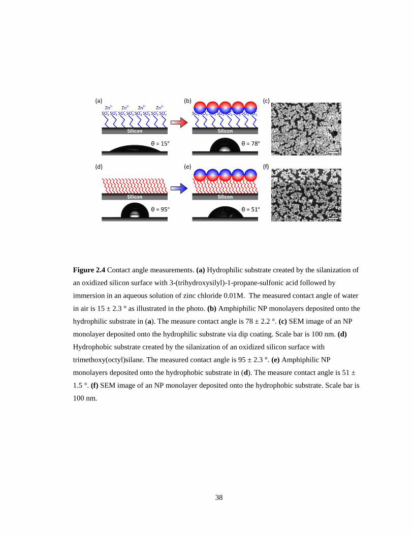
Figure 24 Contact angle measurements (a) Hydrophilic substrate created by the silanization of
an oxidized silicon surface with 3-(trihydroxysilyl)-1-propane-sulfonic acid followed by
immersion in an aqueous solution of zinc chloride 001M The measured contact angle of water
in air is 15 plusmn 23 deg as illustrated in the photo (b) Amphiphilic NP monolayers deposited onto the
hydrophilic substrate in (a) The measure contact angle is 78 plusmn 22 deg (c) SEM image of an NP
monolayer deposited onto the hydrophilic substrate via dip coating Scale bar is 100 nm (d)
Hydrophobic substrate created by the silanization of an oxidized silicon surface with
trimethoxy(octyl)silane The measured contact angle is 95 plusmn 23 deg (e) Amphiphilic NP
monolayers deposited onto the hydrophobic substrate in (d) The measure contact angle is 51 plusmn
15 deg (f) SEM image of an NP monolayer deposited onto the hydrophobic substrate Scale bar is
100 nm middotmiddotmiddotmiddotmiddotmiddotmiddotmiddotmiddotmiddotmiddotmiddotmiddotmiddotmiddotmiddotmiddotmiddotmiddotmiddotmiddotmiddotmiddotmiddotmiddotmiddotmiddotmiddotmiddotmiddotmiddotmiddotmiddotmiddotmiddotmiddotmiddotmiddotmiddotmiddotmiddotmiddotmiddotmiddotmiddotmiddotmiddotmiddotmiddotmiddotmiddotmiddotmiddotmiddotmiddotmiddotmiddotmiddotmiddotmiddotmiddotmiddotmiddotmiddotmiddotmiddotmiddotmiddotmiddotmiddotmiddotmiddotmiddotmiddotmiddotmiddotmiddotmiddotmiddotmiddotmiddotmiddotmiddotmiddotmiddotmiddotmiddotmiddotmiddotmiddotmiddotmiddotmiddotmiddotmiddotmiddotmiddotmiddotmiddotmiddotmiddotmiddotmiddotmiddotmiddotmiddotmiddotmiddotmiddotmiddotmiddotmiddotmiddotmiddotmiddotmiddotmiddotmiddotmiddotmiddotmiddotmiddotmiddotmiddotmiddotmiddotmiddotmiddotmiddotmiddotmiddotmiddotmiddotmiddotmiddotmiddotmiddotmiddot 38
ix
Figure 25 (ab) The measured contact angle of water on hydrophilic substrate as prepared (a)
and on AuMUA monolayer (b) (c) SEM image of an AuMUA mon-olayer deposited onto the
hydrophilic substrate Scale bar is 100 nm (de) The measured contact angle of water on
hydrophobic substrate as prepared (d) and on AuDDT monolayer (e) (f) SEM image of an
AuDDT monolayer bilayer deposited onto the hydrophobic substrate Scale bar is 100nm middotmiddotmiddotmiddotmiddotmiddot 40
Figure 26 Thermodynamic considerations (a) The formation of amphiphilic NPs is independent
of the manner in which they are prepared In one preparation DDT ligands are added first
followed by MUA ligands (b) In another MUA ligands are added first followed by DDT ligands
Both preparations yield the same amphiphilic NPs In both (a) and (b) the initial NP
concentration is 02 mM in 4 ml of toluene or 4 ml of water respectively (c) The formation of
amphiphilic NPs at the liquid ndash liquid interface is a spontaneous process In other words the free
energy G of scenario (2) is less than that of the alternative scenario (1) middotmiddotmiddotmiddotmiddotmiddotmiddotmiddotmiddotmiddotmiddotmiddotmiddotmiddotmiddotmiddotmiddotmiddotmiddotmiddotmiddotmiddotmiddotmiddotmiddotmiddotmiddotmiddotmiddotmiddotmiddotmiddotmiddotmiddotmiddotmiddotmiddotmiddot 42
Figure 27 Results of contact angle measurements on hydrophobic and hydrophilic silicon
substrates coated with various NP monolayers Each column represents experiments using
amphiphilic NPs prepared via different kinetic routes ndash namely (1) MUA and DDT added
simultaneously (2) DDT first then MUA and (3) MUA first then DDT Contact angle estimates
were determined using at least five independent samples the error estimates represent one
standard deviation abovebelow the mean middotmiddotmiddotmiddotmiddotmiddotmiddotmiddotmiddotmiddotmiddotmiddotmiddotmiddotmiddotmiddotmiddotmiddotmiddotmiddotmiddotmiddotmiddotmiddotmiddotmiddotmiddotmiddotmiddotmiddotmiddotmiddotmiddotmiddotmiddotmiddotmiddotmiddotmiddotmiddotmiddotmiddotmiddotmiddotmiddotmiddotmiddotmiddotmiddotmiddotmiddotmiddotmiddotmiddotmiddotmiddotmiddotmiddotmiddotmiddotmiddotmiddotmiddotmiddotmiddotmiddotmiddotmiddotmiddotmiddotmiddotmiddotmiddotmiddotmiddotmiddotmiddotmiddotmiddotmiddotmiddotmiddotmiddotmiddot 43
Figure 28 Effect of varying the NP concentration and ligand ratio (a) At low concentrations
(here [AuNP] = 004 mM on a gold basis) NPs organize exclusively at the toluene ndash water
interface As the concentration is increased ([AuNP] = 01 and 02 mM) amphiphilic NPs coat
the walls of the glass vials within the toluene phase At high concentrations ([AuNP] = 2 mM)
amphiphilic NPs ndash formed under vigorous agitation ndash aggregate at the interface and on the surface
of the glass but remain largely insoluble in either bulk phase The relevant experimental
parameters are V = 3 ml and A = 6 cm2 (b) UV ndash vis absorbance of the surface plasmon
resonance (SPR) band at max = 525 nm for NPs in water and max = 600 nm for aggregated NPs
in toluene as a function of the ligand ratio The concentration of NPs remaining in both bulk
phase is minimal for = 1 middotmiddotmiddotmiddotmiddotmiddotmiddotmiddotmiddotmiddotmiddotmiddotmiddotmiddotmiddotmiddotmiddotmiddotmiddotmiddotmiddotmiddotmiddotmiddotmiddotmiddotmiddotmiddotmiddotmiddotmiddotmiddotmiddotmiddotmiddotmiddotmiddotmiddotmiddotmiddotmiddotmiddotmiddotmiddotmiddotmiddotmiddotmiddotmiddotmiddotmiddotmiddotmiddotmiddotmiddotmiddotmiddotmiddotmiddotmiddotmiddotmiddotmiddotmiddotmiddotmiddotmiddotmiddotmiddotmiddotmiddotmiddotmiddotmiddotmiddotmiddotmiddotmiddotmiddotmiddotmiddotmiddotmiddotmiddotmiddotmiddotmiddotmiddotmiddotmiddotmiddotmiddotmiddotmiddotmiddotmiddotmiddotmiddotmiddotmiddotmiddotmiddotmiddotmiddotmiddotmiddotmiddotmiddot 45
Figure 29 Extension to iron oxide NPs The same methodology can also be applied to iron oxide
NPs using hydrophobic (DCD) and hydrophilic (DCA) diol ligands The turbidity of the aqueous
phases after functionalization derives from small toluene droplets remaining therein (even after
several hours of centrifugation) unlike analogous AuNPs the amphiphilic iron oxide particles
form highly stable emulsions middotmiddotmiddotmiddotmiddotmiddotmiddotmiddotmiddotmiddotmiddotmiddotmiddotmiddotmiddotmiddotmiddotmiddotmiddotmiddotmiddotmiddotmiddotmiddotmiddotmiddotmiddotmiddotmiddotmiddotmiddotmiddotmiddotmiddotmiddotmiddotmiddotmiddotmiddotmiddotmiddotmiddotmiddotmiddotmiddotmiddotmiddotmiddotmiddotmiddotmiddotmiddotmiddotmiddotmiddotmiddotmiddotmiddotmiddotmiddotmiddotmiddotmiddotmiddotmiddotmiddotmiddotmiddotmiddotmiddotmiddotmiddotmiddotmiddotmiddotmiddotmiddotmiddotmiddotmiddotmiddotmiddotmiddotmiddotmiddotmiddotmiddotmiddotmiddotmiddotmiddotmiddotmiddotmiddotmiddotmiddotmiddotmiddotmiddotmiddotmiddotmiddotmiddotmiddot 48
Figure 31 1H NMR spectra of disulfides formed by of iodine oxidation of MUA and DDT
ligands in CD2Cl2 (a) Control sample of 31 mixture of MUA and DDT ligands (no
nanoparticles) (b) Amphiphilic AuMUADDT NPs (f = 025) middotmiddotmiddotmiddotmiddotmiddotmiddotmiddotmiddotmiddotmiddotmiddotmiddotmiddotmiddotmiddotmiddotmiddotmiddotmiddotmiddotmiddotmiddotmiddotmiddotmiddotmiddotmiddotmiddotmiddotmiddotmiddotmiddotmiddotmiddotmiddotmiddotmiddotmiddotmiddotmiddotmiddotmiddotmiddotmiddotmiddotmiddotmiddotmiddotmiddotmiddotmiddot 58
Figure 32 (a) AuNPs functionalized with mixed monolayers of DDT and deprotonated MUA
assemble to form chain-like structures in aqueous solutions at high salt concentrations (csalt = 02
M) when the surface fraction of DDT ligands is f asymp 025 (b) Cryo-TEM images of NP chains
(cAu = 5 mM csalt = 02 M f asymp 025) middotmiddotmiddotmiddotmiddotmiddotmiddotmiddotmiddotmiddotmiddotmiddotmiddotmiddotmiddotmiddotmiddotmiddotmiddotmiddotmiddotmiddotmiddotmiddotmiddotmiddotmiddotmiddotmiddotmiddotmiddotmiddotmiddotmiddotmiddotmiddotmiddotmiddotmiddotmiddotmiddotmiddotmiddotmiddotmiddotmiddotmiddotmiddotmiddotmiddotmiddotmiddotmiddotmiddotmiddotmiddotmiddotmiddotmiddotmiddotmiddotmiddotmiddotmiddotmiddotmiddotmiddotmiddotmiddotmiddotmiddotmiddotmiddotmiddotmiddotmiddotmiddotmiddotmiddotmiddotmiddotmiddotmiddotmiddotmiddotmiddotmiddotmiddotmiddotmiddotmiddotmiddotmiddotmiddot 62
x
Figure 33 Electrostatic titration data (a) The plot shows the reciprocal of the precipitation point
(moles of CTAT added mCTAT) of AuDDTMUA as a function of the ligand ratio The line is
the best linear fit to the data ( with a = 018 plusmn 0016 molminus1
and
b = 447 plusmn 019 molminus1
where the uncertainties plusmn represent standard error) The parameter K
relating the ratio of ligands added to that on the surface of the NPs χsurf = Kχ is
K = a b = 0041 plusmn 0005 (b) The plot shows mCTAT of CoFe2O4 HDDDHUA as a function of χ
The line is the best linear fit to the data (a = 0019 plusmn 0008 μmolminus1
and b = 11906 plusmn 0960
μmolminus1
) The parameter K for CoFe2O4 HDDDHUA is K = a b = 0001 plusmn 00007 middotmiddotmiddotmiddotmiddotmiddotmiddotmiddotmiddotmiddotmiddotmiddotmiddotmiddotmiddotmiddotmiddotmiddotmiddot 63
Figure 34 Effect of salt concentration csalt (a) Cryo-TEM images of 5 mM solutions (on a gold
atom basis) of AuMUADDT NPs with and without added salt (TMACl) for the DDT coverage f
= 025 (b) Cluster size distributions obtained by DLS for the solutio solutions in (a)
Precipitation and redispersal of the particles does not influence the final size distribution
(compare blue triangles vs green squares) middotmiddotmiddotmiddotmiddotmiddotmiddotmiddotmiddotmiddotmiddotmiddotmiddotmiddotmiddotmiddotmiddotmiddotmiddotmiddotmiddotmiddotmiddotmiddotmiddotmiddotmiddotmiddotmiddotmiddotmiddotmiddotmiddotmiddotmiddotmiddotmiddotmiddotmiddotmiddotmiddotmiddotmiddotmiddotmiddotmiddotmiddotmiddotmiddotmiddotmiddotmiddotmiddotmiddotmiddotmiddotmiddotmiddotmiddotmiddotmiddotmiddotmiddotmiddotmiddotmiddotmiddotmiddotmiddotmiddotmiddotmiddotmiddotmiddotmiddotmiddotmiddotmiddotmiddotmiddotmiddotmiddotmiddot 64
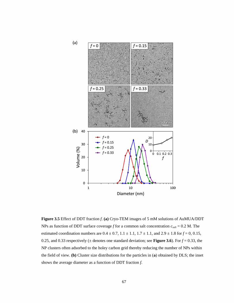
Figure 35 Effect of DDT fraction f (a) Cryo-TEM images of 5 mM solutions of AuMUADDT
NPs as function of DDT surface coverage f for a common salt concentration csalt = 02 M The
estimated coordination numbers are 04 plusmn 07 11 plusmn 11 17 plusmn 11 and 29 plusmn 18 for f = 0 015
025 and 033 respectively (plusmn denotes one standard deviation see Figure 36) For f = 033 the
NP clusters often adsorbed to the holey carbon grid thereby reducing the number of NPs within
the field of view (b) Cluster size distributions for the particles in (a) obtained by DLS the inset
shows the average diameter as a function of DDT fraction f middotmiddotmiddotmiddotmiddotmiddotmiddotmiddotmiddotmiddotmiddotmiddotmiddotmiddotmiddotmiddotmiddotmiddotmiddotmiddotmiddotmiddotmiddotmiddotmiddotmiddotmiddotmiddotmiddotmiddotmiddotmiddotmiddotmiddotmiddotmiddotmiddotmiddotmiddotmiddotmiddotmiddotmiddotmiddotmiddotmiddotmiddotmiddotmiddotmiddotmiddotmiddotmiddotmiddotmiddotmiddot 67
Figure 36 NP coordination number (a) Cryo-TEM image (f = 025 csalt = 02 M) before and
after particle identification different clusters are denoted by different colors (Scale bar = 200 nm)
(b) Nearest neighbor distributions for different DDT fractions f as estimated from cryo-TEM
images The inset shows the average number of nearest neighbors as a function of the DDT
fraction middotmiddotmiddotmiddotmiddotmiddotmiddotmiddotmiddotmiddotmiddotmiddotmiddotmiddotmiddotmiddotmiddotmiddotmiddotmiddotmiddotmiddotmiddotmiddotmiddotmiddotmiddotmiddotmiddotmiddotmiddotmiddotmiddotmiddotmiddotmiddotmiddotmiddotmiddotmiddotmiddotmiddotmiddotmiddotmiddotmiddotmiddotmiddotmiddotmiddotmiddotmiddotmiddotmiddotmiddotmiddotmiddotmiddotmiddotmiddotmiddotmiddotmiddotmiddotmiddotmiddotmiddotmiddotmiddotmiddotmiddotmiddotmiddotmiddotmiddotmiddotmiddotmiddotmiddotmiddotmiddotmiddotmiddotmiddotmiddotmiddotmiddotmiddotmiddotmiddotmiddotmiddotmiddotmiddotmiddotmiddotmiddotmiddotmiddotmiddotmiddotmiddotmiddotmiddotmiddotmiddotmiddotmiddotmiddotmiddotmiddotmiddotmiddotmiddotmiddotmiddotmiddotmiddotmiddotmiddotmiddotmiddotmiddotmiddotmiddotmiddotmiddotmiddotmiddotmiddotmiddotmiddotmiddotmiddotmiddotmiddotmiddotmiddot 68
Figure 37 Effect of assembly time Cryo-TEM images of AuMUADDT NPs with DDT
coverage f = 025 from 5 mM NP solutions (on a gold atom basis) containing 02 M of TMACl
The image was taken at (a) ~1 minute and (b) ~ 24 hours after preparing samples middotmiddotmiddotmiddotmiddotmiddotmiddotmiddotmiddotmiddotmiddotmiddotmiddotmiddotmiddotmiddotmiddotmiddotmiddotmiddotmiddot 71
Figure 38 (a) Idealized geometry of an NP ndash NP contact The ligand length of ~2 nm sets the
range of hydrophobic interactions the particle separation was chosen to accommodate the volume
of the ligands within the contact region (b) Minimum energy ligand distribution for a linear
cluster with coordination parameter v = 16 all hydrophobic ligands are accommodated within
contact regions (c-e) Results of MC simulations with particle density = 2times10minus4
hydrophobic
bond strength = 100 and electrostatic repulsion = 90 for four different values of the
coordination parameter ν corresponding to the conditions of experiments shown in Figure 35 (c)
Equilibrium particle configurations Chain-like assemblies similar to those from Figure 32b
were observed for ν = 18 (d) Average cluster size (black markers) as a function of the parameter
v blue curves denote one standard deviation above and below the mean (e) Average number of
nearest neighbors NN (black markers) as a function of v blue curves denote one standard
deviation above and below the mean middotmiddotmiddotmiddotmiddotmiddotmiddotmiddotmiddotmiddotmiddotmiddotmiddotmiddotmiddotmiddotmiddotmiddotmiddotmiddotmiddotmiddotmiddotmiddotmiddotmiddotmiddotmiddotmiddotmiddotmiddotmiddotmiddotmiddotmiddotmiddotmiddotmiddotmiddotmiddotmiddotmiddotmiddotmiddotmiddotmiddotmiddotmiddotmiddotmiddotmiddotmiddotmiddotmiddotmiddotmiddotmiddotmiddotmiddotmiddotmiddotmiddotmiddotmiddotmiddotmiddotmiddotmiddotmiddotmiddotmiddotmiddotmiddotmiddotmiddotmiddotmiddotmiddotmiddotmiddotmiddotmiddotmiddotmiddotmiddotmiddotmiddotmiddotmiddotmiddotmiddotmiddot 74
xi
Figure 39 (a) CoFe2O4 NPs functionalized with mixed monolayers of HDD and deprotonated
DHUA assemble to form chain-like structures in aqueous solutions at high salt concentrations
(csalt = 02 M) when the surface fraction of HDD ligands is f asymp 02 (b) Cryo-TEM images and (c)
DLS size distributions of NP chains (cCoFe2O4 = 666 mgmL csalt = 02 M and f asymp 0 and 02) middotmiddotmiddotmiddotmiddot 78
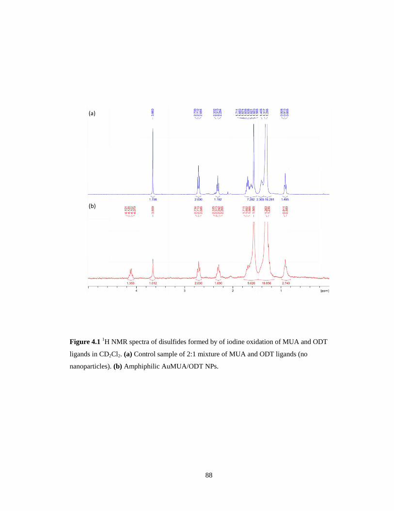
Figure 41 1H NMR spectra of disulfides formed by of iodine oxidation of MUA and ODT
ligands in CD2Cl2 (a) Control sample of 21 mixture of MUA and ODT ligands (no
nanoparticles) (b) Amphiphilic AuMUAODT NPs middotmiddotmiddotmiddotmiddotmiddotmiddotmiddotmiddotmiddotmiddotmiddotmiddotmiddotmiddotmiddotmiddotmiddotmiddotmiddotmiddotmiddotmiddotmiddotmiddotmiddotmiddotmiddotmiddotmiddotmiddotmiddotmiddotmiddotmiddotmiddotmiddotmiddotmiddotmiddotmiddotmiddotmiddotmiddotmiddotmiddotmiddotmiddotmiddotmiddotmiddotmiddotmiddotmiddotmiddotmiddotmiddotmiddotmiddotmiddotmiddotmiddotmiddotmiddotmiddotmiddotmiddotmiddot 88
Figure 42 (a) Gold nanoparticles functionalized with mixed monolayers of ODT and
deprotonated MUA incorporate spontaneously into vesicles formed from 3070 mixtures of
cationic (cetyltrimethylammonium tosylate CTAT) and anionic (sodium dodecyl benzene
sulfonate SDBS) surfactants (b) Cryo-TEM image shows colocalization of AuMUAODT
particles with surfactant vesicles in aqueous solution Amphiphilic NPs interact with the
hydrophobic core of the vesicle walls (cf below) under basic conditions (pH = 11) with 100 mM
added salt (tetramethylammonium chloride TMACl) middotmiddotmiddotmiddotmiddotmiddotmiddotmiddotmiddotmiddotmiddotmiddotmiddotmiddotmiddotmiddotmiddotmiddotmiddotmiddotmiddotmiddotmiddotmiddotmiddotmiddotmiddotmiddotmiddotmiddotmiddotmiddotmiddotmiddotmiddotmiddotmiddotmiddotmiddotmiddotmiddotmiddotmiddotmiddotmiddotmiddotmiddotmiddotmiddotmiddotmiddotmiddotmiddotmiddotmiddotmiddotmiddotmiddotmiddotmiddotmiddotmiddotmiddotmiddotmiddotmiddot 90
Figure 43 (a) Electrostatic titrations quantify the fraction of charged MUA ligands on the
surface of AuMUAODT particles (bc) AuMUAODT particles form stable clusters of ~10 NPs
in water (pH = 11) as revealed by (b) TEM and (c) DLS (d) Water soluble AuMUAODT
particles accumulate at the water-toluene interface middotmiddotmiddotmiddotmiddotmiddotmiddotmiddotmiddotmiddotmiddotmiddotmiddotmiddotmiddotmiddotmiddotmiddotmiddotmiddotmiddotmiddotmiddotmiddotmiddotmiddotmiddotmiddotmiddotmiddotmiddotmiddotmiddotmiddotmiddotmiddotmiddotmiddotmiddotmiddotmiddotmiddotmiddotmiddotmiddotmiddotmiddotmiddotmiddotmiddotmiddotmiddotmiddotmiddotmiddotmiddotmiddotmiddotmiddotmiddotmiddotmiddotmiddotmiddotmiddotmiddotmiddotmiddotmiddotmiddot 92
Figure 44 (left) TEM image of AuMUA NPs In contrast to the AuMUAODT NPs shown in
Figure 43b AuMUA NPs show no sign of aggregation by TEM (right) Particle size distribution
obtained from TEM images like that shown here The average NP diameter is 62 nm the
standard deviation is 08 nm middotmiddotmiddotmiddotmiddotmiddotmiddotmiddotmiddotmiddotmiddotmiddotmiddotmiddotmiddotmiddotmiddotmiddotmiddotmiddotmiddotmiddotmiddotmiddotmiddotmiddotmiddotmiddotmiddotmiddotmiddotmiddotmiddotmiddotmiddotmiddotmiddotmiddotmiddotmiddotmiddotmiddotmiddotmiddotmiddotmiddotmiddotmiddotmiddotmiddotmiddotmiddotmiddotmiddotmiddotmiddotmiddotmiddotmiddotmiddotmiddotmiddotmiddotmiddotmiddotmiddotmiddotmiddotmiddotmiddotmiddotmiddotmiddotmiddotmiddotmiddotmiddotmiddotmiddotmiddotmiddotmiddotmiddotmiddotmiddotmiddotmiddotmiddotmiddotmiddotmiddotmiddotmiddotmiddotmiddotmiddotmiddotmiddotmiddotmiddotmiddotmiddotmiddotmiddotmiddot 94
Figure 45 (ab) Surfactant Vesicles (a) ζ-potential distribution for CTATSDBS surfactant
vesicles in water with no added TMACl (b) ζ-potential distribution for surfactant vesicles in
water with 100 mM added TMACl (cd) AuMUAODT nanoparticles (c) ζ-potential distribution
for AuMUAODT NP clusters in water at pH = 11 with no added TMACl (d) ζ-potential
distribution for AuMUAODT NP clusters in water at pH = 11 with 100 mM added TMACl middotmiddotmiddotmiddot 95
Figure 46 (a) Solutions of AuMUAODT (1) and surfactant vesicles (2) are simply mixed to
form NP-vesicle structures (3) (b) Size distributions obtained by DLS for mixtures 2 3 and 3
with 100 mM TMACl (c) UV-visible extinction spectra for mixtures 1 3 and 3 with 100 mM
TMACl middotmiddotmiddotmiddotmiddotmiddotmiddotmiddotmiddotmiddotmiddotmiddotmiddotmiddotmiddotmiddotmiddotmiddotmiddotmiddotmiddotmiddotmiddotmiddotmiddotmiddotmiddotmiddotmiddotmiddotmiddotmiddotmiddotmiddotmiddotmiddotmiddotmiddotmiddotmiddotmiddotmiddotmiddotmiddotmiddotmiddotmiddotmiddotmiddotmiddotmiddotmiddotmiddotmiddotmiddotmiddotmiddotmiddotmiddotmiddotmiddotmiddotmiddotmiddotmiddotmiddotmiddotmiddotmiddotmiddotmiddotmiddotmiddotmiddotmiddotmiddotmiddotmiddotmiddotmiddotmiddotmiddotmiddotmiddotmiddotmiddotmiddotmiddotmiddotmiddotmiddotmiddotmiddotmiddotmiddotmiddotmiddotmiddotmiddotmiddotmiddotmiddotmiddotmiddotmiddotmiddotmiddotmiddotmiddotmiddotmiddotmiddotmiddotmiddotmiddotmiddotmiddotmiddotmiddotmiddotmiddotmiddotmiddotmiddotmiddotmiddotmiddotmiddotmiddotmiddotmiddotmiddotmiddotmiddotmiddotmiddotmiddotmiddot 97
Figure 47 (a) Mixtures of vesicles and AuMUAODT NPs without added salt show no signs of
NP-vesicle association by cryo-TEM (b) -potential distributions for mixtures of vesicles and
AuMUAODT NPs with and without added salt (c) Schematic illustrations of possible NP-
vesicle configurations middotmiddotmiddotmiddotmiddotmiddotmiddotmiddotmiddotmiddotmiddotmiddotmiddotmiddotmiddotmiddotmiddotmiddotmiddotmiddotmiddotmiddotmiddotmiddotmiddotmiddotmiddotmiddotmiddotmiddotmiddotmiddotmiddotmiddotmiddotmiddotmiddotmiddotmiddotmiddotmiddotmiddotmiddotmiddotmiddotmiddotmiddotmiddotmiddotmiddotmiddotmiddotmiddotmiddotmiddotmiddotmiddotmiddotmiddotmiddotmiddotmiddotmiddotmiddotmiddotmiddotmiddotmiddotmiddotmiddotmiddotmiddotmiddotmiddotmiddotmiddotmiddotmiddotmiddotmiddotmiddotmiddotmiddotmiddotmiddotmiddotmiddotmiddotmiddotmiddotmiddotmiddotmiddotmiddotmiddotmiddotmiddotmiddotmiddotmiddotmiddotmiddotmiddotmiddotmiddotmiddotmiddotmiddotmiddotmiddotmiddotmiddotmiddotmiddotmiddot 99
Figure 51 (a) Cryo-TEM image and (b) DLS size distribution of Au-TMA-ODT NP clusters in
the 73 methanolwater mixtures middotmiddotmiddotmiddotmiddotmiddotmiddotmiddotmiddotmiddotmiddotmiddotmiddotmiddotmiddotmiddotmiddotmiddotmiddotmiddotmiddotmiddotmiddotmiddotmiddotmiddotmiddotmiddotmiddotmiddotmiddotmiddotmiddotmiddotmiddotmiddotmiddotmiddotmiddotmiddotmiddotmiddotmiddotmiddotmiddotmiddotmiddotmiddotmiddotmiddotmiddotmiddotmiddotmiddotmiddotmiddotmiddotmiddotmiddotmiddotmiddotmiddotmiddotmiddotmiddotmiddotmiddotmiddotmiddotmiddotmiddotmiddotmiddotmiddotmiddotmiddotmiddotmiddotmiddotmiddotmiddotmiddotmiddotmiddotmiddotmiddotmiddotmiddotmiddotmiddotmiddotmiddotmiddotmiddotmiddotmiddotmiddot 108
xii
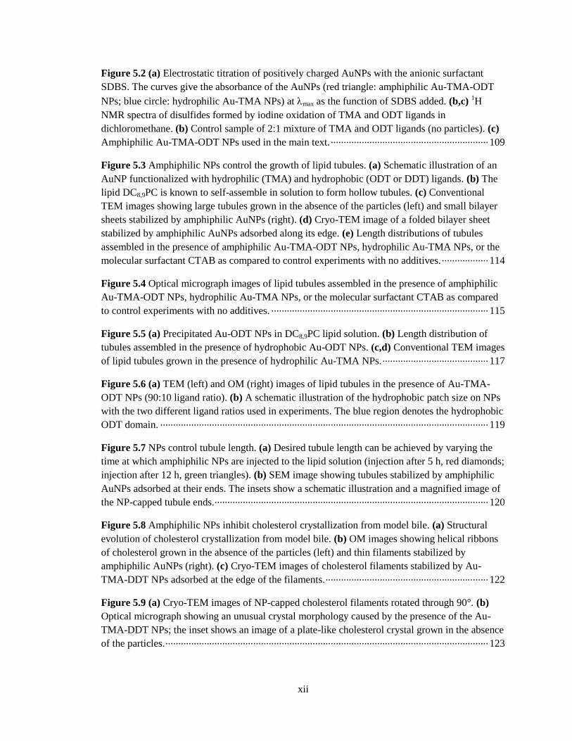
Figure 52 (a) Electrostatic titration of positively charged AuNPs with the anionic surfactant
SDBS The curves give the absorbance of the AuNPs (red triangle amphiphilic Au-TMA-ODT
NPs blue circle hydrophilic Au-TMA NPs) at max as the function of SDBS added (bc) 1H
NMR spectra of disulfides formed by iodine oxidation of TMA and ODT ligands in
dichloromethane (b) Control sample of 21 mixture of TMA and ODT ligands (no particles) (c)
Amphiphilic Au-TMA-ODT NPs used in the main text middotmiddotmiddotmiddotmiddotmiddotmiddotmiddotmiddotmiddotmiddotmiddotmiddotmiddotmiddotmiddotmiddotmiddotmiddotmiddotmiddotmiddotmiddotmiddotmiddotmiddotmiddotmiddotmiddotmiddotmiddotmiddotmiddotmiddotmiddotmiddotmiddotmiddotmiddotmiddotmiddotmiddotmiddotmiddotmiddotmiddotmiddotmiddotmiddotmiddotmiddotmiddotmiddotmiddotmiddotmiddotmiddotmiddotmiddotmiddotmiddot 109
Figure 53 Amphiphilic NPs control the growth of lipid tubules (a) Schematic illustration of an
AuNP functionalized with hydrophilic (TMA) and hydrophobic (ODT or DDT) ligands (b) The
lipid DC89PC is known to self-assemble in solution to form hollow tubules (c) Conventional
TEM images showing large tubules grown in the absence of the particles (left) and small bilayer
sheets stabilized by amphiphilic AuNPs (right) (d) Cryo-TEM image of a folded bilayer sheet
stabilized by amphiphilic AuNPs adsorbed along its edge (e) Length distributions of tubules
assembled in the presence of amphiphilic Au-TMA-ODT NPs hydrophilic Au-TMA NPs or the
molecular surfactant CTAB as compared to control experiments with no additives middotmiddotmiddotmiddotmiddotmiddotmiddotmiddotmiddotmiddotmiddotmiddotmiddotmiddotmiddotmiddotmiddotmiddot 114
Figure 54 Optical micrograph images of lipid tubules assembled in the presence of amphiphilic
Au-TMA-ODT NPs hydrophilic Au-TMA NPs or the molecular surfactant CTAB as compared
to control experiments with no additives middotmiddotmiddotmiddotmiddotmiddotmiddotmiddotmiddotmiddotmiddotmiddotmiddotmiddotmiddotmiddotmiddotmiddotmiddotmiddotmiddotmiddotmiddotmiddotmiddotmiddotmiddotmiddotmiddotmiddotmiddotmiddotmiddotmiddotmiddotmiddotmiddotmiddotmiddotmiddotmiddotmiddotmiddotmiddotmiddotmiddotmiddotmiddotmiddotmiddotmiddotmiddotmiddotmiddotmiddotmiddotmiddotmiddotmiddotmiddotmiddotmiddotmiddotmiddotmiddotmiddotmiddotmiddotmiddotmiddotmiddotmiddotmiddotmiddotmiddotmiddotmiddotmiddotmiddotmiddotmiddotmiddotmiddotmiddot 115
Figure 55 (a) Precipitated Au-ODT NPs in DC89PC lipid solution (b) Length distribution of
tubules assembled in the presence of hydrophobic Au-ODT NPs (cd) Conventional TEM images
of lipid tubules grown in the presence of hydrophilic Au-TMA NPs middotmiddotmiddotmiddotmiddotmiddotmiddotmiddotmiddotmiddotmiddotmiddotmiddotmiddotmiddotmiddotmiddotmiddotmiddotmiddotmiddotmiddotmiddotmiddotmiddotmiddotmiddotmiddotmiddotmiddotmiddotmiddotmiddotmiddotmiddotmiddotmiddotmiddotmiddotmiddotmiddot 117
Figure 56 (a) TEM (left) and OM (right) images of lipid tubules in the presence of Au-TMA-
ODT NPs (9010 ligand ratio) (b) A schematic illustration of the hydrophobic patch size on NPs
with the two different ligand ratios used in experiments The blue region denotes the hydrophobic
ODT domain middotmiddotmiddotmiddotmiddotmiddotmiddotmiddotmiddotmiddotmiddotmiddotmiddotmiddotmiddotmiddotmiddotmiddotmiddotmiddotmiddotmiddotmiddotmiddotmiddotmiddotmiddotmiddotmiddotmiddotmiddotmiddotmiddotmiddotmiddotmiddotmiddotmiddotmiddotmiddotmiddotmiddotmiddotmiddotmiddotmiddotmiddotmiddotmiddotmiddotmiddotmiddotmiddotmiddotmiddotmiddotmiddotmiddotmiddotmiddotmiddotmiddotmiddotmiddotmiddotmiddotmiddotmiddotmiddotmiddotmiddotmiddotmiddotmiddotmiddotmiddotmiddotmiddotmiddotmiddotmiddotmiddotmiddotmiddotmiddotmiddotmiddotmiddotmiddotmiddotmiddotmiddotmiddotmiddotmiddotmiddotmiddotmiddotmiddotmiddotmiddotmiddotmiddotmiddotmiddotmiddotmiddotmiddotmiddotmiddotmiddotmiddotmiddotmiddotmiddotmiddotmiddotmiddotmiddotmiddotmiddotmiddotmiddotmiddotmiddotmiddotmiddot 119
Figure 57 NPs control tubule length (a) Desired tubule length can be achieved by varying the
time at which amphiphilic NPs are injected to the lipid solution (injection after 5 h red diamonds
injection after 12 h green triangles) (b) SEM image showing tubules stabilized by amphiphilic
AuNPs adsorbed at their ends The insets show a schematic illustration and a magnified image of
the NP-capped tubule ends middotmiddotmiddotmiddotmiddotmiddotmiddotmiddotmiddotmiddotmiddotmiddotmiddotmiddotmiddotmiddotmiddotmiddotmiddotmiddotmiddotmiddotmiddotmiddotmiddotmiddotmiddotmiddotmiddotmiddotmiddotmiddotmiddotmiddotmiddotmiddotmiddotmiddotmiddotmiddotmiddotmiddotmiddotmiddotmiddotmiddotmiddotmiddotmiddotmiddotmiddotmiddotmiddotmiddotmiddotmiddotmiddotmiddotmiddotmiddotmiddotmiddotmiddotmiddotmiddotmiddotmiddotmiddotmiddotmiddotmiddotmiddotmiddotmiddotmiddotmiddotmiddotmiddotmiddotmiddotmiddotmiddotmiddotmiddotmiddotmiddotmiddotmiddotmiddotmiddotmiddotmiddotmiddotmiddotmiddotmiddotmiddotmiddotmiddotmiddotmiddotmiddotmiddotmiddotmiddotmiddot 120
Figure 58 Amphiphilic NPs inhibit cholesterol crystallization from model bile (a) Structural
evolution of cholesterol crystallization from model bile (b) OM images showing helical ribbons
of cholesterol grown in the absence of the particles (left) and thin filaments stabilized by
amphiphilic AuNPs (right) (c) Cryo-TEM images of cholesterol filaments stabilized by Au-
TMA-DDT NPs adsorbed at the edge of the filaments middotmiddotmiddotmiddotmiddotmiddotmiddotmiddotmiddotmiddotmiddotmiddotmiddotmiddotmiddotmiddotmiddotmiddotmiddotmiddotmiddotmiddotmiddotmiddotmiddotmiddotmiddotmiddotmiddotmiddotmiddotmiddotmiddotmiddotmiddotmiddotmiddotmiddotmiddotmiddotmiddotmiddotmiddotmiddotmiddotmiddotmiddotmiddotmiddotmiddotmiddotmiddotmiddotmiddotmiddotmiddotmiddotmiddotmiddotmiddotmiddotmiddotmiddot 122
Figure 59 (a) Cryo-TEM images of NP-capped cholesterol filaments rotated through 90deg (b)
Optical micrograph showing an unusual crystal morphology caused by the presence of the Au-
TMA-DDT NPs the inset shows an image of a plate-like cholesterol crystal grown in the absence
of the particles middotmiddotmiddotmiddotmiddotmiddotmiddotmiddotmiddotmiddotmiddotmiddotmiddotmiddotmiddotmiddotmiddotmiddotmiddotmiddotmiddotmiddotmiddotmiddotmiddotmiddotmiddotmiddotmiddotmiddotmiddotmiddotmiddotmiddotmiddotmiddotmiddotmiddotmiddotmiddotmiddotmiddotmiddotmiddotmiddotmiddotmiddotmiddotmiddotmiddotmiddotmiddotmiddotmiddotmiddotmiddotmiddotmiddotmiddotmiddotmiddotmiddotmiddotmiddotmiddotmiddotmiddotmiddotmiddotmiddotmiddotmiddotmiddotmiddotmiddotmiddotmiddotmiddotmiddotmiddotmiddotmiddotmiddotmiddotmiddotmiddotmiddotmiddotmiddotmiddotmiddotmiddotmiddotmiddotmiddotmiddotmiddotmiddotmiddotmiddotmiddotmiddotmiddotmiddotmiddotmiddotmiddotmiddotmiddotmiddotmiddotmiddotmiddotmiddotmiddotmiddotmiddotmiddotmiddotmiddotmiddotmiddotmiddotmiddotmiddot 123
xiii
Figure 510 Amphiphilic NPs stabilize DPPC sheets (a) The lipid DPPC self-assembles in
solution to form spherical vesicles (b) Cryo-TEM images showing large bilayer sheets formed by
shearing a solution of DPPC vesicles (left) and small bilayer sheets stabilized by amphiphilic
AuNPs (right) The inset shows a magnified image of the bilayer edge decorated with AuNPs (c)
DLS size distributions of shear-induced DPPC bilayer sheets with and without addition of
amphiphilic Au-TMA-ODT NPs middotmiddotmiddotmiddotmiddotmiddotmiddotmiddotmiddotmiddotmiddotmiddotmiddotmiddotmiddotmiddotmiddotmiddotmiddotmiddotmiddotmiddotmiddotmiddotmiddotmiddotmiddotmiddotmiddotmiddotmiddotmiddotmiddotmiddotmiddotmiddotmiddotmiddotmiddotmiddotmiddotmiddotmiddotmiddotmiddotmiddotmiddotmiddotmiddotmiddotmiddotmiddotmiddotmiddotmiddotmiddotmiddotmiddotmiddotmiddotmiddotmiddotmiddotmiddotmiddotmiddotmiddotmiddotmiddotmiddotmiddotmiddotmiddotmiddotmiddotmiddotmiddotmiddotmiddotmiddotmiddotmiddotmiddotmiddotmiddotmiddotmiddotmiddotmiddotmiddotmiddotmiddotmiddotmiddotmiddotmiddot 125
Figure 511 Cryo-TEM images of NP-capped DPPC bilayer sheets rotated through 90deg middotmiddotmiddotmiddotmiddotmiddotmiddotmiddotmiddot 126
Figure 61 A schematic of an isothermal titration calorimeter shows the sample cell and the
reference cell in an isothermal jacket The syringe is inserted in the sample cell and a series of
injections are made The injection of one component into the other results in exothermic or
endothermic heat effect middotmiddotmiddotmiddotmiddotmiddotmiddotmiddotmiddotmiddotmiddotmiddotmiddotmiddotmiddotmiddotmiddotmiddotmiddotmiddotmiddotmiddotmiddotmiddotmiddotmiddotmiddotmiddotmiddotmiddotmiddotmiddotmiddotmiddotmiddotmiddotmiddotmiddotmiddotmiddotmiddotmiddotmiddotmiddotmiddotmiddotmiddotmiddotmiddotmiddotmiddotmiddotmiddotmiddotmiddotmiddotmiddotmiddotmiddotmiddotmiddotmiddotmiddotmiddotmiddotmiddotmiddotmiddotmiddotmiddotmiddotmiddotmiddotmiddotmiddotmiddotmiddotmiddotmiddotmiddotmiddotmiddotmiddotmiddotmiddotmiddotmiddotmiddotmiddotmiddotmiddotmiddotmiddotmiddotmiddotmiddotmiddotmiddotmiddotmiddotmiddotmiddotmiddotmiddotmiddotmiddotmiddotmiddotmiddotmiddotmiddot 134
Figure 62 Cryo-TEM images of charged AuTMA NPs loaded in DOPC vesicle interior middotmiddotmiddotmiddotmiddotmiddotmiddotmiddot 136
Figure 63 (a) A schematic of Langmuir trough (b) Surface pressure ndash area isotherm of the
Langmuir monolayer of amphiphilic NPs middotmiddotmiddotmiddotmiddotmiddotmiddotmiddotmiddotmiddotmiddotmiddotmiddotmiddotmiddotmiddotmiddotmiddotmiddotmiddotmiddotmiddotmiddotmiddotmiddotmiddotmiddotmiddotmiddotmiddotmiddotmiddotmiddotmiddotmiddotmiddotmiddotmiddotmiddotmiddotmiddotmiddotmiddotmiddotmiddotmiddotmiddotmiddotmiddotmiddotmiddotmiddotmiddotmiddotmiddotmiddotmiddotmiddotmiddotmiddotmiddotmiddotmiddotmiddotmiddotmiddotmiddotmiddotmiddotmiddotmiddotmiddotmiddotmiddotmiddotmiddotmiddotmiddotmiddotmiddotmiddotmiddotmiddot 138
Figure 64 (a) Magnetic NPs as field responsive amphiphiles (b) Field-induced capillary
attraction middotmiddotmiddotmiddotmiddotmiddotmiddotmiddotmiddotmiddotmiddotmiddotmiddotmiddotmiddotmiddotmiddotmiddotmiddotmiddotmiddotmiddotmiddotmiddotmiddotmiddotmiddotmiddotmiddotmiddotmiddotmiddotmiddotmiddotmiddotmiddotmiddotmiddotmiddotmiddotmiddotmiddotmiddotmiddotmiddotmiddotmiddotmiddotmiddotmiddotmiddotmiddotmiddotmiddotmiddotmiddotmiddotmiddotmiddotmiddotmiddotmiddotmiddotmiddotmiddotmiddotmiddotmiddotmiddotmiddotmiddotmiddotmiddotmiddotmiddotmiddotmiddotmiddotmiddotmiddotmiddotmiddotmiddotmiddotmiddotmiddotmiddotmiddotmiddotmiddotmiddotmiddotmiddotmiddotmiddotmiddotmiddotmiddotmiddotmiddotmiddotmiddotmiddotmiddotmiddotmiddotmiddotmiddotmiddotmiddotmiddotmiddotmiddotmiddotmiddotmiddotmiddotmiddotmiddotmiddotmiddotmiddotmiddotmiddotmiddotmiddotmiddotmiddotmiddotmiddotmiddotmiddotmiddotmiddot 141
xiv
ACKNOWLEDGEMENTS
I would like to thank those who contributed and supported through my graduate studies at
the Pennsylvania State University First of all I would like to express my sincere appreciation to
my advisor Professor Kyle J M Bishop for his guidance and support I am grateful to him for
providing me with the opportunity to work in the research area of colloids and helping me with
great discussions and inspirations Without his patience and advice I would not be able to have
achievements in my research work I would also like to thank the members of my graduate
committee Professor Robert Rioux Professor Darrell Velegol and Professor Raymond E
Schaak all of whom have taught me through my graduate studies
I thank to my friends who made my life in State College enjoyable and all members of
Bishop Group in the past and present who provide a nice environment in which to work The
great friendships that I have made with them have given me things to look forwards to outside of
research as well as countless happy memories that I will always be thankful Finally I would like
to express deep appreciation to my family for their infinite support and love
The research in this dissertation has been supported by Penn State Center for Nanoscale
Science (NSF DMR-0820404) and Non-equilibrium Energy Research Center (DE-SC0000989)
1
Chapter 1
Inorganic Nanoparticle Amphiphiles Preparation and Application
11 Introduction
Surfactants also called surface-active agents are substances that modify the interfacial
free energy between two immiscible phases such as liquid ndash liquid solid ndash liquid or liquid ndash gas
by adsorbing at the interface Surfactants have long been employed in a broad range of industrial
processes as wetting agents emulsifiers foaming agents and dispersants The surfactants usually
contain both hydrophilic (water-soluble) and hydrophobic (water-insoluble) components This
combination is known as an amphiphilic character When surfactants are present in mixture of
water and oil the hydrophilic portions of the surfactants are expected to be oriented toward the
water phase while hydrophobic portions should contact the oil phase As a result surfactants at
water ndash oil interfaces can reduce interfacial tension and stabilize thermodynamically unstable
system
Surfactants can be classified into two groups molecule and particle Molecular
surfactants are usually 1-2 nm and size of particle surfactants range from tens of nanometer to
even micrometer For particle surfactants particles with homogeneous surface coating can have
surface activity (eg Pickering emulsion) but here we consider only particle surfactants which
are amphiphilic While the surface-active properties of molecular surfactants are well studied that
of particle surfactants are less developed and remain a subject of great attention due to their use in
drug delivery emulsification assembly of hierarchical structures coatings and optoelectronic
devices12
Nanoscale particle surfactants are of particular interest because their size is
2
intermediate between molecules and microscale particles As a result nanoparticle (NP)
surfactants can have advantages of both molecular and particle-based surfactants Like
microparticles the NP surfactants are capable of preventing coalescence of emulsion droplets by
controlling the spacing between contacting droplets3 On the other hand NPs can be stable
against sedimentation even at high salt concentrations and have higher rate of mass transfer from
solution to a surface or interface than the microparticles Due to their intermediate size they
adsorb strongly but often reversibly at an interface More importantly the NP surfactants
incorporating inorganic cores offer additional functionalities such as enhanced fluorescence (eg
cadmium selenide)4 plasmonic excitation (eg gold silver and platinum)
5 and
superparamagnetism (eg iron oxide and cobalt ferrite)6 owing to their composition and size
These functionalities cannot be achieved by polymeric particles7 and differ significantly from that
of the corresponding bulk materials and microparticles
In this introductory chapter we outline various strategies used for preparing inorganic NP
with amphiphilic surface chemistries and highlight recent advances in their applications in
nanostructured assemblies interfacial engineering biomedical engineering and stimuli
responsive systems
12 Preparation of Inorganic Nanoparticle Amphiphiles
The majority of previously reported methods for the preparation of particle surfactants
with amphiphilic surface chemistries have focused on microparticles These include metal vapor
deposition stamping gel trapping electroless plating and particle lithography8-11
However such
traditional techniques are less effective for NPs due to the particlesrsquo rotational diffusion (Drot ~
1r3 where Drot is rotational diffusion coefficient and r is particle radius
12) Alternative approaches
are needed for the NP amphiphiles
3
Synthesis of inorganic NPs generally requires organic capping agents which bind to
surface atoms of the NPs as ligands Various functional groups have a specific affinity to certain
inorganic surfaces (eg thiol to gold and catechol to metal oxide) The ligands form self-
assembled monolayers (SAMs) on surface of the NPs when they are adsorbed from solution13
The ligands play a key role in controlling the particle size and shape during the synthesis
stabilizing the particles in solution and mediating interactions of the particles with their
environment Compositions of the ligand monolayers are often modified by ligand-exchange
reactions to tailor surface chemistries of the NPs For example hydrophobic capping ligands are
exchanged for hydrophilic ligands to provide stability in aqueous solution and biocompability14
A common strategy for functionalizing NPs with amphiphilic surfaces is based on spontaneous
self-organization of ligands on the NP surface This includes coadsorption of hydrophilic and
hydrophobic ligands from a solution15-17
adsorption of amphiphilic ligands18-22
or partial
exchange for hydrophilic (hydrophobic) ligand on a preformed hydrophobic (hydrophilic) ligand
monolayer23-26
(Figure 11) As a result the NPs are decorated with mixed SAMs that comprise
well-defined mixtures of different ligands
The challenge in the preparation of NP amphiphiles is to engineer the particles in a
controlled manner Two important issues need to be addressed to produce NP amphiphiles with
desired properties One is the ability to control surface composition ie the numbers of different
ligand types on the NP surface This is analogous to controlling the hydrophilic ndash lipophilic
balance (HLB) of molecular surfactants The mole fraction of a specific ligand in the SAM
reflects ndash but is not necessarily the same as ndash the mole fraction of the ligand in solution The
choice of solvent can also influence the relative ratio of components in the SAMs formed from a
mixture of polar and nonpolar ligand molecules The other one is the ability to control ligand
distribution ie the spatial arrangement of the different ligand types on the NP surface Selective
surface modifications using templates lead to asymmetric ligand distributions forming Janus-
4
Figure 11 Synthesis of NP amphiphiles by (a) adsorption of amphiphilic ligands (b)
coadsorption of hydrophilic and hydrophobic ligands from a solution or (c) partial exchange for
hydrophilic ligand on a preformed hydrophobic ligand Gray bead represents NP Red line
indicates hydrophobic ligand and blue line indicates hydrophilic ligand
5
patterns on the NP surfaces27-29
Our group reported a simple method for the formation of NP
amphiphiles that provides a comprehensive solution to both of these issues as detailed further in
Chapter 216
It is known that mixed SAMs of two alkanethiolates having chemically distinct terminal
groups or different chain lengths exhibit thermodynamically driven phase separation on flat gold
substrates30-34
Similar phase separation of a binary mixture of dissimilar ligands has been
observed on curved substrates Stellacci et al studied the morphology of mixed SAMs of
hydrophilic mercaptopropionic acid (MPA) and hydrophobic 1-octanethiol (OT) ligands on gold
and silver NPs35
They observed discrete and ordered domains of each ligand which were as small
as 5 Aring The domain morphology and dimension were controlled by varying the ligand
composition and the curvature of the underlying substrate In their recent experiments completely
separated distribution of ligands into two bulk phases on the NPs forming Janus-patterns was
confirmed when the NP diameter was smaller than 15 nm36
The phase behavior of mixed SAMs
on the surface of NPs was studied by Glotzer et al using molecular dynamics simulations37
They
demonstrated that dissimilar ligands spontaneously separate into ordered domains on the NPs due
to the interplay between the enthalpy of phase separation and the conformational entropy that
arises at the interface between immiscible ligand molecules The simulations predicted that the
ligand layer presents either microphase separated stripe-like pattern or bulk phase separated Janus
pattern depending on (i) the length difference of the ligands (ii) the bulkiness difference of the
ligands and (iii) the relative ligand length and the radius of curvature of the particle
Various techniques have been used to characterize the ligand domains on the NPs such as
scanning tunneling microscopy (STM)3536
infrared (IR) spectroscopy38
and electron
paramagnetic resonance (EPR) spectroscopy39
Recently the degree of segregation between two
ligands on the NPs has been quantitatively analyzed using mass spectrometry combined with
matrix-assisted laser desorptionionization1540
The gold ndash thiolate complex which is liberated
6
from the surface of gold NP is examined to compare to a theoretical model Cliffel et al
measured different degrees of nanophase separation of various ligand mixtures from no
separation to moderate (domain formation) and complete separation (Janus pattern)40
It has been demonstrated that mixed SAMs decorating the NPs are adaptive as a response
to changes in the particlesrsquo environment by reorganizing themselves Bjoslashrnholm et al explored
adaptive behavior of the mixed alkylthiols with different chain length (C12 vs C5) andor
terminal functionality (hydroxyl- vs methyl-terminated) when the gold NPs were spread onto air
ndash water interface41
Based on the experimental observation that the interparticle spacing in the
monolayer of NP amphiphiles was not affected by the amount of hydrophilic ligands on the NPs
it was suggested that the NP amphiphiles adapt to form hydrophilic domains on their bottom
surface which are in contact with the water and do not take up lateral space in the particle
monolayer When the particle monolayer was compressed in a Langmuir trough the NPs with
mixed long (C12) and short (C5) ligands responded to the changes in lateral pressure by expelling
the long ligands from the plane of the particle monolayer Grazing incidence X-ray diffraction
measurements showed that the area of the peak corresponding to the ligand segregated phase is
increasing compared to the random phase peak as the particle monolayer is compressed41
Consequently the NPs adapted their structures with rearranged ligand monolayer in which the
short ligands are present in the particle monolayer plane while long ligands are present on the top
and bottom of the particles Fiałkowski et al characterized adaptive behavior of NP amphiphiles
by monitoring the methylene stretching modes of alkene chains in the mixed 11-
mercaptoundecyltrimethylammonium chloride (TMA) and1-undecanethiol (UDT) ligand layer
using IR spectroscopy 42 They showed that the rearrangement of the ligands occurs only when
the amphiphilic NPs are brought from organic phase to air ndash water interface The resulting NPs at
the interface exhibited properties of Janus NPs with hydrophobic and hydrophilic hemispheres
suggesting that the ligands are redistributed to form Janus patterns on the NP surface
7
A plausible mechanism for the redistribution of the ligands on the NP surface is the
migration of ligands on the metal surface Thiols have been known to diffuse over the flat gold
substrate43-47
as well as NP surface Chechik et al used a cleavable ester bridge connecting the
two branches of the thiols to probe the lateral diffusion of thiolate ligands on gold NP surface48
EPR spectroscopy revealed that the average distance between the neighboring thiols increases
after cleaving the bridge and thermal treatment A broad range of the time scale for ligand
reorganization has been reported from minutes to days The diffusion coefficient of thiols on flat
gold substrate was estimated to be 10-18
ndash 10-17
cm2s at elevated temperature
49 Rotello et al
observed time evolution of binding sites on the NP surface over a period of ~80 h at room
temperature50
Werts et al proposed that equilibration of ligands on the NP surface takes 1 ndash 2 h
using fluorescence titration51
Bjoslashrnholm et al suggested that the ligand rearrangement can
proceed within minutes in the presence of strong driving forces41
13 Applications of Nanoparticle Amphiphiles
131 Building Blocks for Self-Assembly
Self-assembly of NPs is a process that NPs spontaneously organize into ordered
macroscopic structures by interacting one another through various templates interparticle forces
or external forces52-54
The assembled structures show novel collective properties (eg electronic
magnetic or optical property) which arise from the coupling between individual NP
components55
Inspired by self-assembly in biological system where complex structures are predesigned
by hydrophobic interaction between components directional assembly of particles can be
achieved by decorating the particle surface with discrete hydrophilic and hydrophobic patches
When the amphiphilic particles are added to an aqueous solution the hydrophobic surfaces
8
organize to minimize their unfavorable contact with the surrounding water which drives the
particles to assemble into well-defined one- two- and three-dimensional structures (Figure
12)56-58
Several examples of the self-assembly of NP amphiphiles have been reported192259-61
Schenning et al observed that gold NPs covered with hydrophobic alkyl (C10) and hydrophilic
ethyleneoxide (EO4) ligands assemble to flat micellar disks upon injection of the NPs into
water62
The disk-shaped aggregates had an average height of 85 nm indicating that the disks are
constructed from bilayers of the NPs in which ligands are intercalated based on the gold core size
(37 nm in diameter) and length of the both ligands (22 ndash 25 nm) The authors suggested that
hydrophobic C10 ligands are present in the interior of the bilayers while EO4 ligands are at the
exterior which originate from the dynamic character of thiols on gold surfaces The same concept
was extended to hydrophobic conjugated oligo(p-phenylene vinylene) (OPV) ligands showing
that phase separation between dissimilar ligands on NPs occurs upon self-assembly in water63
Xia et al used silver nanocubes which have a non-spherical shape and demonstrated that the
structure of resultant assemblies can be controlled by functionalizing the six faces of nanocube
with hydrophobic octadecanethiol (ODT) and hydrophilic mercaptohexadecanoic acid (MHA)
SAMs in different schemes64
Depending on the number of faces on each cube that were rendered
hydrophobic (f) a variety of assemblies emerged for example dimers (f = 1) linear chains (f = 2)
sheets (f = 4) and three-dimensional lattice (f = 6) By contrast when the amphiphilic NPs are
added to organic solution the assembled structures are expected through contact with their
hydrophilic patches Due to the asymmetric interactions patterned on the NP surface the
interactions between particles are selective and directional resulting in wormlike aggregates65
Molecular simulations have been performed to study the self-assembly of particles with
discrete patches66
Glotzer et al demonstrated how the specific arrangements of attractive patches
on the particle surface control the relative position of the particles and overall structure of the
9
Figure 12 Self-assembled structures of particles with discrete hydrophobic patchy in water
Depending on the number andor distribution of patches on the particlersquos surface a variety of
assemblies can emerge Red region indicates hydrophobic patchy
10
particle assemblies The simulations predicted that anisotropic interactions direct the organization
of the particles from a disordered state into diverse and complex structures such as chains sheets
rings icosahedra square pyramids tetrahedra and twisted and staircase structures The
simulations further predicted that the formation of sheets and icosahedra occurs via a first-order
transition while the formation of chains is analogous to equilibrium polymerization
Components of self-assembled structures are often desired to have the ability to change
their interactions in a programmable manner as required by emerging applications in energy
capture and storage photonics and electronics67
One of the challenges in the realization of
programmable assembly from NP amphiphiles is designing dynamic interparticle interactions
Our group investigated self-assembly of NP amphiphiles with reconfigurable patches which
enable to tune particle coordination number within self-assembled structures through both
experimental and simulation methods as further described in Chapter 368
The fluid interfaces are often used as platforms for self-assembly of amphiphilic NPs into
two-dimensional structures1769
Fiałkowski et al investigated a spontaneous arrangement of
positively charged gold NPs decorated with a mixture of TMA and UDT into well-ordered
monolayer lattices at dichloromethane ndash water interface69
When water was layered over
dichloromethane containing the NPs the NPs migrated spontaneously from the bulk organic
phase and formed hexagonally close-packed lattices at the interface The formation of the lattice
was attributed to the interplay of the lateral repulsive electrostatic force between the neighboring
NPs and attractive hydrophobic effect between NPs and organic phase Recently Fiałkowski et al
presented dynamic self-assembly of identical NPs at water ndash air interface70
In contrast to typical
self-assembly at thermodynamic equilibrium dynamic self-assembly is non-equilibrium system
which can adapt componentrsquos organization in response to external stimuli54
In this process
external stimulus was surface tension gradient which was created by localized addition or
removal of volatile organic solvent from the interface In response to the surface tension gradient
11
the NPs migrated toward the regions of higher surface tension resulting in the formation of a
dense monolayer At equilibrium when the surface tension gradient vanished the NPs
spontaneously scattered at the interface
132 Interfacial Engineering
Particles can strongly adsorb at liquid ndash liquid interface and stabilize emulsion1 The
interfacial activity of the particles can be described by the desorption energy (E) which is defined
as the energy that is required to move a particle from the fluid interface to bulk phases
(11)
where r is the particle radius is the interfacial tension between oil and water and is
the contact angle that particle makes at the interface In a biphasic mixture of toluene and water
where = 36 mNm the desorption energy for the removal of the NPs of 20 nm in diameter
from the interface is 2750 kBT at = 90deg suggesting that the NP amphiphiles which surface
chemistries are asymmetric adsorb favorably at the interface
The interfacial activity of the NP amphiphiles has been characterized quantitatively using
a pendant drop tensiometry Krauch et al demonstrated that the interfacial tension of amphiphilic
goldiron oxide NPs (225 mNm) at n-hexane ndash water interface is lower than that of
corresponding homogeneous NPs (~34 mNm) of comparable size71
The results indicated that
amphiphilic surface of the NPs leads high interfacial activity Hidalgo-Alvarez et al studied
surface pressure ndash area isotherm for a monolayer of NPs at a water ndash air interface via the growing
and shrinking pendant drop technique72
The experiments showed that the surface pressure at the
most compressed states (lt 2 nm2particle) is about three times higher for the gold NPs decorated
with hydrophobic hexanethiol (HP) and hydrophilic 2-(2-mercapto-ethoxy) ethanol (MEE) than
12
for the homogenous NPs with HP This result is in agreement with theoretical work which
predicts that desorption energy of the amphiphilic particle is three times higher than that of the
homogenous particle (either hydrophilic or hydrophobic) at the maximum73
The relationship between the amphiphilic NPsrsquo surface hydrophobicity and the interfacial
activity has been investigated Striolo et al systematically studied the interfacial activity of silica
NPs with different surface chemistry at water ndash decane interface using a molecular dynamic
simulation74
The overall hydrophobicity of the NP was controlled by the ratio of hydrophilic and
hydrophobic group present at the surface The simulation predicted that the three phase contact
angle increases with the NPsrsquo surface hydrophobicity and the contact angle of the NP with
randomly disperse hydrophobic groups is smaller than that observed in Janus NPs of equal overall
surface chemistry Rotello et al experimentally explored the role of particlersquos hydrophobicity on
their dynamic behavior at toluene ndash water interface using a series of monolayer-protected gold
NPs75
As the hydrophobicity of the ligands in the monolayer increased NPs created a more
stable interphase with an observed a linear decrease in the meso-equilibrium surface tension
Control over the interparticle interactions in two dimensional structures at fluid interfaces
has important implications for molecular electronics emulsion stabilization and optical
devices76-78
The interactions between interfacial particles include van der waals electrostatic
capillary steric and solvation interactions7879
While the interactions between microparticles
have been well studied80-82
nanoscale interactions are more challenging experimentally The lack
of experimental observations leads poor understanding of the interactions governing interfacial
NP assembly
13
133 Biomedicial Engineering
The incorporation of inorganic NPs into the cell membrane transforms the properties of
cells accordingly and offers promising opportunities for utilizing them in a wide variety of
applications such as photothermal therapy8384
cellular imaging85-87
biosensing8889
and druggene
delivery90-92
For example iron oxide NPs are capable of magnetic actuation in remote delivery
and controlled release As imaging agents gold NPs and quantum dots enable to selectively view
specific biological processes in cells with improved detection limits
Translocating inorganic NPs into the membrane requires manipulating interactions of the
NPs with biological components Therefore it is of great importance for precise control over
particle surface chemistry which largely governs the interactions between NPs and cell
membranes Hydrophilic NPs can associate with bilayer membranes either by electrostatic
adsorption at bilayer ndash water interface93-95
or by encapsulation in polar interior of enclosed
membranes (Figure 13a)9596
In the context of developing stimuli-responsive NP ndash liposome
hybrids hydrophobic NPs are appealing because they can be embedded directly in the
hydrophobic bilayer core (Figure 13b) and corresponding response to stimuli should influence
on the properties of the hybrids effectively909197-99
Major concerns in current strategies based on
hydrophobic NPs are inclusion size limitation of NPs between bilayers and the use of stabilizing
detergents that must subsequently removed Our group demonstrated that relatively large
amphiphilic NPs (~6 nm) can be incorporated spontaneously into the bilayer of surfactant
vesicles (~ 25 nm) through hydrophobic interaction in the absence of detergents as detailed
further in Chapter 4100
Similar interactions were studied using coarse-grained molecular simulations aimed at the
effect of surface morphology of NPs on the interaction with lipid membranes101
The results
indicated that a hydrophobic NP can result in the inclusion into the bilayer whereas an
14
Figure 13 (a) Hydrophilic particles adsorbed on vesicle or encapsulated in aqueous vesicle
interior (b) Hydrophobic particles entrapped in the hydrophobic bilayer core
15
amphiphilic NP is found to adsorb onto the membrane The interaction between amphiphilic NPs
and bilayer lipid membranes can be quantified by electrophysiology experiments102
The
adsorption behavior of gold NPs coated with a mixed monolayer of hydrophilic 11-
mercaptoundecanesulfonate (MUS) and hydrophobic OT ligands to the bilayer was monitored
through the capacitive increase of suspended planar lipid membranes upon fusion with the NPs
Using a Langmuir model the partition coefficient and standard free energy gain of NP adsorption
were characterized for a variety of sizes and surface compositions
It has been demonstrated that NP amphiphiles are capable of non-specifically penetrating
cell membranes without disrupting the membrane and potentially triggering cell death103-106
Unlike cellular uptake of homogeneous and charged NPs by energy-dependent mechanisms (eg
endocytosis107-109
or transient membrane poration110-112
) gold NPs with ordered arrangements of
hydrophilic MUS and hydrophobic OT ligands spontaneously diffused through cellular
membranes through an energy-independent process104
Alexander-Katz et al have studied
extensively non-specific interactions between NP amphiphiles and lipid bilayers using a
combination of simulations and experimental studies113-119
The authors proposed that the critical
step in the penetration process is a fusion step in which the NPs incorporate into the hydrophobic
core of the bilayer This fusion step is driven by hydrophobic force assuming that ligands on the
surface of the NPs are capable of rearranging to maximize favorable interactions with the
hydrophobic core The simulations showed that the penetration behavior of charged NP
amphiphiles is thermodynamically favorable The authors explored the effect of particle size
ligand length morphology composition and hydrophobic strength on the change in the free
energy upon insertion of the particles into the bilayers
By taking advantage of the non-specific interaction between hydrophobic patches on
amphiphilic NPs and bilayer cores stabilizing open edges of bilayer membranes can be achieved
Balazes et al showed that NP amphiphiles in solution self-assembled to the edges of pores in
16
lipid bilayers using a coarse-grained numerical simulation120 The NPs stabilized the pore location
and size so that the pores can be controlled in a reliable repeatable manner Recently our group
experimentally demonstrated the NP amphiphiles can stabilize open edges of bilayer and control
growth of transient amphiphile assemblies as further described in Chapter 5
134 Stimuli-Responsive Systems
Stimuli-responsive materials can dramatically change their properties in response to small
variations in environmental conditions121
The combination of chemical anisotropy and response
to stimuli makes responsive amphiphiles appealing as programmable and reconfigurable surface
active agents Compared to typical responsive systems which are triggered by chemical
temperature pH or enzyme particle surfactants which consist of inorganic NPs can respond to
external stimuli such as magnetic field and light without any diffusion limitation
Plasmonic gold NPs are known for enhanced light absorption and scattering attributed to
a unique optical property which is surface plasmon resonance Additionally gold NPs can
convert the adsorbed light into heat Nie et al demonstrated light-triggered rupture and fusion of
giant vesicles which consist of polymer-tethered amphiphilic gold nanorods (NRs)122
Upon
irradiation of near-IR light gold NRs were deformed to spherical gold NPs due to localized
intense heating The photothermally induced shape transformation of gold NRs enabled the
application of such giant vesicles for remote-controlled release Duan et al showed plasmonic
vesicular structures which respond to multiple stimuli59
The destruction of the NR vesicles was
triggered not only by stimulus specific to the amphiphilic polymer grafted but also by the
photothermal property of the NRs
Magnetic particles can transform energy into heat or forces in response to a magnetic
field Superparamagnetic iron oxide NPs (SPIONs) which are exposed to an external alternating
17
current (AC) magnetic field become heated by Neacuteel and Brownian relaxation Magnetically
induced heat can be used for hyperthermia to selectively destroy cancer cells or for controlled
release of drugs from liposomes 9091123
The ability to control translational or rotational motion of magnetic NPs remotely has
implications in many areas of cell biology nanotechnology and interfacial engineering88124125
In
the presence of magnetic field magnetic particles can assemble into desired configurations due to
the field induced polarization and strong dipole-dipole interactions Luzinov et al showed
responsive iron oxide NPs which are capable of reorganization in the external magnetic field126
Unlike typical field-directed NP assemblies which are disrupted by Brownian motion when
external field is removed the NP assembly remained unchanged even after the removal of the
magnetic field due to hydrophobic interaction originating from NPrsquos amphiphilic surface
chemistry The ldquolockedrdquo NP assembly was disassembled in response to pH or temperature
suggesting that responsive NPs are capable of switchable and reconfigurable assembly upon
external stimuli Zhang et al demonstrated that rotational movements of SPIONs in low magnetic
field frequency significantly improved cellular internalization of the NPs127
Shear force created
by oscillatory torques of the NPs which were bound to cell membrane in the presence of magnetic
field tore the membrane resulting in cell apoptosis
Capillary interactions between proximal particles adsorbed at interfaces can direct
assembly of particles by making them attract or repel in specific orientations Theoretical models
have been established for describing the behavior of a magnetic particle amphiphile adsorbed at
fluid interfaces under the influence of an external magnetic field128
In the models the particle
had a dipole moment orthogonal to its boundary of chemical anisotropy and the magnetic field
directed parallel to the interface Lattice Boltzmann simulations predicted that torque acting on
the particle tilts the particle and introduces deformation of the interface in a dipolar manner
These interface deformations can be used to manufacture tunable dipolar capillary interactions
18
between many particles at fluid-fluid interface which could be utilized to assemble
reconfigurable materials Although this study focused on microparticles it is conceivable to
extend it for both micron and nanoscale particles where particle size is smaller than capillary
length and gravity can be neglected
14 Conclusions
Inorganic particle surfactants are hybrid materials designed for elaborative
surfaceinterface property and have become invaluable tools in many fields from cell biology to
industry In this chapter we have introduced the recent advances in the preparation of
amphiphilic NPs and highlighted a number of interesting studies that provide a flavor of the
diverse systems to which inorganic NP amphiphiles can be applied Despite contributions from
many researchers toward a new generation of particle amphiphiles it still remains challenging for
developing responsive amphiphiles which enable to continuous reversible and dynamic control
over surface property More methodologies are needed to elucidate clearly underlying mechanism
of interactions of particle surfactants with biology systems
15 References
1 Binks B P Particles as Surfactants - Similarities and Differences Curr Opin Colloid
Interface Sci2002 7 21-41
2 Yin Y Alivisatos A P Colloidal Nanocrystal Synthesis and the Organic-Inorganic
Interface Nature 2005 437 664-670
3 Horozov T S Binks B P Particle-Stabilized Emulsions A Bilayer or a Bridging
Monolayer Angew Chem Int Ed2006 45 773-776
4 Resch-Genger U Grabolle M Cavaliere-Jaricot S Nitschke R Nann T Quantum
Dots versus Organic Dyes as Fluorescent Labels NatMethods 2008 5 763-775
19
5 Eustis S El-Sayed M AWhy Gold Nanoparticles Are More Precious than Pretty Gold
Noble Metal Surface Plasmon Resonance and Its Enhancement of the Radiative and
Nonradiative Properties of Nanocrystals of Different Shapes ChemSocRev2006 35
209-217
6 Batlle X Labarta A Finite-Size Effects in Fine Particles Magnetic and Transport
Properties J Phys D Appl Phys2002 35 R15-R42
7 Walther A Mueller A H E Janus Particles Soft Matter 2008 4 663-668
8 Cayre O Paunov V N Velev O D Fabrication of Asymmetrically Coated Colloid
Particles by Microcontact Printing Techniques J MaterChem 2003 13 2445-2450
9 Cui J-Q Kretzschmar I Surface-Anisotropic Polystyrene Spheres by Electroless
Deposition Langmuir 2006 22 8281-8284
10 Snyder C E Yake A M Feick J D Velegol D Nanoseale Functionalization and
Site-Specific Assembly of Colloids by Particle Lithography Langmuir 2005 21 4813-
4815
11 Takei H Shimizu N Gradient Sensitive Microscopic Probes Prepared by Gold
Evaporation and Chemisorption on Latex Spheres Langmuir 1997 13 1865-1868
12 Heyes D M Nuevo M J Morales J J Branka A C Translational and Rotational
Diffusion of Model Nanocolloidal Dispersions Studied by Molecular Dynamics
Simulations J PhysCondensMatter 1998 10 10159-10178
13 Love J C Estroff L A Kriebel J K Nuzzo R G Whitesides G M Self-
Assembled Monolayers of Thiolates on Metals as a Form of Nanotechnology Chem Rev
2005 105 1103-1169
14 Jun Y W Huh Y M Choi J S Lee J H Song H T Kim S Yoon S Kim K
S Shin J S Suh J S Cheon J Nanoscale Size Effect of Magnetic Nanocrystals and
Their Utilization for Cancer Diagnosis via Magnetic Resonance Imaging J Am Chem
Soc 2005 127 5732-5733
15 Iida R Kawamura H Niikura K Kimura T Sekiguchi S Joti Y Bessho Y
Mitomo H Nishino Y Ijiro K Synthesis of Janus-Like Gold Nanoparticles with
HydrophilicHydrophobic Faces by Surface Ligand Exchange and Their Self-Assemblies
in Water Langmuir 2015 31 4054-4062
16 Andala D M Shin S H R Lee H-Y Bishop K J M Templated Synthesis of
Amphiphilic Nanoparticles at the Liquid-Liquid Interface Acs Nano 2012 6 1044-1050
17 Cheng L Liu A Peng S Duan H Responsive Plasmonic Assemblies of
Amphiphilic Nanocrystals at Oil-Water Interfaces Acs Nano 2010 4 6098-6104
20
18 Zubarev E R Xu J Sayyad A Gibson J D Amphiphilic Gold Nanoparticles with
V-shaped Arms J Am Chem Soc 2006 128 4958-4959
19 Hu J Wu T Zhang G Liu S Efficient Synthesis of Single Gold Nanoparticle Hybrid
Amphiphilic Triblock Copolymers and Their Controlled Self-Assembly J Am Chem
Soc 2012 134 7624-7627
20 Guo Y Harirchian-Saei S Izumi C M S Moffitt M G Block Copolymer Mimetic
Self-Assembly of Inorganic Nanoparticles Acs Nano 2011 5 3309-3318
21 Mai Y Eisenberg A Controlled Incorporation of Particles into the Central Portion of
Vesicle Walls J Am Chem Soc 2010 132 10078-10084
22 He J Liu Y Babu T Wei Z Nie Z Self-Assembly of Inorganic Nanoparticle
Vesicles and Tubules Driven by Tethered Linear Block Copolymers J Am Chem
Soc 2012 134 11342-11345
23 McIntosh C M Esposito E A Boal A K Simard J M Martin C T Rotello V
M Inhibition of DNA Transcription using Cationic Mixed Monolayer Protected Gold
Clusters J Am Chem Soc 2001 123 7626-7629
24 Simard J Briggs C Boal A K Rotello V M Formation and pH-Controlled
Assembly of Amphiphilic Gold Nanoparticles Chem Comm 2000 1943-1944
25 Vilain C Goettmann F Moores A Le Floch P Sanchez C Study of Metal
Nanoparticles Stabilised by Mixed Ligand Shell A Striking Blue Shift of the Surface-
Plasmon Band Evidencing the Formation of Janus Nanoparticles J MaterChem 2007
17 3509-3514
26 Glogowski E He J B Russell T P Emrick T Mixed Monolayer Coverage on Gold
Nanoparticles for Interfacial Stabilization of Immiscible Fluids Chem Comm 2005
4050-4052
27 Pradhan S Xu L-P Chen S Janus Nanoparticles by Interfacial Engineering Adv
Funct Mater 2007 17 2385-2392
28 Zahn N Kickelbick G Synthesis and Aggregation Behavior of Hybrid Amphiphilic
Titania Janus Nanoparticles via Surface-Functionalization in Pickering Emulsions
Colloids Surf A 2014 461 142-150
29 Wang B Li B Zhao B Li C Y Amphiphilic Janus Gold Nanoparticles via
Combining Solid-State Grafting-to and Grafting-from Methods J Am Chem Soc
2008 130 11594-11595
21
30 Stranick S J Parikh A N Tao Y T Allara D L Weiss P S Phase-Separation of
Mixed-Composition Self-Assembled Monolayers into Nanometer-Scale Molecular
Domains J Phys Chem 1994 98 7636-7646
31 Folkers J P Laibinis P E Whitesides G M Self-Assembled Monolayers of
Alkanethiols on Gold ndash Comparisons of Monolayers Containing Mixtures of Short-Chain
and Long-Chain Constituents with CH3 and CH2OH Terminal Groups Langmuir 1992 8
1330-1341
32 Tamada K Hara M Sasabe H Knoll W Surface Phase Behavior of n-Alkanethiol
Self-Assembled Monolayers Adsorbed on Au(111) An Atomic Force Microscope Study
Langmuir 1997 13 1558-1566
33 Stranick S J Atre S V Parikh A N Wood M C Allara D L Winograd N
Weiss P S Nanometer-Scale Phase Separation in Mixed Composition Self-Assembled
Monolayers Nanotechnology 1996 7 438-442
34 Smith R K Reed S M Lewis P A Monnell J D Clegg R S Kelly K F
Bumm L A Hutchison J E Weiss P S Phase Separation within a Binary Self-
Assembled Monolayer on Au111 Driven by an Amide-Containing Alkanethiol J
Phys Chem B 2001 105 1119-1122
35 Jackson A M Myerson J W Stellacci F Spontaneous Assembly of Subnanometre-
Ordered Domains in the Ligand Shell of Monolayer-Protected Nanoparticles Nat Mater
2004 3 330-336
36 Kim H Carney R P Reguera J Ong Q K Liu X Stellacci F Synthesis and
Characterization of Janus Gold Nanoparticles Adv Mater 2012 24 3857-3863
37 Singh C Ghorai P K Horsch M A Jackson A M Larson R G Stellacci F
Glotzer S C Entropy-Mediated Patterning of Surfactant-Coated Nanoparticles and
Surfaces Phys Rev Lett 2007 99
38 Centrone A Hu Y Jackson A M Zerbi G Stellacci F Phase Separation on Mixed-
Monolayer-Protected Metal Nanoparticles A Study by Infrared Spectroscopy and
Scanning Tunneling Microscopy Small 2007 3 814-817
39 Gentilini C Franchi P Mileo E Polizzi S Lucarini M Pasquato L Formation of
Patches on 3D SAMs Driven by Thiols with Immiscible Chains Observed by ESR
Spectroscopy Angew Chem Int Ed 2009 48 3060-3064
40 Harkness K M Balinski A McLean J A Cliffel D E Nanoscale Phase
Segregation of Mixed Thiolates on Gold Nanoparticles Angew Chem IntEd2011 50
10554-10559
22
41 Norgaard K Weygand M J Kjaer K Brust M Bjornholm T Adaptive Chemistry
of Bifunctional Gold Nanoparticles at the AirWater Interface A Synchrotron X-ray
Study of Giant Amphiphiles Faraday Discuss 2004 125 221-233
42 Sashuk V Holyst R Wojciechowski T Fialkowski M Close-Packed Monolayers of
Charged Janus-Type Nanoparticles at the Air-Water Interface J Colloid Interface Sci
2012 375 180-186
43 McCarley R L Dunaway D J Willicut R J Mobility of the Alkanethiol Gold(111)
Interface Studied by Scanning Probe Microscopy Langmuir 1993 9 2775-2777
44 Poirier G E Tarlov M J Molecular Ordering and Gold Migration Observed in
Butanethiol Self-Assembled Monolayers Using Scanning-Tunneling-Microscopy
J Phys Chem 1995 99 10966-10970
45 Stranick S J Parikh A N Allara D L Weiss P S A New Mechanism for Surface-
Diffusion-Motion of a Substrate-Adsorbate Complex J Phys Chem 1994 98 11136-
11142
46 Bucher J P Santesson L Kern K Thermal Healing of Self-Assembled Organic
Monolayers- Hexadecanethiol and Octadecanethiol on Au(111) and Ag(111) Langmuir
1994 10 979-983
47 Ishida T Yamamoto S Mizutani W Motomatsu M Tokumoto H Hokari H
Azehara H Fujihira M Evidence for Cleavage of Disulfides in the Self-Assembled
Monolayer on Au(111) Langmuir 1997 13 3261-3265
48 Ionita P Volkov A Jeschke G Chechik V Lateral Diffusion of Thiol Ligands on the
Surface of Au Nanoparticles An Electron Paramagnetic Resonance Study Anal Chem
2008 80 95-106
49 Imabayashi S Hobara D Kakiuchi T Voltammetric Detection of the Surface
Diffusion of Adsorbed Thiolate Molecules in Artificially Phase-Separated Binary Self-
Assembled Monolayers on a Au(111) Surface Langmuir 2001 17 2560-2563
50 Boal A K Rotello V M Fabrication and Self-Optimization of Multivalent Receptors
on Nanoparticle Scaffolds J Am Chem Soc 2000 122 734-735
51 Werts M H V Zaim H Blanchard-Desce M Excimer Probe of the Binding of Alkyl
Disulfides to Gold Nanoparticles and Subsequent Monolayer Dynamics Photochem
Photobiol Sci 2004 3 29-32
52 Whitesides G M Grzybowski B Self-Assembly at All Scales Science 2002 295
2418-2421
23
53 Bishop K J M Wilmer C E Soh S Grzybowski B A Nanoscale Forces and Their
Uses in Self-Assembly Small 2009 5 1600-1630
54 Grzybowski B A Wilmer C E Kim J Browne K P Bishop K J M Self-
Assembly From Crystals to Cells Soft Matter 2009 5 1110-1128
55 Nie Z Petukhova A Kumacheva E Properties and Emerging Applications of Self-
Assembled Structures Made from Inorganic Nanoparticles Nat Nanotech 2010 5 15-25
56 Chen Q Whitmer J K Jiang S Bae S C Luijten E Granick S Supracolloidal
Reaction Kinetics of Janus Spheres Science 2011 331 199-202
57 Hong L Cacciuto A Luijten E Granick S Clusters of Amphiphilic Colloidal
Spheres Langmuir 2008 24 621-625
58 Chen Q Bae S C Granick S Directed Self-Assembly of a Colloidal Kagome Lattice
Nature 2011 469 381-384
59 Song J Cheng L Liu A Yin J Kuang M Duan H Plasmonic Vesicles of
Amphiphilic Gold Nanocrystals Self-Assembly and External-Stimuli-Triggered
Destruction J Am Chem Soc 2011 133 10760-10763
60 Zubarev E R Xu J Sayyad A Gibson J D Amphiphilicity-Driven Organization of
Nanoparticles into Discrete Assemblies J Am Chem Soc 2006 128 15098-15099
61 Song J Zhou J Duan H Self-Assembled Plasmonic Vesicles of SERS-Encoded
Amphiphilic Gold Nanoparticles for Cancer Cell Targeting and Traceable Intracellular
Drug Delivery J Am Chem Soc 2012 134 13458-13469
62 van Herrikhuyzen J Portale G Gielen J C Christianen P C M Sommerdijk N A
J M Meskers S C J Schenning A P H J Disk Micelles from Amphiphilic Janus
Gold Nanoparticles Chem Comm 2008 697-699
63 Jakobs R T M van Herrikhuyzen J Gielen J C Christianen P C M Meskers S
C J Schenning A P H J Self-Assembly of Amphiphilic Gold Nanoparticles
Decorated with a Mixed Shell of Oligo(p-phenylene vinylene)s and Ethyleneoxide
Ligands J Mater Chem 2008 18 3438-3441
64 Rycenga M McLellan J M Xia Y Controlling the Assembly of Silver Nanocubes
through Selective Functionalization of Their Faces Adv Mater 2008 20 2416-2420
65 Wang B Li B Dong B Zhao B Li C Y Homo- and Hetero-Particle Clusters
Formed by Janus Nanoparticles with Bicompartment Polymer Brushes Macromolecules
2010 43 9234-9238
66 Zhang Z L Glotzer S C Self-Assembly of Patchy Particles Nano Letters 2004 4
1407-1413
24
67 Cademartiri L Bishop K J M Programmable Self-Assembly Nat Mater 2015 14 2-
9
68 Lee H-Y Shin S H R Drews A M Chirsan A M Lewis S A Bishop K J M
Self-Assembly of Nanoparticle Amphiphiles with Adaptive Surface Chemistry Acs Nano
2014 8 9979-9987
69 Sashuk V Holyst R Wojciechowski T Gorecka E Fialkowski M Autonomous
Self-Assembly of Ionic Nanoparticles into Hexagonally Close-Packed Lattices at a Planar
Oil-Water Interface Chem Eur J 2012 18 2235-2238
70 Sashuk V Winkler K Zywocinski A Wojciechowski T Gorecka E Fialkowski
M Nanoparticles in a Capillary Trap Dynamic Self-Assembly at Fluid Interfaces Acs
Nano 2013 7 8833-8839
71 Glaser N Adams D J Boeker A Krausch G Janus Particles at Liquid-Liquid
Interfaces Langmuir 2006 22 5227-5229
72 Angel Fernandez-Rodriguez M Song Y Angel Rodriguez-Valverde M Chen S
Angel Cabrerizo-Vilchez M Hidalgo-Alvarez R Comparison of the Interfacial Activity
between Homogeneous and Janus Gold Nanoparticles by Pendant Drop Tensiometry
Langmuir 2014 30 1799-1804
73 Walther A Mueller A H E Janus Particles Synthesis Self-Assembly Physical
Properties and Applications Chem Rev 2013 113 5194-5261
74 Fan H Resasco D E Striolo A Amphiphilic Silica Nanoparticles at the Decane-
Water Interface Insights from Atomistic Simulations Langmuir 2011 27 5264-5274
75 Rana S Yu X Patra D Moyano D F Miranda O R Hussain I Rotello V M
Control of Surface Tension at Liquid-Liquid Interfaces Using Nanoparticles and
Nanoparticle-Protein Complexes Langmuir 2012 28 2023-2027
76 Collier C P Saykally R J Shiang J J Henrichs S E Heath J R Reversible
Tuning of Silver Quantum Dot Monolayers through the Metal-Insulator Transition
Science 1997 277 1978-1981
77 Tao A Sinsermsuksakul P Yang P Tunable Plasmonic Lattices of Silver
Nanocrystals Nat Nanotech 2007 2 435-440
78 Bresme F Oettel M Nanoparticles at Fluid Interfaces J Phys Condens Matter 2007
19
79 Garbin V Crocker J C Stebe K J Nanoparticles at Fluid Interfaces Exploiting
Capping Ligands to Control Adsorption Stability and Dynamics J Colloid Interface
Sci2012 387 1-11
25
80 Park B J Vermant J Furst E M Heterogeneity of the Electrostatic Repulsion
between Colloids at the Oil-Water Interface Soft Matter 2010 6 5327-5333
81 Loudet J C Alsayed A M Zhang J Yodh A G Capillary Interactions between
Anisotropic Colloidal Particles Phys Rev Lett 2005 94
82 Botto L Yao L Leheny R L Stebe K J Capillary Bond between Rod-like Particles
and the Micromechanics of Particle-Laden Interfaces Soft Matter 2012 8 4971-4979
83 Kennedy L C Bickford L R Lewinski N A Coughlin A J Hu Y Day E S
West J L Drezek R A A New Era for Cancer Treatment Gold-Nanoparticle-
Mediated Thermal Therapies Small 2011 7 169-183
84 He J Huang X Li Y-C Liu Y Babu T Aronova M A Wang S Lu Z Chen
X Nie Z Self-Assembly of Amphiphilic Plasmonic Micelle-Like Nanoparticles in
Selective Solvents J Am Chem Soc 2013 135 7974-7984
85 Sharma P Brown S Walter G Santra S Moudgil B Nanoparticles for Bioimaging
Adv Colloid Interface Sci 2006 123 471-485
86 Dubertret B Skourides P Norris D J Noireaux V Brivanlou A H Libchaber A
In Vivo Imaging of Quantum Dots Encapsulated in Phospholipid Micelles Science 2002
298 1759-1762
87 Popovic Z Liu W Chauhan V P Lee J Wong C Greytak A B Insin N
Nocera D G Fukumura D Jain R K Bawendi M G A Nanoparticle Size Series for
In Vivo Fluorescence Imaging Angew Chem Int Ed 2010 49 8649-8652
88 Edwards K A Baeumner A J Enhancement of Heterogeneous Assays Using
Fluorescent Magnetic Liposomes Anal Chem 2014 86 6610-6616
89 Peng G Hakim M Broza Y Y Billan S Abdah-Bortnyak R Kuten A Tisch U
Haick H Detection of Lung Breast Colorectal and Prostate Cancers from Exhaled
Breath using a Single Array of Nanosensors Br J Cancer 2010 103 542-551
90 Amstad E Kohlbrecher J Mueller E Schweizer T Textor M Reimhult E
Triggered Release from Liposomes through Magnetic Actuation of Iron Oxide
Nanoparticle Containing Membranes Nano Lett 2011 11 1664-1670
91 Chen Y Bose A Bothun G D Controlled Release from Bilayer-Decorated
Magnetoliposomes via Electromagnetic Heating Acs Nano 2010 4 3215-3221
92 Ghosh P Han G De M Kim C K Rotello V M Gold Nanoparticles in Delivery
Applications Adv Drug DelivRev 2008 60 1307-1315
93 Zhang L F Granick S How to Stabilize Phospholipid Liposomes (Using
Nanoparticles) Nano Lett 2006 6 694-698
26
94 Pornpattananangkul D Olson S Aryal S Sartor M Huang C-M Vecchio K
Zhang L Stimuli-Responsive Liposome Fusion Mediated by Gold Nanoparticles Acs
Nano 2010 4 1935-1942
95 Wang M Zhang M Siegers C Scholes G D Winnik M A Polymer Vesicles as
Robust Scaffolds for the Directed Assembly of Highly Crystalline Nanocrystals
Langmuir 2009 25 13703-13711
96 Beaune G Menager C Cabuil V Location of Magnetic and Fluorescent Nanoparticles
Encapsulated inside Giant Liposomes J Phys Chem B 2008 112 7424-7429
97 Gopalakrishnan G Danelon C Izewska P Prummer M Bolinger P-Y
Geissbuehler I Demurtas D Dubochet J Vogel H Multifunctional LipidQuantum
Dot Hybrid Nanocontainers for Controlled Targeting of Live Cells Angew Chem Int
Ed 2006 45 5478-5483
98 Bonnaud C Monnier C A Demurtas D Jud C Vanhecke D Montet X Hovius
R Lattuada M Rothen-Rutishauser B Petri-Fink A Insertion of Nanoparticle
Clusters into Vesicle Bilayers Acs Nano 2014 8 3451-3460
99 Rasch M R Rossinyol E Hueso J L Goodfellow B W Arbiol J Korgel B A
Hydrophobic Gold Nanoparticle Self-Assembly with Phosphatidylcholine Lipid
Membrane-Loaded and Janus Vesicles Nano Lett 2010 10 3733-3739
100 Lee H-Y Shin S H R Abezgauz L L Lewis S A Chirsan A M Danino D D
Bishop K J M Integration of Gold Nanoparticles into Bilayer Structures via Adaptive
Surface Chemistry J Am Chem Soc 2013 135 5950-5953
101 Li Y Chen X Gu N Computational Investigation of Interaction between
Nanoparticles and Membranes HydrophobicHydrophilic Effect J Phys Chem B 2008
112 16647-16653
102 Carney R P Astier Y Carney T M Voitchovsky K Silva P H J Stellacci F
Electrical Method to Quantify Nanoparticle Interaction with Lipid Bilayers Acs Nano
2013 7 932-942
103 Dubavik A Sezgin E Lesnyak V Gaponik N Schwille P Eychmueller A
Penetration of Amphiphilic Quantum Dots through Model and Cellular Plasma
Membranes Acs Nano 2012 6 2150-2156
104 Verma A Uzun O Hu Y Hu Y Han H-S Watson N Chen S Irvine D J
Stellacci F Surface-Structure-Regulated Cell-Membrane Penetration by Monolayer-
Protected Nanoparticles Nat Mater 2008 7 588-595
27
105 Leduc C Jung J-M Carney R R Stellacci F Lounis B Direct Investigation of
Intracellular Presence of Gold Nanoparticles via Photothermal Heterodyne Imaging Acs
Nano 2011 5 2587-2592
106 Yang Y-S Carney R P Stellacci F Irvine D J Enhancing Radiotherapy by Lipid
Nanocapsule-Mediated Delivery of Amphiphilic Gold Nanoparticles to Intracellular
Membranes Acs Nano 2014 8 8992-9002
107 Jiang W Kim B Y S Rutka J T Chan W C W Nanoparticle-Mediated Cellular
Response is Size-Dependent Nat Nanotech 2008 3 145-150
108 Michel R Kesselman E Plostica T Danino D Gradzielski M Internalization of
Silica Nanoparticles into Fluid Liposomes Formation of Interesting Hybrid Colloids
Angew Chem Int Ed 2014 53 12441-12445
109 Cho E C Xie J Wurm P A Xia Y Understanding the Role of Surface Charges in
Cellular Adsorption versus Internalization by Selectively Removing Gold Nanoparticles
on the Cell Surface with a I-2KI Etchant Nano Lett 2009 9 1080-1084
110 Chen J Hessler J A Putchakayala K Panama B K Khan D P Hong S Mullen
D G DiMaggio S C Som A Tew G N Lopatin A N Baker J R Jr Holl M
M B Orr B G Cationic Nanoparticles Induce Nanoscale Disruption in Living Cell
Plasma Membranes J Phys Chem B 2009 113 11179-11185
111 Lin J Zhang H Chen Z Zheng Y Penetration of Lipid Membranes by Gold
Nanoparticles Insights into Cellular Uptake Cytotoxicity and Their Relationship Acs
Nano 2010 4 5421-5429
112 Hong S P Leroueil P R Janus E K Peters J L Kober M M Islam M T Orr
B G Baker J R Holl M M B Interaction of Polycationic Polymers with Supported
Lipid Bilayers and Cells Nanoscale Hole Formation and Enhanced Membrane
Permeability Bioconjugate Chem 2006 17 728-734
113 Van Lehn R C Alexander-Katz A Penetration of Lipid Bilayers by Nanoparticles with
Environmentally-Responsive Surfaces Simulations and Theory Soft Matter 2011 7
11392-11404
114 Van Lehn R C Atukorale P U Carney R P Yang Y-S Stellacci F Irvine D J
Alexander-Katz A Effect of Particle Diameter and Surface Composition on the
Spontaneous Fusion of Monolayer-Protected Gold Nanoparticles with Lipid Bilayers
Nano Lett 2013 13 4060-4067
115 Van Lehn R C Alexander-Katz A Free Energy Change for Insertion of Charged
Monolayer-Protected Nanoparticles into Lipid Bilayers Soft Matter 2014 10 648-658
28
116 Van Lehn R C Alexander-Katz A Pathway for Insertion of Amphiphilic
Nanoparticles into Defect-Free Lipid Bilayers from Atomistic Molecular Dynamics
Simulations Soft Matter 2015 11 3165-3175
117 Van Lehn R C Alexander-Katz A Fusion of Ligand-Coated Nanoparticles with Lipid
Bilayers Effect of Ligand Flexibility J Phys Chem A 2014 118 5848-5856
118 Van Lehn R C Ricci M Silva P H J Andreozzi P Reguera J Voitchovsky K
Stellacci F Alexander-Katz A Lipid Tail Protrusions Mediate the Insertion of
Nanoparticles into Model Cell Membranes Nat Comm 2014 5 4482
119 Van Lehn R C Alexander-Katz A Membrane-Embedded Nanoparticles Induce Lipid
Rearrangements Similar to Those Exhibited by Biological Membrane Proteins J Phys
Chem B 2014 118 12586-12598
120 Alexeev A Uspal W E Balazs A C Harnessing Janus Nanoparticles to Create
Controllable Pores in Membranes Acs Nano 2008 2 1117-1122
121 Synytska A Ionov L Stimuli-Responsive Janus Particles Part Part Syst Char 2013
30 922-930
122 He J Wei Z Wang L Tomova Z Babu T Wang C Han X Fourkas J T Nie
Z Hydrodynamically Driven Self-Assembly of Giant Vesicles of Metal Nanoparticles for
Remote-Controlled Release AngewChem Int Ed 2013 52 2463-2468
123 Luis Corchero J Villaverde A Biomedical Applications of Distally Controlled
Magnetic Nanoparticles Trends Biotech 2009 27 468-476
124 Dobson J Remote Control of Cellular Behaviour with Magnetic Nanoparticles Nat
Nanotech 2008 3 139-143
125 Klyachko N L Sokolsky-Papkov M Pothayee N Efremova M V Gulin D A
Pothayee N Kuznetsov A A Majouga A G Riffle J S Golovin Y I Kabanov A
V Changing the Enzyme Reaction Rate in Magnetic Nanosuspensions by a Non-Heating
Magnetic Field Angew Chem Int Ed 2012 51 12016-12019
126 Motornov M Malynych S Z Pippalla D S Zdyrko B Royter H Roiter Y
Kahabka M Tokarev A Tokarev I Zhulina E Kornev K G Luzinov I Minko
S Field-Directed Self-Assembly with Locking Nanoparticles Nano Lett 2012 12 3814-
3820
127 Zhang E Kircher M F Koch M Eliasson L Goldberg S N Renstrom E
Dynamic Magnetic Fields Remote-Control Apoptosis via Nanoparticle Rotation Acs
Nano 2014 8 3192-3201
29
128 Xie Q Davies G B Gunther F Harting J Tunable Dipolar Capillary Deformations
for Magnetic Janus Particles at Fluid-Fluid Interfaces Soft Matter 2015 11 3581-3588
30
Chapter 2
Templated Synthesis of Amphiphilic Nanoparticles at the Liquid ndash Liquid Interface
21 Introduction
The ability to functionalize nanoscale colloids with a desired distribution of hydrophobic
and hydrophilic surface regions remains an outstanding challenge in materials chemistry Like
molecular amphiphiles (eg surfactants and peptides1) amphiphilic particles are desirable both
as surface active agents2 and for their propensity to self-assemble
3 into higher order aggregates
including well-defined clusters45
disk-shaped67
and cylindrical89
micelles colloidosomes10
and
lattices11
through the use of directional interactions1213
Unlike their molecular analogues
however amphiphiles incorporating nanoscopic inorganic cores allow for additional
functionalities such as superparamagnetism14
plasmonic excitation15
and enhanced
fluorescence16
among others Unfortunately there exists no general synthetic strategy by which to
prepare such ldquoresponsiverdquo nanoparticles possessing amphiphilic surfaces By contrast the
preparation of micrometer-scale Janus particles17
is readily achieved by immobilizing particles at
an interface (solid ndash air41118ndash20
solid ndash liquid2223
liquid ndash liquid10
) and chemically modifying
their exposed surface for example by metal vapor deposition41118ndash20
electroless plating22
microcontact printing21
or particle lithography23
For smaller nanoscale particles such methods
are generally less effective owing to the rotational diffusion of the particles (the rate of which
scales as ~d-3
where d is the particle diameter) therefore alternative strategies based on
spontaneous self-organization are preferred For example nanoparticle alloys24
and core ndash shell25
This chapter was reproduced with permission from Andala Shin Lee Bishop ACS Nano 2012 6 1044-1050
Copyright 2012 American Chemical Society
31
particles can phase separate to form dumbbell-shaped heterodimers composed of two different
materials fused together13172526
similar behaviors have been demonstrated using block
copolymer micelles27
Alternatively nanoparticles with ldquopatchyrdquo surfaces but homogeneous cores
can be prepared through the phase separation of immiscible ligands bound to the particles
surface5ndash72829
Importantly the geometry of the phase-separated domains can in principle be
controlled by templating monolayer segregation at a fluid interface30
In this chapter we describe a general strategy for the amphiphilic functionalization of
gold nanoparticles based on their spontaneous organization at a liquid ndash liquid interface through
the segregation of hydrophobic and hydrophilic ligands bound to the particles surface Unlike
previous strategies that rely on mixed monolayers3132
to produce Pickering-type surfactants233
with homogeneous surface chemistries our approach yields Janus-type particles with amphiphilic
surfaces Furthermore asymmetric surface functionalization does not rely on particle
immobilization34
Instead we demonstrate that the desired amphiphilic particles correspond to the
thermodynamically favored arrangement of the systems components As described by a simple
thermodynamic model the assembly process is driven by the reduction in interfacial energy and
depends on both the concentration of NPs in the mixture and on the ratio of the competing ligands
Finally we demonstrate how this approach can be extended to other nanoparticle ndash ligand
chemistries by preparing amphiphilic nanoparticles with iron oxide cores
22 Experimental Section
221 Materials
Ultrapure grade tetra-n-butylammoniumborohydride (TBAB) 1-dodecanethiol (DDT
98) tetramethylammonium hydroxide (N(CH3)4OH 25 solution in methanol) trimethylamine
n-oxide dihydrate 3-(34-dihydroxyphenyl)propionic acid (dihydrocinnamic acid DCA) and
32
zinc chloride were obtained from Alfa Aesar 11-Mercaptoundecanoic acid (MUA 99) was
obtained from Asemblon dodecylamine (DDA) from Across didodecyldimethylammonium
bromide (DDAB) from Sigma Aldrich gold chloride trihydrate crystal (HAuCl43H2O) from JT
Baker iron pentacarbonyl from Strem Chemicals 12-dodecanediol (DCD) from 3B Scientific
silicon wafers from Silicon Inc dioctylether from Spectrum Chemicals trimethoxy(octyl)silane
from Sigma Aldrich 3-trihydroxysilylpropane sulfonic acid from Gelest Inc acetone methanol
dichloromethane (CH2Cl2) oleic acid and toluene from EMD Chemicals All reagents were all
used without further purification
222 Nanoparticle Synthesis and Functionalization
Precursor solutions of AuNPs covered with dodecylamine (AuDDA) were prepared
according to a slightly modified literature procedure3135
in which HAuCl43H2O was used instead
of AuCl3 to obtain AuDDA particles dispersed in toluene with average diameters of 92 plusmn 12 nm
(Figure 21a) In a typical preparation of amphiphilic AuMUA-DDT NPs 2 mL of toluene
containing 04 mol AuDDA (on a metal atom basis) was added to 2 mL of deionized water To
this mixture were injected simultaneously 002 mol of MUA (dissolved in 01 mL of toluene)
and 002 mol of DDT (dissolved in 01 mL of toluene) to obtain a molar ratio of 2011 for
AuMUADDT After vigorous shaking andor sonication for 30 min the pH was adjusted to 11
by dropwise addition of an aqueous solution of tetramethylammonium hydroxide After further
shaking the emulsified mixture was allowed to break leaving amphiphilic NPs organized at the
interface
Similarly -Fe2O3 nanoparticles were prepared using a literature procedure36
to give 75 plusmn
09 nm particles dispersed in toluene and stabilized by oleic acid (Figure 21b) As with the
AuNPs the as-prepared iron oxide particles were dispersed in the toluene phase of a toluene ndash
33
Figure 21 (a) TEM image of AuDDA NPs d = 92 plusmn 12 nm in diameter (b) TEM image of
Fe2O3 NPs d = 75 plusmn 09 nm in diameter
34
water mixture to which equal amounts of 12-decanediol (DCD) and dihydrocinnamic acid (DCA)
were injected simultaneously to give a diolFe2O3 ratio of 110 The mixture was agitated
vigorously for 30 min to drive the formation of amphiphilic NPs at the interface
223 Characterization
UV ndash visible spectra were recorded on a Perkin-Elmer Lambda 35 UV ndash vis spectrometer
over the range of 300 ndash 800 nm using a quartz cuvette (1 cm path length) and interfaced with UV
WinLab software for data analysis Scanning electron microscopy (SEM) was done on a JEOL
6700F FESEM at 30 kV on samples supported on Si wafer Transmission electron microscopy
(TEM) measurements were taken on JEOL JEM 1200 EXII operating at 80 kV accelerating
voltage and attached to high-resolution Tietz F224 camera for image collection TEM samples
were drop cast onto a carbon-coated copper grid and allowed to dry at ambient conditions
Measurements of advancing contact angles were performed using a Rameacute-Hart model 250
standard goniometer interfaced with DROPimage advanced software version 25
23 Results and Discussions
Gold nanoparticles (NPs) 92 plusmn 12 nm in diameter were synthesized according to a
modified literature procedure3537
Initially the particles were stabilized by weakly bound
dodecylamine (DDA) ligands and dispersed in the toluene phase of a toluene ndash water mixture (cf
Figure 22a) Simultaneous addition of an equal number of two competing ligands ndash one
hydrophobic (dodecanethiol DDT) the other hydrophilic (mercaptoundecanoic acid MUA fully
deprotonated at pH = 11) ndash followed by vigorous agitation resulted in the accumulation of the
newly functionalized particles at the water ndash toluene interface as verified by UV ndash vis
35
measurements on the respective bulk phases (Figure 22b) At the interface the color of NPs
changed noticeably from red to purple due to surface plasmon coupling between proximal
particles specifically the absorbance maximum max increased from 523 nm in solution to ~540
nm at the interface (Figure 23ab) Once formed these surface-active NPs remained stable for
months without particle merging as verified by TEM Additionally the resulting NP surfactants
could be dried and redispersed without compromising their surface activity (Figure 23cd)
Importantly the propensity of the particles to accumulate at the interface relies on the
presence of both DDT and MUA ligands Addition of DDT ligands alone produced NPs that were
fully soluble in toluene and completely insoluble in water conversely the addition of MUA
ligands alone produced water-soluble NPs that were completely insoluble in toluene Owing to
the large differences in solubility between the two ligands in the respective phases we
hypothesized that the surface-active NPs were not covered uniformly by a mixed monolayer of
DDT and MUA ligands but rather asymmetrically with the water side enriched in negatively
charged MUA ligands and the toluene side enriched in hydrophobic DDT ligands
To test this hypothesis we performed contact angle measurements of water droplets on
different surfaces coated with the functionalized NPs (Figure 24) Specifically we prepared
silicon substrates functionalized with self-assembled monolayers (SAMs) of (i) 3-
(trihydroxysilyl)-1-propanesulfonic acid (hydrophilic substrate Figure 24a) and (ii)
trimethoxy(octyl)silane (hydrophobic substrate Figure 24d) To facilitate the interaction
between the negatively charged carboxylate groups on the NPs with those on hydrophilic
substrates the latter were coordinated with Zn2+
ions by immersion in an aqueous solution of zinc
chloride ([ZnCl2] = 001 M) Amphiphilic surface-active NPs were then deposited onto both
hydrophobic and hydrophilic substrates by dip coating to yield high density NP monolayers as
verified by SEM (Figure 24cf)
36
Figure 22 (a) Schematic illustration of the surface functionalization procedure The combination
of AuNPs with hydrophobic (DDT) and hydrophilic (MUA) ligands in a toluene ndash water mixture
yields amphiphilic nanoparticles at the liquid ndash liquid interface The rightmost image shows water
droplets covered with NPs For clarity the schematic illustration of AuDDTMUA shows
complete segregation between the respective ligands While some degree of asymmetric
functionalization is certain (cf Figure 24) the precise extent of phase separation between DDT
and MUA ligands is unknown (b) UV ndash vis spectra of the NP solutions before and after
functionalization show that nanoparticles ndash initially present in the toluene phase ndash assemble at the
interface with no particles remaining in either bulk phase
37
Figure 23 (a) Amphiphilic NPs at the watertoluene interface (left) and deposited onto a
hydrophilic glass substrate (right) (b) UV-vis spectra of AuMUA particles in water (red) and of
AuMUADDT particles deposited onto a glass substrate (blue) (c) Amphiphilic particles before
drying (d) Redispersed particles after complete drying In these experiments the 04 mol of
AuNPs was dispersed into equal volumes (2 mL) of basic water (pH ~ 11) and toluene the ligand
ratio was 2011 for AuDDTMUA
38
Figure 24 Contact angle measurements (a) Hydrophilic substrate created by the silanization of
an oxidized silicon surface with 3-(trihydroxysilyl)-1-propane-sulfonic acid followed by
immersion in an aqueous solution of zinc chloride 001M The measured contact angle of water
in air is 15 plusmn 23 deg as illustrated in the photo (b) Amphiphilic NP monolayers deposited onto the
hydrophilic substrate in (a) The measure contact angle is 78 plusmn 22 deg (c) SEM image of an NP
monolayer deposited onto the hydrophilic substrate via dip coating Scale bar is 100 nm (d)
Hydrophobic substrate created by the silanization of an oxidized silicon surface with
trimethoxy(octyl)silane The measured contact angle is 95 plusmn 23 deg (e) Amphiphilic NP
monolayers deposited onto the hydrophobic substrate in (d) The measure contact angle is 51 plusmn
15 deg (f) SEM image of an NP monolayer deposited onto the hydrophobic substrate Scale bar is
100 nm
39
Prior to the deposition of the NPs the contact angles of water droplets deposited onto the
hydrophilic and hydrophobic substrates were 15 and 95 deg respectively (Figure 24ad)
Deposition of amphiphilic NPs onto hydrophilic substrates increased the apparent contact angle
of the surface to 78 deg (Figure 24b) deposition of identical NPs onto hydrophobic substrates
decreased the contact angle to 51 deg (Figure 24e) Importantly the measured contact angles for
the NP monolayers were dependent on the chemical functionality of the underlying substrate
suggesting that NPs were asymmetrically functionalized with MUA and DDT ligands
Specifically NPs deposited onto hydrophobic substrates present hydrophilic MUA ligands and
yield smaller contact angles than identical NPs deposited onto hydrophilic substrates which
present hydrophobic DDT ligands
While some degree of asymmetric functionalization is certain the precise extent to which
DDT and MUA ligands segregate between the two hemispheres of the NPs surface is less clear
SEM images of the NP-coated substrates reveal a chemically heterogeneous surface (Figure
24cf) of which ~70 is covered by bifunctional NPs and the remaining ~30 by the
underlying SAM The measured contact angles thus reflect contributions from both of these
regions as approximated by Cassies law38
where is
the measured contact angle is the fractional coverage of NPs (here ~ 070) is the
contact angle of a close-packed NP monolayer and is the original contact angle prior to the
deposition of the particles Using this relation we estimate the ldquotruerdquo contact angle for NPs
presenting DDT ligands (Figure 24b) to be asymp 96 deg similarly for NPs presenting MUA
ligands (Figure 24e) asymp 19 deg These values are similar to those of AuDDT nanoparticle
monolayers ( asymp 110 deg Figure 25) and AuMUA nanoparticle monolayers ( asymp 23 deg Figure
25) respectively Therefore when the chemical heterogeneity of the surfaces is taken into
account contact angle measurements suggest that hydrophobic and hydrophilic ligands are highly
segregated on the NPs surface
40
Figure 25 (ab) The measured contact angle of water on hydrophilic substrate as prepared (a)
and on AuMUA monolayer (b) (c) SEM image of an AuMUA mon-olayer deposited onto the
hydrophilic substrate Scale bar is 100 nm (de) The measured contact angle of water on
hydrophobic substrate as prepared (d) and on AuDDT monolayer (e) (f) SEM image of an
AuDDT monolayer bilayer deposited onto the hydrophobic substrate Scale bar is 100nm
41
Regardless of the precise degree of ligand segregation the assembly of particle cores and
competing ligands at the toluene ndash water interface occurs spontaneously through a
thermodynamically controlled process To demonstrate this we varied the order in which the
MUA and DDT ligands were added and compared the resulting amphiphilic particles (Figure
26) In one case DDT ligands were added first to create toluene soluble particles (Figure 26a)
in another MUA ligands were added first to create water-soluble particles (Figure 26b)
Addition of the remaining ligand followed by vigorous shaking for 30 min gave amphiphilic
particles that were indistinguishable from one another provided the final amounts of each ligand
added were the same To demonstrate this we prepared monolayers of NPs functionalized
through different preparations on both hydrophilic and hydrophobic substrates The contact
angles of water droplets on the resulting NP monolayers were identical to those presented in
Figure 24 to within experimental error (Figure 27) Consequently the propensity for NPs to
assemble at the interface can be rationalized through thermodynamic arguments
To show this we approximate and compare the relative free energies of the following
scenarios (Figure 26c) (1) in which NPs are partitioned equally between the bulk phases and
covered uniformly with a single type of ligand (either AuMUA or AuDDT particles) and (2) in
which all NPs are located at the interface and covered asymmetrically with MUA and DDT
ligands (AuDDTMUA particles) To simplify our estimate we assume that MUA and DDT
ligands are soluble only in the water and toluene phases respectively Furthermore we limit our
analysis to the symmetric case in which the number of hydrophobic and hydrophilic ligands
their respective binding energies and the volumes of the two phases are equal With these
assumptions the difference between the chemical potentials of NPs in scenarios 1 and 2 can be
approximated as follows (cf Appendix A for a detailed derivation)
(21)
42
Figure 26 Thermodynamic considerations (a) The formation of amphiphilic NPs is independent
of the manner in which they are prepared In one preparation DDT ligands are added first
followed by MUA ligands (b) In another MUA ligands are added first followed by DDT ligands
Both preparations yield the same amphiphilic NPs In both (a) and (b) the initial NP
concentration is 02 mM in 4 ml of toluene or 4 ml of water respectively (c) The formation of
amphiphilic NPs at the liquid ndash liquid interface is a spontaneous process In other words the free
energy G of scenario (2) is less than that of the alternative scenario (1)
43
Figure 27 Results of contact angle measurements on hydrophobic and hydrophilic silicon
substrates coated with various NP monolayers Each column represents experiments using
amphiphilic NPs prepared via different kinetic routes ndash namely (1) MUA and DDT added
simultaneously (2) DDT first then MUA and (3) MUA first then DDT Contact angle estimates
were determined using at least five independent samples the error estimates represent one
standard deviation abovebelow the mean
44
Here is the particle diameter is a characteristic surface energy of the toluene ndash water
interface39
( ~40 mJm2) is the thermal energy is the volume of each bulk phase is the
interfacial area between them and is the total number of NPs in the system The first term
describes the decrease in energy accompanying the formation of amphiphilic NPs which
effectively reduces the area of the toluene ndash water interface The second describes the decrease in
entropy associated with the NPs loss of translational freedom upon transferring to the interface
from the respective bulk phases (additional entropic contributions are discussed in the Appendix
A) Importantly the spontaneous formation of amphiphilic NPs can be favorable ( )
or unfavorable ( ) depending on the parameters of the system For example typical
experimental parameters ( = 92 nm = 4 mL = 6 cm2 = 40 mJm
2 and = 4 times 10
12
assuming a gold concentration of 004 mM) result in free energy differences of
per NP that is negative in sign and large in magnitude Consequently the formation of
amphiphilic particles localized at the toluenewater interface is thermodynamically favorable in
qualitative agreement with the experimental observations
Equation (21) also predicts that the formation of amphiphilic particles should depend on
the area of toluene ndash water interface At some point as the number of particles is increased the
interfacial area can no longer accommodate all of the particles namely when
Experimentally this is illustrated in Figure 28a which shows the results of experiments using
different NP concentrations In the first ( = 3 mL = 6 cm2 and [AuNP] = 004 mM
corresponding to ) the number of NPs is low and they all organize at the interface
to form amphiphilic particles As the concentration is increased ( ) the toluene ndash water
interface can no longer accommodate all of the particles which then spread onto the glass ndash
toluene interface to form dense coatings (presumably of similar amphiphilic NPs)
45
Figure 28 Effect of varying the NP concentration and ligand ratio (a) At low concentrations
(here [AuNP] = 004 mM on a gold basis) NPs organize exclusively at the toluene ndash water
interface As the concentration is increased ([AuNP] = 01 and 02 mM) amphiphilic NPs coat
the walls of the glass vials within the toluene phase At high concentrations ([AuNP] = 2 mM)
amphiphilic NPs ndash formed under vigorous agitation ndash aggregate at the interface and on the surface
of the glass but remain largely insoluble in either bulk phase The relevant experimental
parameters are V = 3 ml and A = 6 cm2 (b) UV ndash vis absorbance of the surface plasmon
resonance (SPR) band at max = 525 nm for NPs in water and max = 600 nm for aggregated NPs
in toluene as a function of the ligand ratio The concentration of NPs remaining in both bulk
phase is minimal for = 1
46
When the NP concentration is very high (relative to the available interfacial area eg
in the rightmost image of Figure 28a) most of the NPs remain dissolved in the
respective bulk phases that is AuMUA NPs in water and AuDDT NPs in toluene Following the
formation of a single monolayer of NPs at the interface the creation of additional amphiphilic
NPs is not thermodynamically favorable requiring a spontaneous increase in the interfacial area
Nevertheless it is still possible to drive all particles to accumulate at the interface by vigorously
agitating the two-phase mixture to increase the interfacial area (Figure 28a) In the absence of
continued agitation however the resulting emulsion gradually breaks (over the course of ~1 h)
and the NPs aggregate onto the interface Interestingly the particles resulting from this ldquokineticrdquo
preparation remain largely insoluble in either bulk phase for long periods (up to weeks) before
gradually redispersing Thus while the mixed SAMs of hydrophobic and hydrophilic ligands do
indeed redistribute themselves in time the rate of this process is highly variable and depends on
the local environment surrounding each particle (eg formation of amphiphilic particles occurs in
minutes their disassembly within NP aggregates can take weeks)
In addition to the NP concentration the spontaneous assembly of amphiphilic NPs at the
toluene ndash water interface also depends on the ratio between the number of hydrophobic DDT
ligands and hydrophilic MUA ligands in solution As shown in Figure 28b NPs accumulated at
the interface for all values of investigated (01 to 10) however the amount of NPs remaining in
either bulk phase varied as the ligand ratio deviated from 11 Specifically when MUA ligands
were added in excess some NPs remained dispersed in the aqueous phase similarly some NPs
remained in the toluene phase in the presence of excess DDT ligands The total amount of NPs
remaining in both phases combined was minimal for ligand ratios of ca 11 Furthermore while
NPs remaining in the aqueous phase for lt 1 were well-dispersed (due to stabilizing electrostatic
forces) those found in the toluene phase for gt 1 showed signs of aggregation (namely a red
shift in the UV ndash vis absorption peak from 525 to 600 nm)
47
Finally we note that this general functionalization strategy is not limited to AuNPs and
thiolate ligands but should instead be applicable to any nanoparticle systems for which chemical
ligation methods are well developed To demonstrate this generality we extended the present
approach to synthesize amphiphilic iron oxide nanoparticles (75 plusmn 09 nm in diameter) using a
similar combination of hydrophobic (12-decanediol DCD) and hydrophilic dihydrocinnamic
acid DCA fully deprotonated at pH = 11) ligands (Figure 29) Interestingly the emulsions
formed during the synthesis of amphiphilic iron oxide particles (ie following the agitation of the
two phase NPligand mixtures) were highly stable and did not break within a months time
without prolonged centrifugation or addition of acid to protonate the DCA ligands While the
origins of this behavior remain uncertain it may originate from (i) the use of short divalent
ligands which allow the NPs to pack closer together within interfacial monolayers andor (ii)
additional magnetic interactions between the NPs which should further stabilize the NP
monolayers separating the two phases
24 Conclusions
In sum we have developed a simple and reliable method for the synthesis of amphiphilic
inorganic NPs based on the spontaneous organization of particle cores and competing ligands to
reduce the interfacial free energy between two immiscible liquids We are currently applying this
method to prepare particle surfactants whose surface activity can be modulated by external fields
for example plasmonic particles by optical illumination and ferrite particles by external magnetic
fields Of particular interest is the preparation of magnetic amphiphiles for which the orientation
of the particles asymmetric surface functionality can be modulated dynamically through the
rotation of its magnetic moment Such responsive amphiphiles could find applications as
mediators of temporary emulsions40
and as ldquobreathablerdquo membranes41
with dynamically tunable
48
Figure 29 Extension to iron oxide NPs The same methodology can also be applied to iron oxide
NPs using hydrophobic (DCD) and hydrophilic (DCA) diol ligands The turbidity of the aqueous
phases after functionalization derives from small toluene droplets remaining therein (even after
several hours of centrifugation) unlike analogous AuNPs the amphiphilic iron oxide particles
form highly stable emulsions
49
porosity Additionally by varying the relative amounts of particles and ligands it should also be
possible to control the hydrophilic ndash lipophilic balance of the particles and thereby their
propensity to organize into various micellar assemblies One challenge or potential opportunity
remains to understand control and apply the dynamic nature of the mixed SAMs decorating the
functionalized NPs which can reconfigure themselves in response to changes in the particles
environment
25 References
1 Cui H G Webber M J Stupp S I Self-Assembly of Peptide Amphiphiles From
Molecules to Nanostructures to Biomaterials Biopolymers 2010 94 1-18
2 Binks B P Particles as Surfactants minus Similarities and Differences Curr Opin Colloid
Interface Sci 2002 7 21-41
3 Bishop K J M Wilmer C E Soh S Grzybowski B A Nanoscale Forces and Their
Uses in Self-Assembly Small 2009 5 1600-1630
4 Hong L Cacciuto A Luijten E Granick S Clusters of Amphiphilic Colloidal
Spheres Langmuir 2008 24 621-625
5 Wang B B Li B Dong B Zhao B Li C Y Homo- and Hetero-Particle Clusters
Formed by Janus Nanoparticles with Bicompartment Polymer Brushes Macromolecules
2010 43 9234-9238
6 Jakobs R T M van Herrikhuyzen J Gielen J C Christianen P C M Meskers S
C J Schenning A Self-Assembly of Amphiphilic Gold Nanoparticles Decorated with a
Mixed Shell of Oligo(p-Phenylene Vinylene)s and Ethyleneoxide Ligands J Mater
Chem 2008 18 3438-3441
7 van Herrikhuyzen J Portale G Gielen J C Christianen P C M Sommerdijk N
Meskers S C J Schenning A Disk Micelles from Amphiphilic Janus Gold
Nanoparticles Chem Commun 2008 697-699
8 Hartgerink J D Beniash E Stupp S I Self-Assembly and Mineralization of Peptide-
Amphiphile Nanofibers Science 2001 294 1684-1688
9 Zubarev E R Xu J Sayyad A Gibson J D Amphiphilicity-Driven Organization of
Nanoparticles into Discrete Assemblies J Am Chem Soc 2006 128 15098-15099
50
10 Jiang S Granick S Controlling the Geometry (Janus Balance) of Amphiphilic
Colloidal Particles Langmuir 2008 24 2438-2445
11 Chen Q A Bae S C Granick S Directed Self-assembly of a Colloidal Kagome
Lattice Nature 2011 469 381-384
12 Zhang Z L Glotzer S C Self-Assembly of Patchy Particles Nano Lett 2004 4
1407-1413
13 Wei Y H Bishop K J M Kim J Soh S Grzybowski B A Making Use of Bond
Strength and Steric Hindrance in Nanoscale Synthesis Angew Chem-Int Edit 2009
48 9477-9480
14 Batlle X Labarta A Finite-Size Effects in Fine Particles Magnetic and Transport
Properties J Phys D-Appl Phys 2002 35 R15-R42
15 Eustis S El-Sayed M A Why Gold Nanoparticles are more Precious than Pretty Gold
Noble Metal Surface Plasmon Resonance and Its Enhancement of the Radiative and
Nonradiative Properties of Nanocrystals of Different Shapes Chem Soc Rev 2006 35
209-217
16 Resch-Genger U Grabolle M Cavaliere-Jaricot S Nitschke R Nann T Quantum
Dots versus Organic Dyes as Fluorescent Labels Nat Methods 2008 5 763-775
17 Perro A Reculusa S Ravaine S Bourgeat-Lami E B Duguet E Design and
Synthesis of Janus Micro- and Nanoparticles J Mater Chem 2005 15 3745-3760
18 Takei H Shimizu N Gradient Sensitive Microscopic Probes Prepared by Gold
Evaporation and Chemisorption on Latex Spheres Langmuir 1997 13 1865-1868
19 Paunov V N Cayre O J Supraparticles and Janus Particles Fabricated by
Replication of Particle Monolayers at Liquid Surfaces Using a Gel Trapping Technique
Adv Mater 2004 16 788-791
20 Pawar A B Kretzschmar I Patchy Particles by Glancing Angle Deposition Langmuir
2008 24 355-358
21 Cayre O Paunov V N Velev O D Fabrication of Asymmetrically Coated Colloid
Particles by Microcontact Printing Techniques J Mater Chem 2003 13 2445-2450
22 Cui J Q Kretzschmar I Surface-Anisotropic Polystyrene Spheres by Electroless
Deposition Langmuir 2006 22 8281-8284
23 Snyder C E Yake A M Feick J D Velegol D Nanoscale Functionalization and
Site-Specific Assembly of Colloids by Particle Lithography Langmuir 2005 21 4813-
4815
51
24 Motl N E Ewusi-Annan E Sines I T Jensen L Schaak R E Au-Cu Alloy
Nanoparticles with Tunable Compositions and Plasmonic Properties Experimental
Determination of Composition and Correlation with Theory J Phys Chem C 2010 114
19263-19269
25 Gu H W Zheng R K Zhang X X Xu B Facile One-Pot Synthesis of Bifunctional
Heterodimers of Nanoparticles A Conjugate of Quantum Dot and Magnetic
Nanoparticles J Am Chem Soc 2004 126 5664-5665
26 Yu H Chen M Rice P M Wang S X White R L Sun S H Dumbbell-like
Bifunctional Au-Fe3O4 Nanoparticles Nano Lett 2005 5 379-382
27 Erhardt R Zhang M F Boker A Zettl H Abetz C Frederik P Krausch G
Abetz V Muller A H E Amphiphilic Janus Micelles with Polystyrene and
Poly(Methacrylic Acid) Hemispheres J Am Chem Soc 2003 125 3260-3267
28 Gentilini C Pasquato L Morphology of Mixed-Monolayers Protecting Metal
Nanoparticles J Mater Chem 2010 20 1403-1412
29 Gentilini C Franchi P Mileo E Polizzi S Lucarini M Pasquato L Formation of
Patches on 3D SAMs Driven by Thiols with Immiscible Chains Observed by ESR
Spectroscopy Angew Chem-Int Edit 2009 48 3060-3064
30 Norgaard K Weygand M J Kjaer K Brust M Bjornholm T Adaptive Chemistry
of Bifunctional Gold Nanoparticles at the AirWater Interface A Synchrotron X-ray
Study of Giant Amphiphiles Faraday Discuss 2004 125 221-233
31 Glogowski E He J B Russell T P Emrick T Mixed Monolayer Coverage on Gold
Nanoparticles for Interfacial Stabilization of Immiscible Fluids Chem Commun 2005
4050-4052
32 Kubowicz S Daillant J Dubois M Delsanti M Verbavatz J M Mohwald H
Mixed-Monolayer-Protected Gold Nanoparticles for Emulsion Stabilization Langmuir
2010 26 1642-1648
33 Pickering S U Emulsions J Am Chem Soc 1907 91 2001-2021
34 Pradhan S Xu L P Chen S W Janus Nanoparticles by Interfacial Engineering Adv
Funct Mater 2007 17 2385-2392
35 Kalsin A M Fialkowski M Paszewski M Smoukov S K Bishop K J M
Grzybowski B A Electrostatic Self-assembly of Binary Nanoparticle Crystals with a
Diamond-like Lattice Science 2006 312 420-424
52
36 Hyeon T Lee S S Park J Chung Y Bin Na H Synthesis of Highly Crystalline
and Monodisperse Maghemite Nanocrystallites Without a Size-Selection Process J Am
Chem Soc 2001 123 12798-12801
37 Jana N R Peng X G Single-Phase and Gram-Scale Routes Toward Nearly
Monodisperse Au and Other Noble Metal Nanocrystals J Am Chem Soc 2003 125
14280-14281
38 Cassie A B D Baxter S Wettability of Porous Surfaces Trans Faraday Soc 1944
40 0546-0550
39 Saien J Akbari S Interfacial Tension of Toluene Plus Water Plus Sodium Dodecyl
Sulfate from (20 to 50) Degrees C and pH between 4 and 9 J Chem Eng Data 2006 51
1832-1835
40 Liu Y X Jessop P G Cunningham M Eckert C A Liotta C L Switchable
Surfactants Science 2006 313 958-960
41 Gennes P G D Soft Matter Science 1992 256 495-497
53
Chapter 3
Self-Assembly of Nanoparticle Amphiphiles with Adaptive Surface Chemistrydagger
31 Introduction
Amphiphilic particles self-assemble to form a variety of structures ndash clusters1 ndash 3
helices2
vesicles4 ndash 6
and open lattices7 ndash depending on their shape
89 and on the distribution of hydrophilic
and hydrophobic regions decorating their surface Typically the hydrophobic ldquopatchesrdquo on the
particles surface are fixed in place and the resulting structures can be reasonably anticipated as a
consequence of the hydrophobic attraction between the patches along with steric constraints due
to particle shape or other repulsive forces The preferred number of nearest neighbors that is the
coordination number of an amphiphilic particle is determined by the size and the location of the
hydrophobic regions on the particles surface Despite several elegant strategies for the formation
of patchy colloids10 ndash 12
(eg by polymer encapsulation of microsphere clusters13
masked
functionalization using gold vapor deposition14
DNA-based interactions15 ndash 19
or capillary
bridges2021
) controlling the coordination number of nanoscale particles remains a significant
challenge to achieving the programmable self-assembly of nanostructured materials22 ndash 25
which
promise unique mechanical electronic and magnetic properties26
required by emerging
applications in energy capture and storage photonics and electronics
In this chapter we investigate the self-assembly of gold nanoparticles (AuNPs)
functionalized with mixed monolayers of hydrophobic and hydrophilic ligands which are known
dagger This chapter was reproduced with permission from Lee Shin Drews Chirsan Lewis Bishop ACS Nano 2014
89979-9987 Copyright 2014 American Chemical Society
54
to rearrange dynamically27 ndash 29
on the particles surface in response to changes in the local
environment (eg at liquid interfaces272930
within surfactant28
and lipid31 ndash 33
bilayers) As a
consequence the distribution of hydrophobic ligands on the particles surface both directs and
responds to the organization of the NPs in solution Under appropriate conditions these adaptive
amphiphiles self-assemble to form linear particle chains with neighboring particles linked
together through discrete hydrophobic ldquobondsrdquo More generally by controlling the amount of
hydrophobic ligands on the particles surface the preferred coordination number of the
assembling particles can be varied systematically from ~1 (dimers) to ~2 (linear chains) to ~3
(extended clusters) To explain the experimental observations we present a simplified model of
the particle interactions in which hydrophobic ligands redistribute on the particles surface to
minimize the number of hydrophobe ndash water contacts Using these interactions three-dimensional
lattice-based Monte Carlo simulations reproduce the NP structures observed in experiment by
cryo-electron microscopy Together these results support an assembly mechanism in which the
dynamic formation of hydrophobic ldquostickyrdquo patches guide the formation of NP clusters
characterized by a preferred coordination number The generality of this mechanism is illustrated
through additional experiments on amphiphilic cobalt ferrite nanoparticles which self-assemble
in solution to form linear particle clusters under analogous conditions
32 Experimental Section
321 Materials
Tetra-n-butylammonium borohydride (TBAB 98) 1-dodecanethiol (DDT 98) and
tetramethylammonium choloride (TMACl 97) were obtained from Alfa Aesar 11-
Mercaptoundecanoic acid (MUA 95) dodecylamine (DDA 98) hydrazine (anhydrous 98)
tetramethylammonium hydroxide (TMAOH 25 wt solution in water) cetyltrimethylammonium
55
tosylate (CTAT) iodine deuterated dichloromethane gold chloride trihydrate crystal (HAuCl4
3H2O) cobalt(II) acetylacetonate (Co(acac)2 97) 12-tetradecanediol (TDD 90) benzyl
ether (98) oleic acid (OA 90) oleylamine (OAm gt70) 10-undecanoic acid (98) formic
acid (98) and diethyl ether (gt999) were obtained from Sigma-Aldrich Iron(III)
acetylacetonate (Fe(acac)3 97) was obtained from Strem Chemicals Hydrogen peroxide (30
ww) and sodium hydroxide were obtained from BDH chemicals Didodecyldimethylammonium
bromide (DDAB) and 12-hexadecanediol (HDD gt98) were obtained from TCI
Dichloromethane toluene methanol ethanol chloroform hydrochloric acid and acetone were
obtained from EMD chemicals All chemicals were used without further purification
322 Nanoparticle Synthesis and Surface Functionalization
Gold nanoparticles (AuNPs 62 plusmn 08 nm diameter) were synthesized according to a
modified literature procedure3435
using HAuCl4 3H2O instead of AuCl3 Initially the particles
were stabilized by weakly bound DDA ligands and dispersed in toluene The surface of the
AuDDA particles was then functionalized with mixed monolayers of MUA and DDT via ligand
exchange Briefly 0 ndash 40 mol of DDT dissolved in toluene (0 ndash 2 mL of 20 mM solution) was
added to 10 mol AuDDA (05 mL of 20mM solution on a gold atom basis) and stirred gently for
2 min Subsequent addition of 0 ndash 40 mol of MUA dissolved in dichloromethane (0 ndash 2 mL of
20 mM solution) followed by 12 h of stirring at room temperature caused the particles to
precipitate from solution After the supernatant was decanted the functionalized AuMUADDT
particles were washed with toluene dichloromethane and acetone to remove excess ligands
dried with nitrogen and dissolved in 2 mL of deionized water with pH adjusted to gt11 by
addition of TMAOH Finally the samples were sonicated for 10 min and allowed to equilibrate
for at least 24 h unless otherwise stated
56
Cobalt ferrite nanoparticles (82 plusmn 14 nm diameter) were synthesized according to a
modified literature procedure using TDD instead of HDD36
As with the AuNPs the particles
were initially stabilized by oleic acid and oleylamine ligands in toluene and then functionalized
with mixed monolayers of HDD and DHUA via ligand exchange [DHUA was synthesized
according to a literature procedure37
1H NMR (DMSO-d6) (ppm) = 117 ndash 130 (m 10H
(CH2)5CH2CH2COOH) 138 ndash 139 (m 2H CH2CHOH) 14 ndash 15 (m 2H CH2CH2COOH) 216
ndash 22 (t 2H CH2COOH) 321 ndash 324 (m 2H CH2OH) 335 (m 1H CHOH) 42 ndash 44 (br s 2H
OH) 1199 (br s 1H COOH)] Briefly 0 ndash 109 mol of HDD dissolved in chloroform (0 ndash 16
mL of 70 mM solution) was added to 6 mg of CoFe2O4 stabilized by OA and OAm in toluene and
agitated gently for 2 min Then 0 ndash 110 mol of DHUA dissolved in ethanol (0 ndash 11 mL of 10
mM solution) was added to the solution followed by stirring for 12 h at room temperature
Addition of excess of ethanol caused the particles to precipitate from the solution and after the
supernatant was decanted DHUAHDD-functionalized CoFe2O4 NPs were washed with ethanol
and acetone to remove excess ligands dried with nitrogen and dissolved in deionized water with
pH adjusted to gt11 by addition of TMAOH Finally after the samples were centrifuged to
remove aggregated NPs the samples were sonicated for 10 min and allowed to equilibrate for at
least 24 h
323 Ligand Composition by 1H NMR
The electrostatic titration method use to quantify the ligand ratio has been validated
previously in some detail by Grzybowski et al38
and more recently by Bishop et al28
In these
previous works the results of the electrostatic titrations were validated against the method of
Murray et al39
in which the functionalized NPs were first decomposed via iodine oxidation to
57
quantitatively liberate the ligands as disulfides The resulting disulfides were then analyzed by 1H
NMR
To further validate the ligand ratios presented in the present work we repeated the NMR
analysis for the amphiphilic NPs with DDT fraction f = 025 As a control sample we first
prepared a 31 mixture of MUA and DDT ligands (no nanoparticles) in dichloromethane (DCM)
to which iodine was added to oxidize the thiols to disulfides After 2 h of sonication excess
iodine was removed by washing with methanol and drying at 65 oC to remove the solvent
Subsequently the disulfide mixtures were dried under vacuum for 12 h to remove traces of water
methanol and dichloromethane (DCM) The resulting disulfides were dissolved in deuterated
DCM and analyzed by 300 MHz 1H NMR
As shown in Figure 31a we confirmed the peaks originating from the methylene group
next to the carboxyl group of the MUA thiol (R-CH2-COO-CH3 δ ~ 228 ndash 233 ppm) and the
methylene group next to the disulfide (CH2-SS δ ~ 268 ndash 273 ppm) The ratio of the integrated
peak intensities 142 200 = 071 gives an estimate of the ligand composition nMUA
nDDT+MUA
that agrees to within ~5 of the known value of 075 We note that the carboxyl group on MUA
reacts with methanol upon heating in the presence of I2 to form the corresponding methyl ester40
as evidenced by the singlet at d ~ 365 ppm (R-CH2-COO-CH3)
Guided by the control sample we followed the procedure of Murray et al to quantify the
ligand ratio on the surface of the AuMUADDT particles Briefly AuMUADDT NPs dissolved
in water at pH 11 were acidified with HCl to protonate MUA and precipitate the particles from
solution After decanting the supernatant the precipitated NPs were dissolved in ethanol to
which excess of I2 was added The solution was sonicated for 24 h to fully etch the gold and
liberate MUA and DDT ligands as disulfides As in the control excess iodine was removed by
washing with methanol and drying at 65oC to remove the solvent Subsequently the disulfide
mixtures were then dried under vacuum for 12 h remove traces of water methanol and ethanol
58
Figure 31 1H NMR spectra of disulfides formed by of iodine oxidation of MUA and DDT
ligands in CD2Cl2 (a) Control sample of 31 mixture of MUA and DDT ligands (no
nanoparticles) (b) Amphiphilic AuMUADDT NPs (f = 025)
59
The resulting disulfides were dissolved in deuterated DCM filtered to remove the solid residue of
the decomposed NPs and analyzed by 300 MHz 1H NMR
The ligand composition was estimated from the integrated peak intensities of the
methylene group next to the carboxyl group of the MUA thiol (R-CH2-COO-CH2-CH3 and R-
CH2-COO-CH3 δ ~ 228 ndash 234 ppm) and methylene group next to the disulfides (CH2SS δ ~
269 ndash 274 ppm) From the spectrum show in Figure 31b the surface composition is estimated
to be nMUA
nDDT+MUA
= 078 which is close to value of 075 predicted by electrostatic titrations
Indeed the ~4 difference between the two methods is comparable to the error seen in the
control sample of known composition As in the control the carboxyl groups on MUA reacted
with both ethanol and methanol to form the corresponding ethyl ester (CH2-COO-CH2-CH3 δ
~ 411 ndash 413 ppm) and methyl ester (R-CH2-COO-CH3 δ ~ 367 ppm) respectively
We note that the NMR characterization has several disadvantages as compared to the
simpler titration method (1) it requires large amounts of NPs (ca 10 times that used in titration
experiments) and (2) it requires several purification steps that can introduce additional errors
324 Investigation of Self-Assembly by Cryo-TEM
Cryo-TEM played a critical role in the characterization of self-assembly of amphiphilic
NPs This imaging technique has allowed the study of the morphology formation and
stabilization of micelles vesicles and other self-assembled structures in solution41
To preserve
the assembled structures during sample preparation specimens were prepared in the semi
automated Vitrobot at controlled temperature (25 degC) and humidity (100 ) and vitrified
immediately by plunging into liquid ethane at its freezing temperature (-183 degC) Considering
diffusivity of NP in water (~ 10-12
m2s at 25 degC) and thermal diffusivity of water (~ 10
-7 m
2s at
25 degC) the vitrified specimens reflect native assembled structures in solution
60
325 Characterization
Cryo-TEM measurements were taken on FEI Tecnai G2 Spirit Bio Twin operating at 120
kV accelerating voltage and attached to bottom-mount Eagle 4 k HS camera for image collection
Dynamic light scattering measurements were performed on a Malvern Zetasizer Nano ZS90
equipped with a 4 mW HeNe gas laser operating at a wavelength of 6328 nm and a scattering
angle of 90 deg All samples were measured at room temperature
326 Monte Carlo Simulations
All simulations were performed on a ccp lattice (140 times 140 times 140) containing = 529
particles which were initially distributed at random Before the equilibrium configuration was
sampled the system was first equilibrated over the course of 103 attempted MC moves To
accelerate the equilibration of NP clusters under dilute conditions we developed an efficient MC
move that allows for the direct exchange of particles between clusters while maintaining the
detailed balance condition42
required for sampling the equilibrium distribution (Appendix B3)
33 Results and Discussions
Amphiphilic gold nanoparticles (AuNPs 62 plusmn 08 nm diameter) functionalized with
mixed monolayers of 11-mercaptoundecanoic acid (MUA) and 1-dodecanethiol (DDT) were
prepared by ligand exchange as described previously28
During surface functionalization the total
number of ligands added (DDT + MUA) was maintained constant and the molar ratio of
DDTMUA varied from 0 to 20 For ligand ratios less than ~20 the functionalized particles
formed stable dispersions in water (5 mM on a gold atom basis) with the pH adjusted to ~11 by
addition of tetramethylammonium hydroxide to fully deprotonate the carboxyl groups43
Further
61
increase in the amount of DDT added ( gt 20) caused the particles to aggregate and precipitate
from solution
The ratio of DDT and MUA on the NPs surface surf was determined by titrating the
negatively charged AuMUADDT NPs with a cationic surfactant (cetyltrimethylammonium
tosylate CTAT) which caused the particles to precipitate sharply at the point of overall charge
neutrality35
By assuming that (i) the amount of CTAT added is proportional to number of MUA
ligands on the NPs surface and (ii) the ligand ratios and surf are linearly related as surf = K
the constant of proportionality K can be determined by linear regression of the titration data
(Figure 33a) Here K = 0041 plusmn 0005 indicating that MUA adsorbs preferentially to the NP
surface Using this value we estimated the fraction of DDT on the surface of the NPs as f = K(1
+ K) which ranged from f = 0 ndash 04 in the experiments In addition to its simplicity the
electrostatic titration method has been shown to be as accurate as 1H NMR in determining the
ratio of ligands on the surface of gold NPs (see Experimental Section 323)
For the fractional DDT coverage f = 025 amphiphilic NPs formed extended one-
dimensional chains as evidenced by cryo-TEM images of NPs in solution (Figure 32b) In many
cases the particle chains were only one NP wide which suggests the formation of discrete
ldquobondsrdquo between neighboring particles (cf below) Importantly particle chains formed only at
high salt concentrations (02 M tetramethylammonium chloride TMACl) With no added salt
amphiphilic NPs instead assembled into small clusters (2 ndash 4 NPs) with some particles remaining
unaggregated as observed by cryo-TEM (Figure 34a)
In addition to cryo-TEM we confirmed the existence of stable particle aggregates in
solution by dynamic light scattering (DLS Figure 34b) which revealed a unimodal distribution
of cluster sizes The average size of the NP clusters increased with increasing salt concentration
from ~10 nm with no added salt to ~17 nm for 02 M TMACl (Figure 34b) Importantly
62
Figure 32 (a) AuNPs functionalized with mixed monolayers of DDT and deprotonated MUA
assemble to form chain-like structures in aqueous solutions at high salt concentrations (csalt = 02
M) when the surface fraction of DDT ligands is f asymp 025 (b) Cryo-TEM images of NP chains
(cAu = 5 mM csalt = 02 M f asymp 025)
63
Figure 33 Electrostatic titration data (a) The plot shows the reciprocal of the precipitation point
(moles of CTAT added mCTAT) of AuDDTMUA as a function of the ligand ratio The line is
the best linear fit to the data ( with a = 018 plusmn 0016 mol
minus1 and b = 447 plusmn 019
molminus1
where the uncertainties plusmn represent standard error) The parameter K relating the ratio of
ligands added to that on the surface of the NPs χsurf = Kχ is K = a b = 0041 plusmn 0005 (b) The
plot shows mCTAT of CoFe2O4 HDDDHUA as a function of χ The line is the best linear fit to the
data (a = 0019 plusmn 0008 μmolminus1
and b = 11906 plusmn 0960 μmolminus1
) The parameter K for CoFe2O4
HDDDHUA is K = a b = 0001 plusmn 00007
64
Figure 34 Effect of salt concentration csalt (a) Cryo-TEM images of 5 mM solutions (on a gold
atom basis) of AuMUADDT NPs with and without added salt (TMACl) for the DDT coverage f
= 025 (b) Cluster size distributions obtained by DLS for the solutio solutions in (a)
Precipitation and redispersal of the particles does not influence the final size distribution
(compare blue triangles vs green squares)
65
the cluster size distribution remained stable in solution for several months which suggests that
NP clusters are not kinetic precursors to complete precipitation but rather equilibrium structures
This hypothesis is further supported by experiments in which NPs were first precipitated by
addition of acid to protonate the MUA ligands and then redispersed in a basic salt solution (pH
~11 csalt = 02 M TMACl) The cluster size distribution of the redispersed NPs was equal (within
experimental error) to that of the same particles prior to precipitation under identical conditions
(Figure 34b) This observation that cluster size is independent of the initial conditions (dispersed
vs precipitated) provides additional evidence that NP chains assemble under thermodynamic
control
There are several known mechanisms for achieving chain-like NP aggregates however
we argue that none are capable of explaining the NP clusters observed here (1) The present
assemblies are equilibrium structures and cannot be explained by diffusion-limited growth
mechanisms4445
which lead to structurally similar aggregates46
under kinetic control (2) The
gold particles used here have no significant magnetic4748
or electric4950
dipole moments which
can lead to the formation of NP chains through dipolar interactions2551
(3) NP ndash NP ldquobondsrdquo are
not mediated by chemical cross-linking agents52
but rather by the hydrophobic ligands on the
particles surface as we demonstrate below (4) The Debye screening length characterizing the
range of electrostatic repulsion between like-charged NPs is only ndash1
asymp 07 nm ndash much smaller
than the size of the particles (a asymp 7 where a is the particle radius) Consequently chain-like
structures cannot be explained by a competition of short-ranged hydrophobic attraction and long-
ranged electrostatic repulsion which can lead to the formation of linear particle aggregates53ndash55
for larger screening lengths (a ~ 05) Here chain-like assemblies are guided by electrostatic and
hydrophobic interactions acting only between neighboring particles in close contact with one
another
66
To elucidate the mechanism by which NP chains form at high salt concentrations (02 M
TMACl) we varied the amount of hydrophobic (DDT) ligands on the particles surface and
characterized the resulting structures by cryo-TEM (Figure 35a) and DLS (Figure 35b) In the
absence of DDT ligands (f = 0) the NPs remained unaggregated in solution with an average
hydrodynamic diameter of d0 asymp 9 nm (that of an individual NP) Increasing the DDT fraction to f
= 015 NPs formed small linear aggregates of ~2 ndash 6 particles with many particles remaining
unaggregated in solution The average hydrodynamic diameter increased to d asymp 12 nm from
which the number of NPs per cluster can be estimated as (dd0)3 ~2 NPs We note that the
hydrodynamic diameter should be interpreted as a qualitative metric of cluster size as it assumes
spherical clusters characterized by an isotropic diffusion coefficient At f = 025 longer chains of
up to ~10 NPs were observed as well as large branching aggregates the hydrodynamic diameter
was 17 nm (corresponding to ~7 NPs per cluster) Finally at f = 033 NPs formed larger denser
aggregates ndash often with open loop-like structures the hydrodynamic diameter was 20 nm (~11
NPs per cluster) At this relatively high DDT fraction cryo-TEM imaging became challenging as
the NPs adsorbed strongly to the holey carbon TEM grid thereby reducing the number of NPs
visible in the images At higher DDT fractions the NP clusters were no longer stable in solution
Taken together these experiments reveal that both the size of the NP clusters and the
extent of chain branching increase with increasing DDT fraction These observations can be
attributed to an increase in the number andor strength of hydrophobic interactions mediated by
DDT ligands on the particles surface In particular analysis of the cyro-TEM images reveals that
the average NP coordination number increased monotonically from 04 11 17 to 29 as the
DDT fraction increased from f = 0 015 025 to 033 (Figure 36)
To rationalize these observations we note that the formation of linear clusters at
equilibrium cannot be explained by isotropic short-ranged interactions (electrostatic andor
hydrophobic) which invariably lead to bulk aggregates (Appendix B4) Therefore we propose
67
Figure 35 Effect of DDT fraction f (a) Cryo-TEM images of 5 mM solutions of AuMUADDT
NPs as function of DDT surface coverage f for a common salt concentration csalt = 02 M The
estimated coordination numbers are 04 plusmn 07 11 plusmn 11 17 plusmn 11 and 29 plusmn 18 for f = 0 015
025 and 033 respectively (plusmn denotes one standard deviation see Figure 36) For f = 033 the
NP clusters often adsorbed to the holey carbon grid thereby reducing the number of NPs within
the field of view (b) Cluster size distributions for the particles in (a) obtained by DLS the inset
shows the average diameter as a function of DDT fraction f
68
Figure 36 NP coordination number (a) Cryo-TEM image (f = 025 csalt = 02 M) before and
after particle identification different clusters are denoted by different colors (Scale bar = 200 nm)
(b) Nearest neighbor distributions for different DDT fractions f as estimated from cryo-TEM
images The inset shows the average number of nearest neighbors as a function of the DDT
fraction
69
that the attractive hydrophobic forces induced by DDT ligands are instead directed between
hydrophobic patches that organize dynamically on the surface of the particles during their
assembly The number of these ldquostickyrdquo patches depends on the fractional DDT coverage f and
directly determines the preferred coordination number of the assembling particles A similar
mechanism has been beautifully demonstrated for micron scale particles56
and droplets57
functionalized with surface-mobile DNA linkers which were shown to concentrate at contacts
between particles or droplets presenting DNA with complementary sticky ends
For the proposed mechanism to be viable the time scale for ligand reorganization must
be considerably faster than the equilibration time of 24 h used in all experiments The surface
diffusion coefficient for thiols on gold surfaces has been estimated previously58
to be Dt ~10ndash18
to
10 ndash17
cm2s which corresponds to a characteristic reorganization time of a
2Dt ~3 ndash 30 h where a
is the NP radius Additional reports of ligand reorganization on the surface of gold NPs provide
an even broader range of time scales Chechik et al used EPR spectroscopy to probe the lateral
diffusion of thiols on AuNPs and found that ligand diffusion at room temperature requires several
days59
On the other hand the experiments of Bjoslashrnholm et al on amphiphilic NPs ndash similar to
those used here ndash suggest that ligand rearrangement can proceed within minutes in the presence
of strong driving forces (eg that which motivates the migration of a polar molecule from an
organic phase to water)29
In our experiments NP structures were equilibrated for at least 24 h
and we find no differences in the structures that form when we equilibrate for longer times (up to
months) This suggests that ligand reorganization (assuming it does indeed occur) proceeds on
time scales shorter than 24 h
To demonstrate the importance of ligand reorganization on the formation of NP
aggregates we prepared amphiphilic NPs using short assembly times ndash shorter than that of ligand
reorganization ndash and characterized the resulting structures by cryo-TEM Initially amphiphilic
NPs (DDT fraction f = 025) were dispersed in water with no added salt (see Figure 34a left)
70
Addition of 02 M TMACl effectively screened the electrostatic repulsions between the
negatively charged particles and allowed for their aggregation into chain-like assemblies (see
Figure 34a right) When however this aggregation process was arrested after only ~1 min we
found no such structures under cryo-TEM (Figure 37) Instead the NPs remained largely
unaggregated despite frequent collisions at rate of ~103 s
ndash 1 as estimated from the Smoluchowski
equation Briefly the rate of diffusion limited encounters between spheres of radius a and
concentration C is on the order of DC where D asymp kBTd is the Stokes ndash Einstein
diffusivity of the particles in a liquid with viscosity and temperature T Thus while NPs
encounter one another frequently in solution they rarely stick effectively This result is consistent
with the proposed assembly mechanism in which the rate of aggregation is limited by that of
ligand reorganization on the NPs surface
The proposed assembly mechanism is further supported by 3D lattice Monte Carlo
simulations incorporating a simplified model of the NP interactions that combines (i) short-
ranged hydrophobic attraction (ii) dynamic rearrangement of the DDT ligands on the NPs
surface and (iii) short-ranged electrostatic repulsion As we show below this model reproduces
and explains the formation of linear NP aggregates as well as the observed dependencies on the
DDT fraction
In the model each particle is covered by total ligands of which H are hydrophobic
(such that f = HNL) Each contact between a hydrophobic ligand and the aqueous solvent is
assumed to contribute a constant amount to the total energy of the system (here
assuming a characteristic oil water surface energy of 20 mJm2 and a ligand area
60 of
02 nm2) When two NPs make contact a patch is formed to which a hydrophobic ligand can
move to reduce the energy of the system by an amount To keep track of these interactions
71
Figure 37 Effect of assembly time Cryo-TEM images of AuMUADDT NPs with DDT
coverage f = 025 from 5 mM NP solutions (on a gold atom basis) containing 02 M of TMACl
The image was taken at (a) ~1 minute and (b) ~ 24 hours after preparing samples
72
let denote the number of hydrophobic ligands on particle i located within the ldquopatchrdquo formed
with particle j The numbers of ldquobondingrdquo ligands ( with i plusmn j) and ldquononbondingrdquo ligands ( )
obey the relations
and (31)
where is the total number of particles These relations imply that (i) the number of ligands
contributing to each bond is nonnegative and (ii) the total number of ligands on each particle is H
Furthermore we assume that only neighboring particles can form hydrophobic bonds such that
= 0 when particles i and j are not nearest neighbors Finally to account for the geometric
constraints on the maximum number of hydrophobic ligands located between two spherical
particles we specify an upper bound on the number of ligands that can contribute to a single bond
for (32)
Physically depends on the length of the hydrophobic ligands relative to the size of the
particles for the idealized NP ndash NP contact illustrated in Figure 38a we estimate that asymp
0136H Additionally when three or four NPs come into mutual contact their contact regions
overlap and it is necessary to reduce the effective size of the contact to
asymp 0122H and
asymp 0110H respectively (Appendix B1)
With these preliminaries the total energy due to contacts between hydrophobic
ligands and water is expressed as
(33)
To account for the dynamics of hydrophobic DDT ligands on the particles surface we make an
important simplifying assumption ndash namely that the distribution of hydrophobic ligands
equilibrates to the minimum energy configuration Mathematically we identify the ligand
73
distribution that minimizes the hydrophobic energy (33) subject to the constraints (31) and
(32) The solution to this constrained optimization problem can be obtained rapidly and uniquely
using standard methods of linear programming Figure 38b shows the minimum energy ligand
distribution for a simple linear cluster of five NPs
Interestingly we note that the resulting hydrophobic energy cannot be obtained by
summing over local pairwise interactions The distribution of hydrophobic ligands on the
particles surface is determined by the complete topology of the particle aggregates As a
consequence the addition of one NP to one part of a cluster results in a cascading rearrangement
of the hydrophobic ligands to achieve the new minimum energy configuration Such ldquoglobalrdquo
interactions are necessary to explain the formation of the one-dimensional chains observed in
experiment
At high salt conditions the Debye screening length is small relative to the particle size
and repulsive electrostatic interactions are limited to those between nearest neighbors In
particular we assume that each of the nearest neighbor interactions contributes a fixed amount
to the total electrostatic energy The magnitude of the electrostatic interaction
εes between two charged NPs at contact was estimated using the nonlinear Poisson ndash Boltzmann
equation to be for the experimental conditions (Appendix B2)
Using the interactions above we performed Monte Carlo (MC) simulations (NVT
ensemble) on a collection of = 549 particles interacting on a cubic closepacked (ccp) lattice
For a given particle configuration the total energy of the system was approximated as the sum
of hydrophobic and electrostatic contributions The equilibrium distribution of
particle configurations was sampled using the standard Metropolis ndash Hastings algorithm42
with
single particle ldquoexchangerdquo moves and periodic boundaries (see Experimental Section for details)
These MC moves allowed for the direct exchange of particles between different clusters and were
74
Figure 38 (a) Idealized geometry of an NP ndash NP contact The ligand length of ~2 nm sets the
range of hydrophobic interactions the particle separation was chosen to accommodate the volume
of the ligands within the contact region (b) Minimum energy ligand distribution for a linear
cluster with coordination parameter v = 16 all hydrophobic ligands are accommodated within
contact regions (c-e) Results of MC simulations with particle density = 2times10minus4
hydrophobic
bond strength = 100 and electrostatic repulsion = 90 for four different values of the
coordination parameter ν corresponding to the conditions of experiments shown in Figure 35 (c)
Equilibrium particle configurations Chain-like assemblies similar to those from Figure 32b
were observed for ν = 18 (d) Average cluster size (black markers) as a function of the parameter
v blue curves denote one standard deviation above and below the mean (e) Average number of
nearest neighbors NN (black markers) as a function of v blue curves denote one standard
deviation above and below the mean
75
necessary to accelerate the equilibrium of the system at the dilute NP concentrations used in
experiment (Appendix B3)
The model is characterized by four dimensionless parameters that uniquely specify the
behavior of the system (1) the particle density where is the number of lattice
sites (2) the number of hydrophobic ligands per particle where is the
maximum number of ligands per bond (3) the characteristic strength of a hydrophobic bond
where is the thermal energy and (4) the strength of the electrostatic
repulsion In the simulations the particle density was chosen to correspond to
the experimental value of = 2 times 10-4
The hydrophobic bond strength is estimated to be on the
order of ~ 100 which is similar in magnitude to the electrostatic repulsion ~ 100 While both
attractive and repulsive forces are strong (relative to thermal motion) the former must be
considerably stronger than the latter to induce NP aggregation that is In the
simulations we chose = 100 and = 90 to capture the essential features of the experimental
system
Figure 38c shows simulation snapshots of the equilibrium NP configuration for different
values of the coordination parameter = 0 12 18 and 24 corresponding to DDT fractions f
similar to those used in experiment (cf Figure 35) The model captures the experimentally
observed progression from free NPs ( = 0) to short linear chains ( = 12) to longer branched
chains ( = 18) to denser aggregates ( = 24) The average size of these equilibrium clusters
increased exponentially with increasing number of hydrophobic ligands on the particles surface
(Figure 38d) in particular the cluster size increased from 1 to 2 to 7 to 15 NPs for the
conditions in Figure 38c in good agreement with estimates from DLS measurements In
addition to reproducing the experimental observations the model can offer additional insights
into the structure of the NP clusters In particular we find that the average number of nearest
neighbors increases linearly with the number of hydrophobic ligands on the particles surface
76
that is (Figure 38e) This result suggests that the assembling particles have a preferred
coordination number as determined by the competition between hydrophobic attraction and
electrostatic repulsion
To understand the formation of chain-like aggregates in the model consider that all the
hydrophobic groups can be shielded from the solvent provided each particle has ca or more
nearest neighbors The formation of additional neighbors ndash beyond that needed to shield the
hydrophobic ligands ndash is strongly penalized by the repulsive electrostatic interactions Thus for
= 18 NPs form aggregates with 16 plusmn 07 nearest neighbors (where plusmn refers to one standard
deviation) Under dilute conditions one might expect such particles to form trimers rather than
extended chains as (i) both structures have ca two ldquobondsrdquo per NP and (ii) small clusters are
favored for entropic reasons The formation of chain-like structures is a direct consequence of
ligand redistribution which can enable a linear cluster of particles to shield all of its
hydrophobic ligands within bonds (provided see Figure 38b) By
contrast a trimer of = 3 particles requires = 3 bonds to shield its hydrophobic ligands
resulting in an energy difference of per particle (~90 for = 18) As a result
extended chainlike clusters are favored over smaller trimers At higher DDT fractions ( gt 2) the
formation of more open extended structures is also driven by geometric factors the formation of
large hydrophobic patches between neighboring particles favors NP configurations with few
three-particle (triangular) and four-particle (tetrahedral) contacts This effect leads to the
formation of open looplike structures observed in experiment (f = 033) and in simulations ( =
24)
Finally to demonstrate the generality of this mechanism we applied a similar procedure
to assemble particle chains using cobalt ferrite nanoparticles (CoFe2O4 NPs 82 plusmn 14 nm in
diameter) functionalized with chemically similar hydrophobic (12-hexadecanediol HDD) and
hydrophilic (1011-dihydroxyundecanoic acid DHUA fully deprotonated at pH asymp 11) ligands
77
(Figure 39a) As with the AuNP system we investigated the structures formed by amphiphilic
CoFe2O4 NPs using cryo-TEM and DLS (Figure 39bc) At high salt conditions (02 M TMACl)
and HDD coverage f = 02 amphiphilic CoFe2O4 NPs assemble into short linear chains similar to
those of the gold NPs with an average hydrodynamic diameter of 17 nm (~3 particles) By
contrast little or no particle clustering was observed in the absence of hydrophobic ligands (f = 0)
as evidenced by cryo-TEM the average hydrodynamic diameter was 12 nm (that of single NPs)
These observations suggest that chain formation was mediated by hydrophobic interactions
between HDD ligands rather by dipolar interactions between the NP cores
The strength of magnetic dipole ndash dipole interactions between proximal NPs is not
sufficient to induce chain-like ordering The attractive dipole ndash dipole energy of two NPs in
contact is Udd = m2d
3 asymp 05 kBT where m = 0VMS is the particles magnetic moment 0 is
the vacuum permeability V is the particle volume MS = 27 times 105 Am is the saturation
magnetization61
and d = 82 nm is the particle diameter The formation of chain-like assemblies
via magnetic dipole dipole interactions requires significantly larger particles for which the
strength of the magnetic interactions is several times the thermal energy4862
Instead we attribute
the formation of chain-like structures to hydrophobic interactions between the HDD ligands in
direct analogy to the DDT ligands in the AuNP system
34 Conclusions
Nanoparticle amphiphiles functionalized with both hydrophobic (DDT) and hydrophilic
(MUA) ligands self assemble to form equilibrium clusters characterized by a preferred
coordination number which depends on the number of hydrophobic ligands on the particles
surface In particular NPs at high salt conditions (02 M TMACl) and DDT coverage (f = 025)
assemble to form extended chain-like structures through attractive hydrophobic interactions
78
Figure 39 (a) CoFe2O4 NPs functionalized with mixed monolayers of HDD and deprotonated
DHUA assemble to form chain-like structures in aqueous solutions at high salt concentrations
(csalt = 02 M) when the surface fraction of HDD ligands is f asymp 02 (b) Cryo-TEM images and (c)
DLS size distributions of NP chains (cCoFe2O4 = 666 mgmL csalt = 02 M and f asymp 0 and 02)
79
between DDT ligands bound to proximal NPs To explain the formation of these linear clusters
we propose an assembly mechanism whereby hydrophobic patches organize between neighboring
particles to minimize the number of energetically unfavorable DDT ndash water contacts This
mechanism is supported by Monte Carlo simulations incorporating adaptive hydrophobic
interactions that reproduce the structures and trends observed in the experiment The extension of
these results to different NP materials and ligand chemistries (namely CoFe2O4DHUAHDD
NPs) suggests that this mechanism is general to other materials provided that the ligands are
sufficiently mobile on the time scales of interest By contrast to amphiphilic particles with ldquofixedrdquo
surface chemistries adaptive amphiphiles with mobile ligands may provide an easy route for
controlling particle coordination number within NP assemblies Beyond linear chains such
particles may be capable of forming well-defined ldquoequilibrium gelsrdquo in which particles interact
with a prescribed number of nearest neighbors to yield space-filling networks At present this
strategy can specify only the preferred number of nearest neighbor ldquobondsrdquo but not their
orientations It may however be possible to extend the present approach to create directional
bonds through a combination of adaptive hydrophobic attraction and long-range electrostatic
repulsion for example to guide the formation of non-close-packed nanoparticle assemblies133463
35 References
1 Hong L Cacciuto A Luijten E Granick S Clusters of Amphiphilic Colloidal
Spheres Langmuir 2008 24 621-625
2 Chen Q Whitmer J K Jiang S Bae S C Luijten E Granick S Supracolloidal
Reaction Kinetics of Janus Spheres Science 2011 331 199-202
3 Larson-Smith K Pozzo D C Scalable Synthesis of Self-Assembling Nanoparticle
Clusters Based on Controlled Steric Interactions Soft Matter 2011 7 5339-5347
80
4 He J Huang X L Li Y C Liu Y J Babu T Aronova M A Wang S J Lu Z
Y Chen X Y Nie Z H Self-Assembly of Amphiphilic Plasmonic Micelle-Like
Nanoparticles in Selective Solvents J Am Chem Soc 2013 135 7974-7984
5 He J Liu Y J Babu T Wei Z J Nie Z H Self-Assembly of Inorganic
Nanoparticle Vesicles and Tubules Driven by Tethered Linear Block Copolymers J Am
Chem Soc 2012 134 11342-11345
6 He J Wei Z J Wang L Tomova Z Babu T Wang C Y Han X J Fourkas J
T Nie Z H Hydrodynamically Driven Self-Assembly of Giant Vesicles of Metal
Nanoparticles for Remote-Controlled Release Angew Chem-Int Edit 2013 52 2463-
2468
7 Chen Q Bae S C Granick S Directed Self-Assembly of a Colloidal Kagome Lattice
Nature 2011 469 381-384
8 Cademartiri L Bishop K J M Snyder P W Ozin G A Using Shape for Self-
Assembly Philos Trans R Soc A-Math Phys Eng Sci 2012 370 2824-2847
9 Sacanna S Pine D J Shape-Anisotropic Colloids Building Blocks for Complex
Assemblies Curr Opin Colloid Interface Sci 2011 16 96-105
10 Zhang Z L Glotzer S C Self-Assembly of Patchy Particles Nano Lett 2004 4 1407-
1413
11 Pawar A B Kretzschmar I Fabrication Assembly and Application of Patchy
Particles Macromol Rapid Commun 2010 31 150-168
12 Glotzer S C Solomon M J Anisotropy of Building Blocks and Their Assembly into
Complex Structures Nat Mater 2007 6 557-562
13 Wang Y F Wang Y Breed D R Manoharan V N Feng L Hollingsworth A D
Weck M Pine D J Colloids with Valence and Specific Directional Bonding Nature
2012 491 51-56
14 Pawar A B Kretzschmar I Patchy Particles by Glancing Angle Deposition Langmuir
2008 24 355-358
15 Feng L Dreyfus R Sha R J Seeman N C Chaikin P M DNA Patchy Particles
Adv Mater 2013 25 2779-2783
16 Ke Y G Ong L L Shih W M Yin P Three-Dimensional Structures Self-
Assembled from DNA Bricks Science 2012 338 1177-1183
17 Wei B Dai M J Yin P Complex Shapes Self-Assembled from Single-Stranded DNA
Tiles Nature 2012 485 623-626
81
18 Iinuma R Ke Y G Jungmann R Schlichthaerle T Woehrstein J B Yin P
Polyhedra Self-Assembled from DNA Tripods and Characterized with 3d DNA-Paint
Science 2014 344 65-69
19 Sadowski J P Calvert C R Zhang D Y Pierce N A Yin P Developmental Self-
Assembly of a DNA Tetrahedron ACS Nano 2014 8 3251-3259
20 Kamalasanan K Jhunjhunwala S Wu J M Swanson A Gao D Little S R
Patchy Anisotropic Microspheres with Soft Protein Islets Angew Chem-Int Edit 2011
50 8706-8708
21 Hijnen N Clegg P S Colloidal Aggregation in Mixtures of Partially Miscible Liquids
by Shear-Induced Capillary Bridges Langmuir 2014 30 5763-5770
22 Li F Josephson D P Stein A Colloidal Assembly The Road from Particles to
Colloidal Molecules and Crystals Angew Chem-Int Edit 2011 50 360-388
23 Ozin G A Hou K Lotsch B V Cademartiri L Puzzo D P Scotognella F
Ghadimi A Thomson J Nanofabrication by Self-Assembly Mater Today 2009 12
12-23
24 Velev O D Gupta S Materials Fabricated by Micro- and Nanoparticle Assembly - the
Challenging Path from Science to Engineering Adv Mater 2009 21 1897-1905
25 Bishop K J M Wilmer C E Soh S Grzybowski B A Nanoscale Forces and Their
Uses in Self-Assembly Small 2009 5 1600-1630
26 Cademartiri L Ozin G A Concepts of Nanochemistry Wiley-VCH Weinheim 2009
27 Andala D M Shin S H R Lee H Y Bishop K J M Templated Synthesis of
Amphiphilic Nanoparticles at the Liquid-Liquid Interface ACS Nano 2012 6 1044-
1050
28 Lee H Y Shin S H R Abezgauz L L Lewis S A Chirsan A M Danino D D
Bishop K J M Integration of Gold Nanoparticles into Bilayer Structures Via Adaptive
Surface Chemistry J Am Chem Soc 2013 135 5950-5953
29 Norgaard K Weygand M J Kjaer K Brust M Bjornholm T Adaptive Chemistry
of Bifunctional Gold Nanoparticles at the AirWater Interface A Synchrotron X-Ray
Study of Giant Amphiphiles Faraday Discuss 2004 125 221-233
30 Sashuk V Winkler K Zywocinski A Wojciechowski T Gorecka E Fialkowski
M Nanoparticles in a Capillary Trap Dynamic Self-Assembly at Fluid Interfaces ACS
Nano 2013 7 8833ndash8839
82
31 Carney R P Astier Y Carney T M Voitchovsky K Silva P H J Stellacci F
Electrical Method to Quantify Nanoparticle Interaction with Lipid Bilayers ACS Nano
2013 7 932-942
32 Lehn R C V Atukorale P U Carney R P Yang Y-S Stellacci F Irvine D J
Alexander-Katz A Effect of Particle Diameter and Surface Composition on the
Spontaneous Fusion of Monolayer-Protected Gold Nanoparticles with Lipid Bilayers
Nano Lett 2013 13 4060ndash4067
33 Lehn R C V Ricci M Silva P H J Andreozzi P Reguera J Voiumltchovsky K
Stellacci F Alexander-Katz A Lipid Tail Protrusions Mediate the Insertion of
Nanoparticles into Model Cell Membranes Nature Comm 2014 5 4482
34 Kalsin A M Fialkowski M Paszewski M Smoukov S K Bishop K J M
Grzybowski B A Electrostatic Self-Assembly of Binary Nanoparticle Crystals with a
Diamond-Like Lattice Science 2006 312 420-424
35 Jana N R Peng X G Single-Phase and Gram-Scale Routes toward Nearly
Monodisperse Au and Other Noble Metal Nanocrystals J Am Chem Soc 2003 125
14280-14281
36 Sun S H Zeng H Robinson D B Raoux S Rice P M Wang S X Li G X
Monodisperse MFe2O4 (M = Fe Co Mn) Nanoparticles J Am Chem Soc 2004 126
273-279
37 Swern D Billen G N Findley T W Scanlan J T Hydroxylation of Monosaturated
Fatty Materials with Hydrogen Peroxide J Am Chem Soc 1945 67 1786-1789
38 Kalsin A M Kowalczyk B Wesson P Paszewski M Grzybowski B A Studying
the Thermodynamics of Surface Reactions on Nanoparticles by Electrostatic Titrations J
Am Chem Soc 2007 129 6664-6665
39 Templeton AC Hostetler MJ Kraft CT Murray RWReactivity of Monolayer-
Protected Gold Cluster MoleculesthinspSteric Effects J Am Chem Soc 1998 120 1906ndash
1911
40 Ramalinga K Vijayalakshmi P Kaimal TNB A Mild and Efficient Method for
Esterification and Transesterification Catalyzed by Iodine Tetrahedron Letters 2002 43
879-882
41 Danino D Bernheim-Groswasser A Talmon Y Digital Cryogenic Transmission
Electron Microscopy An Advanced Tool for Direct Imaging of Complex Fluids Colloids
Surf A 2001 183-185 113-122
83
42 Frenkel D Smit B Understanding Molecular Simulation 2nd ed Academic Press
New York 2001
43 Wang D W Nap R J Lagzi I Kowalczyk B Han S B Grzybowski B A
Szleifer I How and Why Nanoparticles Curvature Regulates the Apparent Pk(a) of the
Coating Ligands J Am Chem Soc 2011 133 2192-2197
44 Turkevich L A Scher H Occupancy-Probability Scaling in Diffusion-Limited
Aggregation Phys Rev Lett 1985 55 1026-1029
45 Lin M Y Lindsay H M Weitz D A Klein R Ball R C Meakin P Universal
Diffusion-Limited Aggregation J Phys-Condes Matter 1990 2 3093-3113
46 Enustun B V Turkevich J Coagulation of Colloidal Gold J Am Chem Soc 1963 85
3317-3328
47 Wang H Chen Q W Sun Y B Wang M S Sun L X Yan W S Synthesis of
Necklace-Like Magnetic Nanorings Langmuir 2010 26 5957-5962
48 Klokkenburg M Vonk C Claesson E M Meeldijk J D Erne B H Philipse A P
Direct Imaging of Zero-Field Dipolar Structures in Colloidal Dispersions of Synthetic
Magnetite J Am Chem Soc 2004 126 16706-16707
49 Tang Z Y Ozturk B Wang Y Kotov N A Simple Preparation Strategy and One-
Dimensional Energy Transfer in Cdte Nanoparticle Chains J Phys Chem B 2004 108
6927-6931
50 Tang Z Y Kotov N A Giersig M Spontaneous Organization of Single Cdte
Nanoparticles into Luminescent Nanowires Science 2002 297 237-240
51 Tang Z Y Kotov N A One-Dimensional Assemblies of Nanoparticles Preparation
Properties and Promise Adv Mater 2005 17 951-962
52 DeVries G A Brunnbauer M Hu Y Jackson A M Long B Neltner B T Uzun
O Wunsch B H Stellacci F Divalent Metal Nanoparticles Science 2007 315 358-
361
53 Campbell A I Anderson V J van Duijneveldt J S Bartlett P Dynamical Arrest in
Attractive Colloids The Effect of Long-Range Repulsion Phys Rev Lett 2005 94
208301
54 Toledano J C F Sciortino F Zaccarelli E Colloidal Systems with Competing
Interactions From an Arrested Repulsive Cluster Phase to a Gel Soft Matter 2009 5
2390-2398
55 Zhang T H Klok J Tromp R H Groenewold J Kegel W K Non-Equilibrium
Cluster States in Colloids with Competing Interactions Soft Matter 2012 8 667-672
84
56 van der Meulen S A J Leunissen M E Solid Colloids with Surface-Mobile DNA
Linkers J Am Chem Soc 2013 135 15129ndash15134
57 Feng L Pontani L L Dreyfus R Chaikin P Brujic J Specificity Flexibility and
Valence of DNA Bonds Guide Emulsion Architecture Soft Matter 2013 9 9816-9823
58 Imabayashi S Hobara D Kakiuchi T Voltammetric Detection of the Surface
Diffusion of Adsorbed Thiolate Molecules in Artificially Phase-Separated Binary Self-
Assembled Monolayers on a Au(111) Surface Langmuir 2001 17 2560-2563
59 Ionita P Volkov A Jeschke G Chechik V Lateral Diffusion of Thiol Ligands on the
Surface of Au Nanoparticles An Electron Paramagnetic Resonance Study Anal Chem
2008 80 95-106
60 Leff D V Ohara P C Heath J R Gelbart W M Thermodynamic Control of Gold
Nanocrystal Size - Experiment and Theory J Phys Chem 1995 99 7036-7041
61 Maaz K Mumtaz A Hasanain S K Ceylan A Synthesis and Magnetic Properties of
Cobalt Ferrite (CoFe2O4) Nanoparticles Prepared by Wet Chemical Route J Magn
Magn Mater 2007 308 289-295
62 Nakata K Hu Y Uzun O Bakr O Stellacci F Chains of Superparamagnetic
Nanoplarticles Adv Mater 2008 20 4294-4299
63 Bishop K J M Chevalier N R Grzybowski B A When and Why Like-Sized
Oppositely Charged Particles Assemble into Diamond-Like Crystals J Phys Chem Lett
2013 4 1507-1511
85
Chapter 4
Integration of Gold Nanoparticles into Bilayer Structures via Adaptive Surface
ChemistryDagger
41 Introduction
The incorporation of inorganic nanoparticles (NPs) into lipid bilayers has important
implications for medical imaging1 phototherapy treatments
2 and nanoparticle-actuated
vesicles3minus6
for controlled drug release Depending on the material the nanoparticle core imparts
desirable functionalities such as enhanced fluorescence17
plasmonic excitation8 or magnetic
actuation34
The magnitude of these effects scales strongly with the particle diameter (D) For
example the absorption cross sections for both quantum dots9 and plasmonic NPs
8 scale as D
3 as
do the dipole moments of magnetic particles10
Consequently it is often desirable to use larger
particles with dimensions exceeding the thickness of lipid bilayers (typically ~4 nm11
) Existing
methods131213
for incorporating NPs into bilayer structures are limited to small particles
functionalized with hydrophobic surfaces that ldquofitrdquo within the hydrophobic core of the membrane
Larger particles13
do not fuse with lipid bilayers but rather form lipid-covered NP micelles14
as
explained by thermodynamic models1516
Regardless of their size hydrophobic particles are also
difficult to incorporate into preformed aqueous vesicles (or living cells) without the use of
stabilizing detergents that must subsequently be removed12
Dagger This chapter was reproduced with permission from Lee Shin Abezgauz Lewis Chirsan Danino Bishop J Am
Chem Soc 2013 135 5950-5953 Copyright 2013 American Chemical Society
86
By contrast NPs functionalized with mixed monolayers containing both hydrophobic and
hydrophilic ligands have been shown to penetrate the cell membrane and still dissolve easily in
water17
Such particles have the interesting ability to adapt their surface chemistry in response to
environmental cues as further evidenced by the formation of amphiphilic Janus-type NPs at
liquid interfaces1819
Recent theoretical results20
suggest that such environmentally responsive
particles containing binary mixtures of mobile ligands ndash one hydrophobic one hydrophilic ndash can
penetrate and fuse with lipid bilayers even when the particle dimensions greatly exceed that of the
membrane
42 Experimental Section
421 Surface Functionalization of AuMUAODT Nanoparticles
In a typical preparation 356 mol of ODT dissolved in toluene (178 ml of 20 mM
solution) was added to 10 mol AuDDA (05 ml of 20 mM solution on a gold atom basis) and
stirred for five minutes Subsequent addition of 444 mol of MUA dissolved in dichloromethane
(0222 ml of 20 mM solution) followed by 12 h of stirring at 50 degC caused the particles to
precipitate completely from solution After decanting the supernatant the now functionalized
AuMUAODT particles were washed with toluene dichloromethane and acetone to remove
excess ligands dried with nitrogen and dissolved in 2 ml of deionized water with pH adjusted to
gt11 by addition of tetramethylammonium hydroxide (TMAOH)
422 Ligand Composition by 1H NMR
The electrostatic titration method use to quantify the ligand ratio has been validated
previously in some detail by Grzybowski et al21
In that work electrostatic titrations of gold NPs
87
(6 nm in diameter) functionalized with mixed monolayers of 11-mercaptoundecanoic acid (MUA)
and 11-mercaptoundecanol (MUO) were used to determine the relative binding affinities of the
two ligands The method was validated against that of Murray et al22
in which the
AuMUAMUO NPs were first decomposed via iodine oxidation to quantitatively liberate the
ligands as disulfides The resulting disulfides were then analyzed by 1H NMR The two methods
gave ligand compositions that agreed to within ~7
We have repeated the NMR analysis for the amphiphilic NPs used in the current
manuscript (Figure 41) As a control sample we first prepared a 21 mixture of MUA and ODT
ligands (no nanoparticles) in dichloromethane (DCM) to which iodine was added to oxidize the
thiols to disulfides After 2 h of sonication excess iodine was removed by washing with
methanol and drying at 65oC to remove the solvent Subsequently the disulfide mixtures were
dried under vacuum for 12 h to remove traces of water methanol and DCM The resulting
disulfides were dissolved in deuterated DCM and analyzed by 300 MHz 1H NMR
As shown in Figure 41a we confirmed the peaks originating from the methylene group
next to the carboxyl group of the MUA thiol (R-CH2-COO-CH3 δ ~ 229 ndash 233 ppm) and the
methylene group next to the disulfide (CH2-SS δ ~ 269 ndash 273 ppm) The ratio of the integrated
peak intensities 118 200 = 059 gives an estimate of the ligand composition nMUA
nODT+MUA
that agrees to within ~11 of the known value of 067 We note that the carboxyl group on
MUA reacts with methanol upon heating in the presence of I2 to form the corresponding methyl
ester23
as evidenced by the singlet at d ~ 366 ppm (R-CH2-COO-CH3)
Guided by the control sample we followed the procedure of Murray et al to quantify the
ligand ratio on the surface of the AuMUAODT particles Briefly AuMUAODT NPs dissolved
in water at pH 11 were acidified with HCl to protonate MUA and precipitate the particles from
solution After decanting the supernatant the precipitated NPs were dissolved in ethanol to
which excess of I2 was added The solution was sonicated for 24 h to fully etch the gold and
88
Figure 41 1H NMR spectra of disulfides formed by of iodine oxidation of MUA and ODT
ligands in CD2Cl2 (a) Control sample of 21 mixture of MUA and ODT ligands (no
nanoparticles) (b) Amphiphilic AuMUAODT NPs
89
liberate MUA and ODT ligands as disulfides As in the control excess iodine was removed by
washing with methanol and drying at 65oC to remove the solvent Subsequently the disulfide
mixtures were dried under vacuum for 12 h remove trace of water methanol and ethanol The
resulting disulfides were dissolved in deuterated DCM filtered to remove the solid residue of the
decomposed NPs and analyzed by 300 MHz 1H NMR
The ligand composition was estimated from the integrated peak intensities of the
methylene group next to the carboxyl group of the MUA thiol (R-CH2-COO-CH2-CH3 and R-
CH2-COO-CH3 δ ~ 226 ndash 233 ppm) and methylene group next to the disulfides (CH2SS δ ~
268 ndash 273 ppm) From the spectrum show in Figure 41b the surface composition is estimated
to be nMUA
nODT+MUA
= 084 which is close to value of 074 predicted by electrostatic titrations
Indeed the ~13 difference between the two methods is comparable to the error seen in the
control sample of known composition As in the control the carboxyl groups on MUA reacted
with both ethanol and methanol to form the corresponding ethyl ester (CH2-COO-CH2-CH3 δ
~ 407 ndash 415 ppm) and methyl ester (R-CH2-COO-CH3 δ ~ 365 ppm) respectively
43 Results and Discussions
In this chapter we confirm this prediction experimentally and demonstrate that relatively
large (~6 nm in diameter) gold NPs functionalized with mixed monolayers of hydrophobic
octadecanethiol (ODT) and hydrophilic mercaptoundecanoic acid (MUA) are spontaneously
incorporated into the walls of surfactant vesicles (~25 nm thick2425
) The formation of NP minus
vesicle structures is achieved simply by mixing amphiphilic Au NPs and preformed surfactant
vesicles in aqueous solution (Figure 42) The hydrophobic ODT ligands on the NP surface
interact favorably with the hydrophobic core of the bilayer structure to guide the incorporation of
the NPs into the vesicle walls Furthermore as the NPs and the vesicles are negatively charged
90
Figure 42 (a) Gold nanoparticles functionalized with mixed monolayers of ODT and
deprotonated MUA incorporate spontaneously into vesicles formed from 3070 mixtures of
cationic (cetyltrimethylammonium tosylate CTAT) and anionic (sodium dodecyl benzene
sulfonate SDBS) surfactants (b) Cryo-TEM image shows colocalization of AuMUAODT
particles with surfactant vesicles in aqueous solution Amphiphilic NPs interact with the
hydrophobic core of the vesicle walls (cf below) under basic conditions (pH = 11) with 100 mM
added salt (tetramethylammonium chloride TMACl)
91
the formation of NP minus vesicle structures can be controlled by varying the salt concentration in
solution
Initially as-prepared2627
Au NPs (62 plusmn 08 nm in diameter) were stabilized by weakly
bound dodecylamine (DDA) ligands and dispersed in toluene The surface of the AuDDA NPs
was subsequently functionalized with mixed monolayers of MUA and ODT via ligand exchange
The resulting AuMUAODT NPs were washed to remove excess ligands and dispersed in
deionized water with the pH adjusted to gt11 by addition of tetramethylammonium hydroxide
(TMAOH) to deprotonate the MUA ligands fully28
The ODTMUA ratio ( = 8) was chosen to be as large as possible to yield water-soluble
particles that interact strongly with the hydrophobic core of the surfactant bilayers As this ratio
differs from that on the surface of the particles (surf) the latter was determined independently via
electrostatic titrations21
of the negatively charged NPs with the cationic surfactant
cetyltrimethylammonium tosylate (CTAT) (Figure 43a) Dropwise addition of 1 mM CTAT
solution to a 2 mM solution of AuMUAODT NPs (on a gold atom basis) caused the particles to
precipitate sharply at the point of overall charge neutrality as monitored by UVminus vis spectroscopy
By comparing the precipitation point of AuMUAODT NPs to that of analogous AuMUA
particles we determined the fraction of the NP surface covered with negatively charged MUA
ligands (f) the desired surface ratio is given by surf = (1 minus f) f For the NPs described here f asymp
074 (surf asymp 035) indicating that ~26 of the NP surface was covered with hydrophobic ODT
ligands This ligand composition was further supported by 1H NMR analysis of the amphiphilic
NPs Further increasing the amount of ODT (ie gt 8) caused the particles to precipitate from
solution
Even in the absence of visible precipitation the amphiphilic AuMUAODT NPs interact
in solution to form small NP clusters as evidenced by transmission electron microscopy (TEM)
92
Figure 43 (a) Electrostatic titrations quantify the fraction of charged MUA ligands on the
surface of AuMUAODT particles (bc) AuMUAODT particles form stable clusters of ~10 NPs
in water (pH = 11) as revealed by (b) TEM and (c) DLS (d) Water soluble AuMUAODT
particles accumulate at the water-toluene interface
93
and dynamic light scattering (DLS) (Figure 43bc) The average size of the clusters was
measured to be ~10 NPs by TEM and ~18 nm by DLS By contrast purely hydrophilic
negatively charged AuMUA NPs showed no signs of aggregation or cluster formation (Figure
44) The assembly of AuMUAODT NPs into stable clusters is likely driven by hydrophobic
interactions between the ODT ligands which concentrate within the cores of the clusters to
reduce the number of energetically unfavorable ODT minus water contacts Similar micellar structures
have been observed previously for amphiphilic NPs in water2930
Repulsive electrostatic
interactions between the negatively charged clusters prevented their further aggregation The
potential of the NP clusters was measured to be minus 65 plusmn 15 mV (Figure 45)
It is important to note that the MUA and ODT ligands are not bound irreversibly to the
gold surface but can redistribute18
themselves dynamically in response to environmental changes
This is illustrated in Figure 43d which shows how water-soluble AuMUAODT NPs migrate
rapidly to the water minus toluene interface upon vigorous agitation As we18
and others19
have shown
the hydrophilic MUA ligands become enriched on the water side while the hydrophobic ODT
ligands are enriched on the toluene side to reduce the total interfacial energy We expect these
labile ligands to redistribute themselves in a similar fashion upon incorporation into the bilayer
structure of the vesicles
Independent of the amphiphilic NPs vesicles were prepared from a mixture of the
cationic surfactant CTAT and the anionic surfactant sodium dodecyl benzenesulfonate
(SDBS)242531
Surfactant vesicles were chosen over analogous lipid vesicles both for their
stability at high pH (needed to deprotonate the MUA ligands) and for the simplicity of their
preparation Briefly the two surfactants (3070 ww CTATSDBS) were added to deionized water
to 1 wt and agitated vigorously by vortex mixing until the mixture became homogeneous with
a bluish hue The resulting vesicles were polydisperse with diameters ranging from 50 to 150 nm
as revealed by DLS and cryo-TEM Most of the vesicles had one bilayer however some with
94
Figure 44 (left) TEM image of AuMUA NPs In contrast to the AuMUAODT NPs shown in
Figure 43b AuMUA NPs show no sign of aggregation by TEM (right) Particle size distribution
obtained from TEM images like that shown here The average NP diameter is 62 nm the
standard deviation is 08 nm
95
Figure 45 (ab) Surfactant Vesicles (a) ζ-potential distribution for CTATSDBS surfactant
vesicles in water with no added TMACl (b) ζ-potential distribution for surfactant vesicles in
water with 100 mM added TMACl (cd) AuMUAODT nanoparticles (c) ζ-potential distribution
for AuMUAODT NP clusters in water at pH = 11 with no added TMACl (d) ζ-potential
distribution for AuMUAODT NP clusters in water at pH = 11 with 100 mM added TMACl
96
two or three bilayers were observed by cryo-TEM The bilayer thickness was measured
previously by small-angle neutron scattering (SANS) to be ~25 nm2425
Because of the excess
amount of negatively charged SDBS the potential of the vesicles was negative and equal to
minus123 plusmn 14 mV
The NP minus vesicle structures shown in Figure 42b were prepared simply by mixing a
solution of the functionalized AuMUAODT NPs with a solution containing the preformed
surfactant vesicles (Figure 46a) Specifically 1 mL of 1 wt surfactant vesicle solution was
added to 1 mL of 1 mM AuMUAODT solution and the pH was adjusted to ~11 by addition of
TMAOH this was followed by vortex mixing for 5 min To mitigate the repulsive electrostatic
interactions between the NPs and the vesicles (see below) tetramethylammonium chloride
(TMACl) was added to achieve an ionic strength of 100 mM and the mixture was vortex-mixed
for anadditional 10 min Visually the final solution appeared lightly turbid because of light
scattering by the vesicles and wine-red as a result of absorption by the NPs at the surface plasmon
resonance (SPR) wavelength (~520 nm)
The NP minus vesicle mixtures with added salt were stable for more than 1 year without any
precipitation Furthermore the addition of amphiphilic AuMUAODT NPs and salt did not
significantly affect the size distribution of the surfactant vesicles as quantified by DLS (Figure
46b) This result suggests that the vesicle structures are not significantly disrupted by the
amphiphilic particles and that they do not aggregate upon addition of salt Similarly the UV minus vis
extinction spectrum of the NPminusvesicle structures showed little change from that of the initial NP
solutions (Figure 46c) The lack of a red shift in the SPR peak suggests that the NPs remain
well-dispersed from one another32
To investigate the hypothesized association between amphiphilic AuMUAODT NPs and
surfactant vesicles we used cryo-TEM33
to visualize the relative locations of NPs and vesicles
97
Figure 46 (a) Solutions of AuMUAODT (1) and surfactant vesicles (2) are simply mixed to
form NP-vesicle structures (3) (b) Size distributions obtained by DLS for mixtures 2 3 and 3
with 100 mM TMACl (c) UV-visible extinction spectra for mixtures 1 3 and 3 with 100 mM
TMACl
98
frozen in solution at a particular instant in time As shown in Figure 42b all of the NPs in the
field of view were colocalized with surfactant vesicles This result is highly unlikely to occur by
chance in the absence of attractive interactions between the NPs and the vesicles The probability
(p value) of obtaining these results assuming that the nanoparticles were distributed at random
throughout the image is ~10minus11
(Appendix C) Therefore we conclude that AuMUA ODT NPs
associate strongly with surfactant vesicles to form stable NPminusvesicle complexes
Importantly both the hydrophilic MUA ligands and the hydrophobic ODT ligands were
necessary to observe such NP minus vesicle structures To show this we performed control
experiments on analogous mixtures containing either AuMUA NPs or AuODT NPs Mixtures of
AuODT NPs and surfactant vesicles resulted in the rapid precipitation of the hydrophobic NPs
from solution By contrast AuMUA NPs formed stable mixtures with surfactant vesicles but
showed no association between the NPs and the vesicles under cryo-TEM (p value asymp 07)
Additionally NP minus vesicle structures did not form in the absence of added salt as
evidenced by cryo-TEM (Figure 47a) The few instances of NP minus vesicle colocalization
observed are consistent with expectations based on a random (uniform) distribution of NPs
throughout the field of view (p value asymp 07) The lack of NP minus vesicle association at low salt
concentrations is further supported by -potential measurements on mixtures of surfactant
vesicles and AuMUAODT NPs with and without added TMACl (Figure 47b) Specifically
distributions of the potential for NP minus vesicle mixtures without salt showed two peaks one
near minus120 mV (close to that for the vesicles alone = minus123 plusmn 14 mV) and another near minus60 mV
(close to that for AuMUAODT NPs alone = minus65 plusmn 15 mV) After the addition of 100 mM
TMACl distributions of the ζ potential contained just one peak near minus80 mV close to that of the
vesicles alone in the presence of salt (minus76 plusmn 13 mV) These results are consistent with the cryo-
TEM observations and suggest that the NPs remain separate from the vesicles at lower salt
99
Figure 47 (a) Mixtures of vesicles and AuMUAODT NPs without added salt show no signs of
NP-vesicle association by cryo-TEM (b) -potential distributions for mixtures of vesicles and
AuMUAODT NPs with and without added salt (c) Schematic illustrations of possible NP-
vesicle configurations
100
concentrations (cs asymp 10 mM due to counterions from the ionic surfactants Figure 47a) but
associate at higher salt concentrations (cs asymp 100 mM Figure 42b)
Physically the addition of salt acts to decrease both the magnitude and the range of the
repulsive electrostatic interactions between the negatively charged NPs and the like charged
vesicles10
In particular the Debye screening length which is characteristic of the range of
electrostatic interactions decreases from ~4 nm at low salt concentrations to ~1 nm at high salt
concentrations Consequently under low-salt conditions repulsive electrostatic forces maintain
sufficient distance between the NPs and the bilayer surface as to prevent hydrophobic interactions
between the ODT ligands and the bilayer core Under high-salt conditions however the range of
electrostatic interactions becomes comparable to the length of the ODT ligands allowing the
ligands to interact with the bilayer to guide the formation of NP minus vesicle complexes
Even with added salt the NPs and vesicles remain negatively charged ( = minus51 plusmn 16 mV
and minus76 plusmn 13 mV respectively) and the NP minus vesicle association is attributed solely to
hydrophobic interactions between the ODT ligands and the bilayer core The need for both
hydrophilic and hydrophobic ligands suggests that some parts of the NP surface interact with the
hydrophobic core of the bilayer while other parts remain in contact with the aqueous solution
44 Conclusions
In the terminology of ref 20 it is unclear whether to classify the interaction as symmetric
penetration or asymmetric (ldquoJanusrdquo) penetration (Figure 47c) On the basis of simulations and
theory20
the latter is preferred when repulsive interactions within the monolayer (ie between
MUA and ODT) are strongenough to induce spontaneous phase separation of the two ligands
Additionally mobile ligands can segregate in response to the particlesrsquo local environment (eg a
liquid interface1819
an NP cluster or a bilayer structure) In experiments it is difficult to separate
101
these two contributions For example does spontaneous phase separation of MUA and ODT on
the NP surface drive the formation of NP minus vesicle complexes or is phase separation induced by
the change in ligand environment accompanying complex formation Answering such questions
will require further understanding of hierarchical assembly processes in which organization at one
ldquolevelrdquo (eg ligand segregation) is coupled to that at a higher ldquolevelrdquo (eg NP minus vesicle complex
formation) The amphiphilic NPs described here should provide a useful model system for the
study of such multiscale assembly processes Furthermore our results support a general strategy
for the simple preparation of NPminusbilayer complexes using adaptive surface chemistries that
reconfigure in response to environmental cues We are currently working to extend this concept
to other NP materials (eg magnetic cobalt ferrite) and bilayer structures (eg lipids)
45 References
1 Gopalakrishnan G Danelon C Izewska P Prummer M Bolinger P Y Geissbuhler
I Demurtas D Dubochet J Vogel H Multifunctional LipidQuantum Dot Hybrid
Nanocontainers for Controlled Targeting of Live Cells Angew Chem Int Ed 2006 45
5478
2 Kennedy L C Bickford L R Lewinski N A Coughlin A J Hu Y Day E S
West J L Drezek R A A New Era for Cancer Treatment Gold-Nanoparticle-Mediated
Thermal Therapies Small 2011 7 169
3 Amstad E Kohlbrecher J Muller E Schweizer T Textor M Reimhult E
Triggered Release from Liposomes through Magnetic Actuation of Iron Oxide
Nanoparticle Containing Membranes Nano Lett 2011 11 1664
4 Chen Y J Bose A Bothun G D Controlled Release from Bilayer-Decorated
Magnetoliposomes via Electromagnetic Heating ACS Nano 2010 4 3215
5 Volodkin D V Skirtach A G Mohwald H Near-IR Remote Release from Assemblies
of Liposomes and Nanoparticles Angew Chem Int Ed 2009 48 1807
102
6 Wu G H Milkhailovsky A Khant H A Fu C Chiu W Zasadzinski J A
Remotely Triggered Liposome Release by Near-Infrared Light Absorption via Hollow
Gold Nanoshells J Am Chem Soc 2008 130 8175
7 Michalet X Pinaud F F Bentolila L A Tsay J M Doose S Li J J Sundaresan
G Wu A M Gambhir S S Weiss S Quantum Dots for Live Cells in Vivo Imaging
and Diagnostics Science 2005 307 538
8 Kelly K L Coronado E Zhao L L Schatz G C The Optical Properties of Metal
Nanoparticlesthinsp The Influence of Size Shape and Dielectric Environment J Phys Chem
B 2003 107 668
9 Blanton S A Hines M A GuyotSionnest P Photoluminescence Wandering in Single
CdSe Nanocrystals Appl Phys Lett 1996 69 3905
10 Bishop K J M Wilmer C E Soh S Grzybowski B A Nanoscale Forces and Their
Uses in Self-Assembly Small 2009 5 1600
11 Balgavy P Dubnickova M Kucerka N Kiselev M A Yaradaikin S P Uhrikova
D Bilayer Thickness and Lipid Interface Area in Unilamellar Extruded 12-
Diacylphosphatidylcholine Liposomes A Small-Angle Neutron Scattering Study
Biochem Biophys Acta 2001 1512 40
12 Rasch M R Rossinyol E Hueso J L Goodfellow B W Arbiol J Korgel B A
Hydrophobic Gold Nanoparticle Self-Assembly with Phosphatidylcholine Lipid
Membrane-Loaded and Janus Vesicles Nano Lett 2010 10 3733
13 He J Liu Y J Babu T Wei Z J Nie Z H Self-Assembly of Inorganic Nanoparticle
Vesicles and Tubules Driven by Tethered Linear Block Copolymers J Am Chem Soc
2012 134 11342
14 Dubertret B Skourides P Norris D J Noireaux V Brivanlou A H Libchaber A
In Vivo Imaging of Quantum Dots Encapsulated in Phospholipid Micelles Science 2002
298 1759
15 Ginzburg V V Balijepailli S Modeling the Thermodynamics of the Interaction of
Nanoparticles with Cell Membranes Nano Lett 2007 7 3716
16 Wi H S Lee K Pak H K Interfacial Energy Consideration in the Organization of a
Quantum DotndashLipid Mixed System J Phys Condens Matter 2008 20
17 Verma A Uzun O Hu Y H Hu Y Han H S Watson N Chen S L Irvine D J
Stellacci F Surface-Structure-Regulated Cell-Membrane Penetration by Monolayer-
Protected Nanoparticles Nature Mater 2008 7 588
103
18 Andala D M Shin S H R Lee H Y Bishop K J M Templated Synthesis of
Amphiphilic Nanoparticles at the LiquidndashLiquid Interface ACS Nano 2012 6 1044
19 Norgaard K Weygand M J Kjaer K Brust M Bjornholm T Adaptive Chemistry
of Bifunctional Gold Nanoparticles at the AirWater Interface A Synchrotron X-ray
Study of Giant Amphiphiles Faraday Discuss 2004 125 221
20 Van Lehn R C Alexander-Katz A Penetration of Lipid Bilayers by Nanoparticles with
Environmentally-Responsive Surfaces Simulations and Theory Soft Matter 2011 7
11392
21 Kalsin A M Kowalczyk B Wesson P Paszewski M Grzybowski B A Studying
the Thermodynamics of Surface Reactions on Nanoparticles by Electrostatic Titrations J
Am Chem Soc 2007 129 6664
22 Templeton AC Hostetler MJ Kraft CT Murray RWReactivity of Monolayer-
Protected Gold Cluster MoleculesthinspSteric Effects J Am Chem Soc 1998 120 1906ndash
1911
23 Ramalinga K Vijayalakshmi P Kaimal TNB A mild and efficient method for
esterification and transesterification catalyzed by iodine Tetrahedron Letters 2002 43
879-882
24 Lee J H Gustin J P Chen T H Payne G F Raghavan S R VesicleminusBiopolymer
Gelsthinsp Networks of Surfactant Vesicles Connected by Associating Biopolymers Langmuir
2005 21 26
25 Lee J H Agarwal V Bose A Payne G F Raghavan S R Transition from
Unilamellar to Bilamellar Vesicles Induced by an Amphiphilic Biopolymer Phys Rev
Lett 2006 96
26 Jana N R Peng X G Single-Phase and Gram-Scale Routes toward Nearly
Monodisperse Au and Other Noble Metal Nanocrystals J Am Chem Soc 2003 125
14280
27 Kalsin A M Fialkowski M Paszewski M Smoukov S K Bishop K J M
Grzybowski B A Electrostatic Self-Assembly of Binary Nanoparticle Crystals with a
Diamond-Like Lattice Science 2006 312 420
28 Wang D W Nap R J Lagzi I Kowalczyk B Han S B Grzybowski B A
Szleifer I How and Why Nanoparticlersquos Curvature Regulates the Apparent pKa of the
Coating Ligands J Am Chem Soc 2011 133 2192
29 Chen Q Whitmer J K Jiang S Bae S C Luijten E Granick S Supracolloidal
Reaction Kinetics of Janus Spheres Science 2011 331 199
104
30 Larson-Smith K Pozzo D C Scalable Synthesis of Self-Assembling Nanoparticle
Clusters Based on Controlled Steric Interactions Soft Matter 2011 7 5339
31 Kaler E W Murthy A K Rodriguez B E Zasadzinski J A Spontaneous Vesicle
Formation in Aqueous Mixtures of Single-Tailed Surfactants Science 1989 245 1371
32 Ghosh S K Pal T Interparticle Coupling Effect on the Surface Plasmon Resonance of
Gold Nanoparticlesthinsp From Theory to Applications Chem Rev 2007 107 4797
33 Danino D Cryo-TEM of Soft Molecular Assemblies Curr Opin Colloid Interface Sci
2012 17 316
105
Chapter 5
Amphiphilic Nanoparticles Control the Growth and Stability of Lipid Bilayers with
Open Edges Bilayersect
51 Introduction
Molecular Molecular amphiphiles are prototypical building blocks of many self-
assembled structures such as vesicles and membranes that provide stable compartments essential
for drug delivery1 food products cosmetics and even life itself
2 The architectures formed by
these components are typically limited to closed structures in which the hydrophobic domains are
carefully concealed from their polar surroundings En route to these thermodynamically favored
architectures however amphiphiles can form metastable structures such as bilayer sheets helical
ribbons3 or hollow tubules
4 that present open edges exposing the hydrophobic core of the bilayer
The ability to stabilize such structures is desirable in several contexts from the study and
application of membrane proteins56
to the templated synthesis of inorganic nanomaterials7 While
certain detergents and proteins have been shown to stabilize open lipid structures such as bicelles8
and nanodiscs9 there exists no general strategy for controlling the growth and stability of lipid
assemblies with open edges Nanoparticles (NPs) with appropriate surface chemistries are
attractive candidates for stabilizing such structures as their size can be made commensurate with
the bilayer thickness thereby facilitating multivalent interactions with the bilayer edge Surface-
active nanoparticles are known to adsorb strongly at oil-water interfaces10ndash12
and to various types
of bilayer membranes13ndash17
In particular amphiphilic nanoparticles with hydrophilic and
sect This chapter was reproduced with permission from Shin Lee Bishop AngewChem 2015 In Press
106
hydrophobic domains on their surface can form stable dispersions in water1819
and interact
strongly with the hydrophobic core of bilayer membranes1820-23
52 Experimental Section
521 Synthesis and Functionalization of AuNPs
5211 Au-DDA NPs
Gold nanoparticles stabilized by dodecylamine (DDA) ligands were synthesized
according to a modified literature procedure24
using HAuCl43H2O instead of AuCl3 All reagents
were obtained from Sigma-Aldrich and used as received The average size of the particles was 62
nm with a size polydispersity of 13 as quantified by TEM
5212 Ligand Exchange
Au-DDA NPs were functionalized with mixed monolayers of hydrophilic and
hydrophobic thiolate ligands The hydrophilic ligand was always (11-mercaptoundecyl)-NNN-
trimethylammonium chloride (TMA) which was synthesized according to a literature
procedure25
The hydrophobic ligand was either 1-octadecanethiol (ODT) or 1-dodecanethiol
(DDT) purchased from Sigma Aldrich Au-TMA-ODT NPs were prepared by first adding ODT
dissolved in toluene (80 molml 427 ml 342 mol) to a dispersion of Au-DDA NPs in toluene
(40 molml 2 ml 80 mol in terms of gold atoms) and then stirring for 1 min Subsequent
addition of TMA dissolved in dichloromethane (20 molml 213 ml 426 mol) followed by 2 h
of stirring at 45 degC caused the particles to precipitate completely from solution After decanting
the supernatant the functionalized Au-TMA-ODT NPs were washed with toluene
107
dichloromethane and acetone to remove excess ligands dried with nitrogen and dissolved in 05
ml of deionized water Au-TMA-DDT NPs were prepared in a similar fashion DDT in toluene
(80 molml 512 ml 4096 mol) and TMA in dichloromethane (20 molml 128 ml 256
mol) were added to the same amount of Au-DDA NPs and stirred at room temperature for 12 h
to precipitate before washing and redispersing in water
5213 Characterization of Amphiphilic NPs
In solution amphiphilic particles assembled to form stable dispersions of micelle-like
clusters ca 20 nm in diameter and containing ~10 NPs on average as evidenced by dynamic light
scattering (DLS) and cryo-TEM (Figure 51)
5214 Ligand Composition by Electrostatic Titration and 1H NMR
The relative amounts of hydrophilic TMA and hydrophobic ODT on the NP surface was
characterized using two independent methods (1) electrostatic titrations of the cationic NPs with
an anionic surfactant and (2) 1H NMR analysis of iodine-etched NP dispersions
Electrostatic Titrations The ratio of TMA and ODT ligands on the NPsrsquo surface was determined
by titrating the positively charged Au-TMA-ODT NPs with an anionic surfactant (Sodium
dodecylbenzenesulfonate SDBS) which caused the particles to precipitate sharply at the point of
overall charge neutrality The titration of these plasmonic NPs was monitored by UV-vis
spectroscopy which quantified the absorbance at the surface plasmon resonance peak (max = 519
nm Figure 52a) By comparing the precipitation point of amphiphilic Au-TMA-ODT NPs to
108
Figure 51 (a) Cryo-TEM image and (b) DLS size distribution of Au-TMA-ODT NP clusters in
the 73 methanolwater mixtures
109
Figure 52 (a) Electrostatic titration of positively charged AuNPs with the anionic surfactant
SDBS The curves give the absorbance of the AuNPs (red triangle amphiphilic Au-TMA-ODT
NPs blue circle hydrophilic Au-TMA NPs) at max as the function of SDBS added (bc) 1H
NMR spectra of disulfides formed by iodine oxidation of TMA and ODT ligands in
dichloromethane (b) Control sample of 21 mixture of TMA and ODT ligands (no particles) (c)
Amphiphilic Au-TMA-ODT NPs used in the main text
110
that of analogous hydrophilic Au-TMA NPs we determined the fraction of the NP surface
covered with positively charged TMA ligands For the particles used here ca 77 of the
particle surface was covered with TMA ligands and the remaining 23 with ODT ligands This
method been validated previously by Grzybowski et al26
and more recently by Bishop et al19
1H NMR Analysis The above ligand composition was further supported by
1H NMR analysis of
the amphiphilic NPs using the iodine oxidation method27
As a control sample we first prepared a
21 mixture of TMA and ODT ligands (no particles) in dichloromethane to which iodine was
added to oxidize the thiols to disulfides After 2 h of sonication excess iodine was removed by
washing with methanol and drying at 65 oC to remove the solvent Subsequently the disulfide
mixtures were dried under vacuum for 12 h to remove traces of water methanol and
dichloromethane The resulting disulfides were dissolved in deuterated dichloromethane and
analyzed by 1H NMR spectroscopy (Bruker 300 MHz) As shown in Figure 52b we confirmed
the peaks originating from the methylene group next to the trimethylammonium group of the
TMA thiol (R-CH2-NMe3+ ~ 338 ndash 348 ppm) and the methylene group next to the disulfide
(CH2-SS ~ 272 ndash 276 ppm) The ratio of the integrated peak intensities 143 200 = 072
gives an estimate of the ligand composition nTMA
nTMA+ODT
that agrees to within ~ 7 of the
known value of 067
Guided by the control sample we followed a literature procedure to quantify the ligand
ratio on the surface of the Au-TMA-ODT particles Briefly Au-TMA-ODT NPs were dissolved
in ethanol to which excess of iodine was added The solution was sonicated for 24 h to fully etch
the gold and liberate TMA and ODT ligands as disulfides As in the control excess iodine was
removed by washing with methanol and drying at 65 oC to remove the solvent Subsequently the
disulfide mixtures were dried under vacuum for 12 h to remove traces of water methanol and
111
ethanol The resulting disulfides were dissolved in deuterated dichloromethane filtered to remove
the solid residue of the decomposed NPs and analyzed by 1H NMR spectroscopy The ligand
composition was estimated from the integrated peak intensities of the methylene group next to the
trimethylammonium group of the TMA thiol (R-CH2-NMe 3+ ~ 342 ndash 348 ppm) and
methylene group next to the disulfides (CH2-SS ~ 270 ndash 275 ppm) From the spectrum shown
in Figure 52c the surface composition is estimated to be nTMA
nTMA+ODT
= 081 which is close
to value of 077 predicted by electrostatic titration Indeed the ~5 difference between the two
methods is comparable to the error seen in the control sample of known composition
522 Preparation of Bilayer Membranes
5221 Self-Assembly of Lipid Tubules
Lipid tubules were prepared by first dissolving 054 mol of 12-bis(1012-
tricosadiynoyl)-sn-glycero-3-phosphocholine (DC89PC obtained from Avanti Polar Lipids) in a
mixture of methanol (035 ml) and deionized water (015 ml) at 60 degC to form large vesicles
Rapid cooling of the lipid solution in liquid nitrogen for 1 min resulted in the disruption of the
vesicles and the subsequent formation of hollow tubules as described previously28
After
annealing at room temperature for 24 h lipid tubules ca 500 nm in diameter grew to an average
length of 200 plusmn 08 m (where plusmn indicates a 95 confidence interval about the mean) The
bilayer sheets were prepared by addition of Au-TMA-ODT NPs (08 mol on a gold basis
dispersed in 015 ml of water) to the lipid solution prior to quenching in liquid nitrogen
112
5222 Crystallization of Cholesterol from Model Bile
Model bile solution was prepared according to a modified literature procedure3 We used
12-dioleoyl-sn-glycero-3-phosphocholine as a lecithin (purchased from Avanti Polar Lipids) and
diluted the lipid film in deionized water instead of saline solution to improve colloidal stability of
the charged NPs After 1 day of incubation at 37 degC the supersaturated bile solution (05 ml) was
mixed with the Au-TMA-DDT NP solution (533 molml 0075 ml 04 mol) and incubated at
37 degC for 2 weeks
5223 Shear-Induced DPPC Bilayer Sheets
Lipid vesicles of 12-dipalmitoyl-sn-glycero-3-phosphocholine (DPPC purchased from
Avanti Polar Lipids) were prepared in deionized water according to a literature procedure29
A
solution of DPPC vesicles (2 molml 2 ml 4 mol) was stirred at 1200 rpm at room
temperature for 1 day to make large bilayer sheets The small bilayer sheets were prepared by
addition of Au-TMA-ODT NPs (1 molml 1 ml 1 mol) to the vesicle solution (4 molml 1
ml 4 mol) prior to stirring
523 Characterization
Conventional TEM images were captured on a JEOL-1200 EXII microscope operating at
80 kV accelerating voltage equipped with a Gatan Bioscan 792 digital camera for image
collection Cryo-TEM images were captured on a FEI Tecnai G2 Spirit BioTwin operating at 120
kV accelerating voltage and attached to a bottom-mount Eagle 4k HS camera for image collection
To help preserve the NP-lipid assemblies during sample preparation vitrified specimens were
prepared in the semi-automated Vitrobot a closed chamber at 20 degC and 100 relative humidity
113
SEM images were captured on a Leo 1530 Merlin at 30 kV on samples supported on a Si wafer
Optical microscopy was performed on a Zeiss Axio Imager with brightfield illumination DLS
measurements were performed on a Malvern Zetasizer Nano ZS90 equipped with a 4 mW HeNe
gas laser operating at a wavelength of 6328 nm and a scattering angle of 90deg
53 Results and Discussions
In this chapter we show that amphiphilic NPs can bind selectively to the open edge of
bilayer membranes to stabilize otherwise transient structures and inhibit their further growth The
method we describe is based on gold NPs (62 plusmn 08 nm in diameter) functionalized with mixed
monolayers of hydrophilic (TMA) and hydrophobic (ODT or DDT) ligands in a ratio of ~7723
unless otherwise stated (Figure 53a)1819
As shown below addition of these particles to solutions
containing open bilayer structures resulted in the selective adsorption of particles at the
hydrophobic edge of the membrane The resulting NP capped bilayers were stable against further
growth and remained unaggregated in solution for several weeks To demonstrate the generality
of this mechanism we investigated the effects of amphiphilic NPs on three different bilayer-
forming materials (i) hollow tubules of DC89PC lipid4 (ii) helical ribbons of cholesterol
3 and
(iii) sheared vesicles of DPPC lipid29
Each material is used to highlight a different opportunity
for applying these NP surfactants (i) to precisely control the dimensions of lipid nanostructures
(ii) to inhibit the growth of undesirable assemblies such as cholesterol gallstones and (iii) to
stabilize lipid microdiscs used in the study of membrane proteins Taken together these results
suggest that amphiphilic NPs can act as versatile supramolecular surfactants which bind to select
surfaces and control their growth30
The first material we considered is one of several amphiphiles43132
that self-assemble
from solution to form hollow tube-like architectures (Figure 53b) These micron-scale tubules
114
Figure 53 Amphiphilic NPs control the growth of lipid tubules (a) Schematic illustration of an
AuNP functionalized with hydrophilic (TMA) and hydrophobic (ODT or DDT) ligands (b) The
lipid DC89PC is known to self-assemble in solution to form hollow tubules (c) Conventional
TEM images showing large tubules grown in the absence of the particles (left) and small bilayer
sheets stabilized by amphiphilic AuNPs (right) (d) Cryo-TEM image of a folded bilayer sheet
stabilized by amphiphilic AuNPs adsorbed along its edge (e) Length distributions of tubules
assembled in the presence of amphiphilic Au-TMA-ODT NPs hydrophilic Au-TMA NPs or the
molecular surfactant CTAB as compared to control experiments with no additives
115
Figure 54 Optical micrograph images of lipid tubules assembled in the presence of amphiphilic
Au-TMA-ODT NPs hydrophilic Au-TMA NPs or the molecular surfactant CTAB as compared
to control experiments with no additives
116
are useful as containers for controlled release33
as templates for material synthesis7 and as
colorimetric biosensors34
Although several of these applications depend critically on the tubule
dimensions it remains challenging to prepare tubules of a desired length and to prevent further
growth from their ends In a typical experiment lipid tubules were prepared by quenching a
solution of DC89PC vesicles in liquid nitrogen for 1 min and then annealing at room temperature
for hours to days The rapid cooling induced a phase transition35
in the bilayer structure that
disrupted the vesicles and induced the formation of hollow tubules ca 500 nm in diameter with
an average length of 200 plusmn 08 m as evidenced by transmission electron microscopy (TEM
Figure 53c) and optical microscopy (OM Figure 54)
By contrast when Au-TMA-ODT NPs were added to the lipid solution prior to
quenching in liquid nitrogen (in 32 ratio of Au atoms to lipid) no tubules were observed even
after several weeks of annealing at room temperature Instead the amphiphilic NPs stabilized the
formation of small bilayer sheets (13 plusmn 01 m in diameter) as revealed by conventional TEM
(Figure 53c) and by cryo-TEM (Figure 53d) The electron micrographs show that the
amphiphilic NPs adsorbed selectively along the open edges of the lipid bilayer
Both hydrophilic TMA and hydrophobic ODT ligands were necessary to observe
association between NPs and lipid bilayers Hydrophobic Au-ODT NPs precipitated rapidly upon
addition to the lipid solution (Figure 55a) By contrast hydrophilic Au-TMA NPs were readily
dispersed without aggregation but showed no specific association with the bilayer structure
(Figure 55cd) In the presence of Au-TMA NPs lipids assembled to form long tubules (Figure
54) identical to those observed in the absence of the particles (Figure 53e) These experiments
suggest that hydrophobic ODT ligands are needed to facilitate attractive hydrophobic interactions
between the NPs and the bilayer edge At the same time hydrophilic TMA ligands contribute
both to the solubility of the NPs in solution and to the stability of the NP-capped bilayer sheets
117
Figure 55 (a) Precipitated Au-ODT NPs in DC89PC lipid solution (b) Length distribution of
tubules assembled in the presence of hydrophobic Au-ODT NPs (cd) Conventional TEM images
of lipid tubules grown in the presence of hydrophilic Au-TMA NPs
118
Based on these observations we propose that amphiphilic NPs adsorb strongly at the
bilayer edge via hydrophobic interactions mediated by ODT ligands on the particlesrsquo surface
Previous reports have suggested that mixtures of thiolate ligands presenting hydrophobic and
hydrophilic surface groups can rearrange to form hydrophobic ldquopatchesrdquo in response to chemical
heterogeneity in the particlesrsquo environment (eg at liquid interfaces36-38
within micellar
clusters1939
or in bilayer membranes182122
) We hypothesize that amphiphilic NPs interact with
the bilayer edge through one such hydrophobic domain enriched with ODT ligands As a rough
approximation the strength of this hydrophobic interaction is of the order af ~ 60 kBT where
a asymp 3 nm is the NP radius f asymp 023 is the ODT surface fraction asymp 10 mN mminus2
is the interfacial
tension and kBT is the thermal energy at room temperature These strong attractive interactions
are consistent with cryo-TEM images which show that nearly all NPs in solution were adsorbed
at the bilayer edge Once attached to the bilayer the amphiphilic NPs likely form a physical
barrier that inhibits both the merging of two bilayer sheets as well as the addition of individual
lipids
Consistent with the proposed mechanism the nanoscopic size of the amphiphilic particles
was critical to arresting tubule growth as it allowed for multivalent interactions with the bilayer
edge By contrast the addition of a chemically similar molecular amphiphile ndash the cationic
surfactant cetyltrimethyl-ammonium bromide (CTAB) ndash to the lipid solution had no effect on
tubule growth (Figure 53e) Additionally amphiphilic NPs of identical size but with fewer
hydrophobic ligands (~9010 TMA to ODT) were also ineffective at inhibiting tubule growth
(Figure 56a) This observation can be rationalized qualitatively by considering the maximum
size of the putative hydrophobic patch as compared to the thickness of the lipid bilayer (here 65
nm40
or ~48 nm for the hydrophobic bilayer core) Decreasing the ODT fraction from 23 to 10
reduces the patch size from 52 nm to 37 nm (Figure 56b the maximum diameter d of these
hydrophobic patches depends on the fraction f of ODT ligands on the NPsrsquo surface as
119
Figure 56 (a) TEM (left) and OM (right) images of lipid tubules in the presence of Au-TMA-
ODT NPs (9010 ligand ratio) (b) A schematic illustration of the hydrophobic patch size on NPs
with the two different ligand ratios used in experiments The blue region denotes the hydrophobic
ODT domain
120
Figure 57 NPs control tubule length (a) Desired tubule length can be achieved by varying the
time at which amphiphilic NPs are injected to the lipid solution (injection after 5 h red diamonds
injection after 12 h green triangles) (b) SEM image showing tubules stabilized by amphiphilic
AuNPs adsorbed at their ends The insets show a schematic illustration and a magnified image of
the NP-capped tubule ends
121
approximated by the geometric relation where a is the NP radius) such that it
can no longer span the entire thickness of the hydrophobic bilayer edge As a result the more
hydrophilic particles likely form a weaker and more permeable barrier which is incapable of
preventing further tubule growth
The ability of amphiphilic NPs to rapidly arrest tubule growth can be exploited to control
the length of the tubules by varying the time at which NPs are added (Figure 57) In the absence
of the particles the average tubule length increased steadily to ~20 m during the first 24 h of
annealing and remained so thereafter due to the depletion of free lipid from solution When
however amphiphilic NPs were added to the lipid solution after only 5 h of annealing further
growth of the tubules ceased and the average tubule length remained constant at 86 plusmn 05 m for
several days (Figure 57a) Scanning electron microscope (SEM) images revealed that the NPs
adsorbed preferentially to the growing tubule edges thereby arresting their further growth (Figure
57b) These observations indicate that tubules of desired length can be prepared by simply
injecting NP amphiphiles into the lipid solution at a particular time during their growth
Importantly the above mechanism for controlling the growth and stability of bilayer
structures should be generally applicable to other materials that form metastable intermediates
with open bilayer edges To test this hypothesis we investigated the effects of amphiphilic NPs
on the crystallization of cholesterol from a model bile solution which proceeds through several
structural intermediates ndash filaments helical ribbons and tubules ndash en route to the formation of
bulk crystals3 (Figure 58a) These structures are relevant to the biological formation of
gallstones41
and the ability to arrest their growth could suggest possible strategies for the
prevention of gallstone-associated ailments such as ascending cholangitis or pancreatitis In a
typical experiment a model bile solution containing cholesterol lecithin and bile salts in water
was incubated over a 2-week period Initially cholesterol crystals nucleated and grew to form
122
Figure 58 Amphiphilic NPs inhibit cholesterol crystallization from model bile (a) Structural
evolution of cholesterol crystallization from model bile (b) OM images showing helical ribbons
of cholesterol grown in the absence of the particles (left) and thin filaments stabilized by
amphiphilic AuNPs (right) (c) Cryo-TEM images of cholesterol filaments stabilized by Au-
TMA-DDT NPs adsorbed at the edge of the filaments
123
Figure 59 (a) Cryo-TEM images of NP-capped cholesterol filaments rotated through 90deg (b)
Optical micrograph showing an unusual crystal morphology caused by the presence of the Au-
TMA-DDT NPs the inset shows an image of a plate-like cholesterol crystal grown in the absence
of the particles
124
long filamentous ribbons which evolved in time into helical ribbons closed tubules and
ultimately plate-like crystals After 9 days of incubation mixtures of these various structures
could be observed to coexist by OM (Figure 58b)
By contrast when AuNPs functionalized with hydrophilic TMA and hydrophobic DDT
ligands were added to the supersaturated bile solution after 1 day of incubation (in 21 ratio of Au
atoms to cholesterol) they adsorbed at the growing edge of the cholesterol filaments thereby
inhibiting the formation of ribbons and tubules After 9 days of incubation we observed a
mixture of thin filaments and few plate-like crystals with none of the helical ribbons or tubules
seen in control experiments without the NPs (Figure 58b) Cryo-TEM revealed that amphiphilic
NPs adsorbed selectively along the edges of the cholesterol filaments (Figure 58c 59) which
are known to have a bilayer structure with hydrophilic (hydroxyl-rich) faces and hydrophobic
edges42
Although amphiphilic NPs were effective in arresting the growth of cholesterol filaments
they were unable to completely inhibit the formation of plate-like crystals which can form
directly via nucleation and growth (bypassing the kinetically-controlled ribbon and tubule
structures) Nevertheless crystals grown in the presence of the particles exhibit unusual
morphologies that reflect the retarding effects of NP adsorption on crystal growth (Figure 59b)
With further optimization similar strategies could more effectively inhibit this and other
undesirable crystallization processes
As a third and final demonstration of the stabilizing effects of amphiphilic NPs on open
bilayer membranes we considered the transient lipid sheets formed by shearing dispersions of
DPPC vesicles commonly used in model cell membranes29
(Figure 510a) In the absence of the
NPs vigorous stirring of solutions containing spherical vesicles ca 50 nm in diameter caused the
vesicles to rupture and fuse to form large bilayer sheets up to several microns in size as observed
125
Figure 510 Amphiphilic NPs stabilize DPPC sheets (a) The lipid DPPC self-assembles in
solution to form spherical vesicles (b) Cryo-TEM images showing large bilayer sheets formed by
shearing a solution of DPPC vesicles (left) and small bilayer sheets stabilized by amphiphilic
AuNPs (right) The inset shows a magnified image of the bilayer edge decorated with AuNPs (c)
DLS size distributions of shear-induced DPPC bilayer sheets with and without addition of
amphiphilic Au-TMA-ODT NPs
126
Figure 511 Cryo-TEM images of NP-capped DPPC bilayer sheets rotated through 90deg
127
by cryo-TEM (Figure 510b) By contrast when Au-TMA-ODT NPs were added to the vesicle
solution prior to stirring (in 14 ratio of Au atoms to lipid) we observed that the shear-induced
membrane sheets were much smaller (ca 100 nm) than those prepared in the particlesrsquo absence
(Figure 510bc) Consistent with our previous observations cryo-TEM images show that the
NPs adsorbed along the open edge of the bilayer membranes (Figure 510b 511) which
remained stable in solution even after stirring ceased The final size of the NP-capped bilayers
was comparable to that of the initial vesicles which suggests that the particles adsorbed to the
shear-ruptured vesicles before they were able to grow and merge to form larger sheets These
lipid sheets ndash or ldquomicrodiscsrdquo ndash are potentially useful in the study and application of membrane
proteins56
as they can offer larger sizes than existing nanodiscs9 and greater stability than
detergent-stabilized bicelles8
54 Conclusions
In sum we have shown that amphiphilic NPs can act as versatile supramolecular
surfactants30
that bind strongly and selectively to open bilayer membranes to stabilize amphiphile
assemblies This general strategy is not unlike that of antifreeze proteins43
which adsorb to
selective crystal facets to kinetically arrest the growth of undesired ice within certain organisms
There like here the strength and selectivity of the interactions derive from the coordinated action
of many weaker bonds between chemically heterogeneous nanoscale surfaces44
Future
applications of these NP surfactants could further capitalize on the unique material properties
such as plasmonic or magnetic responses that derive from the particlesrsquo inorganic cores
128
55 References
1 Allen T M Cullis P R Liposomal Drug Delivery Systems From Concept to Clinical
Applications Adv Drug Deliv Rev 2013 65 (1) 36-48
2 Mansy S S Schrum J P Krishnamurthy M Tobe S Treco D A Szostak J W
Template-Directed Synthesis of a Genetic Polymer in a Model Protocell Nature 2008
454 (7200) 122-U10
3 Chung D S Benedek G B Konikoff F M Donovan J M Elastic Free-Energy of
Anisotropic Helical Ribbons as Metastable Intermediates in the Crystallization of
Cholesterol Proc Natl Acad Sci U S A 1993 90 (23) 11341-11345
4 Schnur J M Lipid Tubules - A Paradigm for Molecularly Engineered Structures
Science 1993 262 (5140) 1669-1676
5 Saboe P O Lubner C E McCool N S Vargas-Barbosa N M Yan H J Chan S
Ferlez B Bazan G C Golbeck J H Kumar M Two-Dimensional Protein Crystals
for Solar Energy Conversion Adv Mater 2014 26 (41) 7064-7069
6 Shen Y X Saboe P O Sines I T Erbakan M Kumar M Biomimetic membranes
A review J Membr Sci 2014 454 359-381
7 van Bommel K J C Friggeri A Shinkai S Organic Templates for the Generation of
Inorganic Materials Angew Chem-Int Edit 2003 42 (9) 980-999
8 Durr U H N Gildenberg M Ramamoorthy A The Magic of Bicelles Lights Up
Membrane Protein Structure Chem Rev 2012 112 (11) 6054-6074
9 Bayburt T H Sligar S G Membrane Protein Assembly into Nanodiscs FEBS Lett
2010 584 (9) 1721-1727
10 Binks B P Particles as Surfactants - Similarities and Differences Curr Opin Colloid
Interface Sci 2002 7 (1-2) 21-41
11 Duan H W Wang D Y Kurth D G Mohwald H Directing Self-Assembly of
Nanoparticles at WaterOil Interfaces Angew Chem-Int Edit 2004 43 (42) 5639-5642
12 Glaser N Adams D J Boker A Krausch G Janus Particles at Liquid-Liquid
Interfaces Langmuir 2006 22 (12) 5227-5229
13 Gopalakrishnan G Danelon C Izewska P Prummer M Bolinger P Y Geissbuhler
I Demurtas D Dubochet J Vogel H Multifunctional LipidQuantum Dot Hybrid
Nanocontainers for Controlled Targeting of Live Cells Angew Chem-Int Edit 2006 45
(33) 5478-5483
129
14 Wang M F Zhang M Siegers C Scholes G D Winnik M A Polymer Vesicles
as Robust Scaffolds for the Directed Assembly of Highly Crystalline Nanocrystals
Langmuir 2009 25 (24) 13703-13711
15 Schulz M Olubummo A Binder W H Beyond the Lipid-Bilayer Interaction of
Polymers and Nanoparticles with Membranes Soft Matter 2012 8 (18) 4849-4864
16 Michel R Kesselman E Plostica T Danino D Gradzielski M Internalization of
Silica Nanoparticles into Fluid Liposomes Formation of Interesting Hybrid Colloids
Angew Chem-Int Edit 2014 53 (46) 12441-12445
17 Olubummo A Schulz M Schops R Kressler J Binder W H Phase Changes in
Mixed LipidPolymer Membranes by Multivalent Nanoparticle Recognition Langmuir
2014 30 (1) 259-267
18 Lee H Y Shin S H R Abezgauz L Lewis S A Chirsan A M Danino D
Bishop K J M Integration of Gold Nanoparticles into Bilayer Structures via Adaptive
Surface Chemistry J Am Chem Soc 2013 135 (33) 5950-5953
19 Lee H Y Shin S H R Drews A M Chirsan A M Lewis S A Bishop K J M
Self-Assembly of Nanoparticle Amphiphiles with Adaptive Surface Chemistry ACS
Nano 2014 8 (10) 9979-9987
20 Verma A Uzun O Hu Y H Hu Y Han H S Watson N Chen S L Irvine D
J Stellacci F Surface-Structure-Regulated Cell-Membrane Penetration by Monolayer-
Protected Nanoparticles Nat Mater 2008 7 (7) 588-595
21 Carney R P Astier Y Carney T M Voitchovsky K Silva P H J Stellacci F
Electrical Method to Quantify Nanoparticle Interaction with Lipid Bilayers ACS Nano
2013 7 (2) 932-942
22 Van Lehn R C Atukorale P U Carney R P Yang Y S Stellacci F Irvine D J
Alexander-Katz A Effect of Particle Diameter and Surface Composition on the
Spontaneous Fusion of Monolayer-Protected Gold Nanoparticles with Lipid Bilayers
Nano Lett 2013 13 (9) 4060-4067
23 Yang Y S Carney R P Stellacci F Irvine D J Enhancing Radiotherapy by Lipid
Nanocapsule-Mediated Delivery of Amphiphilic Gold Nanoparticles to Intracellular
Membranes ACS Nano 2014 8 (9) 8992-9002
24 Jana N R Peng X G Single-Phase and Gram-Scale Routes toward Nearly
Monodisperse Au and Other Noble Metal Nanocrystals J Am Chem Soc 2003 125
14280
130
25 Tien J Terfort A Whitesides GM Microfabrication through Electrostatic Self-
Assembly Langmuir 1997 13 5349-5355
26 Kalsin A M Kowalczyk B Wesson P Paszewski M Grzybowski B A Studying
the Thermodynamics of Surface Reactions on Nanoparticles by Electrostatic Titrations J
Am Chem Soc 2007 129 6664
27 Templeton AC Hostetler MJ Kraft CT Murray RWReactivity of Monolayer-
Protected Gold Cluster MoleculesthinspSteric Effects J Am Chem Soc 1998 120 1906ndash
1911
28 Thomas B N Safinya C R Plano R J Clark NA Lipid Tubule Self-Assembly
Length Dependance on Cooling Rate Through a First-Order Phase Transition Science
1995 267 1635-1638
29 Attwood S J Choi Y Leonenko Z Preparation of DOPC and DPPC Supported
Planar Lipid Bilayers for Atomic Force Microscopy and Atomic Force Spectroscopy Int
J Mol Sci 2013 14 (2) 3514-3539
30 Kowalczyk B Bishop K J M Lagzi I Wang D W Wei Y H Han S B
Grzybowski B A Charged Nanoparticles as Supramolecular Surfactants for Controlling
the Growth and Stability of Microcrystals Nat Mater 2012 11 (3) 227-232
31 Lee H Y Oh H Lee J H Raghavan S R Shedding Light on Helical Microtubules
Real-Time Observations of Microtubule Self-Assembly by Light Microscopy J Am
Chem Soc 2012 134 (35) 14375-14381
32 Hamley I W Peptide Nanotubes Angew Chem-Int Edit 2014 53 (27) 6866-6881
33 Kameta N Masuda M Minamikawa H Mishima Y Yamashita I Shimizu T
Functionalizable Organic Nanochannels based on Lipid Nanotubes Encapsulation and
Nanofluidic Behavior of Biomacromolecules Chem Mat 2007 19 (14) 3553-3560
34 Lee S B Koepsel R R Russell A J Surface Dispersion and Hardening of Self-
Assembled Diacetylene Nanotubes Nano Lett 2005 5 (11) 2202-2206
35 Thomas B N Safinya C R Plano R J Clark N A Lipid Tubule Self-Assembly -
Length Dependence on Cooling Rate Through a First-Order Phase-Transition Science
1995 267 (5204) 1635-1638
36 Norgaard K Weygand M J Kjaer K Brust M Bjornholm T Adaptive Chemistry
of Bifunctional Gold Nanoparticles at the AirWater Interface A Synchrotron X-ray
Study of Giant Amphiphiles Faraday Discussions 2004 125 221-233
131
37 Andala D M Shin S H R Lee H Y Bishop K J M Templated Synthesis of
Amphiphilic Nanoparticles at the Liquid-Liquid Interface ACS Nano 2012 6 (2) 1044-
1050
38 Sashuk V Holyst R Wojciechowski T Fialkowski M Close-Packed Monolayers of
Charged Janus-Type Nanoparticles at the Air-Water Interface J Colloid Interface Sci
2012 375 180-186
39 Iida R Kawamura H Niikura K Kimura T Sekiguchi S Joti Y Bessho Y
Mitomo H Nishino Y Ijiro K Synthesis of Janus-Like Gold Nanoparticles with
HydrophilicHydrophobic Faces by Surface Ligand Exchange and Their Self-Assemblies
in Water Langmuir 2015 31 (14) 4054-4062
40 Rhodes D G Blechner S L Yager P Schoen P E Structure of Polymerizable
Lipid Bilayers ndash 12-bis(1012-Tricosadiynoyl)-sn-glycero-3-phosphocholine ndash A Tubule-
Forming Phosphatidylcholine Chem Phys Lipids 1988 49 (1-2) 39-47
41 Portincasa P Moschetta A Palasciano G Cholesterol Gallstone Disease Lancet 2006
368 (9531) 230-239
42 Varsano N Fargion I Wolf S G Leiserowitz L Addadi L Formation of 3D
Cholesterol Crystals from 2D Nucleation Sites in Lipid Bilayer Membranes Implications
for Atherosclerosis J Am Chem Soc 2015 137 (4) 1601-1607
43 Yeh Y Feeney R E Antifreeze Proteins Structures and Mechanisms of Function
Chem Rev 1996 96 (2) 601-617
44 Yoo S I Yang M Brender J R Subramanian V Sun K Joo N E Jeong S H
Ramamoorthy A Kotov N A Inhibition of Amyloid Peptide Fibrillation by Inorganic
Nanoparticles Functional Similarities with Proteins Angew Chem-Int Edit 2011 50
(22) 5110-5115
132
Chapter 6
Nanoparticle Amphiphiles as Multifunctional Surfactants Outlook and Future
Prospects
61 Introduction
The combination of heterogeneous organic ligand shell and inorganic particle core with
diameters ranging from few nanometers to tens of nanometers has important implications for a
new class of hybrid materials with potential applications in a broad range of industrial and
biological processes Exploring new opportunities of novel innovative and functional particle
surfactants requires fundamental understanding of complex nanoscale interactions between
particle surfactants themselves or at the interface of particles and other components This chapter
presents ongoing works aimed at studying thermodynamics of particle surfactants and bilayer
membrane interaction transporting charged particles into bilayer membrane investigating
electrostatic interactions of particle surfactants adsorbed at interface and developing field-
responsive surfactants that enable dynamic control of the interfacial tension
62 Thermodynamics of Amphiphilic Nanoparticles and Lipid Bilayer Interaction
In Chapter 4 we demonstrated the ability to manipulate interaction between inorganic
nanoparticles (NPs) and model membranes using adaptive surface chemistry of NPs Despite
progress in understanding of interactions between NPs and lipid bilayer12
there are few
experimental studies of the basic thermodynamics of these nonspecific adsorptions of particle
133
surfactants onto lipid bilayers We propose to measure the thermodynamic parameters of
interactions between amphiphilic NPs and lipid bilayers using isothermal titration calorimetry
(ITC) (Figure 61) ITC has been used to measure reactions or interactions between biomolecules
such as amphiphilic molecules and lipid bilayers34
Recently Granick et al demonstrated that
positively charged homogeneous NPs bind to DPPC vesicles and their interaction is uniformly
endothermic while negatively charged NPs are initially exothermic and followed by an
endothermic process5
In a typical experiment a solution of lipid vesicles is titrated with a solution of
amphiphilic NPs under isothermal conditions As the particles interact with the vesicle surface in
time heat which is either absorbed or released is monitored As a result we can obtain enthalpy
changes (H) as a function of composition of NPs and vesicles (CNPCV) in solution Additional
thermodynamic parameters such as binding affinity (Ka) binding stoichiometry (n) and free
energy (G) of the interactions can be also determined Based on our preliminary data obtained
for AuNPs (d = 6 nm in diameter) integrating with fluid-phase DOPC liposomes we found that
the interactions between amphiphilic AuNPs decorated with TMA and 1-decanethiol (DT)
ligands and bilayers are exothermic while positively charged hydrophilic AuTMA NPs are
endothermic
We will quantitatively study effects of hydrophilic and hydrophobic ligand ratios on NP
surface hydrophobic ligand length and bilayer compositions on the adsorption of amphiphilic
NPs onto lipid bilayers with thermodynamic data This approach will provide complete
thermodynamic information of the NP ndash bilayer interactions and allow elucidating the mechanism
underlying their interactions
134
Figure 61 A schematic of an isothermal titration calorimeter shows the sample cell and the
reference cell in an isothermal jacket The syringe is inserted in the sample cell and a series of
injections are made The injection of one component into the other results in exothermic or
endothermic heat effect
135
63 Encapsulation of Charged Nanoparticles into Fluid Liposomes
The transport of inorganic NPs into liposome interior is of particular interest for utilizing
them in future medical application While highly elaborative methods for combining NPs and
vesicles are developed higher NP density per vesicle is often required to achieve a higher dose ndash
response in therapy6 higher sensitivity in imaging
7 or higher efficiency for drug delivery
8
Recently insertion of a cluster of hydrophobic iron oxide NPs into a hydrophobic bilayer core has
been reported by removing molecular surfactants gradually9 Hydrophilic silica NPs can be
wrapped by supported lipid bilayer and finally internalized into the fluid vesicles depending on
relative concentration of NPs and vesicles10
We propose a simple approach to load high density
of charged NPs into preformed liposomes using salt
In a typical experiment a solution of homogeneous charged AuNPs is mixed with a
solution of fluid zwitterionic liposomes and a known amount of salt is added to the mixture We
hypothesize that different ion permeability of lipid membrane to cation and anion will result in
imbalance of salt ion concentration across the membrane which serves as a strong driving force to
facilitate transport of oppositely charged NPs into the membrane Cryo-TEM imaging would be a
powerful tool with which to visualize NPs encapsulated in liposome as they occur in solution In
our initial experiment we found that positively charged AuTMA NPs (d = 6 nm in diameter) are
encapsulated in DOPC vesicle interior with a very high density in the presence of salt (Figure
62)
We will investigate transport behavior of charged NPs through membrane for different
signs of surface charge on NPs salt concentrations types of salt (eg valency or permeability
coefficients) and bilayer compositions to control encapsulation efficiency and elucidate the
underlying mechanism
136
Figure 62 Cryo-TEM images of charged AuTMA NPs loaded in DOPC vesicle interior
137
64 Surface Pressure Isotherm for a Monolayer of Charged Nanoparticles at Water ndash Air
Interfaces
Investigating interfacial properties of particle surfactants are important for applications in
optical devices and emulsion stabilization Many studies on the interfacial activity of non-densely
packed monolayers from electrically charged particles have focused on micrometer-sized
particles11-14
while little study has been investigated for nanometer-sized particles
We will characterize interfacial activity of amphiphilic NPs at water ndash air interface using
Langmuir trough experiments (Figure 63a)1516
The surface pressure ndash area isotherm for a
monolayer of amphiphilic NPs at the interface will be investigated to quantify interactions
between electrically charged NPs Since interparticle distance of same charged NPs are
considerably greater than particle diameters due to electrostatic repulsive force their surface
pressure would be large even at low particle number density
In a typical experiment amphiphilic NPs described in Chapter 5 are spread onto water ndash
air interface The direct deposition of the NPs in a volatile spreading solvent at the interface
renders it possible to control number of NPs deposited at interface precisely As the spreading
solvent evaporates the ligands on the surface of the NPs would redistribute to form asymmetric
hydrophobic and hydrophilic domains16
Surface pressure changes are monitored by compressing
the NP monolayer In initial experiment surface pressure isotherm of amphiphilic AuTMADT
NPs was measured (Figure 63b)
Experimentally the electrostatic repulsion between NPs at the interface will be controlled
by varying salt concentration and ligand composition on NP surface We will develop theoretical
adsorption models which reproduce and describe accurately our experimental isotherm data with
few physically meaningful fitting parameters The obtained experimental results and the
developed theoretical model would be useful for prediction and control of the interfacial
138
Figure 63 (a) A schematic of Langmuir trough (b) Surface pressure ndash area isotherm of the
Langmuir monolayer of amphiphilic NPs
139
properties of a monolayer of charged particles which are utilized for wide potential applications
65 Dynamic Tuning of Interfacial Tension
It is often desirable to develop responsive surfactants for which the surface activity can
be modulated in response to environmental changes Particle surfactants incorporating magnetic
cores offer several advantages that make them attractive candidates as responsive amphiphiles
Compared to some surfactants that respond to pH17
temperature18
light or electrostatic fields
magnetic field-responsive surfactants can be addressed rapidly and reversibly without rate
limitations due to mass transfer or screening Since magnetic fields interact with only magnetic
materials magnetic particle surfactants are capable of remote specific control of surface activity
We propose to develop a new class of field-responsive surfactants based on magnetic NP
core with amphiphilic surface chemistry for dynamic tuning of interfacial property (Figure 64a)
Strong coupling between the internal magnetic dipole moment of particle core and the chemical
dipole moment created by the asymmetric functionalization of the particlersquos surface will allow
external magnetic fields to directly influence the properties of the interface Unlike previous
magnetically controllable particle surfactants that respond only to field gradients which direct
their accumulation in regions of highest field strength19
the proposed magnetic NPs will respond
not only to field gradients but also to uniform fields enabling dynamic control over the surface
pressure through field-induced rotation of the particles
In a typical experiment CoFe2O4 NPs up to 20 nm in diameter will be synthesized and
functionalized with hydrophilic (DHUA) and hydrophobic (HDD) ligands according to the
experimental procedure described in Chapter 3 This method can be used to synthesize other
magnetic NPs (namely Fe3O4 and MnO2O4) however CoFe2O4 is particularly desirable both for
its high saturation magnetization (Ms asymp 27times105 Am) and for its high magnetic anisotropy (K asymp
140
38times105 Jm
3)
20 21 We will demonstrate that externally applied magnetic fields can modulate the
surface tension of liquid interfaces containing the amphiphilic magnetic NPs using either pendant
drop tensiometry or the Langmuir trough technique We hypothesize that that application of a
uniform field to a particle monolayer at interface will act to decrease the surface pressure as a
result of magnetically induced capillary interactions between proximal NPs (Figure 64b)
The saturation magnetization (Ms) determines the magnetic moment associated with each
single domain particle where is the particle volume and is the magnetic
constant Application of an external magnetic field H results in a torque acting to align the
particles magnetic moment where is the angle between the vectors m and H
This torque must compete with that due to surface forces which is of order where is
the liquid ndash liquid surface tension and is the size of the particles Balancing the respective
torques suggests that 20 nm particles in a 1 T field can cause surface tension
variations of order ~ 10 mJm2 ndash comparable to that of common liquids Furthermore the
anisotropy constant (K) determines the energy barrier KV associated with the internal reversal of
a particlersquos magnetic moment This barrier must be sufficiently large to ensure that a particlersquos
magnetic moment remains coupled to the orientation of its surface without slipping of the
magnetic gears For 20 nm particles this energy barrier is ~1000 kBT at room temperature such
that thermally activating flipping is negligible
66 Conclusions
Inorganic NP amphiphiles have unique properties due to their size surface chemistry and
composition which make them ideal candidates for use in a broad range of fields Further
achievement in the above areas of research will contribute to progress in the ability to control
interaction of inorganic NPs with organic biological components as well as particles themselves
141
Figure 64 (a) Magnetic NPs as field responsive amphiphiles (b) Field-induced capillary
attraction
142
and in development of a new class of responsive amphiphiles
67 References
1 Van Lehn R C Atukorale PU Carney RP Yang YS Stellacci F Irvine DJ
Alexander-Katz A Effect of Particle Diameter and Surface Composition on the
Spontaneous Fusion of Monolayer-Protected Gold Nanoparticles with Lipid Bilayers
Nano Lett 2013 13 4060-4067
2 Van Lehn R C Alexander-Katz A Penetration of Lipid Bilayers by Nanoparticles with
Environmentally-Responsive Surfaces Simulations and Theory Soft Matter 2011 7
11392-11404
3 Moreno M J Bastos M Velazquez-Campoy A Partition of Amphiphilic Molecules to
Lipid Bilayers by Isothermal Titration Calorimetry Analytical Biochemistry 2010 399
44-47
4 Mertins O Dimova R Binding of Chitosan to Phospholipid Vesicles Studied with
Isothermal Titration Calorimetry Langmuir 2011 27 5506-5515
5 Wang B Zhang L Bae S C Granick S Nanoparticle-Induced Surface
Reconstruction of Phospholipid Membranes Proc Natl Acad Sci U S A 2008 105
18171-18175
6 Kennedy L C Bickford LR Lewinski NA Coughlin AJ Hul Y Day ES West
JL Drezek RA A New Era for Cancer Treatment Gold-Nanoparticle-Mediated
Thermal Therapies Small 201172 169-183
7 Dubertret B Skourides P Norris DJ Noireaux V Brivanlou AH Libchaber A
In Vivo Imaging of Quantum Dots Encapsulated In Phospholipid Micelles Science 2002
298 1759-1762
8 Ghosh P Han G De M Kim C K Rotello V M Gold Nanoparticles in Delivery
Applications Adv Drug Deliv Rev 2008 60 1307-1315
9 Bonnaud C Monnier CA Demurtas D Jud C Vanhecke D Montet X Hovius
R Lattuada M Rothen-Rutishauser B Petri-Fink A Insertion of Nanoparticle
Clusters into Vesicle Bilayers ACS Nano 2014 8 3451-3460
143
10 Michel R Kesselman E Plostica T Danino D Gradzielski M Internalization of
Silica Nanoparticles into Fluid Liposomes Formation of Interesting Hybrid Colloids
Angew Chem-Int Edit 2014 53 12441-12445
11 Park B J Direct Measurements of the Effects of Salt and Surfactant on Interaction
Forces Between Colloidal Particles at Water-Oil Interfaces Langmuir 2008 24 1686-
1694
12 Aveyard R Binks B P Clint J H Fletcher P D I Horozov T S Neumann B
Paunov V N Annesley J Botchway S W Nees D Parker A W Ward A D
Burgess A N Measurement of Long-Range Repulsive Forces Between Charged
Particles at an Oil-Water Interface Phys Rev Let 2002 88 246102
13 Horozov T S Aveyard R Clint J H Binks B P Order-Disorder Transition in
Monolayers of Modified Monodisperse Silica Particles at the Octane-Water Interface
Langmuir 2003 19 2822-2829
14 Petkov P V Danov K D Kralchevsky P A Surface Pressure Isotherm for a Mono
layer of Charged Colloidal Particles at a WaterNonpolar-Fluid Interface Experiment and
Theoretical Model Langmuir 2014 30 2768-2778
15 Pradhan S Xu L-P Chen S Janus Nanoparticles by Interfacial Engineering Adv
Func Mater 2007 17 2385-2392
16 Sashuk V Holyst R Wojciechowski T Fialkowski M Close-Packed Monolayers of
Charged Janus-Type Nanoparticles at the Air-Water Interface J Colloid Interface Sci
2012 375 180-186
17 Du J Z Tang Y Q Lewis A L Armes S P pH-Sensitive Vesicles Based on a
Biocompatible Zwitterionic Diblock Copolymer J Am Chem Soc 2005 127 17982-
17983
18 Qin S Geng Y Discher D E Yang S Temperature-Controlled Assembly and
Release from Polymer Vesicles of Poly(Ethylene Oxide)-Block-Poly(N-
Isopropylacrylamide) Adv Mater 2006 18 2905-2909
19 Katagiri K Imai Y Kuomoto K Kaiden T Kono K Aoshima S
Magnetoresponsive On-Demand Release of Hybrid Liposomes Formed from Fe3O4
Nanoparticles and Thermosensitive Block Copolymers Small 2011 7 1683-1689
20 Liu C Zou B S Rondinone A J Zhang J Chemical Control of Superparamagnetic
Properties of Magnesium and Cobalt Spinel Ferrite Nanoparticles through Atomic Level
Magnetic Couplings J Am Chem Soc 2000 122 6263-6267
144
21 Maaz K Mumtaz A Hasanain S K Ceylan A Synthesis and Magnetic Properties of
Cobalt Ferrite (CoFe2O4) Nanoparticles Prepared by Wet Chemical Route JourMag
Mag Mater 2007 308 289-295
145
Appendices
A Details of the Thermodynamic Model
To understand the spontaneous formation of amphiphilic NPs we consider first the
adsorption equilibria of the hydrophilic and hydrophobic ligands (LW and LT respectively for
water soluble and toluene soluble) onto the binding sites on AuNPs (SW and ST) dispersed in the
water phase W and the toluene phase T respectively Assuming that the thiols are negligibly
soluble in phases of opposing polarity the relevant equilibrium processes are enumerated in
Figure A1
Figure A1 Elementary processes associated with the formation of amphiphilic ligands (i)
hydrophilic ligands adsorb to empty gold sites in the water phase (ii) hydrophobic ligands adsorb
to empty gold sites in the toluene phase (iii) empty gold sites can be transferred back and forth
between the two phases
Importantly the gold sites SW and ST are not independent of one another For example if two
adsorption sites on one particle are located in the water phase it is less likely (in some cases
impossible) to find a certain third site on the same particle located in the toluene phase
Consequently this problem cannot be easily decomposed into independent subsystems
146
The problem becomes easier for the symmetric case in which the number of hydrophobic
and hydrophilic ligands their binding energies and the volumes of the respective phases are
equal Under these conditions (similar to those of the experiments) the number of gold sites
within the water and toluene phases are equal however they can be distributed in different ways
as illustrated in Figure A2
Figure A2 Two possible ways to distribute gold surface sites equitably between the two phases
Because the ligand adsorption equilibrium is identical in both scenarios the primary difference in
energy between these two configurations derives from differences in the interfacial area between
the water phase and the toluene phase For every nanoparticle positioned at the toluene-water
interface the total interfacial energy is reduced by a factor of 214
~ d where d is the
nanoparticle diameter and is the interfacial energy of the toluene-water interface Thus
scenario (2) is favored by energetic arguments alone On the other hand scenario (1) has higher
entropy as there are more ways to distribute the NPs throughout the two bulk phases than on the
interface between them
To estimate such entropic effects more quantitatively we adopt a simple lattice model
illustrated in Figure A3
147
Figure A3 Illustration of a simple lattice model whereby the two bulk phases and the interface
are discretized into cubic cells of dimension dtimesdtimesd
For N particles of diameter d the canonical partition function Q1 for to scenario (1) can be
approximated as
2
1
1 11 12 2
NN M
Q qN N
(A1)
1 1 1 11 1 1 1 1 12 2 2 2
ln ln 2 2 ln ln ln lnQ N M M N N M N M N N q (A2)
Here the first term accounts for the different ways of dividing the N particles into two groups
(ie into water and toluene) M1 = Vd3 is the number of lattice ldquositesrdquo within each bulk phase
and q1 is the partition function for a single NP (assumed to be independent of one another) To
facilitate the comparison with scenario (2) below it is convenient to introduce the chemical
potential 1
3 3
1 11 13
lnln ln ln ln
2(2 ) 4B
Q Nd Ndq q
k T N V Nd V
(A3)
The final form assumes dilute solutions (V gtgt Nd3) like those used experimentally
Similarly the canonical partition function Q2 for to scenario (2) can be approximated as
148
2
2 2
NM
Q qN
(A4)
2 2 2 2 2 2ln ln ln ln lnQ M M N N M N M N N q (A5)
Here M2 = Ad2 is the number of lattice ldquositesrdquo associated with the interfacial region The
chemical potential of particles at the interface is
2
2 222
lnln ln
B
Q Ndq
k T N A Nd
(A6)
The partition function of particles at the interface q2 differs from that of those in the bulk q1 in
two ways (i) q2 includes the energy associated with reduction in area of toluene-water interface
214
d and (ii) q2 accounts for the different orientations of particles at the interface (cf Figure
A4 left)
Figure A4 (left) Each particle can partition its adsorption sites (here numbered 1-9) in a number
of different ways (namely ~NL where NL is the number of ligands per particle) by rotating at the
interface (right) We do not account for the fact that NPs can position themselves slightly above
and below the interface (this most certainly occurs to some extent in the real system)
Specifically each particle can partition its adsorbed ligands in a variety of ways by rotating at the
interface the number of distinct orientations is comparable to the number of ligands NL on each
particle Therefore q1 is related to q2 as
149
2
2 1 exp4
L
B
dq q N
k T
(A7)
Note that for simplicity we only consider configurations in which the particles are covered with
an equal number of hydrophobic and hydrophilic ligands (cf Figure A4 right)
To determine which scenario is thermodynamically favored we evaluate the difference
between the chemical potentials
212 1 43
2ln ln
2
LB B
NVk T k T d
Ad Nd
(A8)
Here the first term describes the entropic penalty associated with particles moving from the bulk
phases onto the interface the second term describes an additional entropic contribution associated
with the different ways of distributing the ligand binding sites between the two phases and the
third term describes the decrease in interfacial energy due to particles adsorbed at the interface
In the main text we present a slightly different expression that omits the second term For
the NPs used in the experiments (d = 6 nm) NL ~ 500 and ln( 2) ~ 55B L Bk T N k T which is
small compared to 214
~ 500 Bd k T Therefore the following expression captures the most
essential features of the system the assembly of amphiphilic NPs at the interface is entropically
unfavorably but occurs nonetheless to reduce the interfacial energy between the two immiscible
liquids
212 1 43
2lnB
Vk T d
Ad Nd
(A9)
150
B1 Nanoparticle Contacts
The contact between two model NPs is illustrated in Figure S6a Nanoparticle cores are
approximated as perfect spheres of radius ac = 31 nm covered with a uniform density of ligands
ΓL asymp 47 ligands nm2 (NL asymp 560 ligands per NP) [Leff D V Ohara P C Heath J R Gelbart
W M J Phys Chem 1995 99 7036-7041] Given the ligand length of δ asymp 2 nm we define an
outer radius ao = ac + δ = 51 nm which characterizes the range of hydrophobic interactions NPs
separated by more than 2ao do not interact At contact the distance between two NPs is assumed
to be d = 2ai = 74 nm which corresponds to a circular contact region of radius r = 35 nm The
interparticle distance d was chosen as to accommodate the volume of ligands VL within the
contact region (assuming a ligand density ρL asymp 085 gmL)
To estimate the maximum number of ligands that contribute to an NP-NP contact we
assume that Hmax is proportional to the solid angle Ω of the contact region For the two particle
geometry detailed above this assumption implies that
2 1 171i
o
a
a
(S1)
max 01364
L LH N N
(S2)
where NL is the total number of ligands on a single NP Importantly when more than two NPs
interact with one another their contact regions can overlap thereby reducing the solid angle of
each interaction In the cubic-close-packed (ccp) lattice used in the MC simulations each NP-NP
bond is flanked by four lattice sites the solid angle of the interactions depends on the presence
absence of particles within these neighboring sites as illustrated in Figure B1b In the
simulations the maximum number of ligands within a contact region Hmax was computed using
equation (S2) with solid angles determined by the geometry of the contact
151
Figure B1 (a) Schematic illustration of two NPs in contact highlighting the relevant quantities
described in the text (b) The contact area between two NPs is characterized by the solid angle Ω
and depends on the presence of neighboring particles (open or closed circles) Three of the 24
possible configurations are illustrated here solid angles for other configurations can be derived
from these values
152
B2 Electrostatic Interactions
In the model presented in the main text electrostatic interactions are characterized by a pairwise
interaction energy εes between nearest neighbors Here we use the nonlinear Poisson Boltzmann
equation to (i) solve for the electric potential in and around two NPs at contact and (ii) compute
the interaction energy under the experimental conditions
Figure B2 (a) Computed electrostatic potential φ (scaled by kBT e) in and around two NPs in
contact Here the screening parameter is 302oa [1 2 12
0( 2 )B ok T n e is the
screening length] charge density is 0 50o Bea k T and the dielectric constant of the
ligands is 0025L (b) Electrostatic interaction energy between two spheres in contact as
a function of charge density σ (on the free particles) for the conditions described in (a)
153
Electric Potential The electric potential φ surrounding the NPs is assumed to obey the Poisson-
Boltzmann (PB) equation
2 0
0
2sinh
B
en e
k T
(S3)
where e is the elementary charge n0 is the (monovalent) salt concentration ε is the dielectric
constant of the solvent ε0 is the vacuum permittivity and kBT is the thermal energy The interior
of the particle is modeled as a conductive sphere of radius ac surrounded by a dielectric medium
corresponding to the ligands (dielectric constant εL asymp 2) The potential φL within the ligand shell
is governed by the Laplace equation
2 0L (S4)
Together equations (S3) and (S4) govern the potential in and around the two particles subject to
the following boundary conditions (1) Far from the particles the potential is zero ( ) 0
(2) At the surface of each particle the potential is continuous (L ) and the surface charge is
related to the potential as
0( )L L n (S5)
where n is the vector normal to the surface and σ is the constant surface charge density
Importantly when two NPs come into contact we assume that none of the charged groups remain
within the contact area due to the strong energetic penalty of moving a charge from a high
dielectric environment (the aqueous electrolyte) to a low dielectric environment (the hydrophobic
ligands) (3) At the surface of the NP cores the potential is constant and the net charge on the
core is zero With these preliminaries equations (S3) and (S4) can be solved numerically using a
commercial finite element solver (COMSOL) to determine the electric potential in and around the
two particles (Figure B2a)
154
Electrostatic Energy For constant surface charge σ the electrostatic energy is expressed as
0
( )es
S
U d dS
x (S6)
where the first integral describes the charging of the particle surface S from zero to σ and the
second is carried out over that surface The electrostatic interaction energy is given by
(2) (1)2es es esU U U (S7)
where (1)
esU is the electrostatic energy of a free particle with charge density σ and (2)
esU is the
electrostatic energy of the two particles in contact (Figure B2a) with charge density
12(3 )i oa a which corresponds to the same total charge distributed over a smaller area
Figure B1b plots this interaction as a function of surface charge for the experimental conditions
Assuming a hydrophobic ligand coverage of f = 014 (corresponding to one hydrophobic patch)
the dimensionless charge density (on the free particles) is estimated to be 0 67o Bea k T
which corresponds to an interaction energy of ΔUes asymp 120kBT
155
B3 Monte Carlo Exchange Moves
To simulate the dilute clusters described in the main text we developed an efficient type of
Monte Carlo move that allows for the direct exchange of particles between two clusters thereby
accelerating their equilibration Here we describe the implementation of this move and show that
it satisfies the detailed balance condition
Exchange Move Implementation
1) Pick one of the Np particles at random
2) With probability p move it to one of the Ns lattice sites chosen at random
Alternatively with probability (1 ndash p) move it to one of the zmax = 12 sites adjacent to
another randomly selected particle
3) Accept the move with probability
1 ( )
( ) min 1 exp [ ( ) ( )]1 ( )
z oacc o n U n U o
z n
(S8)
where z(o) is the number of nearest neighbors of the particle in the ldquooldrdquo
configuration and z(n) is the number of nearest neighbors of the particle in the ldquonewrdquo
configuration U(o) and U(n) refer to the energies of the ldquooldrdquo and ldquonewrdquo
configurations 1 Bk T is the inverse temperature and κ is a tunable parameter
which is related to the probability p as
max
(1 )s
p
N p
z N p
(S9)
We now show that the algorithm outlined above satisfies the detailed balance condition
156
( ) ( ) ( ) ( ) ( ) ( )o o n acc o n n n o acc n o N N (S10)
where ( )oN is the equilibrium probability of being in configuration o ( )o n is the
probability of generating configuration n from configuration o and ( )acc n o is the
probability of accepting this move The equilibrium distribution is simply the Boltzmann
distribution
1
( ) exp ( )o U oZ
N (S11)
where Z is the partition function The probability ( )o n is given by
max
1 (1 ) ( )( )
p s p
p p z no n
N N N z
(S12)
where the two terms in correspond to steps 1 and 2 above To ensure detailed balance the ratio
of the acceptance probabilities must obey
( ) ( ) ( )
( ) ( ) ( )
acc o n n n o
acc n o o o n
N
N (S13)
Substituting equations (S11) and (S12) this ratio becomes
( ) 1 ( )
exp [ ( ) ( )]( ) 1 ( )
acc o n z oU n U o
acc n o z n
(S14)
which is satisfied by equation (S8)
To determine a suitable value for the probability p (or equivalently κ) we consider the scenario in
which a free particle forms one bond of energy ndashε with another particle The rate of this process
is
( ) ( ) ( ) ( )K o n o o n acc o n N (S15)
157
max
1 1( ) min 1 exp
1 ( ) 1p s
K o nz N N
(S16)
Assuming the system is dilute (
) this rate is maximal when
max exp 1 (S17)
For smaller values (max) the probability of attempting such a move becomes increasingly
low for larger values (max) the acceptance probability becomes the limiting factor We
used the estimate (S17) in all simulations
158
B4 ldquoControlrdquo Simulations without Adaptive Surface Chemistry
Extended chain-like clusters do not form from particles with isotropic interactions between
nearest neighbors This is illustrated below in Figure B4 which shows typical equilibrium
configurations for two different pairwise interaction strengths (scaled by the thermal energy kBT)
Weak interactions (α lt 25) lead to a disordered gas-like state strong interactions (α gt 25) lead
to phase separation and the formation of a dense crystalline phase Finite size clusters can be
found at intermediate interaction strengths however no chain-like clusters were observed
Figure B4 Particle configurations obtained by Monte Carlo simulations of 549 particles on a ccp
lattice (140times140times140) with attractive interactions between nearest neighbors α is the pairwise
interaction energy scaled by kBT These snap shots were obtained after equilibration of the
system over the course of 5times106 attempted MC exchange moves
159
B5 Improving Chain Monodispersity
In experiments and simulations the distribution of chain lengths was polydisperse The MC
simulations can offer insights into narrowing the chain length distributions In particular for the
coordination parameter1 2v MC simulations give more monodisperse NP chains when
(Figure B5) Under these conditions the electrostatic penalty for additional
bonds beyond that needed to shield the hydrophobic ligands is large relative to the thermal
energy At the same time there is also a strong penalty for clusters that do not shield all
of their hydrophobic ligands from the aqueous surroundings Together these
conditions drive the formation of minimal energy clusters that shield all hydrophobic ligands
using the fewest possible NP-NP interactions for 1 2v these minimal energy clusters are
often (though not exclusively) particle chains (see Figure B5a) Physically these conditions
imply the need to strengthen the hydrophobic attraction (eg by changing the length of the
hydrophobic ligands on the particlesrsquo surface) or reduce the electrostatic repulsion
160
Figure B5 Increasing the effective attraction between NPs (ie increasing from 10 to
50) acts to narrow the cluster size distribution which is centered on particle chains of length
N = 2 (2 ndash v) = 5 (a) Equilibrium particle configuration for v = 16 α = 50 and β =100 (b)
Cluster size distribution for two different values of α When hydrophobic interactions are
stronger (relative to electrostatic repulsion) the size distribution is more narrow
161
C Analysis of Cryo-TEM Images
To quantify the statistics of NP-vesicle interactions from cryo-TEM images we first
calculated the fraction of the image occupied by the vesicles fvesicles For example in the Figure
C1 fvesicles = 011 the area of the image occupied by the TEM grid was not included in the
analysis as surfactant vesicles were not clearly visible therein We then counted the total number
of NPs in the image Ntotal as well as the number of NPs that overlapped a vesicle Noverlap In the
image below there are Ntotal = 11 NPs all of which overlap a vesicle Noverlap = 11 Using the
binomial probability distribution we calculate the p-value ndash that is the probability of obtaining
these results assuming the nanoparticles were distributed uniformly throughout the image ndash as
(1 )overlap total overlaptotal N N N
vesicle vesicle
overlap
Np f f
N
For the example shown below the p-value is p = 29times10-11
which suggests that the observed
result is highly unlikely to have occurred by chance
Figure C1 Image processing of cryo-TEM images gives the fraction of the image occupied by
vesicles (red region fvesicles = 011) the total number of AuMUAODT nanoparticles in the image
162
(Ntotal = 11) and the number of nanoparticles that overlap the vesicles (Noverlap = 11) The region
of the image occupied by the TEM grid (blue) is omitted in the analysis
Cryo-TEM Images of Vesicle-AuMUAODT NP complexes Representative cryo-TEM images
(in addition to that shown in Figure C1) of mixtures of AuMUAODT particles and surfactant
vesicles with 100 mM TMACl and pH adjusted to 11 by addition of TMAOH Combining the
results from these images the fraction of vesicles within the viewing area was fvesicles = 0071 the
total number of NPs was Ntotal = 31 all of which were found to overlap a vesicle (Noverlap = 31)
The resulting p-value is p ~ 10-36
The colocalization of NPs with the vesicles is not a statistical
anomaly but rather the result of specific interactions between the NPs and the vesicles
Ntotal = 13 Noverlap = 13 fvesicles = 0064 p-value = 30times10-16
163
Ntotal = 7 Noverlap = 7 fvesicles = 0038 p-value = 11times10-10
Cryo-TEM images of surfactant vesicles and AuMUAODT NPs without added salt In the
absence of added salt (TMACl) mixtures of AuMUAODT particles and surfactant vesicles
showed no association between the vesicles and the NPs Combining the results from the cry-
TEM images below the fraction of vesicles within the viewing area was fvesicles = 012 the total
number of NPs was Ntotal = 57 of which Noverlap = 6 were found to overlap a vesicle The
resulting p-value is p ~ 016
164
Ntotal = 26 Noverlap = 3 fvesicles = 0084 p-value = 020
Ntotal = 31 Noverlap = 3 fvesicles = 016 p-value = 014
Cryo-TEM images of surfactant vesicles and AuMUA NPs As a control we characterized
mixtures of AuMUA particles (no ODT ligands) and surfactant vesicles with 100 mM TMACl
and pH adjusted to 11 by addition of TMAOH These particles showed no association between
the vesicles and the NPs Combining the results from the cryo-TEM images below the fraction
of vesicles within the viewing area was fvesicles = 0054 the total number of NPs was Ntotal = 43 of
which Noverlap = 2 were found to overlap a vesicle The resulting p-value is p ~ 027
165
Ntotal = 17 Noverlap = 0 fvesicles = 0067 p-value = 031
Ntotal = 26 Noverlap = 2 fvesicles = 0042 p-value = 020
VITA
Sun Hae Ra Shin
Education
2010 ndash 2015 The Pennsylvania State University University Park PA
PhD Chemical Engineering
2002 ndash 2009 Korea Advanced Institute of Science and Technology Republic of Korea
BS (cum laude) MS Chemical and Biomolecular Engineering
2005 ndash 2006 Institut National des Sciences Appliqueacutees de Lyon France
Exchange student Materials Science and Engineering
Research Experience
2010 ndash 2015 Graduate Research Assistant The Pennsylvania State University
Dissertation advisor Assistant Professor Kyle JM Bishop
2009 ndash 2010 Research Scientist Korea Institute of Science and Technology Republic of
Korea
2007 ndash 2009 Graduate Research Assistant Korea Advanced Institute of Science and
Technology Republic of Korea
Thesis advisor Professor Hee-Tae Jung
Publications
ShinSHRdagger LeeHY
dagger BishopKJM Amphiphilic nanoparticles control the growth and
stability of lipid bilayers with open edges AngewChem2015 In Press
LeeHYdagger ShinSHR
dagger DrewsAM ChirsanAM LewisSA BishopKJM Self-assembly
of nanoparticle amphiphiles with adaptive surface chemistry ACS Nano 2014 8 9979-9987
LeeHY ShinSHR AbezgauzLL LewisSA ChirsanAM DaninoDD BishopKJM
Integration of gold nanoparticles into bilayer structures via adaptive surface chemistry
JAmChemSoc 2013 135 5950-5953
AndalaDM ShinSHR LeeHY BishopKJM Templated synthesis of amphiphilic
nanoparticles at the liquid-liquid interface ACS Nano 2012 6 1044-1050
ii
The dissertation of Sun Hae Ra Shin was reviewed and approved by the following
Kyle JM Bishop
Assistant Professor of Chemical Engineering
Dissertation Adviser
Chair of Committee
Robert Rioux
Friedrich G Helfferich Associate Professor of Chemical Engineering
Darrell Velegol
Distinguished Professor of Chemical Engineering
Raymond E Schaak
Professor of Chemistry
Phillip E Savage
Professor of Chemical Engineering
Department Head and Walter L Robb Family Endowed Chair
Signatures are on file in the Graduate School
iii
ABSTRACT
A wide range of molecules polymers and particles which possess both hydrophilic and
hydrophobic domains are desirable as surface active agents Unlike their molecular analogs
inorganic nanoparticle (NP) amphiphiles have the ability both to adapt their surface chemistry in
response to environmental changes and to respond to external stimuli acting on the particle cores
These unique capabilities enable new strategies for a variety of applications in drug delivery
emulsification assembly of hierarchical structures surface coatings and optoelectronic devices
This dissertation describes experimental methods for preparing and characterizing NP
amphiphiles and also discusses their applications in assembly and biomedical engineering
The synthesis of gold NPs functionalized asymmetrically with domains of hydrophobic
and hydrophilic ligands on their respective hemispheres is described in Chapter 2 These particle
amphiphiles form spontaneously by a thermodynamically controlled process in which the particle
cores and two competing ligands assemble at the interface between two immiscible liquids to
reduce the interfacial energy The asymmetric surface chemistry resulting from this process is
confirmed using contact angle measurements of water droplets on NP monolayers deposited onto
hydrophobic and hydrophilic substrates The spontaneous assembly process is rationalized by a
thermodynamic model which accounts both for the energetic contributions driving the assembly
and for the entropic penalties that must be overcome In addition to gold NPs and thiolate ligands
generality of this approach is demonstrated by extending it to the preparation of amphiphilic iron
oxide NPs using two types of diol-terminated ligands The synthetic strategy described here
provides a comprehensive solution for designing NP amphiphiles with controlled surface
composition and ligand distribution
iv
The self-assembly of gold NP amphiphiles in water is discussed in Chapter 3 Unlike
typical amphiphilic particles with ldquofixedrdquo surface chemistries the ligands used here are not bound
irreversibly but can rearrange dynamically on the particles surface during their assembly from
solution Depending on the assembly conditions these adaptive amphiphiles form compact
micellar clusters or extended chain-like assemblies in aqueous solution By controlling the
amount of hydrophobic ligands on the particles surface the average number of nearest neighbors
ndash that is the preferred coordination number ndash can be varied systematically from ~1 (dimers) to ~2
(linear chains) to ~3 (extended clusters) To explain these experimental findings an assembly
mechanism is presented in which hydrophobic ligands organize dynamically to form discrete
patches between proximal NPs to minimize contact with their aqueous surroundings Monte Carlo
simulations incorporating these adaptive hydrophobic interactions reproduce the three-
dimensional assemblies observed in experiment
The spontaneous incorporation of amphiphilic gold NPs into the walls of surfactant
vesicles is reported in Chapter 4 When the NP amphiphiles are mixed with preformed surfactant
vesicles the hydrophobic ligands on the NP surface interact favorably with the hydrophobic core
of the bilayer structure and guide the incorporation of NPs into the vesicle walls Unlike previous
strategies based on small hydrophobic NPs the approach described here allows for the
incorporation of water soluble particles even when the size of the particles greatly exceeds the
bilayer thickness
Finally a strategy to stabilize open bilayer structures using NP amphiphiles is described
in Chapter 5 Molecular amphiphiles self-assemble in polar media to form ordered structures such
as micelles and vesicles essential to a broad range of industrial and biological processes Some of
these architectures such as bilayer sheets helical ribbons and hollow tubules are potentially
useful but inherently unstable owing to the presence of open edges that expose the hydrophobic
bilayer core The particle amphiphiles described here bind selectively to the open edge of bilayer
v
membranes to stabilize otherwise transient amphiphile assemblies It is demonstrated how such
particles can precisely control the size of lipid tubules how they can inhibit the formation of
undesirable assemblies such as gallstone precursors and how they can stabilize free-floating lipid
microdiscs
vi
TABLE OF CONTENTS
LIST OF FIGURES middotmiddotmiddotmiddotmiddotmiddotmiddotmiddotmiddotmiddotmiddotmiddotmiddotmiddotmiddotmiddotmiddotmiddotmiddotmiddotmiddotmiddotmiddotmiddotmiddotmiddotmiddotmiddotmiddotmiddotmiddotmiddotmiddotmiddotmiddotmiddotmiddotmiddotmiddotmiddotmiddotmiddotmiddotmiddotmiddotmiddotmiddotmiddotmiddotmiddotmiddotmiddotmiddotmiddotmiddotmiddotmiddotmiddotmiddotmiddotmiddotmiddotmiddotmiddotmiddotmiddotmiddotmiddotmiddotmiddotmiddotmiddotmiddotmiddotmiddotmiddotmiddotmiddotmiddotmiddotmiddotmiddotmiddotmiddotmiddotmiddotmiddotmiddot viii
ACKNOWLEDGEMENTS middotmiddotmiddotmiddotmiddotmiddotmiddotmiddotmiddotmiddotmiddotmiddotmiddotmiddotmiddotmiddotmiddotmiddotmiddotmiddotmiddotmiddotmiddotmiddotmiddotmiddotmiddotmiddotmiddotmiddotmiddotmiddotmiddotmiddotmiddotmiddotmiddotmiddotmiddotmiddotmiddotmiddotmiddotmiddotmiddotmiddotmiddotmiddotmiddotmiddotmiddotmiddotmiddotmiddotmiddotmiddotmiddotmiddotmiddotmiddotmiddotmiddotmiddotmiddotmiddotmiddotmiddotmiddotmiddotmiddotmiddotmiddotmiddotmiddotmiddotmiddotmiddotmiddot xiv
Chapter 1 Inorganic Nanoparticle Amphiphiles Preparation and Application middotmiddotmiddotmiddotmiddotmiddotmiddotmiddotmiddotmiddotmiddotmiddotmiddotmiddotmiddotmiddotmiddotmiddotmiddotmiddotmiddotmiddotmiddot 1
11 Introductionmiddotmiddotmiddotmiddotmiddotmiddotmiddotmiddotmiddotmiddotmiddotmiddotmiddotmiddotmiddotmiddotmiddotmiddotmiddotmiddotmiddotmiddotmiddotmiddotmiddotmiddotmiddotmiddotmiddotmiddotmiddotmiddotmiddotmiddotmiddotmiddotmiddotmiddotmiddotmiddotmiddotmiddotmiddotmiddotmiddotmiddotmiddotmiddotmiddotmiddotmiddotmiddotmiddotmiddotmiddotmiddotmiddotmiddotmiddotmiddotmiddotmiddotmiddotmiddotmiddotmiddotmiddotmiddotmiddotmiddotmiddotmiddotmiddotmiddotmiddotmiddotmiddotmiddotmiddotmiddotmiddotmiddotmiddotmiddotmiddotmiddotmiddotmiddotmiddotmiddotmiddotmiddot 1
12 Preparation of Inorganic Nanoparticle Amphiphiles middotmiddotmiddotmiddotmiddotmiddotmiddotmiddotmiddotmiddotmiddotmiddotmiddotmiddotmiddotmiddotmiddotmiddotmiddotmiddotmiddotmiddotmiddotmiddotmiddotmiddotmiddotmiddotmiddotmiddotmiddotmiddotmiddotmiddotmiddotmiddotmiddotmiddotmiddotmiddotmiddotmiddotmiddotmiddotmiddot 2
13 Applications of Nanoparticle Amphiphiles middotmiddotmiddotmiddotmiddotmiddotmiddotmiddotmiddotmiddotmiddotmiddotmiddotmiddotmiddotmiddotmiddotmiddotmiddotmiddotmiddotmiddotmiddotmiddotmiddotmiddotmiddotmiddotmiddotmiddotmiddotmiddotmiddotmiddotmiddotmiddotmiddotmiddotmiddotmiddotmiddotmiddotmiddotmiddotmiddotmiddotmiddotmiddotmiddotmiddotmiddotmiddotmiddotmiddotmiddotmiddot 7
131 Building Blocks for Self-Assembly middotmiddotmiddotmiddotmiddotmiddotmiddotmiddotmiddotmiddotmiddotmiddotmiddotmiddotmiddotmiddotmiddotmiddotmiddotmiddotmiddotmiddotmiddotmiddotmiddotmiddotmiddotmiddotmiddotmiddotmiddotmiddotmiddotmiddotmiddotmiddotmiddotmiddotmiddotmiddotmiddotmiddotmiddotmiddotmiddotmiddotmiddotmiddotmiddotmiddotmiddotmiddotmiddotmiddotmiddotmiddotmiddotmiddotmiddot 7
132 Interfacial Engineering middotmiddotmiddotmiddotmiddotmiddotmiddotmiddotmiddotmiddotmiddotmiddotmiddotmiddotmiddotmiddotmiddotmiddotmiddotmiddotmiddotmiddotmiddotmiddotmiddotmiddotmiddotmiddotmiddotmiddotmiddotmiddotmiddotmiddotmiddotmiddotmiddotmiddotmiddotmiddotmiddotmiddotmiddotmiddotmiddotmiddotmiddotmiddotmiddotmiddotmiddotmiddotmiddotmiddotmiddotmiddotmiddotmiddotmiddotmiddotmiddotmiddotmiddotmiddotmiddotmiddotmiddotmiddotmiddotmiddotmiddotmiddot 11
133 Biomedicial Engineering middotmiddotmiddotmiddotmiddotmiddotmiddotmiddotmiddotmiddotmiddotmiddotmiddotmiddotmiddotmiddotmiddotmiddotmiddotmiddotmiddotmiddotmiddotmiddotmiddotmiddotmiddotmiddotmiddotmiddotmiddotmiddotmiddotmiddotmiddotmiddotmiddotmiddotmiddotmiddotmiddotmiddotmiddotmiddotmiddotmiddotmiddotmiddotmiddotmiddotmiddotmiddotmiddotmiddotmiddotmiddotmiddotmiddotmiddotmiddotmiddotmiddotmiddotmiddotmiddotmiddotmiddotmiddotmiddotmiddot 13
134 Stimuli-Responsive Systems middotmiddotmiddotmiddotmiddotmiddotmiddotmiddotmiddotmiddotmiddotmiddotmiddotmiddotmiddotmiddotmiddotmiddotmiddotmiddotmiddotmiddotmiddotmiddotmiddotmiddotmiddotmiddotmiddotmiddotmiddotmiddotmiddotmiddotmiddotmiddotmiddotmiddotmiddotmiddotmiddotmiddotmiddotmiddotmiddotmiddotmiddotmiddotmiddotmiddotmiddotmiddotmiddotmiddotmiddotmiddotmiddotmiddotmiddotmiddotmiddotmiddotmiddotmiddotmiddotmiddot 16
14 Conclusionsmiddotmiddotmiddotmiddotmiddotmiddotmiddotmiddotmiddotmiddotmiddotmiddotmiddotmiddotmiddotmiddotmiddotmiddotmiddotmiddotmiddotmiddotmiddotmiddotmiddotmiddotmiddotmiddotmiddotmiddotmiddotmiddotmiddotmiddotmiddotmiddotmiddotmiddotmiddotmiddotmiddotmiddotmiddotmiddotmiddotmiddotmiddotmiddotmiddotmiddotmiddotmiddotmiddotmiddotmiddotmiddotmiddotmiddotmiddotmiddotmiddotmiddotmiddotmiddotmiddotmiddotmiddotmiddotmiddotmiddotmiddotmiddotmiddotmiddotmiddotmiddotmiddotmiddotmiddotmiddotmiddotmiddotmiddotmiddotmiddotmiddotmiddotmiddotmiddotmiddotmiddot 18 15 References middotmiddotmiddotmiddotmiddotmiddotmiddotmiddotmiddotmiddotmiddotmiddotmiddotmiddotmiddotmiddotmiddotmiddotmiddotmiddotmiddotmiddotmiddotmiddotmiddotmiddotmiddotmiddotmiddotmiddotmiddotmiddotmiddotmiddotmiddotmiddotmiddotmiddotmiddotmiddotmiddotmiddotmiddotmiddotmiddotmiddotmiddotmiddotmiddotmiddotmiddotmiddotmiddotmiddotmiddotmiddotmiddotmiddotmiddotmiddotmiddotmiddotmiddotmiddotmiddotmiddotmiddotmiddotmiddotmiddotmiddotmiddotmiddotmiddotmiddotmiddotmiddotmiddotmiddotmiddotmiddotmiddotmiddotmiddotmiddotmiddotmiddotmiddotmiddotmiddotmiddotmiddot 18
Chapter 2 Templated Synthesis of Amphiphilic Nanoparticles at the Liquid ndash Liquid Interface middot 30
21 Introductionmiddotmiddotmiddotmiddotmiddotmiddotmiddotmiddotmiddotmiddotmiddotmiddotmiddotmiddotmiddotmiddotmiddotmiddotmiddotmiddotmiddotmiddotmiddotmiddotmiddotmiddotmiddotmiddotmiddotmiddotmiddotmiddotmiddotmiddotmiddotmiddotmiddotmiddotmiddotmiddotmiddotmiddotmiddotmiddotmiddotmiddotmiddotmiddotmiddotmiddotmiddotmiddotmiddotmiddotmiddotmiddotmiddotmiddotmiddotmiddotmiddotmiddotmiddotmiddotmiddotmiddotmiddotmiddotmiddotmiddotmiddotmiddotmiddotmiddotmiddotmiddotmiddotmiddotmiddotmiddotmiddotmiddotmiddotmiddotmiddotmiddotmiddotmiddotmiddotmiddotmiddot 30
22 Experimental Section middotmiddotmiddotmiddotmiddotmiddotmiddotmiddotmiddotmiddotmiddotmiddotmiddotmiddotmiddotmiddotmiddotmiddotmiddotmiddotmiddotmiddotmiddotmiddotmiddotmiddotmiddotmiddotmiddotmiddotmiddotmiddotmiddotmiddotmiddotmiddotmiddotmiddotmiddotmiddotmiddotmiddotmiddotmiddotmiddotmiddotmiddotmiddotmiddotmiddotmiddotmiddotmiddotmiddotmiddotmiddotmiddotmiddotmiddotmiddotmiddotmiddotmiddotmiddotmiddotmiddotmiddotmiddotmiddotmiddotmiddotmiddotmiddotmiddotmiddotmiddotmiddotmiddotmiddotmiddot 31
221 Materials middotmiddotmiddotmiddotmiddotmiddotmiddotmiddotmiddotmiddotmiddotmiddotmiddotmiddotmiddotmiddotmiddotmiddotmiddotmiddotmiddotmiddotmiddotmiddotmiddotmiddotmiddotmiddotmiddotmiddotmiddotmiddotmiddotmiddotmiddotmiddotmiddotmiddotmiddotmiddotmiddotmiddotmiddotmiddotmiddotmiddotmiddotmiddotmiddotmiddotmiddotmiddotmiddotmiddotmiddotmiddotmiddotmiddotmiddotmiddotmiddotmiddotmiddotmiddotmiddotmiddotmiddotmiddotmiddotmiddotmiddotmiddotmiddotmiddotmiddotmiddotmiddotmiddotmiddotmiddotmiddotmiddotmiddotmiddotmiddotmiddotmiddotmiddotmiddot 31
222 Nanoparticle Synthesis and Functionalization middotmiddotmiddotmiddotmiddotmiddotmiddotmiddotmiddotmiddotmiddotmiddotmiddotmiddotmiddotmiddotmiddotmiddotmiddotmiddotmiddotmiddotmiddotmiddotmiddotmiddotmiddotmiddotmiddotmiddotmiddotmiddotmiddotmiddotmiddotmiddotmiddotmiddotmiddotmiddotmiddotmiddotmiddotmiddotmiddotmiddot 32
223 Characterization middotmiddotmiddotmiddotmiddotmiddotmiddotmiddotmiddotmiddotmiddotmiddotmiddotmiddotmiddotmiddotmiddotmiddotmiddotmiddotmiddotmiddotmiddotmiddotmiddotmiddotmiddotmiddotmiddotmiddotmiddotmiddotmiddotmiddotmiddotmiddotmiddotmiddotmiddotmiddotmiddotmiddotmiddotmiddotmiddotmiddotmiddotmiddotmiddotmiddotmiddotmiddotmiddotmiddotmiddotmiddotmiddotmiddotmiddotmiddotmiddotmiddotmiddotmiddotmiddotmiddotmiddotmiddotmiddotmiddotmiddotmiddotmiddotmiddotmiddotmiddotmiddotmiddotmiddotmiddot 34
23 Results and Discussions middotmiddotmiddotmiddotmiddotmiddotmiddotmiddotmiddotmiddotmiddotmiddotmiddotmiddotmiddotmiddotmiddotmiddotmiddotmiddotmiddotmiddotmiddotmiddotmiddotmiddotmiddotmiddotmiddotmiddotmiddotmiddotmiddotmiddotmiddotmiddotmiddotmiddotmiddotmiddotmiddotmiddotmiddotmiddotmiddotmiddotmiddotmiddotmiddotmiddotmiddotmiddotmiddotmiddotmiddotmiddotmiddotmiddotmiddotmiddotmiddotmiddotmiddotmiddotmiddotmiddotmiddotmiddotmiddotmiddotmiddotmiddotmiddotmiddotmiddotmiddotmiddot 34
24 Conclusionsmiddotmiddotmiddotmiddotmiddotmiddotmiddotmiddotmiddotmiddotmiddotmiddotmiddotmiddotmiddotmiddotmiddotmiddotmiddotmiddotmiddotmiddotmiddotmiddotmiddotmiddotmiddotmiddotmiddotmiddotmiddotmiddotmiddotmiddotmiddotmiddotmiddotmiddotmiddotmiddotmiddotmiddotmiddotmiddotmiddotmiddotmiddotmiddotmiddotmiddotmiddotmiddotmiddotmiddotmiddotmiddotmiddotmiddotmiddotmiddotmiddotmiddotmiddotmiddotmiddotmiddotmiddotmiddotmiddotmiddotmiddotmiddotmiddotmiddotmiddotmiddotmiddotmiddotmiddotmiddotmiddotmiddotmiddotmiddotmiddotmiddotmiddotmiddotmiddotmiddotmiddot 47
25 References middotmiddotmiddotmiddotmiddotmiddotmiddotmiddotmiddotmiddotmiddotmiddotmiddotmiddotmiddotmiddotmiddotmiddotmiddotmiddotmiddotmiddotmiddotmiddotmiddotmiddotmiddotmiddotmiddotmiddotmiddotmiddotmiddotmiddotmiddotmiddotmiddotmiddotmiddotmiddotmiddotmiddotmiddotmiddotmiddotmiddotmiddotmiddotmiddotmiddotmiddotmiddotmiddotmiddotmiddotmiddotmiddotmiddotmiddotmiddotmiddotmiddotmiddotmiddotmiddotmiddotmiddotmiddotmiddotmiddotmiddotmiddotmiddotmiddotmiddotmiddotmiddotmiddotmiddotmiddotmiddotmiddotmiddotmiddotmiddotmiddotmiddotmiddotmiddotmiddotmiddotmiddot 49
Chapter 3 Self-Assembly of Nanoparticle Amphiphiles with Adaptive Surface Chemistry middotmiddotmiddotmiddotmiddotmiddotmiddot 53
31 Introductionmiddotmiddotmiddotmiddotmiddotmiddotmiddotmiddotmiddotmiddotmiddotmiddotmiddotmiddotmiddotmiddotmiddotmiddotmiddotmiddotmiddotmiddotmiddotmiddotmiddotmiddotmiddotmiddotmiddotmiddotmiddotmiddotmiddotmiddotmiddotmiddotmiddotmiddotmiddotmiddotmiddotmiddotmiddotmiddotmiddotmiddotmiddotmiddotmiddotmiddotmiddotmiddotmiddotmiddotmiddotmiddotmiddotmiddotmiddotmiddotmiddotmiddotmiddotmiddotmiddotmiddotmiddotmiddotmiddotmiddotmiddotmiddotmiddotmiddotmiddotmiddotmiddotmiddotmiddotmiddotmiddotmiddotmiddotmiddotmiddotmiddotmiddotmiddotmiddotmiddotmiddot 53
32 Experimental Section middotmiddotmiddotmiddotmiddotmiddotmiddotmiddotmiddotmiddotmiddotmiddotmiddotmiddotmiddotmiddotmiddotmiddotmiddotmiddotmiddotmiddotmiddotmiddotmiddotmiddotmiddotmiddotmiddotmiddotmiddotmiddotmiddotmiddotmiddotmiddotmiddotmiddotmiddotmiddotmiddotmiddotmiddotmiddotmiddotmiddotmiddotmiddotmiddotmiddotmiddotmiddotmiddotmiddotmiddotmiddotmiddotmiddotmiddotmiddotmiddotmiddotmiddotmiddotmiddotmiddotmiddotmiddotmiddotmiddotmiddotmiddotmiddotmiddotmiddotmiddotmiddotmiddotmiddotmiddot 54
321 Materials middotmiddotmiddotmiddotmiddotmiddotmiddotmiddotmiddotmiddotmiddotmiddotmiddotmiddotmiddotmiddotmiddotmiddotmiddotmiddotmiddotmiddotmiddotmiddotmiddotmiddotmiddotmiddotmiddotmiddotmiddotmiddotmiddotmiddotmiddotmiddotmiddotmiddotmiddotmiddotmiddotmiddotmiddotmiddotmiddotmiddotmiddotmiddotmiddotmiddotmiddotmiddotmiddotmiddotmiddotmiddotmiddotmiddotmiddotmiddotmiddotmiddotmiddotmiddotmiddotmiddotmiddotmiddotmiddotmiddotmiddotmiddotmiddotmiddotmiddotmiddotmiddotmiddotmiddotmiddotmiddotmiddotmiddotmiddotmiddotmiddotmiddotmiddotmiddot 54
322 Nanoparticle Synthesis and Surface Functionalization middotmiddotmiddotmiddotmiddotmiddotmiddotmiddotmiddotmiddotmiddotmiddotmiddotmiddotmiddotmiddotmiddotmiddotmiddotmiddotmiddotmiddotmiddotmiddotmiddotmiddotmiddotmiddotmiddotmiddotmiddotmiddotmiddotmiddotmiddotmiddot 55
323 Ligand Composition by 1H NMR middotmiddotmiddotmiddotmiddotmiddotmiddotmiddotmiddotmiddotmiddotmiddotmiddotmiddotmiddotmiddotmiddotmiddotmiddotmiddotmiddotmiddotmiddotmiddotmiddotmiddotmiddotmiddotmiddotmiddotmiddotmiddotmiddotmiddotmiddotmiddotmiddotmiddotmiddotmiddotmiddotmiddotmiddotmiddotmiddotmiddotmiddotmiddotmiddotmiddotmiddotmiddotmiddotmiddotmiddotmiddotmiddotmiddotmiddotmiddot 56
324 Investigation of Self-Assembly by Cryo-TEM middotmiddotmiddotmiddotmiddotmiddotmiddotmiddotmiddotmiddotmiddotmiddotmiddotmiddotmiddotmiddotmiddotmiddotmiddotmiddotmiddotmiddotmiddotmiddotmiddotmiddotmiddotmiddotmiddotmiddotmiddotmiddotmiddotmiddotmiddotmiddotmiddotmiddotmiddotmiddotmiddotmiddotmiddotmiddotmiddot 59
325 Characterization middotmiddotmiddotmiddotmiddotmiddotmiddotmiddotmiddotmiddotmiddotmiddotmiddotmiddotmiddotmiddotmiddotmiddotmiddotmiddotmiddotmiddotmiddotmiddotmiddotmiddotmiddotmiddotmiddotmiddotmiddotmiddotmiddotmiddotmiddotmiddotmiddotmiddotmiddotmiddotmiddotmiddotmiddotmiddotmiddotmiddotmiddotmiddotmiddotmiddotmiddotmiddotmiddotmiddotmiddotmiddotmiddotmiddotmiddotmiddotmiddotmiddotmiddotmiddotmiddotmiddotmiddotmiddotmiddotmiddotmiddotmiddotmiddotmiddotmiddotmiddotmiddotmiddotmiddotmiddot 60
326 Monte Carlo Simulations middotmiddotmiddotmiddotmiddotmiddotmiddotmiddotmiddotmiddotmiddotmiddotmiddotmiddotmiddotmiddotmiddotmiddotmiddotmiddotmiddotmiddotmiddotmiddotmiddotmiddotmiddotmiddotmiddotmiddotmiddotmiddotmiddotmiddotmiddotmiddotmiddotmiddotmiddotmiddotmiddotmiddotmiddotmiddotmiddotmiddotmiddotmiddotmiddotmiddotmiddotmiddotmiddotmiddotmiddotmiddotmiddotmiddotmiddotmiddotmiddotmiddotmiddotmiddotmiddotmiddotmiddotmiddotmiddotmiddot 60
33 Results and Discussions middotmiddotmiddotmiddotmiddotmiddotmiddotmiddotmiddotmiddotmiddotmiddotmiddotmiddotmiddotmiddotmiddotmiddotmiddotmiddotmiddotmiddotmiddotmiddotmiddotmiddotmiddotmiddotmiddotmiddotmiddotmiddotmiddotmiddotmiddotmiddotmiddotmiddotmiddotmiddotmiddotmiddotmiddotmiddotmiddotmiddotmiddotmiddotmiddotmiddotmiddotmiddotmiddotmiddotmiddotmiddotmiddotmiddotmiddotmiddotmiddotmiddotmiddotmiddotmiddotmiddotmiddotmiddotmiddotmiddotmiddotmiddotmiddotmiddotmiddotmiddotmiddot 60
34 Conclusionsmiddotmiddotmiddotmiddotmiddotmiddotmiddotmiddotmiddotmiddotmiddotmiddotmiddotmiddotmiddotmiddotmiddotmiddotmiddotmiddotmiddotmiddotmiddotmiddotmiddotmiddotmiddotmiddotmiddotmiddotmiddotmiddotmiddotmiddotmiddotmiddotmiddotmiddotmiddotmiddotmiddotmiddotmiddotmiddotmiddotmiddotmiddotmiddotmiddotmiddotmiddotmiddotmiddotmiddotmiddotmiddotmiddotmiddotmiddotmiddotmiddotmiddotmiddotmiddotmiddotmiddotmiddotmiddotmiddotmiddotmiddotmiddotmiddotmiddotmiddotmiddotmiddotmiddotmiddotmiddotmiddotmiddotmiddotmiddotmiddotmiddotmiddotmiddotmiddotmiddotmiddot 77 35 References middotmiddotmiddotmiddotmiddotmiddotmiddotmiddotmiddotmiddotmiddotmiddotmiddotmiddotmiddotmiddotmiddotmiddotmiddotmiddotmiddotmiddotmiddotmiddotmiddotmiddotmiddotmiddotmiddotmiddotmiddotmiddotmiddotmiddotmiddotmiddotmiddotmiddotmiddotmiddotmiddotmiddotmiddotmiddotmiddotmiddotmiddotmiddotmiddotmiddotmiddotmiddotmiddotmiddotmiddotmiddotmiddotmiddotmiddotmiddotmiddotmiddotmiddotmiddotmiddotmiddotmiddotmiddotmiddotmiddotmiddotmiddotmiddotmiddotmiddotmiddotmiddotmiddotmiddotmiddotmiddotmiddotmiddotmiddotmiddotmiddotmiddotmiddotmiddotmiddotmiddotmiddot 79
Chapter 4 Integration of Gold Nanoparticles into Bilayer Structures via Adaptive Surface
Chemistry middotmiddotmiddotmiddotmiddotmiddotmiddotmiddotmiddotmiddotmiddotmiddotmiddotmiddotmiddotmiddotmiddotmiddotmiddotmiddotmiddotmiddotmiddotmiddotmiddotmiddotmiddotmiddotmiddotmiddotmiddotmiddotmiddotmiddotmiddotmiddotmiddotmiddotmiddotmiddotmiddotmiddotmiddotmiddotmiddotmiddotmiddotmiddotmiddotmiddotmiddotmiddotmiddotmiddotmiddotmiddotmiddotmiddotmiddotmiddotmiddotmiddotmiddotmiddotmiddotmiddotmiddotmiddotmiddotmiddotmiddotmiddotmiddotmiddotmiddotmiddotmiddotmiddotmiddotmiddotmiddotmiddotmiddotmiddotmiddotmiddotmiddotmiddotmiddotmiddotmiddotmiddotmiddotmiddotmiddotmiddotmiddotmiddotmiddotmiddot 85
41 Introductionmiddotmiddotmiddotmiddotmiddotmiddotmiddotmiddotmiddotmiddotmiddotmiddotmiddotmiddotmiddotmiddotmiddotmiddotmiddotmiddotmiddotmiddotmiddotmiddotmiddotmiddotmiddotmiddotmiddotmiddotmiddotmiddotmiddotmiddotmiddotmiddotmiddotmiddotmiddotmiddotmiddotmiddotmiddotmiddotmiddotmiddotmiddotmiddotmiddotmiddotmiddotmiddotmiddotmiddotmiddotmiddotmiddotmiddotmiddotmiddotmiddotmiddotmiddotmiddotmiddotmiddotmiddotmiddotmiddotmiddotmiddotmiddotmiddotmiddotmiddotmiddotmiddotmiddotmiddotmiddotmiddotmiddotmiddotmiddotmiddotmiddotmiddotmiddotmiddotmiddotmiddot 85
42 Experimental Section middotmiddotmiddotmiddotmiddotmiddotmiddotmiddotmiddotmiddotmiddotmiddotmiddotmiddotmiddotmiddotmiddotmiddotmiddotmiddotmiddotmiddotmiddotmiddotmiddotmiddotmiddotmiddotmiddotmiddotmiddotmiddotmiddotmiddotmiddotmiddotmiddotmiddotmiddotmiddotmiddotmiddotmiddotmiddotmiddotmiddotmiddotmiddotmiddotmiddotmiddotmiddotmiddotmiddotmiddotmiddotmiddotmiddotmiddotmiddotmiddotmiddotmiddotmiddotmiddotmiddotmiddotmiddotmiddotmiddotmiddotmiddotmiddotmiddotmiddotmiddotmiddotmiddotmiddotmiddot 86
421 Surface Functionalization of AuMUAODT Nanoparticles middotmiddotmiddotmiddotmiddotmiddotmiddotmiddotmiddotmiddotmiddotmiddotmiddotmiddotmiddotmiddotmiddotmiddotmiddotmiddotmiddotmiddotmiddotmiddotmiddotmiddotmiddotmiddotmiddotmiddotmiddot 86
422 Ligand Composition by 1H NMR middotmiddotmiddotmiddotmiddotmiddotmiddotmiddotmiddotmiddotmiddotmiddotmiddotmiddotmiddotmiddotmiddotmiddotmiddotmiddotmiddotmiddotmiddotmiddotmiddotmiddotmiddotmiddotmiddotmiddotmiddotmiddotmiddotmiddotmiddotmiddotmiddotmiddotmiddotmiddotmiddotmiddotmiddotmiddotmiddotmiddotmiddotmiddotmiddotmiddotmiddotmiddotmiddotmiddotmiddotmiddotmiddotmiddotmiddotmiddot 86
43 Results and Discussions middotmiddotmiddotmiddotmiddotmiddotmiddotmiddotmiddotmiddotmiddotmiddotmiddotmiddotmiddotmiddotmiddotmiddotmiddotmiddotmiddotmiddotmiddotmiddotmiddotmiddotmiddotmiddotmiddotmiddotmiddotmiddotmiddotmiddotmiddotmiddotmiddotmiddotmiddotmiddotmiddotmiddotmiddotmiddotmiddotmiddotmiddotmiddotmiddotmiddotmiddotmiddotmiddotmiddotmiddotmiddotmiddotmiddotmiddotmiddotmiddotmiddotmiddotmiddotmiddotmiddotmiddotmiddotmiddotmiddotmiddotmiddotmiddotmiddotmiddotmiddotmiddot 89
vii
44 Conclusionsmiddotmiddotmiddotmiddotmiddotmiddotmiddotmiddotmiddotmiddotmiddotmiddotmiddotmiddotmiddotmiddotmiddotmiddotmiddotmiddotmiddotmiddotmiddotmiddotmiddotmiddotmiddotmiddotmiddotmiddotmiddotmiddotmiddotmiddotmiddotmiddotmiddotmiddotmiddotmiddotmiddotmiddotmiddotmiddotmiddotmiddotmiddotmiddotmiddotmiddotmiddotmiddotmiddotmiddotmiddotmiddotmiddotmiddotmiddotmiddotmiddotmiddotmiddotmiddotmiddotmiddotmiddotmiddotmiddotmiddotmiddotmiddotmiddotmiddotmiddotmiddotmiddotmiddotmiddotmiddotmiddotmiddotmiddotmiddotmiddotmiddotmiddotmiddotmiddot 100 45 References middotmiddotmiddotmiddotmiddotmiddotmiddotmiddotmiddotmiddotmiddotmiddotmiddotmiddotmiddotmiddotmiddotmiddotmiddotmiddotmiddotmiddotmiddotmiddotmiddotmiddotmiddotmiddotmiddotmiddotmiddotmiddotmiddotmiddotmiddotmiddotmiddotmiddotmiddotmiddotmiddotmiddotmiddotmiddotmiddotmiddotmiddotmiddotmiddotmiddotmiddotmiddotmiddotmiddotmiddotmiddotmiddotmiddotmiddotmiddotmiddotmiddotmiddotmiddotmiddotmiddotmiddotmiddotmiddotmiddotmiddotmiddotmiddotmiddotmiddotmiddotmiddotmiddotmiddotmiddotmiddotmiddotmiddotmiddotmiddotmiddotmiddotmiddotmiddotmiddot 101
Chapter 5 Amphiphilic Nanoparticles Control the Growth and Stability of Lipid Bilayers with
Open Edges Bilayer middotmiddotmiddotmiddotmiddotmiddotmiddotmiddotmiddotmiddotmiddotmiddotmiddotmiddotmiddotmiddotmiddotmiddotmiddotmiddotmiddotmiddotmiddotmiddotmiddotmiddotmiddotmiddotmiddotmiddotmiddotmiddotmiddotmiddotmiddotmiddotmiddotmiddotmiddotmiddotmiddotmiddotmiddotmiddotmiddotmiddotmiddotmiddotmiddotmiddotmiddotmiddotmiddotmiddotmiddotmiddotmiddotmiddotmiddotmiddotmiddotmiddotmiddotmiddotmiddotmiddotmiddotmiddotmiddotmiddotmiddotmiddotmiddotmiddotmiddotmiddotmiddotmiddotmiddotmiddotmiddotmiddotmiddotmiddotmiddotmiddotmiddot 105
51 Introductionmiddotmiddotmiddotmiddotmiddotmiddotmiddotmiddotmiddotmiddotmiddotmiddotmiddotmiddotmiddotmiddotmiddotmiddotmiddotmiddotmiddotmiddotmiddotmiddotmiddotmiddotmiddotmiddotmiddotmiddotmiddotmiddotmiddotmiddotmiddotmiddotmiddotmiddotmiddotmiddotmiddotmiddotmiddotmiddotmiddotmiddotmiddotmiddotmiddotmiddotmiddotmiddotmiddotmiddotmiddotmiddotmiddotmiddotmiddotmiddotmiddotmiddotmiddotmiddotmiddotmiddotmiddotmiddotmiddotmiddotmiddotmiddotmiddotmiddotmiddotmiddotmiddotmiddotmiddotmiddotmiddotmiddotmiddotmiddotmiddotmiddotmiddotmiddotmiddot 105
52 Experimental Section middotmiddotmiddotmiddotmiddotmiddotmiddotmiddotmiddotmiddotmiddotmiddotmiddotmiddotmiddotmiddotmiddotmiddotmiddotmiddotmiddotmiddotmiddotmiddotmiddotmiddotmiddotmiddotmiddotmiddotmiddotmiddotmiddotmiddotmiddotmiddotmiddotmiddotmiddotmiddotmiddotmiddotmiddotmiddotmiddotmiddotmiddotmiddotmiddotmiddotmiddotmiddotmiddotmiddotmiddotmiddotmiddotmiddotmiddotmiddotmiddotmiddotmiddotmiddotmiddotmiddotmiddotmiddotmiddotmiddotmiddotmiddotmiddotmiddotmiddotmiddotmiddotmiddot 106
521 Synthesis and Functionalization of AuNPs middotmiddotmiddotmiddotmiddotmiddotmiddotmiddotmiddotmiddotmiddotmiddotmiddotmiddotmiddotmiddotmiddotmiddotmiddotmiddotmiddotmiddotmiddotmiddotmiddotmiddotmiddotmiddotmiddotmiddotmiddotmiddotmiddotmiddotmiddotmiddotmiddotmiddotmiddotmiddotmiddotmiddotmiddotmiddotmiddotmiddotmiddotmiddot 106
522 Preparation of Bilayer Membranes middotmiddotmiddotmiddotmiddotmiddotmiddotmiddotmiddotmiddotmiddotmiddotmiddotmiddotmiddotmiddotmiddotmiddotmiddotmiddotmiddotmiddotmiddotmiddotmiddotmiddotmiddotmiddotmiddotmiddotmiddotmiddotmiddotmiddotmiddotmiddotmiddotmiddotmiddotmiddotmiddotmiddotmiddotmiddotmiddotmiddotmiddotmiddotmiddotmiddotmiddotmiddotmiddotmiddotmiddotmiddotmiddot 111
523 Characterization middotmiddotmiddotmiddotmiddotmiddotmiddotmiddotmiddotmiddotmiddotmiddotmiddotmiddotmiddotmiddotmiddotmiddotmiddotmiddotmiddotmiddotmiddotmiddotmiddotmiddotmiddotmiddotmiddotmiddotmiddotmiddotmiddotmiddotmiddotmiddotmiddotmiddotmiddotmiddotmiddotmiddotmiddotmiddotmiddotmiddotmiddotmiddotmiddotmiddotmiddotmiddotmiddotmiddotmiddotmiddotmiddotmiddotmiddotmiddotmiddotmiddotmiddotmiddotmiddotmiddotmiddotmiddotmiddotmiddotmiddotmiddotmiddotmiddotmiddotmiddotmiddotmiddot 112
53 Results and Discussions middotmiddotmiddotmiddotmiddotmiddotmiddotmiddotmiddotmiddotmiddotmiddotmiddotmiddotmiddotmiddotmiddotmiddotmiddotmiddotmiddotmiddotmiddotmiddotmiddotmiddotmiddotmiddotmiddotmiddotmiddotmiddotmiddotmiddotmiddotmiddotmiddotmiddotmiddotmiddotmiddotmiddotmiddotmiddotmiddotmiddotmiddotmiddotmiddotmiddotmiddotmiddotmiddotmiddotmiddotmiddotmiddotmiddotmiddotmiddotmiddotmiddotmiddotmiddotmiddotmiddotmiddotmiddotmiddotmiddotmiddotmiddotmiddotmiddotmiddot 113
54 Conclusionsmiddotmiddotmiddotmiddotmiddotmiddotmiddotmiddotmiddotmiddotmiddotmiddotmiddotmiddotmiddotmiddotmiddotmiddotmiddotmiddotmiddotmiddotmiddotmiddotmiddotmiddotmiddotmiddotmiddotmiddotmiddotmiddotmiddotmiddotmiddotmiddotmiddotmiddotmiddotmiddotmiddotmiddotmiddotmiddotmiddotmiddotmiddotmiddotmiddotmiddotmiddotmiddotmiddotmiddotmiddotmiddotmiddotmiddotmiddotmiddotmiddotmiddotmiddotmiddotmiddotmiddotmiddotmiddotmiddotmiddotmiddotmiddotmiddotmiddotmiddotmiddotmiddotmiddotmiddotmiddotmiddotmiddotmiddotmiddotmiddotmiddotmiddotmiddotmiddot 127 55 References middotmiddotmiddotmiddotmiddotmiddotmiddotmiddotmiddotmiddotmiddotmiddotmiddotmiddotmiddotmiddotmiddotmiddotmiddotmiddotmiddotmiddotmiddotmiddotmiddotmiddotmiddotmiddotmiddotmiddotmiddotmiddotmiddotmiddotmiddotmiddotmiddotmiddotmiddotmiddotmiddotmiddotmiddotmiddotmiddotmiddotmiddotmiddotmiddotmiddotmiddotmiddotmiddotmiddotmiddotmiddotmiddotmiddotmiddotmiddotmiddotmiddotmiddotmiddotmiddotmiddotmiddotmiddotmiddotmiddotmiddotmiddotmiddotmiddotmiddotmiddotmiddotmiddotmiddotmiddotmiddotmiddotmiddotmiddotmiddotmiddotmiddotmiddotmiddotmiddot 128
Chapter 6 Nanoparticle Amphiphiles as Multifunctional Surfactants Outlook and Future
Prospects middotmiddotmiddotmiddotmiddotmiddotmiddotmiddotmiddotmiddotmiddotmiddotmiddotmiddotmiddotmiddotmiddotmiddotmiddotmiddotmiddotmiddotmiddotmiddotmiddotmiddotmiddotmiddotmiddotmiddotmiddotmiddotmiddotmiddotmiddotmiddotmiddotmiddotmiddotmiddotmiddotmiddotmiddotmiddotmiddotmiddotmiddotmiddotmiddotmiddotmiddotmiddotmiddotmiddotmiddotmiddotmiddotmiddotmiddotmiddotmiddotmiddotmiddotmiddotmiddotmiddotmiddotmiddotmiddotmiddotmiddotmiddotmiddotmiddotmiddotmiddotmiddotmiddotmiddotmiddotmiddotmiddotmiddotmiddotmiddotmiddotmiddotmiddotmiddotmiddotmiddotmiddotmiddotmiddotmiddotmiddotmiddotmiddotmiddot 132
61 Introductionmiddotmiddotmiddotmiddotmiddotmiddotmiddotmiddotmiddotmiddotmiddotmiddotmiddotmiddotmiddotmiddotmiddotmiddotmiddotmiddotmiddotmiddotmiddotmiddotmiddotmiddotmiddotmiddotmiddotmiddotmiddotmiddotmiddotmiddotmiddotmiddotmiddotmiddotmiddotmiddotmiddotmiddotmiddotmiddotmiddotmiddotmiddotmiddotmiddotmiddotmiddotmiddotmiddotmiddotmiddotmiddotmiddotmiddotmiddotmiddotmiddotmiddotmiddotmiddotmiddotmiddotmiddotmiddotmiddotmiddotmiddotmiddotmiddotmiddotmiddotmiddotmiddotmiddotmiddotmiddotmiddotmiddotmiddotmiddotmiddotmiddotmiddotmiddotmiddot 132
62 Thermodynamics of Amphiphilic Nanoparticles and Lipid Bilayer Interaction middotmiddotmiddotmiddotmiddotmiddotmiddotmiddotmiddotmiddotmiddot 132
63 Encapsulation of Charged Nanoparticles into Fluid Liposomes middotmiddotmiddotmiddotmiddotmiddotmiddotmiddotmiddotmiddotmiddotmiddotmiddotmiddotmiddotmiddotmiddotmiddotmiddotmiddotmiddotmiddotmiddotmiddotmiddotmiddotmiddotmiddotmiddot 135
64 Surface Pressure Isotherm for a Monolayer of Charged Nanoparticles at Water ndash Air
Interfaces middotmiddotmiddotmiddotmiddotmiddotmiddotmiddotmiddotmiddotmiddotmiddotmiddotmiddotmiddotmiddotmiddotmiddotmiddotmiddotmiddotmiddotmiddotmiddotmiddotmiddotmiddotmiddotmiddotmiddotmiddotmiddotmiddotmiddotmiddotmiddotmiddotmiddotmiddotmiddotmiddotmiddotmiddotmiddotmiddotmiddotmiddotmiddotmiddotmiddotmiddotmiddotmiddotmiddotmiddotmiddotmiddotmiddotmiddotmiddotmiddotmiddotmiddotmiddotmiddotmiddotmiddotmiddotmiddotmiddotmiddotmiddotmiddotmiddotmiddotmiddotmiddotmiddotmiddotmiddotmiddotmiddotmiddotmiddotmiddotmiddotmiddotmiddotmiddotmiddotmiddotmiddotmiddotmiddotmiddotmiddot 137
65 Dynamic Tuning of Interfacial Tension middotmiddotmiddotmiddotmiddotmiddotmiddotmiddotmiddotmiddotmiddotmiddotmiddotmiddotmiddotmiddotmiddotmiddotmiddotmiddotmiddotmiddotmiddotmiddotmiddotmiddotmiddotmiddotmiddotmiddotmiddotmiddotmiddotmiddotmiddotmiddotmiddotmiddotmiddotmiddotmiddotmiddotmiddotmiddotmiddotmiddotmiddotmiddotmiddotmiddotmiddotmiddotmiddotmiddotmiddotmiddotmiddot 139
66 Conclusionsmiddotmiddotmiddotmiddotmiddotmiddotmiddotmiddotmiddotmiddotmiddotmiddotmiddotmiddotmiddotmiddotmiddotmiddotmiddotmiddotmiddotmiddotmiddotmiddotmiddotmiddotmiddotmiddotmiddotmiddotmiddotmiddotmiddotmiddotmiddotmiddotmiddotmiddotmiddotmiddotmiddotmiddotmiddotmiddotmiddotmiddotmiddotmiddotmiddotmiddotmiddotmiddotmiddotmiddotmiddotmiddotmiddotmiddotmiddotmiddotmiddotmiddotmiddotmiddotmiddotmiddotmiddotmiddotmiddotmiddotmiddotmiddotmiddotmiddotmiddotmiddotmiddotmiddotmiddotmiddotmiddotmiddotmiddotmiddotmiddotmiddotmiddotmiddotmiddot 140 67 References middotmiddotmiddotmiddotmiddotmiddotmiddotmiddotmiddotmiddotmiddotmiddotmiddotmiddotmiddotmiddotmiddotmiddotmiddotmiddotmiddotmiddotmiddotmiddotmiddotmiddotmiddotmiddotmiddotmiddotmiddotmiddotmiddotmiddotmiddotmiddotmiddotmiddotmiddotmiddotmiddotmiddotmiddotmiddotmiddotmiddotmiddotmiddotmiddotmiddotmiddotmiddotmiddotmiddotmiddotmiddotmiddotmiddotmiddotmiddotmiddotmiddotmiddotmiddotmiddotmiddotmiddotmiddotmiddotmiddotmiddotmiddotmiddotmiddotmiddotmiddotmiddotmiddotmiddotmiddotmiddotmiddotmiddotmiddotmiddotmiddotmiddotmiddotmiddotmiddot 142
Appendicesmiddotmiddotmiddotmiddotmiddotmiddotmiddotmiddotmiddotmiddotmiddotmiddotmiddotmiddotmiddotmiddotmiddotmiddotmiddotmiddotmiddotmiddotmiddotmiddotmiddotmiddotmiddotmiddotmiddotmiddotmiddotmiddotmiddotmiddotmiddotmiddotmiddotmiddotmiddotmiddotmiddotmiddotmiddotmiddotmiddotmiddotmiddotmiddotmiddotmiddotmiddotmiddotmiddotmiddotmiddotmiddotmiddotmiddotmiddotmiddotmiddotmiddotmiddotmiddotmiddotmiddotmiddotmiddotmiddotmiddotmiddotmiddotmiddotmiddotmiddotmiddotmiddotmiddotmiddotmiddotmiddotmiddotmiddotmiddotmiddotmiddotmiddotmiddotmiddotmiddotmiddotmiddotmiddotmiddotmiddotmiddotmiddot 145
A Details of the Thermodynamic Model middotmiddotmiddotmiddotmiddotmiddotmiddotmiddotmiddotmiddotmiddotmiddotmiddotmiddotmiddotmiddotmiddotmiddotmiddotmiddotmiddotmiddotmiddotmiddotmiddotmiddotmiddotmiddotmiddotmiddotmiddotmiddotmiddotmiddotmiddotmiddotmiddotmiddotmiddotmiddotmiddotmiddotmiddotmiddotmiddotmiddotmiddotmiddotmiddotmiddotmiddotmiddotmiddotmiddotmiddotmiddotmiddotmiddotmiddot 145
B1 Nanoparticle Contacts middotmiddotmiddotmiddotmiddotmiddotmiddotmiddotmiddotmiddotmiddotmiddotmiddotmiddotmiddotmiddotmiddotmiddotmiddotmiddotmiddotmiddotmiddotmiddotmiddotmiddotmiddotmiddotmiddotmiddotmiddotmiddotmiddotmiddotmiddotmiddotmiddotmiddotmiddotmiddotmiddotmiddotmiddotmiddotmiddotmiddotmiddotmiddotmiddotmiddotmiddotmiddotmiddotmiddotmiddotmiddotmiddotmiddotmiddotmiddotmiddotmiddotmiddotmiddotmiddotmiddotmiddotmiddotmiddotmiddotmiddotmiddotmiddotmiddotmiddotmiddot 150
B2 Electrostatic Interactions middotmiddotmiddotmiddotmiddotmiddotmiddotmiddotmiddotmiddotmiddotmiddotmiddotmiddotmiddotmiddotmiddotmiddotmiddotmiddotmiddotmiddotmiddotmiddotmiddotmiddotmiddotmiddotmiddotmiddotmiddotmiddotmiddotmiddotmiddotmiddotmiddotmiddotmiddotmiddotmiddotmiddotmiddotmiddotmiddotmiddotmiddotmiddotmiddotmiddotmiddotmiddotmiddotmiddotmiddotmiddotmiddotmiddotmiddotmiddotmiddotmiddotmiddotmiddotmiddotmiddotmiddotmiddotmiddotmiddotmiddotmiddot 152
B3 Monte Carlo Exchange Moves middotmiddotmiddotmiddotmiddotmiddotmiddotmiddotmiddotmiddotmiddotmiddotmiddotmiddotmiddotmiddotmiddotmiddotmiddotmiddotmiddotmiddotmiddotmiddotmiddotmiddotmiddotmiddotmiddotmiddotmiddotmiddotmiddotmiddotmiddotmiddotmiddotmiddotmiddotmiddotmiddotmiddotmiddotmiddotmiddotmiddotmiddotmiddotmiddotmiddotmiddotmiddotmiddotmiddotmiddotmiddotmiddotmiddotmiddotmiddotmiddotmiddotmiddotmiddotmiddotmiddot 155
B4 ldquoControlrdquo Simulations without Adaptive Surface Chemistry middotmiddotmiddotmiddotmiddotmiddotmiddotmiddotmiddotmiddotmiddotmiddotmiddotmiddotmiddotmiddotmiddotmiddotmiddotmiddotmiddotmiddotmiddotmiddotmiddotmiddotmiddotmiddotmiddotmiddotmiddot 158
B5 Improving Chain Monodispersity middotmiddotmiddotmiddotmiddotmiddotmiddotmiddotmiddotmiddotmiddotmiddotmiddotmiddotmiddotmiddotmiddotmiddotmiddotmiddotmiddotmiddotmiddotmiddotmiddotmiddotmiddotmiddotmiddotmiddotmiddotmiddotmiddotmiddotmiddotmiddotmiddotmiddotmiddotmiddotmiddotmiddotmiddotmiddotmiddotmiddotmiddotmiddotmiddotmiddotmiddotmiddotmiddotmiddotmiddotmiddotmiddotmiddotmiddotmiddotmiddotmiddot 159
C Analysis of Cryo-TEM Images middotmiddotmiddotmiddotmiddotmiddotmiddotmiddotmiddotmiddotmiddotmiddotmiddotmiddotmiddotmiddotmiddotmiddotmiddotmiddotmiddotmiddotmiddotmiddotmiddotmiddotmiddotmiddotmiddotmiddotmiddotmiddotmiddotmiddotmiddotmiddotmiddotmiddotmiddotmiddotmiddotmiddotmiddotmiddotmiddotmiddotmiddotmiddotmiddotmiddotmiddotmiddotmiddotmiddotmiddotmiddotmiddotmiddotmiddotmiddotmiddotmiddotmiddotmiddotmiddotmiddotmiddot 161
viii
LIST OF FIGURES
Figure 11 Synthesis of NP amphiphiles by (a) adsorption of amphiphilic ligands (b)
coadsorption of hydrophilic and hydrophobic ligands from a solution or (c) partial exchange for
hydrophilic ligand on a preformed hydrophobic ligand Grey bead represents NP Red line
indicates hydrophobic ligand and blue line indicates hydrophilic ligand middotmiddotmiddotmiddotmiddotmiddotmiddotmiddotmiddotmiddotmiddotmiddotmiddotmiddotmiddotmiddotmiddotmiddotmiddotmiddotmiddotmiddotmiddotmiddotmiddotmiddotmiddotmiddotmiddot 4
Figure 12 Self-assembled structures of particles with discrete hydrophobic patchy in water
Depending on the number andor distribution of patches on the particlersquos surface a variety of
assemblies can emerge Red region indicates hydrophobic patchy middotmiddotmiddotmiddotmiddotmiddotmiddotmiddotmiddotmiddotmiddotmiddotmiddotmiddotmiddotmiddotmiddotmiddotmiddotmiddotmiddotmiddotmiddotmiddotmiddotmiddotmiddotmiddotmiddotmiddotmiddotmiddotmiddotmiddotmiddotmiddot 9
Figure 13 (a) Hydrophilic particles adsorbed to vesicle or encapsulated in aqueous vesicle
interior (b) Hydrophobic particles entrapped in the hydrophobic bilayer core middotmiddotmiddotmiddotmiddotmiddotmiddotmiddotmiddotmiddotmiddotmiddotmiddotmiddotmiddotmiddotmiddotmiddotmiddotmiddotmiddot 14
Figure 21 (a) TEM image of AuDDA NPs d = 92 plusmn 12 nm in diameter (b) TEM image of
Fe2O3 NPs d = 75 plusmn 09 nm in diameter middotmiddotmiddotmiddotmiddotmiddotmiddotmiddotmiddotmiddotmiddotmiddotmiddotmiddotmiddotmiddotmiddotmiddotmiddotmiddotmiddotmiddotmiddotmiddotmiddotmiddotmiddotmiddotmiddotmiddotmiddotmiddotmiddotmiddotmiddotmiddotmiddotmiddotmiddotmiddotmiddotmiddotmiddotmiddotmiddotmiddotmiddotmiddotmiddotmiddotmiddotmiddotmiddotmiddotmiddotmiddotmiddotmiddotmiddotmiddotmiddotmiddotmiddotmiddotmiddotmiddotmiddotmiddotmiddotmiddotmiddotmiddotmiddotmiddotmiddotmiddotmiddotmiddotmiddotmiddotmiddotmiddotmiddotmiddotmiddotmiddot 33
Figure 22 (a) Schematic illustration of the surface functionalization procedure The combination
of AuNPs with hydrophobic (DDT) and hydrophilic (MUA) ligands in a toluene ndash water mixture
yields amphiphilic nanoparticles at the liquid ndash liquid interface The rightmost image shows water
droplets covered with NPs For clarity the schematic illustration of AuDDTMUA shows
complete segregation between the respective ligands While some degree of asymmetric
functionalization is certain (cf Figure 24) the precise extent of phase separation between DDT
and MUA ligands is unknown (b) UV ndash vis spectra of the NP solutions before and after
functionalization show that nanoparticles ndash initially present in the toluene phase ndash assemble at the
interface with no particles remaining in either bulk phase middotmiddotmiddotmiddotmiddotmiddotmiddotmiddotmiddotmiddotmiddotmiddotmiddotmiddotmiddotmiddotmiddotmiddotmiddotmiddotmiddotmiddotmiddotmiddotmiddotmiddotmiddotmiddotmiddotmiddotmiddotmiddotmiddotmiddotmiddotmiddotmiddotmiddotmiddotmiddotmiddotmiddotmiddotmiddotmiddotmiddotmiddotmiddotmiddotmiddotmiddotmiddotmiddotmiddotmiddotmiddotmiddotmiddotmiddot 36
Figure 23 (a) Amphiphilic NPs at the watertoluene interface (left) and deposited onto a
hydrophilic glass substrate (right) (b) UV-vis spectra of AuMUA particles in water (red) and of
AuMUADDT particles deposited onto a glass substrate (blue) (c) Amphiphilic particles before
drying (d) Redispersed particles after complete drying In these experiments the 04 mol of
AuNPs was dispersed into equal volumes (2 mL) of basic water (pH ~ 11) and toluene the ligand
ratio was 2011 for AuDDTMUAmiddotmiddotmiddotmiddotmiddotmiddotmiddotmiddotmiddotmiddotmiddotmiddotmiddotmiddotmiddotmiddotmiddotmiddotmiddotmiddotmiddotmiddotmiddotmiddotmiddotmiddotmiddotmiddotmiddotmiddotmiddotmiddotmiddotmiddotmiddotmiddotmiddotmiddotmiddotmiddotmiddotmiddotmiddotmiddotmiddotmiddotmiddotmiddotmiddotmiddotmiddotmiddotmiddotmiddotmiddotmiddotmiddotmiddotmiddotmiddotmiddotmiddotmiddotmiddotmiddotmiddotmiddotmiddotmiddotmiddotmiddotmiddotmiddotmiddotmiddotmiddotmiddotmiddotmiddotmiddotmiddotmiddotmiddotmiddotmiddotmiddotmiddotmiddotmiddotmiddotmiddotmiddotmiddotmiddot 37
Figure 24 Contact angle measurements (a) Hydrophilic substrate created by the silanization of
an oxidized silicon surface with 3-(trihydroxysilyl)-1-propane-sulfonic acid followed by
immersion in an aqueous solution of zinc chloride 001M The measured contact angle of water
in air is 15 plusmn 23 deg as illustrated in the photo (b) Amphiphilic NP monolayers deposited onto the
hydrophilic substrate in (a) The measure contact angle is 78 plusmn 22 deg (c) SEM image of an NP
monolayer deposited onto the hydrophilic substrate via dip coating Scale bar is 100 nm (d)
Hydrophobic substrate created by the silanization of an oxidized silicon surface with
trimethoxy(octyl)silane The measured contact angle is 95 plusmn 23 deg (e) Amphiphilic NP
monolayers deposited onto the hydrophobic substrate in (d) The measure contact angle is 51 plusmn
15 deg (f) SEM image of an NP monolayer deposited onto the hydrophobic substrate Scale bar is
100 nm middotmiddotmiddotmiddotmiddotmiddotmiddotmiddotmiddotmiddotmiddotmiddotmiddotmiddotmiddotmiddotmiddotmiddotmiddotmiddotmiddotmiddotmiddotmiddotmiddotmiddotmiddotmiddotmiddotmiddotmiddotmiddotmiddotmiddotmiddotmiddotmiddotmiddotmiddotmiddotmiddotmiddotmiddotmiddotmiddotmiddotmiddotmiddotmiddotmiddotmiddotmiddotmiddotmiddotmiddotmiddotmiddotmiddotmiddotmiddotmiddotmiddotmiddotmiddotmiddotmiddotmiddotmiddotmiddotmiddotmiddotmiddotmiddotmiddotmiddotmiddotmiddotmiddotmiddotmiddotmiddotmiddotmiddotmiddotmiddotmiddotmiddotmiddotmiddotmiddotmiddotmiddotmiddotmiddotmiddotmiddotmiddotmiddotmiddotmiddotmiddotmiddotmiddotmiddotmiddotmiddotmiddotmiddotmiddotmiddotmiddotmiddotmiddotmiddotmiddotmiddotmiddotmiddotmiddotmiddotmiddotmiddotmiddotmiddotmiddotmiddotmiddotmiddotmiddotmiddotmiddotmiddotmiddotmiddotmiddotmiddotmiddotmiddot 38
ix
Figure 25 (ab) The measured contact angle of water on hydrophilic substrate as prepared (a)
and on AuMUA monolayer (b) (c) SEM image of an AuMUA mon-olayer deposited onto the
hydrophilic substrate Scale bar is 100 nm (de) The measured contact angle of water on
hydrophobic substrate as prepared (d) and on AuDDT monolayer (e) (f) SEM image of an
AuDDT monolayer bilayer deposited onto the hydrophobic substrate Scale bar is 100nm middotmiddotmiddotmiddotmiddotmiddot 40
Figure 26 Thermodynamic considerations (a) The formation of amphiphilic NPs is independent
of the manner in which they are prepared In one preparation DDT ligands are added first
followed by MUA ligands (b) In another MUA ligands are added first followed by DDT ligands
Both preparations yield the same amphiphilic NPs In both (a) and (b) the initial NP
concentration is 02 mM in 4 ml of toluene or 4 ml of water respectively (c) The formation of
amphiphilic NPs at the liquid ndash liquid interface is a spontaneous process In other words the free
energy G of scenario (2) is less than that of the alternative scenario (1) middotmiddotmiddotmiddotmiddotmiddotmiddotmiddotmiddotmiddotmiddotmiddotmiddotmiddotmiddotmiddotmiddotmiddotmiddotmiddotmiddotmiddotmiddotmiddotmiddotmiddotmiddotmiddotmiddotmiddotmiddotmiddotmiddotmiddotmiddotmiddotmiddotmiddot 42
Figure 27 Results of contact angle measurements on hydrophobic and hydrophilic silicon
substrates coated with various NP monolayers Each column represents experiments using
amphiphilic NPs prepared via different kinetic routes ndash namely (1) MUA and DDT added
simultaneously (2) DDT first then MUA and (3) MUA first then DDT Contact angle estimates
were determined using at least five independent samples the error estimates represent one
standard deviation abovebelow the mean middotmiddotmiddotmiddotmiddotmiddotmiddotmiddotmiddotmiddotmiddotmiddotmiddotmiddotmiddotmiddotmiddotmiddotmiddotmiddotmiddotmiddotmiddotmiddotmiddotmiddotmiddotmiddotmiddotmiddotmiddotmiddotmiddotmiddotmiddotmiddotmiddotmiddotmiddotmiddotmiddotmiddotmiddotmiddotmiddotmiddotmiddotmiddotmiddotmiddotmiddotmiddotmiddotmiddotmiddotmiddotmiddotmiddotmiddotmiddotmiddotmiddotmiddotmiddotmiddotmiddotmiddotmiddotmiddotmiddotmiddotmiddotmiddotmiddotmiddotmiddotmiddotmiddotmiddotmiddotmiddotmiddotmiddotmiddot 43
Figure 28 Effect of varying the NP concentration and ligand ratio (a) At low concentrations
(here [AuNP] = 004 mM on a gold basis) NPs organize exclusively at the toluene ndash water
interface As the concentration is increased ([AuNP] = 01 and 02 mM) amphiphilic NPs coat
the walls of the glass vials within the toluene phase At high concentrations ([AuNP] = 2 mM)
amphiphilic NPs ndash formed under vigorous agitation ndash aggregate at the interface and on the surface
of the glass but remain largely insoluble in either bulk phase The relevant experimental
parameters are V = 3 ml and A = 6 cm2 (b) UV ndash vis absorbance of the surface plasmon
resonance (SPR) band at max = 525 nm for NPs in water and max = 600 nm for aggregated NPs
in toluene as a function of the ligand ratio The concentration of NPs remaining in both bulk
phase is minimal for = 1 middotmiddotmiddotmiddotmiddotmiddotmiddotmiddotmiddotmiddotmiddotmiddotmiddotmiddotmiddotmiddotmiddotmiddotmiddotmiddotmiddotmiddotmiddotmiddotmiddotmiddotmiddotmiddotmiddotmiddotmiddotmiddotmiddotmiddotmiddotmiddotmiddotmiddotmiddotmiddotmiddotmiddotmiddotmiddotmiddotmiddotmiddotmiddotmiddotmiddotmiddotmiddotmiddotmiddotmiddotmiddotmiddotmiddotmiddotmiddotmiddotmiddotmiddotmiddotmiddotmiddotmiddotmiddotmiddotmiddotmiddotmiddotmiddotmiddotmiddotmiddotmiddotmiddotmiddotmiddotmiddotmiddotmiddotmiddotmiddotmiddotmiddotmiddotmiddotmiddotmiddotmiddotmiddotmiddotmiddotmiddotmiddotmiddotmiddotmiddotmiddotmiddotmiddotmiddotmiddotmiddotmiddotmiddot 45
Figure 29 Extension to iron oxide NPs The same methodology can also be applied to iron oxide
NPs using hydrophobic (DCD) and hydrophilic (DCA) diol ligands The turbidity of the aqueous
phases after functionalization derives from small toluene droplets remaining therein (even after
several hours of centrifugation) unlike analogous AuNPs the amphiphilic iron oxide particles
form highly stable emulsions middotmiddotmiddotmiddotmiddotmiddotmiddotmiddotmiddotmiddotmiddotmiddotmiddotmiddotmiddotmiddotmiddotmiddotmiddotmiddotmiddotmiddotmiddotmiddotmiddotmiddotmiddotmiddotmiddotmiddotmiddotmiddotmiddotmiddotmiddotmiddotmiddotmiddotmiddotmiddotmiddotmiddotmiddotmiddotmiddotmiddotmiddotmiddotmiddotmiddotmiddotmiddotmiddotmiddotmiddotmiddotmiddotmiddotmiddotmiddotmiddotmiddotmiddotmiddotmiddotmiddotmiddotmiddotmiddotmiddotmiddotmiddotmiddotmiddotmiddotmiddotmiddotmiddotmiddotmiddotmiddotmiddotmiddotmiddotmiddotmiddotmiddotmiddotmiddotmiddotmiddotmiddotmiddotmiddotmiddotmiddotmiddotmiddotmiddotmiddotmiddotmiddotmiddotmiddot 48
Figure 31 1H NMR spectra of disulfides formed by of iodine oxidation of MUA and DDT
ligands in CD2Cl2 (a) Control sample of 31 mixture of MUA and DDT ligands (no
nanoparticles) (b) Amphiphilic AuMUADDT NPs (f = 025) middotmiddotmiddotmiddotmiddotmiddotmiddotmiddotmiddotmiddotmiddotmiddotmiddotmiddotmiddotmiddotmiddotmiddotmiddotmiddotmiddotmiddotmiddotmiddotmiddotmiddotmiddotmiddotmiddotmiddotmiddotmiddotmiddotmiddotmiddotmiddotmiddotmiddotmiddotmiddotmiddotmiddotmiddotmiddotmiddotmiddotmiddotmiddotmiddotmiddotmiddotmiddot 58
Figure 32 (a) AuNPs functionalized with mixed monolayers of DDT and deprotonated MUA
assemble to form chain-like structures in aqueous solutions at high salt concentrations (csalt = 02
M) when the surface fraction of DDT ligands is f asymp 025 (b) Cryo-TEM images of NP chains
(cAu = 5 mM csalt = 02 M f asymp 025) middotmiddotmiddotmiddotmiddotmiddotmiddotmiddotmiddotmiddotmiddotmiddotmiddotmiddotmiddotmiddotmiddotmiddotmiddotmiddotmiddotmiddotmiddotmiddotmiddotmiddotmiddotmiddotmiddotmiddotmiddotmiddotmiddotmiddotmiddotmiddotmiddotmiddotmiddotmiddotmiddotmiddotmiddotmiddotmiddotmiddotmiddotmiddotmiddotmiddotmiddotmiddotmiddotmiddotmiddotmiddotmiddotmiddotmiddotmiddotmiddotmiddotmiddotmiddotmiddotmiddotmiddotmiddotmiddotmiddotmiddotmiddotmiddotmiddotmiddotmiddotmiddotmiddotmiddotmiddotmiddotmiddotmiddotmiddotmiddotmiddotmiddotmiddotmiddotmiddotmiddotmiddotmiddotmiddot 62
x
Figure 33 Electrostatic titration data (a) The plot shows the reciprocal of the precipitation point
(moles of CTAT added mCTAT) of AuDDTMUA as a function of the ligand ratio The line is
the best linear fit to the data ( with a = 018 plusmn 0016 molminus1
and
b = 447 plusmn 019 molminus1
where the uncertainties plusmn represent standard error) The parameter K
relating the ratio of ligands added to that on the surface of the NPs χsurf = Kχ is
K = a b = 0041 plusmn 0005 (b) The plot shows mCTAT of CoFe2O4 HDDDHUA as a function of χ
The line is the best linear fit to the data (a = 0019 plusmn 0008 μmolminus1
and b = 11906 plusmn 0960
μmolminus1
) The parameter K for CoFe2O4 HDDDHUA is K = a b = 0001 plusmn 00007 middotmiddotmiddotmiddotmiddotmiddotmiddotmiddotmiddotmiddotmiddotmiddotmiddotmiddotmiddotmiddotmiddotmiddotmiddot 63
Figure 34 Effect of salt concentration csalt (a) Cryo-TEM images of 5 mM solutions (on a gold
atom basis) of AuMUADDT NPs with and without added salt (TMACl) for the DDT coverage f
= 025 (b) Cluster size distributions obtained by DLS for the solutio solutions in (a)
Precipitation and redispersal of the particles does not influence the final size distribution
(compare blue triangles vs green squares) middotmiddotmiddotmiddotmiddotmiddotmiddotmiddotmiddotmiddotmiddotmiddotmiddotmiddotmiddotmiddotmiddotmiddotmiddotmiddotmiddotmiddotmiddotmiddotmiddotmiddotmiddotmiddotmiddotmiddotmiddotmiddotmiddotmiddotmiddotmiddotmiddotmiddotmiddotmiddotmiddotmiddotmiddotmiddotmiddotmiddotmiddotmiddotmiddotmiddotmiddotmiddotmiddotmiddotmiddotmiddotmiddotmiddotmiddotmiddotmiddotmiddotmiddotmiddotmiddotmiddotmiddotmiddotmiddotmiddotmiddotmiddotmiddotmiddotmiddotmiddotmiddotmiddotmiddotmiddotmiddotmiddotmiddot 64
Figure 35 Effect of DDT fraction f (a) Cryo-TEM images of 5 mM solutions of AuMUADDT
NPs as function of DDT surface coverage f for a common salt concentration csalt = 02 M The
estimated coordination numbers are 04 plusmn 07 11 plusmn 11 17 plusmn 11 and 29 plusmn 18 for f = 0 015
025 and 033 respectively (plusmn denotes one standard deviation see Figure 36) For f = 033 the
NP clusters often adsorbed to the holey carbon grid thereby reducing the number of NPs within
the field of view (b) Cluster size distributions for the particles in (a) obtained by DLS the inset
shows the average diameter as a function of DDT fraction f middotmiddotmiddotmiddotmiddotmiddotmiddotmiddotmiddotmiddotmiddotmiddotmiddotmiddotmiddotmiddotmiddotmiddotmiddotmiddotmiddotmiddotmiddotmiddotmiddotmiddotmiddotmiddotmiddotmiddotmiddotmiddotmiddotmiddotmiddotmiddotmiddotmiddotmiddotmiddotmiddotmiddotmiddotmiddotmiddotmiddotmiddotmiddotmiddotmiddotmiddotmiddotmiddotmiddotmiddotmiddot 67
Figure 36 NP coordination number (a) Cryo-TEM image (f = 025 csalt = 02 M) before and
after particle identification different clusters are denoted by different colors (Scale bar = 200 nm)
(b) Nearest neighbor distributions for different DDT fractions f as estimated from cryo-TEM
images The inset shows the average number of nearest neighbors as a function of the DDT
fraction middotmiddotmiddotmiddotmiddotmiddotmiddotmiddotmiddotmiddotmiddotmiddotmiddotmiddotmiddotmiddotmiddotmiddotmiddotmiddotmiddotmiddotmiddotmiddotmiddotmiddotmiddotmiddotmiddotmiddotmiddotmiddotmiddotmiddotmiddotmiddotmiddotmiddotmiddotmiddotmiddotmiddotmiddotmiddotmiddotmiddotmiddotmiddotmiddotmiddotmiddotmiddotmiddotmiddotmiddotmiddotmiddotmiddotmiddotmiddotmiddotmiddotmiddotmiddotmiddotmiddotmiddotmiddotmiddotmiddotmiddotmiddotmiddotmiddotmiddotmiddotmiddotmiddotmiddotmiddotmiddotmiddotmiddotmiddotmiddotmiddotmiddotmiddotmiddotmiddotmiddotmiddotmiddotmiddotmiddotmiddotmiddotmiddotmiddotmiddotmiddotmiddotmiddotmiddotmiddotmiddotmiddotmiddotmiddotmiddotmiddotmiddotmiddotmiddotmiddotmiddotmiddotmiddotmiddotmiddotmiddotmiddotmiddotmiddotmiddotmiddotmiddotmiddotmiddotmiddotmiddotmiddotmiddotmiddotmiddotmiddotmiddotmiddot 68
Figure 37 Effect of assembly time Cryo-TEM images of AuMUADDT NPs with DDT
coverage f = 025 from 5 mM NP solutions (on a gold atom basis) containing 02 M of TMACl
The image was taken at (a) ~1 minute and (b) ~ 24 hours after preparing samples middotmiddotmiddotmiddotmiddotmiddotmiddotmiddotmiddotmiddotmiddotmiddotmiddotmiddotmiddotmiddotmiddotmiddotmiddotmiddotmiddot 71
Figure 38 (a) Idealized geometry of an NP ndash NP contact The ligand length of ~2 nm sets the
range of hydrophobic interactions the particle separation was chosen to accommodate the volume
of the ligands within the contact region (b) Minimum energy ligand distribution for a linear
cluster with coordination parameter v = 16 all hydrophobic ligands are accommodated within
contact regions (c-e) Results of MC simulations with particle density = 2times10minus4
hydrophobic
bond strength = 100 and electrostatic repulsion = 90 for four different values of the
coordination parameter ν corresponding to the conditions of experiments shown in Figure 35 (c)
Equilibrium particle configurations Chain-like assemblies similar to those from Figure 32b
were observed for ν = 18 (d) Average cluster size (black markers) as a function of the parameter
v blue curves denote one standard deviation above and below the mean (e) Average number of
nearest neighbors NN (black markers) as a function of v blue curves denote one standard
deviation above and below the mean middotmiddotmiddotmiddotmiddotmiddotmiddotmiddotmiddotmiddotmiddotmiddotmiddotmiddotmiddotmiddotmiddotmiddotmiddotmiddotmiddotmiddotmiddotmiddotmiddotmiddotmiddotmiddotmiddotmiddotmiddotmiddotmiddotmiddotmiddotmiddotmiddotmiddotmiddotmiddotmiddotmiddotmiddotmiddotmiddotmiddotmiddotmiddotmiddotmiddotmiddotmiddotmiddotmiddotmiddotmiddotmiddotmiddotmiddotmiddotmiddotmiddotmiddotmiddotmiddotmiddotmiddotmiddotmiddotmiddotmiddotmiddotmiddotmiddotmiddotmiddotmiddotmiddotmiddotmiddotmiddotmiddotmiddotmiddotmiddotmiddotmiddotmiddotmiddotmiddotmiddotmiddot 74
xi
Figure 39 (a) CoFe2O4 NPs functionalized with mixed monolayers of HDD and deprotonated
DHUA assemble to form chain-like structures in aqueous solutions at high salt concentrations
(csalt = 02 M) when the surface fraction of HDD ligands is f asymp 02 (b) Cryo-TEM images and (c)
DLS size distributions of NP chains (cCoFe2O4 = 666 mgmL csalt = 02 M and f asymp 0 and 02) middotmiddotmiddotmiddotmiddot 78
Figure 41 1H NMR spectra of disulfides formed by of iodine oxidation of MUA and ODT
ligands in CD2Cl2 (a) Control sample of 21 mixture of MUA and ODT ligands (no
nanoparticles) (b) Amphiphilic AuMUAODT NPs middotmiddotmiddotmiddotmiddotmiddotmiddotmiddotmiddotmiddotmiddotmiddotmiddotmiddotmiddotmiddotmiddotmiddotmiddotmiddotmiddotmiddotmiddotmiddotmiddotmiddotmiddotmiddotmiddotmiddotmiddotmiddotmiddotmiddotmiddotmiddotmiddotmiddotmiddotmiddotmiddotmiddotmiddotmiddotmiddotmiddotmiddotmiddotmiddotmiddotmiddotmiddotmiddotmiddotmiddotmiddotmiddotmiddotmiddotmiddotmiddotmiddotmiddotmiddotmiddotmiddotmiddotmiddot 88
Figure 42 (a) Gold nanoparticles functionalized with mixed monolayers of ODT and
deprotonated MUA incorporate spontaneously into vesicles formed from 3070 mixtures of
cationic (cetyltrimethylammonium tosylate CTAT) and anionic (sodium dodecyl benzene
sulfonate SDBS) surfactants (b) Cryo-TEM image shows colocalization of AuMUAODT
particles with surfactant vesicles in aqueous solution Amphiphilic NPs interact with the
hydrophobic core of the vesicle walls (cf below) under basic conditions (pH = 11) with 100 mM
added salt (tetramethylammonium chloride TMACl) middotmiddotmiddotmiddotmiddotmiddotmiddotmiddotmiddotmiddotmiddotmiddotmiddotmiddotmiddotmiddotmiddotmiddotmiddotmiddotmiddotmiddotmiddotmiddotmiddotmiddotmiddotmiddotmiddotmiddotmiddotmiddotmiddotmiddotmiddotmiddotmiddotmiddotmiddotmiddotmiddotmiddotmiddotmiddotmiddotmiddotmiddotmiddotmiddotmiddotmiddotmiddotmiddotmiddotmiddotmiddotmiddotmiddotmiddotmiddotmiddotmiddotmiddotmiddotmiddotmiddot 90
Figure 43 (a) Electrostatic titrations quantify the fraction of charged MUA ligands on the
surface of AuMUAODT particles (bc) AuMUAODT particles form stable clusters of ~10 NPs
in water (pH = 11) as revealed by (b) TEM and (c) DLS (d) Water soluble AuMUAODT
particles accumulate at the water-toluene interface middotmiddotmiddotmiddotmiddotmiddotmiddotmiddotmiddotmiddotmiddotmiddotmiddotmiddotmiddotmiddotmiddotmiddotmiddotmiddotmiddotmiddotmiddotmiddotmiddotmiddotmiddotmiddotmiddotmiddotmiddotmiddotmiddotmiddotmiddotmiddotmiddotmiddotmiddotmiddotmiddotmiddotmiddotmiddotmiddotmiddotmiddotmiddotmiddotmiddotmiddotmiddotmiddotmiddotmiddotmiddotmiddotmiddotmiddotmiddotmiddotmiddotmiddotmiddotmiddotmiddotmiddotmiddotmiddotmiddot 92
Figure 44 (left) TEM image of AuMUA NPs In contrast to the AuMUAODT NPs shown in
Figure 43b AuMUA NPs show no sign of aggregation by TEM (right) Particle size distribution
obtained from TEM images like that shown here The average NP diameter is 62 nm the
standard deviation is 08 nm middotmiddotmiddotmiddotmiddotmiddotmiddotmiddotmiddotmiddotmiddotmiddotmiddotmiddotmiddotmiddotmiddotmiddotmiddotmiddotmiddotmiddotmiddotmiddotmiddotmiddotmiddotmiddotmiddotmiddotmiddotmiddotmiddotmiddotmiddotmiddotmiddotmiddotmiddotmiddotmiddotmiddotmiddotmiddotmiddotmiddotmiddotmiddotmiddotmiddotmiddotmiddotmiddotmiddotmiddotmiddotmiddotmiddotmiddotmiddotmiddotmiddotmiddotmiddotmiddotmiddotmiddotmiddotmiddotmiddotmiddotmiddotmiddotmiddotmiddotmiddotmiddotmiddotmiddotmiddotmiddotmiddotmiddotmiddotmiddotmiddotmiddotmiddotmiddotmiddotmiddotmiddotmiddotmiddotmiddotmiddotmiddotmiddotmiddotmiddotmiddotmiddotmiddotmiddotmiddot 94
Figure 45 (ab) Surfactant Vesicles (a) ζ-potential distribution for CTATSDBS surfactant
vesicles in water with no added TMACl (b) ζ-potential distribution for surfactant vesicles in
water with 100 mM added TMACl (cd) AuMUAODT nanoparticles (c) ζ-potential distribution
for AuMUAODT NP clusters in water at pH = 11 with no added TMACl (d) ζ-potential
distribution for AuMUAODT NP clusters in water at pH = 11 with 100 mM added TMACl middotmiddotmiddotmiddot 95
Figure 46 (a) Solutions of AuMUAODT (1) and surfactant vesicles (2) are simply mixed to
form NP-vesicle structures (3) (b) Size distributions obtained by DLS for mixtures 2 3 and 3
with 100 mM TMACl (c) UV-visible extinction spectra for mixtures 1 3 and 3 with 100 mM
TMACl middotmiddotmiddotmiddotmiddotmiddotmiddotmiddotmiddotmiddotmiddotmiddotmiddotmiddotmiddotmiddotmiddotmiddotmiddotmiddotmiddotmiddotmiddotmiddotmiddotmiddotmiddotmiddotmiddotmiddotmiddotmiddotmiddotmiddotmiddotmiddotmiddotmiddotmiddotmiddotmiddotmiddotmiddotmiddotmiddotmiddotmiddotmiddotmiddotmiddotmiddotmiddotmiddotmiddotmiddotmiddotmiddotmiddotmiddotmiddotmiddotmiddotmiddotmiddotmiddotmiddotmiddotmiddotmiddotmiddotmiddotmiddotmiddotmiddotmiddotmiddotmiddotmiddotmiddotmiddotmiddotmiddotmiddotmiddotmiddotmiddotmiddotmiddotmiddotmiddotmiddotmiddotmiddotmiddotmiddotmiddotmiddotmiddotmiddotmiddotmiddotmiddotmiddotmiddotmiddotmiddotmiddotmiddotmiddotmiddotmiddotmiddotmiddotmiddotmiddotmiddotmiddotmiddotmiddotmiddotmiddotmiddotmiddotmiddotmiddotmiddotmiddotmiddotmiddotmiddotmiddotmiddotmiddotmiddotmiddotmiddotmiddotmiddot 97
Figure 47 (a) Mixtures of vesicles and AuMUAODT NPs without added salt show no signs of
NP-vesicle association by cryo-TEM (b) -potential distributions for mixtures of vesicles and
AuMUAODT NPs with and without added salt (c) Schematic illustrations of possible NP-
vesicle configurations middotmiddotmiddotmiddotmiddotmiddotmiddotmiddotmiddotmiddotmiddotmiddotmiddotmiddotmiddotmiddotmiddotmiddotmiddotmiddotmiddotmiddotmiddotmiddotmiddotmiddotmiddotmiddotmiddotmiddotmiddotmiddotmiddotmiddotmiddotmiddotmiddotmiddotmiddotmiddotmiddotmiddotmiddotmiddotmiddotmiddotmiddotmiddotmiddotmiddotmiddotmiddotmiddotmiddotmiddotmiddotmiddotmiddotmiddotmiddotmiddotmiddotmiddotmiddotmiddotmiddotmiddotmiddotmiddotmiddotmiddotmiddotmiddotmiddotmiddotmiddotmiddotmiddotmiddotmiddotmiddotmiddotmiddotmiddotmiddotmiddotmiddotmiddotmiddotmiddotmiddotmiddotmiddotmiddotmiddotmiddotmiddotmiddotmiddotmiddotmiddotmiddotmiddotmiddotmiddotmiddotmiddotmiddotmiddotmiddotmiddotmiddotmiddotmiddotmiddot 99
Figure 51 (a) Cryo-TEM image and (b) DLS size distribution of Au-TMA-ODT NP clusters in
the 73 methanolwater mixtures middotmiddotmiddotmiddotmiddotmiddotmiddotmiddotmiddotmiddotmiddotmiddotmiddotmiddotmiddotmiddotmiddotmiddotmiddotmiddotmiddotmiddotmiddotmiddotmiddotmiddotmiddotmiddotmiddotmiddotmiddotmiddotmiddotmiddotmiddotmiddotmiddotmiddotmiddotmiddotmiddotmiddotmiddotmiddotmiddotmiddotmiddotmiddotmiddotmiddotmiddotmiddotmiddotmiddotmiddotmiddotmiddotmiddotmiddotmiddotmiddotmiddotmiddotmiddotmiddotmiddotmiddotmiddotmiddotmiddotmiddotmiddotmiddotmiddotmiddotmiddotmiddotmiddotmiddotmiddotmiddotmiddotmiddotmiddotmiddotmiddotmiddotmiddotmiddotmiddotmiddotmiddotmiddotmiddotmiddotmiddotmiddot 108
xii
Figure 52 (a) Electrostatic titration of positively charged AuNPs with the anionic surfactant
SDBS The curves give the absorbance of the AuNPs (red triangle amphiphilic Au-TMA-ODT
NPs blue circle hydrophilic Au-TMA NPs) at max as the function of SDBS added (bc) 1H
NMR spectra of disulfides formed by iodine oxidation of TMA and ODT ligands in
dichloromethane (b) Control sample of 21 mixture of TMA and ODT ligands (no particles) (c)
Amphiphilic Au-TMA-ODT NPs used in the main text middotmiddotmiddotmiddotmiddotmiddotmiddotmiddotmiddotmiddotmiddotmiddotmiddotmiddotmiddotmiddotmiddotmiddotmiddotmiddotmiddotmiddotmiddotmiddotmiddotmiddotmiddotmiddotmiddotmiddotmiddotmiddotmiddotmiddotmiddotmiddotmiddotmiddotmiddotmiddotmiddotmiddotmiddotmiddotmiddotmiddotmiddotmiddotmiddotmiddotmiddotmiddotmiddotmiddotmiddotmiddotmiddotmiddotmiddotmiddotmiddot 109
Figure 53 Amphiphilic NPs control the growth of lipid tubules (a) Schematic illustration of an
AuNP functionalized with hydrophilic (TMA) and hydrophobic (ODT or DDT) ligands (b) The
lipid DC89PC is known to self-assemble in solution to form hollow tubules (c) Conventional
TEM images showing large tubules grown in the absence of the particles (left) and small bilayer
sheets stabilized by amphiphilic AuNPs (right) (d) Cryo-TEM image of a folded bilayer sheet
stabilized by amphiphilic AuNPs adsorbed along its edge (e) Length distributions of tubules
assembled in the presence of amphiphilic Au-TMA-ODT NPs hydrophilic Au-TMA NPs or the
molecular surfactant CTAB as compared to control experiments with no additives middotmiddotmiddotmiddotmiddotmiddotmiddotmiddotmiddotmiddotmiddotmiddotmiddotmiddotmiddotmiddotmiddotmiddot 114
Figure 54 Optical micrograph images of lipid tubules assembled in the presence of amphiphilic
Au-TMA-ODT NPs hydrophilic Au-TMA NPs or the molecular surfactant CTAB as compared
to control experiments with no additives middotmiddotmiddotmiddotmiddotmiddotmiddotmiddotmiddotmiddotmiddotmiddotmiddotmiddotmiddotmiddotmiddotmiddotmiddotmiddotmiddotmiddotmiddotmiddotmiddotmiddotmiddotmiddotmiddotmiddotmiddotmiddotmiddotmiddotmiddotmiddotmiddotmiddotmiddotmiddotmiddotmiddotmiddotmiddotmiddotmiddotmiddotmiddotmiddotmiddotmiddotmiddotmiddotmiddotmiddotmiddotmiddotmiddotmiddotmiddotmiddotmiddotmiddotmiddotmiddotmiddotmiddotmiddotmiddotmiddotmiddotmiddotmiddotmiddotmiddotmiddotmiddotmiddotmiddotmiddotmiddotmiddotmiddotmiddot 115
Figure 55 (a) Precipitated Au-ODT NPs in DC89PC lipid solution (b) Length distribution of
tubules assembled in the presence of hydrophobic Au-ODT NPs (cd) Conventional TEM images
of lipid tubules grown in the presence of hydrophilic Au-TMA NPs middotmiddotmiddotmiddotmiddotmiddotmiddotmiddotmiddotmiddotmiddotmiddotmiddotmiddotmiddotmiddotmiddotmiddotmiddotmiddotmiddotmiddotmiddotmiddotmiddotmiddotmiddotmiddotmiddotmiddotmiddotmiddotmiddotmiddotmiddotmiddotmiddotmiddotmiddotmiddotmiddot 117
Figure 56 (a) TEM (left) and OM (right) images of lipid tubules in the presence of Au-TMA-
ODT NPs (9010 ligand ratio) (b) A schematic illustration of the hydrophobic patch size on NPs
with the two different ligand ratios used in experiments The blue region denotes the hydrophobic
ODT domain middotmiddotmiddotmiddotmiddotmiddotmiddotmiddotmiddotmiddotmiddotmiddotmiddotmiddotmiddotmiddotmiddotmiddotmiddotmiddotmiddotmiddotmiddotmiddotmiddotmiddotmiddotmiddotmiddotmiddotmiddotmiddotmiddotmiddotmiddotmiddotmiddotmiddotmiddotmiddotmiddotmiddotmiddotmiddotmiddotmiddotmiddotmiddotmiddotmiddotmiddotmiddotmiddotmiddotmiddotmiddotmiddotmiddotmiddotmiddotmiddotmiddotmiddotmiddotmiddotmiddotmiddotmiddotmiddotmiddotmiddotmiddotmiddotmiddotmiddotmiddotmiddotmiddotmiddotmiddotmiddotmiddotmiddotmiddotmiddotmiddotmiddotmiddotmiddotmiddotmiddotmiddotmiddotmiddotmiddotmiddotmiddotmiddotmiddotmiddotmiddotmiddotmiddotmiddotmiddotmiddotmiddotmiddotmiddotmiddotmiddotmiddotmiddotmiddotmiddotmiddotmiddotmiddotmiddotmiddotmiddotmiddotmiddotmiddotmiddotmiddotmiddot 119
Figure 57 NPs control tubule length (a) Desired tubule length can be achieved by varying the
time at which amphiphilic NPs are injected to the lipid solution (injection after 5 h red diamonds
injection after 12 h green triangles) (b) SEM image showing tubules stabilized by amphiphilic
AuNPs adsorbed at their ends The insets show a schematic illustration and a magnified image of
the NP-capped tubule ends middotmiddotmiddotmiddotmiddotmiddotmiddotmiddotmiddotmiddotmiddotmiddotmiddotmiddotmiddotmiddotmiddotmiddotmiddotmiddotmiddotmiddotmiddotmiddotmiddotmiddotmiddotmiddotmiddotmiddotmiddotmiddotmiddotmiddotmiddotmiddotmiddotmiddotmiddotmiddotmiddotmiddotmiddotmiddotmiddotmiddotmiddotmiddotmiddotmiddotmiddotmiddotmiddotmiddotmiddotmiddotmiddotmiddotmiddotmiddotmiddotmiddotmiddotmiddotmiddotmiddotmiddotmiddotmiddotmiddotmiddotmiddotmiddotmiddotmiddotmiddotmiddotmiddotmiddotmiddotmiddotmiddotmiddotmiddotmiddotmiddotmiddotmiddotmiddotmiddotmiddotmiddotmiddotmiddotmiddotmiddotmiddotmiddotmiddotmiddotmiddotmiddotmiddotmiddotmiddotmiddot 120
Figure 58 Amphiphilic NPs inhibit cholesterol crystallization from model bile (a) Structural
evolution of cholesterol crystallization from model bile (b) OM images showing helical ribbons
of cholesterol grown in the absence of the particles (left) and thin filaments stabilized by
amphiphilic AuNPs (right) (c) Cryo-TEM images of cholesterol filaments stabilized by Au-
TMA-DDT NPs adsorbed at the edge of the filaments middotmiddotmiddotmiddotmiddotmiddotmiddotmiddotmiddotmiddotmiddotmiddotmiddotmiddotmiddotmiddotmiddotmiddotmiddotmiddotmiddotmiddotmiddotmiddotmiddotmiddotmiddotmiddotmiddotmiddotmiddotmiddotmiddotmiddotmiddotmiddotmiddotmiddotmiddotmiddotmiddotmiddotmiddotmiddotmiddotmiddotmiddotmiddotmiddotmiddotmiddotmiddotmiddotmiddotmiddotmiddotmiddotmiddotmiddotmiddotmiddotmiddotmiddot 122
Figure 59 (a) Cryo-TEM images of NP-capped cholesterol filaments rotated through 90deg (b)
Optical micrograph showing an unusual crystal morphology caused by the presence of the Au-
TMA-DDT NPs the inset shows an image of a plate-like cholesterol crystal grown in the absence
of the particles middotmiddotmiddotmiddotmiddotmiddotmiddotmiddotmiddotmiddotmiddotmiddotmiddotmiddotmiddotmiddotmiddotmiddotmiddotmiddotmiddotmiddotmiddotmiddotmiddotmiddotmiddotmiddotmiddotmiddotmiddotmiddotmiddotmiddotmiddotmiddotmiddotmiddotmiddotmiddotmiddotmiddotmiddotmiddotmiddotmiddotmiddotmiddotmiddotmiddotmiddotmiddotmiddotmiddotmiddotmiddotmiddotmiddotmiddotmiddotmiddotmiddotmiddotmiddotmiddotmiddotmiddotmiddotmiddotmiddotmiddotmiddotmiddotmiddotmiddotmiddotmiddotmiddotmiddotmiddotmiddotmiddotmiddotmiddotmiddotmiddotmiddotmiddotmiddotmiddotmiddotmiddotmiddotmiddotmiddotmiddotmiddotmiddotmiddotmiddotmiddotmiddotmiddotmiddotmiddotmiddotmiddotmiddotmiddotmiddotmiddotmiddotmiddotmiddotmiddotmiddotmiddotmiddotmiddotmiddotmiddotmiddotmiddotmiddotmiddot 123
xiii
Figure 510 Amphiphilic NPs stabilize DPPC sheets (a) The lipid DPPC self-assembles in
solution to form spherical vesicles (b) Cryo-TEM images showing large bilayer sheets formed by
shearing a solution of DPPC vesicles (left) and small bilayer sheets stabilized by amphiphilic
AuNPs (right) The inset shows a magnified image of the bilayer edge decorated with AuNPs (c)
DLS size distributions of shear-induced DPPC bilayer sheets with and without addition of
amphiphilic Au-TMA-ODT NPs middotmiddotmiddotmiddotmiddotmiddotmiddotmiddotmiddotmiddotmiddotmiddotmiddotmiddotmiddotmiddotmiddotmiddotmiddotmiddotmiddotmiddotmiddotmiddotmiddotmiddotmiddotmiddotmiddotmiddotmiddotmiddotmiddotmiddotmiddotmiddotmiddotmiddotmiddotmiddotmiddotmiddotmiddotmiddotmiddotmiddotmiddotmiddotmiddotmiddotmiddotmiddotmiddotmiddotmiddotmiddotmiddotmiddotmiddotmiddotmiddotmiddotmiddotmiddotmiddotmiddotmiddotmiddotmiddotmiddotmiddotmiddotmiddotmiddotmiddotmiddotmiddotmiddotmiddotmiddotmiddotmiddotmiddotmiddotmiddotmiddotmiddotmiddotmiddotmiddotmiddotmiddotmiddotmiddotmiddotmiddot 125
Figure 511 Cryo-TEM images of NP-capped DPPC bilayer sheets rotated through 90deg middotmiddotmiddotmiddotmiddotmiddotmiddotmiddotmiddot 126
Figure 61 A schematic of an isothermal titration calorimeter shows the sample cell and the
reference cell in an isothermal jacket The syringe is inserted in the sample cell and a series of
injections are made The injection of one component into the other results in exothermic or
endothermic heat effect middotmiddotmiddotmiddotmiddotmiddotmiddotmiddotmiddotmiddotmiddotmiddotmiddotmiddotmiddotmiddotmiddotmiddotmiddotmiddotmiddotmiddotmiddotmiddotmiddotmiddotmiddotmiddotmiddotmiddotmiddotmiddotmiddotmiddotmiddotmiddotmiddotmiddotmiddotmiddotmiddotmiddotmiddotmiddotmiddotmiddotmiddotmiddotmiddotmiddotmiddotmiddotmiddotmiddotmiddotmiddotmiddotmiddotmiddotmiddotmiddotmiddotmiddotmiddotmiddotmiddotmiddotmiddotmiddotmiddotmiddotmiddotmiddotmiddotmiddotmiddotmiddotmiddotmiddotmiddotmiddotmiddotmiddotmiddotmiddotmiddotmiddotmiddotmiddotmiddotmiddotmiddotmiddotmiddotmiddotmiddotmiddotmiddotmiddotmiddotmiddotmiddotmiddotmiddotmiddotmiddotmiddotmiddotmiddotmiddotmiddot 134
Figure 62 Cryo-TEM images of charged AuTMA NPs loaded in DOPC vesicle interior middotmiddotmiddotmiddotmiddotmiddotmiddotmiddot 136
Figure 63 (a) A schematic of Langmuir trough (b) Surface pressure ndash area isotherm of the
Langmuir monolayer of amphiphilic NPs middotmiddotmiddotmiddotmiddotmiddotmiddotmiddotmiddotmiddotmiddotmiddotmiddotmiddotmiddotmiddotmiddotmiddotmiddotmiddotmiddotmiddotmiddotmiddotmiddotmiddotmiddotmiddotmiddotmiddotmiddotmiddotmiddotmiddotmiddotmiddotmiddotmiddotmiddotmiddotmiddotmiddotmiddotmiddotmiddotmiddotmiddotmiddotmiddotmiddotmiddotmiddotmiddotmiddotmiddotmiddotmiddotmiddotmiddotmiddotmiddotmiddotmiddotmiddotmiddotmiddotmiddotmiddotmiddotmiddotmiddotmiddotmiddotmiddotmiddotmiddotmiddotmiddotmiddotmiddotmiddotmiddotmiddot 138
Figure 64 (a) Magnetic NPs as field responsive amphiphiles (b) Field-induced capillary
attraction middotmiddotmiddotmiddotmiddotmiddotmiddotmiddotmiddotmiddotmiddotmiddotmiddotmiddotmiddotmiddotmiddotmiddotmiddotmiddotmiddotmiddotmiddotmiddotmiddotmiddotmiddotmiddotmiddotmiddotmiddotmiddotmiddotmiddotmiddotmiddotmiddotmiddotmiddotmiddotmiddotmiddotmiddotmiddotmiddotmiddotmiddotmiddotmiddotmiddotmiddotmiddotmiddotmiddotmiddotmiddotmiddotmiddotmiddotmiddotmiddotmiddotmiddotmiddotmiddotmiddotmiddotmiddotmiddotmiddotmiddotmiddotmiddotmiddotmiddotmiddotmiddotmiddotmiddotmiddotmiddotmiddotmiddotmiddotmiddotmiddotmiddotmiddotmiddotmiddotmiddotmiddotmiddotmiddotmiddotmiddotmiddotmiddotmiddotmiddotmiddotmiddotmiddotmiddotmiddotmiddotmiddotmiddotmiddotmiddotmiddotmiddotmiddotmiddotmiddotmiddotmiddotmiddotmiddotmiddotmiddotmiddotmiddotmiddotmiddotmiddotmiddotmiddotmiddotmiddotmiddotmiddotmiddotmiddot 141
xiv
ACKNOWLEDGEMENTS
I would like to thank those who contributed and supported through my graduate studies at
the Pennsylvania State University First of all I would like to express my sincere appreciation to
my advisor Professor Kyle J M Bishop for his guidance and support I am grateful to him for
providing me with the opportunity to work in the research area of colloids and helping me with
great discussions and inspirations Without his patience and advice I would not be able to have
achievements in my research work I would also like to thank the members of my graduate
committee Professor Robert Rioux Professor Darrell Velegol and Professor Raymond E
Schaak all of whom have taught me through my graduate studies
I thank to my friends who made my life in State College enjoyable and all members of
Bishop Group in the past and present who provide a nice environment in which to work The
great friendships that I have made with them have given me things to look forwards to outside of
research as well as countless happy memories that I will always be thankful Finally I would like
to express deep appreciation to my family for their infinite support and love
The research in this dissertation has been supported by Penn State Center for Nanoscale
Science (NSF DMR-0820404) and Non-equilibrium Energy Research Center (DE-SC0000989)
1
Chapter 1
Inorganic Nanoparticle Amphiphiles Preparation and Application
11 Introduction
Surfactants also called surface-active agents are substances that modify the interfacial
free energy between two immiscible phases such as liquid ndash liquid solid ndash liquid or liquid ndash gas
by adsorbing at the interface Surfactants have long been employed in a broad range of industrial
processes as wetting agents emulsifiers foaming agents and dispersants The surfactants usually
contain both hydrophilic (water-soluble) and hydrophobic (water-insoluble) components This
combination is known as an amphiphilic character When surfactants are present in mixture of
water and oil the hydrophilic portions of the surfactants are expected to be oriented toward the
water phase while hydrophobic portions should contact the oil phase As a result surfactants at
water ndash oil interfaces can reduce interfacial tension and stabilize thermodynamically unstable
system
Surfactants can be classified into two groups molecule and particle Molecular
surfactants are usually 1-2 nm and size of particle surfactants range from tens of nanometer to
even micrometer For particle surfactants particles with homogeneous surface coating can have
surface activity (eg Pickering emulsion) but here we consider only particle surfactants which
are amphiphilic While the surface-active properties of molecular surfactants are well studied that
of particle surfactants are less developed and remain a subject of great attention due to their use in
drug delivery emulsification assembly of hierarchical structures coatings and optoelectronic
devices12
Nanoscale particle surfactants are of particular interest because their size is
2
intermediate between molecules and microscale particles As a result nanoparticle (NP)
surfactants can have advantages of both molecular and particle-based surfactants Like
microparticles the NP surfactants are capable of preventing coalescence of emulsion droplets by
controlling the spacing between contacting droplets3 On the other hand NPs can be stable
against sedimentation even at high salt concentrations and have higher rate of mass transfer from
solution to a surface or interface than the microparticles Due to their intermediate size they
adsorb strongly but often reversibly at an interface More importantly the NP surfactants
incorporating inorganic cores offer additional functionalities such as enhanced fluorescence (eg
cadmium selenide)4 plasmonic excitation (eg gold silver and platinum)
5 and
superparamagnetism (eg iron oxide and cobalt ferrite)6 owing to their composition and size
These functionalities cannot be achieved by polymeric particles7 and differ significantly from that
of the corresponding bulk materials and microparticles
In this introductory chapter we outline various strategies used for preparing inorganic NP
with amphiphilic surface chemistries and highlight recent advances in their applications in
nanostructured assemblies interfacial engineering biomedical engineering and stimuli
responsive systems
12 Preparation of Inorganic Nanoparticle Amphiphiles
The majority of previously reported methods for the preparation of particle surfactants
with amphiphilic surface chemistries have focused on microparticles These include metal vapor
deposition stamping gel trapping electroless plating and particle lithography8-11
However such
traditional techniques are less effective for NPs due to the particlesrsquo rotational diffusion (Drot ~
1r3 where Drot is rotational diffusion coefficient and r is particle radius
12) Alternative approaches
are needed for the NP amphiphiles
3
Synthesis of inorganic NPs generally requires organic capping agents which bind to
surface atoms of the NPs as ligands Various functional groups have a specific affinity to certain
inorganic surfaces (eg thiol to gold and catechol to metal oxide) The ligands form self-
assembled monolayers (SAMs) on surface of the NPs when they are adsorbed from solution13
The ligands play a key role in controlling the particle size and shape during the synthesis
stabilizing the particles in solution and mediating interactions of the particles with their
environment Compositions of the ligand monolayers are often modified by ligand-exchange
reactions to tailor surface chemistries of the NPs For example hydrophobic capping ligands are
exchanged for hydrophilic ligands to provide stability in aqueous solution and biocompability14
A common strategy for functionalizing NPs with amphiphilic surfaces is based on spontaneous
self-organization of ligands on the NP surface This includes coadsorption of hydrophilic and
hydrophobic ligands from a solution15-17
adsorption of amphiphilic ligands18-22
or partial
exchange for hydrophilic (hydrophobic) ligand on a preformed hydrophobic (hydrophilic) ligand
monolayer23-26
(Figure 11) As a result the NPs are decorated with mixed SAMs that comprise
well-defined mixtures of different ligands
The challenge in the preparation of NP amphiphiles is to engineer the particles in a
controlled manner Two important issues need to be addressed to produce NP amphiphiles with
desired properties One is the ability to control surface composition ie the numbers of different
ligand types on the NP surface This is analogous to controlling the hydrophilic ndash lipophilic
balance (HLB) of molecular surfactants The mole fraction of a specific ligand in the SAM
reflects ndash but is not necessarily the same as ndash the mole fraction of the ligand in solution The
choice of solvent can also influence the relative ratio of components in the SAMs formed from a
mixture of polar and nonpolar ligand molecules The other one is the ability to control ligand
distribution ie the spatial arrangement of the different ligand types on the NP surface Selective
surface modifications using templates lead to asymmetric ligand distributions forming Janus-
4
Figure 11 Synthesis of NP amphiphiles by (a) adsorption of amphiphilic ligands (b)
coadsorption of hydrophilic and hydrophobic ligands from a solution or (c) partial exchange for
hydrophilic ligand on a preformed hydrophobic ligand Gray bead represents NP Red line
indicates hydrophobic ligand and blue line indicates hydrophilic ligand
5
patterns on the NP surfaces27-29
Our group reported a simple method for the formation of NP
amphiphiles that provides a comprehensive solution to both of these issues as detailed further in
Chapter 216
It is known that mixed SAMs of two alkanethiolates having chemically distinct terminal
groups or different chain lengths exhibit thermodynamically driven phase separation on flat gold
substrates30-34
Similar phase separation of a binary mixture of dissimilar ligands has been
observed on curved substrates Stellacci et al studied the morphology of mixed SAMs of
hydrophilic mercaptopropionic acid (MPA) and hydrophobic 1-octanethiol (OT) ligands on gold
and silver NPs35
They observed discrete and ordered domains of each ligand which were as small
as 5 Aring The domain morphology and dimension were controlled by varying the ligand
composition and the curvature of the underlying substrate In their recent experiments completely
separated distribution of ligands into two bulk phases on the NPs forming Janus-patterns was
confirmed when the NP diameter was smaller than 15 nm36
The phase behavior of mixed SAMs
on the surface of NPs was studied by Glotzer et al using molecular dynamics simulations37
They
demonstrated that dissimilar ligands spontaneously separate into ordered domains on the NPs due
to the interplay between the enthalpy of phase separation and the conformational entropy that
arises at the interface between immiscible ligand molecules The simulations predicted that the
ligand layer presents either microphase separated stripe-like pattern or bulk phase separated Janus
pattern depending on (i) the length difference of the ligands (ii) the bulkiness difference of the
ligands and (iii) the relative ligand length and the radius of curvature of the particle
Various techniques have been used to characterize the ligand domains on the NPs such as
scanning tunneling microscopy (STM)3536
infrared (IR) spectroscopy38
and electron
paramagnetic resonance (EPR) spectroscopy39
Recently the degree of segregation between two
ligands on the NPs has been quantitatively analyzed using mass spectrometry combined with
matrix-assisted laser desorptionionization1540
The gold ndash thiolate complex which is liberated
6
from the surface of gold NP is examined to compare to a theoretical model Cliffel et al
measured different degrees of nanophase separation of various ligand mixtures from no
separation to moderate (domain formation) and complete separation (Janus pattern)40
It has been demonstrated that mixed SAMs decorating the NPs are adaptive as a response
to changes in the particlesrsquo environment by reorganizing themselves Bjoslashrnholm et al explored
adaptive behavior of the mixed alkylthiols with different chain length (C12 vs C5) andor
terminal functionality (hydroxyl- vs methyl-terminated) when the gold NPs were spread onto air
ndash water interface41
Based on the experimental observation that the interparticle spacing in the
monolayer of NP amphiphiles was not affected by the amount of hydrophilic ligands on the NPs
it was suggested that the NP amphiphiles adapt to form hydrophilic domains on their bottom
surface which are in contact with the water and do not take up lateral space in the particle
monolayer When the particle monolayer was compressed in a Langmuir trough the NPs with
mixed long (C12) and short (C5) ligands responded to the changes in lateral pressure by expelling
the long ligands from the plane of the particle monolayer Grazing incidence X-ray diffraction
measurements showed that the area of the peak corresponding to the ligand segregated phase is
increasing compared to the random phase peak as the particle monolayer is compressed41
Consequently the NPs adapted their structures with rearranged ligand monolayer in which the
short ligands are present in the particle monolayer plane while long ligands are present on the top
and bottom of the particles Fiałkowski et al characterized adaptive behavior of NP amphiphiles
by monitoring the methylene stretching modes of alkene chains in the mixed 11-
mercaptoundecyltrimethylammonium chloride (TMA) and1-undecanethiol (UDT) ligand layer
using IR spectroscopy 42 They showed that the rearrangement of the ligands occurs only when
the amphiphilic NPs are brought from organic phase to air ndash water interface The resulting NPs at
the interface exhibited properties of Janus NPs with hydrophobic and hydrophilic hemispheres
suggesting that the ligands are redistributed to form Janus patterns on the NP surface
7
A plausible mechanism for the redistribution of the ligands on the NP surface is the
migration of ligands on the metal surface Thiols have been known to diffuse over the flat gold
substrate43-47
as well as NP surface Chechik et al used a cleavable ester bridge connecting the
two branches of the thiols to probe the lateral diffusion of thiolate ligands on gold NP surface48
EPR spectroscopy revealed that the average distance between the neighboring thiols increases
after cleaving the bridge and thermal treatment A broad range of the time scale for ligand
reorganization has been reported from minutes to days The diffusion coefficient of thiols on flat
gold substrate was estimated to be 10-18
ndash 10-17
cm2s at elevated temperature
49 Rotello et al
observed time evolution of binding sites on the NP surface over a period of ~80 h at room
temperature50
Werts et al proposed that equilibration of ligands on the NP surface takes 1 ndash 2 h
using fluorescence titration51
Bjoslashrnholm et al suggested that the ligand rearrangement can
proceed within minutes in the presence of strong driving forces41
13 Applications of Nanoparticle Amphiphiles
131 Building Blocks for Self-Assembly
Self-assembly of NPs is a process that NPs spontaneously organize into ordered
macroscopic structures by interacting one another through various templates interparticle forces
or external forces52-54
The assembled structures show novel collective properties (eg electronic
magnetic or optical property) which arise from the coupling between individual NP
components55
Inspired by self-assembly in biological system where complex structures are predesigned
by hydrophobic interaction between components directional assembly of particles can be
achieved by decorating the particle surface with discrete hydrophilic and hydrophobic patches
When the amphiphilic particles are added to an aqueous solution the hydrophobic surfaces
8
organize to minimize their unfavorable contact with the surrounding water which drives the
particles to assemble into well-defined one- two- and three-dimensional structures (Figure
12)56-58
Several examples of the self-assembly of NP amphiphiles have been reported192259-61
Schenning et al observed that gold NPs covered with hydrophobic alkyl (C10) and hydrophilic
ethyleneoxide (EO4) ligands assemble to flat micellar disks upon injection of the NPs into
water62
The disk-shaped aggregates had an average height of 85 nm indicating that the disks are
constructed from bilayers of the NPs in which ligands are intercalated based on the gold core size
(37 nm in diameter) and length of the both ligands (22 ndash 25 nm) The authors suggested that
hydrophobic C10 ligands are present in the interior of the bilayers while EO4 ligands are at the
exterior which originate from the dynamic character of thiols on gold surfaces The same concept
was extended to hydrophobic conjugated oligo(p-phenylene vinylene) (OPV) ligands showing
that phase separation between dissimilar ligands on NPs occurs upon self-assembly in water63
Xia et al used silver nanocubes which have a non-spherical shape and demonstrated that the
structure of resultant assemblies can be controlled by functionalizing the six faces of nanocube
with hydrophobic octadecanethiol (ODT) and hydrophilic mercaptohexadecanoic acid (MHA)
SAMs in different schemes64
Depending on the number of faces on each cube that were rendered
hydrophobic (f) a variety of assemblies emerged for example dimers (f = 1) linear chains (f = 2)
sheets (f = 4) and three-dimensional lattice (f = 6) By contrast when the amphiphilic NPs are
added to organic solution the assembled structures are expected through contact with their
hydrophilic patches Due to the asymmetric interactions patterned on the NP surface the
interactions between particles are selective and directional resulting in wormlike aggregates65
Molecular simulations have been performed to study the self-assembly of particles with
discrete patches66
Glotzer et al demonstrated how the specific arrangements of attractive patches
on the particle surface control the relative position of the particles and overall structure of the
9
Figure 12 Self-assembled structures of particles with discrete hydrophobic patchy in water
Depending on the number andor distribution of patches on the particlersquos surface a variety of
assemblies can emerge Red region indicates hydrophobic patchy
10
particle assemblies The simulations predicted that anisotropic interactions direct the organization
of the particles from a disordered state into diverse and complex structures such as chains sheets
rings icosahedra square pyramids tetrahedra and twisted and staircase structures The
simulations further predicted that the formation of sheets and icosahedra occurs via a first-order
transition while the formation of chains is analogous to equilibrium polymerization
Components of self-assembled structures are often desired to have the ability to change
their interactions in a programmable manner as required by emerging applications in energy
capture and storage photonics and electronics67
One of the challenges in the realization of
programmable assembly from NP amphiphiles is designing dynamic interparticle interactions
Our group investigated self-assembly of NP amphiphiles with reconfigurable patches which
enable to tune particle coordination number within self-assembled structures through both
experimental and simulation methods as further described in Chapter 368
The fluid interfaces are often used as platforms for self-assembly of amphiphilic NPs into
two-dimensional structures1769
Fiałkowski et al investigated a spontaneous arrangement of
positively charged gold NPs decorated with a mixture of TMA and UDT into well-ordered
monolayer lattices at dichloromethane ndash water interface69
When water was layered over
dichloromethane containing the NPs the NPs migrated spontaneously from the bulk organic
phase and formed hexagonally close-packed lattices at the interface The formation of the lattice
was attributed to the interplay of the lateral repulsive electrostatic force between the neighboring
NPs and attractive hydrophobic effect between NPs and organic phase Recently Fiałkowski et al
presented dynamic self-assembly of identical NPs at water ndash air interface70
In contrast to typical
self-assembly at thermodynamic equilibrium dynamic self-assembly is non-equilibrium system
which can adapt componentrsquos organization in response to external stimuli54
In this process
external stimulus was surface tension gradient which was created by localized addition or
removal of volatile organic solvent from the interface In response to the surface tension gradient
11
the NPs migrated toward the regions of higher surface tension resulting in the formation of a
dense monolayer At equilibrium when the surface tension gradient vanished the NPs
spontaneously scattered at the interface
132 Interfacial Engineering
Particles can strongly adsorb at liquid ndash liquid interface and stabilize emulsion1 The
interfacial activity of the particles can be described by the desorption energy (E) which is defined
as the energy that is required to move a particle from the fluid interface to bulk phases
(11)
where r is the particle radius is the interfacial tension between oil and water and is
the contact angle that particle makes at the interface In a biphasic mixture of toluene and water
where = 36 mNm the desorption energy for the removal of the NPs of 20 nm in diameter
from the interface is 2750 kBT at = 90deg suggesting that the NP amphiphiles which surface
chemistries are asymmetric adsorb favorably at the interface
The interfacial activity of the NP amphiphiles has been characterized quantitatively using
a pendant drop tensiometry Krauch et al demonstrated that the interfacial tension of amphiphilic
goldiron oxide NPs (225 mNm) at n-hexane ndash water interface is lower than that of
corresponding homogeneous NPs (~34 mNm) of comparable size71
The results indicated that
amphiphilic surface of the NPs leads high interfacial activity Hidalgo-Alvarez et al studied
surface pressure ndash area isotherm for a monolayer of NPs at a water ndash air interface via the growing
and shrinking pendant drop technique72
The experiments showed that the surface pressure at the
most compressed states (lt 2 nm2particle) is about three times higher for the gold NPs decorated
with hydrophobic hexanethiol (HP) and hydrophilic 2-(2-mercapto-ethoxy) ethanol (MEE) than
12
for the homogenous NPs with HP This result is in agreement with theoretical work which
predicts that desorption energy of the amphiphilic particle is three times higher than that of the
homogenous particle (either hydrophilic or hydrophobic) at the maximum73
The relationship between the amphiphilic NPsrsquo surface hydrophobicity and the interfacial
activity has been investigated Striolo et al systematically studied the interfacial activity of silica
NPs with different surface chemistry at water ndash decane interface using a molecular dynamic
simulation74
The overall hydrophobicity of the NP was controlled by the ratio of hydrophilic and
hydrophobic group present at the surface The simulation predicted that the three phase contact
angle increases with the NPsrsquo surface hydrophobicity and the contact angle of the NP with
randomly disperse hydrophobic groups is smaller than that observed in Janus NPs of equal overall
surface chemistry Rotello et al experimentally explored the role of particlersquos hydrophobicity on
their dynamic behavior at toluene ndash water interface using a series of monolayer-protected gold
NPs75
As the hydrophobicity of the ligands in the monolayer increased NPs created a more
stable interphase with an observed a linear decrease in the meso-equilibrium surface tension
Control over the interparticle interactions in two dimensional structures at fluid interfaces
has important implications for molecular electronics emulsion stabilization and optical
devices76-78
The interactions between interfacial particles include van der waals electrostatic
capillary steric and solvation interactions7879
While the interactions between microparticles
have been well studied80-82
nanoscale interactions are more challenging experimentally The lack
of experimental observations leads poor understanding of the interactions governing interfacial
NP assembly
13
133 Biomedicial Engineering
The incorporation of inorganic NPs into the cell membrane transforms the properties of
cells accordingly and offers promising opportunities for utilizing them in a wide variety of
applications such as photothermal therapy8384
cellular imaging85-87
biosensing8889
and druggene
delivery90-92
For example iron oxide NPs are capable of magnetic actuation in remote delivery
and controlled release As imaging agents gold NPs and quantum dots enable to selectively view
specific biological processes in cells with improved detection limits
Translocating inorganic NPs into the membrane requires manipulating interactions of the
NPs with biological components Therefore it is of great importance for precise control over
particle surface chemistry which largely governs the interactions between NPs and cell
membranes Hydrophilic NPs can associate with bilayer membranes either by electrostatic
adsorption at bilayer ndash water interface93-95
or by encapsulation in polar interior of enclosed
membranes (Figure 13a)9596
In the context of developing stimuli-responsive NP ndash liposome
hybrids hydrophobic NPs are appealing because they can be embedded directly in the
hydrophobic bilayer core (Figure 13b) and corresponding response to stimuli should influence
on the properties of the hybrids effectively909197-99
Major concerns in current strategies based on
hydrophobic NPs are inclusion size limitation of NPs between bilayers and the use of stabilizing
detergents that must subsequently removed Our group demonstrated that relatively large
amphiphilic NPs (~6 nm) can be incorporated spontaneously into the bilayer of surfactant
vesicles (~ 25 nm) through hydrophobic interaction in the absence of detergents as detailed
further in Chapter 4100
Similar interactions were studied using coarse-grained molecular simulations aimed at the
effect of surface morphology of NPs on the interaction with lipid membranes101
The results
indicated that a hydrophobic NP can result in the inclusion into the bilayer whereas an
14
Figure 13 (a) Hydrophilic particles adsorbed on vesicle or encapsulated in aqueous vesicle
interior (b) Hydrophobic particles entrapped in the hydrophobic bilayer core
15
amphiphilic NP is found to adsorb onto the membrane The interaction between amphiphilic NPs
and bilayer lipid membranes can be quantified by electrophysiology experiments102
The
adsorption behavior of gold NPs coated with a mixed monolayer of hydrophilic 11-
mercaptoundecanesulfonate (MUS) and hydrophobic OT ligands to the bilayer was monitored
through the capacitive increase of suspended planar lipid membranes upon fusion with the NPs
Using a Langmuir model the partition coefficient and standard free energy gain of NP adsorption
were characterized for a variety of sizes and surface compositions
It has been demonstrated that NP amphiphiles are capable of non-specifically penetrating
cell membranes without disrupting the membrane and potentially triggering cell death103-106
Unlike cellular uptake of homogeneous and charged NPs by energy-dependent mechanisms (eg
endocytosis107-109
or transient membrane poration110-112
) gold NPs with ordered arrangements of
hydrophilic MUS and hydrophobic OT ligands spontaneously diffused through cellular
membranes through an energy-independent process104
Alexander-Katz et al have studied
extensively non-specific interactions between NP amphiphiles and lipid bilayers using a
combination of simulations and experimental studies113-119
The authors proposed that the critical
step in the penetration process is a fusion step in which the NPs incorporate into the hydrophobic
core of the bilayer This fusion step is driven by hydrophobic force assuming that ligands on the
surface of the NPs are capable of rearranging to maximize favorable interactions with the
hydrophobic core The simulations showed that the penetration behavior of charged NP
amphiphiles is thermodynamically favorable The authors explored the effect of particle size
ligand length morphology composition and hydrophobic strength on the change in the free
energy upon insertion of the particles into the bilayers
By taking advantage of the non-specific interaction between hydrophobic patches on
amphiphilic NPs and bilayer cores stabilizing open edges of bilayer membranes can be achieved
Balazes et al showed that NP amphiphiles in solution self-assembled to the edges of pores in
16
lipid bilayers using a coarse-grained numerical simulation120 The NPs stabilized the pore location
and size so that the pores can be controlled in a reliable repeatable manner Recently our group
experimentally demonstrated the NP amphiphiles can stabilize open edges of bilayer and control
growth of transient amphiphile assemblies as further described in Chapter 5
134 Stimuli-Responsive Systems
Stimuli-responsive materials can dramatically change their properties in response to small
variations in environmental conditions121
The combination of chemical anisotropy and response
to stimuli makes responsive amphiphiles appealing as programmable and reconfigurable surface
active agents Compared to typical responsive systems which are triggered by chemical
temperature pH or enzyme particle surfactants which consist of inorganic NPs can respond to
external stimuli such as magnetic field and light without any diffusion limitation
Plasmonic gold NPs are known for enhanced light absorption and scattering attributed to
a unique optical property which is surface plasmon resonance Additionally gold NPs can
convert the adsorbed light into heat Nie et al demonstrated light-triggered rupture and fusion of
giant vesicles which consist of polymer-tethered amphiphilic gold nanorods (NRs)122
Upon
irradiation of near-IR light gold NRs were deformed to spherical gold NPs due to localized
intense heating The photothermally induced shape transformation of gold NRs enabled the
application of such giant vesicles for remote-controlled release Duan et al showed plasmonic
vesicular structures which respond to multiple stimuli59
The destruction of the NR vesicles was
triggered not only by stimulus specific to the amphiphilic polymer grafted but also by the
photothermal property of the NRs
Magnetic particles can transform energy into heat or forces in response to a magnetic
field Superparamagnetic iron oxide NPs (SPIONs) which are exposed to an external alternating
17
current (AC) magnetic field become heated by Neacuteel and Brownian relaxation Magnetically
induced heat can be used for hyperthermia to selectively destroy cancer cells or for controlled
release of drugs from liposomes 9091123
The ability to control translational or rotational motion of magnetic NPs remotely has
implications in many areas of cell biology nanotechnology and interfacial engineering88124125
In
the presence of magnetic field magnetic particles can assemble into desired configurations due to
the field induced polarization and strong dipole-dipole interactions Luzinov et al showed
responsive iron oxide NPs which are capable of reorganization in the external magnetic field126
Unlike typical field-directed NP assemblies which are disrupted by Brownian motion when
external field is removed the NP assembly remained unchanged even after the removal of the
magnetic field due to hydrophobic interaction originating from NPrsquos amphiphilic surface
chemistry The ldquolockedrdquo NP assembly was disassembled in response to pH or temperature
suggesting that responsive NPs are capable of switchable and reconfigurable assembly upon
external stimuli Zhang et al demonstrated that rotational movements of SPIONs in low magnetic
field frequency significantly improved cellular internalization of the NPs127
Shear force created
by oscillatory torques of the NPs which were bound to cell membrane in the presence of magnetic
field tore the membrane resulting in cell apoptosis
Capillary interactions between proximal particles adsorbed at interfaces can direct
assembly of particles by making them attract or repel in specific orientations Theoretical models
have been established for describing the behavior of a magnetic particle amphiphile adsorbed at
fluid interfaces under the influence of an external magnetic field128
In the models the particle
had a dipole moment orthogonal to its boundary of chemical anisotropy and the magnetic field
directed parallel to the interface Lattice Boltzmann simulations predicted that torque acting on
the particle tilts the particle and introduces deformation of the interface in a dipolar manner
These interface deformations can be used to manufacture tunable dipolar capillary interactions
18
between many particles at fluid-fluid interface which could be utilized to assemble
reconfigurable materials Although this study focused on microparticles it is conceivable to
extend it for both micron and nanoscale particles where particle size is smaller than capillary
length and gravity can be neglected
14 Conclusions
Inorganic particle surfactants are hybrid materials designed for elaborative
surfaceinterface property and have become invaluable tools in many fields from cell biology to
industry In this chapter we have introduced the recent advances in the preparation of
amphiphilic NPs and highlighted a number of interesting studies that provide a flavor of the
diverse systems to which inorganic NP amphiphiles can be applied Despite contributions from
many researchers toward a new generation of particle amphiphiles it still remains challenging for
developing responsive amphiphiles which enable to continuous reversible and dynamic control
over surface property More methodologies are needed to elucidate clearly underlying mechanism
of interactions of particle surfactants with biology systems
15 References
1 Binks B P Particles as Surfactants - Similarities and Differences Curr Opin Colloid
Interface Sci2002 7 21-41
2 Yin Y Alivisatos A P Colloidal Nanocrystal Synthesis and the Organic-Inorganic
Interface Nature 2005 437 664-670
3 Horozov T S Binks B P Particle-Stabilized Emulsions A Bilayer or a Bridging
Monolayer Angew Chem Int Ed2006 45 773-776
4 Resch-Genger U Grabolle M Cavaliere-Jaricot S Nitschke R Nann T Quantum
Dots versus Organic Dyes as Fluorescent Labels NatMethods 2008 5 763-775
19
5 Eustis S El-Sayed M AWhy Gold Nanoparticles Are More Precious than Pretty Gold
Noble Metal Surface Plasmon Resonance and Its Enhancement of the Radiative and
Nonradiative Properties of Nanocrystals of Different Shapes ChemSocRev2006 35
209-217
6 Batlle X Labarta A Finite-Size Effects in Fine Particles Magnetic and Transport
Properties J Phys D Appl Phys2002 35 R15-R42
7 Walther A Mueller A H E Janus Particles Soft Matter 2008 4 663-668
8 Cayre O Paunov V N Velev O D Fabrication of Asymmetrically Coated Colloid
Particles by Microcontact Printing Techniques J MaterChem 2003 13 2445-2450
9 Cui J-Q Kretzschmar I Surface-Anisotropic Polystyrene Spheres by Electroless
Deposition Langmuir 2006 22 8281-8284
10 Snyder C E Yake A M Feick J D Velegol D Nanoseale Functionalization and
Site-Specific Assembly of Colloids by Particle Lithography Langmuir 2005 21 4813-
4815
11 Takei H Shimizu N Gradient Sensitive Microscopic Probes Prepared by Gold
Evaporation and Chemisorption on Latex Spheres Langmuir 1997 13 1865-1868
12 Heyes D M Nuevo M J Morales J J Branka A C Translational and Rotational
Diffusion of Model Nanocolloidal Dispersions Studied by Molecular Dynamics
Simulations J PhysCondensMatter 1998 10 10159-10178
13 Love J C Estroff L A Kriebel J K Nuzzo R G Whitesides G M Self-
Assembled Monolayers of Thiolates on Metals as a Form of Nanotechnology Chem Rev
2005 105 1103-1169
14 Jun Y W Huh Y M Choi J S Lee J H Song H T Kim S Yoon S Kim K
S Shin J S Suh J S Cheon J Nanoscale Size Effect of Magnetic Nanocrystals and
Their Utilization for Cancer Diagnosis via Magnetic Resonance Imaging J Am Chem
Soc 2005 127 5732-5733
15 Iida R Kawamura H Niikura K Kimura T Sekiguchi S Joti Y Bessho Y
Mitomo H Nishino Y Ijiro K Synthesis of Janus-Like Gold Nanoparticles with
HydrophilicHydrophobic Faces by Surface Ligand Exchange and Their Self-Assemblies
in Water Langmuir 2015 31 4054-4062
16 Andala D M Shin S H R Lee H-Y Bishop K J M Templated Synthesis of
Amphiphilic Nanoparticles at the Liquid-Liquid Interface Acs Nano 2012 6 1044-1050
17 Cheng L Liu A Peng S Duan H Responsive Plasmonic Assemblies of
Amphiphilic Nanocrystals at Oil-Water Interfaces Acs Nano 2010 4 6098-6104
20
18 Zubarev E R Xu J Sayyad A Gibson J D Amphiphilic Gold Nanoparticles with
V-shaped Arms J Am Chem Soc 2006 128 4958-4959
19 Hu J Wu T Zhang G Liu S Efficient Synthesis of Single Gold Nanoparticle Hybrid
Amphiphilic Triblock Copolymers and Their Controlled Self-Assembly J Am Chem
Soc 2012 134 7624-7627
20 Guo Y Harirchian-Saei S Izumi C M S Moffitt M G Block Copolymer Mimetic
Self-Assembly of Inorganic Nanoparticles Acs Nano 2011 5 3309-3318
21 Mai Y Eisenberg A Controlled Incorporation of Particles into the Central Portion of
Vesicle Walls J Am Chem Soc 2010 132 10078-10084
22 He J Liu Y Babu T Wei Z Nie Z Self-Assembly of Inorganic Nanoparticle
Vesicles and Tubules Driven by Tethered Linear Block Copolymers J Am Chem
Soc 2012 134 11342-11345
23 McIntosh C M Esposito E A Boal A K Simard J M Martin C T Rotello V
M Inhibition of DNA Transcription using Cationic Mixed Monolayer Protected Gold
Clusters J Am Chem Soc 2001 123 7626-7629
24 Simard J Briggs C Boal A K Rotello V M Formation and pH-Controlled
Assembly of Amphiphilic Gold Nanoparticles Chem Comm 2000 1943-1944
25 Vilain C Goettmann F Moores A Le Floch P Sanchez C Study of Metal
Nanoparticles Stabilised by Mixed Ligand Shell A Striking Blue Shift of the Surface-
Plasmon Band Evidencing the Formation of Janus Nanoparticles J MaterChem 2007
17 3509-3514
26 Glogowski E He J B Russell T P Emrick T Mixed Monolayer Coverage on Gold
Nanoparticles for Interfacial Stabilization of Immiscible Fluids Chem Comm 2005
4050-4052
27 Pradhan S Xu L-P Chen S Janus Nanoparticles by Interfacial Engineering Adv
Funct Mater 2007 17 2385-2392
28 Zahn N Kickelbick G Synthesis and Aggregation Behavior of Hybrid Amphiphilic
Titania Janus Nanoparticles via Surface-Functionalization in Pickering Emulsions
Colloids Surf A 2014 461 142-150
29 Wang B Li B Zhao B Li C Y Amphiphilic Janus Gold Nanoparticles via
Combining Solid-State Grafting-to and Grafting-from Methods J Am Chem Soc
2008 130 11594-11595
21
30 Stranick S J Parikh A N Tao Y T Allara D L Weiss P S Phase-Separation of
Mixed-Composition Self-Assembled Monolayers into Nanometer-Scale Molecular
Domains J Phys Chem 1994 98 7636-7646
31 Folkers J P Laibinis P E Whitesides G M Self-Assembled Monolayers of
Alkanethiols on Gold ndash Comparisons of Monolayers Containing Mixtures of Short-Chain
and Long-Chain Constituents with CH3 and CH2OH Terminal Groups Langmuir 1992 8
1330-1341
32 Tamada K Hara M Sasabe H Knoll W Surface Phase Behavior of n-Alkanethiol
Self-Assembled Monolayers Adsorbed on Au(111) An Atomic Force Microscope Study
Langmuir 1997 13 1558-1566
33 Stranick S J Atre S V Parikh A N Wood M C Allara D L Winograd N
Weiss P S Nanometer-Scale Phase Separation in Mixed Composition Self-Assembled
Monolayers Nanotechnology 1996 7 438-442
34 Smith R K Reed S M Lewis P A Monnell J D Clegg R S Kelly K F
Bumm L A Hutchison J E Weiss P S Phase Separation within a Binary Self-
Assembled Monolayer on Au111 Driven by an Amide-Containing Alkanethiol J
Phys Chem B 2001 105 1119-1122
35 Jackson A M Myerson J W Stellacci F Spontaneous Assembly of Subnanometre-
Ordered Domains in the Ligand Shell of Monolayer-Protected Nanoparticles Nat Mater
2004 3 330-336
36 Kim H Carney R P Reguera J Ong Q K Liu X Stellacci F Synthesis and
Characterization of Janus Gold Nanoparticles Adv Mater 2012 24 3857-3863
37 Singh C Ghorai P K Horsch M A Jackson A M Larson R G Stellacci F
Glotzer S C Entropy-Mediated Patterning of Surfactant-Coated Nanoparticles and
Surfaces Phys Rev Lett 2007 99
38 Centrone A Hu Y Jackson A M Zerbi G Stellacci F Phase Separation on Mixed-
Monolayer-Protected Metal Nanoparticles A Study by Infrared Spectroscopy and
Scanning Tunneling Microscopy Small 2007 3 814-817
39 Gentilini C Franchi P Mileo E Polizzi S Lucarini M Pasquato L Formation of
Patches on 3D SAMs Driven by Thiols with Immiscible Chains Observed by ESR
Spectroscopy Angew Chem Int Ed 2009 48 3060-3064
40 Harkness K M Balinski A McLean J A Cliffel D E Nanoscale Phase
Segregation of Mixed Thiolates on Gold Nanoparticles Angew Chem IntEd2011 50
10554-10559
22
41 Norgaard K Weygand M J Kjaer K Brust M Bjornholm T Adaptive Chemistry
of Bifunctional Gold Nanoparticles at the AirWater Interface A Synchrotron X-ray
Study of Giant Amphiphiles Faraday Discuss 2004 125 221-233
42 Sashuk V Holyst R Wojciechowski T Fialkowski M Close-Packed Monolayers of
Charged Janus-Type Nanoparticles at the Air-Water Interface J Colloid Interface Sci
2012 375 180-186
43 McCarley R L Dunaway D J Willicut R J Mobility of the Alkanethiol Gold(111)
Interface Studied by Scanning Probe Microscopy Langmuir 1993 9 2775-2777
44 Poirier G E Tarlov M J Molecular Ordering and Gold Migration Observed in
Butanethiol Self-Assembled Monolayers Using Scanning-Tunneling-Microscopy
J Phys Chem 1995 99 10966-10970
45 Stranick S J Parikh A N Allara D L Weiss P S A New Mechanism for Surface-
Diffusion-Motion of a Substrate-Adsorbate Complex J Phys Chem 1994 98 11136-
11142
46 Bucher J P Santesson L Kern K Thermal Healing of Self-Assembled Organic
Monolayers- Hexadecanethiol and Octadecanethiol on Au(111) and Ag(111) Langmuir
1994 10 979-983
47 Ishida T Yamamoto S Mizutani W Motomatsu M Tokumoto H Hokari H
Azehara H Fujihira M Evidence for Cleavage of Disulfides in the Self-Assembled
Monolayer on Au(111) Langmuir 1997 13 3261-3265
48 Ionita P Volkov A Jeschke G Chechik V Lateral Diffusion of Thiol Ligands on the
Surface of Au Nanoparticles An Electron Paramagnetic Resonance Study Anal Chem
2008 80 95-106
49 Imabayashi S Hobara D Kakiuchi T Voltammetric Detection of the Surface
Diffusion of Adsorbed Thiolate Molecules in Artificially Phase-Separated Binary Self-
Assembled Monolayers on a Au(111) Surface Langmuir 2001 17 2560-2563
50 Boal A K Rotello V M Fabrication and Self-Optimization of Multivalent Receptors
on Nanoparticle Scaffolds J Am Chem Soc 2000 122 734-735
51 Werts M H V Zaim H Blanchard-Desce M Excimer Probe of the Binding of Alkyl
Disulfides to Gold Nanoparticles and Subsequent Monolayer Dynamics Photochem
Photobiol Sci 2004 3 29-32
52 Whitesides G M Grzybowski B Self-Assembly at All Scales Science 2002 295
2418-2421
23
53 Bishop K J M Wilmer C E Soh S Grzybowski B A Nanoscale Forces and Their
Uses in Self-Assembly Small 2009 5 1600-1630
54 Grzybowski B A Wilmer C E Kim J Browne K P Bishop K J M Self-
Assembly From Crystals to Cells Soft Matter 2009 5 1110-1128
55 Nie Z Petukhova A Kumacheva E Properties and Emerging Applications of Self-
Assembled Structures Made from Inorganic Nanoparticles Nat Nanotech 2010 5 15-25
56 Chen Q Whitmer J K Jiang S Bae S C Luijten E Granick S Supracolloidal
Reaction Kinetics of Janus Spheres Science 2011 331 199-202
57 Hong L Cacciuto A Luijten E Granick S Clusters of Amphiphilic Colloidal
Spheres Langmuir 2008 24 621-625
58 Chen Q Bae S C Granick S Directed Self-Assembly of a Colloidal Kagome Lattice
Nature 2011 469 381-384
59 Song J Cheng L Liu A Yin J Kuang M Duan H Plasmonic Vesicles of
Amphiphilic Gold Nanocrystals Self-Assembly and External-Stimuli-Triggered
Destruction J Am Chem Soc 2011 133 10760-10763
60 Zubarev E R Xu J Sayyad A Gibson J D Amphiphilicity-Driven Organization of
Nanoparticles into Discrete Assemblies J Am Chem Soc 2006 128 15098-15099
61 Song J Zhou J Duan H Self-Assembled Plasmonic Vesicles of SERS-Encoded
Amphiphilic Gold Nanoparticles for Cancer Cell Targeting and Traceable Intracellular
Drug Delivery J Am Chem Soc 2012 134 13458-13469
62 van Herrikhuyzen J Portale G Gielen J C Christianen P C M Sommerdijk N A
J M Meskers S C J Schenning A P H J Disk Micelles from Amphiphilic Janus
Gold Nanoparticles Chem Comm 2008 697-699
63 Jakobs R T M van Herrikhuyzen J Gielen J C Christianen P C M Meskers S
C J Schenning A P H J Self-Assembly of Amphiphilic Gold Nanoparticles
Decorated with a Mixed Shell of Oligo(p-phenylene vinylene)s and Ethyleneoxide
Ligands J Mater Chem 2008 18 3438-3441
64 Rycenga M McLellan J M Xia Y Controlling the Assembly of Silver Nanocubes
through Selective Functionalization of Their Faces Adv Mater 2008 20 2416-2420
65 Wang B Li B Dong B Zhao B Li C Y Homo- and Hetero-Particle Clusters
Formed by Janus Nanoparticles with Bicompartment Polymer Brushes Macromolecules
2010 43 9234-9238
66 Zhang Z L Glotzer S C Self-Assembly of Patchy Particles Nano Letters 2004 4
1407-1413
24
67 Cademartiri L Bishop K J M Programmable Self-Assembly Nat Mater 2015 14 2-
9
68 Lee H-Y Shin S H R Drews A M Chirsan A M Lewis S A Bishop K J M
Self-Assembly of Nanoparticle Amphiphiles with Adaptive Surface Chemistry Acs Nano
2014 8 9979-9987
69 Sashuk V Holyst R Wojciechowski T Gorecka E Fialkowski M Autonomous
Self-Assembly of Ionic Nanoparticles into Hexagonally Close-Packed Lattices at a Planar
Oil-Water Interface Chem Eur J 2012 18 2235-2238
70 Sashuk V Winkler K Zywocinski A Wojciechowski T Gorecka E Fialkowski
M Nanoparticles in a Capillary Trap Dynamic Self-Assembly at Fluid Interfaces Acs
Nano 2013 7 8833-8839
71 Glaser N Adams D J Boeker A Krausch G Janus Particles at Liquid-Liquid
Interfaces Langmuir 2006 22 5227-5229
72 Angel Fernandez-Rodriguez M Song Y Angel Rodriguez-Valverde M Chen S
Angel Cabrerizo-Vilchez M Hidalgo-Alvarez R Comparison of the Interfacial Activity
between Homogeneous and Janus Gold Nanoparticles by Pendant Drop Tensiometry
Langmuir 2014 30 1799-1804
73 Walther A Mueller A H E Janus Particles Synthesis Self-Assembly Physical
Properties and Applications Chem Rev 2013 113 5194-5261
74 Fan H Resasco D E Striolo A Amphiphilic Silica Nanoparticles at the Decane-
Water Interface Insights from Atomistic Simulations Langmuir 2011 27 5264-5274
75 Rana S Yu X Patra D Moyano D F Miranda O R Hussain I Rotello V M
Control of Surface Tension at Liquid-Liquid Interfaces Using Nanoparticles and
Nanoparticle-Protein Complexes Langmuir 2012 28 2023-2027
76 Collier C P Saykally R J Shiang J J Henrichs S E Heath J R Reversible
Tuning of Silver Quantum Dot Monolayers through the Metal-Insulator Transition
Science 1997 277 1978-1981
77 Tao A Sinsermsuksakul P Yang P Tunable Plasmonic Lattices of Silver
Nanocrystals Nat Nanotech 2007 2 435-440
78 Bresme F Oettel M Nanoparticles at Fluid Interfaces J Phys Condens Matter 2007
19
79 Garbin V Crocker J C Stebe K J Nanoparticles at Fluid Interfaces Exploiting
Capping Ligands to Control Adsorption Stability and Dynamics J Colloid Interface
Sci2012 387 1-11
25
80 Park B J Vermant J Furst E M Heterogeneity of the Electrostatic Repulsion
between Colloids at the Oil-Water Interface Soft Matter 2010 6 5327-5333
81 Loudet J C Alsayed A M Zhang J Yodh A G Capillary Interactions between
Anisotropic Colloidal Particles Phys Rev Lett 2005 94
82 Botto L Yao L Leheny R L Stebe K J Capillary Bond between Rod-like Particles
and the Micromechanics of Particle-Laden Interfaces Soft Matter 2012 8 4971-4979
83 Kennedy L C Bickford L R Lewinski N A Coughlin A J Hu Y Day E S
West J L Drezek R A A New Era for Cancer Treatment Gold-Nanoparticle-
Mediated Thermal Therapies Small 2011 7 169-183
84 He J Huang X Li Y-C Liu Y Babu T Aronova M A Wang S Lu Z Chen
X Nie Z Self-Assembly of Amphiphilic Plasmonic Micelle-Like Nanoparticles in
Selective Solvents J Am Chem Soc 2013 135 7974-7984
85 Sharma P Brown S Walter G Santra S Moudgil B Nanoparticles for Bioimaging
Adv Colloid Interface Sci 2006 123 471-485
86 Dubertret B Skourides P Norris D J Noireaux V Brivanlou A H Libchaber A
In Vivo Imaging of Quantum Dots Encapsulated in Phospholipid Micelles Science 2002
298 1759-1762
87 Popovic Z Liu W Chauhan V P Lee J Wong C Greytak A B Insin N
Nocera D G Fukumura D Jain R K Bawendi M G A Nanoparticle Size Series for
In Vivo Fluorescence Imaging Angew Chem Int Ed 2010 49 8649-8652
88 Edwards K A Baeumner A J Enhancement of Heterogeneous Assays Using
Fluorescent Magnetic Liposomes Anal Chem 2014 86 6610-6616
89 Peng G Hakim M Broza Y Y Billan S Abdah-Bortnyak R Kuten A Tisch U
Haick H Detection of Lung Breast Colorectal and Prostate Cancers from Exhaled
Breath using a Single Array of Nanosensors Br J Cancer 2010 103 542-551
90 Amstad E Kohlbrecher J Mueller E Schweizer T Textor M Reimhult E
Triggered Release from Liposomes through Magnetic Actuation of Iron Oxide
Nanoparticle Containing Membranes Nano Lett 2011 11 1664-1670
91 Chen Y Bose A Bothun G D Controlled Release from Bilayer-Decorated
Magnetoliposomes via Electromagnetic Heating Acs Nano 2010 4 3215-3221
92 Ghosh P Han G De M Kim C K Rotello V M Gold Nanoparticles in Delivery
Applications Adv Drug DelivRev 2008 60 1307-1315
93 Zhang L F Granick S How to Stabilize Phospholipid Liposomes (Using
Nanoparticles) Nano Lett 2006 6 694-698
26
94 Pornpattananangkul D Olson S Aryal S Sartor M Huang C-M Vecchio K
Zhang L Stimuli-Responsive Liposome Fusion Mediated by Gold Nanoparticles Acs
Nano 2010 4 1935-1942
95 Wang M Zhang M Siegers C Scholes G D Winnik M A Polymer Vesicles as
Robust Scaffolds for the Directed Assembly of Highly Crystalline Nanocrystals
Langmuir 2009 25 13703-13711
96 Beaune G Menager C Cabuil V Location of Magnetic and Fluorescent Nanoparticles
Encapsulated inside Giant Liposomes J Phys Chem B 2008 112 7424-7429
97 Gopalakrishnan G Danelon C Izewska P Prummer M Bolinger P-Y
Geissbuehler I Demurtas D Dubochet J Vogel H Multifunctional LipidQuantum
Dot Hybrid Nanocontainers for Controlled Targeting of Live Cells Angew Chem Int
Ed 2006 45 5478-5483
98 Bonnaud C Monnier C A Demurtas D Jud C Vanhecke D Montet X Hovius
R Lattuada M Rothen-Rutishauser B Petri-Fink A Insertion of Nanoparticle
Clusters into Vesicle Bilayers Acs Nano 2014 8 3451-3460
99 Rasch M R Rossinyol E Hueso J L Goodfellow B W Arbiol J Korgel B A
Hydrophobic Gold Nanoparticle Self-Assembly with Phosphatidylcholine Lipid
Membrane-Loaded and Janus Vesicles Nano Lett 2010 10 3733-3739
100 Lee H-Y Shin S H R Abezgauz L L Lewis S A Chirsan A M Danino D D
Bishop K J M Integration of Gold Nanoparticles into Bilayer Structures via Adaptive
Surface Chemistry J Am Chem Soc 2013 135 5950-5953
101 Li Y Chen X Gu N Computational Investigation of Interaction between
Nanoparticles and Membranes HydrophobicHydrophilic Effect J Phys Chem B 2008
112 16647-16653
102 Carney R P Astier Y Carney T M Voitchovsky K Silva P H J Stellacci F
Electrical Method to Quantify Nanoparticle Interaction with Lipid Bilayers Acs Nano
2013 7 932-942
103 Dubavik A Sezgin E Lesnyak V Gaponik N Schwille P Eychmueller A
Penetration of Amphiphilic Quantum Dots through Model and Cellular Plasma
Membranes Acs Nano 2012 6 2150-2156
104 Verma A Uzun O Hu Y Hu Y Han H-S Watson N Chen S Irvine D J
Stellacci F Surface-Structure-Regulated Cell-Membrane Penetration by Monolayer-
Protected Nanoparticles Nat Mater 2008 7 588-595
27
105 Leduc C Jung J-M Carney R R Stellacci F Lounis B Direct Investigation of
Intracellular Presence of Gold Nanoparticles via Photothermal Heterodyne Imaging Acs
Nano 2011 5 2587-2592
106 Yang Y-S Carney R P Stellacci F Irvine D J Enhancing Radiotherapy by Lipid
Nanocapsule-Mediated Delivery of Amphiphilic Gold Nanoparticles to Intracellular
Membranes Acs Nano 2014 8 8992-9002
107 Jiang W Kim B Y S Rutka J T Chan W C W Nanoparticle-Mediated Cellular
Response is Size-Dependent Nat Nanotech 2008 3 145-150
108 Michel R Kesselman E Plostica T Danino D Gradzielski M Internalization of
Silica Nanoparticles into Fluid Liposomes Formation of Interesting Hybrid Colloids
Angew Chem Int Ed 2014 53 12441-12445
109 Cho E C Xie J Wurm P A Xia Y Understanding the Role of Surface Charges in
Cellular Adsorption versus Internalization by Selectively Removing Gold Nanoparticles
on the Cell Surface with a I-2KI Etchant Nano Lett 2009 9 1080-1084
110 Chen J Hessler J A Putchakayala K Panama B K Khan D P Hong S Mullen
D G DiMaggio S C Som A Tew G N Lopatin A N Baker J R Jr Holl M
M B Orr B G Cationic Nanoparticles Induce Nanoscale Disruption in Living Cell
Plasma Membranes J Phys Chem B 2009 113 11179-11185
111 Lin J Zhang H Chen Z Zheng Y Penetration of Lipid Membranes by Gold
Nanoparticles Insights into Cellular Uptake Cytotoxicity and Their Relationship Acs
Nano 2010 4 5421-5429
112 Hong S P Leroueil P R Janus E K Peters J L Kober M M Islam M T Orr
B G Baker J R Holl M M B Interaction of Polycationic Polymers with Supported
Lipid Bilayers and Cells Nanoscale Hole Formation and Enhanced Membrane
Permeability Bioconjugate Chem 2006 17 728-734
113 Van Lehn R C Alexander-Katz A Penetration of Lipid Bilayers by Nanoparticles with
Environmentally-Responsive Surfaces Simulations and Theory Soft Matter 2011 7
11392-11404
114 Van Lehn R C Atukorale P U Carney R P Yang Y-S Stellacci F Irvine D J
Alexander-Katz A Effect of Particle Diameter and Surface Composition on the
Spontaneous Fusion of Monolayer-Protected Gold Nanoparticles with Lipid Bilayers
Nano Lett 2013 13 4060-4067
115 Van Lehn R C Alexander-Katz A Free Energy Change for Insertion of Charged
Monolayer-Protected Nanoparticles into Lipid Bilayers Soft Matter 2014 10 648-658
28
116 Van Lehn R C Alexander-Katz A Pathway for Insertion of Amphiphilic
Nanoparticles into Defect-Free Lipid Bilayers from Atomistic Molecular Dynamics
Simulations Soft Matter 2015 11 3165-3175
117 Van Lehn R C Alexander-Katz A Fusion of Ligand-Coated Nanoparticles with Lipid
Bilayers Effect of Ligand Flexibility J Phys Chem A 2014 118 5848-5856
118 Van Lehn R C Ricci M Silva P H J Andreozzi P Reguera J Voitchovsky K
Stellacci F Alexander-Katz A Lipid Tail Protrusions Mediate the Insertion of
Nanoparticles into Model Cell Membranes Nat Comm 2014 5 4482
119 Van Lehn R C Alexander-Katz A Membrane-Embedded Nanoparticles Induce Lipid
Rearrangements Similar to Those Exhibited by Biological Membrane Proteins J Phys
Chem B 2014 118 12586-12598
120 Alexeev A Uspal W E Balazs A C Harnessing Janus Nanoparticles to Create
Controllable Pores in Membranes Acs Nano 2008 2 1117-1122
121 Synytska A Ionov L Stimuli-Responsive Janus Particles Part Part Syst Char 2013
30 922-930
122 He J Wei Z Wang L Tomova Z Babu T Wang C Han X Fourkas J T Nie
Z Hydrodynamically Driven Self-Assembly of Giant Vesicles of Metal Nanoparticles for
Remote-Controlled Release AngewChem Int Ed 2013 52 2463-2468
123 Luis Corchero J Villaverde A Biomedical Applications of Distally Controlled
Magnetic Nanoparticles Trends Biotech 2009 27 468-476
124 Dobson J Remote Control of Cellular Behaviour with Magnetic Nanoparticles Nat
Nanotech 2008 3 139-143
125 Klyachko N L Sokolsky-Papkov M Pothayee N Efremova M V Gulin D A
Pothayee N Kuznetsov A A Majouga A G Riffle J S Golovin Y I Kabanov A
V Changing the Enzyme Reaction Rate in Magnetic Nanosuspensions by a Non-Heating
Magnetic Field Angew Chem Int Ed 2012 51 12016-12019
126 Motornov M Malynych S Z Pippalla D S Zdyrko B Royter H Roiter Y
Kahabka M Tokarev A Tokarev I Zhulina E Kornev K G Luzinov I Minko
S Field-Directed Self-Assembly with Locking Nanoparticles Nano Lett 2012 12 3814-
3820
127 Zhang E Kircher M F Koch M Eliasson L Goldberg S N Renstrom E
Dynamic Magnetic Fields Remote-Control Apoptosis via Nanoparticle Rotation Acs
Nano 2014 8 3192-3201
29
128 Xie Q Davies G B Gunther F Harting J Tunable Dipolar Capillary Deformations
for Magnetic Janus Particles at Fluid-Fluid Interfaces Soft Matter 2015 11 3581-3588
30
Chapter 2
Templated Synthesis of Amphiphilic Nanoparticles at the Liquid ndash Liquid Interface
21 Introduction
The ability to functionalize nanoscale colloids with a desired distribution of hydrophobic
and hydrophilic surface regions remains an outstanding challenge in materials chemistry Like
molecular amphiphiles (eg surfactants and peptides1) amphiphilic particles are desirable both
as surface active agents2 and for their propensity to self-assemble
3 into higher order aggregates
including well-defined clusters45
disk-shaped67
and cylindrical89
micelles colloidosomes10
and
lattices11
through the use of directional interactions1213
Unlike their molecular analogues
however amphiphiles incorporating nanoscopic inorganic cores allow for additional
functionalities such as superparamagnetism14
plasmonic excitation15
and enhanced
fluorescence16
among others Unfortunately there exists no general synthetic strategy by which to
prepare such ldquoresponsiverdquo nanoparticles possessing amphiphilic surfaces By contrast the
preparation of micrometer-scale Janus particles17
is readily achieved by immobilizing particles at
an interface (solid ndash air41118ndash20
solid ndash liquid2223
liquid ndash liquid10
) and chemically modifying
their exposed surface for example by metal vapor deposition41118ndash20
electroless plating22
microcontact printing21
or particle lithography23
For smaller nanoscale particles such methods
are generally less effective owing to the rotational diffusion of the particles (the rate of which
scales as ~d-3
where d is the particle diameter) therefore alternative strategies based on
spontaneous self-organization are preferred For example nanoparticle alloys24
and core ndash shell25
This chapter was reproduced with permission from Andala Shin Lee Bishop ACS Nano 2012 6 1044-1050
Copyright 2012 American Chemical Society
31
particles can phase separate to form dumbbell-shaped heterodimers composed of two different
materials fused together13172526
similar behaviors have been demonstrated using block
copolymer micelles27
Alternatively nanoparticles with ldquopatchyrdquo surfaces but homogeneous cores
can be prepared through the phase separation of immiscible ligands bound to the particles
surface5ndash72829
Importantly the geometry of the phase-separated domains can in principle be
controlled by templating monolayer segregation at a fluid interface30
In this chapter we describe a general strategy for the amphiphilic functionalization of
gold nanoparticles based on their spontaneous organization at a liquid ndash liquid interface through
the segregation of hydrophobic and hydrophilic ligands bound to the particles surface Unlike
previous strategies that rely on mixed monolayers3132
to produce Pickering-type surfactants233
with homogeneous surface chemistries our approach yields Janus-type particles with amphiphilic
surfaces Furthermore asymmetric surface functionalization does not rely on particle
immobilization34
Instead we demonstrate that the desired amphiphilic particles correspond to the
thermodynamically favored arrangement of the systems components As described by a simple
thermodynamic model the assembly process is driven by the reduction in interfacial energy and
depends on both the concentration of NPs in the mixture and on the ratio of the competing ligands
Finally we demonstrate how this approach can be extended to other nanoparticle ndash ligand
chemistries by preparing amphiphilic nanoparticles with iron oxide cores
22 Experimental Section
221 Materials
Ultrapure grade tetra-n-butylammoniumborohydride (TBAB) 1-dodecanethiol (DDT
98) tetramethylammonium hydroxide (N(CH3)4OH 25 solution in methanol) trimethylamine
n-oxide dihydrate 3-(34-dihydroxyphenyl)propionic acid (dihydrocinnamic acid DCA) and
32
zinc chloride were obtained from Alfa Aesar 11-Mercaptoundecanoic acid (MUA 99) was
obtained from Asemblon dodecylamine (DDA) from Across didodecyldimethylammonium
bromide (DDAB) from Sigma Aldrich gold chloride trihydrate crystal (HAuCl43H2O) from JT
Baker iron pentacarbonyl from Strem Chemicals 12-dodecanediol (DCD) from 3B Scientific
silicon wafers from Silicon Inc dioctylether from Spectrum Chemicals trimethoxy(octyl)silane
from Sigma Aldrich 3-trihydroxysilylpropane sulfonic acid from Gelest Inc acetone methanol
dichloromethane (CH2Cl2) oleic acid and toluene from EMD Chemicals All reagents were all
used without further purification
222 Nanoparticle Synthesis and Functionalization
Precursor solutions of AuNPs covered with dodecylamine (AuDDA) were prepared
according to a slightly modified literature procedure3135
in which HAuCl43H2O was used instead
of AuCl3 to obtain AuDDA particles dispersed in toluene with average diameters of 92 plusmn 12 nm
(Figure 21a) In a typical preparation of amphiphilic AuMUA-DDT NPs 2 mL of toluene
containing 04 mol AuDDA (on a metal atom basis) was added to 2 mL of deionized water To
this mixture were injected simultaneously 002 mol of MUA (dissolved in 01 mL of toluene)
and 002 mol of DDT (dissolved in 01 mL of toluene) to obtain a molar ratio of 2011 for
AuMUADDT After vigorous shaking andor sonication for 30 min the pH was adjusted to 11
by dropwise addition of an aqueous solution of tetramethylammonium hydroxide After further
shaking the emulsified mixture was allowed to break leaving amphiphilic NPs organized at the
interface
Similarly -Fe2O3 nanoparticles were prepared using a literature procedure36
to give 75 plusmn
09 nm particles dispersed in toluene and stabilized by oleic acid (Figure 21b) As with the
AuNPs the as-prepared iron oxide particles were dispersed in the toluene phase of a toluene ndash
33
Figure 21 (a) TEM image of AuDDA NPs d = 92 plusmn 12 nm in diameter (b) TEM image of
Fe2O3 NPs d = 75 plusmn 09 nm in diameter
34
water mixture to which equal amounts of 12-decanediol (DCD) and dihydrocinnamic acid (DCA)
were injected simultaneously to give a diolFe2O3 ratio of 110 The mixture was agitated
vigorously for 30 min to drive the formation of amphiphilic NPs at the interface
223 Characterization
UV ndash visible spectra were recorded on a Perkin-Elmer Lambda 35 UV ndash vis spectrometer
over the range of 300 ndash 800 nm using a quartz cuvette (1 cm path length) and interfaced with UV
WinLab software for data analysis Scanning electron microscopy (SEM) was done on a JEOL
6700F FESEM at 30 kV on samples supported on Si wafer Transmission electron microscopy
(TEM) measurements were taken on JEOL JEM 1200 EXII operating at 80 kV accelerating
voltage and attached to high-resolution Tietz F224 camera for image collection TEM samples
were drop cast onto a carbon-coated copper grid and allowed to dry at ambient conditions
Measurements of advancing contact angles were performed using a Rameacute-Hart model 250
standard goniometer interfaced with DROPimage advanced software version 25
23 Results and Discussions
Gold nanoparticles (NPs) 92 plusmn 12 nm in diameter were synthesized according to a
modified literature procedure3537
Initially the particles were stabilized by weakly bound
dodecylamine (DDA) ligands and dispersed in the toluene phase of a toluene ndash water mixture (cf
Figure 22a) Simultaneous addition of an equal number of two competing ligands ndash one
hydrophobic (dodecanethiol DDT) the other hydrophilic (mercaptoundecanoic acid MUA fully
deprotonated at pH = 11) ndash followed by vigorous agitation resulted in the accumulation of the
newly functionalized particles at the water ndash toluene interface as verified by UV ndash vis
35
measurements on the respective bulk phases (Figure 22b) At the interface the color of NPs
changed noticeably from red to purple due to surface plasmon coupling between proximal
particles specifically the absorbance maximum max increased from 523 nm in solution to ~540
nm at the interface (Figure 23ab) Once formed these surface-active NPs remained stable for
months without particle merging as verified by TEM Additionally the resulting NP surfactants
could be dried and redispersed without compromising their surface activity (Figure 23cd)
Importantly the propensity of the particles to accumulate at the interface relies on the
presence of both DDT and MUA ligands Addition of DDT ligands alone produced NPs that were
fully soluble in toluene and completely insoluble in water conversely the addition of MUA
ligands alone produced water-soluble NPs that were completely insoluble in toluene Owing to
the large differences in solubility between the two ligands in the respective phases we
hypothesized that the surface-active NPs were not covered uniformly by a mixed monolayer of
DDT and MUA ligands but rather asymmetrically with the water side enriched in negatively
charged MUA ligands and the toluene side enriched in hydrophobic DDT ligands
To test this hypothesis we performed contact angle measurements of water droplets on
different surfaces coated with the functionalized NPs (Figure 24) Specifically we prepared
silicon substrates functionalized with self-assembled monolayers (SAMs) of (i) 3-
(trihydroxysilyl)-1-propanesulfonic acid (hydrophilic substrate Figure 24a) and (ii)
trimethoxy(octyl)silane (hydrophobic substrate Figure 24d) To facilitate the interaction
between the negatively charged carboxylate groups on the NPs with those on hydrophilic
substrates the latter were coordinated with Zn2+
ions by immersion in an aqueous solution of zinc
chloride ([ZnCl2] = 001 M) Amphiphilic surface-active NPs were then deposited onto both
hydrophobic and hydrophilic substrates by dip coating to yield high density NP monolayers as
verified by SEM (Figure 24cf)
36
Figure 22 (a) Schematic illustration of the surface functionalization procedure The combination
of AuNPs with hydrophobic (DDT) and hydrophilic (MUA) ligands in a toluene ndash water mixture
yields amphiphilic nanoparticles at the liquid ndash liquid interface The rightmost image shows water
droplets covered with NPs For clarity the schematic illustration of AuDDTMUA shows
complete segregation between the respective ligands While some degree of asymmetric
functionalization is certain (cf Figure 24) the precise extent of phase separation between DDT
and MUA ligands is unknown (b) UV ndash vis spectra of the NP solutions before and after
functionalization show that nanoparticles ndash initially present in the toluene phase ndash assemble at the
interface with no particles remaining in either bulk phase
37
Figure 23 (a) Amphiphilic NPs at the watertoluene interface (left) and deposited onto a
hydrophilic glass substrate (right) (b) UV-vis spectra of AuMUA particles in water (red) and of
AuMUADDT particles deposited onto a glass substrate (blue) (c) Amphiphilic particles before
drying (d) Redispersed particles after complete drying In these experiments the 04 mol of
AuNPs was dispersed into equal volumes (2 mL) of basic water (pH ~ 11) and toluene the ligand
ratio was 2011 for AuDDTMUA
38
Figure 24 Contact angle measurements (a) Hydrophilic substrate created by the silanization of
an oxidized silicon surface with 3-(trihydroxysilyl)-1-propane-sulfonic acid followed by
immersion in an aqueous solution of zinc chloride 001M The measured contact angle of water
in air is 15 plusmn 23 deg as illustrated in the photo (b) Amphiphilic NP monolayers deposited onto the
hydrophilic substrate in (a) The measure contact angle is 78 plusmn 22 deg (c) SEM image of an NP
monolayer deposited onto the hydrophilic substrate via dip coating Scale bar is 100 nm (d)
Hydrophobic substrate created by the silanization of an oxidized silicon surface with
trimethoxy(octyl)silane The measured contact angle is 95 plusmn 23 deg (e) Amphiphilic NP
monolayers deposited onto the hydrophobic substrate in (d) The measure contact angle is 51 plusmn
15 deg (f) SEM image of an NP monolayer deposited onto the hydrophobic substrate Scale bar is
100 nm
39
Prior to the deposition of the NPs the contact angles of water droplets deposited onto the
hydrophilic and hydrophobic substrates were 15 and 95 deg respectively (Figure 24ad)
Deposition of amphiphilic NPs onto hydrophilic substrates increased the apparent contact angle
of the surface to 78 deg (Figure 24b) deposition of identical NPs onto hydrophobic substrates
decreased the contact angle to 51 deg (Figure 24e) Importantly the measured contact angles for
the NP monolayers were dependent on the chemical functionality of the underlying substrate
suggesting that NPs were asymmetrically functionalized with MUA and DDT ligands
Specifically NPs deposited onto hydrophobic substrates present hydrophilic MUA ligands and
yield smaller contact angles than identical NPs deposited onto hydrophilic substrates which
present hydrophobic DDT ligands
While some degree of asymmetric functionalization is certain the precise extent to which
DDT and MUA ligands segregate between the two hemispheres of the NPs surface is less clear
SEM images of the NP-coated substrates reveal a chemically heterogeneous surface (Figure
24cf) of which ~70 is covered by bifunctional NPs and the remaining ~30 by the
underlying SAM The measured contact angles thus reflect contributions from both of these
regions as approximated by Cassies law38
where is
the measured contact angle is the fractional coverage of NPs (here ~ 070) is the
contact angle of a close-packed NP monolayer and is the original contact angle prior to the
deposition of the particles Using this relation we estimate the ldquotruerdquo contact angle for NPs
presenting DDT ligands (Figure 24b) to be asymp 96 deg similarly for NPs presenting MUA
ligands (Figure 24e) asymp 19 deg These values are similar to those of AuDDT nanoparticle
monolayers ( asymp 110 deg Figure 25) and AuMUA nanoparticle monolayers ( asymp 23 deg Figure
25) respectively Therefore when the chemical heterogeneity of the surfaces is taken into
account contact angle measurements suggest that hydrophobic and hydrophilic ligands are highly
segregated on the NPs surface
40
Figure 25 (ab) The measured contact angle of water on hydrophilic substrate as prepared (a)
and on AuMUA monolayer (b) (c) SEM image of an AuMUA mon-olayer deposited onto the
hydrophilic substrate Scale bar is 100 nm (de) The measured contact angle of water on
hydrophobic substrate as prepared (d) and on AuDDT monolayer (e) (f) SEM image of an
AuDDT monolayer bilayer deposited onto the hydrophobic substrate Scale bar is 100nm
41
Regardless of the precise degree of ligand segregation the assembly of particle cores and
competing ligands at the toluene ndash water interface occurs spontaneously through a
thermodynamically controlled process To demonstrate this we varied the order in which the
MUA and DDT ligands were added and compared the resulting amphiphilic particles (Figure
26) In one case DDT ligands were added first to create toluene soluble particles (Figure 26a)
in another MUA ligands were added first to create water-soluble particles (Figure 26b)
Addition of the remaining ligand followed by vigorous shaking for 30 min gave amphiphilic
particles that were indistinguishable from one another provided the final amounts of each ligand
added were the same To demonstrate this we prepared monolayers of NPs functionalized
through different preparations on both hydrophilic and hydrophobic substrates The contact
angles of water droplets on the resulting NP monolayers were identical to those presented in
Figure 24 to within experimental error (Figure 27) Consequently the propensity for NPs to
assemble at the interface can be rationalized through thermodynamic arguments
To show this we approximate and compare the relative free energies of the following
scenarios (Figure 26c) (1) in which NPs are partitioned equally between the bulk phases and
covered uniformly with a single type of ligand (either AuMUA or AuDDT particles) and (2) in
which all NPs are located at the interface and covered asymmetrically with MUA and DDT
ligands (AuDDTMUA particles) To simplify our estimate we assume that MUA and DDT
ligands are soluble only in the water and toluene phases respectively Furthermore we limit our
analysis to the symmetric case in which the number of hydrophobic and hydrophilic ligands
their respective binding energies and the volumes of the two phases are equal With these
assumptions the difference between the chemical potentials of NPs in scenarios 1 and 2 can be
approximated as follows (cf Appendix A for a detailed derivation)
(21)
42
Figure 26 Thermodynamic considerations (a) The formation of amphiphilic NPs is independent
of the manner in which they are prepared In one preparation DDT ligands are added first
followed by MUA ligands (b) In another MUA ligands are added first followed by DDT ligands
Both preparations yield the same amphiphilic NPs In both (a) and (b) the initial NP
concentration is 02 mM in 4 ml of toluene or 4 ml of water respectively (c) The formation of
amphiphilic NPs at the liquid ndash liquid interface is a spontaneous process In other words the free
energy G of scenario (2) is less than that of the alternative scenario (1)
43
Figure 27 Results of contact angle measurements on hydrophobic and hydrophilic silicon
substrates coated with various NP monolayers Each column represents experiments using
amphiphilic NPs prepared via different kinetic routes ndash namely (1) MUA and DDT added
simultaneously (2) DDT first then MUA and (3) MUA first then DDT Contact angle estimates
were determined using at least five independent samples the error estimates represent one
standard deviation abovebelow the mean
44
Here is the particle diameter is a characteristic surface energy of the toluene ndash water
interface39
( ~40 mJm2) is the thermal energy is the volume of each bulk phase is the
interfacial area between them and is the total number of NPs in the system The first term
describes the decrease in energy accompanying the formation of amphiphilic NPs which
effectively reduces the area of the toluene ndash water interface The second describes the decrease in
entropy associated with the NPs loss of translational freedom upon transferring to the interface
from the respective bulk phases (additional entropic contributions are discussed in the Appendix
A) Importantly the spontaneous formation of amphiphilic NPs can be favorable ( )
or unfavorable ( ) depending on the parameters of the system For example typical
experimental parameters ( = 92 nm = 4 mL = 6 cm2 = 40 mJm
2 and = 4 times 10
12
assuming a gold concentration of 004 mM) result in free energy differences of
per NP that is negative in sign and large in magnitude Consequently the formation of
amphiphilic particles localized at the toluenewater interface is thermodynamically favorable in
qualitative agreement with the experimental observations
Equation (21) also predicts that the formation of amphiphilic particles should depend on
the area of toluene ndash water interface At some point as the number of particles is increased the
interfacial area can no longer accommodate all of the particles namely when
Experimentally this is illustrated in Figure 28a which shows the results of experiments using
different NP concentrations In the first ( = 3 mL = 6 cm2 and [AuNP] = 004 mM
corresponding to ) the number of NPs is low and they all organize at the interface
to form amphiphilic particles As the concentration is increased ( ) the toluene ndash water
interface can no longer accommodate all of the particles which then spread onto the glass ndash
toluene interface to form dense coatings (presumably of similar amphiphilic NPs)
45
Figure 28 Effect of varying the NP concentration and ligand ratio (a) At low concentrations
(here [AuNP] = 004 mM on a gold basis) NPs organize exclusively at the toluene ndash water
interface As the concentration is increased ([AuNP] = 01 and 02 mM) amphiphilic NPs coat
the walls of the glass vials within the toluene phase At high concentrations ([AuNP] = 2 mM)
amphiphilic NPs ndash formed under vigorous agitation ndash aggregate at the interface and on the surface
of the glass but remain largely insoluble in either bulk phase The relevant experimental
parameters are V = 3 ml and A = 6 cm2 (b) UV ndash vis absorbance of the surface plasmon
resonance (SPR) band at max = 525 nm for NPs in water and max = 600 nm for aggregated NPs
in toluene as a function of the ligand ratio The concentration of NPs remaining in both bulk
phase is minimal for = 1
46
When the NP concentration is very high (relative to the available interfacial area eg
in the rightmost image of Figure 28a) most of the NPs remain dissolved in the
respective bulk phases that is AuMUA NPs in water and AuDDT NPs in toluene Following the
formation of a single monolayer of NPs at the interface the creation of additional amphiphilic
NPs is not thermodynamically favorable requiring a spontaneous increase in the interfacial area
Nevertheless it is still possible to drive all particles to accumulate at the interface by vigorously
agitating the two-phase mixture to increase the interfacial area (Figure 28a) In the absence of
continued agitation however the resulting emulsion gradually breaks (over the course of ~1 h)
and the NPs aggregate onto the interface Interestingly the particles resulting from this ldquokineticrdquo
preparation remain largely insoluble in either bulk phase for long periods (up to weeks) before
gradually redispersing Thus while the mixed SAMs of hydrophobic and hydrophilic ligands do
indeed redistribute themselves in time the rate of this process is highly variable and depends on
the local environment surrounding each particle (eg formation of amphiphilic particles occurs in
minutes their disassembly within NP aggregates can take weeks)
In addition to the NP concentration the spontaneous assembly of amphiphilic NPs at the
toluene ndash water interface also depends on the ratio between the number of hydrophobic DDT
ligands and hydrophilic MUA ligands in solution As shown in Figure 28b NPs accumulated at
the interface for all values of investigated (01 to 10) however the amount of NPs remaining in
either bulk phase varied as the ligand ratio deviated from 11 Specifically when MUA ligands
were added in excess some NPs remained dispersed in the aqueous phase similarly some NPs
remained in the toluene phase in the presence of excess DDT ligands The total amount of NPs
remaining in both phases combined was minimal for ligand ratios of ca 11 Furthermore while
NPs remaining in the aqueous phase for lt 1 were well-dispersed (due to stabilizing electrostatic
forces) those found in the toluene phase for gt 1 showed signs of aggregation (namely a red
shift in the UV ndash vis absorption peak from 525 to 600 nm)
47
Finally we note that this general functionalization strategy is not limited to AuNPs and
thiolate ligands but should instead be applicable to any nanoparticle systems for which chemical
ligation methods are well developed To demonstrate this generality we extended the present
approach to synthesize amphiphilic iron oxide nanoparticles (75 plusmn 09 nm in diameter) using a
similar combination of hydrophobic (12-decanediol DCD) and hydrophilic dihydrocinnamic
acid DCA fully deprotonated at pH = 11) ligands (Figure 29) Interestingly the emulsions
formed during the synthesis of amphiphilic iron oxide particles (ie following the agitation of the
two phase NPligand mixtures) were highly stable and did not break within a months time
without prolonged centrifugation or addition of acid to protonate the DCA ligands While the
origins of this behavior remain uncertain it may originate from (i) the use of short divalent
ligands which allow the NPs to pack closer together within interfacial monolayers andor (ii)
additional magnetic interactions between the NPs which should further stabilize the NP
monolayers separating the two phases
24 Conclusions
In sum we have developed a simple and reliable method for the synthesis of amphiphilic
inorganic NPs based on the spontaneous organization of particle cores and competing ligands to
reduce the interfacial free energy between two immiscible liquids We are currently applying this
method to prepare particle surfactants whose surface activity can be modulated by external fields
for example plasmonic particles by optical illumination and ferrite particles by external magnetic
fields Of particular interest is the preparation of magnetic amphiphiles for which the orientation
of the particles asymmetric surface functionality can be modulated dynamically through the
rotation of its magnetic moment Such responsive amphiphiles could find applications as
mediators of temporary emulsions40
and as ldquobreathablerdquo membranes41
with dynamically tunable
48
Figure 29 Extension to iron oxide NPs The same methodology can also be applied to iron oxide
NPs using hydrophobic (DCD) and hydrophilic (DCA) diol ligands The turbidity of the aqueous
phases after functionalization derives from small toluene droplets remaining therein (even after
several hours of centrifugation) unlike analogous AuNPs the amphiphilic iron oxide particles
form highly stable emulsions
49
porosity Additionally by varying the relative amounts of particles and ligands it should also be
possible to control the hydrophilic ndash lipophilic balance of the particles and thereby their
propensity to organize into various micellar assemblies One challenge or potential opportunity
remains to understand control and apply the dynamic nature of the mixed SAMs decorating the
functionalized NPs which can reconfigure themselves in response to changes in the particles
environment
25 References
1 Cui H G Webber M J Stupp S I Self-Assembly of Peptide Amphiphiles From
Molecules to Nanostructures to Biomaterials Biopolymers 2010 94 1-18
2 Binks B P Particles as Surfactants minus Similarities and Differences Curr Opin Colloid
Interface Sci 2002 7 21-41
3 Bishop K J M Wilmer C E Soh S Grzybowski B A Nanoscale Forces and Their
Uses in Self-Assembly Small 2009 5 1600-1630
4 Hong L Cacciuto A Luijten E Granick S Clusters of Amphiphilic Colloidal
Spheres Langmuir 2008 24 621-625
5 Wang B B Li B Dong B Zhao B Li C Y Homo- and Hetero-Particle Clusters
Formed by Janus Nanoparticles with Bicompartment Polymer Brushes Macromolecules
2010 43 9234-9238
6 Jakobs R T M van Herrikhuyzen J Gielen J C Christianen P C M Meskers S
C J Schenning A Self-Assembly of Amphiphilic Gold Nanoparticles Decorated with a
Mixed Shell of Oligo(p-Phenylene Vinylene)s and Ethyleneoxide Ligands J Mater
Chem 2008 18 3438-3441
7 van Herrikhuyzen J Portale G Gielen J C Christianen P C M Sommerdijk N
Meskers S C J Schenning A Disk Micelles from Amphiphilic Janus Gold
Nanoparticles Chem Commun 2008 697-699
8 Hartgerink J D Beniash E Stupp S I Self-Assembly and Mineralization of Peptide-
Amphiphile Nanofibers Science 2001 294 1684-1688
9 Zubarev E R Xu J Sayyad A Gibson J D Amphiphilicity-Driven Organization of
Nanoparticles into Discrete Assemblies J Am Chem Soc 2006 128 15098-15099
50
10 Jiang S Granick S Controlling the Geometry (Janus Balance) of Amphiphilic
Colloidal Particles Langmuir 2008 24 2438-2445
11 Chen Q A Bae S C Granick S Directed Self-assembly of a Colloidal Kagome
Lattice Nature 2011 469 381-384
12 Zhang Z L Glotzer S C Self-Assembly of Patchy Particles Nano Lett 2004 4
1407-1413
13 Wei Y H Bishop K J M Kim J Soh S Grzybowski B A Making Use of Bond
Strength and Steric Hindrance in Nanoscale Synthesis Angew Chem-Int Edit 2009
48 9477-9480
14 Batlle X Labarta A Finite-Size Effects in Fine Particles Magnetic and Transport
Properties J Phys D-Appl Phys 2002 35 R15-R42
15 Eustis S El-Sayed M A Why Gold Nanoparticles are more Precious than Pretty Gold
Noble Metal Surface Plasmon Resonance and Its Enhancement of the Radiative and
Nonradiative Properties of Nanocrystals of Different Shapes Chem Soc Rev 2006 35
209-217
16 Resch-Genger U Grabolle M Cavaliere-Jaricot S Nitschke R Nann T Quantum
Dots versus Organic Dyes as Fluorescent Labels Nat Methods 2008 5 763-775
17 Perro A Reculusa S Ravaine S Bourgeat-Lami E B Duguet E Design and
Synthesis of Janus Micro- and Nanoparticles J Mater Chem 2005 15 3745-3760
18 Takei H Shimizu N Gradient Sensitive Microscopic Probes Prepared by Gold
Evaporation and Chemisorption on Latex Spheres Langmuir 1997 13 1865-1868
19 Paunov V N Cayre O J Supraparticles and Janus Particles Fabricated by
Replication of Particle Monolayers at Liquid Surfaces Using a Gel Trapping Technique
Adv Mater 2004 16 788-791
20 Pawar A B Kretzschmar I Patchy Particles by Glancing Angle Deposition Langmuir
2008 24 355-358
21 Cayre O Paunov V N Velev O D Fabrication of Asymmetrically Coated Colloid
Particles by Microcontact Printing Techniques J Mater Chem 2003 13 2445-2450
22 Cui J Q Kretzschmar I Surface-Anisotropic Polystyrene Spheres by Electroless
Deposition Langmuir 2006 22 8281-8284
23 Snyder C E Yake A M Feick J D Velegol D Nanoscale Functionalization and
Site-Specific Assembly of Colloids by Particle Lithography Langmuir 2005 21 4813-
4815
51
24 Motl N E Ewusi-Annan E Sines I T Jensen L Schaak R E Au-Cu Alloy
Nanoparticles with Tunable Compositions and Plasmonic Properties Experimental
Determination of Composition and Correlation with Theory J Phys Chem C 2010 114
19263-19269
25 Gu H W Zheng R K Zhang X X Xu B Facile One-Pot Synthesis of Bifunctional
Heterodimers of Nanoparticles A Conjugate of Quantum Dot and Magnetic
Nanoparticles J Am Chem Soc 2004 126 5664-5665
26 Yu H Chen M Rice P M Wang S X White R L Sun S H Dumbbell-like
Bifunctional Au-Fe3O4 Nanoparticles Nano Lett 2005 5 379-382
27 Erhardt R Zhang M F Boker A Zettl H Abetz C Frederik P Krausch G
Abetz V Muller A H E Amphiphilic Janus Micelles with Polystyrene and
Poly(Methacrylic Acid) Hemispheres J Am Chem Soc 2003 125 3260-3267
28 Gentilini C Pasquato L Morphology of Mixed-Monolayers Protecting Metal
Nanoparticles J Mater Chem 2010 20 1403-1412
29 Gentilini C Franchi P Mileo E Polizzi S Lucarini M Pasquato L Formation of
Patches on 3D SAMs Driven by Thiols with Immiscible Chains Observed by ESR
Spectroscopy Angew Chem-Int Edit 2009 48 3060-3064
30 Norgaard K Weygand M J Kjaer K Brust M Bjornholm T Adaptive Chemistry
of Bifunctional Gold Nanoparticles at the AirWater Interface A Synchrotron X-ray
Study of Giant Amphiphiles Faraday Discuss 2004 125 221-233
31 Glogowski E He J B Russell T P Emrick T Mixed Monolayer Coverage on Gold
Nanoparticles for Interfacial Stabilization of Immiscible Fluids Chem Commun 2005
4050-4052
32 Kubowicz S Daillant J Dubois M Delsanti M Verbavatz J M Mohwald H
Mixed-Monolayer-Protected Gold Nanoparticles for Emulsion Stabilization Langmuir
2010 26 1642-1648
33 Pickering S U Emulsions J Am Chem Soc 1907 91 2001-2021
34 Pradhan S Xu L P Chen S W Janus Nanoparticles by Interfacial Engineering Adv
Funct Mater 2007 17 2385-2392
35 Kalsin A M Fialkowski M Paszewski M Smoukov S K Bishop K J M
Grzybowski B A Electrostatic Self-assembly of Binary Nanoparticle Crystals with a
Diamond-like Lattice Science 2006 312 420-424
52
36 Hyeon T Lee S S Park J Chung Y Bin Na H Synthesis of Highly Crystalline
and Monodisperse Maghemite Nanocrystallites Without a Size-Selection Process J Am
Chem Soc 2001 123 12798-12801
37 Jana N R Peng X G Single-Phase and Gram-Scale Routes Toward Nearly
Monodisperse Au and Other Noble Metal Nanocrystals J Am Chem Soc 2003 125
14280-14281
38 Cassie A B D Baxter S Wettability of Porous Surfaces Trans Faraday Soc 1944
40 0546-0550
39 Saien J Akbari S Interfacial Tension of Toluene Plus Water Plus Sodium Dodecyl
Sulfate from (20 to 50) Degrees C and pH between 4 and 9 J Chem Eng Data 2006 51
1832-1835
40 Liu Y X Jessop P G Cunningham M Eckert C A Liotta C L Switchable
Surfactants Science 2006 313 958-960
41 Gennes P G D Soft Matter Science 1992 256 495-497
53
Chapter 3
Self-Assembly of Nanoparticle Amphiphiles with Adaptive Surface Chemistrydagger
31 Introduction
Amphiphilic particles self-assemble to form a variety of structures ndash clusters1 ndash 3
helices2
vesicles4 ndash 6
and open lattices7 ndash depending on their shape
89 and on the distribution of hydrophilic
and hydrophobic regions decorating their surface Typically the hydrophobic ldquopatchesrdquo on the
particles surface are fixed in place and the resulting structures can be reasonably anticipated as a
consequence of the hydrophobic attraction between the patches along with steric constraints due
to particle shape or other repulsive forces The preferred number of nearest neighbors that is the
coordination number of an amphiphilic particle is determined by the size and the location of the
hydrophobic regions on the particles surface Despite several elegant strategies for the formation
of patchy colloids10 ndash 12
(eg by polymer encapsulation of microsphere clusters13
masked
functionalization using gold vapor deposition14
DNA-based interactions15 ndash 19
or capillary
bridges2021
) controlling the coordination number of nanoscale particles remains a significant
challenge to achieving the programmable self-assembly of nanostructured materials22 ndash 25
which
promise unique mechanical electronic and magnetic properties26
required by emerging
applications in energy capture and storage photonics and electronics
In this chapter we investigate the self-assembly of gold nanoparticles (AuNPs)
functionalized with mixed monolayers of hydrophobic and hydrophilic ligands which are known
dagger This chapter was reproduced with permission from Lee Shin Drews Chirsan Lewis Bishop ACS Nano 2014
89979-9987 Copyright 2014 American Chemical Society
54
to rearrange dynamically27 ndash 29
on the particles surface in response to changes in the local
environment (eg at liquid interfaces272930
within surfactant28
and lipid31 ndash 33
bilayers) As a
consequence the distribution of hydrophobic ligands on the particles surface both directs and
responds to the organization of the NPs in solution Under appropriate conditions these adaptive
amphiphiles self-assemble to form linear particle chains with neighboring particles linked
together through discrete hydrophobic ldquobondsrdquo More generally by controlling the amount of
hydrophobic ligands on the particles surface the preferred coordination number of the
assembling particles can be varied systematically from ~1 (dimers) to ~2 (linear chains) to ~3
(extended clusters) To explain the experimental observations we present a simplified model of
the particle interactions in which hydrophobic ligands redistribute on the particles surface to
minimize the number of hydrophobe ndash water contacts Using these interactions three-dimensional
lattice-based Monte Carlo simulations reproduce the NP structures observed in experiment by
cryo-electron microscopy Together these results support an assembly mechanism in which the
dynamic formation of hydrophobic ldquostickyrdquo patches guide the formation of NP clusters
characterized by a preferred coordination number The generality of this mechanism is illustrated
through additional experiments on amphiphilic cobalt ferrite nanoparticles which self-assemble
in solution to form linear particle clusters under analogous conditions
32 Experimental Section
321 Materials
Tetra-n-butylammonium borohydride (TBAB 98) 1-dodecanethiol (DDT 98) and
tetramethylammonium choloride (TMACl 97) were obtained from Alfa Aesar 11-
Mercaptoundecanoic acid (MUA 95) dodecylamine (DDA 98) hydrazine (anhydrous 98)
tetramethylammonium hydroxide (TMAOH 25 wt solution in water) cetyltrimethylammonium
55
tosylate (CTAT) iodine deuterated dichloromethane gold chloride trihydrate crystal (HAuCl4
3H2O) cobalt(II) acetylacetonate (Co(acac)2 97) 12-tetradecanediol (TDD 90) benzyl
ether (98) oleic acid (OA 90) oleylamine (OAm gt70) 10-undecanoic acid (98) formic
acid (98) and diethyl ether (gt999) were obtained from Sigma-Aldrich Iron(III)
acetylacetonate (Fe(acac)3 97) was obtained from Strem Chemicals Hydrogen peroxide (30
ww) and sodium hydroxide were obtained from BDH chemicals Didodecyldimethylammonium
bromide (DDAB) and 12-hexadecanediol (HDD gt98) were obtained from TCI
Dichloromethane toluene methanol ethanol chloroform hydrochloric acid and acetone were
obtained from EMD chemicals All chemicals were used without further purification
322 Nanoparticle Synthesis and Surface Functionalization
Gold nanoparticles (AuNPs 62 plusmn 08 nm diameter) were synthesized according to a
modified literature procedure3435
using HAuCl4 3H2O instead of AuCl3 Initially the particles
were stabilized by weakly bound DDA ligands and dispersed in toluene The surface of the
AuDDA particles was then functionalized with mixed monolayers of MUA and DDT via ligand
exchange Briefly 0 ndash 40 mol of DDT dissolved in toluene (0 ndash 2 mL of 20 mM solution) was
added to 10 mol AuDDA (05 mL of 20mM solution on a gold atom basis) and stirred gently for
2 min Subsequent addition of 0 ndash 40 mol of MUA dissolved in dichloromethane (0 ndash 2 mL of
20 mM solution) followed by 12 h of stirring at room temperature caused the particles to
precipitate from solution After the supernatant was decanted the functionalized AuMUADDT
particles were washed with toluene dichloromethane and acetone to remove excess ligands
dried with nitrogen and dissolved in 2 mL of deionized water with pH adjusted to gt11 by
addition of TMAOH Finally the samples were sonicated for 10 min and allowed to equilibrate
for at least 24 h unless otherwise stated
56
Cobalt ferrite nanoparticles (82 plusmn 14 nm diameter) were synthesized according to a
modified literature procedure using TDD instead of HDD36
As with the AuNPs the particles
were initially stabilized by oleic acid and oleylamine ligands in toluene and then functionalized
with mixed monolayers of HDD and DHUA via ligand exchange [DHUA was synthesized
according to a literature procedure37
1H NMR (DMSO-d6) (ppm) = 117 ndash 130 (m 10H
(CH2)5CH2CH2COOH) 138 ndash 139 (m 2H CH2CHOH) 14 ndash 15 (m 2H CH2CH2COOH) 216
ndash 22 (t 2H CH2COOH) 321 ndash 324 (m 2H CH2OH) 335 (m 1H CHOH) 42 ndash 44 (br s 2H
OH) 1199 (br s 1H COOH)] Briefly 0 ndash 109 mol of HDD dissolved in chloroform (0 ndash 16
mL of 70 mM solution) was added to 6 mg of CoFe2O4 stabilized by OA and OAm in toluene and
agitated gently for 2 min Then 0 ndash 110 mol of DHUA dissolved in ethanol (0 ndash 11 mL of 10
mM solution) was added to the solution followed by stirring for 12 h at room temperature
Addition of excess of ethanol caused the particles to precipitate from the solution and after the
supernatant was decanted DHUAHDD-functionalized CoFe2O4 NPs were washed with ethanol
and acetone to remove excess ligands dried with nitrogen and dissolved in deionized water with
pH adjusted to gt11 by addition of TMAOH Finally after the samples were centrifuged to
remove aggregated NPs the samples were sonicated for 10 min and allowed to equilibrate for at
least 24 h
323 Ligand Composition by 1H NMR
The electrostatic titration method use to quantify the ligand ratio has been validated
previously in some detail by Grzybowski et al38
and more recently by Bishop et al28
In these
previous works the results of the electrostatic titrations were validated against the method of
Murray et al39
in which the functionalized NPs were first decomposed via iodine oxidation to
57
quantitatively liberate the ligands as disulfides The resulting disulfides were then analyzed by 1H
NMR
To further validate the ligand ratios presented in the present work we repeated the NMR
analysis for the amphiphilic NPs with DDT fraction f = 025 As a control sample we first
prepared a 31 mixture of MUA and DDT ligands (no nanoparticles) in dichloromethane (DCM)
to which iodine was added to oxidize the thiols to disulfides After 2 h of sonication excess
iodine was removed by washing with methanol and drying at 65 oC to remove the solvent
Subsequently the disulfide mixtures were dried under vacuum for 12 h to remove traces of water
methanol and dichloromethane (DCM) The resulting disulfides were dissolved in deuterated
DCM and analyzed by 300 MHz 1H NMR
As shown in Figure 31a we confirmed the peaks originating from the methylene group
next to the carboxyl group of the MUA thiol (R-CH2-COO-CH3 δ ~ 228 ndash 233 ppm) and the
methylene group next to the disulfide (CH2-SS δ ~ 268 ndash 273 ppm) The ratio of the integrated
peak intensities 142 200 = 071 gives an estimate of the ligand composition nMUA
nDDT+MUA
that agrees to within ~5 of the known value of 075 We note that the carboxyl group on MUA
reacts with methanol upon heating in the presence of I2 to form the corresponding methyl ester40
as evidenced by the singlet at d ~ 365 ppm (R-CH2-COO-CH3)
Guided by the control sample we followed the procedure of Murray et al to quantify the
ligand ratio on the surface of the AuMUADDT particles Briefly AuMUADDT NPs dissolved
in water at pH 11 were acidified with HCl to protonate MUA and precipitate the particles from
solution After decanting the supernatant the precipitated NPs were dissolved in ethanol to
which excess of I2 was added The solution was sonicated for 24 h to fully etch the gold and
liberate MUA and DDT ligands as disulfides As in the control excess iodine was removed by
washing with methanol and drying at 65oC to remove the solvent Subsequently the disulfide
mixtures were then dried under vacuum for 12 h remove traces of water methanol and ethanol
58
Figure 31 1H NMR spectra of disulfides formed by of iodine oxidation of MUA and DDT
ligands in CD2Cl2 (a) Control sample of 31 mixture of MUA and DDT ligands (no
nanoparticles) (b) Amphiphilic AuMUADDT NPs (f = 025)
59
The resulting disulfides were dissolved in deuterated DCM filtered to remove the solid residue of
the decomposed NPs and analyzed by 300 MHz 1H NMR
The ligand composition was estimated from the integrated peak intensities of the
methylene group next to the carboxyl group of the MUA thiol (R-CH2-COO-CH2-CH3 and R-
CH2-COO-CH3 δ ~ 228 ndash 234 ppm) and methylene group next to the disulfides (CH2SS δ ~
269 ndash 274 ppm) From the spectrum show in Figure 31b the surface composition is estimated
to be nMUA
nDDT+MUA
= 078 which is close to value of 075 predicted by electrostatic titrations
Indeed the ~4 difference between the two methods is comparable to the error seen in the
control sample of known composition As in the control the carboxyl groups on MUA reacted
with both ethanol and methanol to form the corresponding ethyl ester (CH2-COO-CH2-CH3 δ
~ 411 ndash 413 ppm) and methyl ester (R-CH2-COO-CH3 δ ~ 367 ppm) respectively
We note that the NMR characterization has several disadvantages as compared to the
simpler titration method (1) it requires large amounts of NPs (ca 10 times that used in titration
experiments) and (2) it requires several purification steps that can introduce additional errors
324 Investigation of Self-Assembly by Cryo-TEM
Cryo-TEM played a critical role in the characterization of self-assembly of amphiphilic
NPs This imaging technique has allowed the study of the morphology formation and
stabilization of micelles vesicles and other self-assembled structures in solution41
To preserve
the assembled structures during sample preparation specimens were prepared in the semi
automated Vitrobot at controlled temperature (25 degC) and humidity (100 ) and vitrified
immediately by plunging into liquid ethane at its freezing temperature (-183 degC) Considering
diffusivity of NP in water (~ 10-12
m2s at 25 degC) and thermal diffusivity of water (~ 10
-7 m
2s at
25 degC) the vitrified specimens reflect native assembled structures in solution
60
325 Characterization
Cryo-TEM measurements were taken on FEI Tecnai G2 Spirit Bio Twin operating at 120
kV accelerating voltage and attached to bottom-mount Eagle 4 k HS camera for image collection
Dynamic light scattering measurements were performed on a Malvern Zetasizer Nano ZS90
equipped with a 4 mW HeNe gas laser operating at a wavelength of 6328 nm and a scattering
angle of 90 deg All samples were measured at room temperature
326 Monte Carlo Simulations
All simulations were performed on a ccp lattice (140 times 140 times 140) containing = 529
particles which were initially distributed at random Before the equilibrium configuration was
sampled the system was first equilibrated over the course of 103 attempted MC moves To
accelerate the equilibration of NP clusters under dilute conditions we developed an efficient MC
move that allows for the direct exchange of particles between clusters while maintaining the
detailed balance condition42
required for sampling the equilibrium distribution (Appendix B3)
33 Results and Discussions
Amphiphilic gold nanoparticles (AuNPs 62 plusmn 08 nm diameter) functionalized with
mixed monolayers of 11-mercaptoundecanoic acid (MUA) and 1-dodecanethiol (DDT) were
prepared by ligand exchange as described previously28
During surface functionalization the total
number of ligands added (DDT + MUA) was maintained constant and the molar ratio of
DDTMUA varied from 0 to 20 For ligand ratios less than ~20 the functionalized particles
formed stable dispersions in water (5 mM on a gold atom basis) with the pH adjusted to ~11 by
addition of tetramethylammonium hydroxide to fully deprotonate the carboxyl groups43
Further
61
increase in the amount of DDT added ( gt 20) caused the particles to aggregate and precipitate
from solution
The ratio of DDT and MUA on the NPs surface surf was determined by titrating the
negatively charged AuMUADDT NPs with a cationic surfactant (cetyltrimethylammonium
tosylate CTAT) which caused the particles to precipitate sharply at the point of overall charge
neutrality35
By assuming that (i) the amount of CTAT added is proportional to number of MUA
ligands on the NPs surface and (ii) the ligand ratios and surf are linearly related as surf = K
the constant of proportionality K can be determined by linear regression of the titration data
(Figure 33a) Here K = 0041 plusmn 0005 indicating that MUA adsorbs preferentially to the NP
surface Using this value we estimated the fraction of DDT on the surface of the NPs as f = K(1
+ K) which ranged from f = 0 ndash 04 in the experiments In addition to its simplicity the
electrostatic titration method has been shown to be as accurate as 1H NMR in determining the
ratio of ligands on the surface of gold NPs (see Experimental Section 323)
For the fractional DDT coverage f = 025 amphiphilic NPs formed extended one-
dimensional chains as evidenced by cryo-TEM images of NPs in solution (Figure 32b) In many
cases the particle chains were only one NP wide which suggests the formation of discrete
ldquobondsrdquo between neighboring particles (cf below) Importantly particle chains formed only at
high salt concentrations (02 M tetramethylammonium chloride TMACl) With no added salt
amphiphilic NPs instead assembled into small clusters (2 ndash 4 NPs) with some particles remaining
unaggregated as observed by cryo-TEM (Figure 34a)
In addition to cryo-TEM we confirmed the existence of stable particle aggregates in
solution by dynamic light scattering (DLS Figure 34b) which revealed a unimodal distribution
of cluster sizes The average size of the NP clusters increased with increasing salt concentration
from ~10 nm with no added salt to ~17 nm for 02 M TMACl (Figure 34b) Importantly
62
Figure 32 (a) AuNPs functionalized with mixed monolayers of DDT and deprotonated MUA
assemble to form chain-like structures in aqueous solutions at high salt concentrations (csalt = 02
M) when the surface fraction of DDT ligands is f asymp 025 (b) Cryo-TEM images of NP chains
(cAu = 5 mM csalt = 02 M f asymp 025)
63
Figure 33 Electrostatic titration data (a) The plot shows the reciprocal of the precipitation point
(moles of CTAT added mCTAT) of AuDDTMUA as a function of the ligand ratio The line is
the best linear fit to the data ( with a = 018 plusmn 0016 mol
minus1 and b = 447 plusmn 019
molminus1
where the uncertainties plusmn represent standard error) The parameter K relating the ratio of
ligands added to that on the surface of the NPs χsurf = Kχ is K = a b = 0041 plusmn 0005 (b) The
plot shows mCTAT of CoFe2O4 HDDDHUA as a function of χ The line is the best linear fit to the
data (a = 0019 plusmn 0008 μmolminus1
and b = 11906 plusmn 0960 μmolminus1
) The parameter K for CoFe2O4
HDDDHUA is K = a b = 0001 plusmn 00007
64
Figure 34 Effect of salt concentration csalt (a) Cryo-TEM images of 5 mM solutions (on a gold
atom basis) of AuMUADDT NPs with and without added salt (TMACl) for the DDT coverage f
= 025 (b) Cluster size distributions obtained by DLS for the solutio solutions in (a)
Precipitation and redispersal of the particles does not influence the final size distribution
(compare blue triangles vs green squares)
65
the cluster size distribution remained stable in solution for several months which suggests that
NP clusters are not kinetic precursors to complete precipitation but rather equilibrium structures
This hypothesis is further supported by experiments in which NPs were first precipitated by
addition of acid to protonate the MUA ligands and then redispersed in a basic salt solution (pH
~11 csalt = 02 M TMACl) The cluster size distribution of the redispersed NPs was equal (within
experimental error) to that of the same particles prior to precipitation under identical conditions
(Figure 34b) This observation that cluster size is independent of the initial conditions (dispersed
vs precipitated) provides additional evidence that NP chains assemble under thermodynamic
control
There are several known mechanisms for achieving chain-like NP aggregates however
we argue that none are capable of explaining the NP clusters observed here (1) The present
assemblies are equilibrium structures and cannot be explained by diffusion-limited growth
mechanisms4445
which lead to structurally similar aggregates46
under kinetic control (2) The
gold particles used here have no significant magnetic4748
or electric4950
dipole moments which
can lead to the formation of NP chains through dipolar interactions2551
(3) NP ndash NP ldquobondsrdquo are
not mediated by chemical cross-linking agents52
but rather by the hydrophobic ligands on the
particles surface as we demonstrate below (4) The Debye screening length characterizing the
range of electrostatic repulsion between like-charged NPs is only ndash1
asymp 07 nm ndash much smaller
than the size of the particles (a asymp 7 where a is the particle radius) Consequently chain-like
structures cannot be explained by a competition of short-ranged hydrophobic attraction and long-
ranged electrostatic repulsion which can lead to the formation of linear particle aggregates53ndash55
for larger screening lengths (a ~ 05) Here chain-like assemblies are guided by electrostatic and
hydrophobic interactions acting only between neighboring particles in close contact with one
another
66
To elucidate the mechanism by which NP chains form at high salt concentrations (02 M
TMACl) we varied the amount of hydrophobic (DDT) ligands on the particles surface and
characterized the resulting structures by cryo-TEM (Figure 35a) and DLS (Figure 35b) In the
absence of DDT ligands (f = 0) the NPs remained unaggregated in solution with an average
hydrodynamic diameter of d0 asymp 9 nm (that of an individual NP) Increasing the DDT fraction to f
= 015 NPs formed small linear aggregates of ~2 ndash 6 particles with many particles remaining
unaggregated in solution The average hydrodynamic diameter increased to d asymp 12 nm from
which the number of NPs per cluster can be estimated as (dd0)3 ~2 NPs We note that the
hydrodynamic diameter should be interpreted as a qualitative metric of cluster size as it assumes
spherical clusters characterized by an isotropic diffusion coefficient At f = 025 longer chains of
up to ~10 NPs were observed as well as large branching aggregates the hydrodynamic diameter
was 17 nm (corresponding to ~7 NPs per cluster) Finally at f = 033 NPs formed larger denser
aggregates ndash often with open loop-like structures the hydrodynamic diameter was 20 nm (~11
NPs per cluster) At this relatively high DDT fraction cryo-TEM imaging became challenging as
the NPs adsorbed strongly to the holey carbon TEM grid thereby reducing the number of NPs
visible in the images At higher DDT fractions the NP clusters were no longer stable in solution
Taken together these experiments reveal that both the size of the NP clusters and the
extent of chain branching increase with increasing DDT fraction These observations can be
attributed to an increase in the number andor strength of hydrophobic interactions mediated by
DDT ligands on the particles surface In particular analysis of the cyro-TEM images reveals that
the average NP coordination number increased monotonically from 04 11 17 to 29 as the
DDT fraction increased from f = 0 015 025 to 033 (Figure 36)
To rationalize these observations we note that the formation of linear clusters at
equilibrium cannot be explained by isotropic short-ranged interactions (electrostatic andor
hydrophobic) which invariably lead to bulk aggregates (Appendix B4) Therefore we propose
67
Figure 35 Effect of DDT fraction f (a) Cryo-TEM images of 5 mM solutions of AuMUADDT
NPs as function of DDT surface coverage f for a common salt concentration csalt = 02 M The
estimated coordination numbers are 04 plusmn 07 11 plusmn 11 17 plusmn 11 and 29 plusmn 18 for f = 0 015
025 and 033 respectively (plusmn denotes one standard deviation see Figure 36) For f = 033 the
NP clusters often adsorbed to the holey carbon grid thereby reducing the number of NPs within
the field of view (b) Cluster size distributions for the particles in (a) obtained by DLS the inset
shows the average diameter as a function of DDT fraction f
68
Figure 36 NP coordination number (a) Cryo-TEM image (f = 025 csalt = 02 M) before and
after particle identification different clusters are denoted by different colors (Scale bar = 200 nm)
(b) Nearest neighbor distributions for different DDT fractions f as estimated from cryo-TEM
images The inset shows the average number of nearest neighbors as a function of the DDT
fraction
69
that the attractive hydrophobic forces induced by DDT ligands are instead directed between
hydrophobic patches that organize dynamically on the surface of the particles during their
assembly The number of these ldquostickyrdquo patches depends on the fractional DDT coverage f and
directly determines the preferred coordination number of the assembling particles A similar
mechanism has been beautifully demonstrated for micron scale particles56
and droplets57
functionalized with surface-mobile DNA linkers which were shown to concentrate at contacts
between particles or droplets presenting DNA with complementary sticky ends
For the proposed mechanism to be viable the time scale for ligand reorganization must
be considerably faster than the equilibration time of 24 h used in all experiments The surface
diffusion coefficient for thiols on gold surfaces has been estimated previously58
to be Dt ~10ndash18
to
10 ndash17
cm2s which corresponds to a characteristic reorganization time of a
2Dt ~3 ndash 30 h where a
is the NP radius Additional reports of ligand reorganization on the surface of gold NPs provide
an even broader range of time scales Chechik et al used EPR spectroscopy to probe the lateral
diffusion of thiols on AuNPs and found that ligand diffusion at room temperature requires several
days59
On the other hand the experiments of Bjoslashrnholm et al on amphiphilic NPs ndash similar to
those used here ndash suggest that ligand rearrangement can proceed within minutes in the presence
of strong driving forces (eg that which motivates the migration of a polar molecule from an
organic phase to water)29
In our experiments NP structures were equilibrated for at least 24 h
and we find no differences in the structures that form when we equilibrate for longer times (up to
months) This suggests that ligand reorganization (assuming it does indeed occur) proceeds on
time scales shorter than 24 h
To demonstrate the importance of ligand reorganization on the formation of NP
aggregates we prepared amphiphilic NPs using short assembly times ndash shorter than that of ligand
reorganization ndash and characterized the resulting structures by cryo-TEM Initially amphiphilic
NPs (DDT fraction f = 025) were dispersed in water with no added salt (see Figure 34a left)
70
Addition of 02 M TMACl effectively screened the electrostatic repulsions between the
negatively charged particles and allowed for their aggregation into chain-like assemblies (see
Figure 34a right) When however this aggregation process was arrested after only ~1 min we
found no such structures under cryo-TEM (Figure 37) Instead the NPs remained largely
unaggregated despite frequent collisions at rate of ~103 s
ndash 1 as estimated from the Smoluchowski
equation Briefly the rate of diffusion limited encounters between spheres of radius a and
concentration C is on the order of DC where D asymp kBTd is the Stokes ndash Einstein
diffusivity of the particles in a liquid with viscosity and temperature T Thus while NPs
encounter one another frequently in solution they rarely stick effectively This result is consistent
with the proposed assembly mechanism in which the rate of aggregation is limited by that of
ligand reorganization on the NPs surface
The proposed assembly mechanism is further supported by 3D lattice Monte Carlo
simulations incorporating a simplified model of the NP interactions that combines (i) short-
ranged hydrophobic attraction (ii) dynamic rearrangement of the DDT ligands on the NPs
surface and (iii) short-ranged electrostatic repulsion As we show below this model reproduces
and explains the formation of linear NP aggregates as well as the observed dependencies on the
DDT fraction
In the model each particle is covered by total ligands of which H are hydrophobic
(such that f = HNL) Each contact between a hydrophobic ligand and the aqueous solvent is
assumed to contribute a constant amount to the total energy of the system (here
assuming a characteristic oil water surface energy of 20 mJm2 and a ligand area
60 of
02 nm2) When two NPs make contact a patch is formed to which a hydrophobic ligand can
move to reduce the energy of the system by an amount To keep track of these interactions
71
Figure 37 Effect of assembly time Cryo-TEM images of AuMUADDT NPs with DDT
coverage f = 025 from 5 mM NP solutions (on a gold atom basis) containing 02 M of TMACl
The image was taken at (a) ~1 minute and (b) ~ 24 hours after preparing samples
72
let denote the number of hydrophobic ligands on particle i located within the ldquopatchrdquo formed
with particle j The numbers of ldquobondingrdquo ligands ( with i plusmn j) and ldquononbondingrdquo ligands ( )
obey the relations
and (31)
where is the total number of particles These relations imply that (i) the number of ligands
contributing to each bond is nonnegative and (ii) the total number of ligands on each particle is H
Furthermore we assume that only neighboring particles can form hydrophobic bonds such that
= 0 when particles i and j are not nearest neighbors Finally to account for the geometric
constraints on the maximum number of hydrophobic ligands located between two spherical
particles we specify an upper bound on the number of ligands that can contribute to a single bond
for (32)
Physically depends on the length of the hydrophobic ligands relative to the size of the
particles for the idealized NP ndash NP contact illustrated in Figure 38a we estimate that asymp
0136H Additionally when three or four NPs come into mutual contact their contact regions
overlap and it is necessary to reduce the effective size of the contact to
asymp 0122H and
asymp 0110H respectively (Appendix B1)
With these preliminaries the total energy due to contacts between hydrophobic
ligands and water is expressed as
(33)
To account for the dynamics of hydrophobic DDT ligands on the particles surface we make an
important simplifying assumption ndash namely that the distribution of hydrophobic ligands
equilibrates to the minimum energy configuration Mathematically we identify the ligand
73
distribution that minimizes the hydrophobic energy (33) subject to the constraints (31) and
(32) The solution to this constrained optimization problem can be obtained rapidly and uniquely
using standard methods of linear programming Figure 38b shows the minimum energy ligand
distribution for a simple linear cluster of five NPs
Interestingly we note that the resulting hydrophobic energy cannot be obtained by
summing over local pairwise interactions The distribution of hydrophobic ligands on the
particles surface is determined by the complete topology of the particle aggregates As a
consequence the addition of one NP to one part of a cluster results in a cascading rearrangement
of the hydrophobic ligands to achieve the new minimum energy configuration Such ldquoglobalrdquo
interactions are necessary to explain the formation of the one-dimensional chains observed in
experiment
At high salt conditions the Debye screening length is small relative to the particle size
and repulsive electrostatic interactions are limited to those between nearest neighbors In
particular we assume that each of the nearest neighbor interactions contributes a fixed amount
to the total electrostatic energy The magnitude of the electrostatic interaction
εes between two charged NPs at contact was estimated using the nonlinear Poisson ndash Boltzmann
equation to be for the experimental conditions (Appendix B2)
Using the interactions above we performed Monte Carlo (MC) simulations (NVT
ensemble) on a collection of = 549 particles interacting on a cubic closepacked (ccp) lattice
For a given particle configuration the total energy of the system was approximated as the sum
of hydrophobic and electrostatic contributions The equilibrium distribution of
particle configurations was sampled using the standard Metropolis ndash Hastings algorithm42
with
single particle ldquoexchangerdquo moves and periodic boundaries (see Experimental Section for details)
These MC moves allowed for the direct exchange of particles between different clusters and were
74
Figure 38 (a) Idealized geometry of an NP ndash NP contact The ligand length of ~2 nm sets the
range of hydrophobic interactions the particle separation was chosen to accommodate the volume
of the ligands within the contact region (b) Minimum energy ligand distribution for a linear
cluster with coordination parameter v = 16 all hydrophobic ligands are accommodated within
contact regions (c-e) Results of MC simulations with particle density = 2times10minus4
hydrophobic
bond strength = 100 and electrostatic repulsion = 90 for four different values of the
coordination parameter ν corresponding to the conditions of experiments shown in Figure 35 (c)
Equilibrium particle configurations Chain-like assemblies similar to those from Figure 32b
were observed for ν = 18 (d) Average cluster size (black markers) as a function of the parameter
v blue curves denote one standard deviation above and below the mean (e) Average number of
nearest neighbors NN (black markers) as a function of v blue curves denote one standard
deviation above and below the mean
75
necessary to accelerate the equilibrium of the system at the dilute NP concentrations used in
experiment (Appendix B3)
The model is characterized by four dimensionless parameters that uniquely specify the
behavior of the system (1) the particle density where is the number of lattice
sites (2) the number of hydrophobic ligands per particle where is the
maximum number of ligands per bond (3) the characteristic strength of a hydrophobic bond
where is the thermal energy and (4) the strength of the electrostatic
repulsion In the simulations the particle density was chosen to correspond to
the experimental value of = 2 times 10-4
The hydrophobic bond strength is estimated to be on the
order of ~ 100 which is similar in magnitude to the electrostatic repulsion ~ 100 While both
attractive and repulsive forces are strong (relative to thermal motion) the former must be
considerably stronger than the latter to induce NP aggregation that is In the
simulations we chose = 100 and = 90 to capture the essential features of the experimental
system
Figure 38c shows simulation snapshots of the equilibrium NP configuration for different
values of the coordination parameter = 0 12 18 and 24 corresponding to DDT fractions f
similar to those used in experiment (cf Figure 35) The model captures the experimentally
observed progression from free NPs ( = 0) to short linear chains ( = 12) to longer branched
chains ( = 18) to denser aggregates ( = 24) The average size of these equilibrium clusters
increased exponentially with increasing number of hydrophobic ligands on the particles surface
(Figure 38d) in particular the cluster size increased from 1 to 2 to 7 to 15 NPs for the
conditions in Figure 38c in good agreement with estimates from DLS measurements In
addition to reproducing the experimental observations the model can offer additional insights
into the structure of the NP clusters In particular we find that the average number of nearest
neighbors increases linearly with the number of hydrophobic ligands on the particles surface
76
that is (Figure 38e) This result suggests that the assembling particles have a preferred
coordination number as determined by the competition between hydrophobic attraction and
electrostatic repulsion
To understand the formation of chain-like aggregates in the model consider that all the
hydrophobic groups can be shielded from the solvent provided each particle has ca or more
nearest neighbors The formation of additional neighbors ndash beyond that needed to shield the
hydrophobic ligands ndash is strongly penalized by the repulsive electrostatic interactions Thus for
= 18 NPs form aggregates with 16 plusmn 07 nearest neighbors (where plusmn refers to one standard
deviation) Under dilute conditions one might expect such particles to form trimers rather than
extended chains as (i) both structures have ca two ldquobondsrdquo per NP and (ii) small clusters are
favored for entropic reasons The formation of chain-like structures is a direct consequence of
ligand redistribution which can enable a linear cluster of particles to shield all of its
hydrophobic ligands within bonds (provided see Figure 38b) By
contrast a trimer of = 3 particles requires = 3 bonds to shield its hydrophobic ligands
resulting in an energy difference of per particle (~90 for = 18) As a result
extended chainlike clusters are favored over smaller trimers At higher DDT fractions ( gt 2) the
formation of more open extended structures is also driven by geometric factors the formation of
large hydrophobic patches between neighboring particles favors NP configurations with few
three-particle (triangular) and four-particle (tetrahedral) contacts This effect leads to the
formation of open looplike structures observed in experiment (f = 033) and in simulations ( =
24)
Finally to demonstrate the generality of this mechanism we applied a similar procedure
to assemble particle chains using cobalt ferrite nanoparticles (CoFe2O4 NPs 82 plusmn 14 nm in
diameter) functionalized with chemically similar hydrophobic (12-hexadecanediol HDD) and
hydrophilic (1011-dihydroxyundecanoic acid DHUA fully deprotonated at pH asymp 11) ligands
77
(Figure 39a) As with the AuNP system we investigated the structures formed by amphiphilic
CoFe2O4 NPs using cryo-TEM and DLS (Figure 39bc) At high salt conditions (02 M TMACl)
and HDD coverage f = 02 amphiphilic CoFe2O4 NPs assemble into short linear chains similar to
those of the gold NPs with an average hydrodynamic diameter of 17 nm (~3 particles) By
contrast little or no particle clustering was observed in the absence of hydrophobic ligands (f = 0)
as evidenced by cryo-TEM the average hydrodynamic diameter was 12 nm (that of single NPs)
These observations suggest that chain formation was mediated by hydrophobic interactions
between HDD ligands rather by dipolar interactions between the NP cores
The strength of magnetic dipole ndash dipole interactions between proximal NPs is not
sufficient to induce chain-like ordering The attractive dipole ndash dipole energy of two NPs in
contact is Udd = m2d
3 asymp 05 kBT where m = 0VMS is the particles magnetic moment 0 is
the vacuum permeability V is the particle volume MS = 27 times 105 Am is the saturation
magnetization61
and d = 82 nm is the particle diameter The formation of chain-like assemblies
via magnetic dipole dipole interactions requires significantly larger particles for which the
strength of the magnetic interactions is several times the thermal energy4862
Instead we attribute
the formation of chain-like structures to hydrophobic interactions between the HDD ligands in
direct analogy to the DDT ligands in the AuNP system
34 Conclusions
Nanoparticle amphiphiles functionalized with both hydrophobic (DDT) and hydrophilic
(MUA) ligands self assemble to form equilibrium clusters characterized by a preferred
coordination number which depends on the number of hydrophobic ligands on the particles
surface In particular NPs at high salt conditions (02 M TMACl) and DDT coverage (f = 025)
assemble to form extended chain-like structures through attractive hydrophobic interactions
78
Figure 39 (a) CoFe2O4 NPs functionalized with mixed monolayers of HDD and deprotonated
DHUA assemble to form chain-like structures in aqueous solutions at high salt concentrations
(csalt = 02 M) when the surface fraction of HDD ligands is f asymp 02 (b) Cryo-TEM images and (c)
DLS size distributions of NP chains (cCoFe2O4 = 666 mgmL csalt = 02 M and f asymp 0 and 02)
79
between DDT ligands bound to proximal NPs To explain the formation of these linear clusters
we propose an assembly mechanism whereby hydrophobic patches organize between neighboring
particles to minimize the number of energetically unfavorable DDT ndash water contacts This
mechanism is supported by Monte Carlo simulations incorporating adaptive hydrophobic
interactions that reproduce the structures and trends observed in the experiment The extension of
these results to different NP materials and ligand chemistries (namely CoFe2O4DHUAHDD
NPs) suggests that this mechanism is general to other materials provided that the ligands are
sufficiently mobile on the time scales of interest By contrast to amphiphilic particles with ldquofixedrdquo
surface chemistries adaptive amphiphiles with mobile ligands may provide an easy route for
controlling particle coordination number within NP assemblies Beyond linear chains such
particles may be capable of forming well-defined ldquoequilibrium gelsrdquo in which particles interact
with a prescribed number of nearest neighbors to yield space-filling networks At present this
strategy can specify only the preferred number of nearest neighbor ldquobondsrdquo but not their
orientations It may however be possible to extend the present approach to create directional
bonds through a combination of adaptive hydrophobic attraction and long-range electrostatic
repulsion for example to guide the formation of non-close-packed nanoparticle assemblies133463
35 References
1 Hong L Cacciuto A Luijten E Granick S Clusters of Amphiphilic Colloidal
Spheres Langmuir 2008 24 621-625
2 Chen Q Whitmer J K Jiang S Bae S C Luijten E Granick S Supracolloidal
Reaction Kinetics of Janus Spheres Science 2011 331 199-202
3 Larson-Smith K Pozzo D C Scalable Synthesis of Self-Assembling Nanoparticle
Clusters Based on Controlled Steric Interactions Soft Matter 2011 7 5339-5347
80
4 He J Huang X L Li Y C Liu Y J Babu T Aronova M A Wang S J Lu Z
Y Chen X Y Nie Z H Self-Assembly of Amphiphilic Plasmonic Micelle-Like
Nanoparticles in Selective Solvents J Am Chem Soc 2013 135 7974-7984
5 He J Liu Y J Babu T Wei Z J Nie Z H Self-Assembly of Inorganic
Nanoparticle Vesicles and Tubules Driven by Tethered Linear Block Copolymers J Am
Chem Soc 2012 134 11342-11345
6 He J Wei Z J Wang L Tomova Z Babu T Wang C Y Han X J Fourkas J
T Nie Z H Hydrodynamically Driven Self-Assembly of Giant Vesicles of Metal
Nanoparticles for Remote-Controlled Release Angew Chem-Int Edit 2013 52 2463-
2468
7 Chen Q Bae S C Granick S Directed Self-Assembly of a Colloidal Kagome Lattice
Nature 2011 469 381-384
8 Cademartiri L Bishop K J M Snyder P W Ozin G A Using Shape for Self-
Assembly Philos Trans R Soc A-Math Phys Eng Sci 2012 370 2824-2847
9 Sacanna S Pine D J Shape-Anisotropic Colloids Building Blocks for Complex
Assemblies Curr Opin Colloid Interface Sci 2011 16 96-105
10 Zhang Z L Glotzer S C Self-Assembly of Patchy Particles Nano Lett 2004 4 1407-
1413
11 Pawar A B Kretzschmar I Fabrication Assembly and Application of Patchy
Particles Macromol Rapid Commun 2010 31 150-168
12 Glotzer S C Solomon M J Anisotropy of Building Blocks and Their Assembly into
Complex Structures Nat Mater 2007 6 557-562
13 Wang Y F Wang Y Breed D R Manoharan V N Feng L Hollingsworth A D
Weck M Pine D J Colloids with Valence and Specific Directional Bonding Nature
2012 491 51-56
14 Pawar A B Kretzschmar I Patchy Particles by Glancing Angle Deposition Langmuir
2008 24 355-358
15 Feng L Dreyfus R Sha R J Seeman N C Chaikin P M DNA Patchy Particles
Adv Mater 2013 25 2779-2783
16 Ke Y G Ong L L Shih W M Yin P Three-Dimensional Structures Self-
Assembled from DNA Bricks Science 2012 338 1177-1183
17 Wei B Dai M J Yin P Complex Shapes Self-Assembled from Single-Stranded DNA
Tiles Nature 2012 485 623-626
81
18 Iinuma R Ke Y G Jungmann R Schlichthaerle T Woehrstein J B Yin P
Polyhedra Self-Assembled from DNA Tripods and Characterized with 3d DNA-Paint
Science 2014 344 65-69
19 Sadowski J P Calvert C R Zhang D Y Pierce N A Yin P Developmental Self-
Assembly of a DNA Tetrahedron ACS Nano 2014 8 3251-3259
20 Kamalasanan K Jhunjhunwala S Wu J M Swanson A Gao D Little S R
Patchy Anisotropic Microspheres with Soft Protein Islets Angew Chem-Int Edit 2011
50 8706-8708
21 Hijnen N Clegg P S Colloidal Aggregation in Mixtures of Partially Miscible Liquids
by Shear-Induced Capillary Bridges Langmuir 2014 30 5763-5770
22 Li F Josephson D P Stein A Colloidal Assembly The Road from Particles to
Colloidal Molecules and Crystals Angew Chem-Int Edit 2011 50 360-388
23 Ozin G A Hou K Lotsch B V Cademartiri L Puzzo D P Scotognella F
Ghadimi A Thomson J Nanofabrication by Self-Assembly Mater Today 2009 12
12-23
24 Velev O D Gupta S Materials Fabricated by Micro- and Nanoparticle Assembly - the
Challenging Path from Science to Engineering Adv Mater 2009 21 1897-1905
25 Bishop K J M Wilmer C E Soh S Grzybowski B A Nanoscale Forces and Their
Uses in Self-Assembly Small 2009 5 1600-1630
26 Cademartiri L Ozin G A Concepts of Nanochemistry Wiley-VCH Weinheim 2009
27 Andala D M Shin S H R Lee H Y Bishop K J M Templated Synthesis of
Amphiphilic Nanoparticles at the Liquid-Liquid Interface ACS Nano 2012 6 1044-
1050
28 Lee H Y Shin S H R Abezgauz L L Lewis S A Chirsan A M Danino D D
Bishop K J M Integration of Gold Nanoparticles into Bilayer Structures Via Adaptive
Surface Chemistry J Am Chem Soc 2013 135 5950-5953
29 Norgaard K Weygand M J Kjaer K Brust M Bjornholm T Adaptive Chemistry
of Bifunctional Gold Nanoparticles at the AirWater Interface A Synchrotron X-Ray
Study of Giant Amphiphiles Faraday Discuss 2004 125 221-233
30 Sashuk V Winkler K Zywocinski A Wojciechowski T Gorecka E Fialkowski
M Nanoparticles in a Capillary Trap Dynamic Self-Assembly at Fluid Interfaces ACS
Nano 2013 7 8833ndash8839
82
31 Carney R P Astier Y Carney T M Voitchovsky K Silva P H J Stellacci F
Electrical Method to Quantify Nanoparticle Interaction with Lipid Bilayers ACS Nano
2013 7 932-942
32 Lehn R C V Atukorale P U Carney R P Yang Y-S Stellacci F Irvine D J
Alexander-Katz A Effect of Particle Diameter and Surface Composition on the
Spontaneous Fusion of Monolayer-Protected Gold Nanoparticles with Lipid Bilayers
Nano Lett 2013 13 4060ndash4067
33 Lehn R C V Ricci M Silva P H J Andreozzi P Reguera J Voiumltchovsky K
Stellacci F Alexander-Katz A Lipid Tail Protrusions Mediate the Insertion of
Nanoparticles into Model Cell Membranes Nature Comm 2014 5 4482
34 Kalsin A M Fialkowski M Paszewski M Smoukov S K Bishop K J M
Grzybowski B A Electrostatic Self-Assembly of Binary Nanoparticle Crystals with a
Diamond-Like Lattice Science 2006 312 420-424
35 Jana N R Peng X G Single-Phase and Gram-Scale Routes toward Nearly
Monodisperse Au and Other Noble Metal Nanocrystals J Am Chem Soc 2003 125
14280-14281
36 Sun S H Zeng H Robinson D B Raoux S Rice P M Wang S X Li G X
Monodisperse MFe2O4 (M = Fe Co Mn) Nanoparticles J Am Chem Soc 2004 126
273-279
37 Swern D Billen G N Findley T W Scanlan J T Hydroxylation of Monosaturated
Fatty Materials with Hydrogen Peroxide J Am Chem Soc 1945 67 1786-1789
38 Kalsin A M Kowalczyk B Wesson P Paszewski M Grzybowski B A Studying
the Thermodynamics of Surface Reactions on Nanoparticles by Electrostatic Titrations J
Am Chem Soc 2007 129 6664-6665
39 Templeton AC Hostetler MJ Kraft CT Murray RWReactivity of Monolayer-
Protected Gold Cluster MoleculesthinspSteric Effects J Am Chem Soc 1998 120 1906ndash
1911
40 Ramalinga K Vijayalakshmi P Kaimal TNB A Mild and Efficient Method for
Esterification and Transesterification Catalyzed by Iodine Tetrahedron Letters 2002 43
879-882
41 Danino D Bernheim-Groswasser A Talmon Y Digital Cryogenic Transmission
Electron Microscopy An Advanced Tool for Direct Imaging of Complex Fluids Colloids
Surf A 2001 183-185 113-122
83
42 Frenkel D Smit B Understanding Molecular Simulation 2nd ed Academic Press
New York 2001
43 Wang D W Nap R J Lagzi I Kowalczyk B Han S B Grzybowski B A
Szleifer I How and Why Nanoparticles Curvature Regulates the Apparent Pk(a) of the
Coating Ligands J Am Chem Soc 2011 133 2192-2197
44 Turkevich L A Scher H Occupancy-Probability Scaling in Diffusion-Limited
Aggregation Phys Rev Lett 1985 55 1026-1029
45 Lin M Y Lindsay H M Weitz D A Klein R Ball R C Meakin P Universal
Diffusion-Limited Aggregation J Phys-Condes Matter 1990 2 3093-3113
46 Enustun B V Turkevich J Coagulation of Colloidal Gold J Am Chem Soc 1963 85
3317-3328
47 Wang H Chen Q W Sun Y B Wang M S Sun L X Yan W S Synthesis of
Necklace-Like Magnetic Nanorings Langmuir 2010 26 5957-5962
48 Klokkenburg M Vonk C Claesson E M Meeldijk J D Erne B H Philipse A P
Direct Imaging of Zero-Field Dipolar Structures in Colloidal Dispersions of Synthetic
Magnetite J Am Chem Soc 2004 126 16706-16707
49 Tang Z Y Ozturk B Wang Y Kotov N A Simple Preparation Strategy and One-
Dimensional Energy Transfer in Cdte Nanoparticle Chains J Phys Chem B 2004 108
6927-6931
50 Tang Z Y Kotov N A Giersig M Spontaneous Organization of Single Cdte
Nanoparticles into Luminescent Nanowires Science 2002 297 237-240
51 Tang Z Y Kotov N A One-Dimensional Assemblies of Nanoparticles Preparation
Properties and Promise Adv Mater 2005 17 951-962
52 DeVries G A Brunnbauer M Hu Y Jackson A M Long B Neltner B T Uzun
O Wunsch B H Stellacci F Divalent Metal Nanoparticles Science 2007 315 358-
361
53 Campbell A I Anderson V J van Duijneveldt J S Bartlett P Dynamical Arrest in
Attractive Colloids The Effect of Long-Range Repulsion Phys Rev Lett 2005 94
208301
54 Toledano J C F Sciortino F Zaccarelli E Colloidal Systems with Competing
Interactions From an Arrested Repulsive Cluster Phase to a Gel Soft Matter 2009 5
2390-2398
55 Zhang T H Klok J Tromp R H Groenewold J Kegel W K Non-Equilibrium
Cluster States in Colloids with Competing Interactions Soft Matter 2012 8 667-672
84
56 van der Meulen S A J Leunissen M E Solid Colloids with Surface-Mobile DNA
Linkers J Am Chem Soc 2013 135 15129ndash15134
57 Feng L Pontani L L Dreyfus R Chaikin P Brujic J Specificity Flexibility and
Valence of DNA Bonds Guide Emulsion Architecture Soft Matter 2013 9 9816-9823
58 Imabayashi S Hobara D Kakiuchi T Voltammetric Detection of the Surface
Diffusion of Adsorbed Thiolate Molecules in Artificially Phase-Separated Binary Self-
Assembled Monolayers on a Au(111) Surface Langmuir 2001 17 2560-2563
59 Ionita P Volkov A Jeschke G Chechik V Lateral Diffusion of Thiol Ligands on the
Surface of Au Nanoparticles An Electron Paramagnetic Resonance Study Anal Chem
2008 80 95-106
60 Leff D V Ohara P C Heath J R Gelbart W M Thermodynamic Control of Gold
Nanocrystal Size - Experiment and Theory J Phys Chem 1995 99 7036-7041
61 Maaz K Mumtaz A Hasanain S K Ceylan A Synthesis and Magnetic Properties of
Cobalt Ferrite (CoFe2O4) Nanoparticles Prepared by Wet Chemical Route J Magn
Magn Mater 2007 308 289-295
62 Nakata K Hu Y Uzun O Bakr O Stellacci F Chains of Superparamagnetic
Nanoplarticles Adv Mater 2008 20 4294-4299
63 Bishop K J M Chevalier N R Grzybowski B A When and Why Like-Sized
Oppositely Charged Particles Assemble into Diamond-Like Crystals J Phys Chem Lett
2013 4 1507-1511
85
Chapter 4
Integration of Gold Nanoparticles into Bilayer Structures via Adaptive Surface
ChemistryDagger
41 Introduction
The incorporation of inorganic nanoparticles (NPs) into lipid bilayers has important
implications for medical imaging1 phototherapy treatments
2 and nanoparticle-actuated
vesicles3minus6
for controlled drug release Depending on the material the nanoparticle core imparts
desirable functionalities such as enhanced fluorescence17
plasmonic excitation8 or magnetic
actuation34
The magnitude of these effects scales strongly with the particle diameter (D) For
example the absorption cross sections for both quantum dots9 and plasmonic NPs
8 scale as D
3 as
do the dipole moments of magnetic particles10
Consequently it is often desirable to use larger
particles with dimensions exceeding the thickness of lipid bilayers (typically ~4 nm11
) Existing
methods131213
for incorporating NPs into bilayer structures are limited to small particles
functionalized with hydrophobic surfaces that ldquofitrdquo within the hydrophobic core of the membrane
Larger particles13
do not fuse with lipid bilayers but rather form lipid-covered NP micelles14
as
explained by thermodynamic models1516
Regardless of their size hydrophobic particles are also
difficult to incorporate into preformed aqueous vesicles (or living cells) without the use of
stabilizing detergents that must subsequently be removed12
Dagger This chapter was reproduced with permission from Lee Shin Abezgauz Lewis Chirsan Danino Bishop J Am
Chem Soc 2013 135 5950-5953 Copyright 2013 American Chemical Society
86
By contrast NPs functionalized with mixed monolayers containing both hydrophobic and
hydrophilic ligands have been shown to penetrate the cell membrane and still dissolve easily in
water17
Such particles have the interesting ability to adapt their surface chemistry in response to
environmental cues as further evidenced by the formation of amphiphilic Janus-type NPs at
liquid interfaces1819
Recent theoretical results20
suggest that such environmentally responsive
particles containing binary mixtures of mobile ligands ndash one hydrophobic one hydrophilic ndash can
penetrate and fuse with lipid bilayers even when the particle dimensions greatly exceed that of the
membrane
42 Experimental Section
421 Surface Functionalization of AuMUAODT Nanoparticles
In a typical preparation 356 mol of ODT dissolved in toluene (178 ml of 20 mM
solution) was added to 10 mol AuDDA (05 ml of 20 mM solution on a gold atom basis) and
stirred for five minutes Subsequent addition of 444 mol of MUA dissolved in dichloromethane
(0222 ml of 20 mM solution) followed by 12 h of stirring at 50 degC caused the particles to
precipitate completely from solution After decanting the supernatant the now functionalized
AuMUAODT particles were washed with toluene dichloromethane and acetone to remove
excess ligands dried with nitrogen and dissolved in 2 ml of deionized water with pH adjusted to
gt11 by addition of tetramethylammonium hydroxide (TMAOH)
422 Ligand Composition by 1H NMR
The electrostatic titration method use to quantify the ligand ratio has been validated
previously in some detail by Grzybowski et al21
In that work electrostatic titrations of gold NPs
87
(6 nm in diameter) functionalized with mixed monolayers of 11-mercaptoundecanoic acid (MUA)
and 11-mercaptoundecanol (MUO) were used to determine the relative binding affinities of the
two ligands The method was validated against that of Murray et al22
in which the
AuMUAMUO NPs were first decomposed via iodine oxidation to quantitatively liberate the
ligands as disulfides The resulting disulfides were then analyzed by 1H NMR The two methods
gave ligand compositions that agreed to within ~7
We have repeated the NMR analysis for the amphiphilic NPs used in the current
manuscript (Figure 41) As a control sample we first prepared a 21 mixture of MUA and ODT
ligands (no nanoparticles) in dichloromethane (DCM) to which iodine was added to oxidize the
thiols to disulfides After 2 h of sonication excess iodine was removed by washing with
methanol and drying at 65oC to remove the solvent Subsequently the disulfide mixtures were
dried under vacuum for 12 h to remove traces of water methanol and DCM The resulting
disulfides were dissolved in deuterated DCM and analyzed by 300 MHz 1H NMR
As shown in Figure 41a we confirmed the peaks originating from the methylene group
next to the carboxyl group of the MUA thiol (R-CH2-COO-CH3 δ ~ 229 ndash 233 ppm) and the
methylene group next to the disulfide (CH2-SS δ ~ 269 ndash 273 ppm) The ratio of the integrated
peak intensities 118 200 = 059 gives an estimate of the ligand composition nMUA
nODT+MUA
that agrees to within ~11 of the known value of 067 We note that the carboxyl group on
MUA reacts with methanol upon heating in the presence of I2 to form the corresponding methyl
ester23
as evidenced by the singlet at d ~ 366 ppm (R-CH2-COO-CH3)
Guided by the control sample we followed the procedure of Murray et al to quantify the
ligand ratio on the surface of the AuMUAODT particles Briefly AuMUAODT NPs dissolved
in water at pH 11 were acidified with HCl to protonate MUA and precipitate the particles from
solution After decanting the supernatant the precipitated NPs were dissolved in ethanol to
which excess of I2 was added The solution was sonicated for 24 h to fully etch the gold and
88
Figure 41 1H NMR spectra of disulfides formed by of iodine oxidation of MUA and ODT
ligands in CD2Cl2 (a) Control sample of 21 mixture of MUA and ODT ligands (no
nanoparticles) (b) Amphiphilic AuMUAODT NPs
89
liberate MUA and ODT ligands as disulfides As in the control excess iodine was removed by
washing with methanol and drying at 65oC to remove the solvent Subsequently the disulfide
mixtures were dried under vacuum for 12 h remove trace of water methanol and ethanol The
resulting disulfides were dissolved in deuterated DCM filtered to remove the solid residue of the
decomposed NPs and analyzed by 300 MHz 1H NMR
The ligand composition was estimated from the integrated peak intensities of the
methylene group next to the carboxyl group of the MUA thiol (R-CH2-COO-CH2-CH3 and R-
CH2-COO-CH3 δ ~ 226 ndash 233 ppm) and methylene group next to the disulfides (CH2SS δ ~
268 ndash 273 ppm) From the spectrum show in Figure 41b the surface composition is estimated
to be nMUA
nODT+MUA
= 084 which is close to value of 074 predicted by electrostatic titrations
Indeed the ~13 difference between the two methods is comparable to the error seen in the
control sample of known composition As in the control the carboxyl groups on MUA reacted
with both ethanol and methanol to form the corresponding ethyl ester (CH2-COO-CH2-CH3 δ
~ 407 ndash 415 ppm) and methyl ester (R-CH2-COO-CH3 δ ~ 365 ppm) respectively
43 Results and Discussions
In this chapter we confirm this prediction experimentally and demonstrate that relatively
large (~6 nm in diameter) gold NPs functionalized with mixed monolayers of hydrophobic
octadecanethiol (ODT) and hydrophilic mercaptoundecanoic acid (MUA) are spontaneously
incorporated into the walls of surfactant vesicles (~25 nm thick2425
) The formation of NP minus
vesicle structures is achieved simply by mixing amphiphilic Au NPs and preformed surfactant
vesicles in aqueous solution (Figure 42) The hydrophobic ODT ligands on the NP surface
interact favorably with the hydrophobic core of the bilayer structure to guide the incorporation of
the NPs into the vesicle walls Furthermore as the NPs and the vesicles are negatively charged
90
Figure 42 (a) Gold nanoparticles functionalized with mixed monolayers of ODT and
deprotonated MUA incorporate spontaneously into vesicles formed from 3070 mixtures of
cationic (cetyltrimethylammonium tosylate CTAT) and anionic (sodium dodecyl benzene
sulfonate SDBS) surfactants (b) Cryo-TEM image shows colocalization of AuMUAODT
particles with surfactant vesicles in aqueous solution Amphiphilic NPs interact with the
hydrophobic core of the vesicle walls (cf below) under basic conditions (pH = 11) with 100 mM
added salt (tetramethylammonium chloride TMACl)
91
the formation of NP minus vesicle structures can be controlled by varying the salt concentration in
solution
Initially as-prepared2627
Au NPs (62 plusmn 08 nm in diameter) were stabilized by weakly
bound dodecylamine (DDA) ligands and dispersed in toluene The surface of the AuDDA NPs
was subsequently functionalized with mixed monolayers of MUA and ODT via ligand exchange
The resulting AuMUAODT NPs were washed to remove excess ligands and dispersed in
deionized water with the pH adjusted to gt11 by addition of tetramethylammonium hydroxide
(TMAOH) to deprotonate the MUA ligands fully28
The ODTMUA ratio ( = 8) was chosen to be as large as possible to yield water-soluble
particles that interact strongly with the hydrophobic core of the surfactant bilayers As this ratio
differs from that on the surface of the particles (surf) the latter was determined independently via
electrostatic titrations21
of the negatively charged NPs with the cationic surfactant
cetyltrimethylammonium tosylate (CTAT) (Figure 43a) Dropwise addition of 1 mM CTAT
solution to a 2 mM solution of AuMUAODT NPs (on a gold atom basis) caused the particles to
precipitate sharply at the point of overall charge neutrality as monitored by UVminus vis spectroscopy
By comparing the precipitation point of AuMUAODT NPs to that of analogous AuMUA
particles we determined the fraction of the NP surface covered with negatively charged MUA
ligands (f) the desired surface ratio is given by surf = (1 minus f) f For the NPs described here f asymp
074 (surf asymp 035) indicating that ~26 of the NP surface was covered with hydrophobic ODT
ligands This ligand composition was further supported by 1H NMR analysis of the amphiphilic
NPs Further increasing the amount of ODT (ie gt 8) caused the particles to precipitate from
solution
Even in the absence of visible precipitation the amphiphilic AuMUAODT NPs interact
in solution to form small NP clusters as evidenced by transmission electron microscopy (TEM)
92
Figure 43 (a) Electrostatic titrations quantify the fraction of charged MUA ligands on the
surface of AuMUAODT particles (bc) AuMUAODT particles form stable clusters of ~10 NPs
in water (pH = 11) as revealed by (b) TEM and (c) DLS (d) Water soluble AuMUAODT
particles accumulate at the water-toluene interface
93
and dynamic light scattering (DLS) (Figure 43bc) The average size of the clusters was
measured to be ~10 NPs by TEM and ~18 nm by DLS By contrast purely hydrophilic
negatively charged AuMUA NPs showed no signs of aggregation or cluster formation (Figure
44) The assembly of AuMUAODT NPs into stable clusters is likely driven by hydrophobic
interactions between the ODT ligands which concentrate within the cores of the clusters to
reduce the number of energetically unfavorable ODT minus water contacts Similar micellar structures
have been observed previously for amphiphilic NPs in water2930
Repulsive electrostatic
interactions between the negatively charged clusters prevented their further aggregation The
potential of the NP clusters was measured to be minus 65 plusmn 15 mV (Figure 45)
It is important to note that the MUA and ODT ligands are not bound irreversibly to the
gold surface but can redistribute18
themselves dynamically in response to environmental changes
This is illustrated in Figure 43d which shows how water-soluble AuMUAODT NPs migrate
rapidly to the water minus toluene interface upon vigorous agitation As we18
and others19
have shown
the hydrophilic MUA ligands become enriched on the water side while the hydrophobic ODT
ligands are enriched on the toluene side to reduce the total interfacial energy We expect these
labile ligands to redistribute themselves in a similar fashion upon incorporation into the bilayer
structure of the vesicles
Independent of the amphiphilic NPs vesicles were prepared from a mixture of the
cationic surfactant CTAT and the anionic surfactant sodium dodecyl benzenesulfonate
(SDBS)242531
Surfactant vesicles were chosen over analogous lipid vesicles both for their
stability at high pH (needed to deprotonate the MUA ligands) and for the simplicity of their
preparation Briefly the two surfactants (3070 ww CTATSDBS) were added to deionized water
to 1 wt and agitated vigorously by vortex mixing until the mixture became homogeneous with
a bluish hue The resulting vesicles were polydisperse with diameters ranging from 50 to 150 nm
as revealed by DLS and cryo-TEM Most of the vesicles had one bilayer however some with
94
Figure 44 (left) TEM image of AuMUA NPs In contrast to the AuMUAODT NPs shown in
Figure 43b AuMUA NPs show no sign of aggregation by TEM (right) Particle size distribution
obtained from TEM images like that shown here The average NP diameter is 62 nm the
standard deviation is 08 nm
95
Figure 45 (ab) Surfactant Vesicles (a) ζ-potential distribution for CTATSDBS surfactant
vesicles in water with no added TMACl (b) ζ-potential distribution for surfactant vesicles in
water with 100 mM added TMACl (cd) AuMUAODT nanoparticles (c) ζ-potential distribution
for AuMUAODT NP clusters in water at pH = 11 with no added TMACl (d) ζ-potential
distribution for AuMUAODT NP clusters in water at pH = 11 with 100 mM added TMACl
96
two or three bilayers were observed by cryo-TEM The bilayer thickness was measured
previously by small-angle neutron scattering (SANS) to be ~25 nm2425
Because of the excess
amount of negatively charged SDBS the potential of the vesicles was negative and equal to
minus123 plusmn 14 mV
The NP minus vesicle structures shown in Figure 42b were prepared simply by mixing a
solution of the functionalized AuMUAODT NPs with a solution containing the preformed
surfactant vesicles (Figure 46a) Specifically 1 mL of 1 wt surfactant vesicle solution was
added to 1 mL of 1 mM AuMUAODT solution and the pH was adjusted to ~11 by addition of
TMAOH this was followed by vortex mixing for 5 min To mitigate the repulsive electrostatic
interactions between the NPs and the vesicles (see below) tetramethylammonium chloride
(TMACl) was added to achieve an ionic strength of 100 mM and the mixture was vortex-mixed
for anadditional 10 min Visually the final solution appeared lightly turbid because of light
scattering by the vesicles and wine-red as a result of absorption by the NPs at the surface plasmon
resonance (SPR) wavelength (~520 nm)
The NP minus vesicle mixtures with added salt were stable for more than 1 year without any
precipitation Furthermore the addition of amphiphilic AuMUAODT NPs and salt did not
significantly affect the size distribution of the surfactant vesicles as quantified by DLS (Figure
46b) This result suggests that the vesicle structures are not significantly disrupted by the
amphiphilic particles and that they do not aggregate upon addition of salt Similarly the UV minus vis
extinction spectrum of the NPminusvesicle structures showed little change from that of the initial NP
solutions (Figure 46c) The lack of a red shift in the SPR peak suggests that the NPs remain
well-dispersed from one another32
To investigate the hypothesized association between amphiphilic AuMUAODT NPs and
surfactant vesicles we used cryo-TEM33
to visualize the relative locations of NPs and vesicles
97
Figure 46 (a) Solutions of AuMUAODT (1) and surfactant vesicles (2) are simply mixed to
form NP-vesicle structures (3) (b) Size distributions obtained by DLS for mixtures 2 3 and 3
with 100 mM TMACl (c) UV-visible extinction spectra for mixtures 1 3 and 3 with 100 mM
TMACl
98
frozen in solution at a particular instant in time As shown in Figure 42b all of the NPs in the
field of view were colocalized with surfactant vesicles This result is highly unlikely to occur by
chance in the absence of attractive interactions between the NPs and the vesicles The probability
(p value) of obtaining these results assuming that the nanoparticles were distributed at random
throughout the image is ~10minus11
(Appendix C) Therefore we conclude that AuMUA ODT NPs
associate strongly with surfactant vesicles to form stable NPminusvesicle complexes
Importantly both the hydrophilic MUA ligands and the hydrophobic ODT ligands were
necessary to observe such NP minus vesicle structures To show this we performed control
experiments on analogous mixtures containing either AuMUA NPs or AuODT NPs Mixtures of
AuODT NPs and surfactant vesicles resulted in the rapid precipitation of the hydrophobic NPs
from solution By contrast AuMUA NPs formed stable mixtures with surfactant vesicles but
showed no association between the NPs and the vesicles under cryo-TEM (p value asymp 07)
Additionally NP minus vesicle structures did not form in the absence of added salt as
evidenced by cryo-TEM (Figure 47a) The few instances of NP minus vesicle colocalization
observed are consistent with expectations based on a random (uniform) distribution of NPs
throughout the field of view (p value asymp 07) The lack of NP minus vesicle association at low salt
concentrations is further supported by -potential measurements on mixtures of surfactant
vesicles and AuMUAODT NPs with and without added TMACl (Figure 47b) Specifically
distributions of the potential for NP minus vesicle mixtures without salt showed two peaks one
near minus120 mV (close to that for the vesicles alone = minus123 plusmn 14 mV) and another near minus60 mV
(close to that for AuMUAODT NPs alone = minus65 plusmn 15 mV) After the addition of 100 mM
TMACl distributions of the ζ potential contained just one peak near minus80 mV close to that of the
vesicles alone in the presence of salt (minus76 plusmn 13 mV) These results are consistent with the cryo-
TEM observations and suggest that the NPs remain separate from the vesicles at lower salt
99
Figure 47 (a) Mixtures of vesicles and AuMUAODT NPs without added salt show no signs of
NP-vesicle association by cryo-TEM (b) -potential distributions for mixtures of vesicles and
AuMUAODT NPs with and without added salt (c) Schematic illustrations of possible NP-
vesicle configurations
100
concentrations (cs asymp 10 mM due to counterions from the ionic surfactants Figure 47a) but
associate at higher salt concentrations (cs asymp 100 mM Figure 42b)
Physically the addition of salt acts to decrease both the magnitude and the range of the
repulsive electrostatic interactions between the negatively charged NPs and the like charged
vesicles10
In particular the Debye screening length which is characteristic of the range of
electrostatic interactions decreases from ~4 nm at low salt concentrations to ~1 nm at high salt
concentrations Consequently under low-salt conditions repulsive electrostatic forces maintain
sufficient distance between the NPs and the bilayer surface as to prevent hydrophobic interactions
between the ODT ligands and the bilayer core Under high-salt conditions however the range of
electrostatic interactions becomes comparable to the length of the ODT ligands allowing the
ligands to interact with the bilayer to guide the formation of NP minus vesicle complexes
Even with added salt the NPs and vesicles remain negatively charged ( = minus51 plusmn 16 mV
and minus76 plusmn 13 mV respectively) and the NP minus vesicle association is attributed solely to
hydrophobic interactions between the ODT ligands and the bilayer core The need for both
hydrophilic and hydrophobic ligands suggests that some parts of the NP surface interact with the
hydrophobic core of the bilayer while other parts remain in contact with the aqueous solution
44 Conclusions
In the terminology of ref 20 it is unclear whether to classify the interaction as symmetric
penetration or asymmetric (ldquoJanusrdquo) penetration (Figure 47c) On the basis of simulations and
theory20
the latter is preferred when repulsive interactions within the monolayer (ie between
MUA and ODT) are strongenough to induce spontaneous phase separation of the two ligands
Additionally mobile ligands can segregate in response to the particlesrsquo local environment (eg a
liquid interface1819
an NP cluster or a bilayer structure) In experiments it is difficult to separate
101
these two contributions For example does spontaneous phase separation of MUA and ODT on
the NP surface drive the formation of NP minus vesicle complexes or is phase separation induced by
the change in ligand environment accompanying complex formation Answering such questions
will require further understanding of hierarchical assembly processes in which organization at one
ldquolevelrdquo (eg ligand segregation) is coupled to that at a higher ldquolevelrdquo (eg NP minus vesicle complex
formation) The amphiphilic NPs described here should provide a useful model system for the
study of such multiscale assembly processes Furthermore our results support a general strategy
for the simple preparation of NPminusbilayer complexes using adaptive surface chemistries that
reconfigure in response to environmental cues We are currently working to extend this concept
to other NP materials (eg magnetic cobalt ferrite) and bilayer structures (eg lipids)
45 References
1 Gopalakrishnan G Danelon C Izewska P Prummer M Bolinger P Y Geissbuhler
I Demurtas D Dubochet J Vogel H Multifunctional LipidQuantum Dot Hybrid
Nanocontainers for Controlled Targeting of Live Cells Angew Chem Int Ed 2006 45
5478
2 Kennedy L C Bickford L R Lewinski N A Coughlin A J Hu Y Day E S
West J L Drezek R A A New Era for Cancer Treatment Gold-Nanoparticle-Mediated
Thermal Therapies Small 2011 7 169
3 Amstad E Kohlbrecher J Muller E Schweizer T Textor M Reimhult E
Triggered Release from Liposomes through Magnetic Actuation of Iron Oxide
Nanoparticle Containing Membranes Nano Lett 2011 11 1664
4 Chen Y J Bose A Bothun G D Controlled Release from Bilayer-Decorated
Magnetoliposomes via Electromagnetic Heating ACS Nano 2010 4 3215
5 Volodkin D V Skirtach A G Mohwald H Near-IR Remote Release from Assemblies
of Liposomes and Nanoparticles Angew Chem Int Ed 2009 48 1807
102
6 Wu G H Milkhailovsky A Khant H A Fu C Chiu W Zasadzinski J A
Remotely Triggered Liposome Release by Near-Infrared Light Absorption via Hollow
Gold Nanoshells J Am Chem Soc 2008 130 8175
7 Michalet X Pinaud F F Bentolila L A Tsay J M Doose S Li J J Sundaresan
G Wu A M Gambhir S S Weiss S Quantum Dots for Live Cells in Vivo Imaging
and Diagnostics Science 2005 307 538
8 Kelly K L Coronado E Zhao L L Schatz G C The Optical Properties of Metal
Nanoparticlesthinsp The Influence of Size Shape and Dielectric Environment J Phys Chem
B 2003 107 668
9 Blanton S A Hines M A GuyotSionnest P Photoluminescence Wandering in Single
CdSe Nanocrystals Appl Phys Lett 1996 69 3905
10 Bishop K J M Wilmer C E Soh S Grzybowski B A Nanoscale Forces and Their
Uses in Self-Assembly Small 2009 5 1600
11 Balgavy P Dubnickova M Kucerka N Kiselev M A Yaradaikin S P Uhrikova
D Bilayer Thickness and Lipid Interface Area in Unilamellar Extruded 12-
Diacylphosphatidylcholine Liposomes A Small-Angle Neutron Scattering Study
Biochem Biophys Acta 2001 1512 40
12 Rasch M R Rossinyol E Hueso J L Goodfellow B W Arbiol J Korgel B A
Hydrophobic Gold Nanoparticle Self-Assembly with Phosphatidylcholine Lipid
Membrane-Loaded and Janus Vesicles Nano Lett 2010 10 3733
13 He J Liu Y J Babu T Wei Z J Nie Z H Self-Assembly of Inorganic Nanoparticle
Vesicles and Tubules Driven by Tethered Linear Block Copolymers J Am Chem Soc
2012 134 11342
14 Dubertret B Skourides P Norris D J Noireaux V Brivanlou A H Libchaber A
In Vivo Imaging of Quantum Dots Encapsulated in Phospholipid Micelles Science 2002
298 1759
15 Ginzburg V V Balijepailli S Modeling the Thermodynamics of the Interaction of
Nanoparticles with Cell Membranes Nano Lett 2007 7 3716
16 Wi H S Lee K Pak H K Interfacial Energy Consideration in the Organization of a
Quantum DotndashLipid Mixed System J Phys Condens Matter 2008 20
17 Verma A Uzun O Hu Y H Hu Y Han H S Watson N Chen S L Irvine D J
Stellacci F Surface-Structure-Regulated Cell-Membrane Penetration by Monolayer-
Protected Nanoparticles Nature Mater 2008 7 588
103
18 Andala D M Shin S H R Lee H Y Bishop K J M Templated Synthesis of
Amphiphilic Nanoparticles at the LiquidndashLiquid Interface ACS Nano 2012 6 1044
19 Norgaard K Weygand M J Kjaer K Brust M Bjornholm T Adaptive Chemistry
of Bifunctional Gold Nanoparticles at the AirWater Interface A Synchrotron X-ray
Study of Giant Amphiphiles Faraday Discuss 2004 125 221
20 Van Lehn R C Alexander-Katz A Penetration of Lipid Bilayers by Nanoparticles with
Environmentally-Responsive Surfaces Simulations and Theory Soft Matter 2011 7
11392
21 Kalsin A M Kowalczyk B Wesson P Paszewski M Grzybowski B A Studying
the Thermodynamics of Surface Reactions on Nanoparticles by Electrostatic Titrations J
Am Chem Soc 2007 129 6664
22 Templeton AC Hostetler MJ Kraft CT Murray RWReactivity of Monolayer-
Protected Gold Cluster MoleculesthinspSteric Effects J Am Chem Soc 1998 120 1906ndash
1911
23 Ramalinga K Vijayalakshmi P Kaimal TNB A mild and efficient method for
esterification and transesterification catalyzed by iodine Tetrahedron Letters 2002 43
879-882
24 Lee J H Gustin J P Chen T H Payne G F Raghavan S R VesicleminusBiopolymer
Gelsthinsp Networks of Surfactant Vesicles Connected by Associating Biopolymers Langmuir
2005 21 26
25 Lee J H Agarwal V Bose A Payne G F Raghavan S R Transition from
Unilamellar to Bilamellar Vesicles Induced by an Amphiphilic Biopolymer Phys Rev
Lett 2006 96
26 Jana N R Peng X G Single-Phase and Gram-Scale Routes toward Nearly
Monodisperse Au and Other Noble Metal Nanocrystals J Am Chem Soc 2003 125
14280
27 Kalsin A M Fialkowski M Paszewski M Smoukov S K Bishop K J M
Grzybowski B A Electrostatic Self-Assembly of Binary Nanoparticle Crystals with a
Diamond-Like Lattice Science 2006 312 420
28 Wang D W Nap R J Lagzi I Kowalczyk B Han S B Grzybowski B A
Szleifer I How and Why Nanoparticlersquos Curvature Regulates the Apparent pKa of the
Coating Ligands J Am Chem Soc 2011 133 2192
29 Chen Q Whitmer J K Jiang S Bae S C Luijten E Granick S Supracolloidal
Reaction Kinetics of Janus Spheres Science 2011 331 199
104
30 Larson-Smith K Pozzo D C Scalable Synthesis of Self-Assembling Nanoparticle
Clusters Based on Controlled Steric Interactions Soft Matter 2011 7 5339
31 Kaler E W Murthy A K Rodriguez B E Zasadzinski J A Spontaneous Vesicle
Formation in Aqueous Mixtures of Single-Tailed Surfactants Science 1989 245 1371
32 Ghosh S K Pal T Interparticle Coupling Effect on the Surface Plasmon Resonance of
Gold Nanoparticlesthinsp From Theory to Applications Chem Rev 2007 107 4797
33 Danino D Cryo-TEM of Soft Molecular Assemblies Curr Opin Colloid Interface Sci
2012 17 316
105
Chapter 5
Amphiphilic Nanoparticles Control the Growth and Stability of Lipid Bilayers with
Open Edges Bilayersect
51 Introduction
Molecular Molecular amphiphiles are prototypical building blocks of many self-
assembled structures such as vesicles and membranes that provide stable compartments essential
for drug delivery1 food products cosmetics and even life itself
2 The architectures formed by
these components are typically limited to closed structures in which the hydrophobic domains are
carefully concealed from their polar surroundings En route to these thermodynamically favored
architectures however amphiphiles can form metastable structures such as bilayer sheets helical
ribbons3 or hollow tubules
4 that present open edges exposing the hydrophobic core of the bilayer
The ability to stabilize such structures is desirable in several contexts from the study and
application of membrane proteins56
to the templated synthesis of inorganic nanomaterials7 While
certain detergents and proteins have been shown to stabilize open lipid structures such as bicelles8
and nanodiscs9 there exists no general strategy for controlling the growth and stability of lipid
assemblies with open edges Nanoparticles (NPs) with appropriate surface chemistries are
attractive candidates for stabilizing such structures as their size can be made commensurate with
the bilayer thickness thereby facilitating multivalent interactions with the bilayer edge Surface-
active nanoparticles are known to adsorb strongly at oil-water interfaces10ndash12
and to various types
of bilayer membranes13ndash17
In particular amphiphilic nanoparticles with hydrophilic and
sect This chapter was reproduced with permission from Shin Lee Bishop AngewChem 2015 In Press
106
hydrophobic domains on their surface can form stable dispersions in water1819
and interact
strongly with the hydrophobic core of bilayer membranes1820-23
52 Experimental Section
521 Synthesis and Functionalization of AuNPs
5211 Au-DDA NPs
Gold nanoparticles stabilized by dodecylamine (DDA) ligands were synthesized
according to a modified literature procedure24
using HAuCl43H2O instead of AuCl3 All reagents
were obtained from Sigma-Aldrich and used as received The average size of the particles was 62
nm with a size polydispersity of 13 as quantified by TEM
5212 Ligand Exchange
Au-DDA NPs were functionalized with mixed monolayers of hydrophilic and
hydrophobic thiolate ligands The hydrophilic ligand was always (11-mercaptoundecyl)-NNN-
trimethylammonium chloride (TMA) which was synthesized according to a literature
procedure25
The hydrophobic ligand was either 1-octadecanethiol (ODT) or 1-dodecanethiol
(DDT) purchased from Sigma Aldrich Au-TMA-ODT NPs were prepared by first adding ODT
dissolved in toluene (80 molml 427 ml 342 mol) to a dispersion of Au-DDA NPs in toluene
(40 molml 2 ml 80 mol in terms of gold atoms) and then stirring for 1 min Subsequent
addition of TMA dissolved in dichloromethane (20 molml 213 ml 426 mol) followed by 2 h
of stirring at 45 degC caused the particles to precipitate completely from solution After decanting
the supernatant the functionalized Au-TMA-ODT NPs were washed with toluene
107
dichloromethane and acetone to remove excess ligands dried with nitrogen and dissolved in 05
ml of deionized water Au-TMA-DDT NPs were prepared in a similar fashion DDT in toluene
(80 molml 512 ml 4096 mol) and TMA in dichloromethane (20 molml 128 ml 256
mol) were added to the same amount of Au-DDA NPs and stirred at room temperature for 12 h
to precipitate before washing and redispersing in water
5213 Characterization of Amphiphilic NPs
In solution amphiphilic particles assembled to form stable dispersions of micelle-like
clusters ca 20 nm in diameter and containing ~10 NPs on average as evidenced by dynamic light
scattering (DLS) and cryo-TEM (Figure 51)
5214 Ligand Composition by Electrostatic Titration and 1H NMR
The relative amounts of hydrophilic TMA and hydrophobic ODT on the NP surface was
characterized using two independent methods (1) electrostatic titrations of the cationic NPs with
an anionic surfactant and (2) 1H NMR analysis of iodine-etched NP dispersions
Electrostatic Titrations The ratio of TMA and ODT ligands on the NPsrsquo surface was determined
by titrating the positively charged Au-TMA-ODT NPs with an anionic surfactant (Sodium
dodecylbenzenesulfonate SDBS) which caused the particles to precipitate sharply at the point of
overall charge neutrality The titration of these plasmonic NPs was monitored by UV-vis
spectroscopy which quantified the absorbance at the surface plasmon resonance peak (max = 519
nm Figure 52a) By comparing the precipitation point of amphiphilic Au-TMA-ODT NPs to
108
Figure 51 (a) Cryo-TEM image and (b) DLS size distribution of Au-TMA-ODT NP clusters in
the 73 methanolwater mixtures
109
Figure 52 (a) Electrostatic titration of positively charged AuNPs with the anionic surfactant
SDBS The curves give the absorbance of the AuNPs (red triangle amphiphilic Au-TMA-ODT
NPs blue circle hydrophilic Au-TMA NPs) at max as the function of SDBS added (bc) 1H
NMR spectra of disulfides formed by iodine oxidation of TMA and ODT ligands in
dichloromethane (b) Control sample of 21 mixture of TMA and ODT ligands (no particles) (c)
Amphiphilic Au-TMA-ODT NPs used in the main text
110
that of analogous hydrophilic Au-TMA NPs we determined the fraction of the NP surface
covered with positively charged TMA ligands For the particles used here ca 77 of the
particle surface was covered with TMA ligands and the remaining 23 with ODT ligands This
method been validated previously by Grzybowski et al26
and more recently by Bishop et al19
1H NMR Analysis The above ligand composition was further supported by
1H NMR analysis of
the amphiphilic NPs using the iodine oxidation method27
As a control sample we first prepared a
21 mixture of TMA and ODT ligands (no particles) in dichloromethane to which iodine was
added to oxidize the thiols to disulfides After 2 h of sonication excess iodine was removed by
washing with methanol and drying at 65 oC to remove the solvent Subsequently the disulfide
mixtures were dried under vacuum for 12 h to remove traces of water methanol and
dichloromethane The resulting disulfides were dissolved in deuterated dichloromethane and
analyzed by 1H NMR spectroscopy (Bruker 300 MHz) As shown in Figure 52b we confirmed
the peaks originating from the methylene group next to the trimethylammonium group of the
TMA thiol (R-CH2-NMe3+ ~ 338 ndash 348 ppm) and the methylene group next to the disulfide
(CH2-SS ~ 272 ndash 276 ppm) The ratio of the integrated peak intensities 143 200 = 072
gives an estimate of the ligand composition nTMA
nTMA+ODT
that agrees to within ~ 7 of the
known value of 067
Guided by the control sample we followed a literature procedure to quantify the ligand
ratio on the surface of the Au-TMA-ODT particles Briefly Au-TMA-ODT NPs were dissolved
in ethanol to which excess of iodine was added The solution was sonicated for 24 h to fully etch
the gold and liberate TMA and ODT ligands as disulfides As in the control excess iodine was
removed by washing with methanol and drying at 65 oC to remove the solvent Subsequently the
disulfide mixtures were dried under vacuum for 12 h to remove traces of water methanol and
111
ethanol The resulting disulfides were dissolved in deuterated dichloromethane filtered to remove
the solid residue of the decomposed NPs and analyzed by 1H NMR spectroscopy The ligand
composition was estimated from the integrated peak intensities of the methylene group next to the
trimethylammonium group of the TMA thiol (R-CH2-NMe 3+ ~ 342 ndash 348 ppm) and
methylene group next to the disulfides (CH2-SS ~ 270 ndash 275 ppm) From the spectrum shown
in Figure 52c the surface composition is estimated to be nTMA
nTMA+ODT
= 081 which is close
to value of 077 predicted by electrostatic titration Indeed the ~5 difference between the two
methods is comparable to the error seen in the control sample of known composition
522 Preparation of Bilayer Membranes
5221 Self-Assembly of Lipid Tubules
Lipid tubules were prepared by first dissolving 054 mol of 12-bis(1012-
tricosadiynoyl)-sn-glycero-3-phosphocholine (DC89PC obtained from Avanti Polar Lipids) in a
mixture of methanol (035 ml) and deionized water (015 ml) at 60 degC to form large vesicles
Rapid cooling of the lipid solution in liquid nitrogen for 1 min resulted in the disruption of the
vesicles and the subsequent formation of hollow tubules as described previously28
After
annealing at room temperature for 24 h lipid tubules ca 500 nm in diameter grew to an average
length of 200 plusmn 08 m (where plusmn indicates a 95 confidence interval about the mean) The
bilayer sheets were prepared by addition of Au-TMA-ODT NPs (08 mol on a gold basis
dispersed in 015 ml of water) to the lipid solution prior to quenching in liquid nitrogen
112
5222 Crystallization of Cholesterol from Model Bile
Model bile solution was prepared according to a modified literature procedure3 We used
12-dioleoyl-sn-glycero-3-phosphocholine as a lecithin (purchased from Avanti Polar Lipids) and
diluted the lipid film in deionized water instead of saline solution to improve colloidal stability of
the charged NPs After 1 day of incubation at 37 degC the supersaturated bile solution (05 ml) was
mixed with the Au-TMA-DDT NP solution (533 molml 0075 ml 04 mol) and incubated at
37 degC for 2 weeks
5223 Shear-Induced DPPC Bilayer Sheets
Lipid vesicles of 12-dipalmitoyl-sn-glycero-3-phosphocholine (DPPC purchased from
Avanti Polar Lipids) were prepared in deionized water according to a literature procedure29
A
solution of DPPC vesicles (2 molml 2 ml 4 mol) was stirred at 1200 rpm at room
temperature for 1 day to make large bilayer sheets The small bilayer sheets were prepared by
addition of Au-TMA-ODT NPs (1 molml 1 ml 1 mol) to the vesicle solution (4 molml 1
ml 4 mol) prior to stirring
523 Characterization
Conventional TEM images were captured on a JEOL-1200 EXII microscope operating at
80 kV accelerating voltage equipped with a Gatan Bioscan 792 digital camera for image
collection Cryo-TEM images were captured on a FEI Tecnai G2 Spirit BioTwin operating at 120
kV accelerating voltage and attached to a bottom-mount Eagle 4k HS camera for image collection
To help preserve the NP-lipid assemblies during sample preparation vitrified specimens were
prepared in the semi-automated Vitrobot a closed chamber at 20 degC and 100 relative humidity
113
SEM images were captured on a Leo 1530 Merlin at 30 kV on samples supported on a Si wafer
Optical microscopy was performed on a Zeiss Axio Imager with brightfield illumination DLS
measurements were performed on a Malvern Zetasizer Nano ZS90 equipped with a 4 mW HeNe
gas laser operating at a wavelength of 6328 nm and a scattering angle of 90deg
53 Results and Discussions
In this chapter we show that amphiphilic NPs can bind selectively to the open edge of
bilayer membranes to stabilize otherwise transient structures and inhibit their further growth The
method we describe is based on gold NPs (62 plusmn 08 nm in diameter) functionalized with mixed
monolayers of hydrophilic (TMA) and hydrophobic (ODT or DDT) ligands in a ratio of ~7723
unless otherwise stated (Figure 53a)1819
As shown below addition of these particles to solutions
containing open bilayer structures resulted in the selective adsorption of particles at the
hydrophobic edge of the membrane The resulting NP capped bilayers were stable against further
growth and remained unaggregated in solution for several weeks To demonstrate the generality
of this mechanism we investigated the effects of amphiphilic NPs on three different bilayer-
forming materials (i) hollow tubules of DC89PC lipid4 (ii) helical ribbons of cholesterol
3 and
(iii) sheared vesicles of DPPC lipid29
Each material is used to highlight a different opportunity
for applying these NP surfactants (i) to precisely control the dimensions of lipid nanostructures
(ii) to inhibit the growth of undesirable assemblies such as cholesterol gallstones and (iii) to
stabilize lipid microdiscs used in the study of membrane proteins Taken together these results
suggest that amphiphilic NPs can act as versatile supramolecular surfactants which bind to select
surfaces and control their growth30
The first material we considered is one of several amphiphiles43132
that self-assemble
from solution to form hollow tube-like architectures (Figure 53b) These micron-scale tubules
114
Figure 53 Amphiphilic NPs control the growth of lipid tubules (a) Schematic illustration of an
AuNP functionalized with hydrophilic (TMA) and hydrophobic (ODT or DDT) ligands (b) The
lipid DC89PC is known to self-assemble in solution to form hollow tubules (c) Conventional
TEM images showing large tubules grown in the absence of the particles (left) and small bilayer
sheets stabilized by amphiphilic AuNPs (right) (d) Cryo-TEM image of a folded bilayer sheet
stabilized by amphiphilic AuNPs adsorbed along its edge (e) Length distributions of tubules
assembled in the presence of amphiphilic Au-TMA-ODT NPs hydrophilic Au-TMA NPs or the
molecular surfactant CTAB as compared to control experiments with no additives
115
Figure 54 Optical micrograph images of lipid tubules assembled in the presence of amphiphilic
Au-TMA-ODT NPs hydrophilic Au-TMA NPs or the molecular surfactant CTAB as compared
to control experiments with no additives
116
are useful as containers for controlled release33
as templates for material synthesis7 and as
colorimetric biosensors34
Although several of these applications depend critically on the tubule
dimensions it remains challenging to prepare tubules of a desired length and to prevent further
growth from their ends In a typical experiment lipid tubules were prepared by quenching a
solution of DC89PC vesicles in liquid nitrogen for 1 min and then annealing at room temperature
for hours to days The rapid cooling induced a phase transition35
in the bilayer structure that
disrupted the vesicles and induced the formation of hollow tubules ca 500 nm in diameter with
an average length of 200 plusmn 08 m as evidenced by transmission electron microscopy (TEM
Figure 53c) and optical microscopy (OM Figure 54)
By contrast when Au-TMA-ODT NPs were added to the lipid solution prior to
quenching in liquid nitrogen (in 32 ratio of Au atoms to lipid) no tubules were observed even
after several weeks of annealing at room temperature Instead the amphiphilic NPs stabilized the
formation of small bilayer sheets (13 plusmn 01 m in diameter) as revealed by conventional TEM
(Figure 53c) and by cryo-TEM (Figure 53d) The electron micrographs show that the
amphiphilic NPs adsorbed selectively along the open edges of the lipid bilayer
Both hydrophilic TMA and hydrophobic ODT ligands were necessary to observe
association between NPs and lipid bilayers Hydrophobic Au-ODT NPs precipitated rapidly upon
addition to the lipid solution (Figure 55a) By contrast hydrophilic Au-TMA NPs were readily
dispersed without aggregation but showed no specific association with the bilayer structure
(Figure 55cd) In the presence of Au-TMA NPs lipids assembled to form long tubules (Figure
54) identical to those observed in the absence of the particles (Figure 53e) These experiments
suggest that hydrophobic ODT ligands are needed to facilitate attractive hydrophobic interactions
between the NPs and the bilayer edge At the same time hydrophilic TMA ligands contribute
both to the solubility of the NPs in solution and to the stability of the NP-capped bilayer sheets
117
Figure 55 (a) Precipitated Au-ODT NPs in DC89PC lipid solution (b) Length distribution of
tubules assembled in the presence of hydrophobic Au-ODT NPs (cd) Conventional TEM images
of lipid tubules grown in the presence of hydrophilic Au-TMA NPs
118
Based on these observations we propose that amphiphilic NPs adsorb strongly at the
bilayer edge via hydrophobic interactions mediated by ODT ligands on the particlesrsquo surface
Previous reports have suggested that mixtures of thiolate ligands presenting hydrophobic and
hydrophilic surface groups can rearrange to form hydrophobic ldquopatchesrdquo in response to chemical
heterogeneity in the particlesrsquo environment (eg at liquid interfaces36-38
within micellar
clusters1939
or in bilayer membranes182122
) We hypothesize that amphiphilic NPs interact with
the bilayer edge through one such hydrophobic domain enriched with ODT ligands As a rough
approximation the strength of this hydrophobic interaction is of the order af ~ 60 kBT where
a asymp 3 nm is the NP radius f asymp 023 is the ODT surface fraction asymp 10 mN mminus2
is the interfacial
tension and kBT is the thermal energy at room temperature These strong attractive interactions
are consistent with cryo-TEM images which show that nearly all NPs in solution were adsorbed
at the bilayer edge Once attached to the bilayer the amphiphilic NPs likely form a physical
barrier that inhibits both the merging of two bilayer sheets as well as the addition of individual
lipids
Consistent with the proposed mechanism the nanoscopic size of the amphiphilic particles
was critical to arresting tubule growth as it allowed for multivalent interactions with the bilayer
edge By contrast the addition of a chemically similar molecular amphiphile ndash the cationic
surfactant cetyltrimethyl-ammonium bromide (CTAB) ndash to the lipid solution had no effect on
tubule growth (Figure 53e) Additionally amphiphilic NPs of identical size but with fewer
hydrophobic ligands (~9010 TMA to ODT) were also ineffective at inhibiting tubule growth
(Figure 56a) This observation can be rationalized qualitatively by considering the maximum
size of the putative hydrophobic patch as compared to the thickness of the lipid bilayer (here 65
nm40
or ~48 nm for the hydrophobic bilayer core) Decreasing the ODT fraction from 23 to 10
reduces the patch size from 52 nm to 37 nm (Figure 56b the maximum diameter d of these
hydrophobic patches depends on the fraction f of ODT ligands on the NPsrsquo surface as
119
Figure 56 (a) TEM (left) and OM (right) images of lipid tubules in the presence of Au-TMA-
ODT NPs (9010 ligand ratio) (b) A schematic illustration of the hydrophobic patch size on NPs
with the two different ligand ratios used in experiments The blue region denotes the hydrophobic
ODT domain
120
Figure 57 NPs control tubule length (a) Desired tubule length can be achieved by varying the
time at which amphiphilic NPs are injected to the lipid solution (injection after 5 h red diamonds
injection after 12 h green triangles) (b) SEM image showing tubules stabilized by amphiphilic
AuNPs adsorbed at their ends The insets show a schematic illustration and a magnified image of
the NP-capped tubule ends
121
approximated by the geometric relation where a is the NP radius) such that it
can no longer span the entire thickness of the hydrophobic bilayer edge As a result the more
hydrophilic particles likely form a weaker and more permeable barrier which is incapable of
preventing further tubule growth
The ability of amphiphilic NPs to rapidly arrest tubule growth can be exploited to control
the length of the tubules by varying the time at which NPs are added (Figure 57) In the absence
of the particles the average tubule length increased steadily to ~20 m during the first 24 h of
annealing and remained so thereafter due to the depletion of free lipid from solution When
however amphiphilic NPs were added to the lipid solution after only 5 h of annealing further
growth of the tubules ceased and the average tubule length remained constant at 86 plusmn 05 m for
several days (Figure 57a) Scanning electron microscope (SEM) images revealed that the NPs
adsorbed preferentially to the growing tubule edges thereby arresting their further growth (Figure
57b) These observations indicate that tubules of desired length can be prepared by simply
injecting NP amphiphiles into the lipid solution at a particular time during their growth
Importantly the above mechanism for controlling the growth and stability of bilayer
structures should be generally applicable to other materials that form metastable intermediates
with open bilayer edges To test this hypothesis we investigated the effects of amphiphilic NPs
on the crystallization of cholesterol from a model bile solution which proceeds through several
structural intermediates ndash filaments helical ribbons and tubules ndash en route to the formation of
bulk crystals3 (Figure 58a) These structures are relevant to the biological formation of
gallstones41
and the ability to arrest their growth could suggest possible strategies for the
prevention of gallstone-associated ailments such as ascending cholangitis or pancreatitis In a
typical experiment a model bile solution containing cholesterol lecithin and bile salts in water
was incubated over a 2-week period Initially cholesterol crystals nucleated and grew to form
122
Figure 58 Amphiphilic NPs inhibit cholesterol crystallization from model bile (a) Structural
evolution of cholesterol crystallization from model bile (b) OM images showing helical ribbons
of cholesterol grown in the absence of the particles (left) and thin filaments stabilized by
amphiphilic AuNPs (right) (c) Cryo-TEM images of cholesterol filaments stabilized by Au-
TMA-DDT NPs adsorbed at the edge of the filaments
123
Figure 59 (a) Cryo-TEM images of NP-capped cholesterol filaments rotated through 90deg (b)
Optical micrograph showing an unusual crystal morphology caused by the presence of the Au-
TMA-DDT NPs the inset shows an image of a plate-like cholesterol crystal grown in the absence
of the particles
124
long filamentous ribbons which evolved in time into helical ribbons closed tubules and
ultimately plate-like crystals After 9 days of incubation mixtures of these various structures
could be observed to coexist by OM (Figure 58b)
By contrast when AuNPs functionalized with hydrophilic TMA and hydrophobic DDT
ligands were added to the supersaturated bile solution after 1 day of incubation (in 21 ratio of Au
atoms to cholesterol) they adsorbed at the growing edge of the cholesterol filaments thereby
inhibiting the formation of ribbons and tubules After 9 days of incubation we observed a
mixture of thin filaments and few plate-like crystals with none of the helical ribbons or tubules
seen in control experiments without the NPs (Figure 58b) Cryo-TEM revealed that amphiphilic
NPs adsorbed selectively along the edges of the cholesterol filaments (Figure 58c 59) which
are known to have a bilayer structure with hydrophilic (hydroxyl-rich) faces and hydrophobic
edges42
Although amphiphilic NPs were effective in arresting the growth of cholesterol filaments
they were unable to completely inhibit the formation of plate-like crystals which can form
directly via nucleation and growth (bypassing the kinetically-controlled ribbon and tubule
structures) Nevertheless crystals grown in the presence of the particles exhibit unusual
morphologies that reflect the retarding effects of NP adsorption on crystal growth (Figure 59b)
With further optimization similar strategies could more effectively inhibit this and other
undesirable crystallization processes
As a third and final demonstration of the stabilizing effects of amphiphilic NPs on open
bilayer membranes we considered the transient lipid sheets formed by shearing dispersions of
DPPC vesicles commonly used in model cell membranes29
(Figure 510a) In the absence of the
NPs vigorous stirring of solutions containing spherical vesicles ca 50 nm in diameter caused the
vesicles to rupture and fuse to form large bilayer sheets up to several microns in size as observed
125
Figure 510 Amphiphilic NPs stabilize DPPC sheets (a) The lipid DPPC self-assembles in
solution to form spherical vesicles (b) Cryo-TEM images showing large bilayer sheets formed by
shearing a solution of DPPC vesicles (left) and small bilayer sheets stabilized by amphiphilic
AuNPs (right) The inset shows a magnified image of the bilayer edge decorated with AuNPs (c)
DLS size distributions of shear-induced DPPC bilayer sheets with and without addition of
amphiphilic Au-TMA-ODT NPs
126
Figure 511 Cryo-TEM images of NP-capped DPPC bilayer sheets rotated through 90deg
127
by cryo-TEM (Figure 510b) By contrast when Au-TMA-ODT NPs were added to the vesicle
solution prior to stirring (in 14 ratio of Au atoms to lipid) we observed that the shear-induced
membrane sheets were much smaller (ca 100 nm) than those prepared in the particlesrsquo absence
(Figure 510bc) Consistent with our previous observations cryo-TEM images show that the
NPs adsorbed along the open edge of the bilayer membranes (Figure 510b 511) which
remained stable in solution even after stirring ceased The final size of the NP-capped bilayers
was comparable to that of the initial vesicles which suggests that the particles adsorbed to the
shear-ruptured vesicles before they were able to grow and merge to form larger sheets These
lipid sheets ndash or ldquomicrodiscsrdquo ndash are potentially useful in the study and application of membrane
proteins56
as they can offer larger sizes than existing nanodiscs9 and greater stability than
detergent-stabilized bicelles8
54 Conclusions
In sum we have shown that amphiphilic NPs can act as versatile supramolecular
surfactants30
that bind strongly and selectively to open bilayer membranes to stabilize amphiphile
assemblies This general strategy is not unlike that of antifreeze proteins43
which adsorb to
selective crystal facets to kinetically arrest the growth of undesired ice within certain organisms
There like here the strength and selectivity of the interactions derive from the coordinated action
of many weaker bonds between chemically heterogeneous nanoscale surfaces44
Future
applications of these NP surfactants could further capitalize on the unique material properties
such as plasmonic or magnetic responses that derive from the particlesrsquo inorganic cores
128
55 References
1 Allen T M Cullis P R Liposomal Drug Delivery Systems From Concept to Clinical
Applications Adv Drug Deliv Rev 2013 65 (1) 36-48
2 Mansy S S Schrum J P Krishnamurthy M Tobe S Treco D A Szostak J W
Template-Directed Synthesis of a Genetic Polymer in a Model Protocell Nature 2008
454 (7200) 122-U10
3 Chung D S Benedek G B Konikoff F M Donovan J M Elastic Free-Energy of
Anisotropic Helical Ribbons as Metastable Intermediates in the Crystallization of
Cholesterol Proc Natl Acad Sci U S A 1993 90 (23) 11341-11345
4 Schnur J M Lipid Tubules - A Paradigm for Molecularly Engineered Structures
Science 1993 262 (5140) 1669-1676
5 Saboe P O Lubner C E McCool N S Vargas-Barbosa N M Yan H J Chan S
Ferlez B Bazan G C Golbeck J H Kumar M Two-Dimensional Protein Crystals
for Solar Energy Conversion Adv Mater 2014 26 (41) 7064-7069
6 Shen Y X Saboe P O Sines I T Erbakan M Kumar M Biomimetic membranes
A review J Membr Sci 2014 454 359-381
7 van Bommel K J C Friggeri A Shinkai S Organic Templates for the Generation of
Inorganic Materials Angew Chem-Int Edit 2003 42 (9) 980-999
8 Durr U H N Gildenberg M Ramamoorthy A The Magic of Bicelles Lights Up
Membrane Protein Structure Chem Rev 2012 112 (11) 6054-6074
9 Bayburt T H Sligar S G Membrane Protein Assembly into Nanodiscs FEBS Lett
2010 584 (9) 1721-1727
10 Binks B P Particles as Surfactants - Similarities and Differences Curr Opin Colloid
Interface Sci 2002 7 (1-2) 21-41
11 Duan H W Wang D Y Kurth D G Mohwald H Directing Self-Assembly of
Nanoparticles at WaterOil Interfaces Angew Chem-Int Edit 2004 43 (42) 5639-5642
12 Glaser N Adams D J Boker A Krausch G Janus Particles at Liquid-Liquid
Interfaces Langmuir 2006 22 (12) 5227-5229
13 Gopalakrishnan G Danelon C Izewska P Prummer M Bolinger P Y Geissbuhler
I Demurtas D Dubochet J Vogel H Multifunctional LipidQuantum Dot Hybrid
Nanocontainers for Controlled Targeting of Live Cells Angew Chem-Int Edit 2006 45
(33) 5478-5483
129
14 Wang M F Zhang M Siegers C Scholes G D Winnik M A Polymer Vesicles
as Robust Scaffolds for the Directed Assembly of Highly Crystalline Nanocrystals
Langmuir 2009 25 (24) 13703-13711
15 Schulz M Olubummo A Binder W H Beyond the Lipid-Bilayer Interaction of
Polymers and Nanoparticles with Membranes Soft Matter 2012 8 (18) 4849-4864
16 Michel R Kesselman E Plostica T Danino D Gradzielski M Internalization of
Silica Nanoparticles into Fluid Liposomes Formation of Interesting Hybrid Colloids
Angew Chem-Int Edit 2014 53 (46) 12441-12445
17 Olubummo A Schulz M Schops R Kressler J Binder W H Phase Changes in
Mixed LipidPolymer Membranes by Multivalent Nanoparticle Recognition Langmuir
2014 30 (1) 259-267
18 Lee H Y Shin S H R Abezgauz L Lewis S A Chirsan A M Danino D
Bishop K J M Integration of Gold Nanoparticles into Bilayer Structures via Adaptive
Surface Chemistry J Am Chem Soc 2013 135 (33) 5950-5953
19 Lee H Y Shin S H R Drews A M Chirsan A M Lewis S A Bishop K J M
Self-Assembly of Nanoparticle Amphiphiles with Adaptive Surface Chemistry ACS
Nano 2014 8 (10) 9979-9987
20 Verma A Uzun O Hu Y H Hu Y Han H S Watson N Chen S L Irvine D
J Stellacci F Surface-Structure-Regulated Cell-Membrane Penetration by Monolayer-
Protected Nanoparticles Nat Mater 2008 7 (7) 588-595
21 Carney R P Astier Y Carney T M Voitchovsky K Silva P H J Stellacci F
Electrical Method to Quantify Nanoparticle Interaction with Lipid Bilayers ACS Nano
2013 7 (2) 932-942
22 Van Lehn R C Atukorale P U Carney R P Yang Y S Stellacci F Irvine D J
Alexander-Katz A Effect of Particle Diameter and Surface Composition on the
Spontaneous Fusion of Monolayer-Protected Gold Nanoparticles with Lipid Bilayers
Nano Lett 2013 13 (9) 4060-4067
23 Yang Y S Carney R P Stellacci F Irvine D J Enhancing Radiotherapy by Lipid
Nanocapsule-Mediated Delivery of Amphiphilic Gold Nanoparticles to Intracellular
Membranes ACS Nano 2014 8 (9) 8992-9002
24 Jana N R Peng X G Single-Phase and Gram-Scale Routes toward Nearly
Monodisperse Au and Other Noble Metal Nanocrystals J Am Chem Soc 2003 125
14280
130
25 Tien J Terfort A Whitesides GM Microfabrication through Electrostatic Self-
Assembly Langmuir 1997 13 5349-5355
26 Kalsin A M Kowalczyk B Wesson P Paszewski M Grzybowski B A Studying
the Thermodynamics of Surface Reactions on Nanoparticles by Electrostatic Titrations J
Am Chem Soc 2007 129 6664
27 Templeton AC Hostetler MJ Kraft CT Murray RWReactivity of Monolayer-
Protected Gold Cluster MoleculesthinspSteric Effects J Am Chem Soc 1998 120 1906ndash
1911
28 Thomas B N Safinya C R Plano R J Clark NA Lipid Tubule Self-Assembly
Length Dependance on Cooling Rate Through a First-Order Phase Transition Science
1995 267 1635-1638
29 Attwood S J Choi Y Leonenko Z Preparation of DOPC and DPPC Supported
Planar Lipid Bilayers for Atomic Force Microscopy and Atomic Force Spectroscopy Int
J Mol Sci 2013 14 (2) 3514-3539
30 Kowalczyk B Bishop K J M Lagzi I Wang D W Wei Y H Han S B
Grzybowski B A Charged Nanoparticles as Supramolecular Surfactants for Controlling
the Growth and Stability of Microcrystals Nat Mater 2012 11 (3) 227-232
31 Lee H Y Oh H Lee J H Raghavan S R Shedding Light on Helical Microtubules
Real-Time Observations of Microtubule Self-Assembly by Light Microscopy J Am
Chem Soc 2012 134 (35) 14375-14381
32 Hamley I W Peptide Nanotubes Angew Chem-Int Edit 2014 53 (27) 6866-6881
33 Kameta N Masuda M Minamikawa H Mishima Y Yamashita I Shimizu T
Functionalizable Organic Nanochannels based on Lipid Nanotubes Encapsulation and
Nanofluidic Behavior of Biomacromolecules Chem Mat 2007 19 (14) 3553-3560
34 Lee S B Koepsel R R Russell A J Surface Dispersion and Hardening of Self-
Assembled Diacetylene Nanotubes Nano Lett 2005 5 (11) 2202-2206
35 Thomas B N Safinya C R Plano R J Clark N A Lipid Tubule Self-Assembly -
Length Dependence on Cooling Rate Through a First-Order Phase-Transition Science
1995 267 (5204) 1635-1638
36 Norgaard K Weygand M J Kjaer K Brust M Bjornholm T Adaptive Chemistry
of Bifunctional Gold Nanoparticles at the AirWater Interface A Synchrotron X-ray
Study of Giant Amphiphiles Faraday Discussions 2004 125 221-233
131
37 Andala D M Shin S H R Lee H Y Bishop K J M Templated Synthesis of
Amphiphilic Nanoparticles at the Liquid-Liquid Interface ACS Nano 2012 6 (2) 1044-
1050
38 Sashuk V Holyst R Wojciechowski T Fialkowski M Close-Packed Monolayers of
Charged Janus-Type Nanoparticles at the Air-Water Interface J Colloid Interface Sci
2012 375 180-186
39 Iida R Kawamura H Niikura K Kimura T Sekiguchi S Joti Y Bessho Y
Mitomo H Nishino Y Ijiro K Synthesis of Janus-Like Gold Nanoparticles with
HydrophilicHydrophobic Faces by Surface Ligand Exchange and Their Self-Assemblies
in Water Langmuir 2015 31 (14) 4054-4062
40 Rhodes D G Blechner S L Yager P Schoen P E Structure of Polymerizable
Lipid Bilayers ndash 12-bis(1012-Tricosadiynoyl)-sn-glycero-3-phosphocholine ndash A Tubule-
Forming Phosphatidylcholine Chem Phys Lipids 1988 49 (1-2) 39-47
41 Portincasa P Moschetta A Palasciano G Cholesterol Gallstone Disease Lancet 2006
368 (9531) 230-239
42 Varsano N Fargion I Wolf S G Leiserowitz L Addadi L Formation of 3D
Cholesterol Crystals from 2D Nucleation Sites in Lipid Bilayer Membranes Implications
for Atherosclerosis J Am Chem Soc 2015 137 (4) 1601-1607
43 Yeh Y Feeney R E Antifreeze Proteins Structures and Mechanisms of Function
Chem Rev 1996 96 (2) 601-617
44 Yoo S I Yang M Brender J R Subramanian V Sun K Joo N E Jeong S H
Ramamoorthy A Kotov N A Inhibition of Amyloid Peptide Fibrillation by Inorganic
Nanoparticles Functional Similarities with Proteins Angew Chem-Int Edit 2011 50
(22) 5110-5115
132
Chapter 6
Nanoparticle Amphiphiles as Multifunctional Surfactants Outlook and Future
Prospects
61 Introduction
The combination of heterogeneous organic ligand shell and inorganic particle core with
diameters ranging from few nanometers to tens of nanometers has important implications for a
new class of hybrid materials with potential applications in a broad range of industrial and
biological processes Exploring new opportunities of novel innovative and functional particle
surfactants requires fundamental understanding of complex nanoscale interactions between
particle surfactants themselves or at the interface of particles and other components This chapter
presents ongoing works aimed at studying thermodynamics of particle surfactants and bilayer
membrane interaction transporting charged particles into bilayer membrane investigating
electrostatic interactions of particle surfactants adsorbed at interface and developing field-
responsive surfactants that enable dynamic control of the interfacial tension
62 Thermodynamics of Amphiphilic Nanoparticles and Lipid Bilayer Interaction
In Chapter 4 we demonstrated the ability to manipulate interaction between inorganic
nanoparticles (NPs) and model membranes using adaptive surface chemistry of NPs Despite
progress in understanding of interactions between NPs and lipid bilayer12
there are few
experimental studies of the basic thermodynamics of these nonspecific adsorptions of particle
133
surfactants onto lipid bilayers We propose to measure the thermodynamic parameters of
interactions between amphiphilic NPs and lipid bilayers using isothermal titration calorimetry
(ITC) (Figure 61) ITC has been used to measure reactions or interactions between biomolecules
such as amphiphilic molecules and lipid bilayers34
Recently Granick et al demonstrated that
positively charged homogeneous NPs bind to DPPC vesicles and their interaction is uniformly
endothermic while negatively charged NPs are initially exothermic and followed by an
endothermic process5
In a typical experiment a solution of lipid vesicles is titrated with a solution of
amphiphilic NPs under isothermal conditions As the particles interact with the vesicle surface in
time heat which is either absorbed or released is monitored As a result we can obtain enthalpy
changes (H) as a function of composition of NPs and vesicles (CNPCV) in solution Additional
thermodynamic parameters such as binding affinity (Ka) binding stoichiometry (n) and free
energy (G) of the interactions can be also determined Based on our preliminary data obtained
for AuNPs (d = 6 nm in diameter) integrating with fluid-phase DOPC liposomes we found that
the interactions between amphiphilic AuNPs decorated with TMA and 1-decanethiol (DT)
ligands and bilayers are exothermic while positively charged hydrophilic AuTMA NPs are
endothermic
We will quantitatively study effects of hydrophilic and hydrophobic ligand ratios on NP
surface hydrophobic ligand length and bilayer compositions on the adsorption of amphiphilic
NPs onto lipid bilayers with thermodynamic data This approach will provide complete
thermodynamic information of the NP ndash bilayer interactions and allow elucidating the mechanism
underlying their interactions
134
Figure 61 A schematic of an isothermal titration calorimeter shows the sample cell and the
reference cell in an isothermal jacket The syringe is inserted in the sample cell and a series of
injections are made The injection of one component into the other results in exothermic or
endothermic heat effect
135
63 Encapsulation of Charged Nanoparticles into Fluid Liposomes
The transport of inorganic NPs into liposome interior is of particular interest for utilizing
them in future medical application While highly elaborative methods for combining NPs and
vesicles are developed higher NP density per vesicle is often required to achieve a higher dose ndash
response in therapy6 higher sensitivity in imaging
7 or higher efficiency for drug delivery
8
Recently insertion of a cluster of hydrophobic iron oxide NPs into a hydrophobic bilayer core has
been reported by removing molecular surfactants gradually9 Hydrophilic silica NPs can be
wrapped by supported lipid bilayer and finally internalized into the fluid vesicles depending on
relative concentration of NPs and vesicles10
We propose a simple approach to load high density
of charged NPs into preformed liposomes using salt
In a typical experiment a solution of homogeneous charged AuNPs is mixed with a
solution of fluid zwitterionic liposomes and a known amount of salt is added to the mixture We
hypothesize that different ion permeability of lipid membrane to cation and anion will result in
imbalance of salt ion concentration across the membrane which serves as a strong driving force to
facilitate transport of oppositely charged NPs into the membrane Cryo-TEM imaging would be a
powerful tool with which to visualize NPs encapsulated in liposome as they occur in solution In
our initial experiment we found that positively charged AuTMA NPs (d = 6 nm in diameter) are
encapsulated in DOPC vesicle interior with a very high density in the presence of salt (Figure
62)
We will investigate transport behavior of charged NPs through membrane for different
signs of surface charge on NPs salt concentrations types of salt (eg valency or permeability
coefficients) and bilayer compositions to control encapsulation efficiency and elucidate the
underlying mechanism
136
Figure 62 Cryo-TEM images of charged AuTMA NPs loaded in DOPC vesicle interior
137
64 Surface Pressure Isotherm for a Monolayer of Charged Nanoparticles at Water ndash Air
Interfaces
Investigating interfacial properties of particle surfactants are important for applications in
optical devices and emulsion stabilization Many studies on the interfacial activity of non-densely
packed monolayers from electrically charged particles have focused on micrometer-sized
particles11-14
while little study has been investigated for nanometer-sized particles
We will characterize interfacial activity of amphiphilic NPs at water ndash air interface using
Langmuir trough experiments (Figure 63a)1516
The surface pressure ndash area isotherm for a
monolayer of amphiphilic NPs at the interface will be investigated to quantify interactions
between electrically charged NPs Since interparticle distance of same charged NPs are
considerably greater than particle diameters due to electrostatic repulsive force their surface
pressure would be large even at low particle number density
In a typical experiment amphiphilic NPs described in Chapter 5 are spread onto water ndash
air interface The direct deposition of the NPs in a volatile spreading solvent at the interface
renders it possible to control number of NPs deposited at interface precisely As the spreading
solvent evaporates the ligands on the surface of the NPs would redistribute to form asymmetric
hydrophobic and hydrophilic domains16
Surface pressure changes are monitored by compressing
the NP monolayer In initial experiment surface pressure isotherm of amphiphilic AuTMADT
NPs was measured (Figure 63b)
Experimentally the electrostatic repulsion between NPs at the interface will be controlled
by varying salt concentration and ligand composition on NP surface We will develop theoretical
adsorption models which reproduce and describe accurately our experimental isotherm data with
few physically meaningful fitting parameters The obtained experimental results and the
developed theoretical model would be useful for prediction and control of the interfacial
138
Figure 63 (a) A schematic of Langmuir trough (b) Surface pressure ndash area isotherm of the
Langmuir monolayer of amphiphilic NPs
139
properties of a monolayer of charged particles which are utilized for wide potential applications
65 Dynamic Tuning of Interfacial Tension
It is often desirable to develop responsive surfactants for which the surface activity can
be modulated in response to environmental changes Particle surfactants incorporating magnetic
cores offer several advantages that make them attractive candidates as responsive amphiphiles
Compared to some surfactants that respond to pH17
temperature18
light or electrostatic fields
magnetic field-responsive surfactants can be addressed rapidly and reversibly without rate
limitations due to mass transfer or screening Since magnetic fields interact with only magnetic
materials magnetic particle surfactants are capable of remote specific control of surface activity
We propose to develop a new class of field-responsive surfactants based on magnetic NP
core with amphiphilic surface chemistry for dynamic tuning of interfacial property (Figure 64a)
Strong coupling between the internal magnetic dipole moment of particle core and the chemical
dipole moment created by the asymmetric functionalization of the particlersquos surface will allow
external magnetic fields to directly influence the properties of the interface Unlike previous
magnetically controllable particle surfactants that respond only to field gradients which direct
their accumulation in regions of highest field strength19
the proposed magnetic NPs will respond
not only to field gradients but also to uniform fields enabling dynamic control over the surface
pressure through field-induced rotation of the particles
In a typical experiment CoFe2O4 NPs up to 20 nm in diameter will be synthesized and
functionalized with hydrophilic (DHUA) and hydrophobic (HDD) ligands according to the
experimental procedure described in Chapter 3 This method can be used to synthesize other
magnetic NPs (namely Fe3O4 and MnO2O4) however CoFe2O4 is particularly desirable both for
its high saturation magnetization (Ms asymp 27times105 Am) and for its high magnetic anisotropy (K asymp
140
38times105 Jm
3)
20 21 We will demonstrate that externally applied magnetic fields can modulate the
surface tension of liquid interfaces containing the amphiphilic magnetic NPs using either pendant
drop tensiometry or the Langmuir trough technique We hypothesize that that application of a
uniform field to a particle monolayer at interface will act to decrease the surface pressure as a
result of magnetically induced capillary interactions between proximal NPs (Figure 64b)
The saturation magnetization (Ms) determines the magnetic moment associated with each
single domain particle where is the particle volume and is the magnetic
constant Application of an external magnetic field H results in a torque acting to align the
particles magnetic moment where is the angle between the vectors m and H
This torque must compete with that due to surface forces which is of order where is
the liquid ndash liquid surface tension and is the size of the particles Balancing the respective
torques suggests that 20 nm particles in a 1 T field can cause surface tension
variations of order ~ 10 mJm2 ndash comparable to that of common liquids Furthermore the
anisotropy constant (K) determines the energy barrier KV associated with the internal reversal of
a particlersquos magnetic moment This barrier must be sufficiently large to ensure that a particlersquos
magnetic moment remains coupled to the orientation of its surface without slipping of the
magnetic gears For 20 nm particles this energy barrier is ~1000 kBT at room temperature such
that thermally activating flipping is negligible
66 Conclusions
Inorganic NP amphiphiles have unique properties due to their size surface chemistry and
composition which make them ideal candidates for use in a broad range of fields Further
achievement in the above areas of research will contribute to progress in the ability to control
interaction of inorganic NPs with organic biological components as well as particles themselves
141
Figure 64 (a) Magnetic NPs as field responsive amphiphiles (b) Field-induced capillary
attraction
142
and in development of a new class of responsive amphiphiles
67 References
1 Van Lehn R C Atukorale PU Carney RP Yang YS Stellacci F Irvine DJ
Alexander-Katz A Effect of Particle Diameter and Surface Composition on the
Spontaneous Fusion of Monolayer-Protected Gold Nanoparticles with Lipid Bilayers
Nano Lett 2013 13 4060-4067
2 Van Lehn R C Alexander-Katz A Penetration of Lipid Bilayers by Nanoparticles with
Environmentally-Responsive Surfaces Simulations and Theory Soft Matter 2011 7
11392-11404
3 Moreno M J Bastos M Velazquez-Campoy A Partition of Amphiphilic Molecules to
Lipid Bilayers by Isothermal Titration Calorimetry Analytical Biochemistry 2010 399
44-47
4 Mertins O Dimova R Binding of Chitosan to Phospholipid Vesicles Studied with
Isothermal Titration Calorimetry Langmuir 2011 27 5506-5515
5 Wang B Zhang L Bae S C Granick S Nanoparticle-Induced Surface
Reconstruction of Phospholipid Membranes Proc Natl Acad Sci U S A 2008 105
18171-18175
6 Kennedy L C Bickford LR Lewinski NA Coughlin AJ Hul Y Day ES West
JL Drezek RA A New Era for Cancer Treatment Gold-Nanoparticle-Mediated
Thermal Therapies Small 201172 169-183
7 Dubertret B Skourides P Norris DJ Noireaux V Brivanlou AH Libchaber A
In Vivo Imaging of Quantum Dots Encapsulated In Phospholipid Micelles Science 2002
298 1759-1762
8 Ghosh P Han G De M Kim C K Rotello V M Gold Nanoparticles in Delivery
Applications Adv Drug Deliv Rev 2008 60 1307-1315
9 Bonnaud C Monnier CA Demurtas D Jud C Vanhecke D Montet X Hovius
R Lattuada M Rothen-Rutishauser B Petri-Fink A Insertion of Nanoparticle
Clusters into Vesicle Bilayers ACS Nano 2014 8 3451-3460
143
10 Michel R Kesselman E Plostica T Danino D Gradzielski M Internalization of
Silica Nanoparticles into Fluid Liposomes Formation of Interesting Hybrid Colloids
Angew Chem-Int Edit 2014 53 12441-12445
11 Park B J Direct Measurements of the Effects of Salt and Surfactant on Interaction
Forces Between Colloidal Particles at Water-Oil Interfaces Langmuir 2008 24 1686-
1694
12 Aveyard R Binks B P Clint J H Fletcher P D I Horozov T S Neumann B
Paunov V N Annesley J Botchway S W Nees D Parker A W Ward A D
Burgess A N Measurement of Long-Range Repulsive Forces Between Charged
Particles at an Oil-Water Interface Phys Rev Let 2002 88 246102
13 Horozov T S Aveyard R Clint J H Binks B P Order-Disorder Transition in
Monolayers of Modified Monodisperse Silica Particles at the Octane-Water Interface
Langmuir 2003 19 2822-2829
14 Petkov P V Danov K D Kralchevsky P A Surface Pressure Isotherm for a Mono
layer of Charged Colloidal Particles at a WaterNonpolar-Fluid Interface Experiment and
Theoretical Model Langmuir 2014 30 2768-2778
15 Pradhan S Xu L-P Chen S Janus Nanoparticles by Interfacial Engineering Adv
Func Mater 2007 17 2385-2392
16 Sashuk V Holyst R Wojciechowski T Fialkowski M Close-Packed Monolayers of
Charged Janus-Type Nanoparticles at the Air-Water Interface J Colloid Interface Sci
2012 375 180-186
17 Du J Z Tang Y Q Lewis A L Armes S P pH-Sensitive Vesicles Based on a
Biocompatible Zwitterionic Diblock Copolymer J Am Chem Soc 2005 127 17982-
17983
18 Qin S Geng Y Discher D E Yang S Temperature-Controlled Assembly and
Release from Polymer Vesicles of Poly(Ethylene Oxide)-Block-Poly(N-
Isopropylacrylamide) Adv Mater 2006 18 2905-2909
19 Katagiri K Imai Y Kuomoto K Kaiden T Kono K Aoshima S
Magnetoresponsive On-Demand Release of Hybrid Liposomes Formed from Fe3O4
Nanoparticles and Thermosensitive Block Copolymers Small 2011 7 1683-1689
20 Liu C Zou B S Rondinone A J Zhang J Chemical Control of Superparamagnetic
Properties of Magnesium and Cobalt Spinel Ferrite Nanoparticles through Atomic Level
Magnetic Couplings J Am Chem Soc 2000 122 6263-6267
144
21 Maaz K Mumtaz A Hasanain S K Ceylan A Synthesis and Magnetic Properties of
Cobalt Ferrite (CoFe2O4) Nanoparticles Prepared by Wet Chemical Route JourMag
Mag Mater 2007 308 289-295
145
Appendices
A Details of the Thermodynamic Model
To understand the spontaneous formation of amphiphilic NPs we consider first the
adsorption equilibria of the hydrophilic and hydrophobic ligands (LW and LT respectively for
water soluble and toluene soluble) onto the binding sites on AuNPs (SW and ST) dispersed in the
water phase W and the toluene phase T respectively Assuming that the thiols are negligibly
soluble in phases of opposing polarity the relevant equilibrium processes are enumerated in
Figure A1
Figure A1 Elementary processes associated with the formation of amphiphilic ligands (i)
hydrophilic ligands adsorb to empty gold sites in the water phase (ii) hydrophobic ligands adsorb
to empty gold sites in the toluene phase (iii) empty gold sites can be transferred back and forth
between the two phases
Importantly the gold sites SW and ST are not independent of one another For example if two
adsorption sites on one particle are located in the water phase it is less likely (in some cases
impossible) to find a certain third site on the same particle located in the toluene phase
Consequently this problem cannot be easily decomposed into independent subsystems
146
The problem becomes easier for the symmetric case in which the number of hydrophobic
and hydrophilic ligands their binding energies and the volumes of the respective phases are
equal Under these conditions (similar to those of the experiments) the number of gold sites
within the water and toluene phases are equal however they can be distributed in different ways
as illustrated in Figure A2
Figure A2 Two possible ways to distribute gold surface sites equitably between the two phases
Because the ligand adsorption equilibrium is identical in both scenarios the primary difference in
energy between these two configurations derives from differences in the interfacial area between
the water phase and the toluene phase For every nanoparticle positioned at the toluene-water
interface the total interfacial energy is reduced by a factor of 214
~ d where d is the
nanoparticle diameter and is the interfacial energy of the toluene-water interface Thus
scenario (2) is favored by energetic arguments alone On the other hand scenario (1) has higher
entropy as there are more ways to distribute the NPs throughout the two bulk phases than on the
interface between them
To estimate such entropic effects more quantitatively we adopt a simple lattice model
illustrated in Figure A3
147
Figure A3 Illustration of a simple lattice model whereby the two bulk phases and the interface
are discretized into cubic cells of dimension dtimesdtimesd
For N particles of diameter d the canonical partition function Q1 for to scenario (1) can be
approximated as
2
1
1 11 12 2
NN M
Q qN N
(A1)
1 1 1 11 1 1 1 1 12 2 2 2
ln ln 2 2 ln ln ln lnQ N M M N N M N M N N q (A2)
Here the first term accounts for the different ways of dividing the N particles into two groups
(ie into water and toluene) M1 = Vd3 is the number of lattice ldquositesrdquo within each bulk phase
and q1 is the partition function for a single NP (assumed to be independent of one another) To
facilitate the comparison with scenario (2) below it is convenient to introduce the chemical
potential 1
3 3
1 11 13
lnln ln ln ln
2(2 ) 4B
Q Nd Ndq q
k T N V Nd V
(A3)
The final form assumes dilute solutions (V gtgt Nd3) like those used experimentally
Similarly the canonical partition function Q2 for to scenario (2) can be approximated as
148
2
2 2
NM
Q qN
(A4)
2 2 2 2 2 2ln ln ln ln lnQ M M N N M N M N N q (A5)
Here M2 = Ad2 is the number of lattice ldquositesrdquo associated with the interfacial region The
chemical potential of particles at the interface is
2
2 222
lnln ln
B
Q Ndq
k T N A Nd
(A6)
The partition function of particles at the interface q2 differs from that of those in the bulk q1 in
two ways (i) q2 includes the energy associated with reduction in area of toluene-water interface
214
d and (ii) q2 accounts for the different orientations of particles at the interface (cf Figure
A4 left)
Figure A4 (left) Each particle can partition its adsorption sites (here numbered 1-9) in a number
of different ways (namely ~NL where NL is the number of ligands per particle) by rotating at the
interface (right) We do not account for the fact that NPs can position themselves slightly above
and below the interface (this most certainly occurs to some extent in the real system)
Specifically each particle can partition its adsorbed ligands in a variety of ways by rotating at the
interface the number of distinct orientations is comparable to the number of ligands NL on each
particle Therefore q1 is related to q2 as
149
2
2 1 exp4
L
B
dq q N
k T
(A7)
Note that for simplicity we only consider configurations in which the particles are covered with
an equal number of hydrophobic and hydrophilic ligands (cf Figure A4 right)
To determine which scenario is thermodynamically favored we evaluate the difference
between the chemical potentials
212 1 43
2ln ln
2
LB B
NVk T k T d
Ad Nd
(A8)
Here the first term describes the entropic penalty associated with particles moving from the bulk
phases onto the interface the second term describes an additional entropic contribution associated
with the different ways of distributing the ligand binding sites between the two phases and the
third term describes the decrease in interfacial energy due to particles adsorbed at the interface
In the main text we present a slightly different expression that omits the second term For
the NPs used in the experiments (d = 6 nm) NL ~ 500 and ln( 2) ~ 55B L Bk T N k T which is
small compared to 214
~ 500 Bd k T Therefore the following expression captures the most
essential features of the system the assembly of amphiphilic NPs at the interface is entropically
unfavorably but occurs nonetheless to reduce the interfacial energy between the two immiscible
liquids
212 1 43
2lnB
Vk T d
Ad Nd
(A9)
150
B1 Nanoparticle Contacts
The contact between two model NPs is illustrated in Figure S6a Nanoparticle cores are
approximated as perfect spheres of radius ac = 31 nm covered with a uniform density of ligands
ΓL asymp 47 ligands nm2 (NL asymp 560 ligands per NP) [Leff D V Ohara P C Heath J R Gelbart
W M J Phys Chem 1995 99 7036-7041] Given the ligand length of δ asymp 2 nm we define an
outer radius ao = ac + δ = 51 nm which characterizes the range of hydrophobic interactions NPs
separated by more than 2ao do not interact At contact the distance between two NPs is assumed
to be d = 2ai = 74 nm which corresponds to a circular contact region of radius r = 35 nm The
interparticle distance d was chosen as to accommodate the volume of ligands VL within the
contact region (assuming a ligand density ρL asymp 085 gmL)
To estimate the maximum number of ligands that contribute to an NP-NP contact we
assume that Hmax is proportional to the solid angle Ω of the contact region For the two particle
geometry detailed above this assumption implies that
2 1 171i
o
a
a
(S1)
max 01364
L LH N N
(S2)
where NL is the total number of ligands on a single NP Importantly when more than two NPs
interact with one another their contact regions can overlap thereby reducing the solid angle of
each interaction In the cubic-close-packed (ccp) lattice used in the MC simulations each NP-NP
bond is flanked by four lattice sites the solid angle of the interactions depends on the presence
absence of particles within these neighboring sites as illustrated in Figure B1b In the
simulations the maximum number of ligands within a contact region Hmax was computed using
equation (S2) with solid angles determined by the geometry of the contact
151
Figure B1 (a) Schematic illustration of two NPs in contact highlighting the relevant quantities
described in the text (b) The contact area between two NPs is characterized by the solid angle Ω
and depends on the presence of neighboring particles (open or closed circles) Three of the 24
possible configurations are illustrated here solid angles for other configurations can be derived
from these values
152
B2 Electrostatic Interactions
In the model presented in the main text electrostatic interactions are characterized by a pairwise
interaction energy εes between nearest neighbors Here we use the nonlinear Poisson Boltzmann
equation to (i) solve for the electric potential in and around two NPs at contact and (ii) compute
the interaction energy under the experimental conditions
Figure B2 (a) Computed electrostatic potential φ (scaled by kBT e) in and around two NPs in
contact Here the screening parameter is 302oa [1 2 12
0( 2 )B ok T n e is the
screening length] charge density is 0 50o Bea k T and the dielectric constant of the
ligands is 0025L (b) Electrostatic interaction energy between two spheres in contact as
a function of charge density σ (on the free particles) for the conditions described in (a)
153
Electric Potential The electric potential φ surrounding the NPs is assumed to obey the Poisson-
Boltzmann (PB) equation
2 0
0
2sinh
B
en e
k T
(S3)
where e is the elementary charge n0 is the (monovalent) salt concentration ε is the dielectric
constant of the solvent ε0 is the vacuum permittivity and kBT is the thermal energy The interior
of the particle is modeled as a conductive sphere of radius ac surrounded by a dielectric medium
corresponding to the ligands (dielectric constant εL asymp 2) The potential φL within the ligand shell
is governed by the Laplace equation
2 0L (S4)
Together equations (S3) and (S4) govern the potential in and around the two particles subject to
the following boundary conditions (1) Far from the particles the potential is zero ( ) 0
(2) At the surface of each particle the potential is continuous (L ) and the surface charge is
related to the potential as
0( )L L n (S5)
where n is the vector normal to the surface and σ is the constant surface charge density
Importantly when two NPs come into contact we assume that none of the charged groups remain
within the contact area due to the strong energetic penalty of moving a charge from a high
dielectric environment (the aqueous electrolyte) to a low dielectric environment (the hydrophobic
ligands) (3) At the surface of the NP cores the potential is constant and the net charge on the
core is zero With these preliminaries equations (S3) and (S4) can be solved numerically using a
commercial finite element solver (COMSOL) to determine the electric potential in and around the
two particles (Figure B2a)
154
Electrostatic Energy For constant surface charge σ the electrostatic energy is expressed as
0
( )es
S
U d dS
x (S6)
where the first integral describes the charging of the particle surface S from zero to σ and the
second is carried out over that surface The electrostatic interaction energy is given by
(2) (1)2es es esU U U (S7)
where (1)
esU is the electrostatic energy of a free particle with charge density σ and (2)
esU is the
electrostatic energy of the two particles in contact (Figure B2a) with charge density
12(3 )i oa a which corresponds to the same total charge distributed over a smaller area
Figure B1b plots this interaction as a function of surface charge for the experimental conditions
Assuming a hydrophobic ligand coverage of f = 014 (corresponding to one hydrophobic patch)
the dimensionless charge density (on the free particles) is estimated to be 0 67o Bea k T
which corresponds to an interaction energy of ΔUes asymp 120kBT
155
B3 Monte Carlo Exchange Moves
To simulate the dilute clusters described in the main text we developed an efficient type of
Monte Carlo move that allows for the direct exchange of particles between two clusters thereby
accelerating their equilibration Here we describe the implementation of this move and show that
it satisfies the detailed balance condition
Exchange Move Implementation
1) Pick one of the Np particles at random
2) With probability p move it to one of the Ns lattice sites chosen at random
Alternatively with probability (1 ndash p) move it to one of the zmax = 12 sites adjacent to
another randomly selected particle
3) Accept the move with probability
1 ( )
( ) min 1 exp [ ( ) ( )]1 ( )
z oacc o n U n U o
z n
(S8)
where z(o) is the number of nearest neighbors of the particle in the ldquooldrdquo
configuration and z(n) is the number of nearest neighbors of the particle in the ldquonewrdquo
configuration U(o) and U(n) refer to the energies of the ldquooldrdquo and ldquonewrdquo
configurations 1 Bk T is the inverse temperature and κ is a tunable parameter
which is related to the probability p as
max
(1 )s
p
N p
z N p
(S9)
We now show that the algorithm outlined above satisfies the detailed balance condition
156
( ) ( ) ( ) ( ) ( ) ( )o o n acc o n n n o acc n o N N (S10)
where ( )oN is the equilibrium probability of being in configuration o ( )o n is the
probability of generating configuration n from configuration o and ( )acc n o is the
probability of accepting this move The equilibrium distribution is simply the Boltzmann
distribution
1
( ) exp ( )o U oZ
N (S11)
where Z is the partition function The probability ( )o n is given by
max
1 (1 ) ( )( )
p s p
p p z no n
N N N z
(S12)
where the two terms in correspond to steps 1 and 2 above To ensure detailed balance the ratio
of the acceptance probabilities must obey
( ) ( ) ( )
( ) ( ) ( )
acc o n n n o
acc n o o o n
N
N (S13)
Substituting equations (S11) and (S12) this ratio becomes
( ) 1 ( )
exp [ ( ) ( )]( ) 1 ( )
acc o n z oU n U o
acc n o z n
(S14)
which is satisfied by equation (S8)
To determine a suitable value for the probability p (or equivalently κ) we consider the scenario in
which a free particle forms one bond of energy ndashε with another particle The rate of this process
is
( ) ( ) ( ) ( )K o n o o n acc o n N (S15)
157
max
1 1( ) min 1 exp
1 ( ) 1p s
K o nz N N
(S16)
Assuming the system is dilute (
) this rate is maximal when
max exp 1 (S17)
For smaller values (max) the probability of attempting such a move becomes increasingly
low for larger values (max) the acceptance probability becomes the limiting factor We
used the estimate (S17) in all simulations
158
B4 ldquoControlrdquo Simulations without Adaptive Surface Chemistry
Extended chain-like clusters do not form from particles with isotropic interactions between
nearest neighbors This is illustrated below in Figure B4 which shows typical equilibrium
configurations for two different pairwise interaction strengths (scaled by the thermal energy kBT)
Weak interactions (α lt 25) lead to a disordered gas-like state strong interactions (α gt 25) lead
to phase separation and the formation of a dense crystalline phase Finite size clusters can be
found at intermediate interaction strengths however no chain-like clusters were observed
Figure B4 Particle configurations obtained by Monte Carlo simulations of 549 particles on a ccp
lattice (140times140times140) with attractive interactions between nearest neighbors α is the pairwise
interaction energy scaled by kBT These snap shots were obtained after equilibration of the
system over the course of 5times106 attempted MC exchange moves
159
B5 Improving Chain Monodispersity
In experiments and simulations the distribution of chain lengths was polydisperse The MC
simulations can offer insights into narrowing the chain length distributions In particular for the
coordination parameter1 2v MC simulations give more monodisperse NP chains when
(Figure B5) Under these conditions the electrostatic penalty for additional
bonds beyond that needed to shield the hydrophobic ligands is large relative to the thermal
energy At the same time there is also a strong penalty for clusters that do not shield all
of their hydrophobic ligands from the aqueous surroundings Together these
conditions drive the formation of minimal energy clusters that shield all hydrophobic ligands
using the fewest possible NP-NP interactions for 1 2v these minimal energy clusters are
often (though not exclusively) particle chains (see Figure B5a) Physically these conditions
imply the need to strengthen the hydrophobic attraction (eg by changing the length of the
hydrophobic ligands on the particlesrsquo surface) or reduce the electrostatic repulsion
160
Figure B5 Increasing the effective attraction between NPs (ie increasing from 10 to
50) acts to narrow the cluster size distribution which is centered on particle chains of length
N = 2 (2 ndash v) = 5 (a) Equilibrium particle configuration for v = 16 α = 50 and β =100 (b)
Cluster size distribution for two different values of α When hydrophobic interactions are
stronger (relative to electrostatic repulsion) the size distribution is more narrow
161
C Analysis of Cryo-TEM Images
To quantify the statistics of NP-vesicle interactions from cryo-TEM images we first
calculated the fraction of the image occupied by the vesicles fvesicles For example in the Figure
C1 fvesicles = 011 the area of the image occupied by the TEM grid was not included in the
analysis as surfactant vesicles were not clearly visible therein We then counted the total number
of NPs in the image Ntotal as well as the number of NPs that overlapped a vesicle Noverlap In the
image below there are Ntotal = 11 NPs all of which overlap a vesicle Noverlap = 11 Using the
binomial probability distribution we calculate the p-value ndash that is the probability of obtaining
these results assuming the nanoparticles were distributed uniformly throughout the image ndash as
(1 )overlap total overlaptotal N N N
vesicle vesicle
overlap
Np f f
N
For the example shown below the p-value is p = 29times10-11
which suggests that the observed
result is highly unlikely to have occurred by chance
Figure C1 Image processing of cryo-TEM images gives the fraction of the image occupied by
vesicles (red region fvesicles = 011) the total number of AuMUAODT nanoparticles in the image
162
(Ntotal = 11) and the number of nanoparticles that overlap the vesicles (Noverlap = 11) The region
of the image occupied by the TEM grid (blue) is omitted in the analysis
Cryo-TEM Images of Vesicle-AuMUAODT NP complexes Representative cryo-TEM images
(in addition to that shown in Figure C1) of mixtures of AuMUAODT particles and surfactant
vesicles with 100 mM TMACl and pH adjusted to 11 by addition of TMAOH Combining the
results from these images the fraction of vesicles within the viewing area was fvesicles = 0071 the
total number of NPs was Ntotal = 31 all of which were found to overlap a vesicle (Noverlap = 31)
The resulting p-value is p ~ 10-36
The colocalization of NPs with the vesicles is not a statistical
anomaly but rather the result of specific interactions between the NPs and the vesicles
Ntotal = 13 Noverlap = 13 fvesicles = 0064 p-value = 30times10-16
163
Ntotal = 7 Noverlap = 7 fvesicles = 0038 p-value = 11times10-10
Cryo-TEM images of surfactant vesicles and AuMUAODT NPs without added salt In the
absence of added salt (TMACl) mixtures of AuMUAODT particles and surfactant vesicles
showed no association between the vesicles and the NPs Combining the results from the cry-
TEM images below the fraction of vesicles within the viewing area was fvesicles = 012 the total
number of NPs was Ntotal = 57 of which Noverlap = 6 were found to overlap a vesicle The
resulting p-value is p ~ 016
164
Ntotal = 26 Noverlap = 3 fvesicles = 0084 p-value = 020
Ntotal = 31 Noverlap = 3 fvesicles = 016 p-value = 014
Cryo-TEM images of surfactant vesicles and AuMUA NPs As a control we characterized
mixtures of AuMUA particles (no ODT ligands) and surfactant vesicles with 100 mM TMACl
and pH adjusted to 11 by addition of TMAOH These particles showed no association between
the vesicles and the NPs Combining the results from the cryo-TEM images below the fraction
of vesicles within the viewing area was fvesicles = 0054 the total number of NPs was Ntotal = 43 of
which Noverlap = 2 were found to overlap a vesicle The resulting p-value is p ~ 027
165
Ntotal = 17 Noverlap = 0 fvesicles = 0067 p-value = 031
Ntotal = 26 Noverlap = 2 fvesicles = 0042 p-value = 020
VITA
Sun Hae Ra Shin
Education
2010 ndash 2015 The Pennsylvania State University University Park PA
PhD Chemical Engineering
2002 ndash 2009 Korea Advanced Institute of Science and Technology Republic of Korea
BS (cum laude) MS Chemical and Biomolecular Engineering
2005 ndash 2006 Institut National des Sciences Appliqueacutees de Lyon France
Exchange student Materials Science and Engineering
Research Experience
2010 ndash 2015 Graduate Research Assistant The Pennsylvania State University
Dissertation advisor Assistant Professor Kyle JM Bishop
2009 ndash 2010 Research Scientist Korea Institute of Science and Technology Republic of
Korea
2007 ndash 2009 Graduate Research Assistant Korea Advanced Institute of Science and
Technology Republic of Korea
Thesis advisor Professor Hee-Tae Jung
Publications
ShinSHRdagger LeeHY
dagger BishopKJM Amphiphilic nanoparticles control the growth and
stability of lipid bilayers with open edges AngewChem2015 In Press
LeeHYdagger ShinSHR
dagger DrewsAM ChirsanAM LewisSA BishopKJM Self-assembly
of nanoparticle amphiphiles with adaptive surface chemistry ACS Nano 2014 8 9979-9987
LeeHY ShinSHR AbezgauzLL LewisSA ChirsanAM DaninoDD BishopKJM
Integration of gold nanoparticles into bilayer structures via adaptive surface chemistry
JAmChemSoc 2013 135 5950-5953
AndalaDM ShinSHR LeeHY BishopKJM Templated synthesis of amphiphilic
nanoparticles at the liquid-liquid interface ACS Nano 2012 6 1044-1050