Embed Size (px)
Citation preview

1
Adaptation of the Cerebrocortical Circulation to 1 Carotid Artery Occlusion Involves Blood Flow 2 Redistribution between Cortical Regions and is 3
Independent of eNOS 4 5
Andreas Polycarpou1, László Hricisák1, András Iring1,2, Daniel Safar1, 6 Éva Ruisanchez1, Béla Horváth1, Péter Sándor1, Zoltán Benyó1 7
8 1Institute of Clinical Experimental Research, Semmelweis University, Budapest, Hungary 9 2Max Planck Institute for Heart and Lung Research, Department of Pharmacology, Bad 10 Nauheim, Germany 11 12 Authors’ contributions: ZB, PS and BH planned and supervised the project; BH and AI 13 introduced laser-speckle imaging for studying cerebrocortical microcirculation in mice and 14 optimized the experimental procedures in preliminary experiments; AP, LH, AI and DS 15 performed the experiments; AP, LH, DS and ÉR evaluated the experiments and analyzed the 16 data; AP and ÉR prepared the figures and tables; AP, LH, PS and ZB wrote the manuscript. 17 18 Correspondence and request for reprints: Zoltán Benyó, MD, PhD, DSc 19 Institute of Clinical Experimental Research, Semmelweis University 20 Address: Tűzoltó u. 37-47., H-1094 Budapest, Hungary 21 Postal Address: POB 2, H-1428 Budapest, Hungary 22 Tel.: +36-1-210-0306, Fax: +36-1-334-3162 23 E-mail: [email protected] 24 25 26 Running head: Adaptation of the cerebrocortical microcirculation to CAO 27
Articles in PresS. Am J Physiol Heart Circ Physiol (August 5, 2016). doi:10.1152/ajpheart.00197.2016
Copyright © 2016 by the American Physiological Society.

2
Abstract 28 Cerebral circulation is secured by feed-forward and feed-back control pathways to maintain 29 and eventually reestablish the optimal oxygen and nutrient supply of neurons in case of 30 disturbances of the cardiovascular system. Using the high temporal and spatial resolution of 31 laser-speckle imaging we aimed to analyze the pattern of cerebrocortical blood flow (CoBF) 32 changes after unilateral (left) carotid artery occlusion (CAO) in anesthetized mice in order to 33 evaluate the contribution of macrovascular (circle of Willis) vs. pial collateral vessels as well 34 as that of endothelial nitric oxide synthase (eNOS) to the cerebrovascular adaptation to CAO. 35 In wild-type mice CoBF reduction in the left temporal cortex started immediately after CAO, 36 reaching its maximum (-26%) at 5-10 s. Thereafter, CoBF recovered close to the pre-37 occlusion level within 30 s indicating the activation of feed-back pathway(s). Interestingly, 38 the frontoparietal cerebrocortical regions also showed CoBF reduction in the left (-17-19%) 39 but not in the right hemisphere, although these brain areas receive their blood supply from the 40 common azygos anterior cerebral artery in mice. In eNOS-deficient animals the acute CoBF 41 reduction after CAO was unaltered, and the recovery was even accelerated as compared to 42 controls. These results indicate that (i) the Willis circle alone is not sufficient to provide an 43 immediate compensation for the loss of one carotid artery, (ii) pial collaterals attenuate the 44 ischemia of the temporal cortex ipsilateral to CAO at the expense of the blood supply of the 45 frontoparietal region, and (iii) eNOS, surprisingly, does not play an important role in this 46 CoBF redistribution. 47 48 New & Noteworthy 49 Temporal and spatial pattern of cerebrocortical blood flow changes after unilateral carotid 50 artery occlusion has been determined by laser-speckle imaging in mice. The main 51 conclusions are that microvascular feed-back mechanisms involving pial collaterals aid the 52 Willis circle in the cerebrovascular adaptation, and eNOS, surprisingly, is not important in 53 this process. 54 55 Keywords: cerebrocortical microcirculation, carotid artery occlusion, cerebrovascular 56 regulation, pial collateral circulation, eNOS 57 58

3
Introduction 59 An important characteristic of cerebral circulation is the remarkable steadiness of the cerebral 60 blood supply at rest as well as during disturbances of the systemic circulation. In order to 61 meet the high metabolic demands of neurons, astrocytes and pericytes (approx. 7 mg glucose 62 / 100 g grey matter / min), the brain must be supplied continuously by approximately 50 ml / 63 100g / min arterial blood, carrying 3.5 ml / 100 g / min oxygen on average (45). Under 64 physiological conditions, despite fluctuations of the systemic mean arterial blood pressure 65 (MABP) between 60-140 mmHg, changes in the partial pressure of arterial blood gases, and 66 alterations of global or regional neuronal metabolic activity, the necessary rate of cerebral 67 blood flow is ensured by metabolic, myogenic, endothelial and neural regulatory mechanisms 68 (4). 69 The effectiveness of these control systems, however, is not unlimited. Cerebral ischemia is 70 among the most common causes of death: it contributes to 87% of all strokes (36), and 71 approximately 30% of strokes are caused by occlusive diseases of the carotid arteries (20). 72 The overwhelming majority involve the occlusion of the internal carotid arteries (ICA), but 73 the occlusion of the common carotid artery (CCA) is also responsible for approximately 0.24-74 5.0% of stroke cases (1). The pathological mechanisms that can lead to either gradual or 75 sudden carotid artery occlusion include atherosclerosis, thrombosis superimposed on the 76 atherosclerotic plaque, and carotid artery dissection. Among these, atherosclerosis (by far the 77 most common occlusive disease affecting the carotid arteries) may lead to either symptomatic 78 or asymptomatic carotid artery stenosis. The annual stroke rate was found to be 13% in case 79 of high-grade stenosis (luminal occlusion over 70%), and 7% in moderate stenosis (luminal 80 occlusion between 30-69%) (49). 81 It is surprising that following verified partial or complete carotid artery stenosis no serious 82 neurological deficits can be demonstrated in the majority of patients (19). After ligation of 83 the CCA (a method historically used for treating ICA aneurysms), focal neurological deficits 84 were noted only in a minority of the cases and immediate neurological complications 85 developed only in 4.2-6.4% of the patients (31, 37). These important observations indicate 86 that in spite of the closure of one critically important source of the arterial blood supply to the 87 brain (CCA), yet unknown but very efficient compensatory mechanisms step in to maintain 88 sufficient blood flow to the neurons and keep them alive without significant clinical signs. 89

4
Theoretically, there are at least three possible compensatory mechanisms which may be 90 involved in the cerebrovascular adaptation to carotid artery occlusion (CAO). First, 91 compensation may occur in large intracranial vessels within the circle of Willis, where CAO 92 induces pressure, flow and resistance changes. This theory is supported by the observations 93 that in most patients ligation of the CCA results not only in a reduced flow, but also in an 94 immediate reversal of blood flow in the ICA (i.e. away from the brain) for 1-18 hours, after 95 which the flow returns to the normal forward direction with a reduced rate of 24-50% as 96 compared to its pre-occlusion level (55). Interestingly, transgenic mice lacking the anterior 97 connection between the two sides of the circle of Willis were reported to develop severe 98 neurological symptoms and died after unilateral ligation of the CCA indicating the major 99 importance of the intracranial collaterals in the cerebrovascular adaptation to CAO (39). 100 The second possibility for cerebrovascular adaptation to CAO is based on the theory that 101 under physiological conditions there is a balance between the extracranial collateral 102 circulation of the head and neck area and the intracranial collateral circulation (Willis circle) 103 via occipital, facial and maxillary branches of the external carotid artery (ECA). This balance 104 and, consequently, the blood flow in the ICA and in the ECA can change significantly under 105 pathological conditions. The anatomy of the Willis circle varies greatly (27), and in case of 106 an anatomically inadequate circle of Willis, reversal of the flow in the ECA following CCA 107 occlusion may serve as an immediate collateral blood supply for the ICA (55). 108 A third possible mechanism could be the recruitment of pial collateral vessels that form 109 anastomoses between the terminal cortical branches of the major cerebral arteries (i.e. the 110 anterior, middle, and posterior cerebral arteries) throughout the surface of the brain. The aim 111 of the present study was to investigate the potential role of these small pial collateral arteries 112 in the cerebrovascular adaptation to CCA occlusion, with special emphasis on the role of 113 endothelial nitric oxide synthase (eNOS), a constitutively expressed enzyme, producing nitric 114 oxide (NO) in the cerebral circulation. NO is known to play a major role in flow-induced 115 vasodilation of cerebral vessels, in metabolic control of the cerebral blood flow and in 116 neurovascular coupling (12, 13, 51). Therefore, the participation and potential role of eNOS 117 and its product, NO, in cerebrovascular autoregulation following complete occlusion of the 118 left CCA was also investigated. Laser speckle imaging was used to determine and to compare 119 regional cerebrocortical blood flow (CoBF) changes following permanent unilateral CCA 120 occlusion in control wild type (WT) and eNOS-deficient (eNOS-KO) mice. 121

5
Methods 122 The experiments were performed on WT (n=12) and eNOS-KO (n=11) adult male C57Bl6 123 mice (body weight 25-35 g) according to the guidelines of the Hungarian Law of Animal 124 Protection (28/1998). All procedures were approved by the National Scientific Ethical 125 Committee on Animal Experimentation (PEI/001/2706-13/2014). The mice were anesthetized 126 with 2% inhaled isoflurane during femoral artery catheterization, and with intraperitoneally 127 (i.p.) applied ketamine (100 μg / g bw. Calypsol, Richter Gedeon Plc., Budapest, Hungary) 128 and xylazine (10 μg / g bw. CP-Xylazine, CP-Pharma GmbH, Burgdorf, Germany) 129 throughout the rest of the experiment. The depth of the anesthesia was frequently tested 130 during the experiments by checking the plantar nociception or corneal reflex, and additional 131 anesthetic was administered as necessary. The left femoral artery was cannulated under a 132 stereomicroscope, and it was used for continuous systemic arterial pressure measurement, 133 and, at the end of each experiment, the same cannula was used for arterial blood sampling for 134 determination of blood gas tensions and acid/base parameters. Body temperature was 135 maintained between 36 and 37 °C throughout the experiment by using a heating pad, 136 controlled by a rectal probe. 137 Following femoral artery cannulation and intraperitoneal ketamine/xylazine administration, 138 the trachea was exposed and the mice were allowed to breathe spontaneously through an 139 intratracheal cannula. Subsequently, the carotid sheath was gently dissected under 140 microscopic magnification (with particular care to preserve the intact vagus nerve) and a 141 ligature with a loose knot was placed around the left CCA. 142 For the measurement of the CoBF, the head of the mouse was secured in a stereotaxic head 143 holder, and the skull was exposed by retracting the scalp following a midline incision. The 144 CoBF was measured by using the laser-speckle imaging method (PeriCam PSI, Perimed AB, 145 Järfälla, Stockholm, Sweden) in three carefully determined and standardized cortical regions 146 of interest (ROI): frontal, parietal, and temporal cortices of both hemispheres. The reason for 147 choosing these specific cerebral regions for CoBF determinations was that each of these 148 regions is supplied by different cerebral arteries (43). In this way one may more accurately 149 and reliably assess the CoBF alterations and redistributions throughout the entire surface of 150 the cerebral hemispheres following unilateral CAO. The ROIs for the CoBF measurements 151 and the blood supply to these regions are depicted in Figure 1. 152

6
Two key factors were taken into consideration in order to select the required ROIs as 153 accurately as possible: 1) any visible major cerebral arteries, veins and venous sinuses were 154 excluded from the selected ROIs, 2) the cerebrocortical area that is known to have the highest 155 density of microvascular anastomoses between the main cerebral arteries was also excluded 156 from the ROI selection as this area has dual blood supply (28, 29, 50). The localization of the 157 pial anastomoses between the territories of the middle and anterior cerebral arteries (MCA 158 and ACA) has been determined by Maeda et al (28, 29). According to these coordinates we 159 aimed to set the temporal region laterally, whereas the frontal and parietal regions medially 160 from the zone of anastomoses in order to clearly demarcate the territories supplied by the 161 MCA and the ACA (Figure 1). (It has to be noted that in mice the two ACA fuse and give 162 rise to the azygos anterior cerebral artery (AACA) which supplies the frontoparietal cerebral 163 cortex of both hemispheres (43).) The CoBF of the frontal and parietal regions have been 164 evaluated separately because the parietal region may receive additional pial collaterals from 165 the posterior cerebral artery (5), which might improve the capacity of microcirculatory 166 adaptation in this region. 167 Prior to starting the CoBF measurements, atipamezole (Sigma-Aldrich Co., St. Louis, MO, 168 USA; 1 μg/g i.p.) was administered as an antidote to xylazine, to reverse xylazine’s alpha-2 169 agonistic effects, and in this way to ensure a stable blood pressure throughout the experiment. 170 Five to ten minutes were allowed for atipamezole’s effect to get established. Following this, 5 171 to 10 minutes were allowed to acquire baseline data of CoBF and blood pressure. Arterial 172 blood pressure was measured and recorded continuously during the entire time of the 173 experiment. 174 After acquiring the baseline data, the left CCA was occluded by tightening the loose knot 175 around this vessel. The CoBF parameters, measured by the laser-speckle technique were as 176 follows: (i) average steady state CoBF value for one minute preceding CAO that was used as 177 a 100% reference CBF baseline value, (ii) the initial drop of the CBF upon CAO, and (iii) the 178 dynamics of the CBF changes (“recovery”) following the initial drop of CBF, for 5 minutes 179 after the CAO. Before terminating an experiment, arterial blood was sampled via the femoral 180 artery cannula to determine arterial blood gas tensions and acid/base parameters. If arterial O2 181 saturation was less than 90 % or CO2 tension was out of the range of 25-55 mmHg the 182 experiment was excluded from the evaluation. Complete occlusion of the CCA has been 183 verified in each animal by inspection under a stereomicroscope. 184

7
Values in the text, figures and tables are presented as mean ± SEM; n represents the number 185 of mice tested. Statistical analysis for the arterial blood gas and acid/base parameters was 186 performed using Student’s unpaired t-test, whereas for the MABP and CoBF two-way 187 ANOVA with Bonferroni's post hoc test was used. A P value of less than 0.05 was 188 considered to be statistically significant. 189 190 Results 191 Systemic physiological parameters 192 Baseline mean arterial blood pressure (MABP) was stable and within the autoregulatory 193 range of the cerebral circulation both in WT and in eNOS-KO animals (Figure 2A.). 194 However, in accordance with reported observations (46, 47), the MABP of eNOS-KO mice 195 was approximately 25 mmHg higher and more variable as compared to controls. CAO 196 induced only minor MABP changes in both experimental groups, although the elevation of 197 the MABP was more pronounced and sustained in mice deficient in eNOS (Figure 2B.). 198 Importantly, arterial blood gas and acid-base parameters were within the physiological range, 199 and were not different between WT and eNOS-KO animals (Table 1.). 200 Effects of CAO on the regional CoBF in WT mice 201 The first aim of the present study was to analyze the temporal pattern of CAO-induced CoBF 202 changes in the different cerebrocortical regions in order to answer two questions: (i) does the 203 adaptation of cerebral circulation to the altered hemodynamic state after CAO involve active 204 vasodilation, and if so, (ii) do pial collaterals between territories of the main cerebrocortical 205 arteries (AACA and MCA) contribute to this process? We assumed that analysis of the 206 temporal pattern of CoBF changes can be used to answer the first question, whereas 207 development of regional differences within the cerebral cortex can indicate CoBF 208 redistribution via pial anastomic vessels. 209 In WT mice CoBF declined rapidly in all three ipsilateral cerebrocortical regions after CAO 210 (Figures 3, 4A, 4C and 4E). The CoBF reduction was obvious in the temporal cortex within 1 211 s and reached the maximal level at 5-10 s (Figures 3 and 4E). The CoBF of the frontal and 212

8
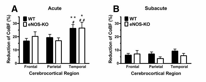
parietal regions ipsilateral to CAO decreased simultaneously with that of the temporal cortex 213 (Figures 4A, 4C and 4E), although their CoBF reduction was significantly less pronounced 214 (Figure 5A). At approximately 10 s after CAO the CoBF started to increase in all cortical 215 regions, and returned close to the baseline level within 30 s (Figures 3, 4A, 4C and 4E). 216 Interestingly, in the subacute phase (i.e. 1-5 min after CAO) the CoBF reduction was less 217 than 10% in all three cortical regions without any significant inter-regional difference (Figure 218 5B). One can conclude from these observations that the existing macrovascular connections 219 (i.e. the circle of Willis) are not sufficient to immediately and completely compensate for the 220 loss of one carotid artery, and active vasodilation is required to accommodate cerebrocortical 221 circulation to the altered hemodynamic situation. In addition, the observation that CAO 222 resulted in a significant CoBF reduction of the frontoparietal region as compared to the 223 contralateral hemisphere in spite of the common blood supply of these brain areas by the 224 AACA indicates redistribution of the CoBF via pial collaterals to the severely ischemic 225 temporal cortex on the side of CAO. 226 Effects of CAO on the regional CoBF in eNOS-KO mice 227 Our observations in WT mice indicated that the adaptation of cerebrocortical circulation to 228 unilateral CAO involves pial and/or microvascular vasodilation. Since endothelial NO is a 229 major regulator of the microvascular resistance in cerebral circulation (12, 13) we tested the 230 hypothesis that eNOS may play a significant role in the adaptation to CAO. Changes of the 231 regional CoBF after CAO in eNOS-KO animals, however, resembled in many ways the 232 findings in WT mice (Figure 4B, 4D, 4F). The temporal pattern showed an acute drop 233 followed by gradual recovery in all three cerebrocortical regions under investigation. In 234 addition, similarly to WT animals, the acute reduction was most pronounced in the temporal 235 cortex (Figure 5A), whereas during the subacute phase this inter-regional difference 236 disappeared (Figure 5B). Surprisingly, the percentual changes of the regional CoBF showed 237 no significant difference between eNOS-KO and WT mice either during the acute (Figure 238 5A) or the subacute (Figure 5B) phase after CAO. In fact, the recovery even appeared to be 239 more rapid in the temporal cortex of eNOS KO mice as compared to WT controls (Figures 240 4E and 4F). 241 242

9
Discussion 243 The present study was designed to investigate the cerebrovascular compensatory mechanisms 244 developing after unilateral occlusion of the CCA. We aimed to answer three basic questions: 245 Does the adaptation of cerebral circulation to the altered hemodynamic state after CAO 246 involve flow changes and active vasodilation in the large arteries of the Willis circle? Do 247 small pial collateral arteries between territories of the main cerebrocortical arteries (AACA 248 and MCA) contribute to the redistribution of the CoBF? Is eNOS involved in the 249 cerebrovascular adaptation to CAO? 250 In our experiments CoBF was reduced rapidly and simultaneously in the ipsilateral frontal, 251 parietal and temporal cerebrocortical regions after CAO (Figure 4.). However, after the acute 252 phase, CoBF started to increase in the affected regions and returned close to the baseline 253 level within 30 s, and 1-5 min after CAO the reduction of CoBF was less than 10% in all 254 three cortical regions without any significant inter-regional differences. Similar dynamics of 255 initial CoBF changes have been reported in the parietal cortex of anesthetized rats (35), 256 followed by an overshoot of the blood flow, which was absent in our present study. One can 257 conclude from these observations that the existing macrovascular connections (i.e. the 258 arteries of the Willis circle) are not sufficient to compensate immediately and completely for 259 the loss of one CCA, and that active cerebral vasodilation is required to adapt cerebrocortical 260 circulation to the altered hemodynamic situation. 261 In our present study unilateral closure of the CCA resulted in instant, significant CoBF 262 reduction in the temporal cortex of the ipsilateral hemisphere. This was expected, since this 263 region is supplied by the MCA originating from the circle of Willis close to the influx of the 264 internal carotid artery. However, it was unexpected that the ipsilateral frontal and parietal 265 cortices also showed reduced blood perfusion as compared to the contralateral ones, although 266 in mice all of these brain regions receive their blood supply from the same artery, namely the 267 AACA. This observation can only be explained by a draining effect through connections 268 between the territories of the MCA and AACA, via pial anastomoses. The presence of such 269 connections (16, 28, 29, 52), as well as their importance after MCA occlusion (50, 56) have 270 already been demonstrated. However, to the best of our knowledge, the present study is the 271 first indication for the involvement of small pial anastomoses in the acute adaptation of 272 cerebrocortical circulation to CCA occlusion. We assume that a steal phenomenon may 273 develop, and the blood flow of pial arteries supplying the fronto-parietal regions is drained 274

10
via pial anastomic vessels to the more ischemic temporal cortex of the hemisphere ipsilateral 275 to the CCA occlusion. Interestingly, 15 days after CAO, markedly enlarged pial anastomic 276 connections have been reported in mice indicating the significant contribution of these 277 collateral vessels also to the chronic adaptation of the cerebrocortical circulation to CAO (16) 278 and similar results have been obtained 6 days after MCA occlusion in mice (56). 279 It is an important question whether the simple existence of collaterals is sufficient for the 280 normalization of cerebrocortical circulation after CAO, or their active dilation is also required 281 for the compensation. To answer this question, the distribution of cerebrovascular resistance 282 along the arterial vessel tree has to be considered. Table 2 gives an overview of the 283 experimental data available. It can be concluded that large cerebral vessels, including the 284 circle of Willis, significantly contribute to the total cerebrovascular resistance, since in 285 normotensive animals the blood pressure in the first order branches of the MCA is 39-54% 286 lower than the systemic mean arterial pressure. The contribution of pial vessels, however, is 287 also significant, evidenced by the additional 10-32% pressure drop from the first order MCA 288 branches to the penetrating arteries/arterioles. These data indicate that vasodilation both in 289 the circle of Willis and pial arteries could improve the blood perfusion of the MCA territory 290 after CAO. The observations that during changes in systemic blood pressure small pial 291 vessels as well as large cerebral arteries simultaneously contribute to the CoBF 292 autoregulation by changing their diameter/resistance (17, 18, 44) suggest that adaptation of 293 the cerebrocortical circulation to CAO may also involve an active vasodilation both in the 294 small and the large cerebral arteries. The temporal pattern of the recovery of CoBF after 295 CAO in our present study also indicates that vasodilation has to develop in order to achieve 296 the optimal level of adaptive responses in the cerebrovascular system. 297 An additional mechanism, which can aid the normalization of the brain’s regional blood 298 perfusion after CAO, is the reduction of the resistance in intraparenchymal microvessels. 299 These changes can be governed by different regulatory pathways, including myogenic, 300 metabolic, neurogenic and endothelial mechanisms. Reduction of the myogenic tone as a 301 response to the smaller transmural pressure, (i.e. weaker wall tension due to the reduced 302 intraluminar pressure), enhanced release of vasodilatory neurotransmitters from the neurons 303 and nerve endings and accumulation of metabolic end-products as a result of insufficient 304 tissue blood perfusion are certainly among the regulatory factors. Several lines of evidence, 305 however, indicate that vasoactive substances - especially NO - released from the 306

11
microvascular endothelium in response to the reduction of cerebral blood supply are crucially 307 important contributors to the maintenance of cerebral blood flow. 308 NO has been shown to play a major, complex role in the regulation of cerebral circulation. A 309 multitude of in vitro as well as in vivo studies support that it contributes significantly to the 310 control of the resting cerebral vascular tone, it has a potent cerebral vasodilatory effect by 311 mediating endothelium-dependent vascular relaxation, it acts directly on vascular smooth 312 muscle, and plays a significant role in the mediation of CO2-induced as well as hypoxia-313 induced cerebral vasodilation. It is well documented that following its release by the 314 endothelium, NO diffuses into the vascular smooth muscle cells, where, by activating soluble 315 guanylyl cyclase (sGC), it increases the intracellular concentration of cyclic guanine 316 monophosphate (cGMP), which in turn eventually leads to smooth muscle relaxation and 317 vasodilation (40). There is also evidence to suggest that NO causes vasodilation not only 318 through cGMP-mediated mechanisms, but in certain species also by activating potassium 319 channels (for review see (13)). 320 In vitro studies, using large cerebral arteries provided most of the experimental evidence 321 regarding endothelium-mediated cerebral vasodilation. It was proved that NO exerts a resting 322 tonic vasodilatory effect on cerebral circulation, since the basal cGMP level was found to be 323 significantly greater in cerebral arteries having intact endothelium, as compared to those from 324 which the endothelium was removed (8, 24, 48). Cerebral vasodilation in response to 325 acetylcholine and to other receptor-mediated agonists (such as serotonin, substance P and 326 ADP), which activate eNOS by increasing intracellular calcium levels, was shown to be NO-327 dependent (for review see (12)). 328 In vivo studies (10) showed that topical application of the NOS inhibitor L-NMMA leads to 329 constriction of the rat basilar artery, an effect that was reversed upon administration of the 330 NO-precursor molecule L-arginine. In several species, under basal conditions, local and 331 systemic application of NOS inhibitors was shown to provoke cerebral vasoconstriction and a 332 decrease in CoBF (12). Due to the fact that systemic NOS inhibitors lead to an increase in the 333 cerebral and the peripheral vascular resistance, it is logical to infer that resting levels of NO 334 are necessary to maintain the resistance vessels in a relaxed state (22). In other studies, which 335 tested mice and piglet pial arterioles, L-arginine administration was shown to dilate pial 336 arterioles in a dose-dependent manner (7, 41). 337

12
NO was also shown to possess a basal inhibitory effect, which buffers spontaneous cerebral 338 vasomotion (9), inhibits vasoconstriction in response to substances such as norepinephrine 339 and serotonin (for review (12)), and thereby contributes to the maintenance of a stable CoBF. 340 Studies have demonstrated that administration of non-selective NOS inhibitors leads to an 341 enhancement of cerebral vascular oscillations (2, 9, 15, 21, 26), whereas NO caused an 342 attenuation of vasomotion (22). 343 Involvement of NO in the regulation of cerebral circulation during ischemia has also been 344 extensively studied. An acute increase in NO concentration within minutes following 345 ischemia has been observed (30), which, due to its rapidity, was attributed to an ischemia-346 induced activation of the constitutive eNOS enzyme (57). Similar observations have also 347 been made in a study, which utilized MCA occlusion to induce cerebral ischemia (23). The 348 potentially beneficial consequence of an increase in NO during ischemia is the maintenance 349 of CoBF through vasodilation and the inhibition of platelet and leukocyte aggregation (13). 350 These effects, therefore, serve to limit the infarct’s size and reduce brain damage. The 351 potential protective effect of endothelium-derived NO during and after cerebral ischemia has 352 been pointed out by studies, which utilized eNOS-deficient mice (13). Administration of L-353 arginine after MCA occlusion leads to the dilation of pial arterioles, to a reduction of infarct 354 size and to an increase of CoBF (33, 34). On the other hand, NOS inhibitors were reported to 355 induce either no effect or an increase in the ischemia-induced cerebral infarct size (for review 356 see (12)). 357 Based on the aforementioned literary data we hypothesized that endothelium-derived NO 358 may play an important role in the cerebrovascular adaptation to CAO. According to our 359 results, however, the CAO-induced acute ipsilateral CoBF reduction in the three investigated 360 cerebrocortical regions of the eNOS-KO animals was not different from that of the control, 361 wild-type mice, neither in the acute, nor in the subacute phase of the CAO. These results 362 indicate that eNOS does not appear to play an important role in the CoBF redistribution after 363 CAO. In fact, the faster recovery of CoBF in the temporal cortex of eNOS-KO compared to 364 WT mice can be attributed to the elevated MABP in these animals. It has been previously 365 described that systematically administered NOS inhibitors can increase arterial pressure in a 366 dose-dependent manner and therefore enhance blood flow in collateral vessels, which supply 367 cerebral regions that were rendered ischemic though arterial occlusion (32). 368

13
Limitations of our experimental approach have also to be considered during the interpretation 369 of our findings. The fact, that eNOS-KO mice showed unaltered cerebrocortical adaptation 370 following CAO-induced reduction of CoBF does not necessarily exclude the role of NO in 371 these mechanisms. Several lines of evidence indicate that endothelium-dependent and -372 independent vasodilator pathways may get activated in order to compensate for the absence 373 of endothelial NO production and therefore the phenotypic consequences of the eNOS gene 374 deletion may underestimate the importance of eNOS under physiological conditions. For 375 instance, neuronal NOS (nNOS) may be upregulated in the absence of eNOS. It has been 376 reported that eNOS- and nNOS-derived NO is simultaneously involved in a variety of 377 cerebrovascular functions, including the regulation of resting cerebral blood flow (3), CO2-378 mediated (38, 53) and neuronally induced vasodilation (6, 14) as well as flow-metabolism 379 coupling (11, 13). Interestingly, it has been shown that nNOS within the brain is not only 380 found in neurons, glial cells and perivascular nerves (12), but also in the endothelium of the 381 cerebral vasculature (3). 382 In conclusion, in the present study, taking advantage of the high temporal and spatial 383 resolution of laser-speckle imaging, attempts were made to gain insight into the mechanisms 384 of the adaptation of cerebrocortical microcirculation to unilateral occlusion of the common 385 carotid artery. The transient reduction of the CoBF in all investigated regions of the 386 ipsilateral hemisphere clearly indicate that, in spite of the well-developed macro- and 387 microvascular collateral network and the robust myogenic control of the vascular tone, the 388 feed-forward mechanisms of cerebrovascular regulation are not sufficient to prevent cerebral 389 ischemia after CAO. The temporal pattern of the CoBF recovery after CAO suggests the 390 significance of an active cerebrovascular vasodilator mechanism driven by metabolic, 391 endothelial or neuronal signals. Surprisingly, eNOS-dependent vasodilation does not appear 392 to be involved in this process. In contrast, intracortical redistribution of the CoBF, 393 presumably via pial anastomoses between the MCA and AACA, appears to attenuate the 394 ischemia of the most severely affected temporal cortex at the expense of reducing the blood 395 perfusion of the frontoparietal regions. 396 397 398
14
Acknowledgements 399 The authors wish to acknowledge the seminal findings and methodological achievements of 400 Prof. Minoru Tomita, MD, PhD (1934–2010) regarding the physiological and 401 pathophysiological functions of pial collateral vessels. The authors are grateful to Dr. 402 Erzsébet Fejes for critically reading the manuscript as well as to Dr. Péter Dancs and András 403 Kucsa for graphic artwork. This study has been supported by the Hungarian Scientific 404 Research Fund (OTKA K-62375, K-101775 and K-112964). 405 406

15
Figure Legends 407 408 Fig. 1. 409 Localization of cerebrocortical regions on a representative laser-speckle image (panel A) and 410 schematic illustration of their supplying vessels (panel B). Small arrows indicate pial 411 anastomoses between the territories supplied by the middle cerebral arteries (MCA) and the 412 azygos anterior cerebral artery (AACA). ICA, internal carotid artery; ACA, anterior cerebral 413 artery; PCA, posterior cerebral artery; FP, frontal pole; B, bregma; λ, lambda 414 415 Fig. 2. 416 Mean arterial blood pressure (MABP) in WT (n=12) and eNOS-KO (n=11) mice (panel A) 417 and its changes after left carotid artery occlusion (CAO) (panel B). CAO was performed at 418 time point „0 s”. MABP was significantly higher in eNOS-KO animals at all time points, 419 whereas ΔMABP differed from 210 s (*P<0.05, **P<0.01, ***P<0.001 between WT and 420 eNOS-KO with 2-way ANOVA and Bonferroni’s post hoc test). Note the enhanced time 421 resolution between (panel A) or before (panel B) the dashed lines. 422 423 Fig. 3. 424 Regional changes of the cerebrocortical blood flow (CoBF) at different time points after left 425 carotid artery occlusion (CAO), shown as differerence images compared to the baseline 426 CoBF, i.e. the averaged CoBF in 1 min preceding CAO. CAO was performed at time point 427 „0”. AU, arbitrary units; F, P and T indicate the frontal, parietal and temporal regions, 428 respectively according to the coordinates described on Fig. 1A. 429 430 Fig. 4. 431 Regional cerebrocortical blood flow (CoBF) in WT (n=12, panels A, C and E) and eNOS-KO 432 (n=11, panels B, D and F) before and after carotid artery occlusion (CAO). CoBF is 433 expressed as percentage of the baseline, i.e. the averaged values in 1 min preceding CAO. 434 Blue and red symbols represent CoBF in the ipsilateral and contralateral hemispheres, 435 respectively. (*P<0.05, **P<0.01, ***P<0.001 vs. „Contralateral” with 2-way ANOVA and 436 Bonferroni’s post hoc test). Note the enhanced time resolution between the dashed lines. 437 438 439

16
Fig. 5. 440 Acute (panel A) and subacute (panel B) reductions of the regional cerebrocortical blood flow 441 (CoBF) in the ipsilateral hemisphere of WT (filled bars, n=12) and eNOS-KO (open bars, 442 n=11) mice after left carotid artery occlusion (CAO). CoBF values have been determined at 443 their minimum („Acute”) or at 5 min after CAO („Subacute”), and reductions were expressed 444 as percentage of the baseline, i.e. the average CoBF in 1 min preceding CAO. (*P<0.05, 445 **P<0.01 vs. „Frontal”; #P<0.05, ##P<0.01 vs. „Parietal” with 2-way ANOVA and 446 Bonferroni’s post hoc test.) 447 448

17
Tables 449 Table 1. Arterial blood gas and acid-base parameters in the 450 experimental groups 451
Variable WT (n=12) eNOS-KO (n=11)
PaO2 (mmHg) 113.5 ± 5.1 112.6 ± 5.1
O2-Saturation (%) 97.5 ± 0.4 97.7 ± 0.3
PaCO2 (mmHg) 41.2 ± 2.1 37.6 ± 2.5
pH 7.30 ± 0.02 7.32 ± 0.02
SBE (mmol/l) -6.0 ± 1.0 -6.4 ± 0.7
[HCO3-] (mmol/l) 19.7 ± 0.9 18.7 ± 0.6
Hematocrit (%) 38.4 ± 1.0 39.4 ± 1.2
452 453 454 Table 2. Blood pressure levels in different segments of the MCA expressed as 455 percentage of the systemic MABP 456 Species Anesthesia
SystemicMABP
(mmHg)
Blood Pressure (% of MABP) in Reference 1st 2nd 3rd 4th*
Order Branch of the MCA
Rat Inactin 122 46% 43% 22% Harper et al. (17, 18)
Cat Pentobarbital 70-140 61% 57% 55% 51% Shapiro et al. (44)
Cat Pentobarbital 120 74%** 42%** Kontos et al. (25)
Cat Ketamine + Halothane +
N2O 87 47%
Schmidt-Kastner et al.
(42)
Cat Chloralose + Urothane +
Pancuronium 115-117 60-63% Yamaguchi et
al. (54)
The level of pressure drop indicates the segmental distribution of cerebrovascular resistance. 457 *Penetrating arterioles 458 **Recalculated from resistance values 459 460

18
REFERENCES 461 1. Bajko Z, Balasa R, Motataianu A, Maier S, Chebut OC, and Szatmari S. 462 Common carotid artery occlusion: a case series. ISRN Neurol 2013: 198595, 2013. 463 2. Behzadi Y, and Liu TT. An arteriolar compliance model of the cerebral blood 464 flow response to neural stimulus. Neuroimage 25: 1100-1111, 2005. 465 3. Benyo Z, Lacza Z, Hortobagyi T, Gorlach C, and Wahl M. Functional 466 importance of neuronal nitric oxide synthase in the endothelium of rat basilar arteries. 467 Brain Res 877: 79-84, 2000. 468 4. Benyo Z, Ruisanchez E, Leszl-Ishiguro M, Sandor P, and Pacher P. 469 Endocannabinoids in cerebrovascular regulation. Am J Physiol Heart Circ Physiol 310: 470 H785-801, 2016. 471 5. Brozici M, van der Zwan A, and Hillen B. Anatomy and functionality of 472 leptomeningeal anastomoses: a review. Stroke 34: 2750-2762, 2003. 473 6. Busija DW, Bari F, Domoki F, and Louis T. Mechanisms involved in the 474 cerebrovascular dilator effects of N-methyl-d-aspartate in cerebral cortex. Brain Res Rev 475 56: 89-100, 2007. 476 7. Busija DW, Leffler CW, and Wagerle LC. Mono-L-arginine-containing 477 compounds dilate piglet pial arterioles via an endothelium-derived relaxing factor-like 478 substance. Circ Res 67: 1374-1380, 1990. 479 8. Cosentino F, Sill JC, and Katusic ZS. Endothelial L-arginine pathway and 480 relaxations to vasopressin in canine basilar artery. Am J Physiol 264: H413-418, 1993. 481 9. Dirnagl U, Lindauer U, and Villringer A. Nitric oxide synthase blockade 482 enhances vasomotion in the cerebral microcirculation of anesthetized rats. Microvasc 483 Res 45: 318-323, 1993. 484 10. Faraci FM. Role of nitric oxide in regulation of basilar artery tone in vivo. Am J 485 Physiol 259: H1216-1221, 1990. 486 11. Faraci FM, and Brian JE, Jr. 7-Nitroindazole inhibits brain nitric oxide synthase 487 and cerebral vasodilatation in response to N-methyl-D-aspartate. Stroke 26: 2172-2175; 488 discussion 2176, 1995. 489 12. Faraci FM, and Brian JE, Jr. Nitric oxide and the cerebral circulation. Stroke 25: 490 692-703, 1994. 491 13. Faraci FM, and Heistad DD. Regulation of the cerebral circulation: role of 492 endothelium and potassium channels. Physiol Rev 78: 53-97, 1998. 493 14. Gotoh J, Kuang TY, Nakao Y, Cohen DM, Melzer P, Itoh Y, Pak H, Pettigrew K, 494 and Sokoloff L. Regional differences in mechanisms of cerebral circulatory response to 495 neuronal activation. Am J Physiol Heart Circ Physiol 280: H821-829, 2001. 496 15. Griffith OW, and Kilbourn RG. Nitric oxide synthase inhibitors: amino acids. 497 Methods Enzymol 268: 375-392, 1996. 498 16. Guo H, Itoh Y, Toriumi H, Yamada S, Tomita Y, Hoshino H, and Suzuki N. 499 Capillary remodeling and collateral growth without angiogenesis after unilateral 500 common carotid artery occlusion in mice. Microcirculation 18: 221-227, 2011. 501

19
17. Harper SL, and Bohlen HG. Microvascular adaptation in the cerebral cortex of 502 adult spontaneously hypertensive rats. Hypertension 6: 408-419, 1984. 503 18. Harper SL, Bohlen HG, and Rubin MJ. Arterial and microvascular contributions 504 to cerebral cortical autoregulation in rats. Am J Physiol 246: H17-24, 1984. 505 19. Hennerici M, Aulich A, Sandmann W, and Freund HJ. Incidence of 506 asymptomatic extracranial arterial disease. Stroke 12: 750-758, 1981. 507 20. Henry M, Polydorou A, Klonaris C, Henry I, Polydorou AD, and Hugel M. 508 Carotid angioplasty and stenting under protection. State of the art. Minerva Cardioangiol 509 55: 19-56, 2007. 510 21. Horvath B, Lenzser G, Benyo B, Nemeth T, Benko R, Iring A, Herman P, 511 Komjati K, Lacza Z, Sandor P, and Benyo Z. Hypersensitivity to thromboxane receptor 512 mediated cerebral vasomotion and CBF oscillations during acute NO-deficiency in rats. 513 PLoS One 5: e14477, 2010. 514 22. Iadecola C, Pelligrino DA, Moskowitz MA, and Lassen NA. Nitric oxide 515 synthase inhibition and cerebrovascular regulation. J Cereb Blood Flow Metab 14: 175-516 192, 1994. 517 23. Kader A, Frazzini VI, Solomon RA, and Trifiletti RR. Nitric oxide production 518 during focal cerebral ischemia in rats. Stroke 24: 1709-1716, 1993. 519 24. Kim P, Schini VB, Sundt TM, Jr., and Vanhoutte PM. Reduced production of 520 cGMP underlies the loss of endothelium-dependent relaxations in the canine basilar 521 artery after subarachnoid hemorrhage. Circ Res 70: 248-256, 1992. 522 25. Kontos HA, Wei EP, Navari RM, Levasseur JE, Rosenblum WI, and Patterson 523 JL, Jr. Responses of cerebral arteries and arterioles to acute hypotension and 524 hypertension. Am J Physiol 234: H371-383, 1978. 525 26. Lacza Z, Herman P, Gorlach C, Hortobagyi T, Sandor P, Wahl M, and Benyo Z. 526 NO synthase blockade induces chaotic cerebral vasomotion via activation of 527 thromboxane receptors. Stroke 32: 2609-2614, 2001. 528 27. Liebeskind DS. Collateral circulation. Stroke 34: 2279-2284, 2003. 529 28. Maeda K, Hata R, Bader M, Walther T, and Hossmann KA. Larger 530 anastomoses in angiotensinogen-knockout mice attenuate early metabolic disturbances 531 after middle cerebral artery occlusion. J Cereb Blood Flow Metab 19: 1092-1098, 1999. 532 29. Maeda K, Hata R, and Hossmann KA. Differences in the cerebrovascular 533 anatomy of C57black/6 and SV129 mice. Neuroreport 9: 1317-1319, 1998. 534 30. Malinski T, Bailey F, Zhang ZG, and Chopp M. Nitric oxide measured by a 535 porphyrinic microsensor in rat brain after transient middle cerebral artery occlusion. J 536 Cereb Blood Flow Metab 13: 355-358, 1993. 537 31. Millikan CH. Cerebral circulation: clinical concepts as effected by vascular 538 anatomy, pathology, and pathophysiology. Clin Neurosurg 16: 419-435, 1969. 539 32. Moncada S, Palmer RM, and Higgs EA. Nitric oxide: physiology, 540 pathophysiology, and pharmacology. Pharmacol Rev 43: 109-142, 1991. 541

20
33. Morikawa E, Huang Z, and Moskowitz MA. L-arginine decreases infarct size 542 caused by middle cerebral arterial occlusion in SHR. Am J Physiol 263: H1632-1635, 543 1992. 544 34. Morikawa E, Rosenblatt S, and Moskowitz MA. L-arginine dilates rat pial 545 arterioles by nitric oxide-dependent mechanisms and increases blood flow during focal 546 cerebral ischaemia. Br J Pharmacol 107: 905-907, 1992. 547 35. Morita Y, Fukuuchi Y, Koto A, Suzuki N, Isozumi K, Gotoh J, Shimizu T, Takao 548 M, and Aoyama M. Rapid changes in pial arterial diameter and cerebral blood flow 549 caused by ipsilateral carotid artery occlusion in rats. Keio J Med 46: 120-127, 1997. 550 36. Mozaffarian D, Benjamin EJ, Go AS, Arnett DK, Blaha MJ, Cushman M, Das SR, 551 de Ferranti S, Despres JP, Fullerton HJ, Howard VJ, Huffman MD, Isasi CR, Jimenez 552 MC, Judd SE, Kissela BM, Lichtman JH, Lisabeth LD, Liu S, Mackey RH, Magid DJ, 553 McGuire DK, Mohler ER, 3rd, Moy CS, Muntner P, Mussolino ME, Nasir K, Neumar 554 RW, Nichol G, Palaniappan L, Pandey DK, Reeves MJ, Rodriguez CJ, Rosamond W, 555 Sorlie PD, Stein J, Towfighi A, Turan TN, Virani SS, Woo D, Yeh RW, and Turner MB. 556 Heart Disease and Stroke Statistics-2016 Update: A Report From the American Heart 557 Association. Circulation 133: e38-e360, 2016. 558 37. Nishioka H. Results of the treatment of intracranial aneurysms by occlusion of 559 the carotid artery in the neck. J Neurosurg 25: 660-704, 1966. 560 38. Okamoto H, Hudetz AG, Roman RJ, Bosnjak ZJ, and Kampine JP. Neuronal 561 NOS-derived NO plays permissive role in cerebral blood flow response to hypercapnia. 562 Am J Physiol 272: H559-566, 1997. 563 39. Proweller A, Wright AC, Horng D, Cheng L, Lu MM, Lepore JJ, Pear WS, and 564 Parmacek MS. Notch signaling in vascular smooth muscle cells is required to pattern 565 the cerebral vasculature. Proc Natl Acad Sci U S A 104: 16275-16280, 2007. 566 40. Robertson BE, Schubert R, Hescheler J, and Nelson MT. cGMP-dependent 567 protein kinase activates Ca-activated K channels in cerebral artery smooth muscle cells. 568 Am J Physiol 265: C299-303, 1993. 569 41. Rosenblum WI, Nishimura H, and Nelson GH. Endothelium-dependent L-Arg- 570 and L-NMMA-sensitive mechanisms regulate tone of brain microvessels. Am J Physiol 571 259: H1396-1401, 1990. 572 42. Schmidt-Kastner R, Hossmann KA, and Ophoff BG. Pial artery pressure after 573 one hour of global ischemia. J Cereb Blood Flow Metab 7: 109-117, 1987. 574 43. Scremin OU, Holschneider DP. Vascular Supply. In: The Mouse Nervous System, 575 edited by Watson C, Paxinos G, Puelles L: Academic Press, 2012. 576 44. Shapiro HM, Stromberg DD, Lee DR, and Wiederhielm CA. Dynamic pressures 577 in the pial arterial microcirculation. Am J Physiol 221: 279-283, 1971. 578 45. Sokoloff L. The metabolism of the central nervous system in vivo. In: Handbook 579 of Physiology-Neurophysiology, edited by Field J, Magoun HW, Hall VE: American 580 Physiological Society, 1960. 581 46. Stauss HM, Godecke A, Mrowka R, Schrader J, and Persson PB. Enhanced 582 blood pressure variability in eNOS knockout mice. Hypertension 33: 1359-1363, 1999. 583

21
47. Stauss HM, Nafz B, Mrowka R, and Persson PB. Blood pressure control in 584 eNOS knock-out mice: comparison with other species under NO blockade. Acta Physiol 585 Scand 168: 155-160, 2000. 586 48. Sugawa M, Koide T, and Takato M. BY-1949 elicits vasodilation via preferential 587 elevation of cyclic GMP levels within the cerebral artery: possible involvement of 588 endothelium-mediated mechanisms. Eur J Pharmacol 215: 57-62, 1992. 589 49. Taylor DW. Beneficial effect of carotid endarterectomy in symptomatic patients 590 with high-grade carotid stenosis. N Engl J Med 325: 445-453, 1991. 591 50. Toriumi H, Tatarishvili J, Tomita M, Tomita Y, Unekawa M, and Suzuki N. 592 Dually supplied T-junctions in arteriolo-arteriolar anastomosis in mice: key to local 593 hemodynamic homeostasis in normal and ischemic states? Stroke 40: 3378-3383, 2009. 594 51. Toth P, Tarantini S, Davila A, Valcarcel-Ares MN, Tucsek Z, Varamini B, 595 Ballabh P, Sonntag WE, Baur JA, Csiszar A, and Ungvari Z. Purinergic glio-endothelial 596 coupling during neuronal activity: role of P2Y1 receptors and eNOS in functional 597 hyperemia in the mouse somatosensory cortex. Am J Physiol Heart Circ Physiol 309: 598 H1837-1845, 2015. 599 52. Vander Eecken HM, and Adams RD. The anatomy and functional significance of 600 the meningeal arterial anastomoses of the human brain. J Neuropathol Exp Neurol 12: 601 132-157, 1953. 602 53. Wang Q, Pelligrino DA, Baughman VL, Koenig HM, and Albrecht RF. The role 603 of neuronal nitric oxide synthase in regulation of cerebral blood flow in normocapnia 604 and hypercapnia in rats. J Cereb Blood Flow Metab 15: 774-778, 1995. 605 54. Yamaguchi S, Kobayashi S, Murata A, Yamashita K, and Tsunematsu T. Effect 606 of aging on collateral circulation via pial anastomoses in cats. Gerontology 34: 157-164, 607 1988. 608 55. Youmans JR, Kindt GW, and Mitchell OC. Extended studies of direction of flow 609 and pressure in the internal carotid artery following common carotid artery ligation. J 610 Neurosurg 27: 250-254, 1967. 611 56. Zhang H, Prabhakar P, Sealock R, and Faber JE. Wide genetic variation in the 612 native pial collateral circulation is a major determinant of variation in severity of stroke. 613 J Cereb Blood Flow Metab 30: 923-934, 2010. 614 57. Zhang ZG, Chopp M, Zaloga C, Pollock JS, and Forstermann U. Cerebral 615 endothelial nitric oxide synthase expression after focal cerebral ischemia in rats. Stroke 616 24: 2016-2021, 1993. 617 618


** **
* *
*** ****
***
**** *********
*
*********
*********
**
Frontal Parietal Temporal0
10
20
30
Acute
Cerebrocortical Region
Redu
ctio
nof
CoBF
(%)
WTeNOS-KO #* * # #*
Frontal Parietal Temporal0
10
20
30
Cerebrocortical Region
Redu
ctio
nof
CoBF
(%)
Subacute
WTeNOS-KO