Embed Size (px)
Citation preview

�
REVISTA ESPAÑOLA DE ESCLEROSIS MÚLTIPLE Nº 13 - Marzo de 2010
original
elapsing neuromyelitis optica (RNMO) is part of a heterogenous spectrum of idiopathic inflammatory demyelinating diseases of the central nervous sys-tem1. This syndrome has been clearly defined over recent years and is characterized clinically by si-multaneous or successive attacks of acute transverse myelitis (ATM) and optic neuritis (ON). Remission is variable, and additional acute attacks occur, almost all of which are restricted to the spinal cord and op-tic nerve. RNMO has a relapsing remitting course si-milar to that of multiple sclerosis (MS), but clinical, laboratory and imaging characteristics are generally able to distinguish between the two conditions2, 3. In a broad sense ATM in patients with RNMO is related to severe spinal cord damage and bilateral motor and sensitive dysfunction4-7.
According to the Transverse Myelitis Consor-tium Working Group8, idiopathic ATM provokes sen-sory, motor or autonomic dysfunction attributable to
an inflammatory, focal spinal cord disease detectable in cerebral spinal fluid (CSF) or by magnetic reso-nance imaging (MRI). Bilateral signs and a clearly defined upper sensory level are the clinical data re-quired for diagnosis, and progression from onset to nadir is generally between 4 hours and 21 days.
ATM is a common initial presentation of NMO2, 4-7. One or more attacks of ATM with spinal-cord MRI showing contiguous T2-weighted signal abnormality extending over three or more vertebral segments (LETM) and normal brain MRI is indi-cative of a high risk for NMO3. In contrast MS pa-tients with transverse myelitis at onset rarely has spinal cord lesions extend over more than two ver-tebral segments9-10.
Some limitations of the 2002 criteria for the diagnosis of ATM have been pointed out11, 12. Taking into consideration patients with asymmetric motor or sensitive dysfunction and those with no evidence of
Acute transverse myelitis in relapsing neuromyelitis optica. Partial myelitis could be a clinical presentation?
Regina M. PaPais-alvaRenga MD, PhD1, 2, MaRina PaPais alvaRenga MD, MsC1, 2, MaRCos PaPais alvaRenga MD, MsC1, 2, 3, 4, luiz ClauDio s. ThuleR2 MD, Ph1Neurology Department. Hospital da Lagoa. Rio de Janeiro. Brazil.2Post graduation Program in Neurology. Federal University of the State of Rio de Janeiro (UNIRIO). Brazil.3Neurology Department. Hospital Regional Universitario Carlos Haya. Málaga. Spain. 4Fundación Carolina / Fundación BBVA.
ABSTRACT. Acute partial transverse myelitis has traditionally been linked to MS while complete transverse myelitis was associated with NMO. We analyze here the spinal cord involvement in sixty patients with relapsing NMO from Rio de Janeiro (Brazil). As the index event, acute transverse myelitis (ATM) was complete in 38 cases and partial in 22 cases. Severe motor impairment occurred in 50% at nadir, 11.6% at recovery and 42.3% at last follow-up. Median time between index events (ATM and optic neuritis [ON]) was 17 months. In 26 cases, the first clinical event was ATM characterized by paraplegia (15), tetraplegia (5) and hemiplegia (1) or sensory dysfunction with marked thoracic level (5); 12 patients developed recurrent myelitis occurred prior to ON; MRI revealed large spinal cord MRI lesions in 87%; CSF showed mild or moderate pleocytosis in 32.5%; elevation of total protein in 35% and intrathecal synthesis of IgG in 37%. Con-clusions: complete and partial transverse myelitis could occur in recurrent NMO.Key words: neuromyelitis optica (NMO), transverse myelitis; natural history, prognosis, Brazil.
RESUMEN. La mielitis transversa aguda parcial se ha asociado tradicionalmente con la EM, mientras que la mielitis transversa completa se ha asociado con la NMO. Analizamos aquí la afectación de la médula espinal en sesenta pacientes con NMO recurrente de Río de Janeiro (Brasil). Como evento índice, la mielitis transversa aguda (ATM) fue completa en 38 casos y parcial en 22 casos. Un trastorno motor grave ocurrió en el 50% en el nadir, 11.6% a la recuperación y 42% durante la última evaluación. El tiempo medio entre el evento índice (ATM y neuritis óptica [ON]) fue de 17 meses. En 26 casos, el primer evento clínico fue la ATM caracterizada por paraplejía (15), tetraplejía (5) y hemiplejía (1) o disfunción sensitiva con un nivel torácico marcado (5); 12 pacientes desarrollaron una mielitis recurrente antes de la NO; la RM mostró lesiones largas de la médula espinal en el 87%; el LCR mostró pleocitosis leve o moderada en el 32,5%; elevación de las proteínas totales en el 35% y síntesis intratecal de IgG en el 37%. Conclusión: la mielitis transversa completa, tanto como parcial, pueden tener lugar en la NMO recurrente.Palabras clave: neuromielitis óptica (NMO), mielitis transversa, historia natural, pronóstico, Brasil.
Corre
spon
denc
ia: R
egina
Mar
ia Pa
pais
Alva
reng
a – N
euro
logy,
UNIR
IO –
Rua M
ariz
e Bar
ros 7
75 –
Tijuc
a – R
io de
Jane
iro
RJ 20
270-
004 B
razil
– Te
lepho
ne: 5
5 21 8
1510
251 –
Fax:
55 21
2264
2123
– E-
: reg
ina_a
lvare
nga@
hotm
ail.co
m
R

13
�
REVISTA ESPAÑOLA DE ESCLEROSIS MÚLTIPLE Nº 13 - Marzo de 2010
original
acute inflammation at CSF or spinal cord MR an al-ternative set of criteria was proposed differentiating acute complete (ACTM) from partial transverse mye-litis (APTM)13. APTM has traditionally been linked to MS13 while ACTM was associated with NMO2, 3.
This study analyzes the spectrum and natural his-tory of spinal cord impairment in a series of patients with RNMO in Rio de Janeiro (Brazil); most of them were of African descent. Full details of the study pro-tocol have been published in a previous study that analyzed the clinical course of the optic neuritis in this group of patients14.
Materials and Methods
A retrospective study was conducted at the Hospital da Lagoa, Rio de Janeiro, Brazil in order to iden-tify all cases of NMO followed up between 198� and 2004 were identified. Clinical data and comple-mentary examination results (MRI of the brain and spinal cord and CSF analysis) were extract for the medical records in order to apply the 1999 Mayo Clinic diagnostic criteria for NMO, which require the presence of three absolute criteria (optic neuri-
❑
tis, ATM and no evidence of clinical disease outside of the optic nerve and spinal cord) and 1 major su-pportive laboratorial criterion (normal brain MRI at onset, extensive vertebral MRI lesion, CSF with < �0 cels.mm3 at acute phase) or 2 minor supportive clinical criteria (bilateral visual deficit, severe mo-tor dysfunction, severe visual dysfunction at least at one eye). The analysis of cerebrospinal fluid (CSF), also included investigation for infectious diseases with specific antibodies for viruses (HIV, cytome-galovirus, herpes simplex and HTLV-I), IgG index in blood and CSF, and oligoclonal IgG band. The brain scan at the onset of the illness was analyzed according to the Paty I-II criteria and classified as suggestive of MS or not. Spinal cord (SC) lesions were classified in small or large [Longitudinally Ex-tensive Transverse Myelitis (LETM)], according to their extent (over three or more vertebral segments). Patients with moderate or severe symmetrical weak-ness and autonomic dysfunction associated with symmetric sensory loss were classified as having ACTM, while those with mild sensorial or motor dysfunction attributable to spinal cord disease, or clearly asymmetric dysfunction, even severe, were
Demographic features and general characteristics of NMOR patients according ethnicity
Features Total White Afro Brazilians p Value*
n 60 25 35Sex (F:M) (54:6) (23:2) (31:4) 0,67Year of onset median (min-max) 1994.5
(1967-2004)1994 (1978-2003)
1996 (1967-2004)
0,35
Age at onset (median; min-max) 30(10-67)
30(10-55)
30(14-67)
0,66
Antecedent event. co-morbidity (n) Viral illness / immunization 4 (6.7) 1 (4.0) 3 (8.6) 0,44 Autoimmune disease 13 (21.7) 3 (12.0) 10 (28.6) 0,12First event (n; %) Myelitis 26 (43.33) 7 (28) 19 (54.28) 0,049† Myelitis + Optic neuritis 2 (3.33) 2 (8) 0 Optic Neuritis 32 (53.33) 16 (64) 16 (57.14%) Total 60 25 35
Time to conversion (n; %) 17 (0.03-240) 34 (0.03-168) 9 (0.16-240) 0,177New events after conversion Myelitis 223 (58.7) 95 (57.0) 129 (60.6) 0,12 Myelitis + Optic neuritis 118 (31.0) 48 (28.1) 68 (31.9) Optic neuritis 39 (10.3) 23 (14.9) 16 (7.5) Total 380 166 213
Death (n) 14 2 12 0,02†
*Chi-square tests were used for statistical analysis except for year and age at onset (ANOVA), and immunization (two-tailed Fisher’s exact test).† p Value given is for the entire distribution of index events in each group (exact chi-square test for pooled data).
Table I

7
REVISTA ESPAÑOLA DE ESCLEROSIS MÚLTIPLE Nº 13 - Marzo de 2010
classified as having APTM. FS/EDSS1� was used to analyze disability.
The statistical significance of the differences ob-served between the dichotomous variables was analy-zed using Pearson’s or Fisher’s chi-square tests, as appropriate, whereas differences between continuo-us variables were analyzed using the Mann Whitney Wilcoxon test. P-values < 0.0� were considered to be statistically significant. We certify that all applicable institutional and governmental regulations concer-ning the ethical use of patient data were followed du-ring the course of this research.
Results
The charts of �0 NMO patients with a recurrent and restricted disease affecting the spinal cord and the optic nerve were analyzed. Demographic and clini-cal data are summarized in Table I. The majority of
❑
patients were female (90%) and African Brazilians (�8.3%). ATM was the most common clinical presen-tation of RNMO and constituted the most frequent event in both ethnic groups during the chronic phase of the disease.
ATM as index eventThe spinal cord index event was clinically clas-
sified as ACTM in 38 patients, most of whom were female (9�.2%) and Afro Brazilians (��.7%) or APTM, that was identified in 22 patients, the majori-ty of whom were female (81.8%) and white (�3.�%). APTM was mainly characterized by Lhermitte’s sign, paresthesia of the limbs with a clearly defined upper sensory level or thoracic pain associated with mild (�0%) or moderate (�0%) sensory loss. All patients had completely recovered within a mean time of 1 month, most spontaneously. Clinical, MRI and CSF findings of these patients are presented at Table II.
Acute partial transverse myelitis (APTM) as Index Event of RNMO
Case GenderEthnia
Number of the Index Event
Clinical description First spinal cordMRI lesion
CSFOCB
1 W/W 1 Hypoesthesia at four limbs, Lhermitte sign No lesion Yes
2 M/W 1 Anesthesia in perineum and constipation No lesion Yes
3 W/W 1 Hypoesthesia at lower limbs, perineum, T10 level One small Yes
4 W/Afro 1 Paresthesia at right thorax No lesion No
5 M/Afro 1 Dorsalgia hypoesthesia at lower limbs, proprioceptive ataxia Large Yes
6 W/W 1 Hypoesthesia at four limbs with T2 level Normal Yes
7 W/W 1 Hypoesthesia at lower limbs, T 10 level Normal Yes
8 W/W 2 Hypoesthesia at left side, mild weakness at right lower limb Large No
9 W/W 2 Hypoesthesia at four limbs, proprioceptive ataxia Large Yes
10 W/W 2 Hypoesthesia at left low limb Large No
11 M/W 2 Hypoesthesia at four limbs ND No
12 W/Afro 2 Paresthesia at right thorax No lesion No
13 W/Afro 2 Hypoesthesia at four limbs, proprioceptive deficit at hands Large No
14 M/Afro 2 Hypoesthesia and weakness at right upper limb Large Yes
15 W/Afro 2 Hypoesthesia at lower limbs, T10 level Large Yes
16 W/W 2 Hypoesthesia at four limbs, T4 level Multiple small No
17 W/Afro 3 Hypoesthesia at lower limbs T4 level Large ND
18 W/Afro 3 Hypoesthesia at four limbs, T2 level Large No
19 W/W 3 Hypoesthesia and weakness at right upper and lower limbs and constipation
Large No
20 W/W 4 Hypoesthesia at four limbs, T2 level Large No
21 W/W 4 Paresthesia, hypoesthesia at upper and lower limbs with T4 level
Large No
22 W/Afro 5 Hemi hypoesthesia Normal Yes
Gender: W: woman, M: man. Ethnia: AB: African Brazilian, WB: White Brazilian.
Table II
Regina M. PaPais-alvaRenga, MaRina PaPais alvaRenga, MaRCos PaPais alvaRenga, luiz ClauDio s. ThuleR

13
8
REVISTA ESPAÑOLA DE ESCLEROSIS MÚLTIPLE Nº 13 - Marzo de 2010
original
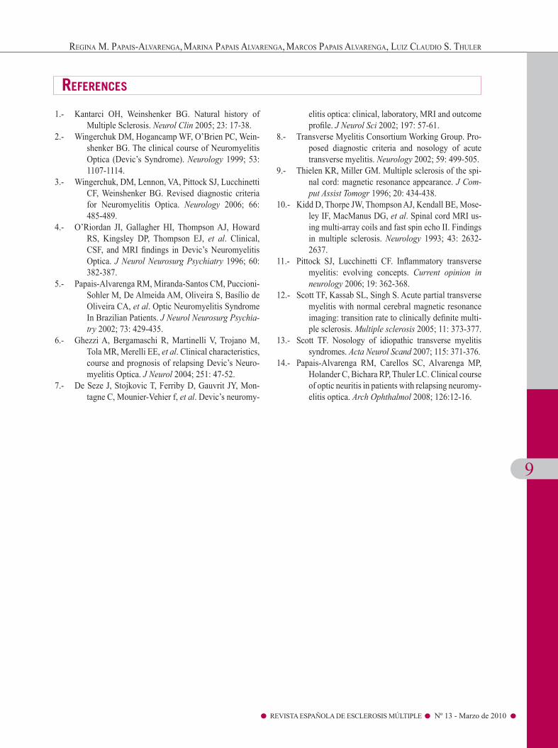
Figure 1 illustrates a large cervical vertebral MRI lesion in a white patient with APTM (case 9).
Last medical assessment At last follow-up, severe weakness (Medical Re-
search Council [MRC] grade 2 or less) was found in 26/60 (42.3%) of the patients. The remaining patients had mild (18.3%) or moderate (1.�%) weakness and 3�.�% had normal motor function. The majority of the patients with motor dysfunction also had some sensory or autonomic dysfunction; however, in 20% of cases se-vere hypoesthesia in the lower limbs or severe sphincter dysfunction requiring catheterization was present.
CSF and MRI findingsThe CSF tests showed mild pleocytosis (� to
20 cells/mm3) in 32.5%, elevation of total protein in 3�% and IgG intrathecal synthesis in 37%. Only 5/29 (17.2%) tests held at acute phase presented more than 50 cels/mm3. All patients had normal bra-in MRI normal or not suggestive of MS. Most of the patients (87%) had longitudinally extensive spinal cord MRI lesion extending over three vertebral seg-ments (LETM). Other abnormalities were multiple small lesions in 4 patients (7.4%) and one small le-sion in 1 patient (1.8%). The SC scan was normal in 2 patients (3.7%) after intravenous methylpredniso-lone treatment.
Discussion
Idiopathic myelitis in ACTM and APTM and sug-gested that the outcome and immunopathogenesis of the two different forms of the disease are different. A complete and severe acute myelitis is considered the characteristic feature of NMO4, 7 and acute par-tial transverse myelopathy has been linked to MS, especially when associated with inflammatory brain MRI lesions suggestive of this disease13. In the pre-sent study all of the patients who presented partial myelitis during the SC index had normal brain MRI at onset or not suggestive of MS, going on to suffer further relapses characterized by complete or partial myelitis. At last follow-up, the majority had mild motor dysfunction and was able to walk unaided in contrast with patients with ACTM that developed severe motor dysfunction. A longitudinally extensi-ve spinal cord lesion at acute phase may be helpful in making a differential diagnosis between RNMO and MS2, 3. In the present study only 12% of the pa-tients were found to have small spinal cord lesions on MRI.
Conclusion
Complete and partial transverse myelitis could occur in recurrent NMO.
❑
❑
A longitudinally extensive cervical vertebral MRI lesion documented during an acute partial transverse myelitis (APTM) in a white Brazilian woman. A: LETM in T2 with edema. B: LETM with contrast enhancement.
Figure 1
A B
9
REVISTA ESPAÑOLA DE ESCLEROSIS MÚLTIPLE Nº 13 - Marzo de 2010
referenceS
1.- Kantarci OH, Weinshenker BG. Natural history of Multiple Sclerosis. Neurol Clin 200�; 23: 17-38.
2.- Wingerchuk DM, Hogancamp WF, O’Brien PC, Wein-shenker BG. The clinical course of Neuromyelitis Optica (Devic’s Syndrome). Neurology 1999; �3: 1107-1114.
3.- Wingerchuk, DM, Lennon, VA, Pittock SJ, Lucchinetti CF, Weinshenker BG. Revised diagnostic criteria for Neuromyelitis Optica. Neurology 200�; ��: 48�-489.
4.- O’Riordan JI, Gallagher HI, Thompson AJ, Howard RS, Kingsley DP, Thompson EJ, et al. Clinical, CSF, and MRI findings in Devic’s Neuromyelitis Optica. J Neurol Neurosurg Psychiatry 199�; �0: 382-387.
�.- Papais-Alvarenga RM, Miranda-Santos CM, Puccioni-Sohler M, De Almeida AM, Oliveira S, Basílio de Oliveira CA, et al. Optic Neuromyelitis Syndrome In Brazilian Patients. J Neurol Neurosurg Psychia-try 2002; 73: 429-43�.
�.- Ghezzi A, Bergamaschi R, Martinelli V, Trojano M, Tola MR, Merelli EE, et al. Clinical characteristics, course and prognosis of relapsing Devic’s Neuro-myelitis Optica. J Neurol 2004; 2�1: 47-�2.
7.- De Seze J, Stojkovic T, Ferriby D, Gauvrit JY, Mon-tagne C, Mounier-Vehier f, et al. Devic’s neuromy-
elitis optica: clinical, laboratory, MRI and outcome profile. J Neurol Sci 2002; 197: �7-�1.
8.- Transverse Myelitis Consortium Working Group. Pro-posed diagnostic criteria and nosology of acute transverse myelitis. Neurology 2002; �9: 499-�0�.
9.- Thielen KR, Miller GM. Multiple sclerosis of the spi-nal cord: magnetic resonance appearance. J Com-put Assist Tomogr 199�; 20: 434-438.
10.- Kidd D, Thorpe JW, Thompson AJ, Kendall BE, Mose-ley IF, MacManus DG, et al. Spinal cord MRI us-ing multi-array coils and fast spin echo II. Findings in multiple sclerosis. Neurology 1993; 43: 2�32-2�37.
11.- Pittock SJ, Lucchinetti CF. Inflammatory transverse myelitis: evolving concepts. Current opinion in neurology 200�; 19: 3�2-3�8.
12.- Scott TF, Kassab SL, Singh S. Acute partial transverse myelitis with normal cerebral magnetic resonance imaging: transition rate to clinically definite multi-ple sclerosis. Multiple sclerosis 200�; 11: 373-377.
13.- Scott TF. Nosology of idiopathic transverse myelitis syndromes. Acta Neurol Scand 2007; 11�: 371-37�.
14.- Papais-Alvarenga RM, Carellos SC, Alvarenga MP, Holander C, Bichara RP, Thuler LC. Clinical course of optic neuritis in patients with relapsing neuromy-elitis optica. Arch Ophthalmol 2008; 12�:12-1�.
Regina M. PaPais-alvaRenga, MaRina PaPais alvaRenga, MaRCos PaPais alvaRenga, luiz ClauDio s. ThuleR