Embed Size (px)
Citation preview

Acute Pancreatitis
Prof .Yaser Rayyan MD,FACP,FASGE
JUSOM
Outline
Introduction
Epidemiology
Pathophysiology
Etiology
Clinical Presentation
Workup
Severity Scoring System
Treatment
Prognosis
Complications

PANCREATITIS
Inflammation in pancreas associated with injury to
exocrine parenchyma
PANCREAS
Stroma
Blood vessels
Ductal system
Parenchyma
Exocrine
Primary injury causing PANCREATITIS
Endocrine
Involved secondarily or as a complication


ACUTE PANCREATITIS
CLINICAL DEFINITION
◦ Abdominal Pain-Typical
◦ Elevated Pancreatic Enzymes
◦ Imaging studies
PATHOLOGICAL DEFINITION
◦ Reversible* pancreatic parenchymal injury associated with inflammation
_____________________________________________________
*if underlying cause of pancreatitis is removed, heal without any impairment of function or morphologic loss of gland
*Recurrent attacks with irreversible parenchymal injury leading to impairment of function and morphologic loss is chronic pancreatitis

CLINICAL CLASSIFICATION
*Considered a phase of chronic pancreatitis
** resulting from fibrosis within pancreas


CLASSIFICATION OF ACUTE
PANCREATITIS
Mild acute pancreatitis (80% cases) (Acute Interstitial/edematous pancreatitis)
◦ Absence of organ failure
◦ Absence of local complications
Severe acute pancreatitis(20 % cases) (Acute Hemorrhagic Necrotizing (fulminant) pancreatitis)
◦ Local complications +/-
◦ Organ failure defined as
SBP < 90 mm Hg
PaO2 ≤ 60 mm Hg
GI bleed ≥ 500 ml/24 hrs
Cret ≥ 2 mg/dL after rehydration
◦ Ranson score ≥ 3
◦ or APACHE ≥ 8


Epidemiology
◦Ranges between 1 and 80 per 100,000 of population
◦The incidence in USA 73.3 per 100,000 per year
◦In Jordan 1.6 per 100,000 of population per year
◦In Saudia Arabia 7.5 per 100,000 of population per year
◦In Egypt 19.1 per 100,000 of population per year
◦In Turkey 22.4 per 100,000n of population per year
http://www.rightdiagnosis.com/a/acute_pancreatitis/stats-country.htm

Epidemiology
Median ages of onset for various etiologies
Etiology Median Ages of onset
Alcohol-related 39 years
Biliary tract–related 69 years
Trauma-related 66 years
Drug-induced etiology 42 years
ERCP-related 58 years
AIDS-related 31 years
Vasculitis-related 36 years

Epidemiology
Gender Predilection
Generally M>F
In males more often related to alcohol
In females more often related to biliary tract disease
Race
3 times higher for blacks than whites

PATHOPHYSIOLOGY
Autodigestion of pancreatic substance by
inappropriately activated pancreatic enzymes
(especially trypsinogen)


Premature enzymes
Trypsin inhibitor
Zymogen granules

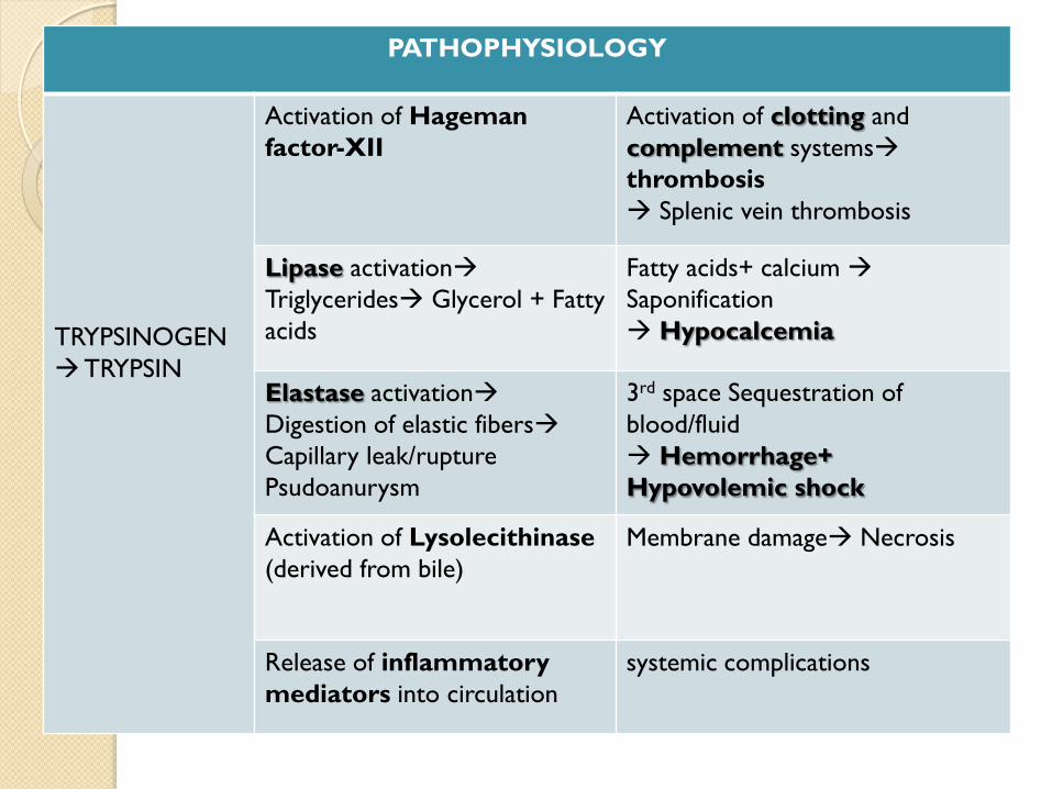
PATHOPHYSIOLOGY
TRYPSINOGEN
TRYPSIN
Activation of Hageman
factor-XII
Activation of clotting and
complement systems
thrombosis
Splenic vein thrombosis
Lipase activation
Triglycerides Glycerol + Fatty
acids
Fatty acids+ calcium
Saponification
Hypocalcemia
Elastase activation
Digestion of elastic fibers
Capillary leak/rupture
Psudoanurysm
3rd space Sequestration of
blood/fluid
Hemorrhage+
Hypovolemic shock
Activation of Lysolecithinase
(derived from bile)
Membrane damage Necrosis
Release of inflammatory
mediators into circulation
systemic complications

Etiology of Pancreatitis
Mechanical
Gall Stone
Ampullary tumor
Pancreatic Ca
Iatrogenic (ERCP)
Trauma
Metabolic
Alcoholism
Hypercalcemia
Hyperlipidimia
Malnutrition
Azotemia
Porphyries
Drugs Tetracycline Azathioprine
Steriods Furosemide Valproic acid
Infective
Mumps
Cocsaki – B
Ascares
Scorpion bite
Snake bite
Genetic
Pancreatic devisim
Annular pancreas
Cystic fibrosis
Autoimmune
Vascular
Shock
Hypothermia
Atheroembolism
Vasculitis (Polyarteritis nodosa, SLE)
Idiopathic
70 % due to
microlithiasis


Ethanol can induce pancreatitis by several methods:
1- Ethanol is a metabolic toxin to pancreatic acinar cells, where it can
interfere with enzyme synthesis and secretion also by Release of free radicals-
superoxide, hydroxyl produced by ethanol metabolism .
2- The "secretion with blockage" mechanism is possible because ethanol causes
spasm of the sphincter of Oddi,
3- Elevation of enzyme proteins that can precipitate within the pancreatic duct.
Calcium then can precipitate within this protein matrix, causing multiple
ductal obstructions by protein bulges .
4- Ethanol also increases ductal permeability, making it possible for
improperly activated enzymes to leak out of the activated enzymes into the
surrounding tissue.

Hyperlipidemia induced AP
• In the absence of gallstones and/or history of significant
history of alcohol use, a serum triglyceride should be
obtained and considered the etiology if > 1,000 mg /dl
• May Lipase without increase of serum amylase
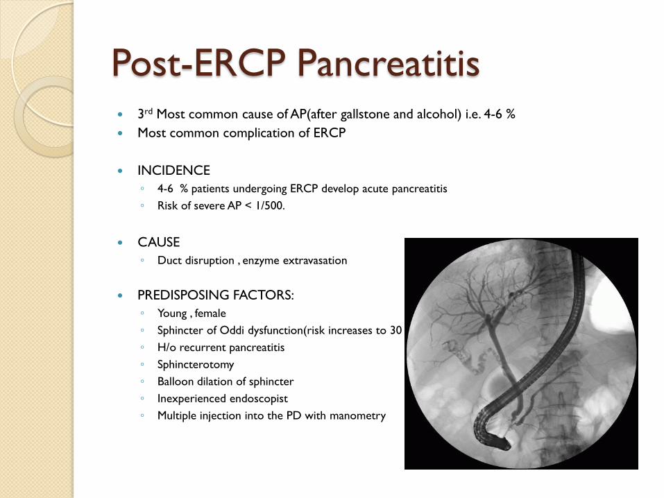
Post-ERCP Pancreatitis 3rd Most common cause of AP(after gallstone and alcohol) i.e. 4-6 %
Most common complication of ERCP
INCIDENCE
◦ 4-6 % patients undergoing ERCP develop acute pancreatitis
◦ Risk of severe AP < 1/500.
CAUSE
◦ Duct disruption , enzyme extravasation
PREDISPOSING FACTORS:
◦ Young , female
◦ Sphincter of Oddi dysfunction(risk increases to 30 %)
◦ H/o recurrent pancreatitis
◦ Sphincterotomy
◦ Balloon dilation of sphincter
◦ Inexperienced endoscopist
◦ Multiple injection into the PD with manometry


ABDOMINAL PAIN-Cardinal Symptom
SITE: usually experienced first in the epigastrium but may be localized to either upper quadrant or felt diffusely throughout the abdomen or lower chest
ONSET: characteristically develops quickly, generally following substantial meal.
SEVERITY: frequently severe, reaching max. intensity within minutes rather than hours
NATURE: “boring through”, “knife like”
DURATION: hours-days
COURSE: constant (refractory to usual doses of analgesics, not relieved by vomiting)
RADIATION: directly to back(50%), chest or flanks
RELEIVING FACTOR: sitting or leaning/stooping forward.
◦ due to shifting forward of abdominal contents and taking pressure off from inflamed pancreas
AGGRAVATING FACTOR: food/alcohol intake, walking, lying supine

OTHER MANIFESTATIONS Nausea, frequent and effortless vomiting, anorexia, diarrhea
◦ Due to reflex pylorospasm
◦ More intense in necrotizing than in edematous pancreatitis
Persistent retching
◦ despite empty stomach
Hiccups
◦ Due to gastric distension/diaphragmatic irritation
Fever
◦ Low grade, seen in infective pancreatitis
Weakness, Anxiety, Sweating
◦ Indicates severe attack.

General Physical Examination Appearance: well gravely ill with profound shock, toxicity and
confusion
Vitals:
◦ Tachypnea(and dyspnea-10%),
◦ Tachycardia(65%).
◦ Hypotension
◦ Temp high(76%)/normal/low (acute swinging pyrexia in cholangitis)
Icterus(28%)
◦ gallstone pancreatitis or due to edema of pancreatic head
Pallor, cold clammy skin, diaphoresis, dehydration

ABDOMEN EXAMINATION Tenderness + Rebound tenderness:
◦ epigastrium/upper abdomen
Distension:
◦ Ileus(BS decreased or absent)
◦ ascites with shifting dullness
Mass in epigastrium(usually absent)
◦ due to inflammation
Guarding(also called “defense musculaire” )-upper abdomen
◦ tensing of the abdominal wall muscles to guard inflamed organs within the abdomen from the pain of pressure upon them(i.e. during palpation)
Rigidity(involuntary stiffness)-unusual
◦ Tensing of the abdominal wall muscles to guard inflamed organs even if patient not touched

SYSTEMIC COMPLICATIONS
CARDIOVASCULAR
◦ Shock- hypovolemic and septic
◦ Arrhythmias/pericardial effusion/sudden death
◦ ST-T nonspecific changes
Pulmonary
◦ Respiratory failure/pneumonia/atelectasis/pleural effusion
◦ Acute Respiratory Distress Syndrome (ARDS)
Renal Failure
◦ Oliguria
◦ Azotemia
◦ Renal artery/vein thrombosis
Hematological
◦ Hemoconcentation
◦ Disseminated Intravascular Coagulopathy (DIC)

SYSTEMIC COMPLICATIONS Metabolic
◦ Hypocalcemia
◦ Hyperglycemia
◦ Hyperlipidemia
Gastrointestinal
◦ Peptic Ulcer/Erosive gastritis
◦ Ileus
◦ Portal vein or splenic vein thrombosis with varices
Neurological
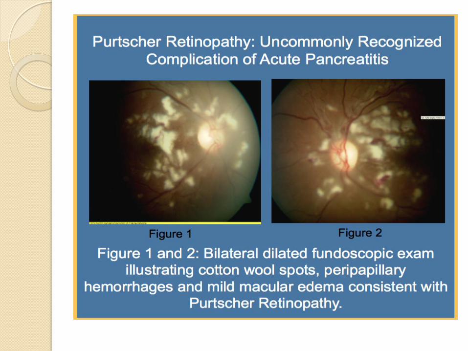
◦ Visual disturbances-Sudden blindness(Purtscher’s retinopathy)
◦ Confusion,irritability,psychosis
◦ Fat emboli
◦ Alcohol withdrawal syndrome
◦ Encephalopathy
Miscellaneous
◦ Subcutaneous fat necrosis
◦ Intra-abdominal saponification
◦ Arthralgia
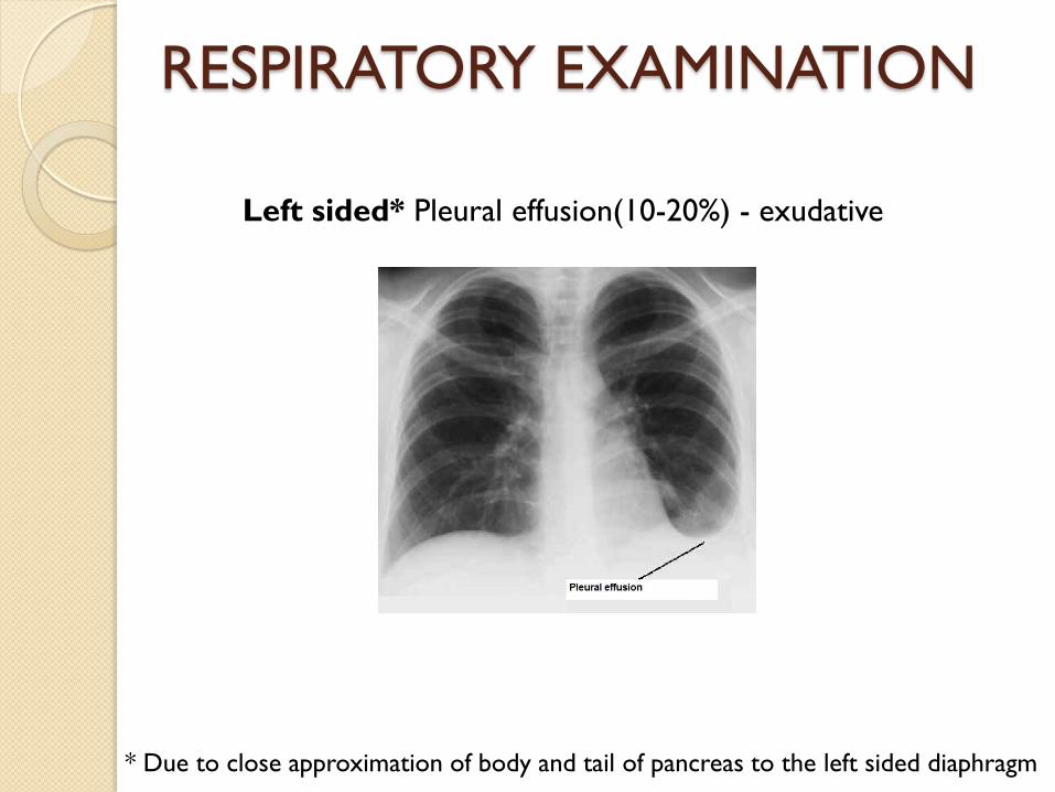
RESPIRATORY EXAMINATION
Left sided* Pleural effusion(10-20%) - exudative
* Due to close approximation of body and tail of pancreas to the left sided diaphragm

MANIFESTAIONS OF COMPLICATIONS
Hypocalcaemia ◦ circumoral numbness or paresthesia (1st symtpom to develop).
◦ carpopedal spasm .
◦ Laryngospasm.
◦ generalized seizures
◦ Chvostek sign : Depending on calcium level, graded response occur: twitching first at
angle of mouth, then by nose, the eye and the facial muscles
Positive in 10 % population in absence of hypocalcaemia
◦ Trousseau sign : BP cuff around arm and inflating to 20 mmHg above SBP for 3-5
minutes
Carpal spasm observed
More specific and sensitive than chvostek sign(postive even before tetany/hyperreflxia)

GREY TURNER1 SIGN CULLEN2 SIGN FOX3 SIGN
1. Named after British surgeon George Grey Turner(1877-1951)
2. Named for Thomas Stephen Cullen (1869-1953), Canadian gynecologist who first
described the sign in ruptured ectopic pregnancy in 1916
3.Named after George Henry Fox(1846-1937), American dermatologist

DIFFERENTIAL DIAGNOSIS
ABDOMINAL CONDITONS THORAX CONDITIONS
Perforated peptic ulcer/gastroentritis
Biliary colic/acute cholecystitis/ Cholangitis
Mesentric Ischemia
Ruptured Aortic Anuerysm
Intestinal Obstruction
Gastric/colon/pancreatic CA
Viral Hepatitis
IBS
Pneumonia/ARDS
Pleuritic pain
MI
GYNECOLOGICAL CONDITONS
• Ectopic pregnancy
• Salpingtis
SYSTEMIC CONDITIONS
DKA

Diagnostic criteria
Most often established by the presence of two of the three following criteria: ◦ (i) abdominal pain consistent with the disease,
◦ (ii) serum amylase and/or lipase greater than three times the upper limit of normal, and/or
◦ (iii) characteristic findings from abdominal imaging.
CT and/or MRI of the pancreas should be reserved for patients ◦ in whom the diagnosis is unclear(typical pain with normal
enzymes)
◦ who fail to improve clinically within the first 48–72 h after hospital admission (e.g., persistent pain, fever, nausea, unable to begin oral feeding)
◦ to evaluate complications

WORKUP
HEMATOLOGICAL investigations
RADIOLOGICAL investigations

HEMATOLOGICAL BASELINES
◦ CBC:
Low Hb: prolonged hemetemesis/melena, internal hemorrhage
Leucocytosis (10,000-30,000/mcL)-infection, non infectious inflammation
Low platelets-DIC
Hct –raised in hemoconcentration
◦ LFT’s:
raised bilirubin, AST/ALT/LDH, ALP, GGTP- gall stone pancreatitis
◦ RFT’s:
raised BUN/cretainine- ATN ARF
◦ Coagulation profile:
increased INR-DIC
◦ Blood sugar:
> 180 mg/dl-diabetes as a sequelae or cause
◦ Serum electrolytes:
Low sodium/potassium: persistent vomiting
Hypocalcemia- saponification/fat necrosis
◦ Serum Protein:
low protein/ albumin

HEMATOLOGICAL ABG’s
Etiology specific investigations ◦ Serum fasting lipid profile
◦ Serum Calcium (Hypercalcemia AP Hypocalcemia)
◦ Autoimmune markers: serum autoantibodies such as anti-nuclear antibody (ANA), IgG4
level, anti-lactoferrin antibody, anti-carbonic anhydrase II antibody, and rheumatoid factor (RF),
Acid-Base Disturbance Etiology
Metabolic (Lactic)acidosis
with high anion gap
Hypovolemic shock
Hypokalemic Hypochloremic
metabolic alkalosis
persistent vomiting
Respiratory acidosis ARDS
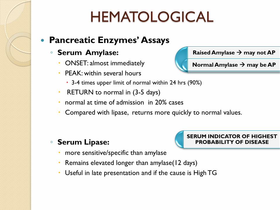
HEMATOLOGICAL
Pancreatic Enzymes’ Assays
◦ Serum Amylase:
ONSET: almost immediately
PEAK: within several hours
3-4 times upper limit of normal within 24 hrs (90%)
RETURN to normal in (3-5 days)
normal at time of admission in 20% cases
Compared with lipase, returns more quickly to normal values.
◦ Serum Lipase:
more sensitive/specific than amylase
Remains elevated longer than amylase(12 days)
Useful in late presentation and if the cause is High TG
Raised Amylase may not AP
Normal Amylase may be AP
SERUM INDICATOR OF HIGHEST PROBABILITY OF DISEASE

Pancreatic Enzymes’ Assays
◦ Urine Amylase
More sensitive than serum levels
Remain elevated for several days after serum levels returned to normal
◦ Pancreatic-specific amylase (p-amylase)
Measuring p-amylase instead to total amylase(also includes salivary amylase) makes diagnosis more specific(88-93%)

CONDITIONS ASSOCIATED WITH RAISED
SERUM AMYLASE
ABDOMEN
Small bowel obstruction
◦ strangulation ileus
◦ mesenteric ischemia
Acute appendicitis
Cholecystitis
Perforated Duodenal Ulcer
Gastroenteritis
Biliary peritonitis
Spasm of sphincter of Oddi
GYNE
Ruptured Ectopic pregnancy
Torsion of an ovarian cyst
OTHERS
Parotitis (Mumps)
Macroamylasaemia
Opioids administration
Low GFR
Brain injury(CVA)- hyperstimulation of pancreas

Plain X-ray abdomen erect AP view
Sentinel* loop sign
◦ Localized isolated Distended gut loop (Ileus) seen near the site of injured viscus or
inflamed organ
◦ RATIONALE: body's effort to localize the traumatic or inflamed lesions
◦ ETIOLOGY: Localized paralysis followed by accumulation of gas
◦ SITE:
Acute Pancreatitis Left hypochondrium (PROXIMAL JEJUNUM)
Acute Appendicitis Right iliac fossa
Acute Cholecystitis Right Hypochondrium
Diverticulitis Left iliac fossa

SENTINEL LOOP SIGN

Plain X-ray abdomen erect AP view
Colon cut-off sign ◦ Gas filled (Distended) segment of proximal(mainly transverse) colon
associated with narrowing of the splenic flexure
◦ with collapse of descending colon
◦ RANTIONALE: Extension of inflammatory process from the pancreas into the phrenicocolic ligament via the transverse mesocolon resulting in functional spasm and/or mechanical narrowing of the splenic
flexure at the level where the colon returns to the retroperitoneum.
◦ Differential DIAGNOSIS: IBD
Carcinoma of colon
Mesenteric Ischemia

COLON CUT-OFF SIGN

Transcutaneous Abdominal Ultrasonography
Not diagnostic
Should be performed within 24 hours in all patients to
◦ detect gall stones* as a potential cause
◦ Rule out acute cholecystits as differential diagnosis
◦ Detect dilated CBD.
* Identification of gallstones as the etiology should prompt referral for cholecystectomy to prevent recurrent attacks and potential biliary sepsis.
Gallstone pancreatitis is usually an acute event and resolves when the stone is removed or passes spontaneously.

IV Contrast enhanced Computed Tomography Scan
Provides over 90 % sensitivity and specificity for the diagnosis of
AP….. BUT
Routine use in patients with AP is unwarranted, as the diagnosis is
apparent in many patients and most have a mild, uncomplicated
course.

IV Contrast enhanced Computed Tomography Scan*
INDICATIONS-DIAGNOSTIC
◦ Diagnostic uncertainty (differentiating pancreatitis from other possible intra-abdominal catastrophes)
◦ Severe acute pancreatitis- distinguish interstitial from necrotizing pancreatitis
Necrosis( non enhancement area > 30 % or 3 cm) done at 72 hrs
◦ Systemic complications:
Progressive deterioration, MOF, sepsis
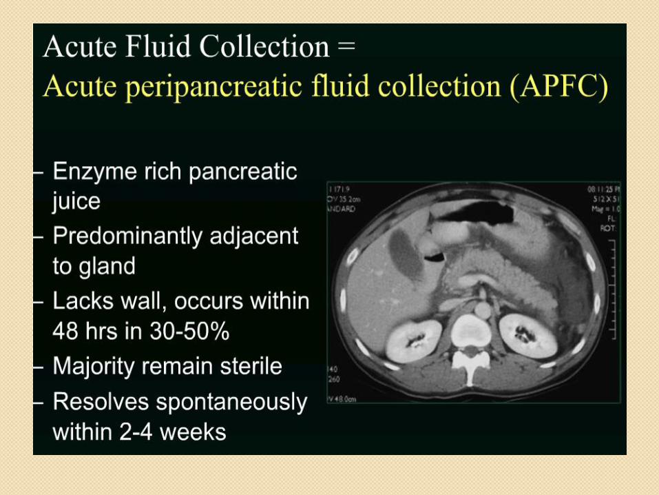
◦ Localized complications:
Altered fat and fascial planes, Fluid collection, pseudocyst, psduoaneurysm,
Bowel distension, mesenteric edema, hemorrhage

Magnetic Resonant Cholangiopancreatography
INDICATION:
◦ diagnosis of suspected biliary and pancreatic duct obstruction in
the setting of pancreatitis.
◦ Repeated attacks of idiopathic acute pancreatitis (Microlithiasis)

Endoscopic Ultrasonography
INDICATIONS
◦ Repeated idiopathic acute pancreatitis*
occult biliary disease- small stones/sludge
secretin-stimulated EUS study may reveal resistance to ductal outflow at the level of the papilla,
as evidenced by dilatation of the pancreatic duct to a greater extent and longer duration than in a healthy population
◦ Age >40 to exclude malignancy
especially those with prolong or recurrent course
RATIONALE: 5 % CA pancreas present as AP

Endoscopic Retrograde Cholangiopancreatography
INDICATION
Severe gallstone AP or AP with concurrent acute cholangitis/biliary obstruction/ biliary sepsis/jaundice (due to persistent stone)
ERCP within 24(-72) h of admission
Sphincterotomy /stent and bile duct clearance
It reduces infective complications/mortality
NOT INDICATED
Not needed early in most patients with gallstone pancreatitis who lack laboratory or clinical evidence of ongoing biliary obstruction
◦ MRCP or EUS recommended if CBD stone still suspected as risk of post-ERCP pancreatitis is greater with normal caliber bile duct and
normal bilirubin
MRCP /EUS as accurate as diagnostic ERCP

SEVERITY SCORING SYSTEMS ACUTE PANCREATITIS SPECIFIC SCORING SYSTEMS
◦ Ranson score
◦ Glagsow score
◦ Bedside Index for Severity in Acute Pancreatitis(BISAP) score
◦ Harmless Acute Pancreatitis Score(HAPS)
◦ Hong Kong Criteria
ACUTE PANCREATITIS NON-SPECIFIC SCORING SYSTEMS
(ICU SCORING SYSTEMS)
◦ Acute Physiology And Chronic Health Evaluation(APACHE) II score
◦ Sequential Organ Failure Assessment(SOFA) score
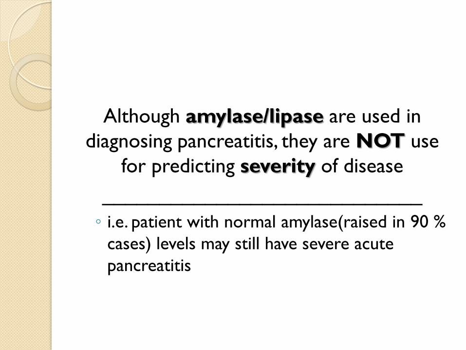
Although amylase/lipase are used in
diagnosing pancreatitis, they are NOT use
for predicting severity of disease
____________________________
◦ i.e. patient with normal amylase(raised in 90 %
cases) levels may still have severe acute
pancreatitis

RANSON SCORE-1974
(for alcohol pancreatitis)
ON ADMISSION AFTER 48 HOURS
Age > 55 yrs
WBC > 16,000/mm3
BSR > 200 mg/dL
AST > 250 IU/L
LDH > 350 IU/L
BUN rise >5 mg/dL
Pa02 < 60 mmHg ( 8 KPa)
Serum Calcium < 8 mg/dL
Base deficit > 4 meq/L
Fluid Sequestration > 6000 mL
Hct fall > 10 %
NOTE: Disease classified as SEVERE when 3 or more factors are present

Revised RANSON SCORE-1979
(for Gallstone pancreatitis)
ON ADMISSION AFTER 48 HOURS
Age > 70 years
WBC > 18,000/mm3
BSR > 220 mg/dL
AST> 250 IU/L
LDH >400 IU/L
BUN rise >5 mg/dL
Pa02 < 60 mmHg ( 8 KPa)
Serum Calcium < 8 mg/dL
Base deficit > 5 meq/L
Fluid Sequestration > 4000 ml
Hct fall > 10 %
NOTE: Disease classified as SEVERE when 3 or more factors are present

RANSON SCORE
Ranson
score
Mortality rate SEVERITY Interpretation
0-2 0-2 % Mild Admit in regular ward
3-5 10-20 % Moderate Admit in ICU/HDU
6-7 40 %
Severe
Associated with more
systemic complications
>7 >50 % Same as above

BALTHAZAR CT severity index(CTSI)-1994
Mild (0-3)
moderate (4-6)
severe (7-10)
CT Severity
Index
Inflammation score + Necrosis score

APACHE Scoring System
(Acute Physiology And Chronic Health Evaluation Score II)
Immediate assessment of the severity of
pancreatitis possible
Unlike ALL pancreatic specific scoring systems,
APACHE includes clinical features of patient besides
laboratory values
(Clinical findings are more important than lab
findings in predicting SIRS, sepsis and other
complications)


DEMERITS OF AP-specific scoring
systems(ACG 2013)
No single laboratory test is accurate to predict severity in patients with AP. ◦ Even the acute-phase reactant CRP, the most
widely studied inflammatory marker in AP, is not practical as it takes 72h to become accurate.
CT and/or MRI imaging also cannot determine severity early in the course of AP, as necrosis usually is not present on admission and may develop after 24 – 48 h.
Thus, in the absence of any available test to determine severity,
close examination to assess early fluid losses, hypovolemic
shock, and symptoms suggestive of organ dysfunction is crucial.


Mild Acute Pancreatitis
mild and self-limiting, needing only brief hospitalization.
Rehydration by IV fluids
Frequent non-invasive observation/monitoring
Brief period of fasting till pain/vomiting settles
◦ Little physiological justification for prolonged NPO
No medication required other than analgesics(important) and anti-emetics
◦ Antibiotics not indicated in absence of signs or documented sources of infection
◦ Pain results in ongoing cholinergic discharge, stimulating gastric and pancreatic secretions
◦ Avoid Morphine-cause sphincter of Oddi spasm
Metabolic support
◦ Correction of electrolyte imbalance

Modified WHO analgesic Ladder

No or little role of………………..
Nasogastric suction
H2-blockers
Secretion-inhibiting drugs
◦ Atropine, calcitonin, somatostatin and its analogue(Octreotide)
◦ glucagon and fluorouracil
Protease inhibiting drugs
◦ Aprotinin, gabexate mesylate,camostate, phospholipase A2 inhibitors, FFP
Indomethacin or PG inhibitors

Monitoring
CLINICAL INVESTIGATIONS
Vitals
UOP
CV pressure
Baselines
Serial ABGs
Serial BSR
Serum calcium/magnesium

ACG 2013 Recommendations
Despite dozens of randomized trials, no medication has been shown to be effective in treating AP.
However, an effective intervention has been well described: EARLY AGRESSIVE IV hydration.

Rationale for EARLY AGRESSIVE IV hydration
Frequent hypovolemia due to
◦ vomiting,
◦ reduced oral intake,
◦ third spacing of fluids(increased vascular permeability)
◦ increased respiratory losses, and
◦ diaphoresis.
Combination of microangiopathic effects and edema of the inflamed pancreas decreases blood flow, leading to increased cellular death, necrosis, and ongoing release of pancreatic enzymes activating numerous cascades.
_________________________________________________________
*provides micro- and macrocirculatory support to prevent serious complications such as pancreatic necrosis

EARLY AGRESSIVE IV hydration
Kon
sa?
Lactated Ringer ’s solution may be the preferred
isotonic crystalloid replacement fluid
• Normal saline given in large volumes may lead to
the development of a non-anion gap,
hyperchloremic metabolic acidosis and
increased chances of SIRS
• Low pH activates the trypsinogen, makes the
acinar cells more susceptible to injury and
increases the severity of established AP
Kab? Early aggressive IV hydration is most beneficial during the
first 12 – 24 h, and may have little benefit beyond this time
period
Kitna?
Aggressive hydration, defined as 250 – 500 ml per hour of
isotonic crystalloid solution should be provided to all
patients, unless cardiovascular, renal, or other related comorbid
factors exist.
EARLY AGRESSIVE IV hydration
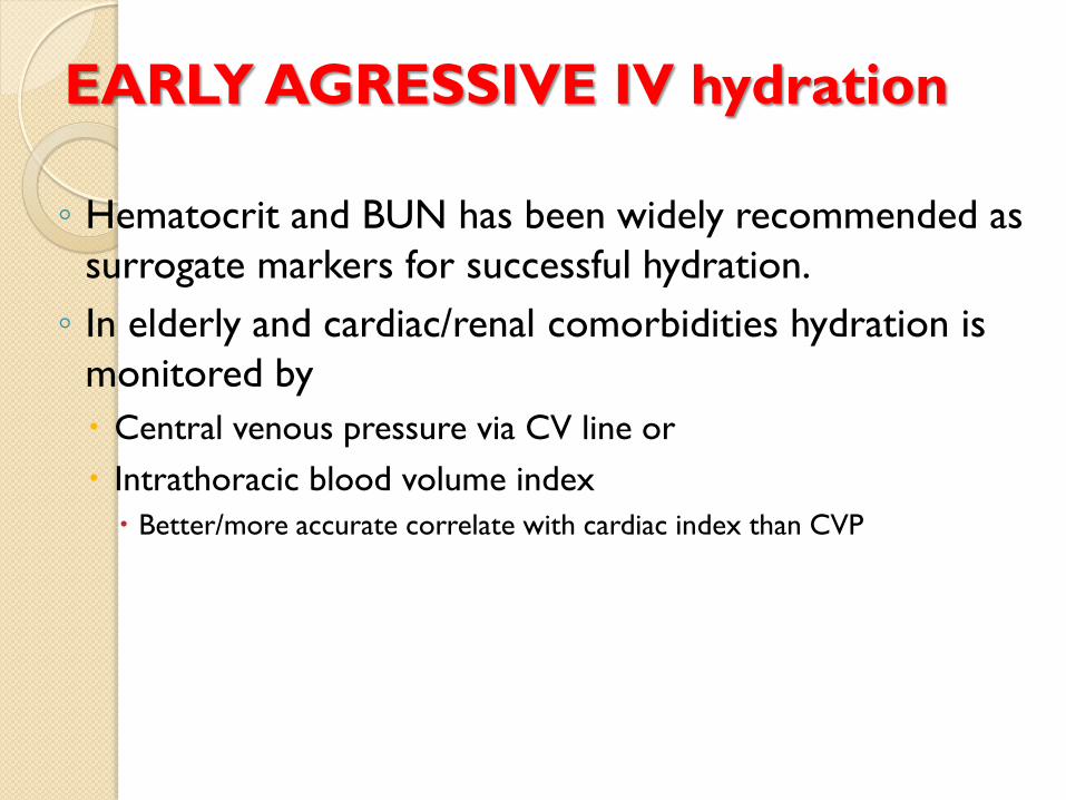
◦ Hematocrit and BUN has been widely recommended as
surrogate markers for successful hydration.
◦ In elderly and cardiac/renal comorbidities hydration is
monitored by
Central venous pressure via CV line or
Intrathoracic blood volume index
Better/more accurate correlate with cardiac index than CVP

Antibiotics
Routine use* NOT recommended(ACG 2013) as ◦ Prophylaxis in severe AP
◦ Preventive measure in sterile necrosis to prevent development of infected necrosis
Indicated in ◦ Established infected pancreatic necrosis or
◦ Extraperitoneal infections Cholangitis, catheter-acquired infections, bacteremia, UTIs, pneumonia
______________________________________________
*Routine use of antifungal agents along with prophylactic or therapeutic antibiotics NOT
recommended(ACG 2013)

Antibiotics
Few antibiotics penetrate due to
consistency of pancreatic necrosis
◦ cefuroxime, or imipenem, or ciprofloxacin
plus metronidazole

Nutrition
In mild AP ◦ oral feedings can be started immediately if there is no nausea/vomiting,
and the abdominal pain/tenderness/Ileus has resolved(amylase return to normal, patient feel hunger)
◦ Initiation of feeding with a small and slowly increasing low-fat (low-protein) soft diet appears as safe as a clear liquid diet, providing more calories Stepwise manner increase from clear liquids to soft diet NOT necessary
In severe AP
◦ Enteral route is recommended to prevent infectious complications
◦ Parenteral nutrition should be avoided, unless enteral route is not available, not tolerated, or not meeting caloric requirements

RATIONALE OF EARLY ENTERAL
NUTRITION
The need to place pancreas at rest until complete resolution of AP no longer seem imperative ◦ Bowel rest associated with intestinal mucosal atrophy and
bacterial translocation from gut and increased infectious complications
Early enteral feeding maintains the gut mucosal barrier, prevents disruption, and prevents translocation of bacteria that seed pancreatic necrosis ◦ Decrease in infectious complications, organ failure and
mortality

………..
Rather than using antibiotics to prevent infected necrosis………….start early enteral feeding to prevent translocation of bacteria
RATIONALE MANAGEMENT
PREVENTION OF STERILE NECROSIS Early aggressive IV hydration
PREVENTION OF INFECTED NECCROSIS Early enteral feeding( NOT antibiotics)
TREATMENT OF INFECTED NECROSIS Antibiotics, drainage, necrosectomy

Route of enteral Nutrition
Traditionally naso-jejunal route has been preferred
to avoid the gastric phase of stimulation BUT
◦ Nasogastric route appears comparable in efficacy and
safety
MERITS OF NASOGASTRIC ROUTE DEMERITS OF NASOGASTRIC ROUTE
NG tube placement is far easier than
nasojejunal tube placement( requiring
interventional radiology or endoscopy, thus
expensive) especially in HDU/ICU setting
Slight increased risk of aspiration
(Can be overcome by placing patient in upright
position and be placed on aspiration
precautions)
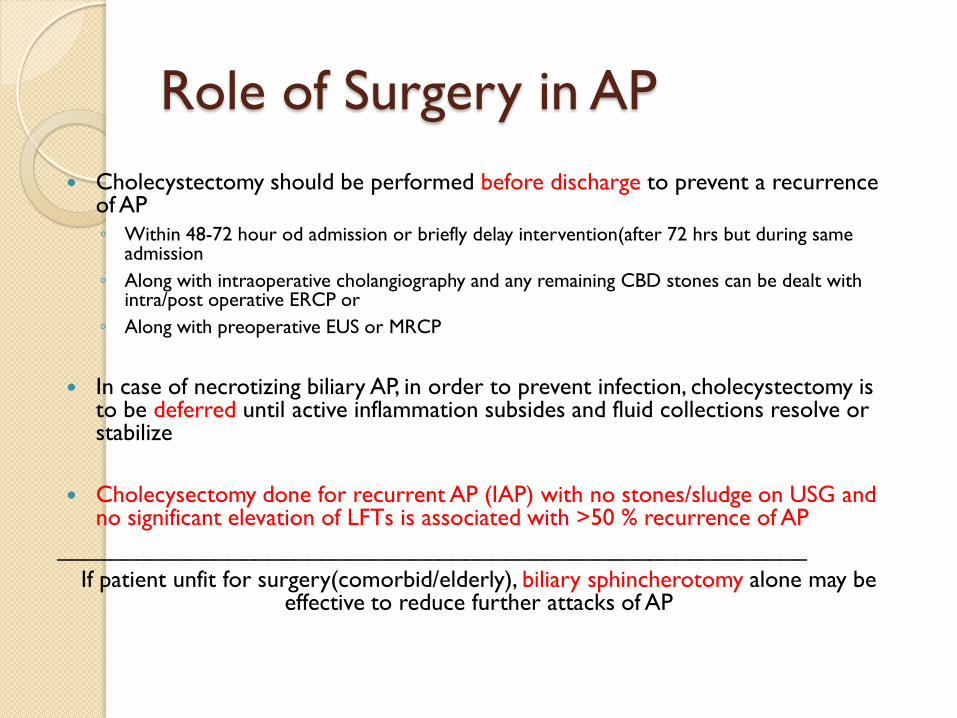
Role of Surgery in AP
Cholecystectomy should be performed before discharge to prevent a recurrence of AP
◦ Within 48-72 hour od admission or briefly delay intervention(after 72 hrs but during same admission
◦ Along with intraoperative cholangiography and any remaining CBD stones can be dealt with intra/post operative ERCP or
◦ Along with preoperative EUS or MRCP
In case of necrotizing biliary AP, in order to prevent infection, cholecystectomy is to be deferred until active inflammation subsides and fluid collections resolve or stabilize
Cholecysectomy done for recurrent AP (IAP) with no stones/sludge on USG and no significant elevation of LFTs is associated with >50 % recurrence of AP
_________________________________________________________
If patient unfit for surgery(comorbid/elderly), biliary sphincherotomy alone may be effective to reduce further attacks of AP
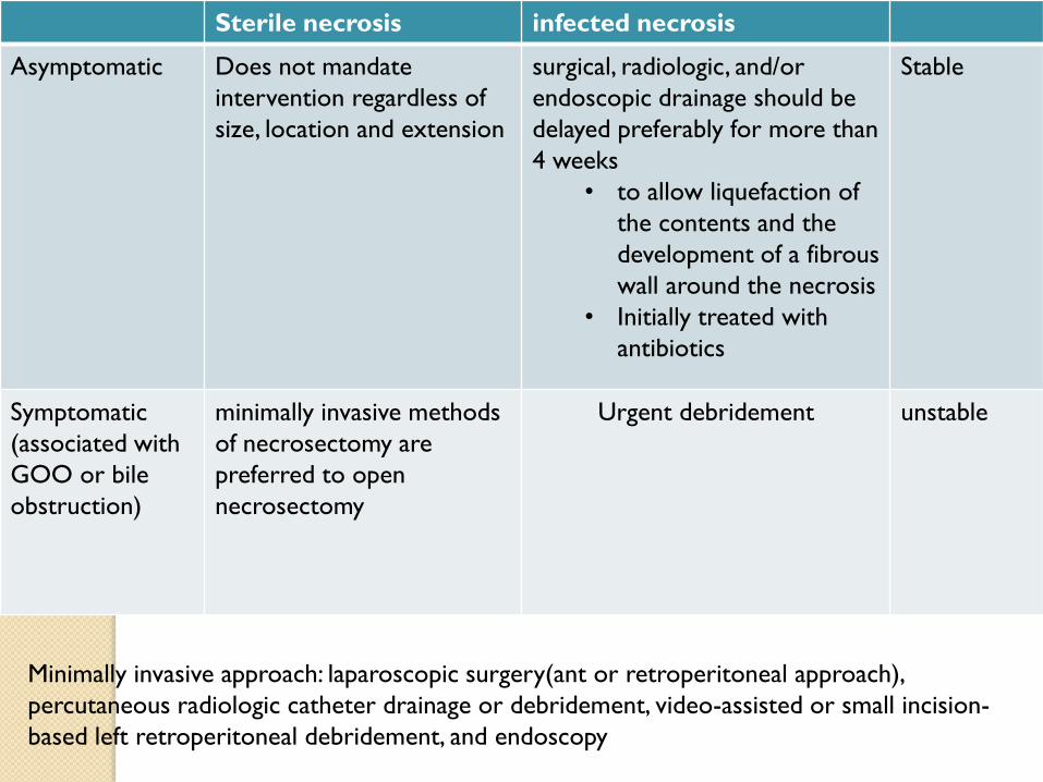
Sterile necrosis infected necrosis
Asymptomatic Does not mandate
intervention regardless of
size, location and extension
surgical, radiologic, and/or
endoscopic drainage should be
delayed preferably for more than
4 weeks
• to allow liquefaction of
the contents and the
development of a fibrous
wall around the necrosis
• Initially treated with
antibiotics
Stable
Symptomatic
(associated with
GOO or bile
obstruction)
minimally invasive methods
of necrosectomy are
preferred to open
necrosectomy
Urgent debridement unstable
Minimally invasive approach: laparoscopic surgery(ant or retroperitoneal approach),
percutaneous radiologic catheter drainage or debridement, video-assisted or small incision-
based left retroperitoneal debridement, and endoscopy

When to Discharge
Pain is well controlled with oral analgesia
Able to tolerate an oral diet that maintains their caloric needs, and
all complications have been addressed adequately
Follow up
Routine clinical follow-up care (typically including physical examination and amylase
and lipase assays) is needed to monitor for potential complications of the
pancreatitis, especially pseudocysts.
Within 4 weeks

Idiopathic Recurrent AP
CT scan
• If neoplasia or chronic pancreatitis is found
• addressed and treated accordingly.
MRCP
• shows developmental abnormalities, strictures, or evidence of chronic pancreatitis
• endoscopic or surgical treatment may be of benefit in a subset of patients
EUS
• Microlithiasis/biliary sludge Cholecystectomy
• Periammpullary mass missed on CT or MRCP
Genetic
• cationic trypsinogen mutations, SPINK1 mutations, or CFTR mutations
ERCP
• sphincter of Oddi manometry
• Placed last because very high rate of post-ERCP pancreatitis(benefits< risk)

Prognosis
TYPE OF AP MORTALITY
Overall 10-15 %
(Biliary>alcholic)
Mild Acute Pancreatitis(80 % cases) 1 %
Severe Acute Pancreatitis(20 % cases) Severe 20-50 %
<1 week 1/3 cases MOF
>1 week 2/3 cases Sepsis
(+MOF)
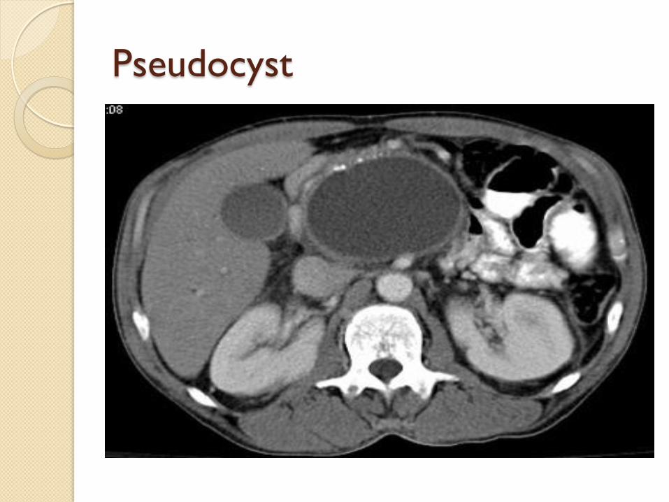
Pseudocyst

Splenic Infarct

ARDS


THANK YOU……….