Embed Size (px)
Citation preview

Umeå University Medical Dissertations New Series No 865 ISSN 0346-6612
ISBN 91-7305-550-6
From the Department of Respiratory Medicine and Allergy, University of Umeå, Sweden,
and the Department of Medical Countermeasures, Divison of NBC Defence, Swedish Defence Research Agency, Umeå, Sweden.
Acute lung injury:
Study of pathogenesis and therapeutic interventions
av
David Rocksén
Akademisk avhandling
som med vederbörligt tillstånd av rektorsämbetet vid Umeå universitet
för avläggande av doktorsexamen i medicinsk vetenskap framläggs till offentligt försvar Fredagen 12 Dec 2003 kl 09.00
i Sal B (Rosa Salen), Byggnad 1 D, Tandläkarhögskolan Norrlands Universitetssjukhus, Umeå.
Fakultetsopponent: Docent Johan Grünewald
Institutionen för medicin, Enheten för lungmedicin, Karolinska sjukhuset, Stockholm
Umeå 2003

David Rocksén 2003
Printed by Solfjädern offset AB, Umeå, Sweden

“We shall not fail or falter; we shall not weaken or tire.... Give us the tools and we will finish the job.”
Sir Winston Churchill (1874-1965)
Contents List of papers Abstract Abbreviations Introduction…………………………………………………………………. 13 Definitions of ALI and ARDS………………………………………………. 15 Incidence…………………………………………………………………….. 16 Risk factors and mortality…………………………………………………... 17 Direct lung injury……………………………………………………... 17 Indirect lung injury……………………………………………………. 17 Lung pathology in ARDS…………………………………………………... 19 Pathogenesis………………………………………………………………… 22 Inflammatory response……………………………………………….. 22 Macrophages………………………………………………... 22 Neutrophils…………………………………………………. 23 Lymphocytes………………………………………………... 24 Cytokines and chemokines………………………………….. 25 Direct injury…………………………………………………………... 26 Pharmacological treatment…………………………………………………. 27 Corticosteroids………………………………………………………... 27 Antioxidants…………………………………………………………... 27 Other pharmacological efforts………………………………………… 28

Animal models of ALI/ARDS……………………………………………… 29 Exposure to endotoxin………………………………………………... 29 Animal models of endotoxin inhalation……………………... 30 Systemic administration of endotoxin……………………….. 31 Two-hit models………………………………………………………. 31 Shwartzman reaction………………………………………... 32 Other animal models of ALI/ARDS…………………………………. 33 Aims of the thesis…………………………………………………………… 35 Methodological considerations…………………………………………….. 37 Main results and discussion………………………………………………... 43 Generalized Shwartzman reaction as an animal model of acute respiratory distress syndrome…………………………. 43 Lung neutrophilia in acute lung inflammation, septic shock and in the generalized Shwarzman reaction……………… 45 Activation of lymphocytes in endotoxin-induced airway inflammation and in the generalized Shwartzman reaction…………….. 47 Treatment with dexamethasone in different experimental animal models of ALI/ARDS…………………………... 50 Treatment with antioxidants in different experimental animal models of ALI/ARDS…………………………... 51 Final comments……………………………………………………………... 55 Acknowledgements…………………………………………………………. 57 References…………………………………………………………………… 60 Papers I-IV


List of papers This thesis is based on the following papers, referred to in text by their Roman numerals: I. Larsson R, Rocksén D, Lilliehöök B, Jonsson Å and Bucht A. Dose-dependent activation of lymphocytes in endotoxin-induced airway inflammation. Infect. Immun. 2000: 68; 6962-69. II. Rocksén D, Lilliehöök B, Larsson R, Johansson T and Bucht A. Differential anti-inflammatory and anti-oxidative effects of dexamethasone and N-acetylcysteine in endotoxin-induced lung inflammation. Clin. Exp. Immunol. 2000: 122; 249-56. III. Rocksén D, Ekstrand-Hammarström B, Johansson L and Bucht A. Vitamin E reduces transendothelial migration of neutrophils and prevents lung injury in endotoxin-induced airway inflammation. Am. J. Respir. Cell Mol. Biol. 2003: 28; 199-207. IV. Rocksén D, Koch B, Sandström T and Bucht A. Generalized Shwartzman reaction as an animal model of acute respiratory distress syndrome. Manuscript.

Abstract Acute lung injury (ALI), denominated acute respiratory distress syndrome (ARDS) in its most severe form, is characterized by lung inflammation, hypoxia, non-cardiogenic pulmonary edema and decreased lung compliance. Despite numerous experimental and clinical studies, the mortality of ARDS remains between 30-70%, and its pathogenesis is still largely unknown. The overall aim of the present thesis was to use experimental mouse models of ALI/ARDS to identify cells and mediators involved in the pathogenesis of the syndrome. Moreover, we have used these models to evaluate the therapeutic effects of the corticosteroid dexamethasone as well as the antioxidants N-acetylcysteine (NAC) and Vitamin E (α-tocopherol: α-toc). In the first study, we investigated the inflammatory response in lung tissue and bronchoalveolar lavage (BAL) after inhalation of bacterial endotoxin (Lipopolysaccharide: LPS), a commonly used experimental animal model of ALI. Our results demonstrated that exposure to LPS induced a dose-dependent increase of neutrophils in BAL fluid (BALF) reaching a maximum after 12h at a low dose and after 24h at a high dose. Furthermore, a low dose induced an early (2h) and transient onset of cytokine and chemokine gene expression in lung tissue, while a high dose caused more delayed and sustained (6-12h) activation. In addition, expression of T-cell derived IFN-γ, IL-2, IL-17 and Rantes was only recorded when mice where exposed to a high dose, indicating a dose-dependent activation of T-cells after LPS inhalation. In the second and third study, we investigated therapeutic interventions in the LPS inhalation model. Significant reduction of neutrophils in BALF was obtained through a single i.p injection of dexamethasone (10mg/kg), whereas treatment with NAC only resulted in reduction of neutrophils when administrated at a high dose (500mg/kg). Measurement of cytokine and chemokine expression in lung tissue revealed marked down-modulation with dexamethasone, while NAC demonstrated poor anti-inflammatory effect. In the third study, we demonstrated that Vitamin E protect against lung tissue damage and exerts anti-inflammatory properties as shown by the decreased number of neutrophils in airspaces when administrated at a dose of 50mg/kg body weight. This effect was due to a reduced transendothelial migration of

neutrophils, but without a profound effect on the early pro-inflammatory response. The fourth study was conducted to investigate if a generalized Shwartzman reaction (GSR) can display similar pathophysiology in mice as observed in clinical ARDS. Our results demonstrated that the generalized Shwartzman reaction induced rapid decline in lung function and 80% mortality. Furthermore, characteristic hallmarks of ARDS, such as lung tissue neutrophilia and edema formation, were observed. However, the lung neutrophilia was not closely associated with the mortality observed in the generalized Shwartzman reaction. Therapeutic effects of 50mg/kg Vitamin E and dexamethasone (10mg/kg) were also evaluated in this model, demonstrating that both drugs improved lung function and exerted anti-inflammatory properties measured as decreased levels of cytokines in serum. In conclusion, the results in this thesis indicated that lymphocytes may be involved in the extensive inflammatory responses observed in ALI/ARDS. Furthermore, we have shown that neutrophils are not solely responsible for the rapid decline in lung function during the progress of experimental ARDS. Therapeutic interventions demonstrated that Vitamin E, alone or in combination with corticosteroids, might be effective for treatment of ALI and ARDS.

Abbreviations ALI acute lung injury ARDS acute respiratory distress syndrome MODS multiple organ dysfunction syndrome BAL bronchoalveolar lavage BALF BAL fluid α-toc alpha-tocopherol (Vitamin E) NAC N-acetylcysteine PBS phosphate buffered saline LPS lipopolysaccharide LBP LPS binding protein GSR generalized Shwartzman reaction LSR local Shwartzman reaction DIC disseminated intravascular coagulation i.p intraperitoneally s.c subcutaneous COPD chronic obstructive pulmonary disease PEEP positive end-expiratory pressure LIS lung injury score PaO2 arterial oxygen data FIO2 amount of oxygen delivered by ventilatory support AECC American-European Consensus Conference on ARDS H2O2 hydrogen peroxide ROS reactive oxygen species NK cells natural killer cells CD4 T cells helper T cells IgE immunoglobulin E TNF-α tumour necrosis factor-alpha IFN-γ interferon-gamma IL- interleukin- MIP- macrophage inflammatory protein- MCP- monocyte chemoattractant protein ICAM-1 intercellular adhesion molecule-1

AP-1 activator protein-1 NF-κB nuclear factor-κB TLR toll-like receptors LDH lactate dehydrogenase KO knock-out H2DCF-DA dichlorodihydrofluorescein diacetate FITC fluorescein isothiocyanate PE phycoerythrin PCR polymerase chain reaction EMSA electrophoretic mobility shift assay ELISA enzyme-linked immunoabsorbent assay


13
Introduction In 1967, at the Colorado General Hospital, Ashbaugh and collegues observed 12 critically ill patients with different etiology who all developed sudden acute respiratory failure. They wondered what was different with these patients compared to the usual cases of respiratory failure observed in chronic obstructive pulmonary disease (COPD), pneumonia and asthma (Petty 2001). Seven of the 12 patients had multiple trauma, 4 had viral pneumonia and 1 patient suffered from acute pancreatitis. Others had previously described acute respiratory failure in patients with separate single diseases. However, Ashbaugh and collegues were the first to describe that patients with a variety of underlying illnesses could develop acute respiratory failure with strikingly similar pulmonary pathophysiology (Simon 1992). All of the 12 patients at the Colorado General Hospital presented a similar pathophysiology characterized by tachypnea, hypoxemia resistant to oxygenation by regular ventilatory support, decreased lung complience and diffuse bilateral infiltrates on chest radiographs. Some patients also demonstrated a deficiency in lung surfactant. These surfactant abnormalities, together with the impaired oxygenation of the blood, had similarities with the neonatal respiratory distress syndrome i.e children born with a deficiency in lung surfactant. Hence Ashbaugh and collegues published their finding in The Lancet in 1967 and named the new syndrome Acute Respiratory Distress Syndrome (ARDS) in adults (Ashbaugh 1967). This article was read by military surgeons treating causulties in the Vietnam War. They had also observed sudden respiratory failure in soldiers suffering from different traumas. A conference on ARDS was quickly planned and held in Washington in 1968, and after that meeting the discovery of ARDS was rapidly spread in the research community. At present, the syndrome is simply denominated ‘the acute respiratory distress syndrome’ so that children are not excluded from the criteria. Even in the first report of the syndrome, one of the 12 patients was 11 years of age.

Background
14
Thirtyfive years since the original description of the syndrome, the mortality of ARDS remains high, resulting in tremendous human and financial costs each year. Since the pathogenesis of the syndrome is still largely unknown, there is a great demand for experimental animal studies and for new therapeutic strategies to be evaluated. Figure 1. Trauma patient at the intensive care unit (Sv: Intensivvårdsavdelningen; IVA) at the University hospital, Umeå, Sweden. Photograph received from MD Jonas Claesson, IVA, Umeå University hospital

Background
15
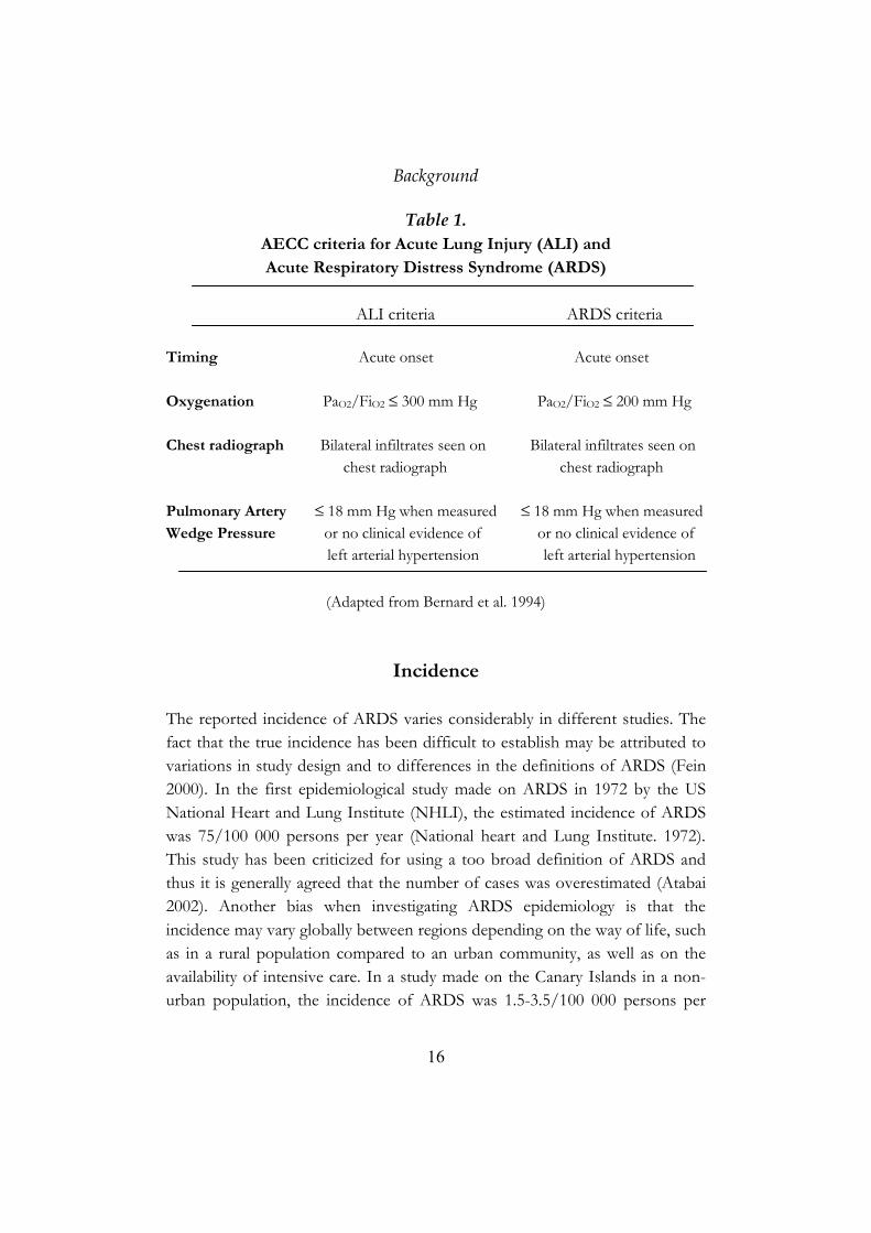
Definitions of ARDS and ALI The most widely used definition of Acute Lung Injury (ALI) and ARDS was published in 1994 by the American-European Consensus Conference (AECC) on ARDS. This committee recommended that ALI and ARDS should be defined as a syndrome of inflammation and increased lung capillary permeability associated with a number of clinical, radiological and physiological abnormalities that cannot be explained by, but may coexist with heart failure. Furthermore, the AECC committee defined ALI and ARDS as syndromes that are acute in onset and characterized by severe hypoxemia. It was agreed that the term ALI could be applied to a broad range of pathological abnormalities in the lung and that the term ARDS should be reserved for the most severe cases of lung injury. To distinguish between ALI and ARDS, the committee suggested the use of the ratio between arterial oxygen data (PaO2) and the amount of oxygen delivered by ventilatory support (FIO2), which is a value commonly used to measure arterial blood oxygenation (PaO2/FIO2). The criteria for ALI would be a PaO2/FIO2 ≤ 300mm Hg (40kPa) and for ARDS PaO2/FIO2
≤ 200mm Hg (26.7kPa) (Bernard 1994). An alternative definition was proposed by Murray et al. in 1988. They developed a system to grade the degree of lung injury in patients with acute respiratory failure which is known as the Lung Injury Score (LIS). This system is based on four parameters which include the chest radiographic score, hypoxemia score (PaO2/FIO2), PEEP score (the Positive End-Expiratory Pressure needed for proper ventilation) and the compliance score. Each parameter was given a number using a scale ranging between 0-4 and the final value is obtained by dividing the sum of the values with the number of parameters used. A LIS score greater than 2.5 in the patient is defined as ALI/ARDS (Murray 1988).
Background
16
Table 1. AECC criteria for Acute Lung Injury (ALI) and Acute Respiratory Distress Syndrome (ARDS)
ALI criteria ARDS criteria Timing Acute onset Acute onset Oxygenation PaO2/FiO2 ≤ 300 mm Hg PaO2/FiO2 ≤ 200 mm Hg Chest radiograph Bilateral infiltrates seen on Bilateral infiltrates seen on chest radiograph chest radiograph Pulmonary Artery ≤ 18 mm Hg when measured ≤ 18 mm Hg when measured Wedge Pressure or no clinical evidence of or no clinical evidence of left arterial hypertension left arterial hypertension
(Adapted from Bernard et al. 1994)
Incidence The reported incidence of ARDS varies considerably in different studies. The fact that the true incidence has been difficult to establish may be attributed to variations in study design and to differences in the definitions of ARDS (Fein 2000). In the first epidemiological study made on ARDS in 1972 by the US National Heart and Lung Institute (NHLI), the estimated incidence of ARDS was 75/100 000 persons per year (National heart and Lung Institute. 1972). This study has been criticized for using a too broad definition of ARDS and thus it is generally agreed that the number of cases was overestimated (Atabai 2002). Another bias when investigating ARDS epidemiology is that the incidence may vary globally between regions depending on the way of life, such as in a rural population compared to an urban community, as well as on the availability of intensive care. In a study made on the Canary Islands in a non-urban population, the incidence of ARDS was 1.5-3.5/100 000 persons per
Background
17
year (Villar 1989). In another study performed in Utah, the incidence of ARDS was 4.8-8.3/100 000 inhibitants per year (Thomsen 1995). Both the study on the Canary Islands and the Utah study have been criticized for excluding patients with a PaO2/FIO2 ≥ 150mm Hg. In recent studies using the AECC criteria (previously described), the corresponding reported incidences were strikingly similar. In a prospective cohort study made in Sweden, Denmark and Island in 1997, the reported incidence was 13.5 for ARDS and 17.9 for ALI/100 000 persons per year (Luhr 1999). Several other studies using the AECC criteria in Japan, Australia and in the USA reported a similar incidence, indicating that the results from the study in Scandinavia are reasonable (Kanazawa 1996, Nolan 1997, Hudson 1999).
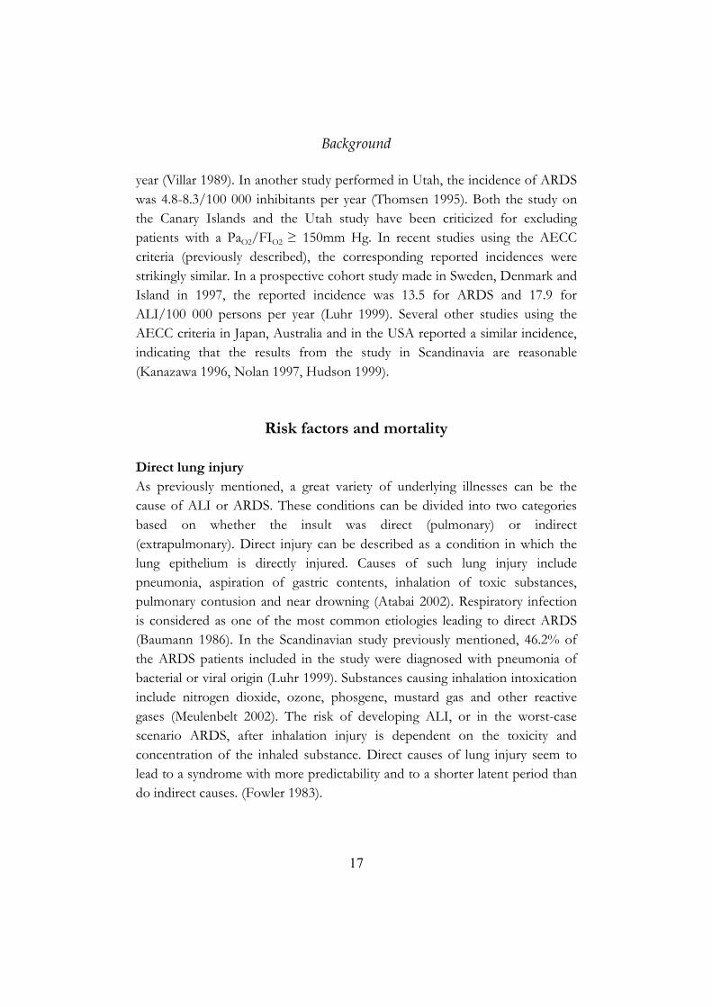
Risk factors and mortality Direct lung injury As previously mentioned, a great variety of underlying illnesses can be the cause of ALI or ARDS. These conditions can be divided into two categories based on whether the insult was direct (pulmonary) or indirect (extrapulmonary). Direct injury can be described as a condition in which the lung epithelium is directly injured. Causes of such lung injury include pneumonia, aspiration of gastric contents, inhalation of toxic substances, pulmonary contusion and near drowning (Atabai 2002). Respiratory infection is considered as one of the most common etiologies leading to direct ARDS (Baumann 1986). In the Scandinavian study previously mentioned, 46.2% of the ARDS patients included in the study were diagnosed with pneumonia of bacterial or viral origin (Luhr 1999). Substances causing inhalation intoxication include nitrogen dioxide, ozone, phosgene, mustard gas and other reactive gases (Meulenbelt 2002). The risk of developing ALI, or in the worst-case scenario ARDS, after inhalation injury is dependent on the toxicity and concentration of the inhaled substance. Direct causes of lung injury seem to lead to a syndrome with more predictability and to a shorter latent period than do indirect causes. (Fowler 1983).
Background
18
Indirect lung injury Indirect lung injuries are conditions that activate a systemic inflammatory reponse. Multiple trauma, burns, sepsis, fat emboli and hemorrhagic shock are all associated with a high risk of developing ARDS. Sepsis has been documented as the condition with the highest risk of progression to ARDS. Several studies have documented that the incidence of ARDS in sepsis patients is approximately 40% (Pepe 1982 and Hudson 1995). Studies have indicated that the incidence of ARDS rose dramatically when several insults were present (Fowler 1983 and Pepe 1982). Other factors that influence the incidence is chronic alcohol abuse (Moss 1996) and age over 65 (Hudson 1995). The fact that chronic alcohol abuse influences the risk of developing ARDS may be attributed to impairment of host immune defence or by hepatic dysfunction which might lead to more severe lung injury (Fein 2000). An inappropriate mode of supportive ventilation is also a risk factor since it may involve mechanical stress and activate an acute inflammatory response (Downey 1997).
Table 2. Clinical disorders associated with the development of ALI/ARDS
Direct Indirect Common Common * Aspiration pneumonia * Sepsis * Pneumonia * Severe trauma with prolonged hypotension and/or multiple fractures * Multiple blood transfusion Less common Less common * Inhalation injury * Acute pancreatitis * Pulmonary contusion * Cardiopulmonary bypass * Fat emboli * Drug overdose * Near drowning * Burns * Reperfusion injury * Head injury
(Adapted from Atabai et al. 2002)

19
Lung pathology in ARDS The most important pathological finding in the lung during the early stages of ARDS is severe pulmonary edema, due to increased permeability of the capillary endothelial and the alveolar epithelial barriers. Furthermore, there is an increase in pulmonary vascular resistance due to vasoconstriction and thromboembolism which further increases the pulmonary edema (Ten Cate 2001). The morphological picture has often been labeled diffuse alveolar damage (DAD) and includes cellular necrosis, inflammation and fibrosis (Welty-Wolf 2002). The disease course of ARDS can be divided into the acute phase (exudative phase), followed by the long-term consequences of ARDS in surviving patients (the proliferative and fibrotic phase). Exudative phase Lungs of patients who die during this phase are rigid, red-blue due to hemmorhage and heavy because of the edema formation (Tomashefski 2000). The first barrier that prevents leakage of fluid from the vascular space are the endothelial cells which are joined together by tight junctions. During the exudative phase, the endothelial cell swell, widening of the tight junctions occurs and there is an increase in the number of vesicles (Tomashefski 2000). The second barrier that prevents leakage into the intra-alveolar space are the alveolar epithelial cells which are joined together by tight junctions that are more intricate than the endothelial junctions and thus form a tighter barrier. The alveolar epithelium consists of two major cell types: the alveolar type I and type II epithelial cells. The alveolar type I cell is a thin flat cell that covers most of the interior alveolar surface. The type II cell is the main cell that produces and secretes surfactant which prevents collapse of the alveoli by lowering the surface tension. During the exudative phase, extensive necrosis of the type I cell occurs and the epithelium may detach, leaving large areas of denuded basement membrane which allows increased plasma leakage through the barrier (Simon 1992). Type II cells may also undergo necrosis although several studies have demonstrated that type 1 cells are more susceptible to injury (Tomashefski 2000). Such damage to the endothelium and the alveolar epithelium produces interstitial and intra-alveolar spaces filled with protein-rich

Lung pathology
20
edema fluid and hyalin membranes. The hyalin membranes are composed largely of fibrin, fibronectin and cellular debris (Simon 1992). Neutrophils are found increasingly during the intitial phase in the capillaries but only sparsely in the interstitium and the airspaces. The alveoli become congested from the hyalin membranes and the inflammatory cells, edematous, and may in some cases collapse because of epithelial necrosis and loss of surfactant tension (Tomashefski 2000). Proliferative phase The proliferative phase is characterized by organization of the edema fluid aquired in the exudative phase and by fibrosis (Bellingan 2002). There is a marked decrease in the number of neutrophils in this phase but lymphocytes are often heavily concentrated in the areas of regeneration (Niederman 1990). Alveolar type II cells proliferate in an attempt to cover the denuded basement membrane and differentiate into type I cells (Bellingan 2002). Fibroblasts proliferate within the alveolar wall and migrate into airspaces where they organize the exudate and deposit collagen into fibrous tissue (Tomashefski 2000). The main site of fibrosis is the intra-alveolar space although some signs of fibrosis are evident in the interstitium (Bellingan 2002). Fibrotic phase If the patient survives the two first stages in the disease course, they may enter a fibrotic phase which can start as soon as 10 days after first signs of respiratory failure (Bellingan 2002). This phase is not well documented due to lack of autopsy data, although some studies have demonstrated that the alveoli and walls of the airspaces are composed of thickened, collagenous connective tissue (Tomashefski 2000).

Lung pathology
21
A. B. C. Figure 2. Lung x-ray images of healthy individual (A), patient with ALI/ARDS (B) and patient with severe ARDS (C). White areas observed in B and C are bilateral lung infiltrates. Images received from MD Jonas Claesson, IVA, Umeå University Hospital
22
Pathogenesis Inflammatory response As previously mentioned, the pathophysiology observed in ALI or ARDS is a consequence of the host response to a wide variety of direct or indirect stimuli. The similarities in lung pathology and pathophysiology after different insults have led many scientists to the conclusion that there is a single pathway for all cases of ALI/ARDS. However, the pathogenesis appears to be more complex and it is likely that several separate mechanisms participate in the disease course. Although the pathogenesis of ALI and ARDS remains unclear, indirect evidence suggest that the pulmonary edema occurs after a direct activation due to acute lung inflammation, or as the pulmonary manifestation of a more generalized (systemic) inflammatory response (Nuytinck 1985 and Dorinsky 1990). The inflammatory response in both cases primarily involves macrophages and neutrophils activated by several pro-inflammatory cytokines and chemokines. Furthermore, there is substancial evidence that other pathways such as the coagulation cascade and the complement system are activated during ARDS (Welty-Wolf 2002). Macrophages Macrophages are of bone marrow origin and mature to become blood monocytes. From the circulation, they migrate into various tissues where they differentiate, and in the lung denominated the alveolar macrophages. The normal lung contains a large number of macrophages both in the alveolar region and in the interstitial compartment. These phagocytic cells represent the first line of defence against invading microbes. Furthermore, they can internalize extracellular fluid, particulates and bind certain molecules on their cell surface (Campbell 1981). However, beside the protective role, there is a general agreement that monocytes and macrophages may amplify the inflammatory reponse in ALI/ARDS through the production and secretion of cytokines, chemokines and other pro-inflammatory mediators. Recent studies have demonstrated that the number of alveolar macrophages increases in the bronchoalveolar lavage fluid of ALI/ARDS patients, a finding which was associated with improved surival rate. This observation raised the hypothesis

Pathogenesis
23
that macrophages also may play a beneficial role in the resolution of ALI/ARDS (Pittet 1997). Support for this theory is the fact that macrophages are involved in the repair process by the production of platelet-derived growth factor (PDGF) which stimulates fibroblast proliferation (Bitterman 1992). Furthermore, they may release transforming growth factor-alpha (TGF-α), of importance for endothelial migration and growth (Madtes 1988). Neutrophils Neutrophils are granular cells which have the capacity to engulf microorganisms by phagocytosis. Lung lavage in normal subjects yields normally only a few percent neutrophils (Sandström 1992). However, a subpopulation of circulating neutrophils is always present within the pulmonary microvasculature. Upon activation, these neutrophils quickly upregulate specific adhesion molecules and may then adhere to the counterreceptors on the endothelium and migrate into lung tissue (Wallace 2002). Several substantial, but indirect pieces of evidence derived from autopsy studies implicate neutrophils in the pathogenesis of ALI/ARDS. Firstly, in most patients with ARDS the percentage of neutrophils in lavage fluid is markedly increased (Simon 1992). Secondly, several studies document that circulating neutrophils are activated in ARDS (Partrick 1996). Activated neutrophils have the capacity to release elastase, myeloperoxidase and reactive oxygen species (ROS) in order to kill invading microorganisms. The ROS include the superoxide anion (O2-), hydrogen peroxide (H2O2), hydroxyl radical (OH), singlet oxygen and other radicals, all of which can be potentially harmful to other cells. When produced in excessive amounts, or when anti-oxidant defence is insufficient, radicals can damage DNA, lipids, proteins and carbohydrates (Gillissen 1998). However, occasional reports have shown that ALI/ARDS can occur in neutropenic patients, a finding that has caused controversies about the role of neutrophils in the pathogenesis. Moreover, experimental animal models have revealed evidence that bacterial endotoxin can produce lung injury independent of neutrophils. The underlying mechanism(s) for these observations has not been thouroughly investigated but a plausible explanation may be a direct action of endotoxin on the endothelium (Winn 1987). These findings suggests

Pathogenesis
24
that the neutrophils are important, but not an essential component in the injurious response. Lymphocytes Three broad classes of lymphocytes are identified: T (thymus-derived) lymphocytes, which have important function in orchestrating immune responses and also involved in direct effector functions, such as the lysis of pathogen-infected cells. B lymphocytes, which are antibody-secreting cells. NK (Natural Killer) cells, acting as important components of innate immunity with a role in the host response to pathogens and defence against tumour cells. T cells may be divided into two distinct classes based on cell surface receptor expression. The majority of T cells express T cell receptors (TCR) consisting of α and β chains (αβ T cells). A small group of T cells express TCRs made up of γ and δ chains (γδ T cells). αβ T cells are composed of two sub-groups: those that express the co-receptor molecule CD4 (helper T cells) and those that express CD8 (cytotoxic T cells). The CD4+ T cell can be divided into two subsets, Th1 and Th2, as defined by their production of different cytokines. Lymphocytes are believed to interplay with the innate immune system to successfully identify and eliminate harmful agents. Cytotoxic T cells have been shown to have a critical role in immune clearance of infections with intracellular pathogens (Walker 2001) and CD4+ T cells are believed to enhance elimination of bacteria from the lungs through recruitment and activation of macrophages and neutrophils (Dunkley 1995 and Dsouza 1994). Furthermore, intraepithelial γδ T cells has been shown to protect the host from pathogenic (Nocardia asteroides) as well as nonpathogenic (ozone exposure) lung inury by modulation of the inflammatory response to epithelial necrosis (King 1999). However, it has also been suggested that lymphocytes are involved in certain airway diseases such as sarcoidosis, asthma and tuberculosis (Berman 1990 and Wahlström 2001). In animals, ALI has been provoked by intravenous administration of activated CD4+ T cells of Th1 phenotype (Clark 1998 and Dixon 2000). Further evidence for an aggravating role of lymphocytes in acute inflammatory responses is the finding that NK cells have a critical role in the development of lethal septic shock in animals (Ogasawara 1998).

Pathogenesis
25
Intriguingly, the concentrations of total IgE have been demonstrated to be significantly elevated in a group of 32 trauma patients when compared to healthy controls. In this trauma group, total IgE concentrations were significantly greater in 18 patients that developed sepsis syndrome compared with those that did not (DiPiro 1992). These data suggest that humoral immunity may be involved in the pathophysiological response to major traumatic insults and the sepsis syndrome. Taken together, the studies reviewed in this section demonstrate that lymphocytes exert beneficial effects in the host response to pathogens, but may in some cases also contribute to lung injury. Surprisingly, there is still a major lack of knowledge about the involvement of lymphocytes in the pathogenesis of ALI and ARDS. Cytokines and chemokines Cytokines are polypeptides that transmit signals between or within cells. A number of different cell types can produce cytokines including macrophages, monocytes, neutrophils, endothelial and epithelial cells. Excessive or unregulated release of cytokines might result in unwanted systemic and/or local effects that may cause ALI or ARDS. The inflammatory process is regulated by the early reponse cytokines (TNF-α and IL-1β) and the chemokine IL-8, which are suggested as key mediators involved in the pathogenesis of ALI/ARDS (Downey 1997). TNF-α is primarily produced and released by alveolar macrophages or monocytes in response to an inflammatory stimulus. IL-1β is produced and released from alveolar macrophages, monocytes or from the vascular endothelium in reponse to TNF-α stimulation. These early reponse cytokines induce an intricate cascade of events which includes the production and release of IL-8 from monocytes, alveolar macrophages, neutrophils, epithelial cells, fibroblasts or endothelial cells. IL-8 mediates its biological effects as a potent chemoattractant for neutrophils and lymphocytes. TNF-α and IL-1β induce sequestration of these neutrophils by inducing synthesis of adhesion molecules (ICAM-1) on the endothelial cell and, furthermore, activate the neutrophil which leads to degranulation (Strieter 1994). Some studies have reported a significant correlation between serum TNF-α levels and the mortality

Pathogenesis
26
associated with sepsis (Waage 1987 and Girardin 1988) In contrast; other studies have shown that plasma concentrations of TNF-α revealed no predictive value for the development of ARDS (Marks 1990, Roten 1991, Hyers 1991). One explanation might be that the release of TNF-α occurs early in the disease course, making its detection difficult. IL-1β has been less investigated than TNF-α, but studies in a group of trauma patients have revealed increased BAL fluid levels of IL-1β in patients who eventually developed ARDS compared with those who did not (Suter 1992 and Siler 1989). Several studies have revealed markedly elevated levels of IL-8 in the edema fluid of patients with ARDS compared with patients with hydrostatic edema (Miller 1996 and Miller 1992). Besides TNF-α, IL-1β and IL-8, a plethora of other cytokines are produced in the host during the inflammatory reponse, all participating more-or-less to the injurious response. Direct injury In some cases of ALI/ARDS, the pathogenesis is more easily understood, as when a toxic agent directly injures cells in the capillary-alveolar wall. As previously mentioned, inhalation exposure to nitrogen dioxide, ozone, phosgene or mustard gas leads to inhalation intoxication that might result in ALI/ARDS. In general, these substances dissolve poorly in mucus and therefore reach distal airways, primarily exerting their damaging effects in the alveoli and bronchial terminates (Meulenbelt 2002). Mild injury causes swelling of type I epithelial cells and endothelial cells, with the development of a more permeable barrier. Severe injury causes epithelial and endothelial destruction, leaving large areas of denuded basement membrane which results in an influx of plasma and subsequently inflammatory cells that causes ALI, or in the worst scenario ARDS (Campbell 1981).

27
Pharmacological treatment Corticosteroids Corticosteroids are anti-inflammatory drugs that have been suggested as possible therapeutic drugs to alleviate the undisputible inflammatory mechanism in the pathogensis of ALI/ARDS. Corticosteroids down-modulate the inflammatory response by reducing the cytokine cascade. They exert this effect by inhibiting the activation of the transcription factors activator protein-1 (AP-1) and nuclear factor-κB (NF-κB) (Barnes 1996 and Karin 1996). Corticosteroids have also the capability of inhibiting IL-1 activity through the induction of soluble IL-1 receptors (Re 1994). Furthermore, they have membrane-stabilizing effects on the endothelium (Cronstein 1992) and downmodulate the expression of adhesion molecules on these cells (Schleimer 1990). Despite these powerful anti-inflammatory effects, numerous clinical trials with corticosteroids have produced poor results. These studies have demonstrated no overall corticosteroid-induced change in mortality associated with ARDS (Bernard 1987), or the incidence secondary to sepsis (Luce 1988). Explanations for these poor results might be an incomplete understanding of the dose and duration of therapy and, furthermore, the optimal time of administration to control the inflammatory response (Cranshaw 2002). Nonetheless, it has been reported in one study that corticosteroids can alleviate fibrosis and facilitate recovery of ARDS when administrated in the fibrotic phase of the disease course (Meduri 1998). Antioxidants There is now convincing evidence that ROS plays a major role in mediating injury to the endothelial barrier of the lung in ALI/ARDS. Support for that scenario is the finding of increased levels of H2O2 in exhaled breath of patients with ARDS (Kietzmann 1993). In normal individuals oxidant productions are normally balanced by a number of antioxidants, including water-soluble molecules such as glutathione and lipid-soluble molecules such as Vitamin E (α-tocopherol: α-toc). It has been proposed that antioxidant defences may fail in ALI/ARDS and expose the lung tissue to oxidant damage. Gluthatione

Pharmacological treatment
28
mediates its antioxidative effect in the reaction: 2 GSH + ROOH → GSSG + ROH + H2O, which is catalyzed by the enzyme gluthatione peroxidase (Conner 1996). N-acetylcysteine (NAC) is a synthetic antioxidant mediating its effect as a precursor for gluthatione (Bernard 1991 and Meyer 1994). Furthermore, several studies have indicated that NAC have direct antioxidative effects and also displays immunomodulating properties (Sagara 1996, Sato 1996). NAC has been shown to protect lung tissue against oxidative damage in different experimental animal models (Wagner 1989, Cuzzocrea 1999, Berend 1985) as well as in a clinical study in which NAC improved static lung complience, vascular resistence and chest radiographic scores. Despite these promising results, further clinical studies with NAC treatment have failed to significantly improve mortality in ARDS patients compared to placebo groups (Bernard 1997, Jepsen 1992, Domenighetti 1997). However, one of these studies revealed a decreased duration of ARDS in surviving patients (Bernard 1997), which suggests that NAC may benefit selected groups of ALI/ARDS patients. Nutritional antioxidants acting as free radical scavengers include Vitamin E, which is a lipid-soluble molecule. Vitamin E is considered as the principal antioxidant defence against lipid peroxidation in the cell membrane of mammals (De La Fuente 2000). Incorporated in the cell membrane, Vitamin E donates and electron and thus scavenges lipid radicals and lipid peroxides (Doelman 1990). Several experimental studies have demonstrated that Vitamin E has both antioxidative and anti-inflammatory properties (Demling 1995, Suntres 1996, Fan 2000). Clinical investigations with Vitamin E have been performed to improve immune responsiveness in middle-aged men and women and reduce the incidence and severity of infectious diseases in the elderly (Meydani 1992). Plasma levels of Vitamin E have been reported to be decreased after infection, trauma, burns and inflammatory reactions, indicating that this antioxidant is exhausted during acute tissue injury (Richard 1990). Taken together, these studies indicate that Vitamin E may be a drug of choice for treatment of ALI/ARDS.

Pharmacological treatment
29
Other pharmacological efforts As mentioned above, systemic inflammatory responses often lead to the development of ARDS. Attempts have been made to reduce the incidence of the syndrome by inhibiting the putative mediators. Hence, administration of IL-1 receptor anatagonist (Fischer 1994) and monoclonal antibodies against endotoxin (McCloskey 1994) and TNF-α (Abraham 1998) have been evaluated in clinical trials, but none of these studies reported any improvement in ARDS mortality. Administration of exogenous surfactant has been an effective treatment for neonatal respiratory distress syndrome. Based on these findings, observed surfactant abnormalities in ARDS encouraged the use of surfactant, but these clinical studies did not result in improved oxygenation or decreased mortality (Anzueto 1996). In recent studies, activated protein C has been shown to reduce the mortality in sepsis patients through anti-inflammatory and anti-coagulant mechanisms (Bernard 2001, Matthay 2001).
Animal models of ALI/ARDS Exposure to endotoxin Experimental animal models are useful tools for better understanding of the underlying mechanisms of ALI/ARDS and may be helpful in development of therapy to prevent the human disease. A plethora of models has been used to elicit ALI/ARDS in animals, but the most extensively used is inhalation exposure or systemic administration of bacterial endotoxin (lipopolysaccharide: LPS). Bacterial LPS is an essential structural component of the outer membrane of gram-negative bacteria. It consists of a lipid (lipid A) linked to a repeating disaccharide unit which varies in length between different bacterial species. Lipid A is the portion of the LPS molecule responsible for its biological properties. Studies in vivo as well as in vitro have demonstrated that the lipid A unit exhibits identical bioactivity as the whole LPS molecule (Morrison 1994, Rietschel 1992, Schletter 1995). In recent years, major advances have been made understanding how host cells recognize endotoxin.
Animal models
30
Circulating LPS binds to LPS binding protein (LBP) which is produced by hepatocytes and is increased 10-20 fold during the acute phase response. The LPS-LBP complex, in turn, binds to membrane-bound CD14 expressed on monocytes, macrophages and to a lesser extent on neutrophils (Rylander 2002 and Bannerman 1999). Many studies have implicated CD14 as the major LPS receptor molecule (Ulevitch 1994, Ulevitch 1995, Viriyakosol 1995). However, CD14 is unable to initiate signal transduction events and subsequent cellular activation due to the lack of a membrane-spanning domain (Read 2001). Recently, a family of toll-like receptors (TLRs) has been identified. Ten human toll receptors have been described to date, and these TLRs respond to different bacterial components (Read 2001). By using C3H/HeJ mice, which are hyporesponsive to LPS, it was discovered that these mice had a point mutation within the coding region of the TLR4 gene which was responsible for the hyporesponsiveness (Quereshi 1999 and Poltorak 1998). There is now a general agreement that CD14 facilitates the contact between LPS+LBP and TLR4 as first suggested by Jiang and coworkers (Jiang 2000). The key feature of TLRs is that they initiate a sequence of cytoplasmic interactions upon activation that results in release of nuclear transcription factors inducing the pro-inflammatory response. Animal models of endotoxin inhalation In animals, lung effects following intratracheal instillation or exposure to aerosolized LPS are characterized by edema formation and production of pro-inflammatory cytokines/chemokines preceding neutrophil recruitment to lung tissue and BALF (Kawabata 2000, Johnston 1998, Ghofrani 1996). Exposure to moderate doses LPS has been used as a prototypic model of ALI in several studies (Kang 2001, Ingenito 2001) and intratracheal administration of high doses in rats has been suggested as a relevant model of ARDS in one study (van Helden 1997). These rats demonstrated increased microvascular permeability reflected by edema and elevated levels of proteins as well as LDH in BALF. Moreover, increased number of neutrophils in BALF, impaired surfactant status, changes in breathing pattern and a gradual development of respiratory failure with decreased compliance was observed.

Animal models
31
Systemic administration of endotoxin LPS is widely considered as the principal agent responsible for the septic shock syndrome in humans, due to its powerful activating effect on the innate immune system. Injection of LPS intraperitoneally (i.p) or intravenously (i.v) into animals induces a septic schock resulting in injury to various organs, including the liver (Jaeschke 1991) and the lung (Heremans 2000). Infusion of live Escherichia coli bacteria has also been used to elicit septic shock syndrome in baboons and rabbits (Matute-Bello 2001, Jansen 1998). The inflammatory response following infusion of live bacteria or systemic injection of LPS is characterized by elevated concentrations of cytokines/chemokines in the circulation and coagulation abnormalities (Kabir 2002, Jourdain 1997, Welty-Wolf 2001). Secondary lung inflammation following the systemic inflammatory response is characterized by lung tissue infiltration of neutrophils, pulmonary edema and impaired oxygenation of the blood (Jourdain 1997, van der Poll 1997, Gatti 1993, Kabir 2002). Altogether, these animal models clearly demonstrate that direct (pulmonary) or indirect (systemic) challenge by endotoxin may cause lung tissue damage that are severe enough to qualify as ALI or ARDS. Two-hit models One theory is that ARDS caused by an indirect insult can be explained through a two-hit hypothesis. The concept of this theory is that an initial trauma increases host vulnerability to a subsequent, otherwise moderately harmful challenge. Several clinical findings exist which support this hypothesis. For example, neutrophils recovered from trauma patients are primed, as shown by the increased release of superoxide anion, proteases and cytokines in response to formyl-methionyl-leucyl-phenylalanine when compared to healthy controls (Zallen 1999, Botha 1995). In animals, several attempts have been made to mimic a clinically relevant situation and thus investigate the validity of the two-hit hypothesis. In rats, hemorrhagic shock has been shown to prime for increased expression of the chemokines CINC and MIP-2 as well as fibrin deposition in lung tissue in response to intratracheal instillation of LPS early after shock (Fan 1998, Fan 2000). In pigs, a two-hit model of hemorrhagic shock and sepsis resulted in
Animal models
32
increased pulmonary arterial pressure and pulmonary vascular resistance (Eissner 2002). Intestinal ischemia/reperfusion followed by septic shock increased mortality in rats when compared to either challenge alone (Grotz 2001). In another study, an attempt was made to evaluate the effect of aspiration of gastric contents followed by nosocomial pneumonia. The results demonstrated that intratracheal instillation of acid in rats followed by LPS enhanced the inflammatory response and edema formation in the lungs when compared to either agent given alone (Yamada 2000). Further support for the hypothesis that various traumatic insults can prime host cells for an exaggered response is the fact that burn injury in rats has been shown to enhance the ex vivo production of TNF-α in macrophages (Williams 1994). However, several studies have demonstrated that exposure to low doses of LPS may down-modulate the inflammatory response after a second LPS challenge. This finding has been denoted “LPS tolerance” and it has been demonstrated that this phenomenon correlates with down-regulation of TLR4 expression on macrophages (Nomura 2000). Thus, it appears that the magnitude of the initial insult and the time-point of the second challenge are crucial for an altered response. Shwartzman reaction Almost 50 years ago, Gregory Shwartzman at the Mount Sinai Hospital in New York discovered an interesting two-hit phenomenon. He observed that subcutaneous (s.c) injection of Salmonella typhi culture filtrate in rabbits caused hemorrhagic necrosis, inflammatory cell recruitment and edema formation at the site of s.c injection when an intravenous challenge of endotoxin was administrated 20-24 h later. This reaction is now universally identified as the “Local Shwartzman Reaction” (LSR). The first subcutaneous injection in the local Shwartzman reaction is called the “preparative challenge” and the systemic challenge is denominated the “provocative challenge”. Intriguingly, pregnancy and tumours have been demonstrated to act as Shwartzman preparative conditions in animals (Mori 1983). Subsequently, a similar kind of phenomenon was described when both the preparative and provocative injections of endotoxin were injected systemically 20-24h apart. This reaction has been denominated the “Generalized
Animal models
33
Shwartzman Reaction” (GSR) (Bronza 1990). The most important pathological finding in the generalized Shwartzman reaction is fibrin thrombosis in many small vessels of various organs with the subsequent development of necrosis, in many cases leading to fatal organ failure. The local and generalized Shwartzman reactions have often been used as basic models in experimental immunology and are often regarded as: “a kind of mad cousin of proper immune reactions” (Bouvier 1972). However, attempts have been made to identify human counterparts of the Shwartzman reaction. Some cases of bilateral renal cortical necrosis and acute hepatic necrosis have been suggested as clinical manifestations of the Shwartzman reaction. Both are characterized by intravascular coagulation, tissue necrosis, hemorrhage and inflammatory cell infiltration. The pathological similarities between these diseases to the Shwartzman reaction has motivated scientists to speculate that the Shwartzman reactions in humans can occur either as a “single organ” type or as multiple organ failure (Mori 1983). A condition often associated with multiple organ failure is disseminated intravascular coagulation. Robbins and Angell stated that “disseminated intravascular coagulation is the human equivalent of the experimentally produced generalized Shwartzman reaction” (Robbins 1976). Thus the generalized Shwartzman reaction has often been used as an experimental animal model of disseminated intravascular coagulation or multiple organ failure (Grossman 1983, Koide 1997, Koide 1996, Toh 1993). In one study, a slightly modified generalized Shwartzman reaction in rabbits, involving the systemic injections of LPS spaced 36h apart, has been used an experimental model of septic lung disease (Miyata 1993). A local Shwartzman reaction has been produced in animal lungs by intratracheal instillation of endotoxin as the preparative challenge followed by a systemic provocative injection. This model has been used as a “human-like” model of idiopathic pulmonary hemorrhage (Shiga 1985). Furthermore, a LSR produced in the lung has been investigated as a model of ALI (Imamura 1997) as well as ARDS (Yokoi 1997). Taken together, these studies demonstrated increased lung edema formation, accumulation of neutrophils in lung tissue, fibrin deposition in the alveolar space and marked destruction of the pulmonary architecture resulting in severe hypoxia.

Animal models
34
Other animal models of ALI/ARDS. Despite the major focus on LPS, it is now generally agreed that other microbial products, structurally different from LPS, may activate an inflammatory response. It has been suggested by Kengatharan et al. that approximately one third of all cases of sepsis are caused by gram-positive organisms (Kengatharan 1998). In animals, bacterial DNA has been demonstrated to cause septic shock and bacterial lipoprotein has been shown to act synergistically with LPS to induce lethal shock (Sparwasser 1997, Zhang 1997, Cowdery 1996). Furthermore, exotoxins produced by both gram-positive and gram-negative bacteria have been demonstrated to synergistically induce lung injury in combination with endotoxin (Schütte 1997, Blank 1997, Walmrath 1996). Another extensively studied model of ALI/ARDS is the systemic administration of oleic acid to animals. This model is characterized by an early (1h) accumulation of neutrophils, altered surfactant composition and profoundly disrupted alveolocapillary barrier (Davidson 2000, Hussain 1998). Inappropriate ventilation in humans needing supportive care might cause ventilator-induced lung injury. This mechanically induced injury has been shown to induce an inflammatory response in the lungs of animals (Chimuello 1999, Imai 1999). Different models have also been used to induce ALI due to inhalation of toxic agents. Inhalation of the cytostaticum bleomycin induces a lung injury often used as an experimental model of pulmonary fibrosis (Maeyama 2001, Savani 2000).

35
Aims of the thesis The overall aim of the present thesis was: * To use experimental murine models of ALI/ARDS to study therapeutic effects of drugs and to describe the inflammatory response, as well as mechanisms involved in the pathogenesis of the syndrome. The specific aims were: * To clarify the role of neutrophils in the pathogenesis of ALI/ARDS by using murine experimental models. * To investigate if lymphocytes are involved in the inflammatory response following inhalation exposure or systemic administration of LPS. * To compare the therapeutic effect of corticosteroid treatment at different time-points and doses in murine models of ALI/ARDS. * To evaluate the antioxidative and anti-inflammatory effects of the antioxidants N-acetylcysteine and Vitamin E. * To examine if a generalized Shwartzman reaction can display similar pathophysiological features in mice as are observed in human ARDS.

37
Methodological considerations Detailed descriptions regarding methods used in this thesis are included in papers I-IV. In this section, only a brief summary of methodological principles and considerations is given. Animals Female C57Bl/6Jbom mice (age: 10-12 weeks) (M&B A/S, Ry, Denmark) were used in all experiments in papers II, III and IV. In paper I, female C57Bl/6Jbom mice from M&B were used for studies of dose-response and kinetics but for investigation of T- and B-lymphocyte involvement, knock-out (KO) mice were purchased from Jackson Laboratories (Bar Harbor, Maine) and bred in our animal facility. The T cell-receptor-deficient mice were of C57Bl6 origin while mice lacking functional B-cells were of strain C57BL/10. Five mice per cage were housed and allowed to acclimatize for seven days prior to the experiments with food and water provided ad libitium. LPS challenge In papers I, II and III, animals were exposed to aerosolized LPS (Escherichia coli, Sigma) for 15min using a nose-only Batelle exposure chamber. The LPS aerosol was generated by a compressed-air Collision six-jet nebulizer at airflow of 7.4 liters/min. This experimental setting yielded a particle size of 0.1 to 0.3µm with a lung deposition of approximately 20% (Koch 1999). In one experiment in paper II, we studied the lung inflammation secondary to septic shock and thus injected 400µg LPS (dissolved in 0.5ml PBS) intraperitoneally (i.p) into the animals. In paper IV, we elicited a generalized Shwartzman reaction in the mice by a preparative i.p injection of 100µg LPS dissolved in 0.5ml of PBS, followed 20 hours later by a provocative injection of 100µg LPS (0.5ml). Administration of drugs. Administration of N-acetylcysteine (NAC) and dexamethasone was performed through i.p injection either 1 hour before LPS challenge or as a therapeutic approach 1 hour after exposure to LPS (paper I). We also administrated NAC

Methods
38
throughout the experimental period in one experiment using 5 injections, starting 1 hour prior to LPS challenge. Instillation of NAC directly into the lungs was also performed in one experiment in paper I. In papers III and IV, Vitamin E (α-tocopherol: α-toc) or dexamethasone was injected i.p 1 hour after LPS challenge, except for the studies of the drug effect on NF-κB and AP-1 activation and the subsequent production of cytokine/chemokine mRNA expression in lung tissue (paper III). Results in paper II showed that pre-treatment with dexamethasone inhibited the mRNA expression of cytokines and chemokines in lung tissue. Based on these results, we also used a pre-tretreatment protocol for those experiments in paper III, to be able to compare our results with the results from paper II. NAC and dexamethasone was dissolved and administered in 400µl saline (paper I). Different doses of NAC were studied and the highest concentration caused an acidic pH of the solution that was adjusted by adding NaOH. Given the fact that Vitamin E is a lipid-soluble molecule that is poorly soluble in saline, this drug was incorporated into liposomes (papers III, IV). Vitamin E-encapsuled liposomes were prepared as described by Suntres and Shek (Suntres 1996) with some minor modifications. One ml of liposomes dissolved in PBS was injected into the mice. In one experiment, a combination treatment was performed with 400µl dexamethasone and 1 ml Vitamin E. Appropriate control groups receiving saline (paper I) or empty liposomes (papers III and IV) were included to the protocol in each experiment. Flow cytometry Cells obtained from bronchoalveolar lavage (BAL) were analyzed with a FACSort flow cytometer (Becton Dickinson). The number of granulocytes accumulated in BALF and the spontaneous granulocyte production of oxygen radicals was analysed by flow cytometric double-staining using the GR-1 MoAb labelled with phycoerythrin (PE) combined with the ROS-sensitive probe H2DCF-DA (FITC). In preliminary experiments, H2DCF-DA, hydroethidine and dihydrorhodamine were investigated as possible indicators for the oxidative burst produced by granulocytes. Based on the fact that H2DCF-DA demonstrated the most reproducible results, this molecule was used in all experiments involving oxidative burst in this thesis. In addition to

Methods
39
neutrophils, the GR-1 MoAb might bind to eosinophils and bone marrow monocytes. We therefore performed a standard cytospin staining (May-Grünewald Giemsa) confirming that the GR-1+ cells were neutrophils. After BAL, total leukocytes in BAL fluid were counted and 2x10⁵ cells from each animal were used. The cells were preincubated with H2DCF-DA before adding the granulocyte-specific GR-1 antibody. The number of granulocytes in BALF was determined by analysing the percentage of positive cells in FL2. The production of oxygen radicals in gated granulocytes was determined by calculating the median fluorescence in FL1. Immunohistochemistry In papers III and IV the accumulation of neutrophils in lung tissue was determined by immunofluorescence staining. Frozen lung tissue (7µm) was serially sectioned and mounted on superfrost slides. In paper III, we observed that most alveoli were collapsed, which made the scoring procedure somewhat difficult. Thus, in paper IV we inflated the lungs with 300µl O.C.T (Tissue-Tek O.C.T compound) diluted 1:3 in PBS before dissection of the lungs. This method maintained the pulmonary structure and prevented collapse of the alveoli. After mounting, slides where incubated with the granulocyte-specific GR-1 primary antibody or anti-CD11b antibody followed by incubation with a FITC-conjugated or PE-conjugated secondary antibody respectively. In paper III, the numbers of GR-1+ and CD11b+ cells in lung blood vessels and in the lung tissue were scored. In paper IV, only scoring of lung tissue neutrophilia was performed. Edema formation Microvascular permeability can be determined by the in vivo administration of Evans blue dye, analyses of protein content in BAL or as the wet-to-dry lung weight ratio. All these methods are valuable tools to determine microvascular permeability and the method chosen is a matter of personal preference. In papers III and IV in this thesis, we measured lung edema formation by calculating the wet/dry lung weight ratio. Lungs were dissected and cleared of extrapulmonary tissue followed by rinsing in PBS to remove contaminating blood. Excessive PBS was removed by careful drying on tissue paper which is

Methods
40
crucial for achieving reproducible results. Lungs were weighed (wet weight) followed by a second weighing after an overnight drying at 50°C (dry weight). LDH Lactatedehydrogenase (LDH) is a cytoplasmatic enzyme that is only detected in BALF in the presence of dead or damaged lung cells (Drent 1996). Measurement of the extracellular appearance of LDH in BALF is a useful tool to determine pulmonary epithelial cell injury, which may arise as a consequence of lipid peroxidation and release of other reactive oxygen species. In paper III we evaluated the protective efficiency of treatment with Vitamin E on lung cell injury. We used a commersially available LDH assay (Sigma) to determine the activity of LDH in the supernatant of BAL fluid. This method determines LDH activity based upon the oxidation of lactate to pyruvate and reduced nicotinamide adenine dinucleotide as end-products. The reaction results in an increase absorbance at 340nm, which is directly proportional to the LDH activity in the sample. PCR The reverse-transcription polymerase chain reaction (RT-PCR) is a method to amplify and measure levels of mRNA expression in cells. The specifity of PCR amplification is based on two oligonucleotides primers flanking the DNA segment of interest. Temperature cycling induces annealing (binding of primers to template), extension (synthesis of complementary DNA) and denaturation (heat-induced separation of the synthesized double strands). After multiple rounds of amplification, the material produced during the PCR reaction can be run on a gel and visualised in UV-light by ethidium bromide staining. In papers I and II we used this method to investigate cytokine and chemokine mRNA expression in lung tissue. In paper III, we used real-time PCR (TaqMan), which is a method based on the ordinary PCR but with sybr green added to the reaction, which gives the opportunity to detect fluorescent-amplified products as they accumulate instead of at the end of the rection. A great advantage using this technology is the fact that the real-time reaction is insensitive to number of cycles so that a semi-quantitative estimation can always be obtained from the reaction curve.

Methods
41
EMSA Electrophoretic Mobility Shift Assay (EMSA) is used to detect the interaction of DNA binding proteins with their DNA recognition sequences. In paper III, nuclear protein extracts prepared from frozen lung tissue were incubated with a radiolabelled DNA probe. The protein-DNA complex was separated from the free probe by migration through a polyacrylamide gel, with the complexes migrating more slowly. Intensity of bands corresponding to the transcription factors NF-κB and AP-1 were determined using an image analyzis system. ELISA Measurement of the systemic concentrations of cytokines in paper IV was performed using commercially avalible cytokine ELISA kits (R&D Systems). Mice were anesthetized and blood was recovered from the animals using orbital puncture. Blood was allowed to clot at room temperature and the concentrations of cytokines in serum was analysed as recommended by the manufacturer, with some minor modifications. Analysis of respiratory function In paper IV we analysed the respiratory function in animals with a generalized Shwartzman reaction. To assess lung function we used plastic animal holders and rubber membranes around the neck of the animals that separated the front nose-only, head-out exposure section from the rear plethysmographic compartment of the holder. The benefit of using this experimental setting is that we were able to determine respiratory function in conscious, spontaneously breathing mice. The breathing pattern was determined using the Lfx-system and enabled us to record the tidal and minute volumes of five animals simultaneously. After acclimatisation in the holders, breathing patterns were recorded for 10min. Final data from each animal was expressed as the mean of ten minute values.


43
Main results and discussion
Generalized Shwartzman reaction as an animal model of
acute respiratory distress syndrome (IV). As previously mentioned, the incidence of ARDS is especially high in sepsis. The inflammatory response during this condition often leads to a widespread endothelial injury named the Multiple Organ Dysfunction Syndrome (MODS), and it has been suggested that ARDS is simply the pulmonary manifestation of the MODS (Nuytinck 1985). One hypothesis is that MODS may occur after sequential insults. As described earlier, the concept of this two-hit theory is that a traumatic insult can prime for a response rendering the host more susceptible to a second, otherwise moderately harmful challenge (Sauaia 1996). This scenario can be fitted into a clinically relevant situation where a trauma patient is at risk for a second insult, either extra-pulmonary or nosocomial infections, or through ventilator-associated pneumonia. Consistent with this hypothesis, autopsy studies of patients who failed to survive ARDS have revealed that pneumonia is present in as many as 70% of the patients (Niederman 1996). Other studies have pin-pointed that the incidence of ARDS rose dramatically when several predisposing risk factors were present (Fowler 1983). Based on these findings, we hypothesized that a generalized Shwartzman reaction (GSR) could accurately portray a clinically relevant situation. Experimental GSR is an often lethal two-hit shock model induced by two consecutive systemic injections of endotoxin administered 18-24 hours apart. Pathophysiology of the generalized Shwartzman reaction is characterized by perivascular accumulation of neutrophils, hemorrhage, necrosis and disseminated intravascular coagulation (DIC), a clinical picture often evident in ARDS and MODS (Levi 2001). It is thus possible that some cases of ARDS in humans occur as a Shwartzman-like reaction due to sequential hits of predisposing factors. In paper IV we used the generalized Shwartzman reaction and investigated if this experimental setting could prime for acute respiratory distress syndrome. Our aim was to investigate a possible relationship between the generalized Shwartzman reaction in mice and human ARDS. We induced

Results and discussion
44
the GSR in mice by administrating a preparative injection of 100µg LPS, followed 20 hours later by a provocative challenge of 100µg LPS. Our data revealed that the first injection of LPS resulted in a decreased tidal and minute respiratory volume when measured after 4 hours. After 20 hours, the minute volume was partly normalized, which can be interpreted as an ability of the mice to compensate the low tidal volume with a fast and shallow breading. The second injection resulted in dramatic decrease in tidal volume and minute respiratory volume 4 hours later, but these mice were unable to fully compensate the diminished tidal volume with an increased respiratory frequency. This unability to compensate most likely leads to a depressed oxygen saturation and may explain the 80% mortality observed after 30 hours in the group of mice receiving 100 + 100µg LPS. These observations motivated us to further study the lung pathophysiology during a GSR, in order to define the mechanism(s) for the dramatic decline in lung function. One hypothetical explanation for the loss of respiratory function might be formation of a massive lung edema after the provocative LPS challenge. Our data demonstrated that animals prepared for a generalized Shwartzman reaction developed a significant lung edema subsequent to the systemic LPS injection, a result which is in agreement with other studies on the lung effects of sepsis (Kabir 2002). However, contrary to our initial hypothesis, the rapid decline in lung function of mice with lethal GSR was not associated with aggravated lung edema when compared to animals receiving a single dose of LPS. A plausible explanation for this observation might be that mice injected with high doses of LPS reached a maximal response in wet lung weight and were thus unable to further increase the edema. Our results also demonstrated a widespread distribution of neutrophils in the lung interstitium 7 hours after the injection of LPS, which is in agreement with a previous study (Kabir 2002). However, the provocative injection of LPS did not further increase the infiltration of neutrophils into the lung interstitium when compared to animals injected with a preparative dose alone (described in detail in the next section). In conclusion, we have demonstrated that the generalized Shwartzman reaction in mice exhibits a lung pathology that resembles clinical features of ARDS, as evidenced by acutely impaired respiratory function leading to mortality as well

Results and discussion
45
as characteristic hallmarks of ARDS such as lung edema and interstitial neutrophil infiltration. Furthermore, previous studies of the GSR have demonstrated intravascular coagulation and tissue necrosis often occuring in a widespread distribution, which are pathological features often observed in human ARDS. Taken together, the results in our study combined with previous studies present pathophysiological and morphological similarities to human ARDS. It is thus possible that some cases of human ARDS occur due to a Shwartzman-like reaction. However, one must bear in mind that the generalized Shwartzman reaction model has some limitations before extrapolating our results to the human syndrome. For example, an optimal Shwartzman reaction is produced when the provocative challenge is administered 18-24h after the preparative challenge, implicating that the secondary insult in trauma patients must occur during this time interval. However, animal studies have demonstrated that less severe Shwartzman reactions can occur if the provocative challenge is injected before or after the 18-24h interval. We argue that our experimental model can be used in future studies to evaluate new therapeutic strategies and to more thoroughly investigate the pathogenesis of respiratory distress following trauma.
Lung neutrophilia in acute lung inflammation, septic shock and in the generalized Shwarzman reaction (I, II, III, IV).
Inhalation of endotoxin is a commonly used experimental model of ALI caused by inhalation exposure. Inflammatory events following inhalation of endotoxin are characterized by the production of pro-inflammatory cytokines and chemokines in lung tissue with a subsequent recruitment of neutrophils. This inflammatory reponse has close resemblance to the observed pathology in many cases of ALI, which motivated us to systematically characterize this model in paper I. We exposed mice to various nebulizer concentrations of LPS, ranging between 0-1000µg/ml. At concentrations of 100 and 1000µg/ml, the number of neutrophils in BALF was significantly increased. The high dose (1000µg/ml) provoked a more severe and sustained inflammation than exposure to low-dose LPS (100µg/ml). The two doses did not differ significantly in neutrophil accumulation during the first 12h. However, at the

Results and discussion
46
high dose, granulocytes continued to accumulate for a further 12h, while animals exposed to a low dose started to resolve the neutrophilic inflammation 12h after LPS exposure. The BALF number of neutrophils were nearly normalized to pre-exposure levels within 48h for both doses. The powerful neutrophilic inflammation observed in mice exposed for the high dose LPS was sufficient to cause lung cell damage as measured by the increased LDH activity in BALF (paper III). Measurement of the extracellular appearance of the enzyme LDH is a useful parameter to determine pulmonary epithelial cell injury, which may arise as a consequence of ROS production by neutrophils. Our results in paper III demonstrated that a detectable increase of LDH activity in BALF was observed 22h after provocation, but only when mice where exposed to the high dose LPS (1000µg/ml). This time-point corresponds to the maximal amount of neutrophils in BALF observed at 24h in paper I, indicating that neutrophils are involved in tissue damaging events during an extensive acute lung inflammation. Administration of LPS intraperitoneally evokes a systemic shock, followed by a secondary lung inflammation. This experimental protocol is a commonly used murine model of ARDS (Kabir 2002). In one experiment in paper II, we used a single systemic injection of 400µg LPS and evaluated the number of neutrophils in BALF 20h after induction of the systemic shock. Our results demonstrated a moderate recruitment of neutrophils into airspaces using injection of LPS i.p as the inflammatory stimulus. The number of neutrophils in BALF was approximately 10 times less than the number observed in mice exposed to a low-dose aerosolized LPS, and 30 times less when compared to a high dose. However, in paper IV we observed a significant interstitial accumulation of neutrophils 7h after the mice were injected with a single dose of 100µg LPS. These results clearly demonstrate that the lung inflammation caused by a systemic injection of LPS is characterized by numerous neutrophils in the interstitium and a limited accumulation of neutrophils into airspaces. In our generalized Shwartzman reaction protocol (paper IV), we compared lung tissue neutrophilia in mice injected with two consecutive i.p injections of 100µg (t=0h and t=20h) to mice injected with 200µg at t=0h or 100µg at

Results and discussion
47
t=20h. Immunofluorescence staining demonstrated a widespread distribution of neutrophils in the lung interstitium 7h after a single injection of 100µg LPS. These data indicate that the fast formation of lung edema in the GSR model (paper IV) is accompanied by accumulation of neutrophils in lung tissue. In animals injected with 200µg at t=0 h, the neutrophilia declined 27h later. Our respiratory data of these animals indicate that lung function improves when the inflammatory response resolves, suggesting that neutrophils play a role in the early edema formation and subsequent lung function decline. However, the provocative injection of LPS did not further increase the infiltration of neutrophils into the lung interstitium when compared to animals injected with the preparative dose alone. These data implicate that neutrophils are not solely responsible for the lethal decline in lung function after the second challenge. A similar lack of correlation has been demonstrated in other animal models (Delclaux 1997), as well as in sepsis patients, where it is reported that the severity of ALI is not associated with number of neutrophils in lung tissue (Kinoshito 1999). Furthermore, some studies have shown that ALI/ARDS may occur in neutropenic patients (Ognibene 1986). Altogether, our results in this thesis indicate that neutrophils cause tissue damage as indicated by edema formation and LDH release into BALF, but that the neutrophils are not the only mechanism involved in the lethal lung function decline observed during a generalized Shwartzman reaction.
Activation of lymphocytes in endotoxin-induced airway inflammation and in the generalized Shwartzman reaction (I, IV).
Inhalation of aerosolized endotoxin is assumed to primarily involve the innate immune system. Most research in this area has thus focused on the role of macrophages and their production of cytokines and chemokines. However, both synthesis of TNF-α in macrophages and recruitment of inflammatory cells are controlled, at least in part, by T cells, as demonstrated by the consequences of depletion of CD4+ cells before LPS challenge (Dsouza 1994). In that study, the decreased production of TNF-α following T cell depletion could be fully restored with the macrophage-activating cytokine IFN-γ, a cytokine primarily produced by T cells and NK cells. Enhanced levels of IFN-γ have been observed in sepsis, and administration of anti-IFN-γ antibody

Results and discussion
48
enhanced survival through modulation of macrophages (Redmond 1991). Based on these findings, it is tempting to speculate that lymphocytes may have a role in the modulation of inflammatory responses occuring during the disease course of ALI/ARDS. However, the role of lymphocytes in ALI/ARDS is still remarkably unknown. In the first paper in this thesis, we investigated the role of different lymphocyte subsets in the acute inflammation caused by inhalation of endotoxin. The kinetics of gene expression in lung tissue of pro-inflammatory cytokines and chemokines was analyzed after exposure to aerosolized LPS. Our results demonstrated that a low-dose LPS (100µg/ml) induced an early (2h after exposure) and transient onset of genes for the chemokines MIP-1α, MIP-1β, MIP-2, MCP-1 and MCP-3. We also detected a similar pattern for TNF-α, IL-1α, IL-1β, and IL-6, which are pro-inflammatory cytokines generally associated with early macrophage activation. At high dose exposure (1000µg/ml) we observed a significant but somewhat delayed mRNA expression of these cytokines and chemokines. Furthermore, we recorded a strong induction of the chemokines RANTES and eotaxin, the cytokines IL-12, IFN-γ, IL-10, IL-17, IL-2, as well as IL-2Ra, which is a cell surface marker for activated T cells. The cytokine IL-12, which is primarily produced by macrophages, induces cytotoxic activity and production of IFN-γ by T cells and NK cells (Wolf 1994). The chemokine RANTES as well as the cytokine IL-2 are primarily produced by activated T cells. Interestingly, we also recorded a strong expression of the T cell-derived cytokine IL-17 after exposure of high-dose LPS. This cytokine has recently been identified and has been shown in vitro to induce production of IL-8, the major chemoattractant for neutrophils in humans (Yao 1995). In addition, it has been reported that instillation of human IL-17 in rats significantly increases the number of neutrophils in the airways (Hoshino 1999, Laan 1999). To our knowledge, paper I in this thesis was the first study demonstrating that gene expression of IL-17 is induced in lung tissue after exposure to aerosolized LPS. Exposure to high dose LPS did not activate the Th2-associated cytokines IL-4 and IL-5, implying a contribution of T cells to the inflammatory process through a type 1 response. Our results indicate that low-dose exposure to endotoxin induces a macrophage-driven inflammatory response, while a high dose activated both macrophages and lymphocytes.

Results and discussion
49
The enhanced and prolonged accumulation of neutrophils in BALF in animals exposed to a high dose (described in detail in the previous section) may thus be explained by a significant contribution of lymphocytes to the inflammatory response. This motivated us to investigate which lymphocyte subsets where involved in this response. We therefore used genetically modified mice (knock-out mice: KO) which lacked functional αβ T cells, γδ T-cells, or both subsets (double KO mice), or B cells, respectively. Unfortunately, we were forced to exclude the αβ T cell and the double knock-out mice from the study because a health test revealed infection of Pneumocystis carinii in these animals. We also depleted NK cells in vivo through an i.p. injection of the pk136 Mab which recognizes the NK1.1 receptor. Our results demonstrated that neither γδ T cells, B cells nor NK cells, respectively, influenced the inflammatory response as measured by number of neutrophils in BALF. These results, together with a previous study (Dsouza 1994), indicate an exclusive contribution of CD4+ αβ T cells to the inflammatory response following LPS inhalation. In paper IV, we investigated the kinetics of cytokines in serum during the Shwartzman reaction. Injection of the preparative doses LPS (100 and 200µg) resulted in a cytokine response in serum characterized by increased levels of IL-1β, IL-6, IL-10 and IFN-γ after 4 hours, reaching a maximal concentration at t=20 hours (IL-6, IL-10 and IFN-γ) or t=4 hours (IL-1β). The provocative injection at t=20 did not further increase the serum concentrations of any cytokine analyzed in this study. The observation of a delayed peak concentration for IFN-γ indicates that Th 1 T cells and/or NK cells are activated by the systemic injection of LPS, probably as a consequence of the initial burst of pro-inflammatory cytokines. It is suggested by others that the hypersensitivity to the provocative LPS challenge in the Shwartzman reaction is attributed to IFN-γ (Ogasawara 1998). The results in our study support that hypothesis, given the fact that the peak of IFN-γ, 20 hours after the preparative injection, coincides with the optimal time-point for the provocative challenge (Brozna 1990). The definite role of IFN-γ in the generalized Shwartzman reaction is not yet defined, although other investigators have reported that IFN-γ stimulates the expression of important cell surface molecules on endothelial cells (Dustin 1986). Taken together, our results

Results and discussion
50
support the hypothesis that lymphocytes are involved in the host response during acute inflammatory reactions, such as in sepsis and severe acute lung injury. The results in this thesis indicate that lymphocytes may potentiate or regulate the inflammatory response during the pathogenesis of human ARDS.
Treatment with dexamethasone in experimental murine models of ALI/ARDS (II, III, IV).
The model of acute lung inflammation characterized in paper I was used in paper II for treatment studies with the drugs dexamethasone and N-acetylcysteine (NAC). We exposed the mice to an aerosol with the low dose LPS (0.1mg/ml), and used the accumulation of neutrophils in BALF 18h after provocation as the disease endpoint. Furthermore, spontaneous granulocyte production of oxygen radicals were analyzed by flow cytometric double-staining using the ROS-sensitive probe H2DCF-DA. We also investigated the ability of the drugs to down-modulate the expression of cytokine and chemokines in lung tissue. A previous study in mice had revealed that treatment of dexamethasone at a dose of 2 x 5mg/kg efficiently inhibits LPS-induced inflammation (Lefort 1998). Our results demonstrated a powerful reduction of neutrophils in BALF when mice were treated with dexamethasone (10mg/kg). Both prophylatic administration 1h before LPS exposure and, more importantly, therapeutic treatment 1h after disease onset, almost completely inhibited the accumulation of neutrophils in BALF. In papers II and III we also demonstrated that relatively low doses of dexamethasone down-modulate the neutrophilic response in airways after LPS exposure. In paper II, we treated mice with 1mg/kg and in paper III we injected a suboptimal dose of 0.5mg/kg. Both these doses significantly decreased the number of recovered neutrophils in BALF when compared to control animals. Interestingly, the results in paper II also demonstrated that dexamethasone significantly reduced the spontaneous production of ROS in granulocytes. Our data indicated that this effect is probably due to blocking of early events in the inflammatory response. Previous studies have revealed that blocking of MIP-1α might inhibit the activation of neutrophils. Besides MIP-1α, dexamethasone also down-modulated gene expression in lung tissue of

Results and discussion
51
TNF-α, IL-1α, IL-1β, IL-6, IL-12, but not that of IL-10 or the chemokines MCP-3 and MIP-2. In paper II we also evaluated the effects of dexamethasone on secondary lung inflammation caused by septic shock. Animals pretreated with 10mg/kg dexamethasone and injected with 400µg LPS demonstrated significantly lower number of neutrophils in BALF compared to control mice. Furthermore, results from paper IV indicated that dexamethasone also down-modulated the serum concentrations of cytokines after a systemic injection of 100µg LPS. Treatment with 10mg/kg dexamethasone in the generalized Shwartzman reaction revealed a clear-cut anti-inflammatory effect as indicated by the attenuated levels of IL-1β, IL-6 and IFN-γ. Taken together, these results from papers II and IV indicate that dexamethasone inhibits the systemic production of cytokines during sepsis and thus down-modulates the recruitment of neutrophils to the airways. However, our results in the two-hit generalized Shwartzman reaction model demonstrated that dexamethasone must be administered early in the disease course to be effective (paper IV). Late treatment with 10mg/kg dexamethasone revealed weak and not statistically significant improvement of respiratory function. In conclusion, our results in this thesis clearly document the powerful anti-inflammatory effects of dexamethasone in various experimental murine models of ALI/ARDS. We have also demonstrated that dexamethasone can exert anti-inflammatory effects at much lower doses than has been used in previous studies. Furthermore, we have shown that besides its anti-inflammatory properties, dexamethasone also exerts antioxidative effects. Our results also pin-point the importance of early treatment with corticosteroids following a traumatic insult that activates an inflammatory response.
Results and discussion
52
Treatment with antioxidants in experimental murine models of ALI/ARDS (II, III, IV).
Our aim in paper II was to evaluate the therapeutic effects of dexamethasone and N-acetylcysteine (NAC). Other investigators have reported significant anti-inflammatory and anti-oxidative effects by NAC in various inflammatory conditions in animals, such as in collagen-induced arthritis (Kröger 1997, Tsuji 1999), ARDS induced by hemorrhage (Fan 1998) or endotoxin shock (Blackwell 1996) and phosgene-induced lung injury (Sciuto 1995). As discussed in the introduction, NAC has also been used in clinical studies without improving mortality, but with some beneficial effects. Based on these results and combined with the results in animal studies, we considered that NAC could be a drug of interest as a therapeutic agent for ALI/ARDS, and thus thoroughly evaluated the anti-inflammatory and antioxidative effects of NAC in paper II. Treatment with NAC demonstrated reduction of granulocytes only when treated with the highest dose used in this study (NAC 500mg/kg). This result could not be reproduced in a subsequent experiment, which suggests that NAC displays only mild anti-inflammatory effects using this experimental protocol. An explanation for this result can be the fact that the half-life of NAC in the body is only 5-6h, which indicates a fast metabolism of the drug. In order to study if effective NAC treatment required a high level of the drug during the entire disease course, we administered NAC repeatedly 5 times with the doses NAC 100mg/kg or NAC 500mg/kg, starting with the first injection 1h prior to LPS exposure. This protocol resulted in one experiment in which we observed a tendency to reduced neutrophil number in BALF, and one repeated experiment in which we observed a significant effect of both doses. We also investigated the effects of NAC on indirect lung inflammation caused by septic shock. Treatment with NAC resulted in a small, but statistically significant, reduction of neutrophils in BALF when compared to control animals. However, when compared to dexamethasone, the anti-inflammatory effect of NAC was weak, possibly explained by the fact that NAC did not down-regulate the expression of pro-inflammatory cytokine and chemokine genes controlled by NF-κB. In contrast to our initial hypothesis, treatment with NAC demonstrated only weak antioxidant effects as measured by the spontaneous ex vivo production of oxygen radicals by BALF neutrophils.
Results and discussion
53
In paper III we investigated the therapeutic effects of the drug Vitamin E (α-tocopherol: α-toc). Other investigators have demonstrated that in addition to its antioxidative properties, Vitamin E also exerts anti-inflammatory effects (Demling 1995, Suntres 1996, Fan 2000, Vojdovic 1985, Suntres 1997). Furthermore, plasma levels of Vitamin E have been reported to be decreased after infection, trauma, burns and inflammatory reactions, indicating that this antioxidant is exhausted during acute tissue injury (Richard 1990). Our results in paper III demonstrated that Vitamin E administered 1 h after aerosolized LPS significantly down-modulates neutrophilia in a dose-dependent manner. To our knowledge, this study was the first to document that therapeutic treatment with Vitamin E is useful in counteracting neutrophilia following inhalation of compounds activating an acute inflammatory response. Furthermore, we demonstrated that treatment with 50mg/kg Vitamin E protects against lung cell injury as measured by LDH activity in BALF and edema formation. Our results indicate that Vitamin E down-modulates airway inflammation through mechanisms different from those of corticosteroids. Analysis using electrophoretic mobility shift assays (EMSA) demonstrated that Vitamin E had no effect on either NF-κB or AP-1 binding activity. Neither did pre-treatment with Vitamin E down-modulate the LPS-induced expression of TNF-α, IL-1β or MIP-1α in lung tissue. But interestingly, in paper IV we analyzed the serum production of IL-1β, IL-6, IL-10 and IFN-γ in animals receiving a GSR (100+100µg LPS) and treated with 50mg/kg Vitamin E injected i.p 1h after the preparative injection, demonstrating a strong inhibition of IL-1β, IL-6 and IFN-γ in animals treated with Vitamin E. These results indicate a limited pulmonary uptake of Vitamin E when injected systemically and that the anti-inflammatory effects in the lung could be enhanced if the drug is delivered directly into the target tissue. In an attempt to elucidate the mechanism for the decreased number of neutrophils in BALF after exposure to aerosolized LPS and treatment with Vitamin E, we investigated the number of neutrophils in peripheral blood and in lung tissue. Our results showed that the number of cells in peripheral blood and lung blood vessels was unchanged compared to control animals, but that Vitamin E prevented the transendothelial migration of the neutrophils, as measured by the decreased

Results and discussion
54
lung tissue neutrophilia in mice treated with Vitamin E (paper III). The mechanism(s) for blocking of transendothelial migration in mice treated with Vitamin E remains to be elucidated. However, previous studies have shown that Vitamin E blocks the adhesion of neutrophils to endothelial cells. Expression of the adhesion molecules ICAM-1 and VCAM-1 on endothelial cells as well as their counter-receptors CD11b/18 and VLA-4 on neutrophils and monocytes have been reported to be inhibited by Vitamin E in vitro (Islam 1998, Yoshida 1999). We also investigated if treatment with Vitamin E could inhibit the spontaneous production of intracellular oxygen radicals in neutrophils. However, the inhibition of intracellular ROS formation was weak, indicating that this antioxidative effect is not a major mechanism for the protective properties of Vitamin E on lung injury (paper III). It is thus probable that the main antioxidative effect of Vitamin E is defence against lipid peroxidation in the cell membrane. Altogether, our results demonstrate that NAC has poor anti-inflammatory activity and that the anti-oxidative effects in our model were weak. But interestingly, Vitamin E demonstrated a dose-dependent down-modulation of the transendothelial migration of neutrophils and, in addition, protected against lung injury. These results suggest that Vitamin E, either alone or in combination with low doses of corticosteroids, might be effective for treatment of ALI/ARDS.

55
Final comments
In this thesis, experimental animal models of ALI/ARDS have been used in order to achieve a comprehensive knowledge of which cells and mediators are involved in the pathogenesis of the syndrome. In addition, a major part of the work was focused on investigating the therapeutic effects of antioxidant and corticosteroid treatment in these models. Although this thesis has not identified the precise mechanism(s) underlying ALI/ARDS, the papers included have added new insights about involvement of neutrophils and lymphocytes in the disease course. The role of lymphocytes in the pathogenesis of ALI/ARDS is a research area that is still mainly unexplored. Results in this thesis have implicated participation of lymphocytes in some situations in the response to a major insult. Whether this involvement is beneficial to the host or contributes to the lung damaging events in patients with ALI/ARDS remains to be elucidated in future studies. The results also support the hypothesis that neutrophils may cause adverse effects when accumulated in excessive amounts in lung tissue. However, neutrophils appear not to be exclusively involved in the mortality during a generalized Shwartzman reaction. These data indicate that some other mechanism(s) also participate in the disease course during indirect ALI/ARDS. It is argued that the generalized Shwartzman reaction can be used in future studies as a complement to clinical investigations to fully characterize the events leading to indirect ALI/ARDS. Besides the definite role of lymphocytes and neutrophils, other mechanisms of possible importance such as intravascular coagulation and necrosis should be thoroughly investigated in future studies. Efforts to reduce the mortality of ALI/ARDS by treatment with antioxidants and corticosteroids have been fruitless to date. The results in this thesis confirm the hypothesis that corticosteroids are ineffective when administered during the acute phase of ARDS. However, the data also indicate that corticosteroids, when administrated very early after a direct or indirect insult, may be beneficial.

Final comments
56
N-acetylcysteine has been administered to trauma patients in several clinical investigations without improving mortality. This is consistent with the results in this thesis, given the fact that we have only observed mild anti-inflammatory and antioxidative properties of N-acetylcysteine. However, this thesis also documented clear-cut anti-inflammatory and antioxidative effects of the antioxidant Vitamin E. This result motivates investigations of Vitamin E as a therapeutic drug in clinical studies aimed at protection of lung tissue during ALI/ARDS. One can speculate that the optimal treatment approach for ALI/ARDS will involve several separate drugs to alleviate different aspects of the pathogenesis. Future studies will reveal if Vitamin E is one of those drugs. Finally, although the disease course of ALI/ARDS appears to be complex, especially when caused by an indirect trauma, it is not an impossible task to elucidate the pathogenesis and the optimal treatment protocol to finally improve the chances of survival for patients with acute respiratory distress.
“Now this is not the end. It is not even the beginning of the end. But it is, perhaps, the end of the beginning.”
Sir Winston Churchill (1874-1965)

57
Acknowledgements It takes more than one person to make a doctor so I would like to thank some people who have all, in different ways, helped me finish this thesis. My gratitude in no particular order whatsoever: Anders Bucht My head supervisor. First of all, for choosing to see the best in me from the start. Secondly, for sharing your knowledge to me, and for supporting me with never-ending enthusiasm. I would also like to thank you for always blaming the reagents, and not me, when an experiment fails. Thank you Anders! Thomas Sandström My assistant supervisor. With your great knowledge, enthusiasm, encouragment and your warm-hearted personality have you really inspired me! Bo Lilliehöök My first supervisor at FOI back in the early days when I was an undergraduate student and Bosse was studying neuropeptides. There would have been no thesis without Bosse that is for sure. I would also like to thank you for all the questions about mice and men during the years. I think you have been able to answer almost all of them. Well done Bosse! Bo Koch It is really nice to see someone have as much love for their work as you do. It has really inspired and motivated me. Thank you also for all the nice discussions about life in general and research in particular. Barbro Ekstrand-Hammarström Thanks for your help and advice about all and everything, but also for encouraging me and for your kind words now and then. FOI is a happier place to be since you came around! Thorsten Johansson. Thorsten taught me much about basic laboratory work during my first time at FOI and he was always supportive and made me feel welcome. He also taught me flow cytometry which resulted in one or two evenings in the lab with Gyllene tider, flow cytometry and a can of beer. Get well soon and come back to us Thorsten! Roland Larsson and Åsa Jonsson. Two persons who unfortunately left our lab a little bit too soon in an attempt to find the meaning of life elsewhere. They really helped me during my first time at FOI and made me feel welcome in the group.
Acknowledgements
58
Lenore Johansson, Elisabet Artursson and Marléne Lundström Three skillful ladies who have taught me much about immunohistochemistry, liposomes and work with animals. Thank you for all your help and the nice times in the lab! Linda Svensson My room-mate (in numerous different rooms unfortunately) during the years at FOI. Thanks for helping me in the lab and for the nice times we have had together, which includes dinners, movies, parties and our road-trip across the USA in a monster-car. Thank you Linda! Sandra Holt A nice girl from the department of pharmacology who brought some cannabinoids into my lab (just for experimental use). Jonas Claesson and Stefan Lehtipalo Doctors and fellow ARDS researchers at IVA for allowing me to see some real trauma patients and for introducing me into their laboratory work. Anders Blomberg, Ragnberth Helleday and Nicolai Stenfors Doctors and fellow lung scientists at the dept of Respiratory medicine and allergy. For giving me the opportunity to see the real world of lung research, and for interesting discussions at different meetings abroad. Marie Lindgren and Lena Åström. Secretaries at FOI and the dept of Respiratory medicine and allergy. Two nice ladies who have professionally assisted me with small, as well as big things during the years. Carin Stenlund At the FOI library. A woman that has helped me with search and ordering of articles with a speed that had made superman jealous. Lena Boberg, Katarina Fors, Ronald Mella and Karin Wallgren At the animal facility for shipping up healthy and happy animals to me. Great work guys! Camilla Österlund, Daniel Johansson and Åsa Gustafsson members of the inflammation group who have all helped in different ways to make FOI a vital place to be. I would like to thank you for all the nice times in the lab as well as dinners, movies, parties and even some bird-watching now and then.

Acknowledgements
59
Other persons at FOI in general and medicinskt skydd in particular. It is impossible to thank all of you in this section but I have appreciated all the help with questions as well as support from numerous people. No one mentioned and certainly no one forgotten. I would also like to thank some of you for the times outside FOI as well. Thank you! Family and friends For your support and patience with me. Hopefully we will see a little more of each other now when I get my life back. Mom and Dad For giving me these fighting spirit genes and for always believing in me. In good, as well as bad times during the years. And last but not least. One warm hug goes to Camilla, Hanna and Josef (Camilla gets a bonus-kiss as well). I honestly don´t think that I could have made it to the end without the weekends to look forward to! This thesis has been financially supported by the Swedish Ministry of Defence, the Swedish Heart Lung foundation and the Swedish Society of Medicine.
60
References Abraham E, Anzueto A, Gutierrez G et al. 1998. Double-blind, randomized controlled trial of monoclonal antibody to human tumour necrosis factor in treatment of septic shock. Lancet. 351: 929-933. Anzueto A, Baughman R.P, Guntupalli K.K, Weg J.G, Wiedermann H.P, Raventos A.A et al. 1996. Aerosolized surfactant in adults with sepsis-induced acute respiratory distress syndrome. N. Engl. J. Med. 334: 1417-1421. Ashbaugh D.G, Bigelow D.B, Petty T.L and Levine B.E. 1967. Acute respiratory distress in adults. Lancet. 2: 319-323. Atabai K and Matthay M.A. 2002. The pulmonary physician in critical care. 5: Acute lung injury and the acute respiratory distress syndrome: definitions and epidemiology. Thorax. 57: 452-458. Bannerman D.D and Goldblum S.E. 1999. Direct effects of endotoxin on the endothelium: Barrier function and injury. Lab. Invest. 79: 1181-1199. Barnes P.J. 1996. Mechanism of action of glucocorticoids in asthma. Am. J. Respir. Crit. Care Med. 154: S21-S26. Baumann W.R, Jung R.C, Koss M. 1986. Incidence and mortality of adult respiratory distress syndrome: A prospective analysis from a large metropolitan hospital. Crit. Care Med. 14: 1-4. Bellingan G.J. 2002. The pulmonary physician in critical care. 6: The pathogenesis of ALI/ARDS. Thorax. 57: 540-546. Berend N. 1985. Inhibition of bleomycin lung toxicity by N-acetylcysteine in the rat. Pathology. 17: 108-110. Berman J.S, Beer D.J, Theodore A.C, Kornfeld H, Bernardo J and Center D.M. 1990. Lymphocyte recruitment to the lung. Am. Rev. Respir. Dis. 142: 238-257.

61
Bernard G.R, Artigas A, Brigham K.L, Carlet J, Falke K, Hudson L, Lamy M, Legall J.R, Morris A, Spragg R et. Al. 1994. The American-European consensus conference on ARDS. Am. J. Respir. Crit. Care Med. 149: 818-824. Definitions, Mechanism, Relevant outcomes, and clinical trial coordination. Am. J. Respir. Crit. Care Med. 149: 818-824. Bernard G.R, Luce J.M, Sprung C.L et al. 1987. High dose corticosteroids in patients with the adult repiratory distress syndrome. N. Engl. J. Med. 317: 1565-1570. Bernard G.R, Wheeler A.P, Arons MM. et al. 1997. A trial of antioxidants N-acetylcysteine and procysteine in ARDS. The Antioxidant in ARDS study group. Chest. 112: 164-172. Bernard G.R. 1991. N-acetylcysteine in experimental and clinical acute lung injury. Am. J. Med. 91: 54-59. Bernard G.R, Vincent J.L, Laterre P-F et al. 2001. Efficacy and safety of recombinant human activated protein C for severe sepsis. N. Engl. J. Med. 344: 699-709. Bitterman P.B. 1992. Pathogenesis of fibrosis in acute lung injury. Am. J. Med. 92: 395-435. Blackwell T.S, Blackwell T.R and Holden E.P et al. 1996. In vivo antioxidant treatment suppresses nuclear factor-κB activation and neutrophilic lung inflammation. J. Immunol. 157: 1630-1637. Blank C, Luz A, Bendigs S, Erdmann A, Wagner H and Heeg K. 1997. Superantigen and endotoxin synergize in the induction of lethal shock. Eur. J. Immunol. 27: 825-833. Botha A.J, Moore F.A, Moore E.E, Kim F.J, Banerjee A and Peterson V.M. 1995. Postinjury neutrophil priming and activation: an early vulnerable window. Surgery. 118: 358-364.

62
Bouvier C.A. 1972. Endothelium and the generalized Shwartzman reaction. 7th Europ. Conf. Microcirculation. Part II. Bibl. Anat. 12: 92-97. Brozna J.P. 1990. Shwartzman reaction. Semin. Thromb. Hemost. 16: 326-332. Campbell E.J and Senior R.M. 1981. Cell injury and repair. Clin. Chest Med. 2: 357-375. Chiumello D, Pristine G and Slutsky A.S. 1999. Mechanical ventilation affects local and systemic cytokines in an animal model of acute respiratory distress syndrome. Am. J. Respir. Crit. Care Med. 160: 109-116. Clark J.G, Madtes D.K, Hackman R.C, Chen W, Cheever M.A and Martin P.J. 1998. Lung injury induced by alloreactive Th1 cells is characterized by host-derived mononuclear cell inflammation and activation of alveolar macrophages. J. Immunol. 161: 1913-1920. Conner E.M and Grisham M.B. 1996. Inflammation, free radicals, and antioxidants. Nutrition. 12: 274-277. Cowdery J.S, Chace J.H, Yi A-K and Krieg A.M. 1996. Bacterial DNA induces NK cells to produce IFN-γ in vivo and increases the toxicity of lipopolysaccharides. J. Immunol. 156: 4570-4575. Cranshaw J and Evans T.W. 2002. Prospects for pharmacotherapy in acute respiratory distress syndrome. Eur. Respir. Monogr. 7: 247-265. Cronstein B.N, Kimmel S.C, Levin R.I, Martiniuk F and Weissman G. 1992. A mechanism for the anti-inflammatory effects of corticosteroids: the glucocorticoid receptor regulates leukocyte adhesion to endothelial cells and expression of endothelial-leukocyte adhesion molecule-1 and intracellular molecule. Proc. Natl. Acad. Sci. USA. 89: 9991-9995.

63
Cuzzocrea S, Costantino G, Mazzon E and Caputi A.P. 1999. Protective effects of N-acetylcysteine on multiple organ failure induced by zymosan in the rat. Crit. Care. Med. 27: 1524-1532. Davidson K.G, Bersten A.D, Barr H.A, Dowling K.D, Nicholas T.E and Doyle I.R. 2000. Lung function, permeability, and surfactant composition in oleic acid-induced acute lung injury in rats. Am. J. Physiol. Lung Cell Mol. Physiol. 279: L1091-L1102. Delclaux C, Rezaiguia-Delclaux S, Delacourt C Brun-Buisson C, Lafuma C and Harf A. 1997. Alveolar neutrophils in endotoxin-induced and bacteria-induced acute lung injury in rats. Am. J. Physiol. 273: 104-112. Demling R, LaLonde C, Ikegami K, Picard L and Nayak U. 1995. Alpha-tocopherol attenuates lung edema and lipid peroxidation caused by acute zymosane-induced peritonitis. Surgery. 117: 226-231. DiPiro J.T, Hamilton R.G, Howdieshell T.R, Adkinson N.F and Mansber A.R. 1992. Total IgE in plasma is elevated after traumatic injury and is associated with sepsis syndrome. Ann. Surg. 215: 460-465. Dixon A.E, Mandac J.B, Madtes D.K, Martin P.J and Clark J.G. 2000. Chemokine expression in Th1 cell-induced lung injury: prominence of IFN-γ-inducible chemokines. Am. J. Physiol. Lung Cell Mol. Physiol. 279: L592-L599. Doelman C.J.A and Bast A. 1990. Oxygen radicals in lung pathology. Free Radic. Biol. Med. 9: 381-400. Domenighetti G, Suter P.M, Schaller M.D, Ritz R, Perret C. 1997. Treatment with N-acetylcysteine during acute respiratory distress syndrome: a randomized double-blind, placebo-controlled study. J. Crit. Care Med. 12: 177-182. Dorinsky P and Gadek J. 1990. Multiple organ failure. Clin. Chest Med. 11: 581-591.

64
Downey G.P and Granton J.T. 1997. Mechanism of acute lung injury. Curr.Opin. Pulm. Med. 3: 234-241. Drent M, Cobben N.A.M, Henderson R.F, Wouters E.F.M and van Dieijen-Visser M. 1996. Usefulness of lactate dehydrogenase and its isoenzymes as indicators of lung damage or inflammation. Eur. Respir. J. 9:1736-1742. D´souza N.B, Mandujano F.J, Nelson S, Summer W.R, Shellito J.E. 1994. CD4(+) T Lymphocyte depletion attenuates lipopolysaccharide-iduced tumour necrosis secretion by alveolar macrophages in the mouse. Lymphokine Cytokine Res. 13: 359-366. Dunkley M, Pabst R and Cripps A. 1995. An important role for intestinally derived T cells in respiratory defence. Immunol. Today. 16: 231-236. Dustin M.L, Rothlein R, Bhan A.K, Dinarello C.A and Springer T.A. 1986. Induction by IL 1 and Interferon-γ: Tissue distribution, biochemistry and function of a natural adherence molecule (ICAM-1). J. Immunol. 137: 245-254. Eissner B, Matz K, Smorodchenko A, Röschmann A and Specht B.U.V. 2002. Chronic porcine two-hit model with hemorrhagic shock and Pseudomonas aeruginosa sepsis. Eur. Surg. Res. 34: 61-67. Fan J, Marshall J.C, Jimenez M et al. 1998. Hemorrhagic shock primes for increased expression of cytokine-induced neutrophil chemoattractant in the lung: role in pulmonary inflammation following lipopolysaccharide. J. Immunol. 161: 440-447. Fan J, Kapus A, Li Y.H, Rizoli S, Marshall J.C and Rotstein O.D. 2000. Priming for enhanced alveolar fibrin deposition after hemorrhagic shock. Role of tumour necrosis factor. Am. J. Respir. Cell Mol. Biol. 22: 412-421. Fan J, Shek P.N, Suntres Z.E, Li Y.H, Oreoploulos G.D and Rotstein O.D. 2000. Liposomal antioxidants provide prolonged protection against acute respiratory distress syndrome. Surgery. 128: 332-338.

65
Fein A.M, Calalang-Colucci M.G. 2000. Acute lung injury and acute respiratory distress syndrome in sepsis and septic shock. Crit. Care Clin. 16: 289-317. Fisher C.J, Dhainaut J.F.A, Opla S.M, et al. 1994. Recombinant human interleukin-1 receptor antagonist in the treatment of patients with sepsis syndrome: Results from a randomized, double-blind, placebo-controlled trial. JAMA. 271: 1836-1843. Fowler A.A, Hamman R.F, Good J.T, Benson K.N, Baird M, Eberle D.J, Petty, T.L and Hyers T.M. 1983. Adult respiratory distress syndrome: risk with common predispositions. Ann. Intern. Med. 98: 593-597. De la Fuente M and Victor VM. 2000. Anti-oxidants as modulators of immune function. Immunol. Cell Biol. 78: 49-54. Gatti S, Faggioni R, Echtenacher B and Ghezzi P. 1993. Role of tumour necrosis factor and reactive oxygen intermediates in lipopolysaccharide-induced pulmonary oedema and lethality. Clin. Exp. Immunol. 91: 456-461. Ghofrani H.A, Rosseau S, Walmrath D, Kaddus W, Kramer A, Grimminger F, Lohmeyer J and Seeger W. 1996. Compartmentalized lung cytokine release in response to intravascular and alveolar enodtoxin challenge. Am. J. Physiol. 270: L62-L68. Gillissen A. and Nowak D. 1998. Characterization of N-acetylcysteine and ambroxol in anti-oxidant therapy. Respir. Med. 92: 609-623. Girardin E, Grau G.E, Dayer J.M, Roux-Lombard P, The J5 study group and Lambert P.H. 1988. Tumour necrosis factor and interleukin-1 in the serum of children with severe infectious purpura. N. Engl. J. Med. 319: 397-400. Grossman J, Pohlman T, Koerner F and Mosher D. 1983. Plasma fibronectin concentration in animal models of sepsis and endotoxemia. J. Surg. Res. 34: 145-150.

66
Grotz M.R.W, Pape H-C, van Griensven M, Stalp M, Rohde F, Bock D and Krettek C. 2001. Glycine reduces the inflammatory response and organ damage in a two-hit sepsis model in rats. Shock. 16: 116-121. Heremans H, Dillen C, Groenen M, Matthys P and Billiau A. 2000. Role of interferon-γ and nitric oxide in pulmonary edema and death induced by lipopolysaccharide. Am. J. Respir. Crit. Care Med. 161: 110-117. Hoshino H, Lotvall J, Skoogh B.E and Linden A. 1999. Neutrophil recruitment by interleukin-17 into rat airways in vivo. Role of tachykinins. Am. J. Respir. Crit. Care Med. 159: 1423-1428. Hudson L.D, Milberg J.A, AnardiHudson L.D, Steinberg K.P, 1999. Epidemiology of acute lung injury and ARDS. Chest. 116: L74-L82. Hunninghake G.W, Gallin J.I, Fauci A.S. 1979. Inflammatory and immune processes in the human lung in health and disease: Evaluation by bronchoalveolar lavage. Am. J. Pathol. 97: 149-206. Hussain N, Wu F, Zhu L, Thrall R.S and Kresch M.J. 1998. Neutrophil apoptosis during the development and resolution of oleic acid-induced acute lung injury in the rat. Am. J. Respir. Cell Mol. Biol. 19: 867-874. Hyers T.M, Tricomi S.M, Dettenmeier P.A and Fowler A.A. 1991. Tumor necrosis factor levels in serum and bronchoalveolar lavage fluid of patients with the adult repiratory distress syndrome. Am. Rev. Respir. Dis. 144: 268-271. Imai Y, Kawano T, Iwamoto S, Nakagawa S, Takata M and Miyasaka K. 1999. Intratracheal anti-tumour necrosis factor-α antibody attenuates ventilator-induced lung injury in rabbits. J. Appl. Physiol. 87: 510-515. Imamura S, Matsukawa A, Ohkawara S, Kagayama M and Yoshinaga M. 1997. Involvement of tumour necrosis factor-α, interleukin-1β, interleukin-8 and

67
interleukin-1 receptor antagonist in acute lung injury caused by local Shwartzman reaction. Pathol Int.47: 16-24 Ingenito E.P, Mora R, Cullivan M, Marzan Y, Haley K, Mark L and Sonna L.A. 2001. Decreased surfactant protein-B expression and surfactant dysfunction in a murine model of acute lung injury. Am. J. Respir. Cell Mol. Biol. 25: 35-44. Islam K.N, Devaraj S and Jialal I. 1998. α-Tocopherol enrichment of monocytes decreases agonist-induced adhesion to human endothelial cells. Circulation. 98: 2255-2261. Jaeschke H, Farhood A and Smith C.W. 1991. Neutrophil-induced liver cell injury in endotoxin shock is a CD11b/CD18-dependent mechanism. Am. J. Physiol. 261: G1051-G1056. Jansen P.M, Eisele B, de Jong I.W, Chang A, Delvos U, Taylor F.B and Hack E. 1998. Effect of C1 inhibitor on inflammatory and physiologic response patterns in primates suffering from lethal septic shock. J. Immunol. 160: 475-484. Jepsen S, Herlevsen P, Knudsen P, Bud M.I and Klausen N.O. 1992. Antioxidant treatment with N-acetylcysteine during adult respiratory distress syndrome: a prospective, randomized, placebo-controlled study. Crit. Care. Med. 20: 918-923. Jiang Q, Akashi S, Miyake K, and Petty H. 2000. Lipopolysaccharide induces physical proximity between CD14 and Toll-like receptor 4 (TLR4) prior to nuclear translocation of NFκB. J. Immunol. 165: 3541-3544. Johnston C.J, Finkelstein J.N, Gelein R and Oberdörster G. 1998. Pulmonary cytokine and chemokine mRNA levels after inhalation of lipopolysaccharide in C57BL/6 mice. Toxicol. Sci. 46: 300-307. Jourdain M, Carrette O, Tournoys A, Fourrier F, Mizon C, Mangalaboyi J, Goudemand J, Mizon J and Chopin C. 1997. Effects of inter-α-inhibitor in

68
experimental endotoxic shock and disseminated intravascular coagulation. Am. J. Respir. Crit. Care Med. 156: 1825-1833. Kabir K, Gelinas J-P, Chen M, Chen D, Zhang D, Luo X, Yang J-H, Carter D and Rabinovici. 2002. Characterization of a murine model of endotoxin-induced acute lung injury. Shock. 17: 300-303. Kanazawa M. 1996. Acute lung injury: clinical concept and experimental approaches to pathogenesis. Keio J. Med. 45: 131-139. Kang J.L, Lee H.W, Lee H.S, Pack I.S, Chong Y, Castranova V and Koh Y. 2001. Genistein prevents nuclear factor-kappa B activation and acute lung injury induced by lipopolysaccharide. Am. J. Respir. Crit. Care Med. 164: 2206-2212. Karin M, Saatcioglue F. 1996. Negative transcriptional regulation by the glucocorticoid receptor is responsible for the antiinflammatory activity of the glucocorticoids. Inhaled corticoids in asthma-mechansim and clinical action. New York, Marcel Dekker. 23-52. Kawabata K, Hagio T, Matsumoto S, Nakao S, Orita S, Aze Y and Ohno H. 2000. Delayed neutrophil elastase inhibition prevents subsequent progression of acute lung injury induced by endotoxin inhalation in hamsters. Am. J. Respir. Crit. Care Med. 161: 2013-2018. Kengatharan K.M, Kimpe S.D, Robson C, Foster S.J and Thiermann C. 1998. Mechanism of gram-positive shock: Identification of peptidoglycan and lipoteichoic acid moities essential in the induction of nitric oxide synthase, shock, and multiple organ failure. J. Exp. Med. 188: 305-315. Kietzmann D, Kahl R, Muller M, Burchardi H and Kettler D. 1993. Hydrogen peroxide in expired breath condensate of patients with acute respiratory failure and ARDS. Intensive Care Med. 19: 78-81.

69
King D.P, Hyde D.M, Jackson K.A, Novosad D.M, Ellis T.N, Putney L, Stovall M.Y, Van Winkle L.S, Beaman B.L and Ferrick D.A. 1999. Cutting edge: Protective response to pulmonary injury requires γδ T lymphocytes. J. Immunol. 162: 5033-5036. Kinoshito M, Mochizuki H and Ono S. 1999. Pulmonary neutrophil accumulation following human endotoxemia. Chest. 116: 1709-1715. Koch B.L, Edvinsson Å.A and Koskinen L-O. 1999. Inhalation of substance P and thriorphan: Acute toxicity and effects on respiration in conscious guinea pigs. J. Appl. Toxicol. 19: 19-23. Koide N, Abe K, Narita K, Kato Y, Sugiyama T, Jiang G-Z and Yokochi T. 1996. Apoptotic cell death of vascular endothelial cells and renal tubular cells in the generalized Shwartzman reaction. FEMS. Immunol. Med. Microbiol. 16: 205-211. Koide N, Abe K, Narita K, Kato Y, Sugiyama T, Yoshida T and Yokochi T. 1997. Expression of intracellular adhesion molecule-1 (ICAM-1) on vascular endothelial cells and renal tubular cells in the generalized Shwartzman reaction as an experimental disseminated intravascular coagulation model. FEMS. Immunol. Med. Microbiol. 18: 67-74. Kröger H, Miesel R, Dietrich A et al. 1997. Suppression of type II collagen-induced arthritis by N-acetylcysteine in mice. Gen Pharmacol. 29: 671-674. Laan M, Cui Z.H, Hoshino H, Lotvall J, Sjostrand M, Gruenert D.C, Skoogh B.E and Linden A. 1999. Neutrophil recruitment by human IL-17 via C-X-C chemokine release in airways. J. Immunol. 162: 2347-2352. Lefort J, Singer M, Leduc D et al. 1998. Systemic administration of endotoxin induces bronchopulmonary hyperreactivity dissociated from TNF-α formation and neutrophil sequestration into the murine lungs. J. Immunol. 161: 474-480. Levi M. 2001. Pathogenesis and treatment of disseminated intravascular coagulation in the septic patient. J. Crit. Care. 16: 167-177.
70
Luce J.M, Montgomery A.B, Marks J.D, Turner J, Metz C.A and Murray J.F. 1988. Ineffectiveness of high-dose methylprednisolone in preventing parenchymal lung injury and improving mortality in patients with sepsis. Am. Rev. Respir. Dis. 138: 62-68. Luhr O.R, Antonsen K, Karlsson M, Aardal S, Thorsteinsson A, Frostell C.G, Bonde J and the ARF study group. 1999. Incidence and mortality after acute respiratory failure and acute respiratory distress syndrome in Sweden, Denmark, and Island. Am. J. Respir. Crit. Care Med. 159: 1849-1861. Madtes D.K, Raines E.W, Sakariassen K.S, Assoian R.K, Sporn M.B, Bell G.I and Ross R. 1988. Induction of transforming growth factor – alpha in activated human alveolar macrophages. Cell. 53: 285-293. Maeyama T, Kuwano K, Kawasaki M, Kunitake R, Hagimoto N and Hara N. 2001. Attenuation of bleomycin-induced pneumopathy in mice by monoclonal antibody to interleukin-12. Am. J. Physiol. Lung Cell Mol. Physiol. 280: L1128-L1137. Marks J.D, Marks C.B, Luce J.M, Montgomery B, Turner J, Metz C.A and Murray J.F. 1990. Plasma tumor necrosis factor in patients with septic shock: mortality rate, incidence of adult respiratory distress syndrome , effects of methylprednisolone administration. Am. Rev. Respir. Dis. 141: 94-97. Matthay M.A. 2001. Severe sepsis: a new treatment with both anticoagulant and anti-inflammatory properties. N. Engl. J. Med. 344: 759-762. Matute-Bello G, Frevert C.W, Kajikawa O, Skerrett S.J, Goodman R.B, Park D.R and Martin T.R. 2001. Septic shock and acute lung injury in rabbits with peritonitis. Am. J. Respir. Crit. Care Med. 163: 234-243. McCloskey R.V, Straube R.C, Sanders C, Smith S.M and Smith C.R. 1994. Treatment of septic shock with human monoclonal antibody HA-1A. A randomized double-blind placebo-controlled trial. Ann. Intern. Med. 121: 1-5.
71
Meduri G.U, Headley A.S, Golden E, Carson S.J, Umberger R.A, Kelso T et al. 1998. Effect of prolonged methylprednisolone therapy in unresolving acute respiratory distress syndrome: a randomized controlled trial. JAMA. 280: 159-165. Meulenbelt J and Spoelstra F.M. 2002. Lung damaging agents: relevant factors to consider in mass exposure, practical guidelines for differential diagnosis and treatment. J. Toxicol. Clin. Toxicol. 40: 248-250. Meydani S.K, Hayek M and Coleman L.1992. Influence of vitamins E and B6 on immune response. Ann. N.Y. Acad. Sci. 669: 125-139. Meyer A, Buhl R. and Magussen H. 1994. The effect of oral N-acetylcysteine on lung glutathione levels in idiopathic pulmonary fibrosis. Eur. Respir. J. 7: 431-436. Miller E.J, Cohen A.B, Nagao S, Griffith D, Maudner R.J, Martin T.R, Wiener-Kronish J.P, Sticherling M, Christophers E and Matthay M.A. 1992. Elevated levels of NAP-1/interleukin-8 are present in the airspaces of patients with adult repiratory distress syndrome and are associated with increased mortality. Am. Rev. Respir. Dis. 146: 427-432. Miller E.J, Cohen A.B and Matthay M.A. 1996. Increased interleukin-8 concentrations in the pulmonary edema fluid of patients with acute respiratory distress syndrome from sepsis. Crit. Care Med. 24: 1448-1454. Miyata T, Torisu M, Toh H and Goya T. 1993. Prolonged lung edema formation during the generalized Shwartzman reaction in rabbits-can it be a model for sptic lung disease? Circ. Shock. 39: 44-51. Mori W. 1983. Intravascular coagulation as a clinical manifestation of the Shwartzman reaction. Bibl. Haematol. 49: 41-48.
72
Morrison D.C, Dinarello C.A, Munford R.S, Natanson C, Danner R, Pollack M, Spitzer J.J, Ulevitch R.J, Vogel S.N and McSweegan E. 1994. Current status of bacterial endotoxins. ASM News. 60: 479-484. Moss M, Bucher B, Moore F.S et. Al. 1996. The role of chronic alcohol abuse in the development of acute repiratory distress syndrome. JAMA. 275: 50-54. Murray J.F, Matthay M.A, Luce J.M. 1988. An expanded definition of the adult respiratory distress syndrome. Am. Rev. Respir. Dis. 138: 720-723. National heart and Lung Institute. 1972. Respiratory diseases. Task force report on problems, research approaches, needs. U.S. Government Printing Office, Washington DC: DHEW. 165-180. Niederman M.S, Fein A.M. 1986. The interaction of infection and the adult respiratory distress syndrome. Crit. Care Clin. 3: 471-495. Niederman M.S, Fein A.M. 1990. Sepsis syndrome, the adult respiratory distress syndrome, and nosocomial pneumonia. A common clinical sequence. Clin. Chest Med. 11: 633-656. Nolan S, Burgess K, Hopper L and Braude S. 1997. Acute respiratory distress syndrome in a community hospital ICU. Intensive Care Med. 23: 530-538. Nomura F, Akashi S, Sakao Y, Sato S, Kawai T, Matsumoto M, Nakanashi K, Kimoto M, Miyake K, Takeda K and Akira S. 2000. Cutting edge: Endotoxin tolerance in mouse peritotoneal macrophages correlates with down-regulation of surface toll-like receptor 4 expression. J. Immunol. 164: 3476-3479. Nuytinck J.K.S and Goris J.A. 1985. Pathophysiology of the adult repiratory distress syndrome (ARDS) and multiple organ failure (MOF) - a hypothesis. Neth. J. Surg. 37: 131-136.
73
Ogasawara K, Takeda K, Hashimoto W, et al. 1998. Involvement of NK1+ cells and their IFN-γ production in the generalized Shwartzman reaction. J. Immunol. 160: 3522-3527. Ognibene F.P, Martin S.E, Parker M.M et al. Adult respiratory distress syndrome in patients with severe neutropenia. 1986. N. Engl. J. Med. 315: 547-51. Partrick D.A, Moore F.A, Moore E.E, Barnett C.C and Silliman C.C. 1996. Neutrophil priming and activation in the pathogensis of postinjury multiple organ failure. New Horizons 4: 194-210. Pepe P.E, Potkin R.T, Reus D.H Hudson L.D and Carrico C.J. 1982. Clinical predictors of the adult respiratory distress syndrome. Am. J. Surg. 144: 124-130. Petty T.L. 2001. In the cards was ARDS. Am. J. Respir. Crit. Care Med. 163: 602-603. Pittet J.F, Mackersie R.C, Martin, T.R and Matthay M.A. 1997. Biological markers of acute lung injury: Prognostic and pathogenetic significance. Am. J. Respir. Crit. Care Med. 155: 1187-1205. Poltorak A, He X, Smirnova I et al. 1998. Defective LPS signaling in C3H/HeJ and C57BL/10ScCr mice: mutations in Tlr4 gene. Science. 282: 2085-2088. Qureshi S.T, Lariviere L, Leveque G et al. 1999. Endotoxin-tolerant mice have mutations in Toll-like receptor 4 (TLR4). J. Exp. Med. 189: 615-625. Re F, Muzio M, De Rossi M. et al. 1994. The type II ”receptor” as a decoy target for interleukin 1 in polymorphonuclear leukocytes: characterization of induction by dexamethasone and ligand binding properties of the released decoy receptor. J. Exp. Med. 179: 739-743. Read R.C and Wyllie D.H. 2001. Toll receptors and sepsis. Curr. Opin. Crit. Care 7: 371-375.

74
Redmond H.P, Chavin K.D, Bromberg J.S and Daly J.M. 1991. Inhibition of macrophage-activating cytokines is beneficial in the acute septic response. Ann. Surg. 214: 502-508. Richard C, Lemonnier F, Thibault M, Couturier M and Auzepy P. 1990. Vitamin E deficiency and lipoperoxidation during adult respiratory distress syndrome. Crit. Care Med. 18: 4-9. Rietschel E.T and Brade H. 1992. Bacterial endotoxins. Sci. Am. 267: 54-61. Robbins S.L and Angell M. 1976. Disseminated intravascular coagulation. Basic pathology, 2 ed. W.B. Saunders, Philadelphia. 999-1012. Roten R, Markert M, Feihl F, Schalle M.D Tagan M.C and Perret C. 1991. Plasma levels of tumor necrosis factor in the adult respiratory distress syndrome. Am. Rev. Respir. Dis. 143: 590-592. Rylander R. 2002. Endotoxins in the environment-exposure and effects. J. Endotoxin. Res. 8: 241-251. Sagara M, Satoh J, Wada R, Yagihashi S, Takahashi K, Fukuzawa M, Muto G, Muto Y, and Toyota T. 1996. Inhibition of development of peripheral neuropathy in streptozotocin-induced diabetic rats with N-acetylcysteine. Diabetologia. 39: 263-269. Sandström T, Bjermer L, Rylander R. 1992. Lipopolysaccharide (LPS) inhalation in healthy subjects increases neutrophils, lymphocytes and fibronectin levels in bronchoalveolar lavage fluid. Eur. Respir. J. 5: 992-996. Sato M, Miyazaki T, Nagaya T, Murata Y, Ida N, Maeda K and Seo H. 1996. Antioxidants inhibit tumour necrosis factor-alpha mediated stimulate of interleukin-8, monocyte chemoattractant protein-1, and collagen expression in cultured human synovial cells. J. Rheumatol. 23: 432-438.

75
Sauaia A, Moore F.A, Moore E.E and Lezotte D.C. 1996. Early risk factors for postinjury multiple organ failure. World J. Surg. 20: 392-400. Savani R.C, Hou G, Liu P, Wang C, Simons E, Grimm P.C, Stern R, Greenberg A.H, Delisser H.M and Khalil N. 2000. A role for hyaluronan in macrophage accumulation and collagen deposition after bleomycin-induced lung injury. Am. J. Respir. Cell Mol. Biol. 23: 475-484. Schleimer R.P. 1990. Effect of glucocorticoids on inflammatory cells relevant to their therapeutic applications in asthma. Am. Rev. Respir. Dis. 141: 59-69. Schletter J, Heine H, Ulmer A.J, and Rietschel E.T. 1995. Molecular mechansims of endotoxin activity. Arch. Microbiol. 164: 383-389. Schütte H, Rosseau S, Czymek R, Ermert L, Walmrath D, Krämer H-J, Seeger W and Grimminger F. 1997. Synergism between endotoxin priming and exotoxin challenge in provoking severe vascular leakage in rabbit lungs. Am. J. Respir. Crit. Care Med. 156: 819-824. Sciuto A.M, Strickland P.T, Kennedy T.P et al. 1995. Protective effects of N-acetylcysteine treatment after phosgene exposue in rabbits. Am. J. Respir. Crit. Care Med. 151: 768-772. Shiga J and Mori W. 1985. A study on pulmonary haemorrhage experimentally produced by the Shwartzman mechanism in rabbits. Acta. Pathol. Jpn. 35: 849-861. Suntres Z.E. and Shek P.N. 1996. Treatment of LPS-induced tissue injury: role of liposomal antioxidants. Shock. 6:57-64. Siler T.M, Swierkosz J.E, Hyers T.M, Fowler A.A, and Webster R.O. 1989. Immunoreactive interleukin-1 in bronchoalveolar lavage fluid of high-risk patients and patients with the adult repiratory distress syndrome. Exp. Lung. Res. 15: 881-894.

76
Simon R.H and Ward P.A. 1992. Adult respiratory distress syndrome. Inflammation: Basic principles and clinical correlates. 2 ed. New York, Raven Press. 999-1012. Sparwasser T, Miethke T, Lipford G, Borschert K, Häcker H, Heeg K and Wagner H. 1997. Bacterial DNA causes septic shock. Nature. 386: 336-337. Strieter R.M and Kunkel S.L. 1994. Acute lung injury: The role of cytokines in the elicitation of neutrophils. J. Invest. Med. 42: 640-651. Suntres Z.E, and Shek P.N. 1996. Treatment of LPS-induced tissue injury: role of liposomal antioxidants. Shock. 6: S57-S64. Suntres Z.E and Shek P.N. 1997. Protective effect of liposomal α-tocopherol against bleomycin-induced lung injury. Biomed. Environ. Sci. 10: 47-59. Suter P.M, Suter S, Girardin E, Roux-Lombard P, Grau G.E and Dayer J.M. 1992. High bronchoalveolar levels of tumour necrosis factor and its inhibitors, interleukin-1, interferon and elastase, in patients with adult repiratory distress syndrome after trauma, shock or sepsis. Am. Rev. Respir. Dis. 145: 1016-1022. Ten Cate H, Saskia H, Schoenmakers H.F, Franco R, Timmerman J.J, Groot A.P, Spek A and Reitsma P.H. 2001. Microvascular coagulopathy and disseminated intravascular coagulation. Crit. Care Med. 29: S95-S98. Thomsen G.E and Morris A.H. 1995. Incidence of the adult repiratory distress syndrome in the state of Utah. Am. J. Respir. Crit. Care Med. 152: 965-971. Toh H, Miyata T and Torisu M. 1993. Characterization of neutrophil activation by repeated injection of endotoxin in rabbits. Role of neutrophils in the generalized Shwartzman reaction. J. Leukoc. Biol. 53: 256-263. Tomashefski J.F. 2000. Pulmonary pathology of acute respiratory distress syndrome. Clin. Chest Med. 21: 435-466.

77
Tsuji F, Miyake Y, Aono H et al. 1999. Effects of bucillamine and N-acetyl-L-cysteine on cytokine production and collagen-induced arthritis (CIA). Clin. Exp. Immunol. 115: 26-31. Ulevitch R.J and Tobias P.S. 1994. Recognition of endotoxin by cells leading to transmembrane signalling. Curr. Opin. Immunol. 6: 125-130. Ulevitch R.J and Tobias P.S. 1995. Receptor-dependent mechanisms of cell stimulation by bacterial endotoxin. Annu. Rev. Immunol. 13: 437-457. van Helden H.P.M, Kuijpers W.C, Steenvoorden D, Go C and Bruijnzeel P.L.B. 1997. Intratracheal aerosolization of endotoxin (LPS) in the rat: a comprehensive animal model to study adult (acute) respiratory distress syndrome. Exp. Lung Res. 23: 297-316. Van der Poll T, Jansen P.M, Montegut W.J, Braxton C.C, Calvano S.E, Stackpole S.A, Smith S.R, Swanson S.W, Hack C.E, Lowry S.F and Moldawer L.L. 1997. Effects of IL-10 on systemic inflammatory responses during sublethal primate endotoxemia. J. Immunol. 158: 1971-1975. Villar J and Slutsky A.S. 1989. The incidence of the adult respiratory distress syndrome. Am. Rev. Respir. Dis. 140: 814-816. Viriyakosol S and Kirkland T. 1995. Knowledge of cellular receptors for bacterial endotoxin. Clin. Infect. Dis. 21: S190-S195. Vojvodic V, Milosavljevic Z, Boscovic B and Bojanic N. 1985. The protective effect of different drugs in rats poisoned by sulfur and nitrogen mustards. Fundam. Appl. Toxicol. 5: S160-S168. Waage A.A, Halstensen A, Espevik T. 1987. Association between tumour necrosis factor in serum and fatal outcome in patients with meningococcal disease. Lancet. 1: 355-357.

78
Wagner P.D, Mathieu-Costello O, Bebout D.E, Gray A.T, Natterson P.D and Glennow C. 1989. Protection against pulmonary O2 toxicity by N-acetylcysteine. Eur. Respir. J. 2: 116-126. Wahlstrom J, Katchar K, Wigzell H, Olerup O, Eklund A, Grunewald J. 2001. Analysis of intracellular cytokines in CD4+ and CD8+ lung and blood T cells in sarcoidosis. Am. J. Respir. Crit. Care Med. 163: 115-121. Walker D.H, Olona J.P and Feng H-M. 2001. Critical role of cytotoxic T lymphocytes in immune clearance of rickettsial infection. Infect. Immun. 69: 1841-1846. Wallace W.A.H and Donelly S.C. 2002. Pathogenesis of acute microvascular lung injury and the acute respiratory distress syndrome. Eur. Respir. Monogr. 7: 22-32. Walmrath D, Ghofrani H-A, Grimminger F, and Seeger W. 1996. Synergism of alveolar endotoxin “priming” and intravascular exotoxin challenge in lung injury. Am. J. Respir. Crit. Care Med. 154: 460-468. Welty-Wolf K.E, Carraway M.S, Ortel T.L and Piantadosi C.A. 2002. Coagulation and inflammation in acute lung injury. Thromb. Haemost. 88: 17-25. Welty-Wolf K.E, Carraway M.S, Miller D.L, Ortel T.L, Ezben M, Ghio A.J, Idell S and Piantadosi C.A. 2001. Coagulation blockade prevents sepsis-induced respiratory and renal failure in baboons. Am. J. Respir. Crit. Care Med. 164: 1988-1996. Williams J.G, Bankey P, Minei J.P, McIntyre K and Turbeville T. 1994. Burn injury enhances alveolar macrophage endotoxin sensitivity. J. Burn Care Rehabil. 15: 493-498. Winn R, Maunder E, Chi E, Harlan J. 1987. Neutrophil depletion does not prevent lung edema after endotoxin infusion in goats. J. Appl. Physiol. 62: 116-121. Wolf S.F, Sieburth D and Sypek J. 1994. Interleukin-12-a key modulator of immune function. Stem Cells. 12: 154-168.

79
Yamada H, Miyazaki H, Kikiuchi T, Fujimoto J and Kudoh I. 2000. Acid instillation enhances the inflammatory response to subsequent lipopolysaccharide challenge in rats. Am. J. Respir. Crit. Care Med. 162: 1366-1371. Yao Z, Painter S.L, Fanslow W.C, Ulrich D, Macduff B.M, Spriggs M.K and Armitage R.J. 1995. Human IL-17: a novel cytokine derived from T cells. J Immunol. 155: 5483-5486. Yokoi K, Mukaida N, Harada A, Watanabe Y and Matsushima K. 1997. Prevention of endotoxemia-induced acute respiratory distress syndrome-like lung injury in rabbits by a monoclonal antibody to IL-8. Lab. Invest. 76: 375-384. Yoshida N, Yoshikawa T, Manabe H, Terasawa Y, Kondo M, Noguchi N and Niki E. 1999. Vitamin E protects against polymorphonuclear leukocyte-dependent adhesion to endothelial cells. J. Leukoc. Biol. 65: 757-763. Zallen G, Moore E.E, Johnson J.L, Tamura D.Y, Aiboshi J and Biffl W.L.S. 1999. Circulating postinjury neutrophils are primed for the release of proinflammatory cytokines. J. Trauma. 46: 42-48. Zhang H, Peterson J.W, Niesel D.W and Klimpel G.R. 1997. Bacterial lipoprotein and lipopolysaccharide act synergistically to induce lethal shock and proinflammatory cytokine production. J. Immunol. 159: 4868-4878.