Embed Size (px)
Citation preview

CNEA / Key Choice 4/7/2012
www.cardionursing.com 1
1
Acute Coronary Syndrome (ACS):
Evidence Based Trends and Treatment
Presented By:
Karen Marzlin DNP, RN, CCNS, CCRN-CMC
Cynthia Webner DNP, RN, CCNS, CCRN-CMC
CNEA / Key Choice
2
Opening Questions
� Patient arrives in ED with chest pain. ECG with no ST changes. Chest pain continues. How often is it beneficial to repeat ECG?
� How do you definitively differentiate unstable angina from NSTEMI (Non ST Elevation MI)?
� Patient non NSTEMI admitted to CCU. When do you proceed to catheterization?
� Name 3 “high risk” clinical features that place the ACS patient with no ST elevation at increase risk of death.
� Which of the following medications have mortality benefit in the treatment of STEMI (ST Elevation MI)? � Nitroglycerin� Morphine Sulfate� Metoprolol� Diltiazem

CNEA / Key Choice 4/7/2012
www.cardionursing.com 2
Hot Off the Press
3
� ARMYDA-6 � 1sr prospective, randomized trial comparing 600 mg loading of clopidogrel with a standard 300-mg dose in STEMI patients undergoing PCI.
� Results confirm that the higher dose is preferable.
�Bivalrudin� Randomized PROBI-VIRI trial (Prolonged Bivalirudin Infusion Versus Intraprocedural only) compared to heparin plus abciximab.
� Continuing bivalirudin for four hours after a percutaneous intervention for ST-segment elevation myocardial infarction (STEMI) improves microvascularreperfusion without extra bleeding complications
� Follow Up Study: Multicenter randomized clinical trial called the Effect in Reducing Infarct Area of a prolonged infusion of Bivalirudin in Primary PCI study
Hot Off the Press
4
� Focus on DIDO time in addition to Door to Device Time
� New sensitive troponin assays
� GI Bleeding common after PCI in STEMI
� Gaps persist in under treatment of women with ACS
�ACE-I
� Lipid lowering agents

CNEA / Key Choice 4/7/2012
www.cardionursing.com 3
Acute Coronary Syndrome refers to any rupture of plaque or thrombotic event that leads to symptomatic ischemia or infarction.
5
STEMI NonSTEMI
6
Acute Coronary Syndrome (ACS)
No ST Elevation
Non STEMI
Unstable Angina
ST Elevation
STEMI

CNEA / Key Choice 4/7/2012
www.cardionursing.com 4
7
Hospitalizations in the U.S. Due to ACS
Acute Coronary
Syndromes*
1.57 Million Hospital Admissions - ACS
UA/NSTEMI† STEMI
1.24 millionAdmissions per year
0.33 millionAdmissions per year
*Primary and secondary diagnoses. †About 0.57 million NSTEMI and 0.67 million UA.Heart Disease and Stroke Statistics – 2007 Update. Circulation 2007; 115:69–171.
8
Pathophysiology of ACS � Deposit of lipids, calcium, fibrin, and other cellular substances within the lining of the arteries.
� Initiates a progressive inflammatory response in an effort to heal the endothelium.
� End result of inflammatory process: the production of a fibrous atherosclerotic plaque.
� Plaque can progress to cause coronary stenosis� Plaque can also rupture prior to causing significant stenosis

CNEA / Key Choice 4/7/2012
www.cardionursing.com 5
Acute Myocardial Infarction
9
Development of myocardial necrosis caused by a critical imbalance between the oxygen supply and demand of the myocardium
10 seconds of oxygen deprivation: Ischemia
1 minutes of Ischemia: Myocardial function affected
20 minutes of oxygen deprivation: Irreversible cell damage
STEMI
NSTEMI
Plaque
� Stable plaque of stable angina� Thick fibrous caps separate the lipid core from the endothelium
� Less complicated than vulnerable plaques
� Tend to have smooth outlines
� Vunerable plaque of ACS � Thin caps� Edge of the fibrous cap is a particularly vulnerable area and is commonly the location of ruptured plaque
� Limitations of stress testing and cardiac catheterization
� Intravascular ultrasound 10

CNEA / Key Choice 4/7/2012
www.cardionursing.com 6
Assessment of Pain
Linking Patient History and Risk factors
Cardiac Biomarkers
ECG Findings
11
Differential Diagnosis of Chest Pain
ACS Symptoms ACS Symptoms ACS Symptoms ACS Symptoms
12
Classic Symptoms
Stable angina
Unstable angina MI
Symptom Variations
Women Elderly Diabetics
CNEA / Key Choice 4/7/2012
www.cardionursing.com 7
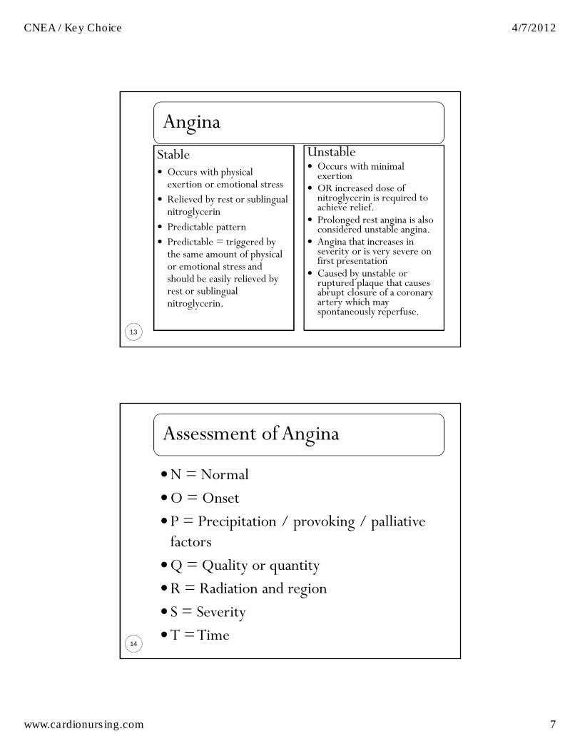
Angina
13
Stable � Occurs with physical exertion or emotional stress
� Relieved by rest or sublingual nitroglycerin
� Predictable pattern
� Predictable = triggered by the same amount of physical or emotional stress and should be easily relieved by rest or sublingual nitroglycerin.
Unstable � Occurs with minimal exertion
� OR increased dose of nitroglycerin is required to achieve relief.
� Prolonged rest angina is also considered unstable angina.
� Angina that increases in severity or is very severe on first presentation
� Caused by unstable or ruptured plaque that causes abrupt closure of a coronary artery which may spontaneously reperfuse.
Assessment of Angina
14
�N = Normal
�O = Onset
�P = Precipitation / provoking / palliative factors
�Q = Quality or quantity
�R = Radiation and region
� S = Severity
�T = Time
CNEA / Key Choice 4/7/2012
www.cardionursing.com 8
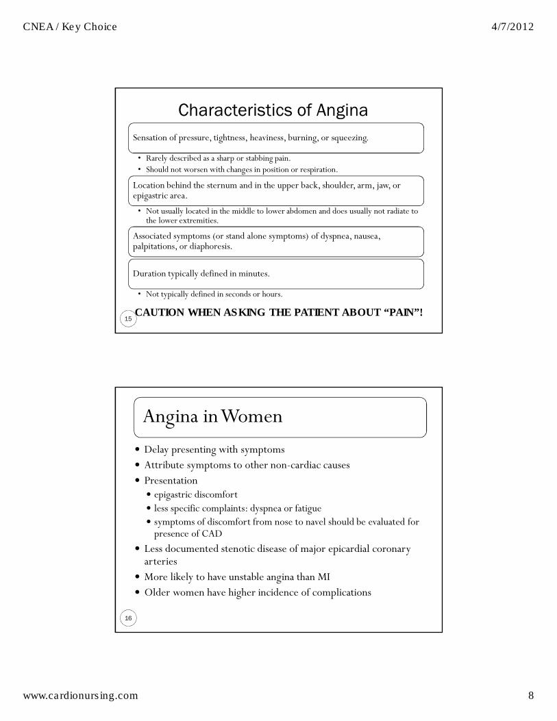
Characteristics of Angina
15
Sensation of pressure, tightness, heaviness, burning, or squeezing.
• Rarely described as a sharp or stabbing pain. • Should not worsen with changes in position or respiration.
Location behind the sternum and in the upper back, shoulder, arm, jaw, or epigastric area.
• Not usually located in the middle to lower abdomen and does usually not radiate to the lower extremities.
Associated symptoms (or stand alone symptoms) of dyspnea, nausea, palpitations, or diaphoresis.
Duration typically defined in minutes.
• Not typically defined in seconds or hours.
CAUTION WHEN ASKING THE PATIENT ABOUT “PAIN”!
Angina in Women
16
� Delay presenting with symptoms� Attribute symptoms to other non-cardiac causes � Presentation
� epigastric discomfort� less specific complaints: dyspnea or fatigue � symptoms of discomfort from nose to navel should be evaluated for presence of CAD
� Less documented stenotic disease of major epicardial coronary arteries
� More likely to have unstable angina than MI� Older women have higher incidence of complications
CNEA / Key Choice 4/7/2012
www.cardionursing.com 9
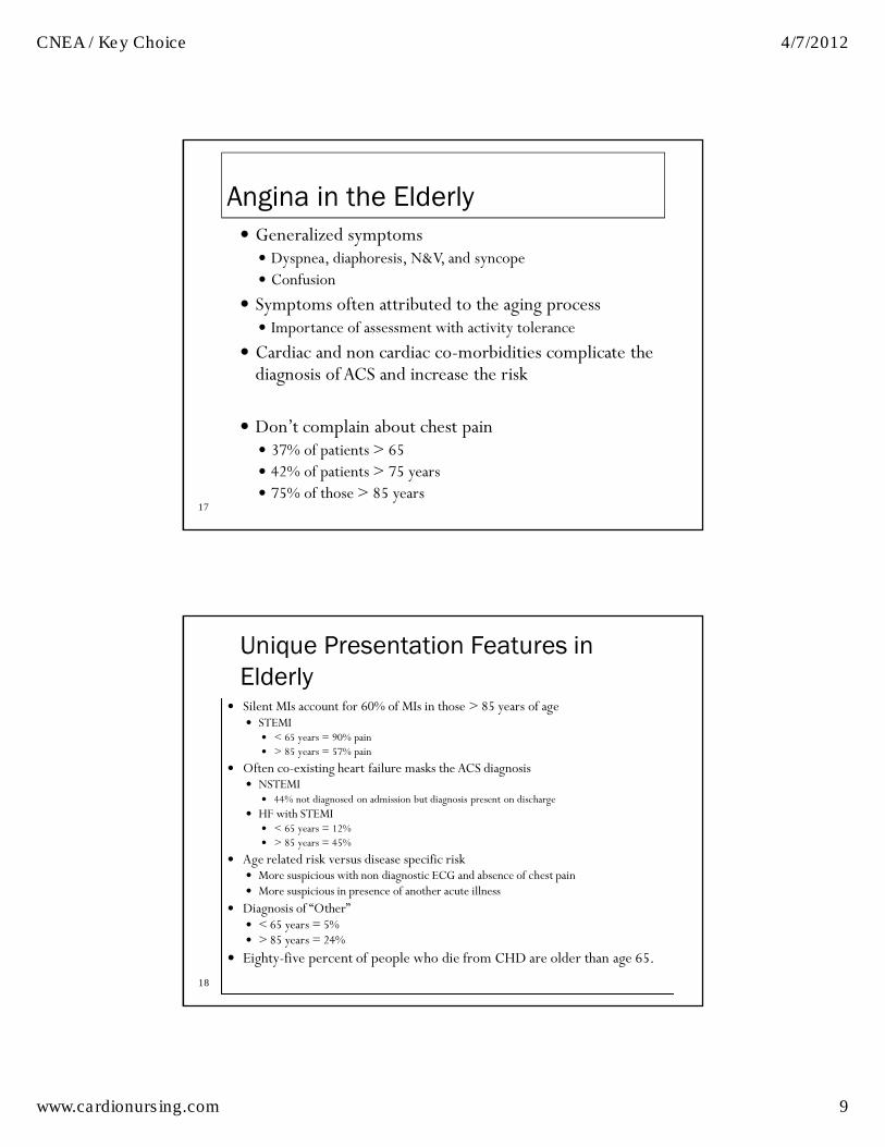
17
Angina in the Elderly� Generalized symptoms
� Dyspnea, diaphoresis, N&V, and syncope � Confusion
� Symptoms often attributed to the aging process� Importance of assessment with activity tolerance
� Cardiac and non cardiac co-morbidities complicate the diagnosis of ACS and increase the risk
� Don’t complain about chest pain� 37% of patients > 65� 42% of patients > 75 years � 75% of those > 85 years
18
Unique Presentation Features in
Elderly � Silent MIs account for 60% of MIs in those > 85 years of age
� STEMI � < 65 years = 90% pain� > 85 years = 57% pain
� Often co-existing heart failure masks the ACS diagnosis � NSTEMI
� 44% not diagnosed on admission but diagnosis present on discharge � HF with STEMI
� < 65 years = 12% � > 85 years = 45%
� Age related risk versus disease specific risk � More suspicious with non diagnostic ECG and absence of chest pain � More suspicious in presence of another acute illness
� Diagnosis of “Other” � < 65 years = 5%� > 85 years = 24%
� Eighty-five percent of people who die from CHD are older than age 65.

CNEA / Key Choice 4/7/2012
www.cardionursing.com 10
Angina in Diabetics
19
� Autonomic dysfunction can affect symptoms experienced with angina
� Less likely to experience pain. � 25% of all patients presenting with ACS are diabetic
� Have severe multi-vessel disease� Have higher rates of complications � Have a greater proportion of ulcerated plaques resulting in intracoronary thrombi
Acute MI Symptoms
20
Symptoms occur spontaneously and are not relieved by rest or nitroglycerin
Chest pressure or discomfort may be accompanied by nausea, vomiting, or diaphoresis
Patient may have hemodynamic instability or cardiac arrest from ventricular fibrillation
Acute MI patients have positive biomarkers and are classified as STEMI or NSTEMI based on ECG presentation

CNEA / Key Choice 4/7/2012
www.cardionursing.com 11
< 25% of ACS patients
Complete occlusion of a vessel by a thrombus Fibrin stable clot (red clot)
Classified more specifically by the portion of the left ventricle suffering injury.
Mortality is greatest within the first 24 to 48 hours of symptom onset
TREATMENT FOCUS = REPERFUSION 21
STEMI
Higher mortality and morbidity than STEMI Nationally under treated according to evidence based practice
guidelines (Crusade Registry)Pathophysiology often involves a platelet plug or white clot
Less stable clot Opportunity for spontaneous reperfusion
Differentiated from unstable angina by troponin levelsTREATMENT FOCUS = ANTIPLATELET THERAPY
22
NSTEMI

CNEA / Key Choice 4/7/2012
www.cardionursing.com 12
23
Causes of UA/NSTEMI*Causes of UA/NSTEMI*Causes of UA/NSTEMI*Causes of UA/NSTEMI*
� Thrombus or thromboembolism, usually arising on disrupted or eroded plaque� Occlusive thrombus, usually with collateral vessels� Subtotally occlusive thrombus on pre-existing plaque� Distal microvascular thromboembolism from plaque-associated
thrombus � Thromboembolism from plaque erosion
� Non–plaque-associated coronary thromboembolism
� Dynamic obstruction (coronary spasm or vascoconstriction) of epicardial and/or microvascular vessels
� Progressive mechanical obstruction to coronary flow
� Coronary arterial inflammation
� Secondary UA
� Coronary artery dissection
*These causes are not mutually exclusive; some patients have 2 or more causes. †DeWood MA, et al. N Engl J Med 1986;315:417–23. ‡May occur on top of an atherosclerotic plaque, producing missed-etiology angina or UA/NSTEMI. §Rare. Modified with permission from Braunwald E. Circulation 1998;98:2219–22. Anderson JL, et al. J Am Coll Cardiol. 2007;50:e1-e157, Table 3.
24
Physiological Changes in the Elderly
� Decreased arterial compliance
� Increased cardiac afterload
� Diastolic dysfunction
� Co-morbid diseases� Aortic stenosis
� Renal dysfunction
� Frailty � 25% of those > 85 years of age
� Inflammatory dysregulation
� Alterations in drug metabolism

CNEA / Key Choice 4/7/2012
www.cardionursing.com 13
25
Evaluation of Oxygen
Supply and Demand
� Increase myocardial oxygen demand:� Hyperthermia� Hypertension� Tachycardia� Conditions producing over stimulation of the sympathetic nervous system (cocaine use, hyperthyroidism)
� Decrease myocardial oxygen delivery:� Anemia� Pulmonary disease.
� Increase myocardial oxygen demand and decrease myocardial oxygen supply:� Aortic stenosis� Hypertrophic cardiomyopathy
Elderly are at risk for secondary coronary events related to supply and demand imbalance.
Cardiac Risk Factors
26
� Non-Modifiable Risk Factors� Previous history� Family history
� 1st degree relative (parents, siblings)
� Men < 55; Women < 65� Age � Gender� Socioeconomic Factors and Ethnicity
9 easily measured and potentially modifiable risk factors account for over 90% of the risk of an initial acute MI
� Smoking� Hypertension� Dyslipidemia� Diabetes� Obesity� Metabolic Syndrome� Inactivity� Alcohol

CNEA / Key Choice 4/7/2012
www.cardionursing.com 14
Other Pertinent History
27
CADCerebral Vascular Disease
Peripheral Vascular Disease
Cardiac Biomarkers
28
� Released into the blood when necrosis occurs as a result of membrane rupture of the myocytes
� Used in the evaluation of ACS� Myoglobin
� Rises the earliest� Within 2 hours after damage� Very sensitive, not specific
� CK (creatine kinase)� Enzyme present in the heart, brain, and skeletal muscle� Elevations are not specific to myocardial damage.
� CK-MB� More specific to the heart� Helpful in identifying more than minor amounts of myocardial damage� Rapidly rises in the presence of myocardial damage.

CNEA / Key Choice 4/7/2012
www.cardionursing.com 15
Cardiac Biomarkers
29
� Troponin I and T� Found only in cardiac muscle � Most sensitive indicator of myocardial damage
� Capable of diagnosing small amounts of myocardial necrosis not measured by rises in CK-MB levels
� Approximately 30% of patients with NSTEMI and normal CKMB levels will test positive
� Of equal sensitivity and specificity� Troponin remains elevated for a long period (late presentation) � Positive troponin + ECG changes of injury / ischemia = infarct � Non CAD causes of troponin elevation (sepsis, pulmonary emboli and chronic kidney disease)
� Troponin I more specific in renal dysfunction
30
Cardiac Biomarker Summary
Cardiac
Biomarker
Specificity /
Sensitivity
Rise Peak Duration
Myoglobin Sensitive but
not specific
Within 2 hours 4 to 10 hours < 24 hours
CK-MB Highly specific 4 to 6 hours 18 to 24 hours 2 to 3 days
Troponin I or T Highly specific
and sensitive
4 to 6 hours 18 to 24 hours 10 or more days

CNEA / Key Choice 4/7/2012
www.cardionursing.com 16
Timing of Release of Various Biomarkers After Timing of Release of Various Biomarkers After Timing of Release of Various Biomarkers After Timing of Release of Various Biomarkers After Acute Myocardial InfarctionAcute Myocardial InfarctionAcute Myocardial InfarctionAcute Myocardial Infarction
31
Shapiro BP, Jaffe AS. Cardiac biomarkers. In: Murphy JG, Lloyd MA, editors. Mayo Clinic Cardiology: Concise Textbook. 3rd ed. Rochester, MN: Mayo Clinic Scientific Press and New York: Informa Healthcare USA, 2007:773–80. Anderson JL, et al. J Am Coll Cardiol 2007;50:e1–e157, Figure 5.
Emerging Biomarkers for ACS and Risk
Stratification
32
� BNPt
� C Reactive Protein
�Myeloperoxidase
� Ischemia modified albumin
CNEA / Key Choice 4/7/2012
www.cardionursing.com 17
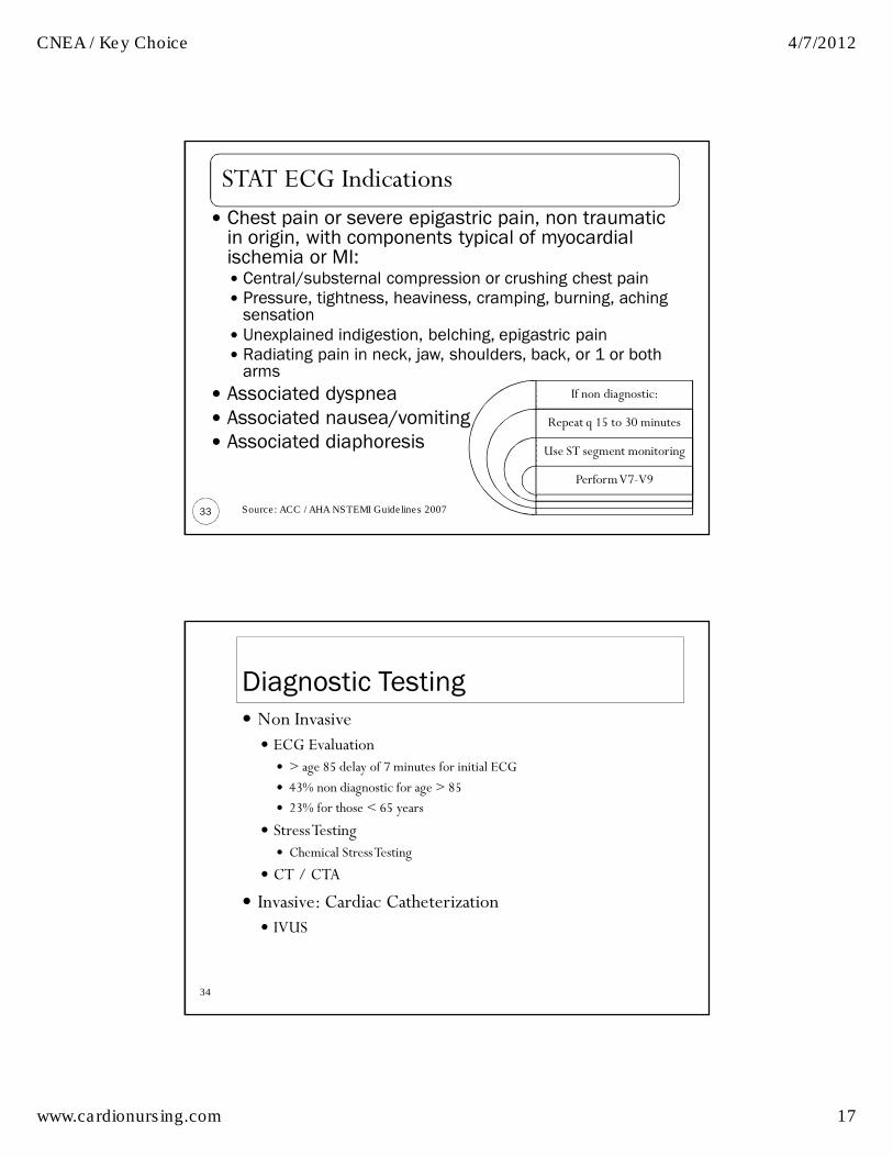
STAT ECG Indications
33
� Chest pain or severe epigastric pain, non traumatic in origin, with components typical of myocardial ischemia or MI:� Central/substernal compression or crushing chest pain� Pressure, tightness, heaviness, cramping, burning, aching
sensation� Unexplained indigestion, belching, epigastric pain� Radiating pain in neck, jaw, shoulders, back, or 1 or both
arms
� Associated dyspnea� Associated nausea/vomiting� Associated diaphoresis
If non diagnostic:
Repeat q 15 to 30 minutes
Use ST segment monitoring
Perform V7-V9
Source: ACC / AHA NSTEMI Guidelines 2007
34
Diagnostic Testing� Non Invasive
� ECG Evaluation � > age 85 delay of 7 minutes for initial ECG
� 43% non diagnostic for age > 85
� 23% for those < 65 years
� Stress Testing� Chemical Stress Testing
� CT / CTA
� Invasive: Cardiac Catheterization� IVUS

CNEA / Key Choice 4/7/2012
www.cardionursing.com 18
Stress Testing in Patients Presenting with Chest Pain
35
� Indicated when ECG and biomarkers are not diagnostic
� Should be done before discharge or within 72 hours as outpatient
� Precautionary pharmacotherapy for low risk patients being done on outpatient basis
�ASA�SL NTG�Beta blockers
Stress Testing
36
� Exercise Stress Test with or without myocardial imaging�Nuclear Scanning� Echocardiogram� Future
� Patient conditions requiring myocardial imaging with stress testing due to lack of reliable ECG interpretation include: � Left bundle branch block�> 1 mm ST-segment depression at rest� Paced ventricular rhythm�Wolf-Parkinson-White syndrome

CNEA / Key Choice 4/7/2012
www.cardionursing.com 19
Exercise Stress Testing
37
� Treadmills or bicycles� Able to exercise on a treadmill for 6 to 12 minutes� While exercising
� Myocardial oxygen demand increases� Coronary arteries dilate in response to increased demand
� If CAD� Coronary arteries not able to adequately dilate to meet the needs of the increased myocardial oxygen demand
� Abnormalities occur on 12-lead ECG or imaging studies
� Consideration with beta-blockers� Hold beta-blockers approximately 48 hours prior to testing� May not hold if determining effectiveness
� Exercise stress testing is less sensitive in women than in men
Chemical Stress Testing
38
� Dipyridamole, adenosine, regadenoson� Done in conjunction with myocardial imaging
� Dipyridamole and adenosine � Non specific adenosine receptor blockers
� Stimulation of these other receptors is what causes the unwanted side effective of atrioventricular (AV) block (A1 receptor) and bronchospasm (A2b and A3 receptors).
� Contraindications:� Severe lung disease or if wheezing
� All 3 agents: Stimulation of Adenosine A2a receptor causes coronary vasodilation � Causes coronary microvascular dilatation
similar to the coronary artery vasodilatation that occurs with exercise
� Regadenoson is A2a selective � Another advantage over other two agents: � Rapid dosing � Non weight based
� Antidote: Aminophylline for all 3 agents
� Patients should be off aminophylline or related products prior to testing with these chemical agents

CNEA / Key Choice 4/7/2012
www.cardionursing.com 20
Chemical Stress Testing
39
� Dobutamine
�High-dose dobutamine increases contractility and heart rate
� Increasing myocardial oxygen demand�More closely mimics exercise stress testing
�May be done with Echo instead of nuclear scan�Side effect: Tachyarrhythmias�Antidote: Beta blocker
Contraindications to Stress Testing
40
� Acute MI <_ 2 days old
� Acute myocarditis or pericarditis
� Acute pulmonary embolism
� Acute aortic dissection
� Symptomatic heart failure
� Severe aortic stenosis
� Symptomatic arrhythmias
� High-risk unstable angina

CNEA / Key Choice 4/7/2012
www.cardionursing.com 21
CT Angiography “FAST CT”
� 64 slice and beyond� Detailed 3D Image� Fast� Coronary artery calcium scoring
� Shows calcified plaque� Predictor of non-calcified plaque
� Coronary artery anatomy� Myocardial function� Need to lower heart rate� Radiation exposure� Good negative predictor
41
Cardiac Catheterization
42
� Indications� Patients with disabling angina despite medical treatment
� Patients with high-risk criteria for coronary heart disease (CHD) on noninvasive testing
� Patients who have survived sudden cardiac death
� Patients with angina and clinical signs of CHD
� Patients with low ejection fraction and ischemia on noninvasive testing
� Patients with inadequate information obtained from noninvasive testing
CNEA / Key Choice 4/7/2012
www.cardionursing.com 22
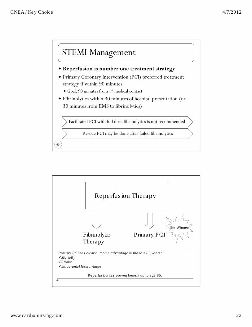
STEMI Management
43
� Reperfusion is number one treatment strategy
� Primary Coronary Intervention (PCI) preferred treatment strategy if within 90 minutes� Goal: 90 minutes from 1st medical contact
� Fibrinolytics within 30 minutes of hospital presentation (or 30 minutes from EMS to fibrinolytics)
Facilitated PCI with full dose fibrinolytics is not recommended.
Rescue PCI may be done after failed fibrinolytics
44
Reperfusion Therapy
Primary PCIFibrinolytic
Therapy
Primary PCI has clear outcome advantage in those > 65 years: �Mortality �Stroke �Intracranial Hemorrhage
Reperfusion has proven benefit up to age 85.
The Winner!
CNEA / Key Choice 4/7/2012
www.cardionursing.com 23
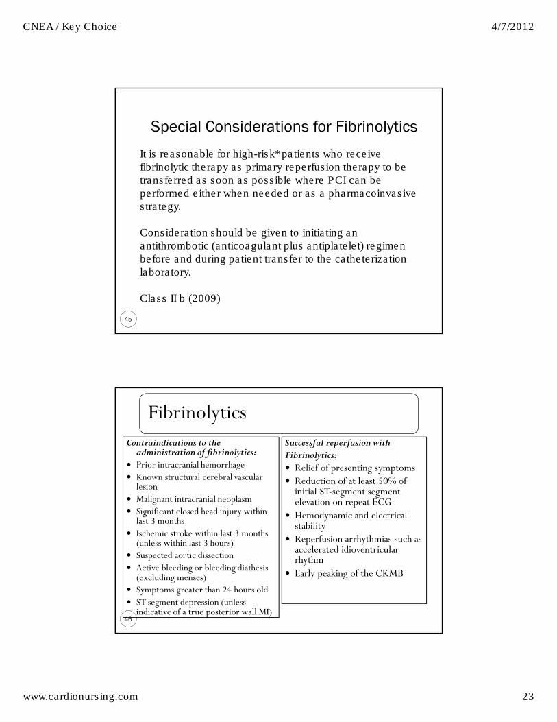
Special Considerations for Fibrinolytics
45
It is reasonable for high-risk* patients who receive
fibrinolytic therapy as primary reperfusion therapy to be
transferred as soon as possible where PCI can be
performed either when needed or as a pharmacoinvasive
strategy.
Consideration should be given to initiating an
antithrombotic (anticoagulant plus antiplatelet) regimen
before and during patient transfer to the catheterization
laboratory.
Class II b (2009)
Fibrinolytics
46
Contraindications to the administration of fibrinolytics:
� Prior intracranial hemorrhage� Known structural cerebral vascular lesion
� Malignant intracranial neoplasm� Significant closed head injury within last 3 months
� Ischemic stroke within last 3 months (unless within last 3 hours)
� Suspected aortic dissection� Active bleeding or bleeding diathesis (excluding menses)
� Symptoms greater than 24 hours old � ST-segment depression (unless indicative of a true posterior wall MI)
Successful reperfusion with Fibrinolytics: � Relief of presenting symptoms� Reduction of at least 50% of initial ST-segment segment elevation on repeat ECG
� Hemodynamic and electrical stability
� Reperfusion arrhythmias such as accelerated idioventricularrhythm
� Early peaking of the CKMB
CNEA / Key Choice 4/7/2012
www.cardionursing.com 24
47
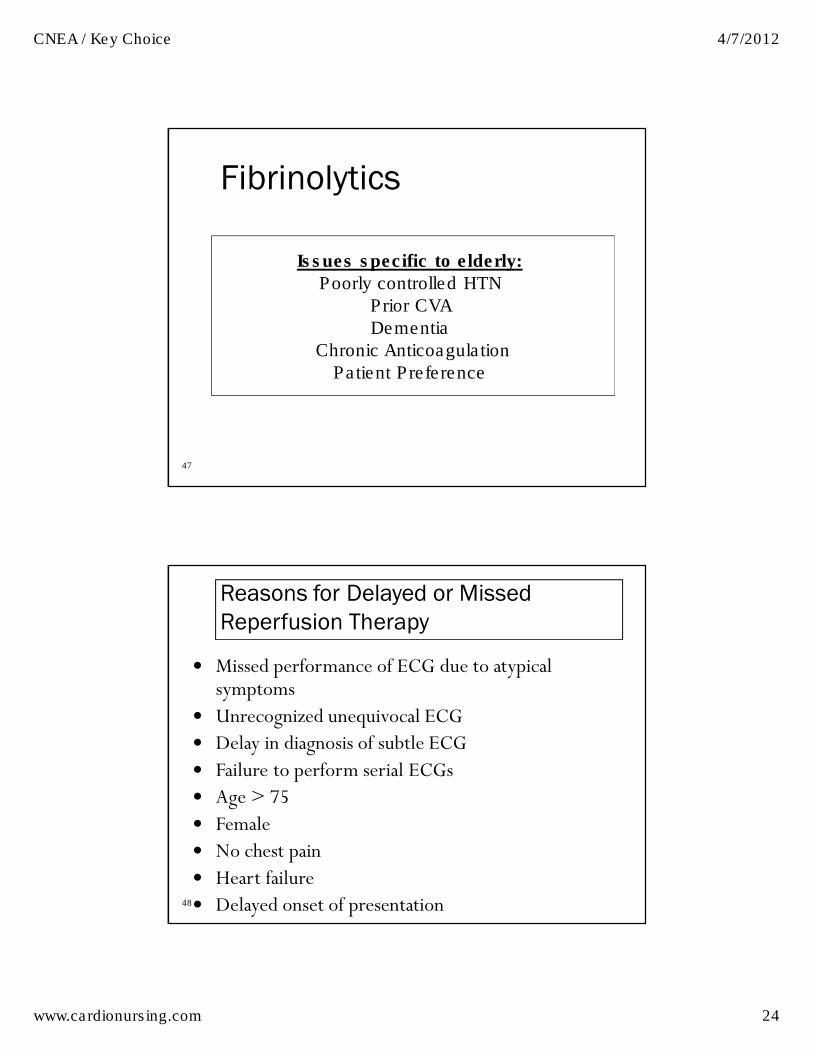
Fibrinolytics
Issues specific to elderly:
Poorly controlled HTN
Prior CVA
Dementia
Chronic Anticoagulation
Patient Preference
48
Reasons for Delayed or Missed
Reperfusion Therapy
� Missed performance of ECG due to atypical symptoms
� Unrecognized unequivocal ECG � Delay in diagnosis of subtle ECG� Failure to perform serial ECGs � Age > 75 � Female� No chest pain � Heart failure � Delayed onset of presentation

CNEA / Key Choice 4/7/2012
www.cardionursing.com 25
49
50
CNEA / Key Choice 4/7/2012
www.cardionursing.com 26
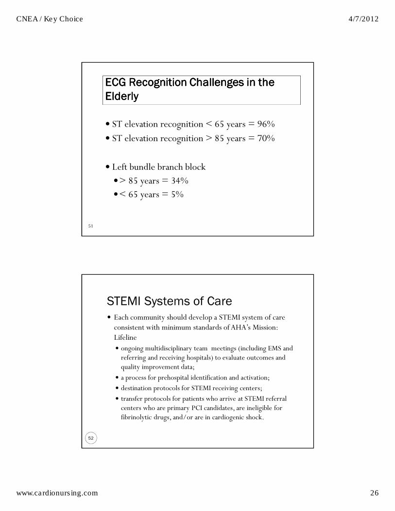
51
ECG Recognition Challenges in the ECG Recognition Challenges in the ECG Recognition Challenges in the ECG Recognition Challenges in the
Elderly Elderly Elderly Elderly
� ST elevation recognition < 65 years = 96%
� ST elevation recognition > 85 years = 70%
� Left bundle branch block
�> 85 years = 34% �< 65 years = 5%
STEMI Systems of Care
52
� Each community should develop a STEMI system of care consistent with minimum standards of AHA’s Mission: Lifeline� ongoing multidisciplinary team meetings (including EMS and referring and receiving hospitals) to evaluate outcomes and quality improvement data;
� a process for prehospital identification and activation;
� destination protocols for STEMI receiving centers;
� transfer protocols for patients who arrive at STEMI referral centers who are primary PCI candidates, are ineligible for fibrinolytic drugs, and/or are in cardiogenic shock.
CNEA / Key Choice 4/7/2012
www.cardionursing.com 27
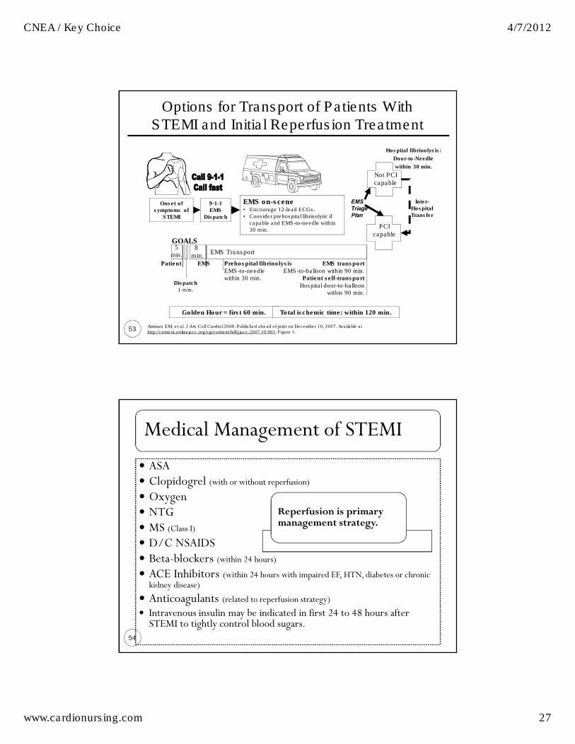
53
Options for Transport of Patients With STEMI and Initial Reperfusion Treatment
EMS Transport
Onset of
symptoms of
STEMI
9-1-1
EMS
Dispatch
EMS on-scene• Encourage 12-lead ECGs.
• Consider prehospital fibrinolytic if
capable and EMS-to-needle within
30 min.
GOALS
PCI
capable
Not PCI
capable
Hospital fibrinolysis:
Door-to-Needle
within 30 min.
Inter-
Hospital
Transfer
Golden Hour = first 60 min. Total ischemic time: within 120 min.
Patient EMS Prehospital fibrinolysis
EMS-to-needlewithin 30 min.
EMS transport
EMS-to-balloon within 90 min.Patient self-transport
Hospital door-to-balloon within 90 min.
Dispatch
1 min.
5
min.8
min.
Antman EM, et al. J Am Coll Cardiol 2008. Published ahead of print on December 10, 2007. Available at http://content.onlinejacc.org/cgi/content/full/j.jacc.2007.10.001. Figure 1.
Medical Management of STEMI
54
� ASA � Clopidogrel (with or without reperfusion) � Oxygen � NTG � MS (Class I) � D/C NSAIDS � Beta-blockers (within 24 hours) � ACE Inhibitors (within 24 hours with impaired EF, HTN, diabetes or chronic kidney disease)
� Anticoagulants (related to reperfusion strategy) � Intravenous insulin may be indicated in first 24 to 48 hours after STEMI to tightly control blood sugars.
Reperfusion is primary management strategy.

CNEA / Key Choice 4/7/2012
www.cardionursing.com 28
New 2009 Recommendation for New 2009 Recommendation for New 2009 Recommendation for New 2009 Recommendation for
Glucose Control Glucose Control Glucose Control Glucose Control
55
� It is reasonable to use an insulin-based regimen to achieve and maintain glucose levels less than 180 mg/dL while avoiding hypoglycemia* for patients with STEMI with either a complicated or uncomplicated course.
56
Treatment of Non STEMI / Unstable Angina:
New Guidelines
� Attacking Platelet is number one treatment strategy
� Dual antiplatelet therapy therapy for invasive strategies in medium to high risk patients �ASA (and one of following) � Clopidogrel (loading) � Prasugral (loading) � GP II b / III a Inhibitors (eptifibatide or tirofiban)
� Antiplatelet therapy also in conservative treatment � Prasugrel not unless PCI is planned �Abciximab not unless PCI is planned

CNEA / Key Choice 4/7/2012
www.cardionursing.com 29
Treatment of Non STEMI / Unstable
Angina: New Guidelines
57
� Anticoagulation options in NSTEMI: �Unfractionated heparin� LMWH (enoxaparin) � Factor Xa inhibitor (fondaparinux) �Direct thrombin inhibitor (bivalrudin)
� Duration of anticoagulation � Enoxaparin or fonaparinux for duration (in conservative) or up to 8 days
� UFH for 48 hours in conservative
Discussion Points
58
�Class III recommendations �No Abciximab if no PCI Planned �No GPIIb/IIIa in low risk patient on dual antiplatetNo prasugrel if previous stroke or TiA
�Oral antiplatelets�Clopidogrel for 1 month preferably a year after conservative treatment
� Ischemic protection versus bleeding risk
CNEA / Key Choice 4/7/2012
www.cardionursing.com 30
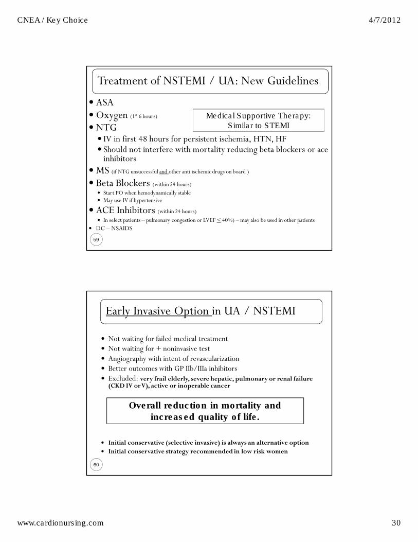
Treatment of NSTEMI / UA: New Guidelines
59
� ASA �Oxygen (1st 6 hours)� NTG
� IV in first 48 hours for persistent ischemia, HTN, HF � Should not interfere with mortality reducing beta blockers or ace inhibitors
�MS (if NTG unsuccessful and other anti ischemic drugs on board ) � Beta Blockers (within 24 hours)
� Start PO when hemodynamically stable � May use IV if hypertensive
� ACE Inhibitors (within 24 hours)� In select patients – pulmonary congestion or LVEF < 40%) – may also be used in other patients
� DC – NSAIDS
Medical Supportive Therapy:
Similar to STEMI
Early Invasive Option in UA / NSTEMI
60
� Not waiting for failed medical treatment � Not waiting for + noninvasive test � Angiography with intent of revascularization � Better outcomes with GP IIb/IIIa inhibitors � Excluded: very frail elderly, severe hepatic, pulmonary or renal failure (CKD IV or V), active or inoperable cancer
� Initial conservative (selective invasive) is always an alternative option � Initial conservative strategy recommended in low risk women
Overall reduction in mortality and
increased quality of life.

CNEA / Key Choice 4/7/2012
www.cardionursing.com 31
61
Early Invasive Indications
�Refractory angina or hemodynamic or electrical instability� Without serious co-morbidities or contraindications to
such procedures
� May be reasonable in patients with chronic renal insufficiency
�Initially stabilized with high risk for clinical events
62
High Risk Features in UA / NSTEMI
� Recurrent angina / ischemia � Rest or low level activity with medical treatment
� Troponin + � New or presumed new ST depression � S&S HF or worsening mitral regurgitation � High risk findings on noninvasive testing
� EF < 35%, large anterior perfusion defect, multiple perfusion defects)
� Hemodynamic instability � Sustained VT � PCI within 6 months � Prior CABG � Reduced LV Function � High risk TIMI or GRACE Score
Elderly: cancer, renal insufficiency, lung disease,
anemia, and heart failure are common co morbid conditions
Population > 75 years: 80% are high risk

CNEA / Key Choice 4/7/2012
www.cardionursing.com 32
Risk Assessment in UA / NSTEMI
63
� TIMI Risk Score � Age > 65
� 3 or > risk factors for CAD
� Prior 50% or > stenosis
� ST deviation on ECG
� 2 or > anginal events in previous 24 hours
� Use of ASA in prior 7 days
� Elevated cardiac biomarkers
� GRACE �Older age
� Killip class
� Systolic BP
� Cardiac arrest during presentation
� Serum creatinine
� Positive initial cardiac markers
� HR
64
Markers of Risk: Specific to Elderly � Mobility and function
� Activities of daily living
� Strength
� Physiological reserves � Frailty
� Poor Nutrition Status � Albumin
�Weight loss
� Cognitive Impairment
� Hearing Alterations
� Vision Alterations
� Isolation
� Resources / Education
� Socioeconomic

CNEA / Key Choice 4/7/2012
www.cardionursing.com 33
65
Risk of In Hospital Death Specific to Age Risk of In Hospital Death Specific to Age Risk of In Hospital Death Specific to Age Risk of In Hospital Death Specific to Age
� < 65 years: 1 in 100
� > 85 years: 1 in 10
1 year mortality rate:
�75 years: 1 in 5
� 85 years: 1 in 4
66
Feature High RiskHigh RiskHigh RiskHigh Risk≥ 1 of the
features
below must
be present:
Intermediate RiskIntermediate RiskIntermediate RiskIntermediate RiskNo high-risk features, but must have
1 of the following:
Low RiskLow RiskLow RiskLow RiskNo high- or intermediate-
risk features but may
have any features below:
HistoryHistoryHistoryHistory Accelerating
tempo of
ischemic sx
in preceding
48 h
Prior MI, peripheral or
cerebrovascular disease, or CABG;
prior ASA use
Character Character Character Character
of painof painof painof pain
Prolonged
ongoing (>
20 min) rest
pain
• Prolonged (> 20 min) rest angina,
now resolved, w/ moderate/high
likelihood of CAD
• Rest angina (> 20 min) or relieved
with rest or sublingual NTG
• Nocturnal angina
• New-onset or progressive CCS
class III/IV angina in past 2 wks
w/o prolonged (> 20 min) rest pain
but with intermediate/high
likelihood of CAD
• ↑ Angina frequency,
severity or duration
• Angina provoked at
lower threshold
• New onset angina with
onset 2 wks to 2 mos
prior to presentation
Short-Term Risk of Death/Nonfatal MI in Patients With UA/NSTEMI

CNEA / Key Choice 4/7/2012
www.cardionursing.com 34
67
Feature High riskHigh riskHigh riskHigh risk Intermediate riskIntermediate riskIntermediate riskIntermediate risk Low riskLow riskLow riskLow risk
Clinical Clinical Clinical Clinical
findingsfindingsfindingsfindings
• Pulmonary edema, most
likely due to ischemia
• New/worsening MR
murmur
• S3 or new/worsening rales
• Hypotension, bradycardia,
tachycardia
• Age > 75 y
Age > 70 y
ECGECGECGECG •Angina @ rest with
transient ST-segment
changes > 0.5 mm
•BBB, new/presumed new
•Sustained VT
• T-wave changes
• Pathological Q-waves/resting ST-
depression < 1 mm in multiple
lead groups (anterior, inferior,
lateral)
Normal or
unchanged
ECG
Cardiac Cardiac Cardiac Cardiac
markersmarkersmarkersmarkers
↑ Cardiac TnT, TnI, or CK-MB
(e.g., TnT/TnI > 0.1 ng/mL)
Slightly ↑ cardiac TnT, TnI, or CK-
MB (e.g., TnT > 0.01, but < 0.1
ng/mL)
Normal
Estimation of the short-term risk of death and nonfatal cardiac ischemic events in UA/NSTEMI is a complex multivariable problem that cannot be fully specified in a table such as this; this table is mean to offer general guidance & illustration rather than rigid algorithms. Braunwald E, et al. AHCPR Publication No. 94-0602:1–154. Anderson JL, et al. J Am Coll Cardiol 2007;50:e1–e157, Table 7.
Short-Term Risk of Death/Nonfatal MI in Patients With UA/NSTEMI, Continued
68
Beta Blockers Considerations � Oral Beta Blockers
�Within 24 hours
� IV Beta Blockers � Reasonable in patients who are hypertensive
� May be harmful in patients with high risk for cardiogenic shock
� Beta blockers have greater benefit in elderly for reduction of future MI and death than in younger patient populations
� Contraindications� Signs of HF � Low cardiac output state � Increased risk for cardiogenic shock � Age > 70 years is a risk factor
�Relative contraindications � PR > .24 seconds � 2nd or 3rd degree block � Active asthma � Reactive airway disease

CNEA / Key Choice 4/7/2012
www.cardionursing.com 35
69
Nitrate Considerations
Contraindications � Systolic BP < 90 mm Hg or < 30 mm Hg below baseline
� Bradycardia < 50 BPM� Tachycardia > 100 BPM (in absence of clinical HF)
� Right ventricular infarct� Within 24 hours of sildenafil � Within 48 hours of taldalafil
� Nitrates may be more helpful in patients > 70 years in reduction of death and heart failure @ 6 month follow up � Nitrates not mortality reducing in younger populations
Include female
patients:
Pulmonary HTN
Other Medication Considerations
70
� Hold ace inhibitors for BP < 100 mm Hg systolic or < 30 mm Hg below baseline
� No IV ace inhibitor within 24 hours due to risk of hypotension
� No immediate release dihydropyridine calcium channel blockers without beta blockade on board
� NSAIDS (except for ASA), whether nonselective or COX-2–selective agents increase risk of mortality, reinfarction, hypertension, HF, and myocardial rupture
� Proton Pump Inhibitors should be prescribed to patients at risk for GI bleed – However: Caution with Clopidogrel
CNEA / Key Choice 4/7/2012
www.cardionursing.com 36
71
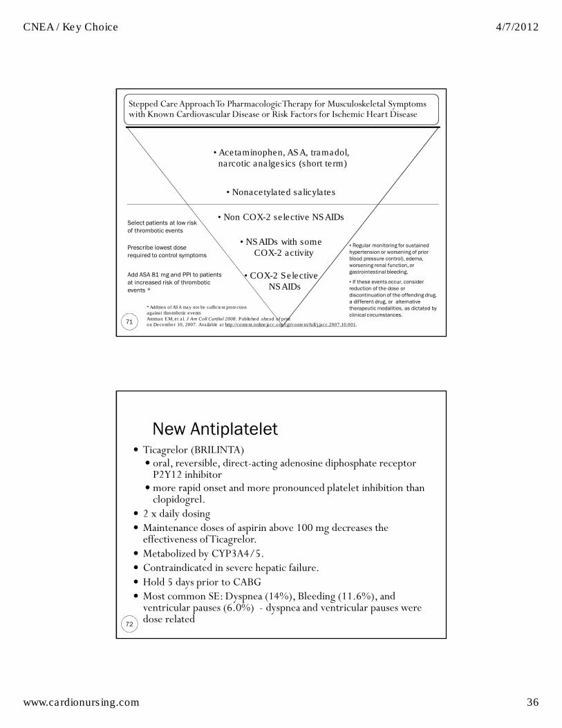
• Acetaminophen, ASA, tramadol,narcotic analgesics (short term)
• COX-2 Selective NSAIDs
• Nonacetylated salicylates
• Non COX-2 selective NSAIDs
• NSAIDs with someCOX-2 activity
Stepped Care Approach To Pharmacologic Therapy for Musculoskeletal Symptoms with Known Cardiovascular Disease or Risk Factors for Ischemic Heart Disease
Select patients at low risk
of thrombotic events
Prescribe lowest dose
required to control symptoms
Add ASA 81 mg and PPI to patients
at increased risk of thrombotic
events *
• Regular monitoring for sustained
hypertension or worsening of prior
blood pressure control), edema,
worsening renal function, or
gastrointestinal bleeding.
• If these events occur, consider
reduction of the dose or
discontinuation of the offending drug,
a different drug, or alternative
therapeutic modalities, as dictated by
clinical circumstances.
* Addition of ASA may not be sufficient protection
against thrombotic events
Antman EM, et al. J Am Coll Cardiol 2008. Published ahead of print on December 10, 2007. Available at http://content.onlinejacc.org/cgi/content/full/j.jacc.2007.10.001.
New Antiplatelet
72
� Ticagrelor (BRILINTA) � oral, reversible, direct-acting adenosine diphosphate receptor P2Y12 inhibitor
�more rapid onset and more pronounced platelet inhibition than clopidogrel.
� 2 x daily dosing � Maintenance doses of aspirin above 100 mg decreases the effectiveness of Ticagrelor.
� Metabolized by CYP3A4/5.� Contraindicated in severe hepatic failure. � Hold 5 days prior to CABG � Most common SE: Dyspnea (14%), Bleeding (11.6%), and ventricular pauses (6.0%) - dyspnea and ventricular pauses were dose related

CNEA / Key Choice 4/7/2012
www.cardionursing.com 37
More on Ticagrelor
73
� PLATO: Multicenter, double-blind, randomized trial� Ticagrelor (180-mg loading dose, 90 mg twice daily thereafter) to clopidogrel (300-to-600-mg loading dose, 75 mg daily thereafter)
� 18,624 patients admitted to the hospital with an acute coronary syndrome, (with or without ST-segment elevation).
� Primary end point — a composite of death from vascular causes, myocardial infarction, or stroke (9.8% to 11.7%; p = <0.001
�No significant difference in rates of major bleeding � Ticagrelor was associated with a higher rate of major bleeding not related to coronary-artery bypass grafting
� Indications � Reduce rate of thrombotic cardiovascular events in ACS. � Reduce rate of stent thrombosis post PCI.
74
Long Term Management of ACS Medications to improve prognosis� Aspirin
� ASA benefits > in those > 65 years� Generally no dose adjusting
� Clopidogrel / Prasugrel� Caution age > 75 with prasugrel� Higher risk of bleeding dual antiplatelet therapy
� No elderly sub group data for clopiodgrel
� *Beta-blockers � *ACE inhibitors (in select patients)
� ARBs (may be used with ACE-I in systolic dysfunction) � Aldactone (EF < 40 with HF or diabetes)
� Lipid-lowering drugs (statins) � Have greater benefit in elderly for reduction of future MI and death than in younger patient populations
* Beta blockers and ACE inhibitors impact long term ventricular remodeling

CNEA / Key Choice 4/7/2012
www.cardionursing.com 38
SL NTG Instruction
75
� No more than 1 dose of SL NTG � If chest discomfort is unimproved or is worsening
5 min after 1 NTG call 9-1-1 immediately before
taking additional NTG.
� May take additional NTG while waiting EMS.
� Chew ASA while waiting EMS.
� In chronic stable angina if symptoms are significantly
improved by 1 dose of NTG may repeat NTG every 5 min
for a maximum of 3 doses and call 9-1-1 if symptoms
have not resolved completely.
76
Medical Therapy Issues in the Elderly Medical Therapy Issues in the Elderly Medical Therapy Issues in the Elderly Medical Therapy Issues in the Elderly
� Altered responses and vulnerability to drugs with:
�Hypotensive action (nitrates, calcium blockers) �Cerebral effects (beta blockers)
� Caution with renally cleared drugs
� START LOW and GO SLOW!!
CNEA / Key Choice 4/7/2012
www.cardionursing.com 39
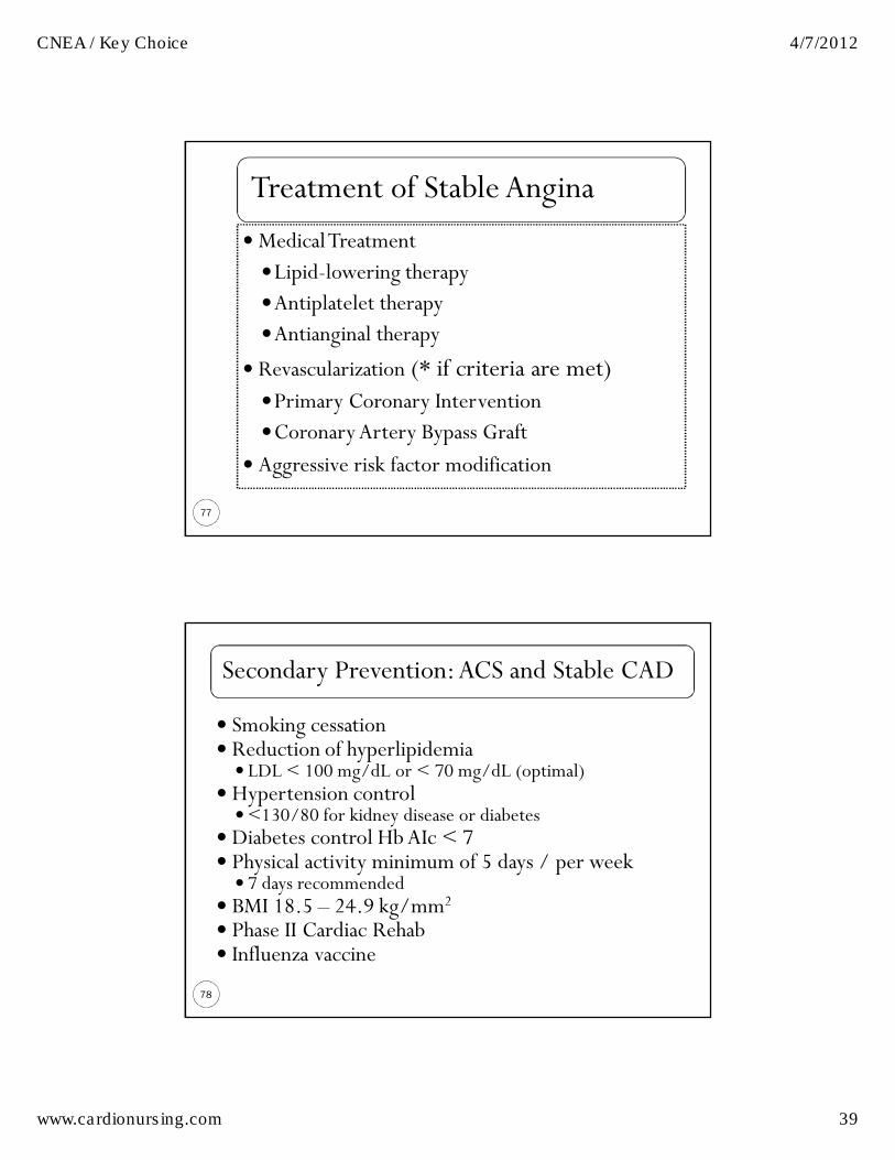
Treatment of Stable Angina
77
�Medical Treatment
�Lipid-lowering therapy �Antiplatelet therapy�Antianginal therapy
� Revascularization (* if criteria are met) �Primary Coronary Intervention�Coronary Artery Bypass Graft
� Aggressive risk factor modification
Secondary Prevention: ACS and Stable CAD
78
� Smoking cessation� Reduction of hyperlipidemia
� LDL < 100 mg/dL or < 70 mg/dL (optimal) � Hypertension control
�<130/80 for kidney disease or diabetes � Diabetes control HbAIc < 7� Physical activity minimum of 5 days / per week
� 7 days recommended� BMI 18.5 – 24.9 kg/mm2� Phase II Cardiac Rehab � Influenza vaccine

CNEA / Key Choice 4/7/2012
www.cardionursing.com 40
79
Key Nursing Care Considerations
� Assess response to beta-blocker therapy� HR / BP � Arrhythmia control� Need for higher / lower dose
� Reassess oxygen saturation after 6 hours and discontinue O2 if saturation is more than 90%
� Assess for complications related to specific type of MI� Assess heart sounds for new holosystolic murmurs
� Risk for myocardial rupture
� Observe for signs of left ventricular dysfunction, including hypotension or clinical signs of heart failure.
� Monitor ECG for conduction disturbances and arrhythmias� Assess for presence of RV infarct
80
Key Nursing Care Considerations
� Restrict activity for at least the first 12 hours, and then begin Phase I Cardiac Rehabilitation� Referral to Phase II Cardiac Rehabilitation
� Utilize cardiac monitoring� ST-segment monitoring � Uninterrupted monitoring for first 24-48 hours
� Focus on holistic approach to anxiety reduction� Include the family. Family visits do not have a negative impact on vital signs or cardiac rhythm
� Address addiction to nicotine� Consideration for nicotine withdrawal� Specific smoking cessation plan

CNEA / Key Choice 4/7/2012
www.cardionursing.com 41
Complications of MI
81
� Hemodynamic Alterations � Ventricular Arrhythmias � Atrial Arrhythmias � Pericarditis � Ventricular Aneurysms � Mechanical Complications
� Myocardial Rupture (free wall or VSD) � Papillary Muscle Rupture
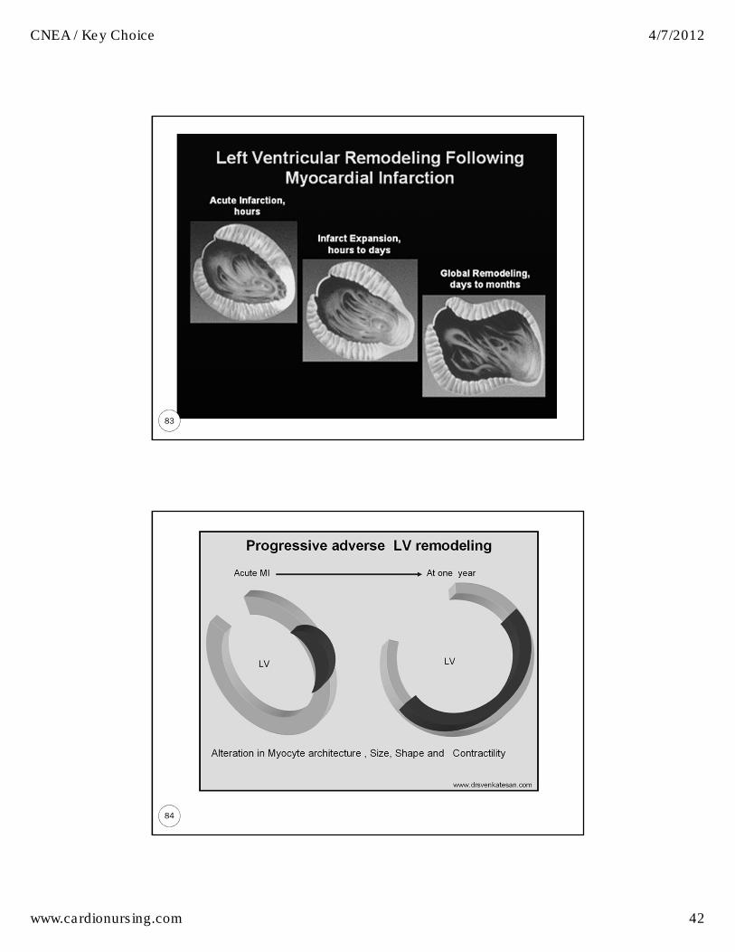
� Long Term: Ventricular Remodeling
82
Complications Specific to Elderly
� STEMI 30 day mortality � < 65 years = 3%
� > 85 years = 30%
� Death related to electrical and mechanical (free wall or papillary muscle rupture) catastrophes
� Reinfarction
� HF development
� Need for transfusion
� PCI – Elderly women and groin complications
CNEA / Key Choice 4/7/2012
www.cardionursing.com 42
83
84
CNEA / Key Choice 4/7/2012
www.cardionursing.com 43
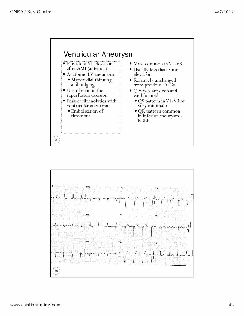
Ventricular Aneurysm
85
� Persistent ST elevation after AMI (anterior)
� Anatomic LV aneurysm �Myocardial thinning and bulging
� Use of echo in the reperfusion decision
� Risk of fibrinolytics with ventricular aneurysm� Embolization of thrombus
� Most common in V1-V3 � Usually less than 3 mm elevation
� Relatively unchanged from previous ECGs
� Q waves are deep and well formed �QS pattern in V1-V3 or very minimal r
�QR pattern common in inferior aneurysm / RBBB
86

CNEA / Key Choice 4/7/2012
www.cardionursing.com 44
87
Myocardial Rupture
� Incidence � 10% MI deaths
� Definition � Myocardial leakage – hemipericardium – tamponade� Perceived sudden; often slow tear
� Associated Factors � Late fibrinolytics� Delayed hospital admission
� Free wall rupture = tamponade� Septal involvement = VSD � Posterior wall = Risk for papillary muscle rupture
88

CNEA / Key Choice 4/7/2012
www.cardionursing.com 45
Myocardial Rupture � Post-infarction regional pericarditis precedes rupture (94% of the time)
89
T Wave Patterns in Post-infarction Regional Pericarditis
Persistently positive T waves 48 hours after an MI
Premature reversal of T wave inversion to
positive
ST segment reelevation
90

CNEA / Key Choice 4/7/2012
www.cardionursing.com 46
91
9292

CNEA / Key Choice 4/7/2012
www.cardionursing.com 47
ST Segment Monitoring
A SUCCESS Story!!The next 2 slides show the following: 1. Admission ECG for a patient with an anteroseptal / lateral wall STEMI.2. ECG post intervention for same patient.
1. Note: The T waves have not yet inverted post intervention. Ideally T waves will begin to invert after an intervention showing evidence of reperfusion.
REMEMBER: T wave must invert within 48-72 hours after a STEMI (the sooner the better). Failure of T waves to invert after a STEMI is indicative of post infarction regional pericarditis and the patient is at higher risk for myocardial rupture.
93
94

CNEA / Key Choice 4/7/2012
www.cardionursing.com 48
95
The strip below assessing ST segments in V3 was done 48 hours post STEMI (same patient as previous 2 ECGs.). The failure of the T waves to invert is indicative of post infarction regional pericarditis with increased risk of myocardial rupture. The patient was hypotensive, which raises the concern for cardiac tamponade as the etiology of the hypotension. This assessment finding was
communicated to the cardiologist.
The patient’s echocardiogram showed a large pericardial effusion and the patient subsequently underwent a surgical pericardial window.
96

CNEA / Key Choice 4/7/2012
www.cardionursing.com 49
97
Cardiac Tamponade
� Clinical syndrome caused by accumulation of fluid in the pericardial space
� Same causes as pericarditis / pericardial effusion � Increase capillary permeability due to inflammation may cause fluid leak into pericardial space � >120cc can cause tamponade if rapid� 2 Liters may not cause tamponade if slow
� Results in reduction in ventricular filling and ultimately hemodynamic compromise
� Differentiation between pericardial effusion and tamponade is hemodynamic status.
98
Practice EKG 1of 3
98

CNEA / Key Choice 4/7/2012
www.cardionursing.com 50
99
Practice EKG 2 of 3
99
100
Practice EKG 3 of 3
100

CNEA / Key Choice 4/7/2012
www.cardionursing.com 51
Papillary Muscle Rupture
101
102
Final Quote:
Our grand business in life
is not to see what lies
dimly at a distance,
but to do what lies clearly at hand.
Thomas Carlyle (1795-1881)