Embed Size (px)
Citation preview

doi: 10.1136/gut.2009.191320 published online November 18, 2009Gut
Alexander Swidsinski, Yvonne Doerffel, Vera Loening-Baucke, et al. nucleatum/necrophoruminvasion with Fusobacterium Acute appendicitis is characterized by local
http://gut.bmj.com/content/early/2009/11/18/gut.2009.191320Updated information and services can be found at:
These include:
P<P Published online November 18, 2009 in advance of the print journal.
serviceEmail alerting
the top right corner of the online article.Receive free email alerts when new articles cite this article. Sign up in the box at
Notes
digital object identifier (DOIs) and date of initial publication. theindexed by PubMed from initial publication. Citations to Advance online articles must include
final publication). Advance online articles are citable and establish publication priority; they areappeared in the paper journal (edited, typeset versions may be posted when available prior to Advance online articles have been peer reviewed and accepted for publication but have not yet
http://gut.bmj.com/cgi/reprintformTo order reprints of this article go to:
http://gut.bmj.com/subscriptions go to: GutTo subscribe to
group.bmj.com on March 10, 2010 - Published by gut.bmj.comDownloaded from
1
Title
Acute appendicitis is characterized by local invasion with Fusobacterium
nucleatum/necrophorum
Short title
Fusobacteria in acute appendicitis
A Swidsinski,1 Y Dörffel,2 V Loening-Baucke,1 F Theissig,3 J C Rückert,4, M Ismail,4 W A
Rau,5 D Gaschler,5 M Weizenegger,6 S Kühn,7 J Schilling,1 W V Dörffel8
1 Humboldt University, Charité Hospital, CCM, Laboratory for Molecular
Genetics, Polymicrobial Infections and Bacterial Biofilms and Department of
Medicine, Section of Gastroenterology, Charité Universitätsmedizin Berlin,
CCM, 10117 Berlin, Germany
2 Outpatient Clinic, Luisenstr. 11-13, Charité Universitätsmedizin Berlin, 10117
Berlin, Germany
3 Carl-Thiem-Klinikum, Pathology, Thiemstr. 111, 03048 Cottbus, Germany
4 Department of General, Visceral, Thoracic and Vascular Surgery, Charité
Universitätsmedizin Berlin, Charitéplatz 1, 10117 Berlin, Germany
5 Department of Surgery, Oberhavel Kliniken Hennigsdorf, Marwitzer Str.91,
16761 Hennigsdorf, Germany
6 Limbach Laboratory, Im Breitspiel 15, 69126 Heidelberg, Germany
7 Department of General, Visceral and Thoracic Surgery, Helios Klinikum Bad
Saarow, Pieskower Strasse 33, 15526 Bad Saarow, Germany
8 Department of Cardiology, Pulmonology, Helios Klinikum Bad Saarow,
Pieskower Strasse 33, 15526 Bad Saarow, Germany
Gut Online First, published on November 18, 2009 as 10.1136/gut.2009.191320
Copyright Article author (or their employer) 2009. Produced by BMJ Publishing Group Ltd (& BSG) under licence.
group.bmj.com on March 10, 2010 - Published by gut.bmj.comDownloaded from

2
Corresponding author: Dr. med. Alexander Swidsinski, Laboratory for Molecular Genetics,
Polymicrobial Infections and Bacterial Biofilms, Charité Universitätsmedizin Berlin , Campus
Mitte, 10098 Berlin, Germany, Tel: 49 30 450 514 003, Fax: 49 30 450 514 039 e-mail:
Keywords: FISH, acute appendicitis, infection, Fusobacteria, Fusobacterium nucleatum,
Fusobacterium necrophorum, inflammatory bowel disease, invasive bacteria, mucosal flora,
intestinal microbiota, polymicrobial infection
Words Abstract: 242
Body: 3182
Abbreviations:
FISH - fluorescence in situ hybridizationAlexa488, Cy3, Cy5, DAPI – different
fluorescent dyes corresponding to green, orange, dark red, and blue colours
FISH probes abbreviations used in text:
Ato291 - Atopobium,
Bac303 - Bacteroides,
Bif164 - Bifidobacteriaceae,
Chis150 - Clostridium histolyticum,
Ebac1790 - Enterobacteriaceae,
Ecyl1387 - Eubacterium cylindroides,
Erec482 - Clostridium group XIVa,
Eub338 - Eubacteria (virtually all bacteria),
Fprau - Faecalibacterium prausnitzii,
group.bmj.com on March 10, 2010 - Published by gut.bmj.comDownloaded from

3
Fnec - Fusobacterium necrophorum,
Fnuc - Fusobacterium nucleatum,
Fuso - Fusobacteria,
Lab158 - Lactobacillus,
Muc1437 - Akkermansia muciniphila
Pasco - Phascolarctobacterium faecium,
Pnig657 - Prevotella nigrescens,
Ser1410 - Serpulina,
Veil223 - Veillonella
The Corresponding Author has the right to grant on behalf of all authors, an exclusive licence
on the worldwide basis to the BMJ Publishing Group Ltd and its Licensees to permit this
article if accepted to be published in Gut editions and any other BMJPGL products to exploit
to all subsidiary rights, as set out in the licence (http://group.bmj.com/products/
journals/instructions-for-authors/licence-forms).
There are no competing interests for any of the authors.
The manuscript includes no personal information about any of the investigated patients which
would make a patient informed consent necessary.
The investigations were approved by the Institutional Review Board of the Charité
Universitätsmedizin Berlin.
group.bmj.com on March 10, 2010 - Published by gut.bmj.comDownloaded from
4
ABSTRACT
Background: Acute appendicitis is a local intestinal inflammation with unclear origin. The
aim was to test whether bacteria in appendicitis differ in composition to bacteria found in
cecal biopsies from healthy and disease controls.
Methods and patients: We investigated sections of 70 appendices using rRNA-based
fluorescence in situ hybridization. Four hundred cecal biopsies and 400 faecal samples from
patients with inflammatory bowel disease and other conditions were used as controls. A set of
73 group-specific bacterial probes was applied for the study.
Results: The mucosal surface in catarrhal appendicitis showed characteristic lesions of single
epithelial cells filled with a mixed bacterial population (“pinned cells”) without ulceration of
the surroundings. Bacteria deeply infiltrated the tissue in suppurative appendicitis.
Fusobacteria (mainly Fusobacterium nucleatum and necrophorum) were a specific
component of these epithelial and submucosal infiltrates in 62% of patients with proven
appendicitis. The presence of Fusobacteria in mucosal lesions correlated positively with the
severity of the appendicitis and was completely absent in cecal biopsies from healthy and
disease controls. Main faecal microbiota represented by Bacteroides, Eubacterium rectale
(Clostridium group XIVa), Faecalibacterium prausnitzii groups and Akkermansia
muciniphila were inversely related to the severity of the disease. The occurrence of other
bacterial groups within mucosal lesions of acute appendicitis was not related to the severity of
the appendicitis. No Fusobacteria were found in rectal swabs of patients with acute
appendicitis.
Conclusions: Local infection with Fusobacterium nucleatum/necrophorum is responsible for
the majority of cases of acute appendicitis.
group.bmj.com on March 10, 2010 - Published by gut.bmj.comDownloaded from
5
INTRODUCTION
Acute appendicitis is the most common surgical emergency. After more than 250 years of
intensive research, the disease is clinically well characterized and appears uniform and simple
from the clinical point of view. There is, however, astonishingly little known about causal
factors for this disease. The most often mentioned cause listed in older textbooks is
obstruction of the appendix by a foreign body. In the age of molecular genetics and modern
imaging, however, obstruction as a cause can be ruled out.1 Upon histopathological
evaluation, evidence of obstruction of the appendix is demonstrable in only a minority of
resected appendices and seems to be the result rather than the cause of appendiceal
inflammation.1 Bacteria have been repeatedly considered as protagonists of inflammation.
However, while microbial cultures are efficient in isolating single bacterial species, they are
unable to characterize polymicrobial processes. Previous studies confirmed the polymicrobial
involvement in acute appendicitis, but were unable to designate a leading microorganism.2,3
This is not surprising, because the diversity of the colonic microbiota is extremely high, with
up to 5000 different species.4 Cultures are not able to quantitatively or qualitatively cover this
diversity. We studied the in situ composition of mucosal and invading bacteria in acute
appendicitis using rRNA-based fluorescence in situ hybridization (FISH).
Our aim was to test whether bacteria in appendicitis differ in composition to bacteria found in
cecal biopsies and faecal samples from healthy and disease controls, and whether the changes
in composition of microbiota are local or can be found also in rectal swabs of individuals with
acute appendicitis prior to surgery.
PATIENTS AND METHODS
Appendices
Materials from 70 appendices removed during laparoscopic emergent appendectomy were
investigated. None of the patients had received pre-operative antibiotic treatment. All patients
group.bmj.com on March 10, 2010 - Published by gut.bmj.comDownloaded from

6
were operated on within 24 hours after onset of symptoms for suspected acute appendicitis.
They had no history of previous episodes, ruling out chronic appendicitis.
Cecal biopsies and samples of faecal cylinders
Paraffin embedded cecal biopsies and faecal cylinders (400 each) that were previously
investigated in our clinic for the study of mucosal5 and faecal6 microbiota, were used as
controls. While selecting controls, we chose at random 100 patients with ulcerative colitis,
100 patients with Crohn’s disease, 50 patients with self-limiting colitis, 50 patients with
diverticulosis, 50 patients with irritable bowel syndrome and 50 healthy controls.
Rectal swabs
Rectal swabs were taken from 30 patients immediately before appendectomy.
Intraoperative isolation of bacteria
Intraoperative isolation of bacteria was performed in 5 patients with acute appendicitis. Pieces
of appendices were washed with physiologic saline, changing the solution 4 times, and then
hypotonically lysed. The lysates of appendiceal tissues were plated on Columbia and
Schaedler blood agar in series of dilutions and incubated anaerobically for 48 hours. Then a
smear from the plate without dilution was taken and hybridized. If Fusobacteria were found
within this smear, single colonies (20 to 200) were tested until bacteria that positively
hybridized with the Fusobacterium (Fuso,7 Fnec, Fnuc8) probe were confirmed and isolated.
FISH
All materials (appendices, biopsies, heads of the rectal swabs) were fixed in Carnoy
solution5,6 and then embedded into paraffin using standard techniques. Sections of 4 µm
thickness were placed on SuperFrost slides (R. Langenbrinck, Emmendingen, Germany).
group.bmj.com on March 10, 2010 - Published by gut.bmj.comDownloaded from

7
Multicolour FISH was performed according to previously described protocols for evaluation
of tissue specimens and identification of bacteria9 with a Nikon e600 fluorescence microscope
(Nikon, Tokyo, Japan) and Nikon DXM1200 camera. Altogether 73 FISH probes (see
supplementary material) were applied.10,11
The Cy3 labelled probe that characterized a bacterial group of interest (orange fluorescence)
was simultaneously hybridized with the universal for virtually all bacteria Eubacteria specific
Eub38812 Alexa488 probe (green fluorescence) and a mix of Cy5 labelled probes (red
fluorescence) that included the main faecal bacterial groups such as Faecalibacterium
prausnitzii (Fprau10), Bacteroides (Bac30313) and Clostridium group XIVa (Erec48214). The
fluorescence signals of specific probes, universal probe and probes for
Fprau+Bac303+Erec482 groups were overlaid within the same microscopic field. The
difference in colour allowed to calculate the proportion of the selected bacterial group (orange
fluorescence) within all bacteria (Eub338, green signals) and to the main faecal bacterial
groups (Fprau+Bac303+Erec482, red signals) and to allocate single bacteria groups spatially
in relation to each other and histological structures.
The counter hybridizations with Alexa488 and Cy5 stained probes allowed further the
exclusion of false positive signals. In case of reduced intensity of the specific hybridization
signals and high background fluorescence it is often difficult to distinguish between true
signals and unspecific binding of the probe to bacteria or complex eukaryotic cell structures.
To avoid these biases, signals that were apparent both with the probe of interest and with the
Fprau/Erec482/Bac303 probes were regarded as unspecific and not further evaluated. Only
signals that had no counterpart with hybridization signals representing the main faecal
bacterial groups were regarded as genuine and used for evaluation and referenced to the
Eub338 signals.9
group.bmj.com on March 10, 2010 - Published by gut.bmj.comDownloaded from

8
RESULTS
The clinical diagnosis of acute appendicitis was confirmed intra-operatively and after
histopathological evaluation in 52 patients; 25 patients had catarrhal appendicitis and 27 had
suppurative appendicitis. In 18 patients the appendix showed no histological signs of acute
appendicitis.
Composition of the microbiota in the appendix
Dense bacterial masses typical for faeces were found in the lumen of 6 of the 18 patients
without appendicitis, 2 of the 25 patients with catarrhal appendicitis and in none of the
patients with suppurative appendicitis. In all other samples, the lumen was filled either with
leukocytes or mucus. Despite lack of gut content, intestinal bacteria could be nevertheless
abundantly seen either between luminal leukocytes (Figure 1), adherent to the mucosal
surface and invading single epithelial cells (Figure 2) or spreading into the subepithelium
(Figure 3).
The proportions of bacterial groups composing more than 1% of the total number of bacteria
are presented in Table 1 for each of the investigated appendices. The Fusobacteria were
mainly represented by Fusobacterium nucleatum (79%), Fusobacterium necrophorum (12%)
and Fusobacteria (9%) that could not be determined at species level as they were positive with
Fuso but negative with the Fnuc and Fnec FISH probes.
Ten of the 72 investigated group and species specific FISH probes hybridized with ≥10% of
the total bacteria in at least one appendix: Bacteroides (Bac303), Clostridium group XIVa
(Erec482), Faecalibacterium prausnitzii (Fprau), Fusobacteria (Fuso, Fnec, Fnuc),
Enterobacteriaceae (Ebac1790),15 Bifidobacteriaceae (Bif164),16 Akkermansia muciniphila
(Muc1437)17 and Serpulina (Ser1410).18 All other investigated bacterial groups were found in
less than 5% of the samples and in concentrations usually around 1% or less: Clostridium
histolyticum (Chis150), 14 Veillonella (Veil223), 19 Atopobium (Ato291), 20 Lactobacillus
group.bmj.com on March 10, 2010 - Published by gut.bmj.comDownloaded from

9
(Lab158), 21 Prevotella nigrescens (Pnig657), 22 Eubacterium cylindroides (Ecyl1387) 19
Phascolarctobacterium faecium (Phasco). 19 The occurrence (Table 2) and mean proportion of
Fusobacteria (Table 3) increased significantly with increasing severity of the inflammation.
The mean proportions of main faecal microbiota represented by a pool of Bacteroides,
Eubacterium rectale and Faecalibacterium prausnitzii groups (Bac303+Fprau+Erec482) to
the total bacteria (Eub338) were inversely related to the severity of the appendicitis (Table 3).
With increasing degree of inflammation the cumulative proportion of these three main faecal
groups fell from 87% in patients with no appendicitis to 51% in patients with suppurative
appendicitis (P<0.001). When investigated using singular FISH probes, the reduction of
Faecalibacterium prausnizii (Fprau) was more profound than that of the Clostridium group
XIVa (Erec482). Bacteroides was less affected. In some cases particular habitual bacterial
groups were completely eradicated (Table 1). Bacteroides was absent only in two patients
with catarrhal and one patient with suppurative appendicitis. Faecalibacterium prausnizii was
completely depleted in 11% of the patients with no appendicitis, in 28% of patients with
catarrhal and 54% of patients with suppurative appendicitis. Clostridium group XIVa
(Erec482) was completely depleted in 4% of patients with catarrhal and 33% of patients with
suppurative appendicitis.
The mean proportions of Akkermansia muciniphila, similar to the habitual faecal bacterial
groups, were inversely related to the severity of the appendicitis (Table 3).
The occurrence and mean proportions of all other groups did not correlate with the severity of
disease (Table 1).
Occurrence of single bacterial groups in lumen and mucosal lesions of patients with
acute appendicitis and controls
Bacterial groups that composed more than 10% of the bacterial population in the lumen were
usually found in mucosa adjacent regions and within mucosal lesions of the same sample. The
group.bmj.com on March 10, 2010 - Published by gut.bmj.comDownloaded from

10
occurrence of single bacterial groups in lumen and mucosal lesions of patients with acute
appendicitis and controls differed markedly, despite the polymicrobial nature of the bacterial
infiltrates and the high diversity of bacteria in each patient. Invasive Fusobacteria (Fnec,
Fnuc, Fuso) were the only bacteria, which were present in the mucosal lesions of patients with
acute appendicitis but not in controls (Table 2). The occurrence of Fusobacteria that infiltrated
the submucosa increased with the progression of the disease from catarrhal to suppurative
appendicitis (Table 2). Fusobacteria were found in the mucus of only 2 of the 400 cecal
biopsies and in none of the stool samples investigated from disease controls and healthy
controls, and they were not invasive. One of the two patients was diagnosed with irritable
bowel syndrome and the other with Crohn’s disease.
Histomorphologic appearance of the bacterial infiltrates
The most frequent lesion in acute appendicitis was a needle like infiltration of single epithelial
cells by a mixed bacterial population within an intact appearing epithelial layer. We called
them “pinned cells” because of the marked appearance of these infiltrates (Figure 2). No such
lesions were observed in repeated hybridizations of cecal biopsies in controls. The “pinned
cells” contained a bacterial mix, with a significant part being Fusobacteria (Fnec, Fnuc or
Fuso positive bacteria). The long filamentous form of the Fusobacteria gave the impression of
bacteria needling the epithelial cell layer. The “pinned cells” were most common in acute
catarrhal appendicitis. The superficial defects of single epithelial cells were less often seen in
suppurative appendicitis, due to the progressive destruction of the epithelial surface, but the
bacterial infiltration of the submucosa was increased (Figure 3, Table 2).
One of the appendices removed for suspected acute appendicitis and without obvious histo-
pathologic signs of appendicitis had pinned cells with invasion of Fusobacteria, indicating
that the preoperative clinical diagnosis was probably correct: the operation was probably
performed at a time when the inflammation had not reached its climax (Table 1).
group.bmj.com on March 10, 2010 - Published by gut.bmj.comDownloaded from

11
Rectal swabs
Rectal swabs from 30 patients taken prior to appendectomy contained faecal flora but no
Fusobacteria.
Bacterial culture from material of resected appendices
Fusobacterium nucleatum was cultured from 3 of the 5 washed acutely inflamed appendices.
The bacteria had long filaments in the FISH hybridizations that were identical to
Fusobacterium nucleatum observed in similar cases within appendices.
Controls
Although Fusobacterium nucleatum could be observed in 0.5% of the cecal biopsies and 2%
of the faecal cylinders from controls, their proportion never consisted of more than 1% of the
total population. Fusobacteria were never invasive in any of the control biopsies (Table 2,3).
The difference to patients with acute appendicitis was highly significant p<0.001.
DISCUSSION
We found that the presence of Fusobacteria in mucosal lesions correlates positively with the
severity of the acute appendicitis when comparing spatial distribution of bacteria within 70
appendices, 400 cecal biopsies, and 400 faecal samples from healthy and inflammatory
controls with a large set of fluorescence in situ hybridization probes specific for different
bacterial groups. Invasive Fusobacteria were completely absent in the controls. Despite clear
traits, the interpretation of appendicitis being an infectious disease remains difficult.
The aetiology of infectious diseases is usually deduced from the so called Koch’s postulates:
1. the pathogen should occur in diseased but not in healthy subjects. 2. the pathogen must be
isolated and 3. the pathogen should lead to disease after transfection. Koch himself never
group.bmj.com on March 10, 2010 - Published by gut.bmj.comDownloaded from

12
spoke about postulates but rather investigative criteria that he continuously adopted. We
should do the same.
1. In case of indigenous microbiota, the isolation of bacteria from diseased persons is of
no significance. To be of importance, indigenous bacteria should be found in locations were
they are normally absent, reach concentrations they never normally achieve or be
accompanied by unique histomorphologic changes. In other words, there must be a clear link
between a pathogen and disease.
2. The molecular genetic research of the last 50 years demonstrated that the colonic flora
of each person contains about 5000 different species.4 Most of them can not be cultured or
quantitatively characterized by culture methods. In modern terms, the second postulate would
be therefore not the isolation, but the identification and characterization of the pathogenic
microorganism. These could be achieved by culture and phenotypical characterization, but
also by PCR, DNA sequencing, FISH or any other reliable method.
3. Ethical restrictions do not allow us to fulfil the third of Koch’s postulates as it was
originally formulated, as transfection is unethical in human beings. Animal experiments are
not always transferable to humans and increasingly difficult to justify ethically as well. The
essence of the third postulate is, however, not a transfection but positive evidence of the chain
of infection. This proof may come from individual transfections or it can also be performed
on an epidemiologic basis (for example in the case of Helicobacter pylori infection).
4. A feature which was not mentioned by Koch, but implemented throughout his work, is
the impact of knowledge of a specific pathogen for development of new diagnostic methods
and treatment.
So is appendicitis an infectious disease? We believe that our data strongly indicate that this is
the case.
1. A link between disease and pathogen: The infiltration of the epithelial layer by
Fusobacteria in acute appendicitis had a marked appearance we termed “pinned cells”. This
group.bmj.com on March 10, 2010 - Published by gut.bmj.comDownloaded from
13
appearance is unknown in any other mucosal pathology. Although Fusobacteria may be
indigenous in the human oral cavity, in patients with acute appendicitis Fusobacteria were
found in proportions that were much higher than those observed in healthy subjects and
disease controls. The Fusobacteria were found in 52% of patients with catarrhal and 70% of
patients with suppurative appendicitis. The mean + SD proportion of Fusobacteria of the total
intestinal microbiota in patients with suppurative appendicitis was 24±29%. The proportion of
invasive Fusobacteria reached 70-90% in 6 patients. In contrast, in 400 cecal biopsies and 400
faecal cylinders from healthy and disease controls, Fusobacteria could be detected by FISH
only in 2 cecal biopsies and 8 faecal cylinders. In all these cases, the proportion of
Fusobacteria was ≤ 1% and Fusobacteria were located strictly within the colonic lumen.
Besides Fusobacteria, seven other faecal bacterial groups were found within epithelial or
subepithelial lesions, however Fusobacteria (Fusobacterium nucleatum and less often
Fusobacterium necrophorum and other Fusobacteria) were the only bacterial groups that
positively correlated to the severity of appendicitis and were absent in cecal biopsies from
healthy and inflammatory controls. The proportions and occurrence of bacterial groups
representing the main faecal microbiota within mucosal lesions correlated inversely to the
severity of appendicitis, indicating the secondary nature of their involvement.
We did not find Fusobacteria in 38% of patients with histologically proven acute appendicitis.
However, since appendicitis is a clinical diagnosis, its uniformity can hardly be assumed. The
absence of Fusobacteria in some cases does not contradict the causal role of Fusobacteria, but
rather indicates that the symptoms of acute intestinal inflammation may be shared between
different infections and diseases and that other pathogens may also be involved.
2. Identification of pathogen: The identification of Fusobacteria was performed with three
Fusobacteria related FISH probes. Bacteria positive for Fusobacterium necrophorum (Fnec)
and Fusobacterium nucleatum (Fnuc) were also positive with the group specific Fusobacteria
(Fuso) probe and with the universal Eub338 probe, but were negative for the other 69 probes.
group.bmj.com on March 10, 2010 - Published by gut.bmj.comDownloaded from

14
The results were reproducible. Fusobacterium nucleatum was the most often involved
bacterium followed by Fusobacterium necrophorum and other Fusobacteria, which could not
be identified at species level at present. We could also cultivate Fusobacteria from 3 of 5
randomly investigated acute inflamed appendices. The cultivation of Fusobacteria from
inflamed appendices was also reported by other groups,23, 24 in up to 44% in acute
appendicitis. 24
3. Infectious properties: The epidemiologic data indicate that acute appendicitis does occur
in outbreaks and temporal clustering of geographically close cases.25,26 We do not know how
Fusobacteria spread and how it comes to the local infection, however, the infectious potential
of Fusobacterium necrophorum in human and animal is well known.27,28 Of the periodontal
species that are statistically associated with periodontal disease, it is the most common
in clinical infections of other body sites. It has been isolated from several parts of the
body and from infections such as tropical skin ulcers, peritonsillar abscesses,
pyomyositis and septic arthritis, bacteremia and liver abscesses, intrauterine infections,
bacterial vaginosis, urinary tract infections, pericarditis and endocarditis and lung and
pleuropulmonary infections.29
The pathogenic mechanisms of Fusobacteria are complex. Several toxins or secreted products,
such as leukotoxin, endotoxin, hemolysin, hemagglutinin, proteases, porin, polyglutamate and
adhesin, etc., have been implicated as virulence factors. The major virulence factor produced
by Fusobacterium necrophorum appears to be leukotoxin, a secreted protein of high
molecular weight, active specifically against leukocytes and induces signaling for apoptosis in
neutrophils. Fusobacterium nucleatum does not express a true leukotoxin, but it can adhere to
epithelial cells and invade them by exploiting the cell signalling and the cytoskeletal elements
of the host cells. 29,30,31
4. Utility: How can the knowledge of the infectious nature of appendicitis contribute to its
treatment and diagnosis? Preliminary data demonstrate that the use of antibiotics is an
group.bmj.com on March 10, 2010 - Published by gut.bmj.comDownloaded from

15
effective prophylaxis32 in acute nonperforated appendicitis. However, the applicability of such
studies to clinical practice was previously very low and the data scarce. If Fusobacteria are
responsible for acute appendicitis, their detection in stool could be of diagnostic significance
and would make the infectious nature of acute appendicitis even more likely. Unfortunately,
our considerable efforts to develop a preoperative test for detection of Fusobacteria in stool of
patients with suspected acute appendicitis failed. Because of intensive abdominal pain,
patients with acute appendicitis repeatedly visit the toilet prior to hospital admission in search
of relief. Because of their empty rectums, it proved impossible to obtain stool samples from
patients with acute appendicitis pre-operatively for example by using digital investigation. We
therefore investigated the mucus obtained by rectal swabs taken from patients preoperatively
but were not able to find Fusobacteria in those samples. We do not think that it is likely due to
the isolation technique, since using the same protocol, we were able to culture Fusobacteria
from 3 of 5 appendices. It appears that the increase in the proportion of Fusobacteria in acute
appendicitis is locally restricted and can not be seen in the rectum. We are not discouraged.
Most of infectious diseases that were described at the time of Koch remained untreatable for
50 years. However, the awareness of their infectious nature led to the development of
prophylactic measures and therapies, which in the end completely eradicated many of such
diseases. Similarly to tracheotomy for croup due to diphtheria, which was once the only life
saving measure, the resection of acute appendicitis presently has no real alternative. And like
tracheotomy for the treatment of diphtheria or stomach surgery for the treatment of peptic
ulcer in the past, today the removal of infectious foci of any kind is anachronistically and
indicates a continuous imperfection of our present medical abilities.
group.bmj.com on March 10, 2010 - Published by gut.bmj.comDownloaded from

16
ACKNOWLEDGEMENT
We thank Hermie Harmsen, Gjalt Welling, Rudolph Amann, Joël Doré for the development
of FISH probes that characterize the main faecal microbiota, without which this study would
have not been possible.
REFERENCES
1. Carr NJ. The pathology of acute appendicitis. Ann Diag Pathol 2000;4:46-58.
2. Roberts JP. Quantitative bacterial flora of acute appendicitis. Arch Dis Child
1988;63:536-40.
3. Moawad MR, Dasmohapatra S, Justin T, Keeling N. Value of intraoperative
abdominal cavity culture in appendicectomy: a retrospective study. Int J Clin Pract
2006;60:1588-90.
4. Dethlefsen, L, Huse, S, Mitchell, L, et al. The pervasive effects of an antibiotic on the
human gut microbiota, as revealed by deep 16S rRNA sequencing. PLoS Biol
2008;November; 6(11): e280.
5. Swidsinski, A, Weber, J, Loening-Baucke, V, Hale LP, Lochs H. Spatial organization
and composition of the mucosal flora in patients with inflammatory bowel disease. J
Clin Microbiol 2005;43:3380-9.
6. Swidsinski, A, Loening-Baucke, V, Vaneechoutte, M, Doerffel Y. Active Crohn's
disease and ulcerative colitis can be specifically diagnosed and monitored based on the
biostructure of the fecal flora. Inflamm Bowel Dis 2008;14:147-61.
7. Sunde PT, Olsen I, Gobel UB, et al. Fluorescence in situ hybridization (FISH) for
direct visualization of bacteria in periapical lesions of asymptomatic root-filled
teeth. Microbiology 2003;149:1095-102.
group.bmj.com on March 10, 2010 - Published by gut.bmj.comDownloaded from
17
8. Gmür R, Wyss C, Xue Y, Thurnheer T, Guggenheim B. Gingival crevice microbiota
from Chinese patients with gingivitis or necrotizing ulcerative gingivitis. Eur J Oral
Sci 2004;112, 33-41.
9. Swidsinski A. Standards for bacterial identification by fluorescence in situ
hybridization within eukaryotic tissue using ribosoma rRNA-based probes. Inflamm
Bowel Dis 2006;12:824-6.
10. Suau A, Rochet V, Sghir A, et al. Fusobacterium prausnitzii and related species
represent a dominant group within the human fecal flora. Syst Appl Microbiol
2001;24:139-45.
11. Loy A, Maixner F, Wagner M, et al. probeBase - an online resource for rRNA-targeted
oligonucleotide probes: new features 2007. Nucleic Acids Res 2007;35:800-4.
12. Amann R, Krumholz L, Stahl DA. Fluorescent-oligonucleotide probing of whole cells
for determinative, phylogenetic, and environmental studies in microbiology. J
Bacteriol 1990;172:762-70.
13. Manz W, Amann R, Ludwig W, Vancanneyt M, Schleifer KH. Application of a suite
of 16S rRNA-specific oligonucleotide probes designed to investigate bacteria of the
phylum cytophaga-flavobacter-bacteroides in the natural environment. Microbiology
1996;142:1097-106.
14. Franks AH, Harmsen HJ, Raangs GC, Jansen GJ, Schut F, Welling GW. Variations of
bacterial populations in human feces measured by fluorescent in situ hybridization
with group-specific 16S rRNA-targeted oligonucleotide probes. Appl Environ
Microbiol 1998;64:3336-45.
15. Bohnert J, Hübner B, Botzenhart K. Rapid identification of Enterobacteriaceae using a
novel 23S rRNA-targeted oligonucleotide probe. Int J Hyg Environ Health
2002;203:77-82.
group.bmj.com on March 10, 2010 - Published by gut.bmj.comDownloaded from

18
16. Takada T, Matsumoto K, Nomoto K. Development of multi-color FISH method for
analysis of seven Bifidobacterium species in human feces. J Microbiol Methods
2004;58:413-21.
17. Derrien M, Collado MC, Ben-Amor K, Salminen S, de Vos WM. The mucin degrader
Akkermansia muciniphila is an abundant resident of the human intestinal tract. Appl
Environ Microbio 2008;74:1646-8.
18. Boye M, Jensen TK, Moller K, Leser TD, and Jorsal SE. Specific detection of the
genus Serpulina, Serpulina hyodysenteriae and Serpulina pilosicoli in porcine
intestines by fluorescent rRNA in situ hybridization. Mol Cell Probes 1998;12:323-30.
19. Harmsen HJ, Raangs GC, He T, Degener J E. and Welling GW. Extensive set of 16S
rRNA-based probes for detection of bacteria in human feces. Appl Environ Microbiol
2002;68:2982-90.
20. Harmsen HJ, Wildeboer-Veloo AC, Grijpstra J, Knol J, Degener JE, Welling GW.
Development of 16S rRNA-based probes for the Coriobacterium group and the
Atopobiumcluster and their application for enumeration of Coriobacteriaceae in
human feces from volunteers of different age groups. Appl Environ Microbiol
2000;66:4523-27.
21. Harmsen HJ, Elfferich P, Schut F, Welling GW. A 16S rRNA-targeted probe for
detection of lactobacilli and enterococci in fecal samples by fluorescent in situ
hybridization. Microbiol Ecol Health Dis 1999;11:3-12.
22. Gmür R, Thurnheer T. Direct quantitative differentiation between Prevotella
intermedia and Prevotella nigrescens in clinical samples. Microbiology 2002;148,
1379-87.
23. Hagelskjaer Kristensen L, Prag J. Localised Fusobacterium necrophorum infections: a
prospective laboratory-based Danish study. Eur J Microbiol Infect Dis 2008 27:733-9.
group.bmj.com on March 10, 2010 - Published by gut.bmj.comDownloaded from
19
24. Rautio M, Saxén H, Siitonen A, Nikku R, Jousimies-Somer H. Bacteriology of
histopathologically defined appendicitis in children. Infect Dis J 2000;19:1078-
83.
25. Andersson R, Hugander A, Thulin A, Nyström PO, Olaison GP. Clusters of acute
appendicitis: further evidence for an infectious aetiology. Int J Epidemiol
1995;24:829-33.
26. Guo Y, Xiao S-Y, Yan H, Sun N-D, Jiang MS, Liu Y. Cluster of acute hemorrhagic
appendicitis among high school students in Wuhan, China. Am J Surg 2004;188:115-
21.
27. Mira A, Pushker R, Legault BA, Moreira D, Rodriguez-Valera F. Evolutionary
relationships of Fusobacterium nucleatum based on phylogenetic analysis and
comparative genomics. BMC Evol Biol 2004;4:50.
28. Bolstad AI, Jensen HB, Bakken V. Taxonomy, biology, and periodontal aspects of
Fusobacterium nucleatum. Clin Microbiol Rev 1996;9:55-71.
29. Huggan P, Murdoch D. Fusobacterial infections: Clinical spectrum and incidence of
invasive disease. J Infect 2008;57:283-9.
30. Brazier JS Human infections with Fusobacterium necrophorum. Anaerobe
2006;12:65-72.
31 Roberts GL. Fusobacterial infections: an underestimated threat. Br J Biomed Sci
2000;57:156-62.
32. Busuttil RW, Davidson RK, Fine M, Tompkins RK. Effect of prophylactic antibiotics
in acute nonperforated appendicitis: a prospective, randomised, double-blind clinical
study. Ann Surg 1981;194:502-9.
group.bmj.com on March 10, 2010 - Published by gut.bmj.comDownloaded from
20
Figure legends
Figure 1.
Section of appendix from a patient with catarrhal appendicitis. The lumen of the
appendix is completely filled with leukocytes (Dapi stain, large blue nuclei,
photograph above). The multicolor FISH of the same microscopic field (photograph
below) demonstrates mixed microbiota located between leukocytes. Long bacterial
rods of Fusobacterium nucleatum are orange, bacteria hybridizing with universal
Eub338 probe are green. Other groups are not shown.
Figure 2.
Typical examples of “pinned” epithelial cells in a patient with catarrhal appendicitis.
Single epithelial cells are filed with mixed bacteria within the otherwise intact
appearing epithelial layer. Bacteria pierce the epithelial layer and start to spread
subepithelially. Fusobacterium necrophorum (the panel above) and Fusobacterium
nucleatum (the panel below) are orange, bacteria hybridizing with universal Eub338
probe are green. Other groups are not shown.
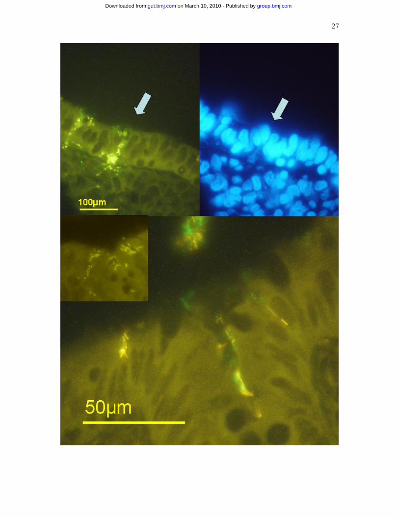
Figure 3.
Subepithelial infiltration by Fusobacterium necrophorum (photograph above) and
Fusobacterium nucleatum (photograph below) in suppurative appendicitis. Bacteria of
Fusobacterium nucleatum appear as long filaments, Fusobacterium necrophorum has
the shape of shorter rods.
group.bmj.com on March 10, 2010 - Published by gut.bmj.comDownloaded from

21
Table 1.
Percent of some bacterial groups to the total bacterial population in material resected for acute appendicitis
Pat. Nr.
Bac 303
EREC 482
Fprau
Fnuc
Fnec
Ebac 1790
Bif 164
Muc 1437
Others Leuko. lumen
Infiltr. of epithel
Adherence
Infiltr. of crypt
Infiltr. of Sub-epith.
Catarrhal appendicitis 1 10 5 2 0 0 10 10 0 30% Ser1410, Veil223 +
2 45 20 10 15 0 1 0
0 3% Ato291 + + + + +
3 50 10 30 0 0 1 0 2 + + +
4 30 5 20 40 0 0 2
0 2% Chis150,Lab158, Ato291
++
++ + + ++
5 40 10 0 40 0 1 0
0 + + + ++
6 40 25 0 20 0 1 0
0 3% Ato291, Pnig657
++ + + -
7 50 5 15 10 0 0 5
1
++ + + +
8 20 45 25 0 0 0 0 10 + + + 9 30 45 20 0 0 0 0 5 Ecyl1387, Lab158 + + +
10 20 10 3 0 0 60 0 0 + + - 11 30 40 10 0 0 0 12 1 + + + - +
12 10 10 1 0 70 0 0 0
++ + + - ++
13 35 50 0 0 0 10 0 0
++ + +
14 70 20 5 0 0 0 0 1
++ +
15 40 40 15 0 0 5 0 1
+ + ++ +
16 30 12 0 50 0 2 0
0
++ +
17 0 15 1 0 0 60 0 3
Lab158 ++ + +
18 20 40 0 1 0 10 0 0 + +
group.bmj.com on March 10, 2010 - Published by gut.bmj.comDownloaded from

22
5 +
19 40 20 20 2 0 0 0 1
5% Ato291 ++ +
20 0 0 0 0 90 10 0 0
++ + +
21 20 30 40 0 0 0 2 1 4% Ato291 + 22 40 20 35 2 0 0 0 0 + + + 23 50 30 5 0 0 0 2 0 Ato291 + + + ++
24 30 10 0 20 0 20 0
0 Ato291
++ ++
25 20 8 10 50 0 10 0
0
++ + + + ++
Suppurative appendicitis
26 60 15 25 0 0 0 0 3
++ +
27 95 0 0 3 0 0 1 0
++ + +
28 100 0 0 0 0 0 0 0
++ ++
29 50 0 0 30 0 20 0
0
++ +
+
30 30 0 0 50 0 10 0
0 3% Ato291 + + +
31 70 5 5 20 0 0 0
0
++ + + +
32 5 20 0 50 0 0 0
0 + + + ++
33 50 20 0 0 0 0 0 0 10% Ecyl1387 + + + 34 20 60 0 0 0 20 0 0.1 + + ++
35 35 15 30 0 0 10 0
0
no lumen ++
36 20 1 20 0 0 0 0
0
40% Fuso
no lumen + ++
37 55 25 10 0 0 1 0 0.1 5% Veil223, Lab158,
Ecyl ++ + + ++
38 35 5 5 0 0 0 30
0
no lumen ++
39 70 20 0 0 10 0 0
0
no lumen ++
40 40 0 0 35 0 0 0
0
++ + +
41 15 5 0 70 0 0 0
0 Veil, Phasco, Lab158
++ ++
42 10 10 0 0 0 1 0 0
70% Fuso, Lab158 ++ +
43 15 2 30 0 0 30 2 1
3% Ecyl1387, Lab158 ++ + + +
44 10 1 15 70 0 1 0
0 Veil
++
++ + ++
45 30 0 0 70 0 0 0
0
++ ++
group.bmj.com on March 10, 2010 - Published by gut.bmj.comDownloaded from

23
46 0 0 10 90 0 0 0
0 + ++
47 20 10 10 50 0 1 0
0.1 Veil
++ ++
48 10 0 0 0 0 1 0
0
90% Fuso
no lumen ++
49 12 0 20 50 0 8 0
1
++
++ ++
50 30 15 0 20 0 20 0
0
++
++ + ++
51 10 30 1 0 50 0 0 0 + + ++
52 20 20 0 50 0 1 0
0
++ + + ++
No appendicitis 53 40 40 0 0 0 20 0 0 54 35 25 25 0 0 0 0 15 + 55 60 25 10 0 0 0 0 3 + + 56 20 40 20 0 0 5 0 0 Veil + +
57 40 10 25 10 0 10 0
1 + + + ++
58 40 30 25 0 0 0 0 5 + + 59 70 20 5 0 0 0 5 1 + + 60 50 „25 15 0 0 0 0 10 + 61 5 50 10 0 0 0 0 0 Ato291, Veil 62 40 10 40 0 0 5 0 3 + + 63 50 10 0 0 0 30 0 0 + + 64 30 30 30 0 0 0 0 10 + 65 50 30 15 0 0 0 0 1 + 66 40 30 25 0 0 0 0 5 Ato291
67 40 40 10 0 0 1 0 8
++ + + +
68 30 40 20 0 0 0 0 0 Chis150
69 30 40 15 0 0 0 0 10
++
70 60 20 10 0 0 0 0 1 + +
Table 2. Occurrence of Fusobacteria (Fnuc/Fnec/Fuso) at specific locations
No appendicitis
Catarrhal appendicitis
Suppurative appendicitis
Cecal biopsy
Faecal cylinder
All locations % Number
6% (1/18)
52%* (13/25)
70%*
(19/27)
0.5% (2/400)
2% (8/400)
Lumen 5% 44%* 52%*
0.5% 2%
Infiltration of epithelial cells
5% 40%* 29%*
0
group.bmj.com on March 10, 2010 - Published by gut.bmj.comDownloaded from
24
Submucosal infiltration 0 36%* 56%*
0
*P< 0.01 from operated patients without appendicitis and from controls
Table 3. Mean ±SD proportion of single bacterial groups on intestinal microbiota in appendicitis and controls
Bacterial group
No appendicitis N=18
Catarrhal appendicitis N= 25
Suppurative appendicitis N=27
Cecal biopsy N=400
Faecal cylinder N=400
Fusobacteria <1 11±17* 24±29* <1 <1 Bac303+Fprau+Erec482
87±11
63±30** 51±29** 90±11 70±18
Muc1437 4.0 ±4.6
1.0 ±2.1** 0.2 ±0.6** Not determined
Not determined
Ebac 3.9±8
8±16 5±8 Not determined
Not determined
*P< 0.01 from operated patients without appendicitis and from controls **P<0.001 from operated patients without appendicitis
group.bmj.com on March 10, 2010 - Published by gut.bmj.comDownloaded from

25
group.bmj.com on March 10, 2010 - Published by gut.bmj.comDownloaded from

26
group.bmj.com on March 10, 2010 - Published by gut.bmj.comDownloaded from
27
group.bmj.com on March 10, 2010 - Published by gut.bmj.comDownloaded from

28
group.bmj.com on March 10, 2010 - Published by gut.bmj.comDownloaded from

29
group.bmj.com on March 10, 2010 - Published by gut.bmj.comDownloaded from


![Acute Appendicitis[1]](https://img.dokumen.tips/doc/110x75/577cd3341a28ab9e7896e8e0/acute-appendicitis1.jpg)


