Embed Size (px)
Citation preview
1
Acute Aortic Syndromes
Massachusetts General Hospital
Harvard Medical School
No disclosures
Michael H. Picard, M.D.
For everything you need to know about the aorta see
Circulation 2010;121:e266-e369
And…….

2
2010 ACCF/AHA/AATS/ACR/ASA/SCAI/SIR/STS/SVM Guidelines for
the diagnosis and management of patients with thoracic aortic disease.
Circ 2010;121:e266-e369
Normal range for size of aorta
2010 ACCF/AHA/AATS/ACR/ASA/SCAI/SIR/STS/SVM Guidelines for
the diagnosis and management of patients with thoracic aortic disease.
Circ 2010;121:e266-e369
Recommendations for aortic imaging techniques to
determine the presence and progression of thoracic
aortic disease
• Measurements of aortic diameter should be taken at reproducible anatomic landmarks, perpendicular to the axis of flow
• For CT and MR measurements, the external diameter should be measured. For aortic root measurements, the widest diameter, typically at the mid sinus level should be used.
• For echo, the internal diameter should be measured. For aortic root measurements, the widest diameter, typically at the mid sinus level should be used.
Acute aortic syndromes chest pain due to aortic disease
• Aortic dissection
• Intramural hematoma – Variant of classic aortic dissection
– Typically no intimal tear
• Penetrating atherosclerotic ulcer
• Rapidly expanding aortic aneurysm
• Rupture or contained rupture of an aortic aneurysm
• Traumatic aortic transection

3
Acute aortic dissection
• Incidence 3/100,000 per year
• Life-threatening condition
– Early mortality 1% per hour
– Survival improved with prompt therapy
• Presentation may be non-specific and risk factors may
not be recognized
– Presentation can range from chest pain (?MI, PE, pericarditis)
to syncope to paralysis to stroke
Chest Pain of aortic dissection
– Acute chest and/or infrascapular back pain
– Pain is abrupt in onset
– Pain is severe, worst ever
– Worst at onset not crescendo
– Can be described as tearing or stabbing in quality
– Pain may migrate
– Patients are restless, cannot get comfortable
Classic features of aortic dissection
• Hypertension
• BP differential between the 2 arms
• Murmur of AI
• Wide mediastinum on CXR
• Older patients have history of hypertension
• Younger patients have evidence of Marfan’s syndrome
4
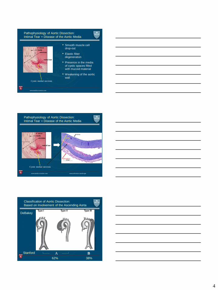
Pathophysiology of Aortic Dissection:
Intimal Tear + Disease of the Aortic Media
• Smooth muscle cell drop-out
• Elastic fiber degeneration
• Presence in the media of cystic spaces filled with mucoid material
• Weakening of the aortic wall
Cystic medial necrosis
www.medicscientist.com
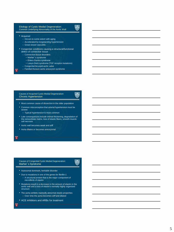
Pathophysiology of Aortic Dissection:
Intimal Tear + Disease of the Aortic Media
www.medicscientist.com
Cystic medial necrosis
intima
media
adventitia
www.reference.medscape
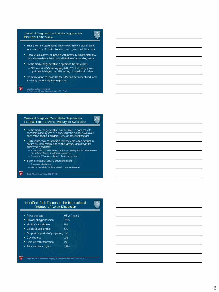
Classification of Aortic Dissection:
Based on Involvement of the Ascending Aorta
DeBakey I II III
Stanford
62% 38%
A B
ww
w.c
edars
-sin
ai.edu
5
Etiology of Cystic Medial Degeneration: Common Underlying Abnormality of the Aortic Wall
• Acquired
– Occurs to some extent with aging
– Accelerated by longstanding hypertension
– Great vessel vasculitis
• Congenital conditions causing a structural/functional
defect of connective tissue
– Connective tissue disorders
• Marfan’s syndrome
• Ehlers-Danlos syndrome
• Loeys-Dietz syndrome (TGF receptor mutations)
– Congential bicuspid aortic valve
– Familial thoracic aortic aneurysm syndrome
Causes of Acquired Cystic Medial Degeneration:
Chronic Hypertension
• Most common cause of dissection in the older population
• Common misconception that arterial hypertension must be severe
– Typical hypertension is most common
• Late consequences include intimal thickening, degradation of the extracellular matrix, loss of elastic fibers, smooth muscle cell necrosis
• Aortic wall becomes weak and stiff
• Aorta dilates or becomes aneurysmal
Causes of Congenital Cystic Medial Degeneration:
Marfan’s Syndrome
• Autosomal dominant, heritable disorder
• Due to mutations in one of the genes for fibrillin-1
– A structural protein that is the major component of
microfibrils of elastin
• Mutations result in a decrease in the amount of elastin in the aortic wall and a loss of elastin's normally highly organized structure
• The aorta exhibits markedly abnormal elastic properties
– Over time the aorta becomes stiff and dilated
• ACE inhibitors and ARBs for treatment
6
Causes of Congenital Cystic Medial Degeneration:
Bicuspid Aortic Valve
• Those with bicuspid aortic valve (BAV) have a significantly
increased risk of aortic dilatation, aneurysm, and dissection
• Echo studies of young people with normally functioning BAV
have shown that 50% have dilatation of ascending aorta
• Cystic medial degeneration appears to be the culprit
– Of those with BAV undergoing AVR, 75% had biopsy proven
cystic medial degen., vs. 14% among tricuspid aortic valves
• No single gene responsible for BAV has been identified, and
it is likely genetically heterogenous
Nistri S, et al. Heart 1999;82:19
DeSa M, et al. J Thorac Cardiovasc Surg 1999;118:588.
Causes of Congenital Cystic Medial Degeneration:
Familial Thoracic Aortic Aneurysm Syndrome
• Cystic medial degeneration can be seen in patients with ascending aneurysms or dissection who do not have overt connective tissue disorders, BAV, or other risk factors
• Such cases may be sporadic, but they are often familial in nature are now referred to as the familial thoracic aortic aneurysm syndrome
– At least 19% of those with thoracic aortic aneurysms in Yale database
had a family history of a thoracic aneurysm
– Screening 1st degree-relatives should be advised
• Several mutations have been identified
– Dominant inheritance
– Marked variability in the expression and penetrance
Coady MA, et al. Arch Surg 1999;134:361.
Identified Risk Factors in the International
Registry of Aortic Dissection
• Advanced age 63 yr (mean)
• History of hypertension 72%
• Marfan’s syndrome 5%
• Bicuspid aortic valve 5%
• Peripartum period of pregnancy 1%
• Cocaine use 1%
• Cardiac catheterization 2%
• Prior cardiac surgery 18%
Hagan PG, et al. International Registry of Aortic Dissection, JAMA 2000;283:897.
7
Non invasive imaging for diagnosis all have high sensitivity and specificity but each have other
advantages and disadvantages
• TEE
– Bedside, rapid (if personnel available)
– Can look at other cardiac structures
• Contrast enhanced multi-detector CT
– Rapid scan, can also look at pulm embolism, coronary anatomy
– Large dose of contrast, lots of radiation
– Artifacts more likely with older devices
• cMR
– High resolution images of various aortic pathologies
– Difficult in an emergency situation or unstable patient
– Concerns about gadolinium but can image without contrast
Local expertise should dictate
Echocardiographic findings
• Intimal flap
– High frequency, low amplitude motion
– flow respects the boundary
– Artifacts
• Motion - Low frequency (or same frequency as adjacent structure)
and high amplitude
• Color flow goes through
• +/- aortic dilation
• Aortic insufficiency
• Wall motion abnormality if flap obstructs coronary ostia
Echo for Aortic Dissection
TEE
• Sensitivity 98-100% and specificity 77-100% in different series
• False negative TEE is rare: few dissections are limited to the “blind spot” at the inferior portion of the arch
• False positive TEE: Artifacts are very common (23-55%), especially in the ascending aorta.

8
Type I aortic dissection
10 days post-partum after uneventful delivery of 2nd child
• Sudden onset 10/10 back pain radiating to the chest while breast-feeding
PMH
• Migraines
• Allergic rhinitis
Family history
• Father with Marfan’s, s/p AVR at age 38
• Brother with Marfan’s, s/p AVR at age 28
TEE – dissection or artifact ?
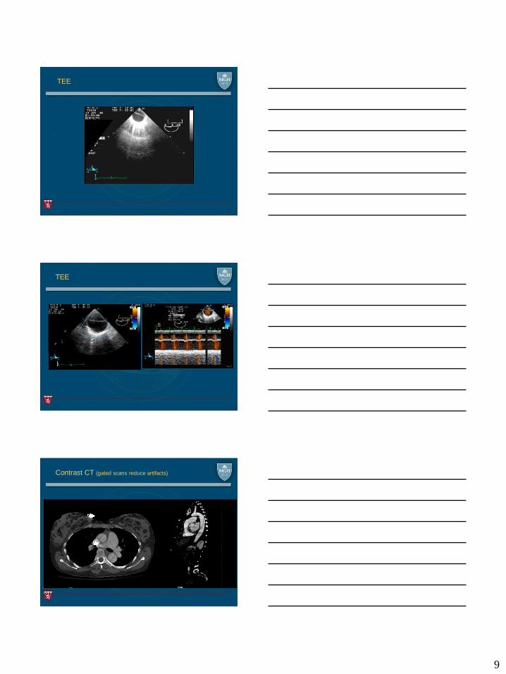
9
TEE
TEE
Contrast CT (gated scans reduce artifacts)

10
• CT: Type B dissection extending from just distal to L subclavian artery to aortic bifurcation
• Hospital course: Medically managed on beta-blocker and discharged home.
Complications of Acute Aortic Dissection:
Aortic Rupture
• Type A
– The ascending aorta is prone
to rupture into the pericardium
causing cardiac tamponade
– 2/3 of all deaths from AoD
– Surgery to indicated to
prevent rupture
• Type B
– Into the pleural space or
mediastinum
– Rupture is uncommon
– Preventative surgery is not
necessary
Specific Complications
of Acute Aortic Dissection
Aortic insufficiency
32%
Acute MI
2%
Rupture into
pericardium
Stroke 5%
BP differential
15%
Paraplegia
<1%
Renal ischemia
5-8%
Mesenteric ischemia
5%
Lower extremity
ischemia 3%

11
Complications of Acute Aortic Dissection:
Branch Artery Compromise
Dissection extends into
branch artery
Distension of the false lumen
compresses true lumen
Mechanisms of AI in acute type A aortic
dissection
• leaflet thickening *
• bicuspid aortic valve *
• incomplete leaflet closure *
• aortic leaflet prolapse *
• intimal flap prolapse
• multiples of above
• * not specific to dissection
Movsowitz et al, JACC 2000;36:884-90
normal
Incomplete
leaflet closure
due to dilation
of STJ
Aortic leaflet
prolapse
Intimal flap
prolapse
Artifacts
• Side-by-side:
– lateral resolution, side lobe, lens effect
• Behind, parallel motion:
– Reverberation
• Behind, opposite motion:
– Mirror image

12
Linear TEE artifacts artifacts in the aorta seen
in the presence of dilated aortas
Appelbe et al, JACC 1993;21:754-60
Artifact distance = 2 times that of interface
Transverse TEE image of
dilated ascending aorta
compressing LA. Note the
artifact crosses borders
Clues to artifacts
• Cross borders
• motion identical to another real structure
– amplitude and frequency at a multiple of the real structure
• indistinct edges
• not reproduced in an orthogonal or other view
• color flow passes through it
• clues - foreign materials present
– catheters, prosthetic valves, grafts
– these are not always in the plane of view
Clues to real structures
• respects borders
• distinct edges (unless thrombus)
• motion
– intimal flaps - amplitude and frequency of motion different
than cardiac cycle, respiration
• seen in multiple views
• color flow respects true borders
• intimal flaps keep company other pathologic processes
13
Late Complications of Aortic Dissection
• Progressive expansion of dissected portions of aorta
– Large aneurysms
– Aortic rupture
• Development of new aneurysms elsewhere in
a non-dissected segments of the thoracic aorta
• Aortic insufficiency from aortic root dilatation
• Careful medical follow-up and surveillance imaging of aorta is
essential
Acute Intramural Hematoma:
Variant of Aortic Dissection
• Risk factors and the presenting signs / symptoms are the same
as classic aortic dissection
• Natural history of acute IMH (in the West) appears to be similar
to classic aortic dissection
– Type A is prone to rupture and death Urgent surgery
• However, data suggests that Asian populations may have a
more favorable outcome from Type A ? No surgery
– Type B is at low risk of rupture Medical management
Intramural Hematoma:
Variant of Aortic Dissection
• Unlike classic aortic dissection it is not caused by a visible
tear in the intima
• Two possible causes:
– Rupture of the vasa vasorum within the aortic media, producing
a hematoma within the aortic wall
– A small tear in the intima that permits only transient blood flow
from the lumen into the aortic wall, which then thromboses
• No active communication between the aortic lumen and the
hematoma and therefore no flow within the aortic wall
• Has a distinctly different and less obvious appearance on
aortic imaging studies (especially TEE)
14
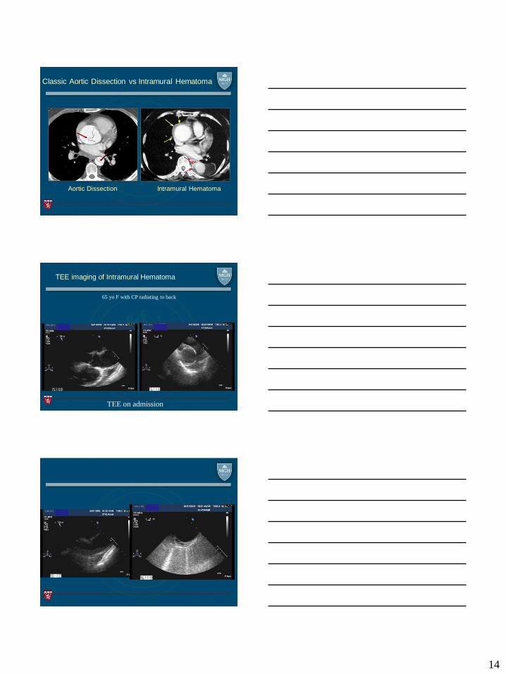
Classic Aortic Dissection vs Intramural Hematoma
Intramural Hematoma Aortic Dissection
TEE imaging of Intramural Hematoma
65 yo F with CP radiating to back
TEE on admission
15
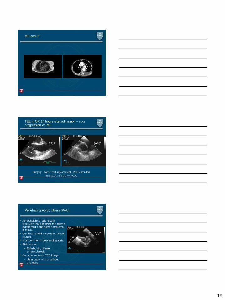
MR and CT
TEE in OR 14 hours after admission – note
progression of IMH
Surgery: aortic root replacement. IMH extended
into RCA so SVG to RCA.
Penetrating Aortic Ulcers (PAU)
• Atherosclerotic lesions with ulceration that penetrate the internal elastic media and allow hematoma in media
• Can lead to IMH, dissection, vessel rupture
• Most common in descending aorta
• Risk factors
– Elderly, htn, diffuse
atherosclerosis
• On cross sectional TEE image
– Ulcer crater with or without
thrombus

16
Penetrating aortic ulcers with superimposed
thrombus
Ascending aortic thrombus due to aortic ulcer
37 yo M with stroke, TTE notes aortic clot
2 days later develops CP and STE. In
OR clot in RCA Anticoagulated and BP
controlled
Ascending aortic thrombus due to atherosclerotic
ulcer
57 yo F presents with RUE arterial insufficiency. Emboli extracted
from upper extremity. TEE showed ao clot. Recurrence of emboli so
taken to surgery

17
Acute aortic syndromes
• Presentations can mimic other conditions
• High early mortality but prompt recognition improves survival
• Location of dissection determines complications and should dictate therapy
• Hypertension most common etiology
• Dissection and hematoma
– Risk factors, presenting signs/symptoms, natural history are
similar
• Early imaging (TEE, CT, MR) critical for quick diagnosis