Embed Size (px)
DESCRIPTION
Guideline mengenai sinusitis akut dan kronis
Citation preview
REVIEW Open Access
Canadian clinical practice guidelines for acuteand chronic rhinosinusitisMartin Desrosiers1*, Gerald A Evans2, Paul K Keith3, Erin D Wright4, Alan Kaplan5, Jacques Bouchard6,Anthony Ciavarella7, Patrick W Doyle8, Amin R Javer9, Eric S Leith10, Atreyi Mukherji11, R Robert Schellenberg12,Peter Small13, Ian J Witterick14
Abstract
This document provides healthcare practitioners with information regarding the management of acuterhinosinusitis (ARS) and chronic rhinosinusitis (CRS) to enable them to better meet the needs of this patientpopulation. These guidelines describe controversies in the management of acute bacterial rhinosinusitis (ABRS) andinclude recommendations that take into account changes in the bacteriologic landscape. Recent guidelines inABRS have been released by American and European groups as recently as 2007, but these are either limited intheir coverage of the subject of CRS, do not follow an evidence-based strategy, or omit relevant stakeholders inguidelines development, and do not address the particulars of the Canadian healthcare environment.Advances in understanding the pathophysiology of CRS, along with the development of appropriate therapeuticstrategies, have improved outcomes for patients with CRS. CRS now affects large numbers of patients globally andprimary care practitioners are confronted by this disease on a daily basis. Although initially considered a chronicbacterial infection, CRS is now recognized as having multiple distinct components (eg, infection, inflammation),which have led to changes in therapeutic approaches (eg, increased use of corticosteroids). The role of bacteria inthe persistence of chronic infections, and the roles of surgical and medical management are evolving. Althoughevidence is limited, guidance for managing patients with CRS would help practitioners less experienced in this areaoffer rational care. It is no longer reasonable to manage CRS as a prolonged version of ARS, but rather, specifictherapeutic strategies adapted to pathogenesis must be developed and diffused.Guidelines must take into account all available evidence and incorporate these in an unbiased fashion intomanagement recommendations based on the quality of evidence, therapeutic benefit, and risks incurred. Thisdocument is focused on readability rather than completeness, yet covers relevant information, offers summaries ofareas where considerable evidence exists, and provides recommendations with an assessment of strength of theevidence base and degree of endorsement by the multidisciplinary expert group preparing the document.These guidelines have been copublished in both Allergy, Asthma & Clinical Immunology and the Journal ofOtolaryngology-Head and Neck Surgery.
IntroductionSinusitis refers to inflammation of a sinus, while rhinitisis inflammation of the nasal mucous membrane. Theproximity between the sinus cavities and the nasal pas-sages, as well as their common respiratory epithelium,lead to frequent simultaneous involvement of both
structures (such as with viral infections). Given the diffi-culty separating the contributions of deep structure tosigns and symptoms, the term rhinosinusitis is fre-quently used to describe this simultaneous involvement,and will be used in this text. Rhinosinusitis refers toinflammation of the nasal cavities and sinuses. Whenthe inflammation is due to bacterial infection, it is calledbacterial rhinosinusitis.Rhinosinusitis is a frequently occurring disease, with
significant impact on quality of life and health carespending, and economic impact in terms of absenteeism
* Correspondence: [email protected] of Otolaryngology - Head and Neck Surgery Centre Hospitalier del’Université de Montréal, Université de Montréal Hotel-Dieu de Montreal, andDepartment of Otolaryngology - Head and Neck Surgery and Allergy,Montreal General Hospital, McGill University, Montreal, QC, CanadaFull list of author information is available at the end of the article
Desrosiers et al. Allergy, Asthma & Clinical Immunology 2011, 7:2http://www.aacijournal.com/content/7/1/2 ALLERGY, ASTHMA & CLINICAL
IMMUNOLOGY
© 2011 Desrosiers et al; licensee BioMed Central Ltd. This is an Open Access article distributed under the terms of the CreativeCommons Attribution License (http://creativecommons.org/licenses/by/2.0), which permits unrestricted use, distribution, andreproduction in any medium, provided the original work is properly cited.
and productivity. It is estimated that approximately6 billion dollars is spent in the United States annuallyon therapy for rhinosinusitis [1]. A recent study inCanada described the impact of chronic rhinosinusitis(CRS) on patients and healthcare utilization [2]. Patientswith CRS had a health status similar to patients witharthritis, cancer, asthma, and inflammatory bowel dis-ease. Compared with people without CRS, those withCRS reported more days spent bedridden and more vis-its to family physicians, alternative healthcare providers,and mental health experts. These findings underscorethe significant impact of this disease on patient qualityof life, as well as costs of care to patients and society.In Canada, 2.89 million prescriptions were dispensed
for acute rhinosinusitis (ARS) or CRS in 2006, withapproximately 2/3 for ARS and 1/3 for CRS [3]. Despitewell-established differences between these 2 diseases inpathophysiology, bacteriology, and standard specialisttreatment strategies, an assessment of therapies pre-scribed in Canada for CRS has shown that medicationsprescribed for CRS exactly paralleled those prescribedfor ARS [3].The incidence of bacterial rhinosinusitis is difficult to
obtain precisely given that not all patients will seek medi-cal help. In the United States in 2007, ARS affected 26million individuals and was responsible for 12.9 millionoffice visits [4]. Although no specific Canadian data isavailable, extrapolation from US data suggests an occur-rence of 2.6 million cases in Canada annually. This is inline with prescription data from 2004. This high inci-dence is not unexpected given that acute bacterial rhino-sinusitis (ABRS) usually develops as a complication in0.5%-2% of upper respiratory tract infections (URTIs) [5].A survey of Canadian households reported the preva-
lence of CRS to be 5% [6]. The prevalence was higher inwomen compared with men (5.7% vs 3.4% for subjectsaged ≥12 years) and increased with age. CRS was asso-ciated with smoking, lower income, history of allergy,asthma, or chronic obstructive pulmonary disease(COPD), and was slightly higher for those living in theeastern region or among native Canadians.Guidelines for ARS have been developed over the past
5 years by both a European group (E3POS) and theAmerican Academy of Otolaryngology-Head and NeckSurgery (AAO-HNS). Both guidelines have limitationsthat we believe are improved upon by the current docu-ment. This current document provides healthcare prac-titioners with a brief, easy-to-read review of informationregarding the management of ARS and CRS. Theseguidelines are meant to have a practical focus, directedat first-line practitioners with an emphasis on patient-centric issues. The readership is considered to be familyphysicians, emergency physicians, or other point-of-careproviders, as well as specialists in otolaryngology-head
and neck surgery, allergy and immunology, or infectiousdisease who dispense first-line care or teach colleagueson the subject. This document is specifically adapted forthe needs of the Canadian practice environment andmakes recommendations that take into account factorssuch as wait times for computed tomography scans orspecialist referral. These guidelines are intended to pro-vide useful information for CRS by addressing this areawhere controversy is unresolved and evidence is typi-cally Grade D - requiring incorporation of expert opi-nion based on pathophysiology and current treatmentregimens. Thus, the main thrust is to provide a compre-hensive guide to CRS and to address changes in themanagement of ABRS.
Guideline Preparation ProcessAn increased emphasis on evidence-based recommenda-tions over the past decade has significantly improvedthe overall quality of most published guidelines, but pre-sent significant difficulties in developing guidelineswhere the evidence base for long-standing, traditionalremedies is often weak or anecdotal, or in emergingentities such as chronic rhinosinusitis (CRS) where con-troversy remains and evidence is sparse. In developingthese guidelines, standard evidence-based developmenttechniques have been combined with the Delphi votingprocess in order to offer the reader the opinion of amultidisciplinary expert group in areas where evidenceis weak.Funding was obtained via an unrestricted grant
obtained from 5 pharmaceutical manufacturers, witheach contributing equally to this project. In order tominimize any appearance of conflict of interest, allfunds were administered via a trust account held at theCanadian Society of Otolaryngology-Head and NeckSurgery (CSO-HNS). No contact with industry wasmade during the guidelines development or reviewprocess.An English-language Medline® search was conducted
using the terms acute bacterial rhinosinusitis (ABRS),chronic rhinosinusitis (CRS), and nasal polyposis (lim-ited to the adult population, human, clinical trials, itemswith abstracts) and further refined based on the indivi-dual topics. This is a multi-disciplined condition andtherefore input from all appropriate associations wasrequired. Inclusion criteria: most current evidence-baseddata, relevance, subject specifics, caliber of the abstract,Canadian data preferred but not exclusive. Exclusioncriteria: newer abstract of the same subject available,non-human, not relevant.The quality of retrieved articles was assessed by
Society Team Leaders along with the principal authorbased on area of expertise. Where necessary, the princi-pal author invited input from the External Content
Desrosiers et al. Allergy, Asthma & Clinical Immunology 2011, 7:2http://www.aacijournal.com/content/7/1/2
Page 2 of 38
Experts. Articles were graded for strength of evidence bydrawing upon strategies adapted from the AmericanAcademy of Pediatrics Steering Committee on QualityImprovement and Management (AAP SCQIM) guide-lines [7], the Grades of Recommendation, Assessment,Development and Evaluation (GRADE) grading system[8], and the AAO-HNS guidelines in sinusitis [9], all ofwhich use similar strategies by classifying strength ofevidence recommendations according to the balance ofthe benefits and downsides after considering the qualityof the evidence. Accordingly, grades of evidence weredefined as:
Grade A. Well-designed, randomized, controlled stu-dies or diagnostic studies on relevant populationsGrade B. Randomized controlled trials or diagnosticstudies with minor limitations; overwhelmingly con-sistent evidence from observational studiesGrade C. Observational studies (case control orcohort design)Grade D. Expert opinion, case reports, reasoningfrom first principlesGrade X. Exceptional situations where validating stu-dies cannot be done and there is a clear predomi-nance of benefit or harm [7].
Strength of EvidenceDefinitions for the strength of evidence recommenda-tions combine the balance of benefit versus harm oftreatment with the grade of the evidence, as follows:
Strong Recommendation: Benefits of treatmentclearly exceed harm; quality of evidence is excellent(Grade A or B). A strong recommendation shouldbe followed unless there is a clear and compellingreason for a different approach.Recommendation: Benefits exceeded harm, but qual-ity of evidence is not as strong (Grade B or C). Arecommendation should generally be followed, butclinicians should remain alert to new informationand consider patient preferences.Option: Quality of evidence is suspect (Grade D) orwell-done studies (Grade A, B or C) show little clearadvantage. An option reflects flexibility in decision-making regarding appropriate practice, but cliniciansmay set limits on alternatives. The preference of thepatient should influence the decision.No Recommendation: A lack of relevant evidence(Grade D) and an unclear balance between benefitsand harm. No recommendation reflects no limita-tions on decision-making and clinicians should bevigilant regarding new information on the balance ofbenefit versus harm. The preference of the patientshould influence the decision.
In situations where high-quality evidence is impossible toobtain and anticipated benefits strongly outweigh the harm,the recommendation may be based on lesser evidence [9].Thus, policy recommendations were formulated based
on evidence quality and the balance of potential benefitsand harm. As many therapies have not been subjectedto safety evaluation in a clinical trial setting, the poten-tial for harm was assessed for each therapy and weighsin the recommendation. The guidelines presented usedthese approaches to formulate strength of evidencerecommendations, with options to recommend denotedas:
• Strong• Moderate• Weak• An option for therapy, or• Not recommended as either clinical trial data of agiven therapy did not support its use or a concernfor toxicity was noted.
Strength of RecommendationRecommendations were assessed according to a Delphivoting process, whereby voting options included toaccept completely, to accept with some reservation, toaccept with major reservation, to reject with reservation,or to reject completely [7,10]. Only statements that wereaccepted by over 50% of the group were retained.Strength of the recommendation by the multidisciplin-ary group of experts was denoted as:
• Strong (for accept completely)• Moderate (for accept with some reservation), or• Weak (for accept with major reservation).
Thus, strength of recommendation is a measure ofendorsement by the group of experts.These guidelines have been developed from the outset
to meet the AGREE criteria [11] to ensure maximumimpact.DISCLAIMER: These guidelines are designed to offer
evidence-based strategies in the management of acuteand chronic rhinosinusitis. They are, however, notintended to replace clinical judgment or establish a pro-tocol for all individuals with suspected rhinosinusitis. Dif-ferent presentations, associated comorbidities, oravailability of resources may require adaptation of theseguidelines, thus there may be other appropriateapproaches to diagnosing and managing these conditions.
Summary of Guideline Statements and StrengthsStatements and their ratings for strength of evidenceand recommendation are summarized in Table 1.
Desrosiers et al. Allergy, Asthma & Clinical Immunology 2011, 7:2http://www.aacijournal.com/content/7/1/2
Page 3 of 38
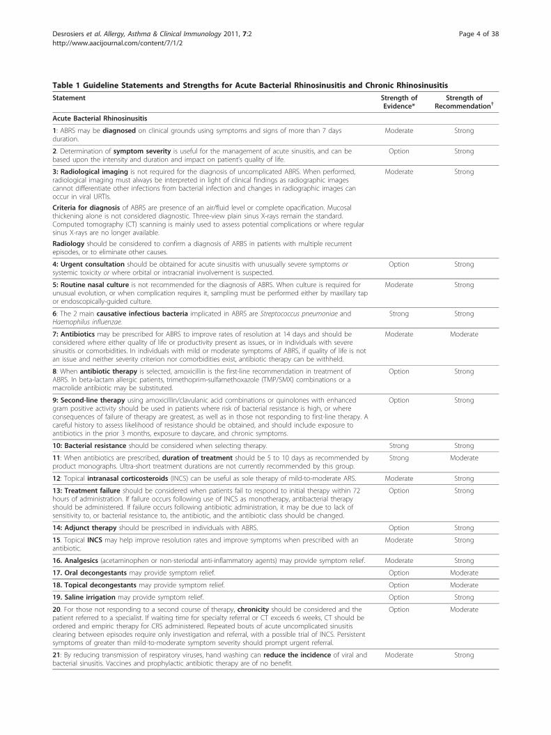
Table 1 Guideline Statements and Strengths for Acute Bacterial Rhinosinusitis and Chronic Rhinosinusitis
Statement Strength ofEvidence*
Strength ofRecommendation†
Acute Bacterial Rhinosinusitis
1: ABRS may be diagnosed on clinical grounds using symptoms and signs of more than 7 daysduration.
Moderate Strong
2. Determination of symptom severity is useful for the management of acute sinusitis, and can bebased upon the intensity and duration and impact on patient’s quality of life.
Option Strong
3: Radiological imaging is not required for the diagnosis of uncomplicated ABRS. When performed,radiological imaging must always be interpreted in light of clinical findings as radiographic imagescannot differentiate other infections from bacterial infection and changes in radiographic images canoccur in viral URTIs.
Moderate Strong
Criteria for diagnosis of ABRS are presence of an air/fluid level or complete opacification. Mucosalthickening alone is not considered diagnostic. Three-view plain sinus X-rays remain the standard.Computed tomography (CT) scanning is mainly used to assess potential complications or where regularsinus X-rays are no longer available.
Radiology should be considered to confirm a diagnosis of ARBS in patients with multiple recurrentepisodes, or to eliminate other causes.
4: Urgent consultation should be obtained for acute sinusitis with unusually severe symptoms orsystemic toxicity or where orbital or intracranial involvement is suspected.
Option Strong
5: Routine nasal culture is not recommended for the diagnosis of ABRS. When culture is required forunusual evolution, or when complication requires it, sampling must be performed either by maxillary tapor endoscopically-guided culture.
Moderate Strong
6: The 2 main causative infectious bacteria implicated in ABRS are Streptococcus pneumoniae andHaemophilus influenzae.
Strong Strong
7: Antibiotics may be prescribed for ABRS to improve rates of resolution at 14 days and should beconsidered where either quality of life or productivity present as issues, or in individuals with severesinusitis or comorbidities. In individuals with mild or moderate symptoms of ABRS, if quality of life is notan issue and neither severity criterion nor comorbidities exist, antibiotic therapy can be withheld.
Moderate Moderate
8: When antibiotic therapy is selected, amoxicillin is the first-line recommendation in treatment ofABRS. In beta-lactam allergic patients, trimethoprim-sulfamethoxazole (TMP/SMX) combinations or amacrolide antibiotic may be substituted.
Option Strong
9: Second-line therapy using amoxicillin/clavulanic acid combinations or quinolones with enhancedgram positive activity should be used in patients where risk of bacterial resistance is high, or whereconsequences of failure of therapy are greatest, as well as in those not responding to first-line therapy. Acareful history to assess likelihood of resistance should be obtained, and should include exposure toantibiotics in the prior 3 months, exposure to daycare, and chronic symptoms.
Option Strong
10: Bacterial resistance should be considered when selecting therapy. Strong Strong
11: When antibiotics are prescribed, duration of treatment should be 5 to 10 days as recommended byproduct monographs. Ultra-short treatment durations are not currently recommended by this group.
Strong Moderate
12: Topical intranasal corticosteroids (INCS) can be useful as sole therapy of mild-to-moderate ARS. Moderate Strong
13: Treatment failure should be considered when patients fail to respond to initial therapy within 72hours of administration. If failure occurs following use of INCS as monotherapy, antibacterial therapyshould be administered. If failure occurs following antibiotic administration, it may be due to lack ofsensitivity to, or bacterial resistance to, the antibiotic, and the antibiotic class should be changed.
Option Strong
14: Adjunct therapy should be prescribed in individuals with ABRS. Option Strong
15. Topical INCS may help improve resolution rates and improve symptoms when prescribed with anantibiotic.
Moderate Strong
16. Analgesics (acetaminophen or non-steriodal anti-inflammatory agents) may provide symptom relief. Moderate Strong
17. Oral decongestants may provide symptom relief. Option Moderate
18. Topical decongestants may provide symptom relief. Option Moderate
19. Saline irrigation may provide symptom relief. Option Strong
20. For those not responding to a second course of therapy, chronicity should be considered and thepatient referred to a specialist. If waiting time for specialty referral or CT exceeds 6 weeks, CT should beordered and empiric therapy for CRS administered. Repeated bouts of acute uncomplicated sinusitisclearing between episodes require only investigation and referral, with a possible trial of INCS. Persistentsymptoms of greater than mild-to-moderate symptom severity should prompt urgent referral.
Option Moderate
21: By reducing transmission of respiratory viruses, hand washing can reduce the incidence of viral andbacterial sinusitis. Vaccines and prophylactic antibiotic therapy are of no benefit.
Moderate Strong
Desrosiers et al. Allergy, Asthma & Clinical Immunology 2011, 7:2http://www.aacijournal.com/content/7/1/2
Page 4 of 38
Acute Bacterial Rhinosinusitis (ABRS)Definition and DiagnosisStatement 1: ABRS may be diagnosed on clinical groundsusing symptoms and signs of more than 7 days duration.Strength of evidence: ModerateStrength of recommendation: StrongRationale: ABRS is a clinical diagnosis that must be
differentiated from uncomplicated viral infections of theupper respiratory passages. Although no single symptomaccurately predicts the presence or absence of bacterialinfection, the presence of several signs and symptomsincreases the predictive value.
DefinitionThe common cold is caused by a rhinovirus, and in mostcases peak symptom severity is reached by 3 days [12].However, the same virus can activate an inflammatoryprocess that can lead to bronchitis, pharyngitis, and rhino-sinusitis [13]. Thus, the term rhinosinusitis has been usedto distinguish this more severe phenotypic entity from thecommon cold, which is associated with sinusitis [14].Despite the frequency of the common cold, 0.5% to 2% ofindividuals with the common cold will develop ABRS [5].ABRS is defined as a bacterial infection of the parana-
sal sinuses, described as a sudden onset of symptomatic
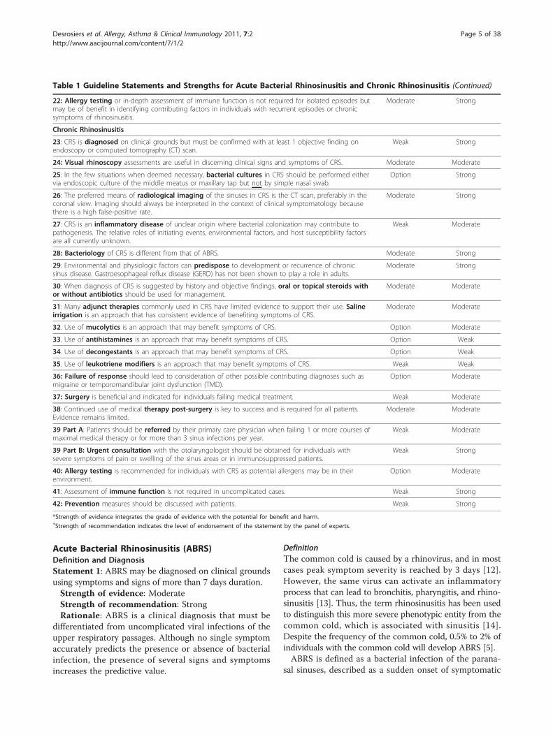
Table 1 Guideline Statements and Strengths for Acute Bacterial Rhinosinusitis and Chronic Rhinosinusitis (Continued)
22: Allergy testing or in-depth assessment of immune function is not required for isolated episodes butmay be of benefit in identifying contributing factors in individuals with recurrent episodes or chronicsymptoms of rhinosinusitis.
Moderate Strong
Chronic Rhinosinusitis
23: CRS is diagnosed on clinical grounds but must be confirmed with at least 1 objective finding onendoscopy or computed tomography (CT) scan.
Weak Strong
24: Visual rhinoscopy assessments are useful in discerning clinical signs and symptoms of CRS. Moderate Moderate
25: In the few situations when deemed necessary, bacterial cultures in CRS should be performed eithervia endoscopic culture of the middle meatus or maxillary tap but not by simple nasal swab.
Option Strong
26: The preferred means of radiological imaging of the sinuses in CRS is the CT scan, preferably in thecoronal view. Imaging should always be interpreted in the context of clinical symptomatology becausethere is a high false-positive rate.
Moderate Strong
27: CRS is an inflammatory disease of unclear origin where bacterial colonization may contribute topathogenesis. The relative roles of initiating events, environmental factors, and host susceptibility factorsare all currently unknown.
Weak Moderate
28: Bacteriology of CRS is different from that of ABRS. Moderate Strong
29: Environmental and physiologic factors can predispose to development or recurrence of chronicsinus disease. Gastroesophageal reflux disease (GERD) has not been shown to play a role in adults.
Moderate Strong
30: When diagnosis of CRS is suggested by history and objective findings, oral or topical steroids withor without antibiotics should be used for management.
Moderate Moderate
31: Many adjunct therapies commonly used in CRS have limited evidence to support their use. Salineirrigation is an approach that has consistent evidence of benefiting symptoms of CRS.
Moderate Moderate
32. Use of mucolytics is an approach that may benefit symptoms of CRS. Option Moderate
33. Use of antihistamines is an approach that may benefit symptoms of CRS. Option Weak
34. Use of decongestants is an approach that may benefit symptoms of CRS. Option Weak
35. Use of leukotriene modifiers is an approach that may benefit symptoms of CRS. Weak Weak
36: Failure of response should lead to consideration of other possible contributing diagnoses such asmigraine or temporomandibular joint dysfunction (TMD).
Option Moderate
37: Surgery is beneficial and indicated for individuals failing medical treatment. Weak Moderate
38: Continued use of medical therapy post-surgery is key to success and is required for all patients.Evidence remains limited.
Moderate Moderate
39 Part A: Patients should be referred by their primary care physician when failing 1 or more courses ofmaximal medical therapy or for more than 3 sinus infections per year.
Weak Moderate
39 Part B: Urgent consultation with the otolaryngologist should be obtained for individuals withsevere symptoms of pain or swelling of the sinus areas or in immunosuppressed patients.
Weak Strong
40: Allergy testing is recommended for individuals with CRS as potential allergens may be in theirenvironment.
Option Moderate
41: Assessment of immune function is not required in uncomplicated cases. Weak Strong
42: Prevention measures should be discussed with patients. Weak Strong
*Strength of evidence integrates the grade of evidence with the potential for benefit and harm.†Strength of recommendation indicates the level of endorsement of the statement by the panel of experts.
Desrosiers et al. Allergy, Asthma & Clinical Immunology 2011, 7:2http://www.aacijournal.com/content/7/1/2
Page 5 of 38
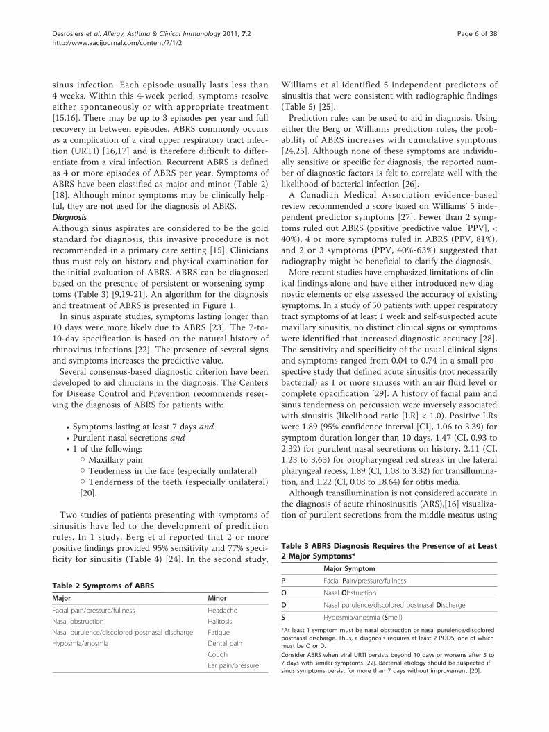
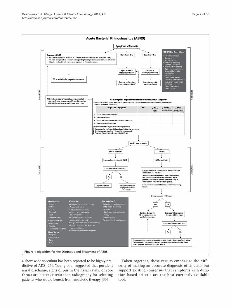
sinus infection. Each episode usually lasts less than4 weeks. Within this 4-week period, symptoms resolveeither spontaneously or with appropriate treatment[15,16]. There may be up to 3 episodes per year and fullrecovery in between episodes. ABRS commonly occursas a complication of a viral upper respiratory tract infec-tion (URTI) [16,17] and is therefore difficult to differ-entiate from a viral infection. Recurrent ABRS is definedas 4 or more episodes of ABRS per year. Symptoms ofABRS have been classified as major and minor (Table 2)[18]. Although minor symptoms may be clinically help-ful, they are not used for the diagnosis of ABRS.DiagnosisAlthough sinus aspirates are considered to be the goldstandard for diagnosis, this invasive procedure is notrecommended in a primary care setting [15]. Cliniciansthus must rely on history and physical examination forthe initial evaluation of ABRS. ABRS can be diagnosedbased on the presence of persistent or worsening symp-toms (Table 3) [9,19-21]. An algorithm for the diagnosisand treatment of ABRS is presented in Figure 1.In sinus aspirate studies, symptoms lasting longer than
10 days were more likely due to ABRS [23]. The 7-to-10-day specification is based on the natural history ofrhinovirus infections [22]. The presence of several signsand symptoms increases the predictive value.Several consensus-based diagnostic criterion have been
developed to aid clinicians in the diagnosis. The Centersfor Disease Control and Prevention recommends reser-ving the diagnosis of ABRS for patients with:
• Symptoms lasting at least 7 days and• Purulent nasal secretions and• 1 of the following:
○ Maxillary pain○ Tenderness in the face (especially unilateral)○ Tenderness of the teeth (especially unilateral)[20].
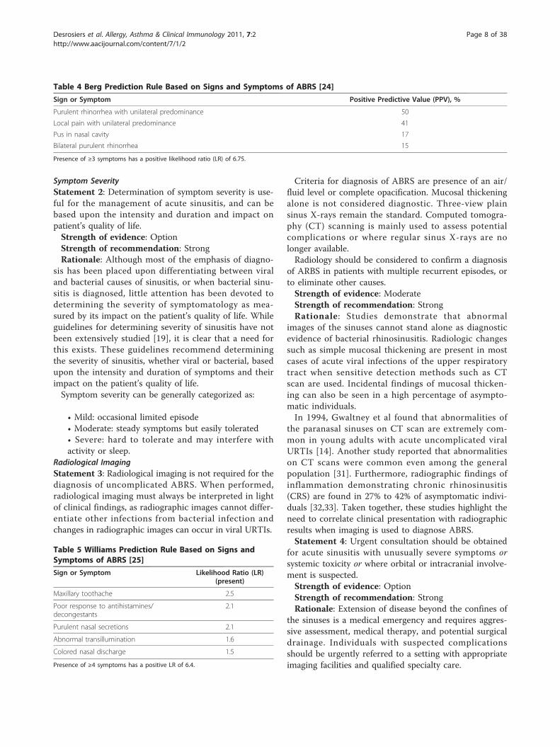
Two studies of patients presenting with symptoms ofsinusitis have led to the development of predictionrules. In 1 study, Berg et al reported that 2 or morepositive findings provided 95% sensitivity and 77% speci-ficity for sinusitis (Table 4) [24]. In the second study,
Williams et al identified 5 independent predictors ofsinusitis that were consistent with radiographic findings(Table 5) [25].Prediction rules can be used to aid in diagnosis. Using
either the Berg or Williams prediction rules, the prob-ability of ABRS increases with cumulative symptoms[24,25]. Although none of these symptoms are individu-ally sensitive or specific for diagnosis, the reported num-ber of diagnostic factors is felt to correlate well with thelikelihood of bacterial infection [26].A Canadian Medical Association evidence-based
review recommended a score based on Williams’ 5 inde-pendent predictor symptoms [27]. Fewer than 2 symp-toms ruled out ABRS (positive predictive value [PPV], <40%), 4 or more symptoms ruled in ABRS (PPV, 81%),and 2 or 3 symptoms (PPV, 40%-63%) suggested thatradiography might be beneficial to clarify the diagnosis.More recent studies have emphasized limitations of clin-
ical findings alone and have either introduced new diag-nostic elements or else assessed the accuracy of existingsymptoms. In a study of 50 patients with upper respiratorytract symptoms of at least 1 week and self-suspected acutemaxillary sinusitis, no distinct clinical signs or symptomswere identified that increased diagnostic accuracy [28].The sensitivity and specificity of the usual clinical signsand symptoms ranged from 0.04 to 0.74 in a small pro-spective study that defined acute sinusitis (not necessarilybacterial) as 1 or more sinuses with an air fluid level orcomplete opacification [29]. A history of facial pain andsinus tenderness on percussion were inversely associatedwith sinusitis (likelihood ratio [LR] < 1.0). Positive LRswere 1.89 (95% confidence interval [CI], 1.06 to 3.39) forsymptom duration longer than 10 days, 1.47 (CI, 0.93 to2.32) for purulent nasal secretions on history, 2.11 (CI,1.23 to 3.63) for oropharyngeal red streak in the lateralpharyngeal recess, 1.89 (CI, 1.08 to 3.32) for transillumina-tion, and 1.22 (CI, 0.08 to 18.64) for otitis media.Although transillumination is not considered accurate in
the diagnosis of acute rhinosinusitis (ARS),[16] visualiza-tion of purulent secretions from the middle meatus using
Table 2 Symptoms of ABRS
Major Minor
Facial pain/pressure/fullness Headache
Nasal obstruction Halitosis
Nasal purulence/discolored postnasal discharge Fatigue
Hyposmia/anosmia Dental pain
Cough
Ear pain/pressure
Table 3 ABRS Diagnosis Requires the Presence of at Least2 Major Symptoms*
Major Symptom
P Facial Pain/pressure/fullness
O Nasal Obstruction
D Nasal purulence/discolored postnasal Discharge
S Hyposmia/anosmia (Smell)
*At least 1 symptom must be nasal obstruction or nasal purulence/discoloredpostnasal discharge. Thus, a diagnosis requires at least 2 PODS, one of whichmust be O or D.
Consider ABRS when viral URTI persists beyond 10 days or worsens after 5 to7 days with similar symptoms [22]. Bacterial etiology should be suspected ifsinus symptoms persist for more than 7 days without improvement [20].
Desrosiers et al. Allergy, Asthma & Clinical Immunology 2011, 7:2http://www.aacijournal.com/content/7/1/2
Page 6 of 38
a short wide speculum has been reported to be highly pre-dictive of ARS [25]. Young et al suggested that purulentnasal discharge, signs of pus in the nasal cavity, or sorethroat are better criteria than radiography for selectingpatients who would benefit from antibiotic therapy [30].
Taken together, these results emphasize the diffi-culty of making an accurate diagnosis of sinusitis butsupport existing consensus that symptoms with dura-tion-based criteria are the best currently availabletool.
Figure 1 Algorithm for the Diagnosis and Treatment of ABRS.
Desrosiers et al. Allergy, Asthma & Clinical Immunology 2011, 7:2http://www.aacijournal.com/content/7/1/2
Page 7 of 38
Symptom SeverityStatement 2: Determination of symptom severity is use-ful for the management of acute sinusitis, and can bebased upon the intensity and duration and impact onpatient’s quality of life.Strength of evidence: OptionStrength of recommendation: StrongRationale: Although most of the emphasis of diagno-
sis has been placed upon differentiating between viraland bacterial causes of sinusitis, or when bacterial sinu-sitis is diagnosed, little attention has been devoted todetermining the severity of symptomatology as mea-sured by its impact on the patient’s quality of life. Whileguidelines for determining severity of sinusitis have notbeen extensively studied [19], it is clear that a need forthis exists. These guidelines recommend determiningthe severity of sinusitis, whether viral or bacterial, basedupon the intensity and duration of symptoms and theirimpact on the patient’s quality of life.Symptom severity can be generally categorized as:
• Mild: occasional limited episode• Moderate: steady symptoms but easily tolerated• Severe: hard to tolerate and may interfere withactivity or sleep.
Radiological ImagingStatement 3: Radiological imaging is not required for thediagnosis of uncomplicated ABRS. When performed,radiological imaging must always be interpreted in lightof clinical findings, as radiographic images cannot differ-entiate other infections from bacterial infection andchanges in radiographic images can occur in viral URTIs.
Criteria for diagnosis of ABRS are presence of an air/fluid level or complete opacification. Mucosal thickeningalone is not considered diagnostic. Three-view plainsinus X-rays remain the standard. Computed tomogra-phy (CT) scanning is mainly used to assess potentialcomplications or where regular sinus X-rays are nolonger available.Radiology should be considered to confirm a diagnosis
of ARBS in patients with multiple recurrent episodes, orto eliminate other causes.Strength of evidence: ModerateStrength of recommendation: StrongRationale: Studies demonstrate that abnormal
images of the sinuses cannot stand alone as diagnosticevidence of bacterial rhinosinusitis. Radiologic changessuch as simple mucosal thickening are present in mostcases of acute viral infections of the upper respiratorytract when sensitive detection methods such as CTscan are used. Incidental findings of mucosal thicken-ing can also be seen in a high percentage of asympto-matic individuals.In 1994, Gwaltney et al found that abnormalities of
the paranasal sinuses on CT scan are extremely com-mon in young adults with acute uncomplicated viralURTIs [14]. Another study reported that abnormalitieson CT scans were common even among the generalpopulation [31]. Furthermore, radiographic findings ofinflammation demonstrating chronic rhinosinusitis(CRS) are found in 27% to 42% of asymptomatic indivi-duals [32,33]. Taken together, these studies highlight theneed to correlate clinical presentation with radiographicresults when imaging is used to diagnose ABRS.Statement 4: Urgent consultation should be obtained
for acute sinusitis with unusually severe symptoms orsystemic toxicity or where orbital or intracranial involve-ment is suspected.Strength of evidence: OptionStrength of recommendation: StrongRationale: Extension of disease beyond the confines of
the sinuses is a medical emergency and requires aggres-sive assessment, medical therapy, and potential surgicaldrainage. Individuals with suspected complicationsshould be urgently referred to a setting with appropriateimaging facilities and qualified specialty care.
Table 4 Berg Prediction Rule Based on Signs and Symptoms of ABRS [24]
Sign or Symptom Positive Predictive Value (PPV), %
Purulent rhinorrhea with unilateral predominance 50
Local pain with unilateral predominance 41
Pus in nasal cavity 17
Bilateral purulent rhinorrhea 15
Presence of ≥3 symptoms has a positive likelihood ratio (LR) of 6.75.
Table 5 Williams Prediction Rule Based on Signs andSymptoms of ABRS [25]
Sign or Symptom Likelihood Ratio (LR)(present)
Maxillary toothache 2.5
Poor response to antihistamines/decongestants
2.1
Purulent nasal secretions 2.1
Abnormal transillumination 1.6
Colored nasal discharge 1.5
Presence of ≥4 symptoms has a positive LR of 6.4.
Desrosiers et al. Allergy, Asthma & Clinical Immunology 2011, 7:2http://www.aacijournal.com/content/7/1/2
Page 8 of 38
Red flags for urgent referral include:
• Systemic toxicity• Altered mental status• Severe headache• Swelling of the orbit or change in visual acuity.
Orbital and intracranial complications are the mostfeared complications of both acute and chronic rhinosi-nusitis. In the pre-antibiotic era, 20% of patients withorbital cellulitis went blind and 17% of patients diedfrom intracranial sepsis [34]. Even in the current era,complications can result in permanent blindness ordeath if not treated appropriately and aggressively.Visual loss from sinusitis was reported at a rate of up to10% in a 1991 study [35].Periorbital or orbital cellulitis is the most common
complication of ABRS and most often caused by acuteethmoid and/or frontal disease [36,37]. Infection spreadsfrom the sinuses to the orbit with relative ease [38,39].Periorbital cellulitis is seen on CT as soft tissue swellingand manifests as orbital pain, edema, and high fever. Ifnot aggressively treated, it may spread beyond the orbi-tal septum. Postseptal inflammation involves structuresof the orbit with the development of proptosis, limita-tion of ocular motion, pain and tenderness, and con-junctival chemosis. A subperiosteal or orbital abscessmay result in ophthalmoplegia (globe becomes fixed as aresult of extra-ocular muscle paralysis) and diminishedvisual acuity. A CT scan showing evidence of an abscess,or lack of clinical improvement after 24 to 48 hrs ofintravenous antibiotics are indications for surgicalexploration and drainage. Blindness may result fromcentral retinal artery occlusion, optic neuritis, cornealulceration, or pan-ophthalmitis.Altered mental status and non-specific signs charac-
terized by high fever, frontal or retro-orbital migraine,and the presence of generic signs of meningeal irritationwarrant immediate consultation with an Ear NoseThroat (ENT) specialist and CT scanning (with con-trast). Infection can spread from the sinuses to theintracranial structures [40]. Intracranial complicationscan include osteomyelitis of the frontal bone (Pott’spuffy tumor), meningitis, subdural empyema, epiduralabscess, brain abscess, and cavernous sinus thrombosis.The mortality rate for intracranial complications rangesfrom 20% to 60% [41]. High-dose, long-term intravenousantibiotic therapy followed by endoscopic drainage orcraniotomy and surgical drainage are usually requiredfor successful treatment [42].Because of the serious nature of complications,
patients with suspected complications of ABRS shouldbe immediately referred to an otolaryngologist withappropriate consultation from other services, including
(but not limited to) ophthalmology, neurosurgery, andinfectious diseases.Microbiology of ABRSStatement 5: Routine nasal culture is not recommendedfor the diagnosis of ABRS. When culture is required forunusual evolution, or when complication requires it,sampling must be performed either by maxillary tap orendoscopically-guided culture.Strength of evidence: ModerateStrength of recommendation: StrongRationale: Sinus puncture and aspiration remain the gold
standard for determining the etiology of ABRS. Howeverbecause of the invasive nature of sinus puncture requiredfor bacterial studies, this procedure is rarely performed.The bacterial etiology of ABRS has been well defined
by numerous studies dating back almost 50 years. Typi-cally, the findings between investigators have been con-cordant [5,43-46]:
• Sinus puncture and aspiration remain the goldstandard for determining the etiology of ABRS, butare rarely performed due to the invasive nature ofsinus puncture• Cultures obtained from the nasal passages do notprovide any diagnostic value• ABRS can be differentiated from viral etiology by asinus aspirate that shows the presence of >104 col-ony forming units of bacteria/mL or if polymorphnuclear cells in sinus fluid exceeds 5000 cells/mL• Lower quantities of bacteria may represent earlystages of infection.
Comparisons of endoscopically-directed middle meatuscultures (EDMM), a less invasive approach to bacterialsampling, with maxillary sinus aspirate (MSA; the goldstandard) have reported similar results [47-49]. A meta-analysis comparing the sensitivity and specificity ofEDMM with MSA for ABRS reported that EDMM had asensitivity of 81%, specificity of 91%, and overall accuracyof 87% compared with MSA [50]. Study authors con-cluded that EDMM was a reliable alterative to MSA forobtaining cultures from patients with suspected ABRS.Take Home Points
ABRS is a bacterial infection of the paranasal sinusescharacterized by:
• Sudden onset of symptomatic sinusinfection• Symptom duration > 7 days• Length of episode < 4 weeks.
Major symptoms (PODS):• Facial Pain/pressure/fullness• Nasal Obstruction• Nasal purulence/discolored postnasalDischarge
Desrosiers et al. Allergy, Asthma & Clinical Immunology 2011, 7:2http://www.aacijournal.com/content/7/1/2
Page 9 of 38
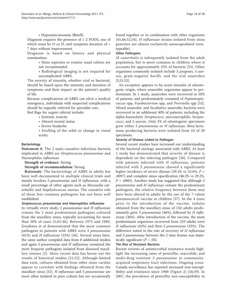
• Hyposmia/anosmia (Smell).Diagnosis requires the presence of ≥ 2 PODS, one ofwhich must be O or D, and symptom duration of >7 days without improvement.Diagnosis is based on history and physicalexamination:
• Sinus aspirates or routine nasal culture arenot recommended• Radiological imaging is not required foruncomplicated ABRS.
The severity of sinusitis, whether viral or bacterial,should be based upon the intensity and duration ofsymptoms and their impact on the patient’s qualityof life.Because complications of ABRS can elicit a medicalemergency, individuals with suspected complicationsshould be urgently referred for specialist care.Red flags for urgent referral include:
• Systemic toxicity• Altered mental status• Severe headache• Swelling of the orbit or change in visualacuity.
BacteriologyStatement 6: The 2 main causative infectious bacteriaimplicated in ABRS are Streptococcus pneumoniae andHaemophilus influenzae.Strength of evidence: StrongStrength of recommendation: StrongRationale: The bacteriology of ABRS in adults has
been well documented in multiple clinical trials andmainly involves S pneumoniae and H influenzae, with asmall percentage of other agents such as Moraxella cat-arhallis and Staphylococcus aureus. The causative roleof these less common pathogens has not been wellestablished.Streptococcus pneumoniae and Haemophilus influenzaeIn virtually every study, S pneumoniae and H influenzaeremain the 2 most predominant pathogens culturedfrom the maxillary sinus, typically accounting for morethan 50% of cases [5,43-46]. Between 1975 and 1989,Gwaltney et al demonstrated that the most commonpathogens in patients with ABRS were S pneumoniae(41%) and H influenzae (35%) [44]. Several years later,the same author compiled data from 8 additional studiesand again S pneumoniae and H influenzae remained themost frequent pathogens isolated from diseased maxil-lary sinuses [5]. More recent data has borne out theresults of historical studies [51,52]. Although limiteddata exist, cultures obtained from other sinus cavitiesappear to correlate with findings obtained from themaxillary sinus [53]. H influenzae and S pneumoniae aremost often isolated in pure culture but are occasionally
found together or in combination with other organisms[45,46,52,54]. H influenzae strains isolated from sinuspuncture are almost exclusively unencapsulated (non-typeable).Other PathogensM catarrhalis is infrequently isolated from the adultpopulation, but is more common in children where itaccounts for approximately 25% of bacteria [55]. Otherorganisms commonly isolated include S pyogenes, S aur-eus, gram-negative bacilli, and the oral anaerobes[5,51,52].An exception appears to be acute sinusitis of odonto-
genic origin, where anaerobic organisms appear to pre-dominate. In 1 study, anaerobes were recovered in 50%of patients, and predominately consisted of Peptostrepto-coccus spp, Fusobacterium spp, and Prevotella spp [53].Mixed anaerobic and facultative anaerobic bacteria wererecovered in an additional 40% of patients, including thealpha-haemolytic Streptococci, microaerophilic Strepto-cocci, and S aureus. Only 5% of odontogenic specimensgrew either S pneumoniae or H influenzae. Beta-lacta-mase producing bacteria were isolated from 10 of 20specimens.Severity of Disease Linked to PathogenSeveral recent studies have increased our understandingof the bacterial etiology associated with ABRS. At least1 study has demonstrated that severity of disease isdependent on the infecting pathogen [56]. Comparedwith patients infected with H influenzae, patientsinfected with S pneumoniae showed a significantlyhigher incidence of severe disease (39.2% vs 23.6%, P =.0097) and complete sinus opacification (46.2% vs 29.2%,P = .0085). Another study has suggested that although Spneumoniae and H influenzae remain the predominantpathogens, the relative frequency between them mayhave been altered in adults by the use of the 7-valentpneumococcal vaccine in children [57]. In the 4 yearsprior to the introduction of the vaccine, isolatesobtained from the maxillary sinus of 156 adults predo-minately grew S pneumoniae (46%), followed by H influ-enzae (36%). After introduction of the vaccine, the mostpredominant organisms recovered from 229 adults wereH influenzae (43%) and then S pneumoniae (35%). Thedifference noted in the rate of recovery of H influenzaeand S pneumoniae between the 2 time frames was statis-tically significant (P < .05).The Rise of Resistant BacteriaRecent reviews of antimicrobial resistance trends high-light the increasing rates of penicillin, macrolide, andmulti-drug resistant S pneumoniae in community-acquired respiratory tract infections. Ongoing cross-Canada surveillance has reported increased non-suscept-ibility and resistance since 1988 (Figure 2) [58,59]. In2007, the prevalence of penicillin non-susceptibility in
Desrosiers et al. Allergy, Asthma & Clinical Immunology 2011, 7:2http://www.aacijournal.com/content/7/1/2
Page 10 of 38
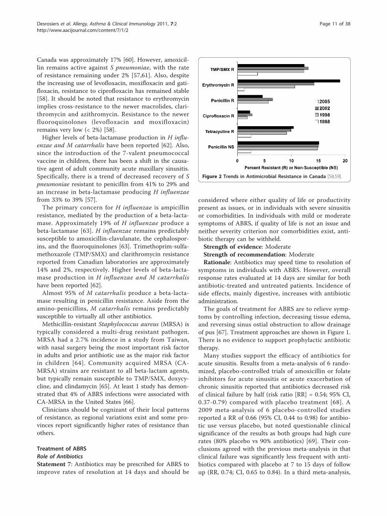
Canada was approximately 17% [60]. However, amoxicil-lin remains active against S pneumoniae, with the rateof resistance remaining under 2% [57,61]. Also, despitethe increasing use of levofloxacin, moxifloxacin and gati-floxacin, resistance to ciprofloxacin has remained stable[58]. It should be noted that resistance to erythromycinimplies cross-resistance to the newer macrolides, clari-thromycin and azithromycin. Resistance to the newerfluoroquinolones (levofloxacin and moxifloxacin)remains very low (< 2%) [58].Higher levels of beta-lactamase production in H influ-
enzae and M catarrhalis have been reported [62]. Also,since the introduction of the 7-valent pneumococcalvaccine in children, there has been a shift in the causa-tive agent of adult community acute maxillary sinusitis.Specifically, there is a trend of decreased recovery of Spneumoniae resistant to penicillin from 41% to 29% andan increase in beta-lactamase producing H influenzaefrom 33% to 39% [57].The primary concern for H influenzae is ampicillin
resistance, mediated by the production of a beta-lacta-mase. Approximately 19% of H influenzae produce abeta-lactamase [63]. H influenzae remains predictablysusceptible to amoxicillin-clavulanate, the cephalospor-ins, and the fluoroquinolones [63]. Trimethoprim-sulfa-methoxazole (TMP/SMX) and clarithromycin resistancereported from Canadian laboratories are approximately14% and 2%, respectively. Higher levels of beta-lacta-mase production in H influenzae and M catarrhalishave been reported [62].Almost 95% of M catarrhalis produce a beta-lacta-
mase resulting in penicillin resistance. Aside from theamino-penicillins, M catarrhalis remains predictablysusceptible to virtually all other antibiotics.Methicillin-resistant Staphylococcus aureus (MRSA) is
typically considered a multi-drug resistant pathogen.MRSA had a 2.7% incidence in a study from Taiwan,with nasal surgery being the most important risk factorin adults and prior antibiotic use as the major risk factorin children [64]. Community acquired MRSA (CA-MRSA) strains are resistant to all beta-lactam agents,but typically remain susceptible to TMP/SMX, doxycy-cline, and clindamycin [65]. At least 1 study has demon-strated that 4% of ABRS infections were associated withCA-MRSA in the United States [66].Clinicians should be cognizant of their local patterns
of resistance, as regional variations exist and some pro-vinces report significantly higher rates of resistance thanothers.
Treatment of ABRSRole of AntibioticsStatement 7: Antibiotics may be prescribed for ABRS toimprove rates of resolution at 14 days and should be
considered where either quality of life or productivitypresent as issues, or in individuals with severe sinusitisor comorbidities. In individuals with mild or moderatesymptoms of ABRS, if quality of life is not an issue andneither severity criterion nor comorbidities exist, anti-biotic therapy can be withheld.Strength of evidence: ModerateStrength of recommendation: ModerateRationale: Antibiotics may speed time to resolution of
symptoms in individuals with ABRS. However, overallresponse rates evaluated at 14 days are similar for bothantibiotic-treated and untreated patients. Incidence ofside effects, mainly digestive, increases with antibioticadministration.The goals of treatment for ABRS are to relieve symp-
toms by controlling infection, decreasing tissue edema,and reversing sinus ostial obstruction to allow drainageof pus [67]. Treatment approaches are shown in Figure 1.There is no evidence to support prophylactic antibiotictherapy.Many studies support the efficacy of antibiotics for
acute sinusitis. Results from a meta-analysis of 6 rando-mized, placebo-controlled trials of amoxicillin or folateinhibitors for acute sinusitis or acute exacerbation ofchronic sinusitis reported that antibiotics decreased riskof clinical failure by half (risk ratio [RR] = 0.54; 95% CI,0.37-0.79) compared with placebo treatment [68]. A2009 meta-analysis of 6 placebo-controlled studiesreported a RR of 0.66 (95% CI, 0.44 to 0.98) for antibio-tic use versus placebo, but noted questionable clinicalsignificance of the results as both groups had high curerates (80% placebo vs 90% antibiotics) [69]. Their con-clusions agreed with the previous meta-analysis in thatclinical failure was significantly less frequent with anti-biotics compared with placebo at 7 to 15 days of followup (RR, 0.74; CI, 0.65 to 0.84). In a third meta-analysis,
Figure 2 Trends in Antimicrobial Resistance in Canada [58,59].
Desrosiers et al. Allergy, Asthma & Clinical Immunology 2011, 7:2http://www.aacijournal.com/content/7/1/2
Page 11 of 38
16 randomized, placebo-controlled studies of antibioticsfor the treatment of presumed ABRS were included[70]. This study used a random effect model odds ratio(OR) and reported a higher proportion of improvementor cure (OR = 1.60, 95% CI, 1.31 to 1.96), but also ahigher rate of adverse events (OR = 1.94, 95% CI, 1.29-2.92) for the antibiotic group versus the placebo group.Although antimicrobial therapy is recommended for
the management of ABRS, this recommendation is notwithout controversy [15,16,69-71]. In a meta-analysis ofstudies enrolling patients with suspected ABRS not con-firmed by imaging, laboratory testing, or cultures, analy-sis of individual patient data resulted in an OR of 1.37(95% CI, 1.13 to 1.66) for antibiotic use versus placebo[72]. The calculated number needed to treat was 15.Study authors concluded that clear justification for anti-biotic treatment was lacking when ABRS was based onclinical signs and symptoms. However, because the ana-lysis included studies of patients who had not hadX-rays of the sinuses, and studies enrolled patients withobvious viral infection, the meta-analysis missed anopportunity to assess antibiotic efficacy in patients whowere clearly likely to benefit from treatment [73]. Inanother meta-analysis of patients with symptoms ofacute sinusitis or rhinitis (10 studies) or acute rhinor-rhea (3 studies), symptom duration averaged 8.1 days(studies ranged from a median of 4.5 days to a mean of15.4 days), and diagnosis was made from signs andsymptoms in over half of the studies. Although cure orimprovement rates were significantly better for the anti-biotic group at 7 to 12 days, there was no differencebetween treatment groups at 15 days, suggesting thatthere was no difference between antibiotics and placeboon patient outcomes. However, the meta-analysisincluded studies of patients who likely had viral rhinosi-nusits, in which antibiotics would be ineffective, thusreducing the ability to assess drug efficacy on patientsmost likely to benefit from treatment [74]. A long-termobjection to interpretation of placebo versus antibioticstudies of acute sinusitis has been that the presumedeffectiveness of antibiotics in the management of bacter-ial rhinosinusitis is diluted by the large number of indi-viduals with viral disease participating in these trials.However, a recent study has suggested that even incases of bacterial rhinosinusitis confirmed by sinus aspi-rate obtained via puncture, antibiotics are no betterthan placebo. In this study, patients with positive bacter-ial cultures for ABRS reported that while 5-day moxi-floxacin treatment led to numerically fewer clinicalfailure rates versus placebo (19.2% vs 33.3%, respec-tively), the difference was not statistically significant(P = .122) [75]. Although the findings suggested a trendfor faster symptom resolution and lower failure rates forantibiotic-treated individuals, they did not confirm the
absolute utility of antibiotic treatment compared withplacebo.Combined, the various studies and meta-analyses do
suggest that antibiotic use, in the setting of ABRS, mayspeed time to symptom resolution, but that little effectis noted upon ultimate outcome, with similar rates ofresolution.Take Home Points
Microbiology of ABRS:• Main causative bacteria are S pneumoniae andH influenzae• Minor causative bacteria are Moraxella catar-hallis and S aureus
○ M catarrhalis is infrequent in the adultpopulation, but accounts for about 25% ofbacteria in children
• Anaerobic organisms appear to predominate inacute sinusitis of odontogenic origin.
Role of antibiotic therapy in individuals with ABRS:• Goals of treatment are to relieve symptoms by:
○ Controlling infection○ Decreasing tissue edema○ Reversing sinus ostial obstruction to allowdrainage of pus
• Antibiotics may be prescribed to improve ratesof symptom resolution
○ Overall response rates are similar for anti-biotic-treated and untreated individuals.
• Antibiotics should be considered forindividuals:
○ With severe sinusitis or comorbidities○ Where quality of life or productivity areissues
• Incidence of side effects, mainly digestive,increases with antibiotic administration.
Choosing an AntibioticStatement 8: When antibiotic therapy is selected, amox-icillin is the first-line recommendation in treatment ofABRS. In beta-lactam allergic patients, trimethoprim-sulfamethoxazole (TMP/SMX) combinations or amacrolide antibiotic may be substituted.Strength of evidence: OptionStrength of recommendation: StrongStatement 9: Second-line therapy using amoxicillin/
clavulanic acid combinations or quinolones withenhanced gram positive activity should be used inpatients where risk of bacterial resistance is high, orwhere consequences of failure of therapy are greatest, aswell as in those not responding to first-line therapy. Acareful history to assess likelihood of resistance shouldbe obtained, and should include exposure to antibioticsin the prior 3 months, exposure to daycare, and chronicsymptoms.Strength of evidence: Option
Desrosiers et al. Allergy, Asthma & Clinical Immunology 2011, 7:2http://www.aacijournal.com/content/7/1/2
Page 12 of 38
Strength of recommendation: StrongRationale: A comprehensive knowledge of the com-
mon etiologies associated with ABRS and the prevalenceof antibiotic resistance among these pathogens is para-mount to select appropriate treatment. Because antibio-tic selection will almost always be made in the absenceof bacterial cultures to guide management, activityagainst the suspected pathogen should be considered.Some important considerations for choosing an anti-
biotic include: the suspected or confirmed etiology,medical history, Canadian patterns of antimicrobialresistance, tolerability, convenience, and cost of treat-ment. It should also be noted that an individual’s medi-cal history is an important factor in treatment strategy.Patients who are at increased risk of bacterial resistanceand complications due to underlying disease (eg, dia-betes, chronic renal failure, immune deficiency) shouldnot be treated the same as otherwise healthy adults withABRS. Underlying systemic disorders place patients withABRS at increased risk of recurrence, antibiotic resis-tance, and complications.Studies have reported that expensive antibiotics were
no more effective than amoxicillin or folate inhibitorsfor acute uncomplicated sinusitis in otherwise healthyadults [68]. A meta-analysis of 3338 patients from 16randomized comparative non-placebo studies concludedthat differences between antimicrobial agents are smallin otherwise healthy adults and adolescents, and there-fore an inexpensive antibiotic should initially be chosen[61]. Current evidence based on randomized controlledtrials suggest comparable efficacy amongst the antibio-tics that have been approved for ABRS in Canada[15,16,76-85]. These include amoxicillin, amoxicillin/cla-vulanate, cefuroxime axetil, clindamycin, TMP/SMX,clarithromycin, ciprofloxacin, levofloxacin, and moxi-floxacin [86].Selection between these different options may be diffi-
cult. Current recommendations are made on the basisof presumed efficacy, risk of bacterial resistance, pre-sence of complications, or cost of therapy.First-line therapy is amoxicillin. Surveillance studies
demonstrate that resistance rates to amoxicillin bystreptococci remain low and a consistent responseremains predicted. Higher doses of amoxicillin are sug-gested in suspected cases of penicillin-resistant S pneu-moniae [62]. In patients with a questionable history ofbeta-lactam allergy, skin testing may be appropriate toconfirm or deny sensitivity, as restricting use of penicil-lin and penicillin derivatives may result in disadvantagesto the patient (ie, costs, side effects) [87]. First-line useof macrolides should probably be limited to patientsallergic to penicillin.Individuals with no clinical response within 72 hours
may be presumed to be unresponsive to therapy. The
possibility of bacterial resistance should be suspected,and therapy should be changed to a second-lineantibiotic.Second-line therapy using fluoroquinolones with
enhanced gram-positive activity (ie, levofloxacin, moxi-floxacin) or amoxicillin-clavulanic acid inhibitors asinitial management may be needed when there are con-cerns of bacterial resistance or risk of complications incases of failure due to underlying disease.Some populations have been found to be at greater
risk of harboring penicillin- and macrolide-resistantstreptococci. Depending on geographic location andenvironment, S pneumoniae may be resistant to macro-lides and TMP/SMX in nearly one third of cases [88].Compared with control subjects, those with exposure todaycare settings had a 3.79 (CI, 0.85 to 7.77) higherodds of having penicillin-resistant infection [89]. It hasbeen demonstrated that individuals with invasive strep-tococcal infections and antibiotic use within the past3 months have a higher rate of antibiotic resistance, par-ticularly in those treated with TMP-SMX (OR, 5.97) orthe macrolide azithromycin (OR, 2.78) [90]. Individualswith antibiotic use within the past 3 months, chronicsymptoms greater than 4 weeks, or parents of childrenin daycare have a higher risk of harboring penicillin-and macrolide-resistant bacteria and should be treatedaccordingly.Second-line therapy used as initial management is also
needed in situations where a higher risk of complicationis associated with treatment failure because of underly-ing systemic disease. Bacterial sinusitis of the frontaland sphenoid sinuses pose a higher risk of complicationthan maxillary and ethmoid sinusitis and require moreaggressive management and surveillance, with first-linetherapy consisting of a second-line agent [16]. Indivi-duals with underlying immunosuppressive sites or medi-cations, or with chronic medical conditions, are atincreased risk of complications if failure of therapyoccurs.Statement 10: Bacterial resistance should be consid-
ered when selecting therapy.Strength of evidence: StrongStrength of recommendation: StrongRationale: Bacterial resistance rates to penicillin and
macrolide/streptogramin/licosamide families haveincreased rapidly over the past decade to the extent thatpenicillin and macrolide resistance is now common.Failure of therapy secondary to resistant organisms hasled to poor clinical outcomes in several well-documen-ted instances.There is increasing evidence for the association
between antimicrobial resistance and adverse patientoutcomes [91,92]. Clinicians should enquire aboutrecent antibiotic use and choose an alternate class of
Desrosiers et al. Allergy, Asthma & Clinical Immunology 2011, 7:2http://www.aacijournal.com/content/7/1/2
Page 13 of 38
antibiotic from that used in the past 3 months [93].Supporting this approach are new data that have shownthat therapy within the past 3 months is a risk factor forpneumococcal resistance. The Toronto Bacterial Net-work evaluated data from patients in 3339 cases of inva-sive pneumococcal infection, of whom 563 had a historyof antibiotic therapy in the preceding 3 months wherethe identity of the therapy was known [90]. In the study,recent therapy with penicillin, macrolides, trimetho-prim-sulfa, and quinolones (but not cephalosporin) wasassociated with a higher frequency of resistance to thatsame agent. Other patient subgroups identified as at riskfor infection with resistant bacterial strains included theyoung (< 2 years of age), the elderly (> 65 years of age),and those with severe underlying disease. These findingsemphasize the importance of taking a history of recentantibiotic use and choosing an agent that differs fromwhat the patient had recently received.Take Home Points
There are increasing rates of antibiotic resistance:• Penicillin-, macrolide-, and multi-drug resistantS pneumoniae in community-acquired respiratorytract infections• Be cognizant of local patterns of antibioticresistance, as regional variations exist.
Medical history influences treatment choice:• Identify patients at increased risk of bacterialresistance and complications
○ Those with underlying disease (eg, diabetes,chronic renal failure, immune deficiency)○ Those with underlying systemic disorders.
Considerations for choosing an antibiotic:• Suspected or confirmed etiology• Medical history• Presence of complications• Canadian patterns of antimicrobial resistance• Risk of bacterial resistance• Tolerability• Convenience• Cost of treatment.
Antibiotic choice:• First-line: amoxicillin
○ In beta-lactam allergy: TMP/SMX ormacrolide
• Second-line: amoxicillin/clavulanic acid combina-tion, or quinolones with enhanced gram-positiveactivity (ie, levofloxacin, moxifloxacin)
○ For use where first-line therapy failed(defined as no clinical response within72 hours), risk of bacterial resistance is high,or where consequences of therapy failure aregreatest (ie, because of underlying systemicdisease).
For uncomplicated ABRS in otherwise healthyadults, antibiotics show comparable efficacy.
Statement 11: When antibiotics are prescribed, dura-tion of treatment should be 5 to 10 days as recom-mended by product monographs. Ultra-short treatmentdurations are not currently recommended by this group.Strength of evidence: StrongStrength of recommendation: ModerateRationale: Some data support efficacy of shorter dura-
tions of therapy; however, none of these short durationshave been approved in Canada, and are thus not recom-mended by this group.Traditional approaches to antimicrobial management
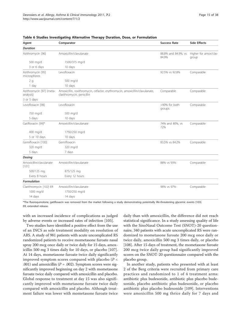
of ABRS focus on courses of therapy of at least 10 daysduration [94]. The rationale for this length of therapyoriginated from studies in tonsillopharyngitis. Potentialbenefits of short-course therapy include improved com-pliance, fewer adverse events, reduced risk of treatmentfailure and bacterial resistance, and reduced cost. Anumber of studies have investigated short-course ther-apy with various antibiotics and have demonstratedsimilar benefit as comparators (Table 6). These studieshave been performed using a variety of antibiotics, somerecommended, some not presently recommended inthese guidelines, and several either not or no longermarketed in Canada. Of note is that in the UnitedStates, a 1-day course of azithromycin reported compar-able efficacy to the comparator [95]. Despite this result,it is the opinion of the group that a recommendationfor ultra-short courses of therapy be reserved untilfurther supporting trials are performed.It is of the opinion of the group that 10 days of ther-
apy with an antibiotic is sufficient. Evolution of the dis-ease and symptom response remains similar regardlessof shorter or longer courses of antibiotics [104]. Thus,absence of complete cure (improvement in symptomswithout complete disappearance of symptoms) at theend of therapy should be expected and should not causean immediate prescription of a second antibiotic.Alternatives to Antibiotics: Intranasal Corticosteroids (INCS)as MonotherapyStatement 12: Topical INCS can be useful as sole ther-apy of mild-to-moderate ARS.Strength of evidence: ModerateStrength of recommendation: StrongRationale: Topical INCS offer an approach to hasten
resolution of sinus episodes and clearance of infectiousorganisms by promoting drainage and reducing mucosalswelling [105]. They are also used to decrease the fre-quency and severity of recurrent episodes [106]. Con-cerns regarding safety of treatment with INCS have notbeen borne out as their use has not been associated
Desrosiers et al. Allergy, Asthma & Clinical Immunology 2011, 7:2http://www.aacijournal.com/content/7/1/2
Page 14 of 38
with an increased incidence of complications as judgedby adverse events or increased rates of infection [105].Two studies have identified a positive effect from the use
of an INCS as sole treatment modality on resolution ofARS. A study of 981 patients with acute uncomplicated RSrandomized patients to receive mometasone furoate nasalspray 200 mcg once daily or twice daily for 15 days, amox-icillin 500 mg 3 times daily for 10 days, or placebo [107].At 14 days, mometasone furoate twice daily significantlyimproved symptom scores compared with placebo (P <.001) and amoxicillin (P = .002). Symptom scores were sig-nificantly improved beginning on day 2 with mometasonefuroate twice daily compared with amoxicillin and placebo.Global response to treatment at day 15 was also signifi-cantly improved with mometasone furoate twice dailycompared with amoxicillin and placebo. Although treat-ment failure was lower with mometasone furoate twice
daily than with amoxicillin, the difference did not reachstatistical significance. In a study assessing quality of lifewith the SinoNasal Outcome Test (SNOT)-20 question-naire, 340 patients with acute uncomplicated RS were ran-domized to mometasone furoate 200 mcg once daily ortwice daily, amoxicillin 500 mg 3 times daily, or placebo[108]. After 15 days of treatment, the mometasone furoate200 mcg twice daily group had significantly improvedscores on the SNOT-20 questionnaire compared with theplacebo group.In another study, patients who presented with at least
2 of the Berg criteria were recruited from primary carepractices and randomized to 1 of 4 treatment arms:antibiotic plus budesonide, antibiotic plus placebo bude-sonide, placebo antibiotic plus budesonide, or placeboantibiotic plus placebo budesonide [109]. Interventionswere amoxicillin 500 mg thrice daily for 7 days and
Table 6 Studies Investigating Alternative Therapy Duration, Dose, or Formulation
Agent Comparator Success Rate Side Effects
Duration
Azithromycin [96] Amoxicillin/clavulanate 88.8% and 84.9%, vs84.9%
Higher for amox/clavgroup
500 mg/d 1500/375 mg/d
3 or 6 days 10 days
Azithromycin [95]microspheres
Levofloxacin 92.5% vs 92.8% Comparable
2 g 500 mg/d
1 day 10 days
Azithromycin [97] (meta-analysis)
Amoxicillin, roxithromycin, cefaclor, erythromycin, amoxicillin/clavulanate,clarithromycin, penicillin
Comparable Comparable
3 or 5 days
Levofloxacin [98] Levofloxacin >90% for bothgroups
Comparable
750 mg/d 500 mg/d
5 days 10 days
Gatifloxacin [99]* Amoxicillin/clavulanate 74% and 80%, vs72%
Comparable
400 mg/d 1750/250 mg/d
5 or 10 days 10 days
Gemifloxacin [100] Gemifloxacin 83.5% vs 84.2% Comparable
320 mg/d 320 mg/d
5 days 7 days
Dosing
Amoxicillin/clavulanate[101]
Amoxicillin/clavulanate 88% vs 93% Comparable
500/125 mg 875/125 mg
Every 8 hours Every 12 hours
Formulation
Clarithromycin [102] ER Amoxicillin/clavulanate 98% vs 97% Comparable
1000 mg/d 1750/250 mg/d
14 days 14 days
*The fluoroquinolone, gatifloxacin was removed from the market following a study demonstrating potentially life-threatening glycemic events [103].
ER, extended release.
Desrosiers et al. Allergy, Asthma & Clinical Immunology 2011, 7:2http://www.aacijournal.com/content/7/1/2
Page 15 of 38
200 mg of budesonide per nostril once daily for 10 days.Results showed no significant difference between treat-ment arms (OR = 0.99, 95% CI, 0.57 to 1.73 for antibio-tic vs placebo; OR = 0.93, 95% CI, 0.54 to 1.62 forbudesonide vs placebo). Authors concluded there wasno place for these agents in the treatment of ARS in pri-mary care. However, because the median days of symp-tom duration at presentation was shorter (7 days, with arange of 4 to 14 days) than currently recommended, thepatient population may have included a greater propor-tion than usual of viral rather than bacterial sinusitis[110], thus limiting the ability to detect the benefit oftreatments on bacterial episodes.Although there is limited evidence for and against the
use of INCS as monotherapy in the treatment of ABRS,it remains an interesting treatment approach. INCS cur-rently offers a novel option that may be explored basedon limited evidence suggesting benefit. In the context ofconflicting results between different trials, the use ofINCS with established dosing requirements indicated forABRS may be preferable. Additional clinical trials andfurther experience in coming years will better discern itsrole in the management of ABRS.Management of Failures of First-Line TherapyStatement 13: Treatment failure should be consideredwhen patients fail to respond to initial therapy within72 hours of administration. If failure occurs followinguse of INCS as monotherapy, antibacterial therapyshould be administered. If failure occurs following anti-biotic administration, it may be due to lack of sensitivityto, or bacterial resistance to, the antibiotic, and the anti-biotic class should be changed.Strength of evidence: OptionStrength of recommendation: StrongRationale: In patients managed with a topical corti-
costeroid as sole therapy, persistent bacterial infectionmay be presumed and an antibiotic should be instituted,according to guidelines for selection of an antibiotic.Bacteriologic response to antibiotics should be expectedwithin 48 hours, thus symptoms should at least partiallyattenuate by 72 hours. If symptoms persist unchangedat this time, failure of response to antibiotic therapymust be considered along with possible resistance [71].Antibiotic therapy must be adjusted by switching to asecond-line antibiotic such as moxifloxacin or amoxicil-lin/clavulanic acid combination or, in the case of a sec-ond-line failure, to another antibiotic class.Studies using in-dwelling catheters for serial sampling
of sinus fluid have reported the time course of antibio-tics to eradicate pathogens as ranging from 24 to 72hours [111-113]. In the absence of at least a partial clin-ical response by 72 hours, bacterial resistance should besuspected as one of the causes of failure and appropriatemeasures should be instituted.
Take Home PointsFactors suggesting greater risk of penicillin- andmacrolide-resistant streptococci:
• Antibiotic use within the past 3 months○ Choose an alternate class of antibiotic fromthat used in the past 3 months
• Chronic symptoms greater than 4 weeks• Parents of children in daycare.
When antibiotics are prescribed, treatment durationshould be 5 to 10 days as recommended by productmonographs.
• Improvement in symptoms without completedisappearance of symptoms at the end of therapyshould be expected and should not cause animmediate prescription of a second antibiotic.
Topical INCS can be useful as sole therapy of mild-to-moderate ARS.Treatment of first-line therapy failure:
• If symptoms do not at least partially attenuateby 72 hours after INCS monotherapy:
○ Administer antibiotic therapy• If symptoms do not at least partially attenuateby 72 hours after antibiotic administration:
○ Bacterial resistance should be considered,and○ Antibiotic class should be changed○ Switch to a second-line antibiotic, such as▪ Moxifloxacin▪ Amoxicillin/clavulanic acid combination○ In the case of a second-line failure, switchto another antibiotic class.
Adjunct TherapyStatement 14: Adjunct therapy should be prescribed inindividuals with ABRS.Strength of evidence: OptionStrength of recommendation: StrongStatement 15. Topical intranasal corticosteroids
(INCS) may help improve resolution rates and improvesymptoms when prescribed with an antibiotic.Strength of evidence: ModerateStrength of recommendation: StrongStatement 16. Analgesics (acetaminophen or non-
steriodal anti-inflammatory agents) may provide symp-tom relief.Strength of evidence: ModerateStrength of recommendation: StrongStatement 17. Oral decongestants may provide symp-
tom relief.Strength of evidence: OptionStrength of recommendation: ModerateStatement 18. Topical decongestants may provide
symptom relief.Strength of evidence: OptionStrength of recommendation: Moderate
Desrosiers et al. Allergy, Asthma & Clinical Immunology 2011, 7:2http://www.aacijournal.com/content/7/1/2
Page 16 of 38
Statement 19. Saline irrigation may provide symptomrelief.Strength of evidence: OptionStrength of recommendation: StrongRationale: Analgesics, oral and topical decongestants,
topical INCS, and saline sprays or rinses can all helprelieve symptoms of both viral and bacterial infectionsof the upper respiratory passages and can all be sug-gested for symptomatic relief.Ancillary and Alternative Therapies Recent reviewssuggest that the evidence for use of ancillary therapies isrelatively weak, as few prospective randomized clinicaltrials have been performed to assess their effectiveness.This does not necessarily mean that the therapies are ofno benefit, as these have long been a part of clinicalpractice and may offer benefits. However, the lack ofgood quality trials supporting their use requires theincorporation of weaker levels of evidence, thus recom-mendations are derived from extension from first princi-ples and expert opinion.Based on its effects on inflammation, topical INCS in
conjunction with antibiotic therapy have been assessedfor their effectiveness in improving resolution of signsand symptoms of rhinosinusitis. In the Cochrane reviewon this topic, 3 randomized, placebo-controlled studiesof the efficacy of 15- to 21-day courses of mometasonefuroate, fluticasone propionate, or budesonide for nasalendoscopy-confirmed ARS found limited but positiveevidence for INCS as an adjuvant to antibiotics [105].The symptoms of cough and nasal discharge were signif-icantly improved (P < .05) through the second week oftreatment for patients receiving budesonide (50 mcg)plus amoxicillin-clavulanate potassium compared withthose receiving placebo plus the antibiotic [114]. Inpatients receiving mometasone furoate (200 mcg or 400mcg twice daily) plus amoxicillin/clavulanate potassium,total symptom score days 1 to 15 averaged and over the21-day study period were significantly improved (P ≤.017) compared with patients receiving placebo plusantibiotic [115]. In a third study, a 21-day course of flu-ticasone propionate (200 mcg) plus cefuroximeimproved the clinical success rate compared with pla-cebo plus antibiotic (93.5% vs 73.9%, P = .009) as wellas the speed of recovery (6 days vs 9.5 days, P = .01)[106]. No significant steroid-related adverse effects orrecurrence rates were reported. Topical INCS thusappear to be safe and to afford an additional benefitwhen antibiotics are used.Oral decongestants have been shown to improve nasal
congestion and can be used until symptoms resolve,provided there are no contraindications to their use.Topical decongestant use is felt to be controversial andshould not be used for longer than 72 hours due to thepotential for rebound congestion [9].
There are no clinical studies supporting the use ofantihistamines in ABRS [71]. Although 1 randomizedcontrolled trial of human immunodeficiency virus-infected patients with acute or chronic sinusitis reportedbenefit with the mucolytic agent guaifenesin [116], nobenefit was reported in a randomized controlled trial inhealthy subjects [117].There is limited evidence suggesting benefit of saline
irrigation in patients with acute sinusitis. Many studiessupport the role of buffered hypertonic and bufferednormal nasal saline to promote mucociliary clearance.In a study of patients with ABRS, thrice-daily irrigationwith 3% nasal saline improved mucociliary clearancebeginning in week 1 [118]. Moreover, subjects usingonce daily hypertonic saline nasal irrigation reportedsignificantly improved symptoms, quality of life, anddecreased medication use compared with control sub-jects [119]. However, the impact of saline sprays onnasal airway patency is less clear, with studies variouslyreporting no impact of saline sprays [120] and improvedpatency with buffered physiological saline spray [121].Their impact on symptom improvement is also uncer-tain, with a study of hypertonic saline spray reportingno improvement in nasal symptoms or illness duration[122]. Saline therapy, either as a spray or high-volumeirrigation, has seen widespread use as adjunct treatmentdespite a limited evidence base. Although the utility ofsaline sprays remains unclear, the use of saline irrigationas ancillary therapy is based on evidence of modestsymptomatic benefit and good tolerability.Complementary and Alternative Medicine Recentreviews have found limited evidence for alternative andcomplementary medicine for ABRS [71,123]. Some ofthese therapies include home-based foods such as soups,fruit juices, teas, nutritional supplements, and herbalremedies. Alternative practices that have failed to showefficacy under scientific trial conditions include acu-puncture, chiropracty, naturopathy, aromatherapy, mas-sage, and therapeutic touch. Vitamin C preparations andzinc lozenges are also felt to be controversial [71,123].Studies of zinc lozenges for the common cold have pro-duced mixed results. A recent meta-analysis of Echina-cea preparations has shown some positive effects inreducing duration of respiratory tract symptoms [124].A recent systematic review comparing placebo with
the herbal medications Sinupret or Bromelain as adjuncttherapy reported limited evidence of improved symp-toms. Further, single randomized controlled trials onEsbetritox, Mytrol, Cineole, and Bi Yuan Shu showedsome initial positive evidence [125]. A prospective ran-domized controlled trial compared the homeopathicmedication Sinfrontal with placebo among 56 casesand controls with radiograph-confirmed acute maxillarysinusitis [126]. Participants were allowed saline
Desrosiers et al. Allergy, Asthma & Clinical Immunology 2011, 7:2http://www.aacijournal.com/content/7/1/2
Page 17 of 38
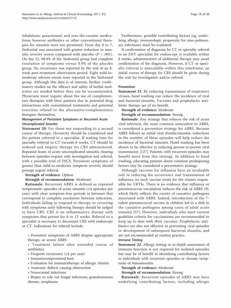
inhalations, paracetemol, and over-the-counter medica-tions; however antibiotics or other conventional thera-pies for sinusitis were not permitted. From day 0 to 7,Sinfrontal was associated with greater reduction in sinu-sitis severity scores compared with placebo (P < .001).On day 21, 68.4% of the Sinfrontal group had completeresolution of symptoms versus 8.9% of the placebogroup. No recurrence was reported by the end of an 8-week post-treatment observation period. Eight mild-to-moderate adverse events were reported in the Sinfrontalgroup. Although this data is of interest, further confir-matory studies on the efficacy and safety of herbal med-icines are needed before they can be recommended.Physicians must inquire about the use of complemen-tary therapies with their patients due to potential druginteractions with conventional treatments and potentialtoxicities related to the alternative/complementarytherapies themselves.Management of Persistent Symptoms or Recurrent AcuteUncomplicated SinusitisStatement 20: For those not responding to a secondcourse of therapy, chronicity should be considered andthe patient referred to a specialist. If waiting time forspecialty referral or CT exceeds 6 weeks, CT should beordered and empiric therapy for CRS administered.Repeated bouts of acute uncomplicated sinusitis clearingbetween episodes require only investigation and referral,with a possible trial of INCS. Persistent symptoms ofgreater than mild-to-moderate symptom severity shouldprompt urgent referral.Strength of evidence: OptionStrength of recommendation: ModerateRationale: Recurrent ABRS is defined as repeated
symptomatic episodes of acute sinusitis (≥4 episodes peryear) with clear symptom-free periods in between thatcorrespond to complete resolution between infections.Individuals failing to respond to therapy or recurringwith symptoms early following therapy should be judgedto have CRS. CRS is an inflammatory disease withsymptoms that persist for 8 to 12 weeks. Referral to aspecialist is necessary to document CRS with endoscopyor CT. Indications for referral include:
• Persistent symptoms of ABRS despite appropriatetherapy, or severe ABRS• Treatment failure after extended course ofantibiotics• Frequent recurrence (≥4 per year)• Immunocompromised host• Evaluation for immunotherapy of allergic rhinitis• Anatomic defects causing obstruction• Nosocomial infections• Biopsy to rule out fungal infections, granulomatousdisease, neoplasms.
Furthermore, possible contributing factors (eg, under-lying allergy, immunologic propensity for sino-pulmon-ary infections) must be evaluated.If confirmation of diagnosis by CT or specialty referral
to an ENT specialist for endoscopy is available within6 weeks, administration of additional therapy may awaitconfirmation of the diagnosis. However, if CT or speci-alty referral is unavailable within this timeframe, aninitial course of therapy for CRS should be given duringthe wait for investigation and/or referral.
PreventionStatement 21: By reducing transmission of respiratoryviruses, hand washing can reduce the incidence of viraland bacterial sinusitis. Vaccines and prophylactic anti-biotic therapy are of no benefit.Strength of evidence: ModerateStrength of recommendation: StrongRationale: Any strategy that reduces the risk of acute
viral infection, the most common antecedent to ABRS,is considered a prevention strategy for ABRS. BecauseABRS follows an initial viral rhinitis/sinusitis, reductionsin the number of these episodes will help reduce theincidence of bacterial sinusitis. Hand washing has beenshown to be effective in reducing person-to-person viraltransmission [127]. Patients with recurrent episodes maybenefit more from this strategy. In addition to handwashing, educating patients about common predisposingfactors may be considered a preventative strategy.Although vaccines for influenza have an invaluable
role in reducing the occurrence and transmission ofinfluenza, no such vaccine exists for the viruses respon-sible for URTIs. There is no evidence that influenza orpneumococcus vaccination reduces the risk of ABRS [9],which likely reflects the variety of causative pathogensassociated with ABRS. Indeed, introduction of the 7-valent pneumococcal vaccine in children led to a shift inthe causative pathogens among cases of adult acutesinusitis [57]. However, individuals who meet currentguideline criteria for vaccinations are recommended tokeep up to date with their vaccines. Prophylactic anti-biotics are also not effective in preventing viral episodesor development of subsequent bacterial sinusitis, andare not recommended as routine practice.Immune TestingStatement 22: Allergy testing or in-depth assessment ofimmune function is not required for isolated episodesbut may be of benefit in identifying contributing factorsin individuals with recurrent episodes or chronic symp-toms of rhinosinusitis.Strength of evidence: ModerateStrength of recommendation: StrongRationale: Recurrent episodes of ABRS may have
underlying contributing factors, including allergic
Desrosiers et al. Allergy, Asthma & Clinical Immunology 2011, 7:2http://www.aacijournal.com/content/7/1/2
Page 18 of 38
rhinitis and immune deficiencies. In 1 study, patientswith CRS or frequent episodes of ARS had a 57% preva-lence of positive skin allergy tests [128]. Another studyshowed that 84% of patients who had surgery for CRShad a positive allergy test, and 58% had multiple aller-gen sensitivities [129]. Such patients may have increasedsusceptibility to inflammation of the nose and paranasalsinuses [130]. However, in the treatment of ABRS in pri-mary care, allergy testing is not required for investigat-ing or resolving acute episodes.Take Home Points
Adjunct therapy may provide symptom relief andshould be prescribed in individuals with ABRS:
• Topical intranasal corticosteroids (INCS)• Analgesics (acetaminophen or non-steriodalanti-inflammatory agents)• Oral decongestants• Topical decongestants• Saline irrigation.
The goal of prevention strategies is to reduce the ofrisk acute viral infection, the most common antece-dent to ABRS.
• Techniques:○ Handwashing○ Educating patients on common predispos-ing factors.
For patients with recurrent episodes of ABRS, con-sider underlying contributing factors:
• Allergy testing to detect allergic rhinitis• In-depth assessment of immune function todetect immune deficiencies.
Chronic Rhinosinusitis (CRS)Adult CRS prompts an estimated 18 to 22 millionannual office visits and 545 000 annual emergency roomvisits in the United States [131]. In a survey study,patients with CRS reported more bodily pain and worsesocial functioning than patients with other chronic con-ditions such as chronic obstructive pulmonary disease,congestive heart failure, and back pain [132]. Theimpact of CRS on patient quality of life is comparable inseverity to that of other chronic conditions. As withother chronic diseases, CRS should be proactivelymanaged.
Definition and DiagnosisStatement 23: CRS is diagnosed on clinical grounds butmust be confirmed with at least 1 objective finding onendoscopy or computed tomography (CT) scan.Strength of evidence: WeakStrength of recommendation: StrongRationale: Symptoms of CRS alone are not sufficient
to diagnose CRS because they can be nonspecific andmimicked by several disease entities (eg, upper
respiratory tract infection [URTI], migraine). Confir-mation of sinus disease using an objective measure isrequired. Conversely, in the absence of symptoms,diagnosis of CRS based on radiology alone is notappropriate because of a high incidence of radiologicalanomalies on CT scans in normal individuals. Thus,the presence of symptoms plus an objective findingare necessary.CRS may be defined as an inflammatory disease invol-
ving the nasal mucosa and paranasal sinuses [133]. CRSis a symptom-based diagnosis supported by objectivedocumentation of disease by physical findings and diag-nostic imaging or sinonasal endoscopy [9,18,133-137].An algorithm for the diagnosis and management of CRSis presented in Figure 3.Take Home Points
Impact of CRS on patients:• Significant bodily pain and impaired socialfunctioning• Quality of life is comparable in severity to thatof other chronic conditions• As a chronic condition, CRS should be proac-tively managed.
CRS is an inflammatory disease involving the nasalmucosa and paranasal sinuses.
• Symptoms are usually of lesser intensity thanthose of ABRS• Symptoms present for 8-12 weeks.
Symptoms of CRSSymptoms of CRS are usually of lesser intensity thanthose of acute bacterial rhinosinusitis (ABRS) but theirduration exceeds the 4 weeks commonly used as theupper limit for the diagnosis of ABRS. A diagnosis ofCRS is probable if 2 or more major symptoms are pre-sent for at least 8 to 12 weeks along with documentedinflammation of the paranasal sinuses or nasal mucosa(Table 7) [9,21,138].CRS can be further categorized based on the
absence or presence of nasal polyps (CRS withoutnasal polyps, CRSsNP; or CRS with nasal polyps,CRSwNP) [137]. Although both are characterized bymucopurulent drainage and nasal obstruction,CRSsNP is frequently associated with facial pain/pres-sure/fullness whereas CRSwNP is frequently character-ized by hyposmia.A diagnosis of CRSsNP requires the presence of the
following:
• At least 2 symptoms and• Inflammation (eg, discolored mucus, edema ofmiddle meatus or ethmoid area) documented byendoscopy and• Absence of polyps in the middle meatus (by endo-scopy) and/or
Desrosiers et al. Allergy, Asthma & Clinical Immunology 2011, 7:2http://www.aacijournal.com/content/7/1/2
Page 19 of 38
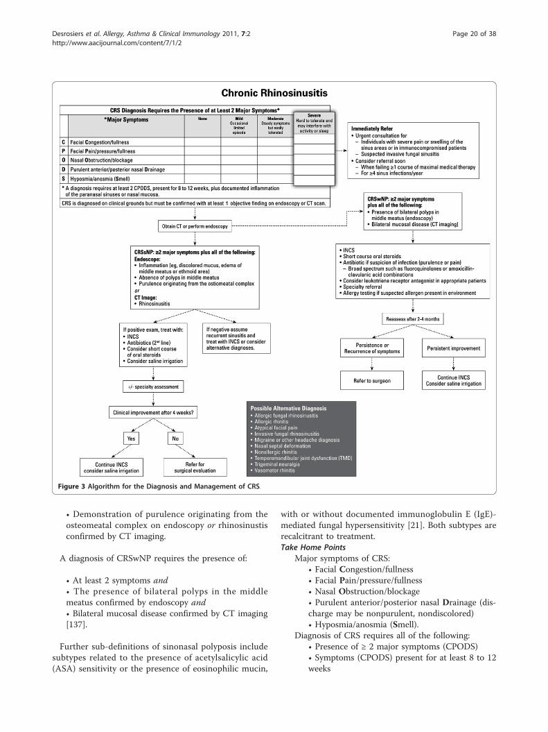
• Demonstration of purulence originating from theosteomeatal complex on endoscopy or rhinosinustisconfirmed by CT imaging.
A diagnosis of CRSwNP requires the presence of:
• At least 2 symptoms and• The presence of bilateral polyps in the middlemeatus confirmed by endoscopy and• Bilateral mucosal disease confirmed by CT imaging[137].
Further sub-definitions of sinonasal polyposis includesubtypes related to the presence of acetylsalicylic acid(ASA) sensitivity or the presence of eosinophilic mucin,
with or without documented immunoglobulin E (IgE)-mediated fungal hypersensitivity [21]. Both subtypes arerecalcitrant to treatment.Take Home Points
Major symptoms of CRS:• Facial Congestion/fullness• Facial Pain/pressure/fullness• Nasal Obstruction/blockage• Purulent anterior/posterior nasal Drainage (dis-charge may be nonpurulent, nondiscolored)• Hyposmia/anosmia (Smell).
Diagnosis of CRS requires all of the following:• Presence of ≥ 2 major symptoms (CPODS)• Symptoms (CPODS) present for at least 8 to 12weeks
Figure 3 Algorithm for the Diagnosis and Management of CRS.
Desrosiers et al. Allergy, Asthma & Clinical Immunology 2011, 7:2http://www.aacijournal.com/content/7/1/2
Page 20 of 38
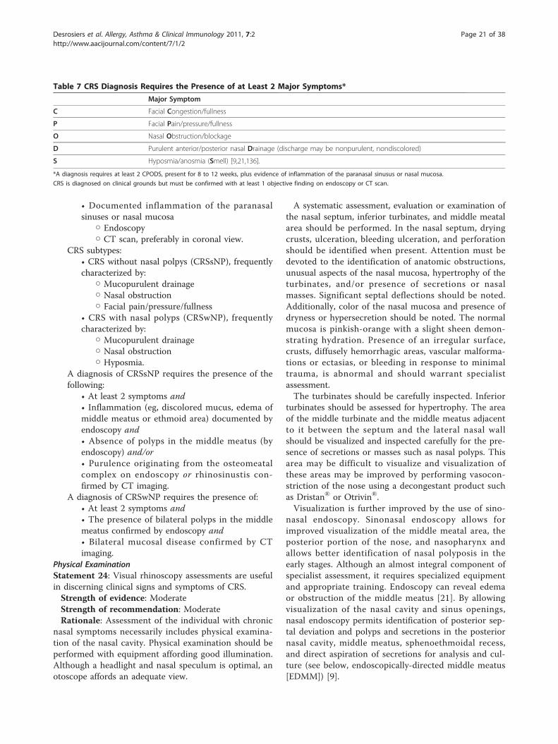
• Documented inflammation of the paranasalsinuses or nasal mucosa
○ Endoscopy○ CT scan, preferably in coronal view.
CRS subtypes:• CRS without nasal polpys (CRSsNP), frequentlycharacterized by:
○ Mucopurulent drainage○ Nasal obstruction○ Facial pain/pressure/fullness
• CRS with nasal polyps (CRSwNP), frequentlycharacterized by:
○ Mucopurulent drainage○ Nasal obstruction○ Hyposmia.
A diagnosis of CRSsNP requires the presence of thefollowing:
• At least 2 symptoms and• Inflammation (eg, discolored mucus, edema ofmiddle meatus or ethmoid area) documented byendoscopy and• Absence of polyps in the middle meatus (byendoscopy) and/or• Purulence originating from the osteomeatalcomplex on endoscopy or rhinosinustis con-firmed by CT imaging.
A diagnosis of CRSwNP requires the presence of:• At least 2 symptoms and• The presence of bilateral polyps in the middlemeatus confirmed by endoscopy and• Bilateral mucosal disease confirmed by CTimaging.
Physical ExaminationStatement 24: Visual rhinoscopy assessments are usefulin discerning clinical signs and symptoms of CRS.Strength of evidence: ModerateStrength of recommendation: ModerateRationale: Assessment of the individual with chronic
nasal symptoms necessarily includes physical examina-tion of the nasal cavity. Physical examination should beperformed with equipment affording good illumination.Although a headlight and nasal speculum is optimal, anotoscope affords an adequate view.
A systematic assessment, evaluation or examination ofthe nasal septum, inferior turbinates, and middle meatalarea should be performed. In the nasal septum, dryingcrusts, ulceration, bleeding ulceration, and perforationshould be identified when present. Attention must bedevoted to the identification of anatomic obstructions,unusual aspects of the nasal mucosa, hypertrophy of theturbinates, and/or presence of secretions or nasalmasses. Significant septal deflections should be noted.Additionally, color of the nasal mucosa and presence ofdryness or hypersecretion should be noted. The normalmucosa is pinkish-orange with a slight sheen demon-strating hydration. Presence of an irregular surface,crusts, diffusely hemorrhagic areas, vascular malforma-tions or ectasias, or bleeding in response to minimaltrauma, is abnormal and should warrant specialistassessment.The turbinates should be carefully inspected. Inferior
turbinates should be assessed for hypertrophy. The areaof the middle turbinate and the middle meatus adjacentto it between the septum and the lateral nasal wallshould be visualized and inspected carefully for the pre-sence of secretions or masses such as nasal polyps. Thisarea may be difficult to visualize and visualization ofthese areas may be improved by performing vasocon-striction of the nose using a decongestant product suchas Dristan® or Otrivin®.Visualization is further improved by the use of sino-
nasal endoscopy. Sinonasal endoscopy allows forimproved visualization of the middle meatal area, theposterior portion of the nose, and nasopharynx andallows better identification of nasal polyposis in theearly stages. Although an almost integral component ofspecialist assessment, it requires specialized equipmentand appropriate training. Endoscopy can reveal edemaor obstruction of the middle meatus [21]. By allowingvisualization of the nasal cavity and sinus openings,nasal endoscopy permits identification of posterior sep-tal deviation and polyps and secretions in the posteriornasal cavity, middle meatus, sphenoethmoidal recess,and direct aspiration of secretions for analysis and cul-ture (see below, endoscopically-directed middle meatus[EDMM]) [9].
Table 7 CRS Diagnosis Requires the Presence of at Least 2 Major Symptoms*
Major Symptom
C Facial Congestion/fullness
P Facial Pain/pressure/fullness
O Nasal Obstruction/blockage
D Purulent anterior/posterior nasal Drainage (discharge may be nonpurulent, nondiscolored)
S Hyposmia/anosmia (Smell) [9,21,136].
*A diagnosis requires at least 2 CPODS, present for 8 to 12 weeks, plus evidence of inflammation of the paranasal sinusus or nasal mucosa.
CRS is diagnosed on clinical grounds but must be confirmed with at least 1 objective finding on endoscopy or CT scan.
Desrosiers et al. Allergy, Asthma & Clinical Immunology 2011, 7:2http://www.aacijournal.com/content/7/1/2
Page 21 of 38
Methods of Bacterial RecoveryStatement 25: In the few situations when deemednecessary, bacterial cultures in CRS should be per-formed either via endoscopic culture of the middle mea-tus or maxillary tap but not by simple nasal swab.Strength of evidence: OptionStrength of recommendation: StrongRationale: Endoscopic cultures of the middle meatus
are less invasive than maxillary tap. To be of value,these cultures require skilled practitioners to performthe endoscopic examination and sampling to avoid con-tamination from normal nasal flora or from adjacentstructures. Simple swab nasal cultures are of little pre-dictive value. Of note is that when purulent secretionsare observed, these should be sampled directly, as cul-ture resulting from a thin stream of pus and an adjacentarea can differ. Also, since all specimens are potentiallycontaminated to varying degrees, proper specimen col-lection, transport, storage, and processing are key.Involvement of bacterial biofilm can be difficult to
detect, and identification of pathogens relies on analyz-ing tissue samples with either electron or confocal scan-ning laser microscopy (CSLM), or indirectly byanalyzing the DNA signature for biofilm-forming genes[139,140]. These tests remain primarily research toolsand are not currently available in the clinical setting.Maxillary Sinus Aspirate (MSA) MSA is consideredthe gold standard for bacterial recovery in acute andchronic sinusitis, but there are still issues of concern.Because the procedure is invasive, it is usually per-formed by specialists rather than in family practice, andeven then only rarely. MSA is recommended if there areserious complications, such as orbital infection, intracra-nial extension of the infection, or in patients with noso-comial sinusitis. MSAs are usually obtained by puncturethrough the canine fossa or inferior meatus and are sub-ject to contamination by oral or nasal flora during col-lection. Because pathogens in various sinuses may differ,MSAs are limited in only being representative of themaxillary sinus. Other concerns with MSAs includepatient compliance, discomfort, bleeding, secondaryinfection, and (rarely) injury to the infraorbital nerve ororbit.Nasal and Postnasal Discharge Culture Most studieshave shown poor correlation of nose and throat cultureswith MSAs, and these cultures are generally not recom-mended in patients with acute or chronic sinusitis.Endoscopically-Directed Middle Meatus Culture(EDMM) EDMM requires specialist expertise andequipment. It is safe and usually painless. The areasampled (the middle meatus) contains the ostiomeatalcomplex, which provides a common drainage pathwayfor the maxillary, ethmoid, and frontal sinuses. EDMMcultures are therefore representative of frontal, ethmoid,
and maxillary sinuses. However, as with MSAs, EDMMsare subject to contamination with resident nasal flora(including anaerobes), which makes their interpretationsubject to clinical situation. Also, pathogenic bacteriasuch as S. pneumoniae, H. influenzae, and S. aureus maybe isolated from asymptomatic patients in the carrierstate. The significance of this carrier status is uncertainbut in the absence of symptoms, treatment is rarelyinitiated. EDMM swab and MSA have been shown to beequivalent for detection of pathogens and likelihood ofcontamination [140,141].Fungal Pathogens If invasive fungal sinusitis is sus-pected, prompt diagnosis and treatment are essential.Culture must be requested promptly because theseinfections are life threatening and usually require emer-gency surgery. However, results of culture are rarelyavailable to assist with decision-making, and diagnosis ismost frequently made on the basis of Gram staining andfrozen sections demonstrating the characteristic branch-ing hyphae arrangement. Biopsies for Gram stain andculture (aerobic and anaerobic bacterial culture plusfungal culture) and for histopathology and special stainsare key.Conclusions for Bacterial Recovery Although postna-sal/nasal discharge is common, routine cultures of suchare discouraged and empiric therapy is recommended. Ifa patient fails multiple courses of empiric therapy, theyshould be referred to an otolaryngologist for evaluation,which usually includes sinonasal endoscopy. If purulentmaterial is identified, diagnostic culture may be made byEDMM. When situation warrants it, such as for compli-cations or in nosocomial intensive care unit sinusitis,MSA may be performed.Radiological ImagingStatement 26: The preferred means of radiological ima-ging of the sinuses in CRS is the CT scan, preferably inthe coronal view. Imaging should always be interpretedin the context of clinical symptomatology because thereis a high false-positive rate.Strength of evidence: ModerateStrength of recommendation: StrongRationale: Conventional X-ray images do not ade-
quately image the ethmoid sinuses or the osteomeatalcomplexes, which are key to the development and per-sistence of CRS, and when clinically indicated, may thusbe assessed with CT scanning. However, positive CTfindings alone are not indicative of CRS in the absenceof signs or symptoms given the high prevalence ofmucosal changes accompanying URTIs and/or asympto-matic changes in the non-diseased population.CT is an important means of providing objective evi-
dence for the diagnosis of CRS but must absolutely becorrelated with clinical and endoscopic findings tointerpret them meaningfully. Several studies have
Desrosiers et al. Allergy, Asthma & Clinical Immunology 2011, 7:2http://www.aacijournal.com/content/7/1/2
Page 22 of 38
demonstrated that CT cannot be used as a sole indica-tor for CRS [31-33]. Further studies have reportedeither no correlation [142] or a low correlation[143-145] between symptoms and CT findings. Theseresults reflect the need to interpret imaging results inthe context of persistent symptoms to provide an accu-rate diagnosis. Thus CT should not be used as the solecriteria for determining the need for surgical interven-tion, but rather should be used as an objective tool forconfirming the diagnosis of CRS and for surgical plan-ning. CT scans should be ordered after failure of maxi-mal medical management and/or for the planning ofsurgery. A complete CT scan should be obtained if thephysician is contemplating surgical intervention, and thescan ideally should be at a minimum resolution of3 mm coronal slices, if not more detailed. Reconstruc-tion in the sagittal plane may help with performance ofsurgery, particularly in the area of the frontal sinus.Take Home Points
Visual assessments include:• Physical examination of the nasal cavity withequipment affording good illumination
○ Headlight and nasal speculum○ Otoscope.
Examination of:• The nasal septum
○ Identify drying crusts, ulceration, bleedingulceration, and perforation, anatomicobstructions, unusual aspects of the nasalmucosa, and/or presence of secretions ornasal masses○ Note significant septal deflections, andcolor of the nasal mucosa and presence ofdryness or hypersercretion. The normalmucosa is pinkish-orange with a slight sheendemonstrating hydration○ Presence of an irregular surface, crusts, dif-fusely hemorrhagic areas, vascular malforma-tions or ectasias, or bleeding in response tominimal trauma, is abnormal and shouldwarrant specialist assessment
• Inferior turbinates:○ Assess for hypertrophy
• Middle meatal area:○ Inspect the area of the middle turbinateand the middle meatus adjacent to it betweenthe septum and the lateral nasal wall for thepresence of secretions or masses such asnasal polyps○ Visualization of these areas may beimproved by performing vasoconstriction ofthe nose using a decongestant product (eg,Dristan® or Otrivin®). Sinonasal endoscopymay improve visualization.
PathophysiologyStatement 27: CRS is an inflammatory disease ofunclear origin where bacterial colonization may contri-bute to pathogenesis. The relative roles of initiatingevents, environmental factors, and host susceptibilityfactors are all currently unknown.Strength of evidence: WeakStrength of recommendation: ModerateRationale: Intense inflammation with eosinophilic,
neutrophilic, and lymphocytic infiltrations and upregula-tion of numerous T helper (Th) cell type 2-associatedcytokines has been well documented in biopsy samplesof CRS. The disease process has several similarities withasthma, including infiltration of a similar population ofinflammatory cells, cytokine profile, and evidence of tis-sue remodeling. The role of bacteria remains imprecisein the face of frequent negative cultures, however a rolefor colonization of S. aureus contributing to disease viasuperantigenic stimulation has been proposed. Emergingevidence on bacterial biofilms may explain frequentnegative cultures and change our future understandingof the role of bacteria in CRS.The last 10 years have witnessed new insights into the
inflammatory mechanisms of CRS. Investigation of theinflammatory roles of cytokines and chemokines hasshed considerable light on the pathogenesis of this dis-ease. It is now widely documented that T lymphocytesand the activated eosinophils are prominent within thesinus mucosa of patients with CRS, especially in atopicpatients. Recruitment and activation of the inflammatorycell infiltrate has largely been attributed to the effects ofTh2 cytokines (namely interleukin [IL]-4, IL-5, IL-13,and granulocyte monocyte-colony stimulating factor),and the eosinophil-associated chemokines, eotaxin, andmonocyte chemotactic proteins.Evidence that CRS subtypes have distinct pathogenetic
mechanisms, and may represent distinct diseases, hasbeen suggested in biomarker studies of nasal secretions.In 1 study, IL-5 and nasal IgE were significantly asso-ciated with CRSwNP but not with CRSsNP or acute rhi-nosinusitis (ARS) [146]. Another study extended thesefindings, reporting that CRSwNP had Th2 polarizationand a higher prevalence of IL-5, IgE, eosinophils,eotaxin, and eosinophil cationic protein, whereasCRSsNP had Th1 polarization and higher levels of inter-feron-gamma and transforming growth factor (TGF)-beta [147]. These results indicate that cytokine andmediator profiles may be useful in differentiatingbetween disease entities.Recruitment and persistence of adaptive immune
responses resulting in the development of the clinicalsymptoms characteristic for CRS may reflect dysfunctionof the nasal epithelium and its subsequent inability toproperly coordinate immune responses to foreign matter
Desrosiers et al. Allergy, Asthma & Clinical Immunology 2011, 7:2http://www.aacijournal.com/content/7/1/2
Page 23 of 38
[148]. Epithelial cells in patients with CRS were found tohave altered expression and function of Toll-like recep-tors, production of factors involved in control of innateimmunity, and in the functional regulation of localadaptive immunity [148-151].Due to the heterogeneity of the pathogenesis and the
clinical presentation of CRS, it has been suggested thatCRS be considered a syndrome with persistent charac-teristic symptoms instead of as a discrete disease entity[148].Allergy and InflammationThe inflammatory disease of the nasal and paranasalsinus mucosa is classified as allergic and non-allergic,depending on the presence or absence of atopy. Theimmunopathologic mechanisms underlying the develop-ment of CRS in allergic patients are largely related tothe effects of Th2 cytokines and their correspondingreceptors. In contrast, a combination of Th1 and Th2cytokines seems to orchestrate the inflammatoryresponse in non-allergic CRS patients. Similar observa-tions have been made in CRS with and without nasalpolyposis [152,153]. Despite these distinct mechanisms,the common outcome in CRS, in both atopic and non-atopic patients, is an intense eosinophilic infiltration.Production of IgE, while present in allergic CRS, hasalso been reported in CRS even in the absence of historyof allergy and the presence of a negative skin test [154].Remodeling, or structural changes, associated with
chronic inflammation include epithelial changes,increased deposition of extracellular matrix proteins (eg,collagen), and increased expression of growth factorsand profibrotic cytokines (eg, IL-6, IL-11, IL-17, TGF-beta, and platelet-derived growth factor) [155,156].The Upper - Lower Airway RelationshipThe current one-airway or united airway concept is sup-ported by anatomical links and similarities in histology,pathophysiology, and immune mechanisms. Approxi-mately 40% of patients with CRS have asthma [157,158]and many more demonstrate bronchial hyperreactivitywithout overt symptoms, supporting a clinical linkbetween these 2 conditions. Conversely, asthmaticpatients often report the presence of upper airway dis-ease and the frequency increases with severity ofasthma. The mechanism of the relationship is unclear.Eosinophilic inflammation is a common link betweenthese 2 diseases, which could be consistent with the the-ory of united airways. The systemic nature of airwayinflammation is supported by data showing that immuneresponses within the airway are paralleled by similarimmuno-inflammatory events in peripheral blood andbone marrow. Allergen provocation of either the upperor lower airway induces not only local changes but simi-lar findings in the other airway, peripheral blood, andbone marrow [159]. Eosinophilic inflammation, airway
remodeling, and cytokine patterns are similar through-out the airway. Studies have shown that increased eosi-nophils in blood and sputum and elevated nitric oxidelevels in asthmatics correlate with the severity of sinusCT abnormalities (reflected by sinus CT scores).
BacteriologyStatement 28: Bacteriology of CRS is different from thatof ABRS.Strength of evidence: ModerateStrength of recommendation: StrongRationale: Bacteriology of CRS is not as well under-
stood as that of ABRS. Frequent negative cultures, highlevels of S aureus and coagulase-negative isolates, and aquestionable role of anaerobes complicate the picture ofCRS. Although the presence of S aureus and coagulase-negative Staphylococci (CNS) have long been believed tosuggest contamination, demonstration of S aureus-derived enterotoxin thought to participate in the devel-opment of CRS potentially implicates this agent as animportant pathogen in CRS. Association of in vitro bio-film-producing capacity and poor outcomes in post-endoscopic sinus surgery (ESS) patients also support arole for these bacteria in disease pathogenesis [160,161].Normally, the nasal vestibule is colonized with skin
flora and frequently contains S aureus. In healthy con-trol subjects, the middle meatus contains a mixture ofskin and mucosal flora, such as CNS, diphtheroids, viri-dans group streptococci, P acnes and other anaerobes,and also contains bacteria capable of behaving as patho-gens in disease settings, such as S aureus, H influenzae,and S pneumoniae.The main pathogens recovered in chronic sinusitis
include S aureus, Enterobacteriaceae spp, and Pseudo-monas spp, and less commonly S pneumoniae, H influ-enzae, and beta hemolytic streptococci. It is thoughtthat CNS may be pathogenic when present in largeamounts, and when seen with neutrophils in the Gramstain or on histopathology.The role of bacteria in CRS has been difficult to
understand because bacteria have been cultured in only50% of patients undergoing primary ESS [162]. Addi-tionally, the flora recovered is different from that inABRS, with high recovery rates of S aureus and Pseudo-monas aeruginosa. The effect by which these knownpathogens exert their effect is only beginning to beexplained. Despite the fact that S aureus can be identi-fied in 20% to 30% of nasal or sinus cultures in healthyCaucasians, S aureus has nevertheless been suggested toact as a pathogen in CRS with nasal polyposis, either viaa superantigen-driven mechanism [163-165], interfer-ence with tissue metalloproteinase function [147], orinduction of the low-affinity glucocorticoid receptor-beta [166]. Pseudomonas aeruginosa is a frequent
Desrosiers et al. Allergy, Asthma & Clinical Immunology 2011, 7:2http://www.aacijournal.com/content/7/1/2
Page 24 of 38
colonizer of the diseased respiratory tract and it isalmost ubiquitous in adult patients with cystic fibrosis(CF). Its action is via a number of toxins and proteases.Haemophilus influenza, a respiratory pathogen pre-viously believed to be important mainly in acute infec-tions, may also be involved. In a study of bacterialbiofilms in CRS using CSLM with fluorescent in-situhybridization, the principal pathogen identified was Hinfluenza, despite the fact that it was not recovered inany of the simultaneously performed conventional sinuscultures [139]. However, it was also recovered in 2 of 5of the asymptomatic control specimens, reinforcing theimportance of other factors such as host susceptibilityto the development and persistence of inflammation inCRS. These reports require additional confirmation.Bacterial resistance alone cannot explain persistence
of disease. Persistence of bacteria intracellularly or asbacterial biofilms may provide some answers by fur-nishing what seems to many as the ‘missing link ’between bacterial presence and inflammation in CRS.The intracellular persistence of S aureus has beenshown to occur between exacerbations of disease inpatients colonized with this agent [167]. The presenceof bacterial biofilms has been demonstrated in CRSpatients in several studies, and may explain negativecultures [168-170]. Arguing for a functional linkbetween bacterial biofilms and CRS, 2 studies havereported poor outcomes in post-ESS patients harboringS aureus or Pseudomonas aeruginosa with the capacityto form a biofilm in vitro [160,171]. This was not thecase for CNS, reinforcing the concept that it is not thepresence of the biofilm itself but the specific patho-genic bacteria that is responsible for this phenomenon.This finding was confirmed by a separate group ofinvestigators [171].Fungi in CRSFungi frequently colonize the nasal airways in healthysubjects, and there have been conflicting reports of therole of fungi in CRS [172,173]. The presence of severaldifferent species of fungi in both individuals with CRSand healthy controls has been reported, with responsesto Alternaria sp only in those individuals suffering fromCRS. Large-scale placebo-controlled trials have failed todemonstrate a beneficial effect of topical irrigation withan antifungal.Invasive fungi (eg, Aspergillus spp and Zygomycetes
[Rhizopus, Mucor, Absidia]) can be aggressive and aremore commonly seen in immunocompromised patients(eg, bone marrow transplant, diabetic, immunosuppres-sive agents); these are uncommon in immunocompetenthosts. Chronic invasive fungal sinusitis, a less severe dis-ease, can be caused by Candida spp, Aspergillus spp,Pseudallescheria boydii, and is seen mostly in immuno-compromised hosts.
Take Home PointsCRS is an inflammatory disease of unclear origin.Contributors may include:
• Bacterial colonization• Bacterial biofilms• Eosinophilic, neutrophilic, and lymphocyticinfiltrations• Upregulation of numerous Th2-associatedcytokines• Tissue remodeling (epithelial changes,increased extracellular matrix proteins, growthfactors, and profibrotic cytokines)• Atopy determines allergic versus nonallergicclassification.
Bacteriology of CRS is different from that of ABRS:• Not as well understood as that of ABRS• The main pathogens include:
○ S aureus○ Enterobacteriaceae spp○ Pseudomonas spp
• Less common:○ S pneumoniae○ H influenzae○ Beta hemolytic streptococci.○ Coagulase-negative Staphylococci (CNS).
Predisposing FactorsStatement 29: Environmental and physiologic factorscan predispose to development or recurrence of chronicsinus disease. Gastroesophageal reflux disease (GERD)has not been shown to play a role in adults.Strength of evidence: ModerateStrength of recommendation: StrongRationale: Although the mechanism has not been
fully explained, a high prevalence of allergic rhinitis hasbeen documented in CRS patients. In addition, asthmaco-occurs in 40% to 70% of patients with CRS [158].Ciliary dysfunction and immune dysfunction have alsobeen associated with CRS [135].Physiologic factors include conditions in which muco-
ciliary clearance is defective (due to either an abnormal-ity of the cilia or mucus rheology), ostia patency is lost,or immune deficiency is present [174]. Key factors ofsinonasal defense (cilia, mucus, ostia) may becomeabnormal in conditions such as allergic rhinitis, non-allergic rhinitis, atrophic rhinitis, hormonally-induced ordrug-induced rhinitis, occupational rhinitis, ciliary dyski-nesia, and nasal polyposis obstructing the ostia.AllergyEpidemiological data show an increased prevalence ofallergic rhinitis in patients with CRS, but the role ofallergy in the development of CRS remains unclear[129,175]. The theory that swelling of the nasal mucosain allergic rhinitis at the site of the sinus ostia
Desrosiers et al. Allergy, Asthma & Clinical Immunology 2011, 7:2http://www.aacijournal.com/content/7/1/2
Page 25 of 38
predisposes to mucus retention and infection and subse-quent rhinosinusitis has not been confirmed. Occupa-tional exposures may include workplace allergens(animals, foodstuffs, chemicals), irritants, and cigarettesmoke. The contribution of these irritants is unclear.Aspirin SensitivityAspirin (acetylsalicylic acid [ASA])-exacerbated respira-tory disease is an inflammatory disease with underlyingasthma, nasal polyps, and sinusitis [176]. The combinedpresence of asthma, nasal polyps, and ASA sensitivity istermed Samter’s triad. ASA intolerance is found withvariable frequency and severity in patients with rhinosi-nusitis with or without nasal polyps. Patients with thistriad generally are sensitive to all cross-reacting non-steroidal anti-inflammatory agents (eg, ibuprofen).Patients may have life-threatening asthma attacks inaddition to severe recalcitrant sinusitis. Although theprecise mechanism is unclear, it may be related to inhi-bition of an enzyme cyclooxygenase (COX) with subse-quent shunting of arachidonic acid metabolism to thelipoxygenase pathway, culminating in massive leuko-triene release. Other inflammatory products may also beinvolved.Gastroesophageal Reflux Disease (GERD)GERD is a common gastrointestinal problem possiblyassociated with both upper and lower airway disease. Aproposed mechanism suggests that GERD causes refluxof gastric acid into the pharynx and subsequently to thenasopharynx causing inflammation of the sinus ostiumleading to sinusitis [177]. Although an associationbetween gastroesophageal reflux and sinusitis has beensuggested [178], no definitive causal relationship hasbeen shown in a well-performed controlled study inadults [179]. Due to this lack of evidence, the hypothesisthat GERD contributes to sinusitis cannot be supported.Cystic FibrosisCF is caused by mutations within the CF transmem-brane conductance regulator gene leading to alteredchloride transport in secretions. In addition to the vis-cous mucus that blocks the lungs and digestive system,CF is also characterized by inflammation, sinus block-age, and polyposis. An estimated 5% to 86% of childrenwith CF have nasal polyposis [180]. Approximately halfof CF carriers also reported CRS, suggesting an interac-tion between the CF gene mutation and CRS [181]. Ofinterest is that biopsy samples in CF show a predomi-nantly neutrophilic infiltrate, suggesting that develop-ment of nasal polyposis may occur via a differentpathogenic mechanism, however both Th1 and Th2cytokine profiles have been reported in patients withCRS [182].Take Home Points
Both environmental and physiologic factors that canpredispose to/be associated with CRS:
• Allergic rhinitis• Asthma• Ciliary dysfunction• Immune dysfunction• Aspirin-exacerbated respiratory disease• Defective mucociliary clearance• Lost ostia patency• Cystic fibrosis.
Management of CRSStatement 30: When diagnosis of CRS is suggested byhistory and objective findings, oral or topical steroidswith or without antibiotics should be used formanagement.Strength of evidence: ModerateStrength of recommendation: ModerateRationale: Once a diagnosis based upon symptoms
and confirmed by imaging or endoscopy is made, contri-buting or predisposing factors must be identified andaddressed. Unless faced with a complication or severeillness putting adjacent structures or individual’s overallhealth in jeopardy, initial treatment for individuals withCRS is medical (Figure 3). CRSsNP is managed withnasal or oral corticosteroid and oral antibiotics. InCRSwNP, topical INCS and short courses of oral ster-oids are the mainstay of management, with simulta-neous oral antibiotic therapy indicated only in thepresence of symptoms suggesting infection (eg, pain orrecurrent episodes of sinusitis, or when purulence isdocumented on rhinoscopy/endoscopy).Selection of antibiotic therapy differs from ABRS as
bacteriology is different, and tends to be broader spec-trum. Duration of therapy has not been defined buttrends towards a slightly longer duration than that ofABRS.Take Home Points
General management strategies for CRS:• Identify and address contributing or predispos-ing factors• Oral or topical steroids with or withoutantibiotics:
○ Antibiotic therapy should be broader spec-trum than for ABRS▪ Empiric therapy should target entericGram-negative organisms, S aureus, andanaerobes in addition to the most commonencapsulated organisms associated with anABRS (S pneumoniae, H influenzae, Mcatarrhalis)○ Use antibiotics with broad-spectrum cover-age (eg, amoxicillin-clavulanic acid inhibitors,fluoroquinolones such as moxifloxacin)○ Antibiotic therapy duration tends to beslightly longer than for ABRS.
Desrosiers et al. Allergy, Asthma & Clinical Immunology 2011, 7:2http://www.aacijournal.com/content/7/1/2
Page 26 of 38
In the absence of complication or severe illness,initial treatment is medical:
• CRSsNP: nasal or oral corticosteroid and oralantibiotics• CRSwNP: topical INCS and short courses oforal steroids○ Simultaneous oral antibiotics indicated only inthe presence of symptoms suggesting infection.
Adapting Therapy to Pathophysiologic DifferencesCRS without polyps Bacterial infections are believed toplay an important role in patients with CRSsNP. Whenpossible, cultures for bacteria and fungi should beobtained using methods to minimize nasal contamination.Despite the frequent presence of positive bacterial cul-
tures in CRS, INCS are of benefit and should be pre-scribed for all patients when diagnosis is confirmed byobjective means. Maximal medical therapy consisting ofantibiotics with or without a short course of oral ster-oids should be prescribed at the initiation of therapy.Ancillary measures such as saline irrigation may be ofhelp. A short course of oral corticosteroid may berequired for more severe symptoms or persistent dis-ease, according physician assessment.CRS with polyps INCS remain the mainstay of therapy.These may be complemented by a short course of oralsteroids in symptomatic subjects. Leukotriene receptorantagonists may warrant a clinical trial, especially inpatients with ASA sensitivity. In CRSwNP, the presenceof symptoms suggesting infection (eg, pain or recurrentepisodes of sinusitis, or when purulence is documentedon endoscopy) warrants combined therapy with empiricor culture-directed antibiotics.Medical TherapyAnti-infective Agents Antibiotic therapy is consideredan important component in managing exacerbations ofCRS but should be combined with anti-inflammatorytherapy to manage both the inflammatory and infectiouscomponents that contribute to the development andpersistence of CRSsNP.Although studies of antibiotics show their utility in the
treatment of ABRS, antibiotic use in CRS is based onextension from first principles. Antibiotic selection mustbe judicious; however, guidance as to selection of opti-mal agent is still unclear and should, for the moment,be based on logic of bacterial flora. Empiric antibiotictherapy must be broader than in the treatment of ABRSbecause of the greater likelihood of infectious agentssuch as S aureus, Gram-negative enteric organisms, andanaerobic organisms. Thus, if appropriate bacteriologicsamples cannot be obtained and empiric therapy isrequired, as in most clinical situations, considerationshould be given to therapies that target enteric Gram-negative organisms, S aureus, and anaerobes in additionto the most common encapsulated organisms associated
with an ABRS (S pneumoniae, H influenzae, M catar-rhalis). Thus, amoxicillin-clavulanic acid inhibitors orfluoroquinolones (eg, moxifloxacin) may be prescribed.There are no high-quality, large-scale, placebo-
controlled studies of antibiotics for CRS. Quality evi-dence for the use of antibiotics in CRS is thus somewhatlimited. In 2 comparator studies of short-term antibioticuse for CRS comparing amoxicillin clavulanate to cipro-floxacin [183] or cefuroxime axetil [184], clinical curerates were 51% and 50% for amoxicillin clavulanate andciprofloxacin, respectively, and 95% and 88% for amoxi-cillin clavulanate and cefuroxime. This difference inoutcomes may be a reflection of the criteria used todiagnose the disease and to determine success.A number of studies have reported that long-term (eg,
3 months) treatment with low-dose macrolides (eg, roxi-thromycin, clarithromycin) is effective in improvingsymptoms of CRS in adults [185-188]. However, theseseries were small, and with the exception of 1 study, didnot include a placebo arm. Additionally, the mechanismof the effect is not well understood, but may be relatedto the ability of macrolides to inhibit the local hostimmune response and diminish the virulence of bacteria[19], rather than via the eradication of bacteria. Despitethe potential interest of this approach to medical ther-apy, macrolide therapy in its current status has signifi-cant limitations and is not recommended as standardtherapy in these guidelines. It is of particular interestthat subgroup analysis in at least one of these studieshas shown this therapy to be active only in those indivi-duals with low serum IgE [188], suggesting thatthis effect might be limited to those individuals with“neutrophilic” chronic sinus disease, as opposed to the“eosinophilic” disease. However, the optimal patientphenotype for this type of therapy remains to be betterdefined.The use of topical antibiotics has been studied, with
results ranging from modest benefit to no benefit[189-191]. However, these studies were performed inpost-ESS settings, where sinus ostia are widely patentand therapy is capable of directly penetrating the surgi-cally-widened ostia of the sinuses. Larger, well-designedstudies are needed to clarify the role of this approach totreat unoperated patients with CRS because penetrationof the antibiotic into the sinus may not be optimal inthe that setting.Nasal Corticosteroids The benefits of INCS in CRS areattributed to their anti-inflammatory properties andeffects in relieving nasal congestion and shrinking nasalpolyps. Early studies of INCS in patients with CRSwNPreported benefit in reducing polyp size and improvingnasal symptoms [192-194]. Recent large-scale rando-mized trials have confirmed the efficacy of INCS inpatients with CRSwNP [195-199]. One study reported
Desrosiers et al. Allergy, Asthma & Clinical Immunology 2011, 7:2http://www.aacijournal.com/content/7/1/2
Page 27 of 38
that compared with placebo, once or twice daily mome-tasone furoate significantly reduced polyp grade scoreand improved the symptoms of congestion/obstruction,anterior rhinorrea, postnasal drip, and loss of smell[197]. Other large, double-blind studies reported signifi-cant improvements in nasal congestion/obstruction andreduced polyp size with mometasone compared withplacebo [198,199]. Studies of budesonide have also beenreported to produce significant reductions in polyp sizeand improve symptoms compared with placebo[195,196]. These therapies have been well tolerated.Inconsistent results have been reported in patients with
CRSsNP. Studies of INCS for CRSsNP are fewer in numberand suffer from a lack of standardized patient definitionsand trial design, making comparisons difficult. In a studyof patients failing antibiotic therapy, a 20-week course ofbudesonide nasal spray significantly decreased nasal con-gestion and discharge and improved sense of smell com-pared with placebo [200]. However, in a small study offluticasone propionate in patients with CRS, no benefit ver-sus placebo was reported [201]. Larger, well-defined studiesof INCS effectiveness in patients with CRSsNP are needed[202]. The safety of long-term therapy has neverthelessbeen documented. Long-term treatment with INCS maycause minor epistaxis, but is not associated with adversestructural changes or thinning of the epithelium [203].Despite the absence of strong supporting evidence,
given the pronounced inflammatory component in bothCRSwNP and CRSsNP, it is the consideration of thegroup that treatment with INCS is an important part ofthe management of CRS and should be included in allpatients with CRS with or without nasal polyps.For severe polypoid disease not responding to INCS,
studies have reported that a short course (2 weeks) ofprednisone is effective to reduce polyp size, followed bylong-term INCS to maintain the benefit [204-206].Short-course systemic steroids have also been of benefitprior to endoscopic surgery. The minimal effective doseof systemic corticosteroids should be used to minimizepotentially serious side effects [19].Take Home Points
CRS without polyps:• INCS should be prescribed for all patients
○ Benefits include:▪ Addressing inflammatory component ofCRS
• Antibiotics with or without a short course oforal steroids should be prescribed at the initia-tion of therapy• Ancillary measures such as saline irrigationmay be of help• A short course of oral corticosteroid may berequired for more severe symptoms or persistentdisease.
CRS with polyps:• INCS are the mainstay of therapy
○ Benefits include:▪ Addressing inflammatory component ofCRS▪ Relieving nasal congestion▪ Shrinking nasal polyps
• A short course of oral steroids may be pre-scribed in symptomatic subjects
○ 2-week course of prednisone may reducepolyp size in patients unresponsive to INCS
• Leukotriene receptor antagonists may warrant atrial, especially in patients with ASA sensitivity• Combined therapy with empiric or culture-directed antibiotics are indicated in the presenceof symptoms suggesting infection (eg, pain orrecurrent episodes of sinusitis, or when puru-lence is documented on endoscopy).
Adjunct Therapy Statement 31: Many adjunct thera-pies commonly used in CRS have limited evidence tosupport their use. Saline irrigation is an approach thathas consistent evidence of benefiting symptoms of CRS.Strength of evidence: ModerateStrength of recommendation: ModerateStatement 32. Use of mucolytics is an approach that
may benefit symptoms of CRS.Strength of evidence: OptionStrength of recommendation: ModerateStatement 33. Use of antihistamines is an approach
that may benefit symptoms of CRS.Strength of evidence: OptionStrength of recommendation: WeakStatement 34. Use of decongestants is an approach
that may benefit symptoms of CRS.Strength of evidence: OptionStrength of recommendation: WeakStatement 35. Use of leukotriene modifiers is an
approach that may benefit symptoms of CRS.Strength of evidence: WeakStrength of recommendation: Weak
RationaleSaline Buffered saline irrigation facilitates mechanicalremoval of mucus, decreases crusting, and is thought tofacilitate removal of infective agents and inflammatorymediators, and increase ciliary beat frequency [207].Thus, it is a valuable adjunctive therapy that is used in avariety of sino-nasal conditions ranging from CRS andallergic rhinitis to post-operative care. Although well-designed studies in CRS are lacking, a recent Cochranereview reported that nasal saline irrigation was effectivein relieving symptoms of CRS [207].Mucolytics Guaifenesin has been demonstrated to be aneffective expectorant and theoretically should benefitremoval of tenacious mucus from sinuses. A small study
Desrosiers et al. Allergy, Asthma & Clinical Immunology 2011, 7:2http://www.aacijournal.com/content/7/1/2
Page 28 of 38
of patients with human immunodeficiency virus andsinonasal disease reported less congestion and thinnerpostnasal drainage with guaifenesin versus placebo[116]. However, no clinical trials have evaluated the useof mucolytic agents in patients with CRS, so its useremains empiric. Recommended doses of mucolytics arehigh (eg, guaifenesin 2400 mg/d) [208] and are notavailable in these concentrations in Canada.Antihistamines No clinical trials have demonstratedthat antihistamines improve symptoms in patients withCRS, but antihistamines have reported benefit inpatients with documented inhalant allergies [209].Decongestants Due to the concern of aggravating CRSdue to development of rhinitis medicamentosa fromlong-term use of topical decongestants [210], prolongeduse of these agents should be avoided. Oral deconge-stants have not been adequately evaluated in CRS butconcerns remain regarding systemic effects with long-term use. They may be of benefit during short-termexacerbations from presumed viral episodes.Leukotriene modifiers Small studies of the leukotrienemodifiers, zileuton and zafirlukast, have supported apotential role for these agents in alleviating symptomsin patients with sinus symptoms and nasal polyps[211,212]. Montelukast has also reported symptomaticimprovement in patients with asthma and nasal polypo-sis [213], as well as preventing recurrence of polyps inpatients with aspirin sensitivity [214,215]. However, lar-ger randomized studies are needed to explore patientsubtypes likely to benefit from this approach. Leuko-triene modifiers are not currently recommended for thetreatment of CRS.Anti-mycotic agents Although antimycotic agents havebeen used in the treatment of invasive fungal rhinosinu-sitis and allergic fungal rhinosinusitis, they have notshown efficacy in treating patients with CRS with orwithout nasal polyps [216-219]. A recent large, rando-mized, placebo-controlled trial in patients with CRSsNPreported no difference in symptom improvementbetween intranasal amphotericin B and nasal saline irri-gation [220].Anti-inflammatory agents COX-1 and COX-2 inhibi-tors have not been demonstrated to be effective in CRSaside from modifying associated pain. Macrolides havebeen shown to have anti-inflammatory properties inother respiratory conditions and limited data suggeststhese may be beneficial in CRS. Further studies areneeded.Immunomodulatory agents Minimal data exists for useof agents such as interferon gamma [221]. At presentthere is no data to support the use of any specific cyto-kine or anti-cytokine in CRS.Aspirin desensitization In individuals with ASA sensi-tivity, treatment for aspirin-exacerbated respiratory
disease (AERD) has included aspirin desensitization.Studies have demonstrated efficacy of a 3-day desensi-tization protocol followed by daily high dose ASA(usually 650 mg bid) in treating severe nasal polyposisin patients with AERD [222]. Some improvements havealso been demonstrated with anti-leukotriene therapyin patients with AERD [223], but more studies areneeded. Of note is that compliance to therapy is essen-tial, as therapy must be reinitiated if more than 2 dosesare missed. There are very few centers experienced inthis therapy and none currently perform this inCanada, and thus this therapy is not recommended atthis juncture.Failure of responseStatement 36: Failure of response should lead to con-sideration of other possible contributing diagnoses suchas migraine or temporomandibular joint dysfunction(TMD)Strength of evidence: OptionStrength of recommendation: ModerateRationale: Alternate diagnoses that can be considered
and may need to be distinguished from CRS include:allergic rhinitis, nonallergic rhinitis, vasomotor rhinitis,allergic fungal rhinosinusitis, invasive fungal rhinosinus-tis, nasal septal deformation, atypical facial pain,migraine or other headache diagnosis, TMD, and tri-geminal neuralgia [9].Take Home Points
Adjunct therapies may benefit symptoms of CRS:• Approaches with consistent evidence of bene-fiting symptoms:
○ Saline irrigation• Approaches with limited evidence of benefitingsymptoms:
○ Mucolytics○ Antihistamines○ Leukotriene modifiers.
Failure of response should prompt consideration ofother possible/contributing diagnoses:
• Allergic fungal rhinosinusitis• Allergic rhinitis• Atypical facial pain• Invasive fungal rhinosinustis• Migraine or other headache diagnosis• Nasal septal deformation• Nonallergic rhinitis• Temporomandibular joint dysfunction (TMD)• Trigeminal neuralgia• Vasomotor rhinitis.
SurgeryStatement 37: Surgery is beneficial and indicated forindividuals failing medical treatment.Strength of evidence: WeakStrength of recommendation: Moderate
Desrosiers et al. Allergy, Asthma & Clinical Immunology 2011, 7:2http://www.aacijournal.com/content/7/1/2
Page 29 of 38
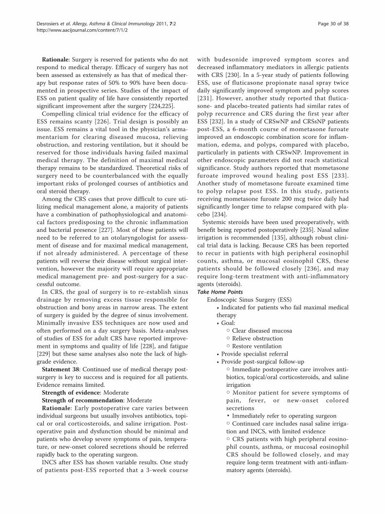
Rationale: Surgery is reserved for patients who do notrespond to medical therapy. Efficacy of surgery has notbeen assessed as extensively as has that of medical ther-apy but response rates of 50% to 90% have been docu-mented in prospective series. Studies of the impact ofESS on patient quality of life have consistently reportedsignificant improvement after the surgery [224,225].Compelling clinical trial evidence for the efficacy of
ESS remains scanty [226]. Trial design is possibly anissue. ESS remains a vital tool in the physician’s arma-mentarium for clearing diseased mucosa, relievingobstruction, and restoring ventilation, but it should bereserved for those individuals having failed maximalmedical therapy. The definition of maximal medicaltherapy remains to be standardized. Theoretical risks ofsurgery need to be counterbalanced with the equallyimportant risks of prolonged courses of antibiotics andoral steroid therapy.Among the CRS cases that prove difficult to cure uti-
lizing medical management alone, a majority of patientshave a combination of pathophysiological and anatomi-cal factors predisposing to the chronic inflammationand bacterial presence [227]. Most of these patients willneed to be referred to an otolaryngologist for assess-ment of disease and for maximal medical management,if not already administered. A percentage of thesepatients will reverse their disease without surgical inter-vention, however the majority will require appropriatemedical management pre- and post-surgery for a suc-cessful outcome.In CRS, the goal of surgery is to re-establish sinus
drainage by removing excess tissue responsible forobstruction and bony areas in narrow areas. The extentof surgery is guided by the degree of sinus involvement.Minimally invasive ESS techniques are now used andoften performed on a day surgery basis. Meta-analysesof studies of ESS for adult CRS have reported improve-ment in symptoms and quality of life [228], and fatigue[229] but these same analyses also note the lack of high-grade evidence.Statement 38: Continued use of medical therapy post-
surgery is key to success and is required for all patients.Evidence remains limited.Strength of evidence: ModerateStrength of recommendation: ModerateRationale: Early postoperative care varies between
individual surgeons but usually involves antibiotics, topi-cal or oral corticosteroids, and saline irrigation. Post-operative pain and dysfunction should be minimal andpatients who develop severe symptoms of pain, tempera-ture, or new-onset colored secretions should be referredrapidly back to the operating surgeon.INCS after ESS has shown variable results. One study
of patients post-ESS reported that a 3-week course
with budesonide improved symptom scores anddecreased inflammatory mediators in allergic patientswith CRS [230]. In a 5-year study of patients followingESS, use of fluticasone propionate nasal spray twicedaily significantly improved symptom and polyp scores[231]. However, another study reported that flutica-sone- and placebo-treated patients had similar rates ofpolyp recurrence and CRS during the first year afterESS [232]. In a study of CRSwNP and CRSsNP patientspost-ESS, a 6-month course of mometasone furoateimproved an endoscopic combination score for inflam-mation, edema, and polyps, compared with placebo,particularly in patients with CRSwNP. Improvement inother endoscopic parameters did not reach statisticalsignificance. Study authors reported that mometasonefuroate improved wound healing post ESS [233].Another study of mometasone furoate examined timeto polyp relapse post ESS. In this study, patientsreceiving mometasone furoate 200 mcg twice daily hadsignificantly longer time to relapse compared with pla-cebo [234].Systemic steroids have been used preoperatively, with
benefit being reported postoperatively [235]. Nasal salineirrigation is recommended [135], although robust clini-cal trial data is lacking. Because CRS has been reportedto recur in patients with high peripheral eosinophilcounts, asthma, or mucosal eosinophil CRS, thesepatients should be followed closely [236], and mayrequire long-term treatment with anti-inflammatoryagents (steroids).Take Home Points
Endoscopic Sinus Surgery (ESS)• Indicated for patients who fail maximal medicaltherapy• Goal:
○ Clear diseased mucosa○ Relieve obstruction○ Restore ventilation
• Provide specialist referral• Provide post-surgical follow-up
○ Immediate postoperative care involves anti-biotics, topical/oral corticosteroids, and salineirrigation○ Monitor patient for severe symptoms ofpain, fever, or new-onset coloredsecretions▪ Immediately refer to operating surgeon○ Continued care includes nasal saline irriga-tion and INCS, with limited evidence○ CRS patients with high peripheral eosino-phil counts, asthma, or mucosal eosinophilCRS should be followed closely, and mayrequire long-term treatment with anti-inflam-matory agents (steroids).
Desrosiers et al. Allergy, Asthma & Clinical Immunology 2011, 7:2http://www.aacijournal.com/content/7/1/2
Page 30 of 38
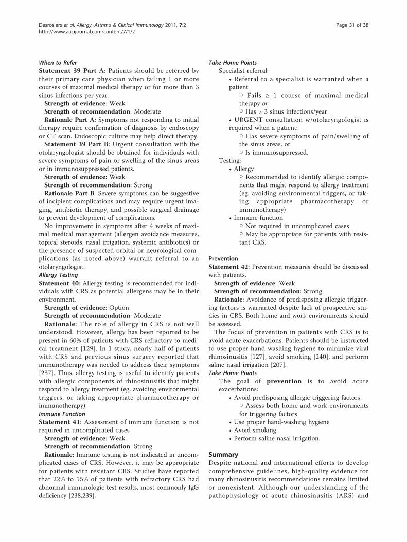
When to ReferStatement 39 Part A: Patients should be referred bytheir primary care physician when failing 1 or morecourses of maximal medical therapy or for more than 3sinus infections per year.Strength of evidence: WeakStrength of recommendation: ModerateRationale Part A: Symptoms not responding to initial
therapy require confirmation of diagnosis by endoscopyor CT scan. Endoscopic culture may help direct therapy.Statement 39 Part B: Urgent consultation with the
otolaryngologist should be obtained for individuals withsevere symptoms of pain or swelling of the sinus areasor in immunosuppressed patients.Strength of evidence: WeakStrength of recommendation: StrongRationale Part B: Severe symptoms can be suggestive
of incipient complications and may require urgent ima-ging, antibiotic therapy, and possible surgical drainageto prevent development of complications.No improvement in symptoms after 4 weeks of maxi-
mal medical management (allergen avoidance measures,topical steroids, nasal irrigation, systemic antibiotics) orthe presence of suspected orbital or neurological com-plications (as noted above) warrant referral to anotolaryngologist.Allergy TestingStatement 40: Allergy testing is recommended for indi-viduals with CRS as potential allergens may be in theirenvironment.Strength of evidence: OptionStrength of recommendation: ModerateRationale: The role of allergy in CRS is not well
understood. However, allergy has been reported to bepresent in 60% of patients with CRS refractory to medi-cal treatment [129]. In 1 study, nearly half of patientswith CRS and previous sinus surgery reported thatimmunotherapy was needed to address their symptoms[237]. Thus, allergy testing is useful to identify patientswith allergic components of rhinosinusitis that mightrespond to allergy treatment (eg, avoiding environmentaltriggers, or taking appropriate pharmacotherapy orimmunotherapy).Immune FunctionStatement 41: Assessment of immune function is notrequired in uncomplicated casesStrength of evidence: WeakStrength of recommendation: StrongRationale: Immune testing is not indicated in uncom-
plicated cases of CRS. However, it may be appropriatefor patients with resistant CRS. Studies have reportedthat 22% to 55% of patients with refractory CRS hadabnormal immunologic test results, most commonly IgGdeficiency [238,239].
Take Home PointsSpecialist referral:
• Referral to a specialist is warranted when apatient
○ Fails ≥ 1 course of maximal medicaltherapy or○ Has > 3 sinus infections/year
• URGENT consultation w/otolaryngologist isrequired when a patient:
○ Has severe symptoms of pain/swelling ofthe sinus areas, or○ Is immunosuppressed.
Testing:• Allergy
○ Recommended to identify allergic compo-nents that might respond to allergy treatment(eg, avoiding environmental triggers, or tak-ing appropriate pharmacotherapy orimmunotherapy)
• Immune function○ Not required in uncomplicated cases○ May be appropriate for patients with resis-tant CRS.
PreventionStatement 42: Prevention measures should be discussedwith patients.Strength of evidence: WeakStrength of recommendation: StrongRationale: Avoidance of predisposing allergic trigger-
ing factors is warranted despite lack of prospective stu-dies in CRS. Both home and work environments shouldbe assessed.The focus of prevention in patients with CRS is to
avoid acute exacerbations. Patients should be instructedto use proper hand-washing hygiene to minimize viralrhinosinusitis [127], avoid smoking [240], and performsaline nasal irrigation [207].Take Home Points
The goal of prevention is to avoid acuteexacerbations:
• Avoid predisposing allergic triggering factors○ Assess both home and work environmentsfor triggering factors
• Use proper hand-washing hygiene• Avoid smoking• Perform saline nasal irrigation.
SummaryDespite national and international efforts to developcomprehensive guidelines, high-quality evidence formany rhinosinusitis recommendations remains limitedor nonexistent. Although our understanding of thepathophysiology of acute rhinosinusitis (ARS) and
Desrosiers et al. Allergy, Asthma & Clinical Immunology 2011, 7:2http://www.aacijournal.com/content/7/1/2
Page 31 of 38
chronic rhinosinusitis (CRS) has dramatically improvedduring the past decade, our understanding of the under-lying mechanisms remains limited. This incomplete evi-dence base translates into continued difficulty withclassifying the various forms of rhinosinusitis, selectingamong available therapies, and developing new thera-peutic options. Selecting appropriate therapy thusremains a challenge for both ARS and CRS. Professionalexperience and expert opinions are required to developrecommendations because of the absence of well-designed prospective clinical trials of therapeuticoptions. Future research must improve our understand-ing of rhinosinusitis and provide a strong evidence basefor therapeutic recommendations.Much remains to be settled in ARS to improve diag-
nostic criteria and direct treatment, including a betterunderstanding of the timelines of pathophysiologicalchanges during rhinovirus infection, host factors asso-ciated with the transition to bacterial infection, and therole of mucosal immunity, as well as severity of sympto-matology and predictors of non-resolution/complica-tions. The role of antibiotic therapy in ARS has beenunder scrutiny, which will likely lead to changes in clini-cal trial design. Improving objective methods of identify-ing ARS cases that warrant therapy will permit morerigorous and meaningful patient selection. Randomized,placebo-controlled studies should test the effectivenessof antibiotics and other treatments using direct mea-sures of bacterial presence and viability at the beginningand end of therapy, rather than relying on symptomaticimprovement. Trial designs that include identification ofpredictors of positive response to therapy would facili-tate the development of recommendations.Many aspects of CRS remain controversial and a bet-
ter understanding of the pathophysiology, definitions,and role of causative factors will improve treatmentapproaches. As more information about CRS subtypesand novel therapies are discovered, large-scale, prospec-tive, placebo-controlled trials of therapies will need tobe repeated in the context of the various subtypes.Delivery methods that improve coverage of the nasalpassages and/or penetration of the sinus cavities areneeded. As results from studies of medical and surgicalapproaches increase, attempts should be made to iden-tify the optimal therapy according to subtypes of diseaseand time point in the disease evolution. Post-operativemedical management needs to be better recognized.Because there is a large number of patients with persis-tent signs and symptoms of the disease despite medicaland surgical intervention, management guidelines speci-fic to this group of patients should be developed.The past 2 decades have seen a rapid increase in our
knowledge of the pathogenesis, diagnosis, and manage-ment of ARS and CRS. It is hoped that over the next
decade, study findings will expand and build upon thecurrent foundation to bring scientific credibility andimproved outcomes to our field.
AbbreviationsAAO-HNS: American Academy of Otolaryngology-Head and Neck Surgery;AAP SCQIM: American Academy of Pediatrics Steering Committee on QualityImprovement and Management; ABRS: acute bacterial rhinosinusitis; AERD:aspirin-exacerbated respiratory disease; ARS: acute rhinosinusitis; ASA:acetylsalicylic acid (aspirin); CA-MRSA: community acquired methicillin-resistant Staphylococcus aureus; CF: cystic fibrosis; CI: confidence interval;CNS: coagulase-negative Staphylococci; COPD: chronic obstructivepulmonary disease; COX: cyclooxygenase; CRS: chronic rhinosinusitis; CRSsNP:chronic rhinosinusitis without nasal polyps; CRSwNP: chronic rhinosinusitiswith nasal polyps; CSLM: confocal scanning laser microscopy; CSO-HNS:Canadian Society of Otolaryngology-Head and Neck Surgery; CT: computedtomography; EDMM: endoscopically-directed middle meatus; ENT: Ear, Nose,and Throat; ESS: endoscopic sinus surgery; GERD: gastroesophageal refluxdisease; GRADE: Grades of Recommendation, Assessment, Development andEvaluation; IgE: immunoglobulin E; IL: interleukin; INCS: intranasalcorticosteroids; LR: likelihood ratio; MRSA: methicillin-resistant Staphylococcusaureus; MSA: maxillary sinus aspirate; OR: odds ratio; PPV: positive predictivevalue; RR: risk ratio; SNOT: SinoNasal Outcome Test; TGF: transforminggrowth factor; Th: T helper cell; TMD: temporomandibular joint dysfunction;TMP/SMX: trimethoprim-sulfamethoxazole; URTI: upper respiratory tractinfection.
AcknowledgementsFinancial assistance for the development of these guidelines was generouslyprovided as unrestricted grants to the Canadian Society of Otolaryngology-Head Neck Surgery by Bayer Inc, GlaxoSmithKline Inc, MERCK Canada Inc,Nycomed Canada Inc, and sanofi-aventis Canada Inc. Funding was obtainedvia an unrestricted grant, with each contributing equally to this project. Allfunds were administered via a trust account held at the CSO-HNS. Nocontact with industry was made during the guidelines development orreview process.The authors wish to thank our external reviewers Ken Bayly, BSc, MD,Matthew J Neskar, BSc Pharm, Brian Rotenberg, MD, FRCSC, EthanRubinstein, MD, FRCPC and Fanny Silviu-Dan, MD, FRCPC. The authors alsowish to thank our external content experts, Ross Davidson, PhD, QutaybaHamid MD, PhD, FRCPC, Donald E Low, MD, FRCPC and our methodologist,Pierre Ernst, MD, MSc, FRCPC. Finally the authors wish to thank Helen BuckieLloyd (Toronto, ON) and Lynne Isbell, PhD (Carmel, IN, USA) for editorialsupport.These guidelines have been endorsed by the Association of MedicalMicrobiology and Infectious Disease Canada, Canadian Society of Allergyand Clinical Immunology, Canadian Society of Otolaryngology - Head andNeck Surgery, Canadian Association of Emergency Physicians and The FamilyPhysicians Airways Group of Canada.
Author details1Division of Otolaryngology - Head and Neck Surgery Centre Hospitalier del’Université de Montréal, Université de Montréal Hotel-Dieu de Montreal, andDepartment of Otolaryngology - Head and Neck Surgery and Allergy,Montreal General Hospital, McGill University, Montreal, QC, Canada. 2Divisionof Infectious Diseases, Department of Medicine, and Departments ofMicrobiology & Immunology and Pathology & Molecular Medicine, Queen’sUniversity and Kingston General Hospital, Kingston, ON, Canada.3Deparmtent of Medicine, Division of Allergy and Clinical Immunology,McMaster University Hamilton, ON, Canada. 4Division of Otolaryngology -Head and Neck Surgery, University of Alberta, Edmonton, AB, Canada.5Family Physician Airways Group of Canada and Brampton Civic Hospital,Richmond Hill, ON, Canada. 6Clinical Medicine, Laval’s University Quebec andDepartment of Medicine, Hôpital de la Malbaie La Malbaie, QC, Canada.7Family Physician Airways Group of Canada, Aldergrove, BC, Canada.8Division of Medical Microbiology and Infection Control, Vancouver GeneralHospital and Department of Pathology and Laboratory Medicine, Universityof British Columbia, Vancouver, BC, Canada. 9St Paul’s Sinus Center andDivision of Otolaryngology-Head and Neck Surgery, University of BritishColumbia, Vancouver, BC, Canada. 10Department of Medicine, University of
Desrosiers et al. Allergy, Asthma & Clinical Immunology 2011, 7:2http://www.aacijournal.com/content/7/1/2
Page 32 of 38
Toronto and Women’s College Hospital, Halton Healthcare Services (OakvilleTrafalger Site), Toronto, ON, Canada. 11Department of Medicine, Division ofInfectious Diseases, McMaster University and Department of Medicine/Infectious Diseases, Hamilton General Hospital, McMaster Wing Hamilton,ON, Canada. 12Department of Medicine, Division of Allergy and Immunology,University of British Columbia, and James Hogg iCAPTURE Centre forCardiovascular and Pulmonary Research, St. Paul’s Hospital Vancouver, BC,Canada. 13Division of Allergy and Clinical Immunology, Jewish GeneralHospital and Department of Medicine, Department of Medicine, McGillUniversity, Montreal, QC, Canada. 14Department of Otolaryngology-Head &Neck Surgery, University of Toronto, Toronto, ON, Canada.
Authors’ contributionsMD conceived of the need for guidelines and coordinated the societies forrepresentation. GE, PK, and EW participated in its design and coordinatedand identified contributing authors. MD, GE, PK, EW, AK, JB, AC, PD, AJ, EL,AM, RS, PS, and IW participated in a full day meeting to review theguidelines format and content. MD, GE, PK, EW, AK, JB, AC, PD, AJ, EL, AM,RS, PS, and IW participated in the Delphi voting. MD, GE, PK, EW, AK, JB, AC,PD, AJ, EL, AM, RS, PS, and IW reviewed drafts and supplied revisions. MD,GE, PK, EW, RD, QH, and DL assessed quality of retrieved articles. MD, AK, JB,AC, PD, AJ, EL, AM, RS, PS, and IW provided first draft manuscripts forcontent. MD, GE, PK, EW, AK, JB, AC, PD, AJ, EL, AM, RS, PS, and IW providedreview of drafts to final manuscript. MD, AK, JB, and AC provided revisionsappropriate for the primary care community. MD, AK, AC and GE designedand refined the algorithms. MD and PE defined methodology. RD, QH, andDL provided expert content as required. All authors read and approved thefinal manuscript.
Competing interestsMD - Speakers Bureau: Merck Canada, Advisory board: GlaxoSmithKline,Merck Canada, Ethicon Surgical, Consultant: MedtronicXomed (bacterialbiofilms), Research funding: Fondation Antoine Turmel, Fonds de rechercheen santé du Québec, MedtronicXomed, PK - Advisory Boards:GlaxoSmithKline, Merck Canada, Talecris, CSL Behring, Research funding:GlaxoSmithKline, Merck Canada, Affexa Life Sciences, AK - Advisory Boards:Merck Canada, AstraZeneca Canada and Nycomed Canada, AC - AdvisoryBoards: Pfizer, AJ - Speaker: Merck Canada, Bayer, Abbott Canada, NycomedCanada, MedtronicXomed, PS - Advisor to Merck Canada, GlaxoSmithKline,King, IW - Advisor: Abbott Canada, GlaxoSmithKline, Merck Canada,Pharmascience Inc. GE, EW, JB, PD, EL, AM, RS declare that they have nocompeting interests.
Received: 18 October 2010 Accepted: 10 February 2011Published: 10 February 2011
References1. Anand VK: Epidemiology and economic impact of rhinosinusitis. Ann Otol
Rhinol Laryngol Suppl 2004, 193:3-5.2. Macdonald KI, McNally JD, Massoud E: The health and resource utilization
of Canadians with chronic rhinosinusitis. Laryngoscope 2009, 119:184-189.3. IMS Health: Canadian Disease and Therapeutic Index (CDTI) database.
2006.4. Pleis J, Lucas J: Summary health statistics for U.S. adults: National Health
Interview Survey, 2007. National Center for Health Statistics. Vital HealthStat 2009, 10.
5. Gwaltney JM Jr: Acute community-acquired sinusitis. Clin Infect Dis 1996,23:1209-1223, quiz 1224-1205.
6. Chen Y, Dales R, Lin M: The epidemiology of chronic rhinosinusitis inCanadians. Laryngoscope 2003, 113:1199-1205.
7. Classifying recommendations for clinical practice guidelines. Pediatrics2004, 114:874-877.
8. Schunemann HJ, Jaeschke R, Cook DJ, Bria WF, El-Solh AA, Ernst A, Fahy BF,Gould MK, Horan KL, Krishnan JA, et al: An official ATS statement: gradingthe quality of evidence and strength of recommendations in ATSguidelines and recommendations. Am J Respir Crit Care Med 2006,174:605-614.
9. Rosenfeld RM, Andes D, Bhattacharyya N, Cheung D, Eisenberg S,Ganiats TG, Gelzer A, Hamilos D, Haydon RC, Hudgins PA, et al: Clinicalpractice guideline: adult sinusitis. Otolaryngol Head Neck Surg 2007, 137:S1-31.
10. Barkun A, Bardou M, Marshall JK: Consensus recommendations formanaging patients with nonvariceal upper gastrointestinal bleeding.Ann Intern Med 2003, 139:843-857.
11. Appraisal of guidelines for research and evaluation (AGREE) instrument.[http://www.agreecollaboration.org/instrument/].
12. Gwaltney JM Jr: Rhinovirus infection of the normal human airway. Am JRespir Crit Care Med 1995, 152:S36-39.
13. Gwaltney JM: Viral respiratory infection therapy: historical perspectivesand current trials. Am J Med 2002, 112(Suppl 6A):33S-41S.
14. Gwaltney JM Jr, Phillips CD, Miller RD, Riker DK: Computed tomographicstudy of the common cold. N Engl J Med 1994, 330:25-30.
15. Infectious rhinosinusitis in adults: classification, etiology andmanagement. International Rhinosinusitis Advisory Board. Ear NoseThroat J 1997, 76:1-22.
16. Desrosiers M, Frenkiel S, Hamid QA, Low D, Small P, Carr S, Hawke M,Kirkpatrick D, Lavigne F, Mandell L, et al: Acute bacterial sinusitis in adults:management in the primary care setting. J Otolaryngol 2002, 31(Suppl2):2S2-14.
17. Piccirillo JF: Clinical practice. Acute bacterial sinusitis. N Engl J Med 2004,351:902-910.
18. Lanza DC, Kennedy DW: Adult rhinosinusitis defined. Otolaryngol HeadNeck Surg 1997, 117:S1-7.
19. Fokkens W, Lund V, Mullol J: European position paper on rhinosinusitisand nasal polyps 2007. Rhinol Suppl 2007, 1-136.
20. Hickner JM, Bartlett JG, Besser RE, Gonzales R, Hoffman JR, Sande MA:Principles of appropriate antibiotic use for acute rhinosinusitis in adults:background. Ann Emerg Med 2001, 37:703-710.
21. Scadding GK, Durham SR, Mirakian R, Jones NS, Drake-Lee AB, Ryan D,Dixon TA, Huber PA, Nasser SM: BSACI guidelines for themanagement of rhinosinusitis and nasal polyposis. Clin Exp Allergy2008, 38:260-275.
22. Antimicrobial treatment guidelines for acute bacterial rhinosinusitis.Sinus and Allergy Health Partnership. Otolaryngol Head Neck Surg 2000,123:5-31.
23. Evans FO Jr, Sydnor JB, Moore WE, Moore GR, Manwaring JL, Brill AH,Jackson RT, Hanna S, Skaar JS, Holdeman LV, et al: Sinusitis of the maxillaryantrum. N Engl J Med 1975, 293:735-739.
24. Berg O, Carenfelt C: Analysis of symptoms and clinical signs in themaxillary sinus empyema. Acta Otolaryngol 1988, 105:343-349.
25. Williams JW Jr, Simel DL, Roberts L, Samsa GP: Clinical evaluation forsinusitis. Making the diagnosis by history and physical examination. AnnIntern Med 1992, 117:705-710.
26. Hadley JA, Schaefer SD: Clinical evaluation of rhinosinusitis: history andphysical examination. Otolaryngol Head Neck Surg 1997, 117:S8-11.
27. Low DE, Desrosiers M, McSherry J, Garber G, Williams JW Jr, Remy H,Fenton RS, Forte V, Balter M, Rotstein C, et al: A practical guide for thediagnosis and treatment of acute sinusitis. CMAJ 1997, 156(Suppl 6):S1-14.
28. Blomgren K, Hytonen M, Pellinen J, Relander M, Pitkaranta A: Diagnosticaccuracy of acute maxillary sinusitis in adults in primary care. Scand JPrim Health Care 2002, 20:40-44.
29. Thomas C, Aizin V: Brief report: a red streak in the lateral recess of theoropharynx predicts acute sinusitis. J Gen Intern Med 2006, 21:986-988.
30. Young J, Bucher H, Tschudi P, Periat P, Hugenschmidt C, Welge-Lussen A:The clinical diagnosis of acute bacterial rhinosinusitis in general practiceand its therapeutic consequences. J Clin Epidemiol 2003, 56:377-384.
31. Ashraf N, Bhattacharyya N: Determination of the “incidental” Lund scorefor the staging of chronic rhinosinusitis. Otolaryngol Head Neck Surg 2001,125:483-486.
32. Bolger WE, Butzin CA, Parsons DS: Paranasal sinus bony anatomicvariations and mucosal abnormalities: CT analysis for endoscopic sinussurgery. Laryngoscope 1991, 101:56-64.
33. Flinn J, Chapman ME, Wightman AJ, Maran AG: A prospective analysis ofincidental paranasal sinus abnormalities on CT head scans. ClinOtolaryngol Allied Sci 1994, 19:287-289.
34. Gamble R: Acute inflammations of the orbit in children. Arch Ophthalmol1933, 10:483-497.
35. Patt BS, Manning SC: Blindness resulting from orbital complications ofsinusitis. Otolaryngol Head Neck Surg 1991, 104:789-795.
36. Gungor A, Corey JP: Pediatric sinusitis: a literature review with emphasison the role of allergy. Otolaryngol Head Neck Surg 1997, 116:4-15.
Desrosiers et al. Allergy, Asthma & Clinical Immunology 2011, 7:2http://www.aacijournal.com/content/7/1/2
Page 33 of 38
37. Kuranov NI: [Orbital and intracranial complications of rhinosinusitis].Vestn Otorinolaringol 2001, 46-47.
38. Eustis HS, Mafee MF, Walton C, Mondonca J: MR imaging and CT of orbitalinfections and complications in acute rhinosinusitis. Radiol Clin North Am1998, 36:1165-1183, xi.
39. Rumelt S, Rubin PA: Potential sources for orbital cellulitis. Int OphthalmolClin 1996, 36:207-221.
40. Gallagher RM, Gross CW, Phillips CD: Suppurative intracranialcomplications of sinusitis. Laryngoscope 1998, 108:1635-1642.
41. Broberg T, Murr A, Fischbein N: Devastating complications of acutepediatric bacterial sinusitis. Otolaryngol Head Neck Surg 1999, 120:575-579.
42. Giannoni CM, Stewart MG, Alford EL: Intracranial complications of sinusitis.Laryngoscope 1997, 107:863-867.
43. Axelsson A, Brorson JE: The correlation between bacteriological findingsin the nose and maxillary sinus in acute maxillary sinusitis. Laryngoscope1973, 83:2003-2011.
44. Gwaltney JM Jr, Scheld WM, Sande MA, Sydnor A: The microbial etiologyand antimicrobial therapy of adults with acute community-acquiredsinusitis: a fifteen-year experience at the University of Virginia andreview of other selected studies. J Allergy Clin Immunol 1992, 90:457-461,discussion 462.
45. Gwaltney JM Jr, Sydnor A Jr, Sande MA: Etiology and antimicrobialtreatment of acute sinusitis. Ann Otol Rhinol Laryngol Suppl 1981, 90:68-71.
46. Van Cauwenberge P, Verschraegen G, Van Renterghem L: Bacteriologicalfindings in sinusitis (1963-1975). Scand J Infect Dis Suppl 1976, 72-77.
47. Joniau S, Vlaminck S, Van Landuyt H, Kuhweide R, Dick C: Microbiology ofsinus puncture versus middle meatal aspiration in acute bacterialmaxillary sinusitis. Am J Rhinol 2005, 19:135-140.
48. Talbot GH, Kennedy DW, Scheld WM, Granito K: Rigid nasal endoscopyversus sinus puncture and aspiration for microbiologic documentationof acute bacterial maxillary sinusitis. Clin Infect Dis 2001, 33:1668-1675.
49. Vogan JC, Bolger WE, Keyes AS: Endoscopically guided sinonasal cultures:a direct comparison with maxillary sinus aspirate cultures. OtolaryngolHead Neck Surg 2000, 122:370-373.
50. Benninger MS, Payne SC, Ferguson BJ, Hadley JA, Ahmad N: Endoscopicallydirected middle meatal cultures versus maxillary sinus taps in acutebacterial maxillary rhinosinusitis: a meta-analysis. Otolaryngol Head NeckSurg 2006, 134:3-9.
51. Sande MA, Gwaltney JM: Acute community-acquired bacterial sinusitis:continuing challenges and current management. Clin Infect Dis 2004,39(Suppl 3):S151-158.
52. Sokol W: Epidemiology of sinusitis in the primary care setting: resultsfrom the 1999-2000 respiratory surveillance program. Am J Med 2001,111(Suppl 9A):19S-24S.
53. Brook I: Bacteriology of acute and chronic ethmoid sinusitis. J ClinMicrobiol 2005, 43:3479-3480.
54. Jousimies-Somer HR, Savolainen S, Ylikoski JS: Bacteriological findings ofacute maxillary sinusitis in young adults. J Clin Microbiol 1988,26:1919-1925.
55. Wald ER: Microbiology of acute and chronic sinusitis in children. J AllergyClin Immunol 1992, 90:452-456.
56. Benninger M, Brook I, Farrell DJ: Disease severity in acute bacterialrhinosinusitis is greater in patients infected with Streptococcuspneumoniae than in those infected with Haemophilus influenzae.Otolaryngol Head Neck Surg 2006, 135:523-528.
57. Brook I, Foote PA, Hausfeld JN: Frequency of recovery of pathogenscausing acute maxillary sinusitis in adults before and after introductionof vaccination of children with the 7-valent pneumococcal vaccine.J Med Microbiol 2006, 55:943-946.
58. Green K, Pong-Porter S, et al: Trends in antimicrobial prescribing andpneumococcal resistance, Canada, 1993-2005. 46th Annual InterscienceConference on Antimicrobial Agents and Chemotherapy San Francisco 2006.
59. Green K, McGreer A, et al: Trends in antimicrobial resistance in Canadianstrains of Streptococcus pneumoniae, 1988-2003. 43rd Annual InterscienceConference on Antimicrobial Agents and Chemotherapy Chicago 2003.
60. Percentage of penicillin non-susceptible S. pneumoniae in Canada:1988-2007. [http://microbiology.mtsinai.on.ca/data/sp/sp_2007.shtml#figure1].
61. de Bock GH, Dekker FW, Stolk J, Springer MP, Kievit J, van Houwelingen JC:Antimicrobial treatment in acute maxillary sinusitis: a meta-analysis.J Clin Epidemiol 1997, 50:881-890.
62. Marple BF, Brunton S, Ferguson BJ: Acute bacterial rhinosinusitis: a reviewof U.S. treatment guidelines. Otolaryngol Head Neck Surg 2006,135:341-348.
63. Zhanel GG, Palatnick L, Nichol KA, Low DE, Hoban DJ: Antimicrobialresistance in Haemophilus influenzae and Moraxella catarrhalisrespiratory tract isolates: results of the Canadian Respiratory OrganismSusceptibility Study, 1997 to 2002. Antimicrob Agents Chemother 2003,47:1875-1881.
64. Huang WH, Hung PK: Methicillin-resistant Staphylococcus aureusinfections in acute rhinosinusitis. Laryngoscope 2006, 116:288-291.
65. Barton M, Hawkes M, Moore D, Conly J, Nicolle L, Allen U, Boyd N,Embree J, Horne LV, Saux NL, et al: Guidelines for the prevention andmanagement of community-associated methicillin-resistantStaphylococcus aureus: A perspective for Canadian health carepractitioners. Can J Infect Dis Med Microbiol 2006, 17(suppl C):4C-24C.
66. Fridkin SK, Hageman JC, Morrison M, Sanza LT, Como-Sabetti K, Jernigan JA,Harriman K, Harrison LH, Lynfield R, Farley MM: Methicillin-resistantStaphylococcus aureus disease in three communities. N Engl J Med 2005,352:1436-1444.
67. Osguthorpe JD: Adult rhinosinusitis: diagnosis and management. Am FamPhysician 2001, 63:69-76.
68. de Ferranti SD, Ioannidis JP, Lau J, Anninger WV, Barza M: Are amoxycillinand folate inhibitors as effective as other antibiotics for acute sinusitis?A meta-analysis. BMJ 1998, 317:632-637.
69. Ahovuo-Saloranta A, Borisenko OV, Kovanen N, Varonen H, Rautakorpi UM,Williams JW Jr, Makela M: Antibiotics for acute maxillary sinusitis.Cochrane Database Syst Rev 2009, CD000243.
70. Falagas ME, Giannopoulou KP, Vardakas KZ, Dimopoulos G,Karageorgopoulos DE: Comparison of antibiotics with placebo fortreatment of acute sinusitis: a meta-analysis of randomised controlledtrials. Lancet Infect Dis 2008, 8:543-552.
71. Scheid DC, Hamm RM: Acute bacterial rhinosinusitis in adults: part II.Treatment. Am Fam Physician 2004, 70:1697-1704.
72. Young J, De Sutter A, Merenstein D, van Essen GA, Kaiser L, Varonen H,Williamson I, Bucher HC: Antibiotics for adults with clinically diagnosedacute rhinosinusitis: a meta-analysis of individual patient data. Lancet2008, 371:908-914.
73. Lindbaek M, Butler CC: Antibiotics for sinusitis-like symptoms in primarycare. Lancet 2008, 371:874-876.
74. Rosenfeld RM, Singer M, Jones S: Systematic review of antimicrobialtherapy in patients with acute rhinosinusitis. Otolaryngol Head Neck Surg2007, 137:S32-45.
75. Hadley JA, Mosges R, Desrosiers M, Haverstock D, van Veenhuyzen D,Herman-Gnjidic Z: Moxifloxacin five-day therapy vs. placebo in acutebacterial sinusitis. The Laryngoscope 2010, 120:1057-1062.
76. Adelglass J, DeAbate CA, McElvaine P, Fowler CL, LoCocco J, Campbell T:Comparison of the effectiveness of levofloxacin and amoxicillin-clavulanate for the treatment of acute sinusitis in adults. OtolaryngolHead Neck Surg 1999, 120:320-327.
77. Arrieta JR, Galgano AS, Sakano E, Fonseca X, Amabile-Cuevas CF,Hernandez-Oliva G, Vivar R, Gonzalez G, Torres A: Moxifloxacin vsamoxicillin/clavulanate in the treatment of acute sinusitis. Am JOtolaryngol 2007, 28:78-82.
78. Buchanan P, Roos K, Tellier G, Rangaraju M, Leroy B: Bacteriological efficacyof 5-day therapy with telithromycin in acute maxillary sinusitis. Int JAntimicrob Agents 2005, 25:237-246.
79. Bucher HC, Tschudi P, Young J, Periat P, Welge-Luussen A, Zust H,Schindler C: Effect of amoxicillin-clavulanate in clinically diagnosed acuterhinosinusitis: a placebo-controlled, double-blind, randomized trial ingeneral practice. Arch Intern Med 2003, 163:1793-1798.
80. Burke T, Villanueva C, Mariano H Jr, Huck W, Orchard D, Haverstock D,Heyd A, Church D: Comparison of moxifloxacin and cefuroxime axetil inthe treatment of acute maxillary sinusitis. Sinusitis Infection StudyGroup. Clin Ther 1999, 21:1664-1677.
81. Ferguson BJ, Guzzetta RV, Spector SL, Hadley JA: Efficacy and safety of oraltelithromycin once daily for 5 days versus moxifloxacin once daily for 10days in the treatment of acute bacterial rhinosinusitis. Otolaryngol HeadNeck Surg 2004, 131:207-214.
82. Henry DC, Kapral D, Busman TA, Paris MM: Cefdinir versus levofloxacin inpatients with acute rhinosinusitis of presumed bacterial etiology: a
Desrosiers et al. Allergy, Asthma & Clinical Immunology 2011, 7:2http://www.aacijournal.com/content/7/1/2
Page 34 of 38

multicenter, randomized, double-blind study. Clin Ther 2004,26:2026-2033.
83. Lasko B, Lau CY, Saint-Pierre C, Reddington JL, Martel A, Anstey RJ: Efficacyand safety of oral levofloxacin compared with clarithromycin in thetreatment of acute sinusitis in adults: a multicentre, double-blind,randomized study. The Canadian Sinusitis Study Group. J Int Med Res1998, 26:281-291.
84. Siegert R, Gehanno P, Nikolaidis P, Bagger-Sjoback D, Ibanez JM, Hampel B,Sommerauer B: A comparison of the safety and efficacy of moxifloxacin(BAY 12-8039) and cefuroxime axetil in the treatment of acute bacterialsinusitis in adults. The Sinusitis Study Group. Respir Med 2000, 94:337-344.
85. Upchurch J, Rosemore M, Tosiello R, Kowalsky S, Echols R: Randomizeddouble-blind study comparing 7- and 10-day regimens of faropenemmedoxomil with a 10-day cefuroxime axetil regimen for treatment ofacute bacterial sinusitis. Otolaryngol Head Neck Surg 2006, 135:511-517.
86. Canadian Pharmacists Association: Compendium of pharmaceuticals andspecialties. The Canadian drug reference for health professionals Ottawa:Canadian Pharmacists Association; 2007.
87. MacLaughlin EJ, Saseen JJ, Malone DC: Costs of beta-lactam allergies:selection and costs of antibiotics for patients with a reported beta-lactam allergy. Arch Fam Med 2000, 9:722-726.
88. Jacobs MR: Streptococcus pneumoniae: epidemiology and patterns ofresistance. Am J Med 2004, 117(Suppl 3A):3S-15S.
89. Levine OS, Farley M, Harrison LH, Lefkowitz L, McGeer A, Schwartz B: Riskfactors for invasive pneumococcal disease in children: a population-based case-control study in North America. Pediatrics 1999, 103:E28.
90. Vanderkooi OG, Low DE, Green K, Powis JE, McGeer A: Predictingantimicrobial resistance in invasive pneumococcal infections. Clin InfectDis 2005, 40:1288-1297.
91. Daneman N, McGeer A, Green K, Low DE: Macrolide resistance inbacteremic pneumococcal disease: implications for patientmanagement. Clin Infect Dis 2006, 43:432-438.
92. Fuller JD, McGeer A, Low DE: Drug-resistant pneumococcal pneumonia:clinical relevance and approach to management. Eur J Clin MicrobiolInfect Dis 2005, 24:780-788.
93. Low D: Managing antibiotic resistance. Canadian Bacterial SurveillanceNetwork Newsletter 2005.
94. Gwaltney JM Jr, Wiesinger BA, Patrie JT: Acute community-acquiredbacterial sinusitis: the value of antimicrobial treatment and the naturalhistory. Clin Infect Dis 2004, 38:227-233.
95. Murray JJ, Emparanza P, Lesinskas E, Tawadrous M, Breen JD: Efficacy andsafety of a novel, single-dose azithromycin microsphere formulationversus 10 days of levofloxacin for the treatment of acute bacterialsinusitis in adults. Otolaryngol Head Neck Surg 2005, 133:194-200.
96. Henry DC, Riffer E, Sokol WN, Chaudry NI, Swanson RN: Randomizeddouble-blind study comparing 3- and 6-day regimens of azithromycinwith a 10-day amoxicillin-clavulanate regimen for treatment of acutebacterial sinusitis. Antimicrob Agents Chemother 2003, 47:2770-2774.
97. Ioannidis JP, Contopoulos-Ioannidis DG, Chew P, Lau J: Meta-analysis ofrandomized controlled trials on the comparative efficacy and safety ofazithromycin against other antibiotics for upper respiratory tractinfections. J Antimicrob Chemother 2001, 48:677-689.
98. Poole M, Anon J, Paglia M, Xiang J, Khashab M, Kahn J: A trial of high-dose, short-course levofloxacin for the treatment of acute bacterialsinusitis. Otolaryngol Head Neck Surg 2006, 134:10-17.
99. Sher LD, McAdoo MA, Bettis RB, Turner MA, Li NF, Pierce PF: A multicenter,randomized, investigator-blinded study of 5- and 10-day gatifloxacinversus 10-day amoxicillin/clavulanate in patients with acute bacterialsinusitis. Clin Ther 2002, 24:269-281.
100. Ferguson BJ, Anon J, Poole MD, Hendrick K, Gilson M, Seltzer EG: Shorttreatment durations for acute bacterial rhinosinusitis: Five days ofgemifloxacin versus 7 days of gemifloxacin. Otolaryngol Head Neck Surg2002, 127:1-6.
101. Seggev JS, Enrique RR, Brandon ML, Larsen LS, Van Tuyl RA, Rowinski CA: Acombination of amoxicillin and clavulanate every 12 hours vs every 8hours for treatment of acute bacterial maxillary sinusitis. Arch OtolaryngolHead Neck Surg 1998, 124:921-925.
102. Riffer E, Spiller J, Palmer R, Shortridge V, Busman TA, Valdes J: Once dailyclarithromycin extended-release vs twice-daily amoxicillin/clavulanate inpatients with acute bacterial sinusitis: a randomized, investigator-blinded study. Curr Med Res Opin 2005, 21:61-70.
103. Park-Wyllie LY, Juurlink DN, Kopp A, Shah BR, Stukel TA, Stumpo C,Dresser L, Low DE, Mamdani MM: Outpatient gatifloxacin therapy anddysglycemia in older adults. N Engl J Med 2006, 354:1352-1361.
104. Ip S, Fu L, Balk E, Chew P, Devine D, Lau J: Update on acute bacterialrhinosinusitis. Evid Rep Technol Assess (Summ) 2005, 1-3.
105. Zalmanovici A, Yaphe J: Steroids for acute sinusitis. Cochrane DatabaseSyst Rev 2007, CD005149.
106. Dolor RJ, Witsell DL, Hellkamp AS, Williams JW Jr, Califf RM, Simel DL:Comparison of cefuroxime with or without intranasal fluticasone for thetreatment of rhinosinusitis. The CAFFS Trial: a randomized controlledtrial. JAMA 2001, 286:3097-3105.
107. Meltzer EO, Bachert C, Staudinger H: Treating acute rhinosinusitis:comparing efficacy and safety of mometasone furoate nasal spray,amoxicillin, and placebo. J Allergy Clin Immunol 2005, 116:1289-1295.
108. Bachert C, Meltzer EO: Effect of mometasone furoate nasal spray onquality of life of patients with acute rhinosinusitis. Rhinology 2007,45:190-196.
109. Williamson IG, Rumsby K, Benge S, Moore M, Smith PW, Cross M, Little P:Antibiotics and topical nasal steroid for treatment of acute maxillarysinusitis: a randomized controlled trial. JAMA 2007, 298:2487-2496.
110. Meltzer EO: Antibiotics and nasal steroids for acute sinusitis. JAMA 2008,299:1422-1423, author reply 1423.
111. Ambrose PG, Anon JB, Owen JS, Van Wart S, McPhee ME, Bhavnani SM,Piedmonte M, Jones RN: Use of pharmacodynamic end points in theevaluation of gatifloxacin for the treatment of acute maxillary sinusitis.Clin Infect Dis 2004, 38:1513-1520.
112. Ambrose PG, Anon JB, Bhavnani SM, Okusanya OO, Jones RN, Paglia MR,Kahn J, Drusano GL: Use of pharmacodynamic endpoints for theevaluation of levofloxacin for the treatment of acute maxillary sinusitis.Diagn Microbiol Infect Dis 2008, 61:13-20.
113. Anon JB, Paglia M, Xiang J, Ambrose PG, Jones RN, Kahn JB: Serial sinusaspirate samples during high-dose, short-course levofloxacin treatmentof acute maxillary sinusitis. Diagn Microbiol Infect Dis 2007, 57:105-107.
114. Barlan IB, Erkan E, Bakir M, Berrak S, Basaran MM: Intranasal budesonidespray as an adjunct to oral antibiotic therapy for acute sinusitis inchildren. Ann Allergy Asthma Immunol 1997, 78:598-601.
115. Nayak AS, Settipane GA, Pedinoff A, Charous BL, Meltzer EO, Busse WW,Zinreich SJ, Lorber RR, Rikken G, Danzig MR: Effective dose range ofmometasone furoate nasal spray in the treatment of acuterhinosinusitis. Ann Allergy Asthma Immunol 2002, 89:271-278.
116. Wawrose SF, Tami TA, Amoils CP: The role of guaifenesin in the treatmentof sinonasal disease in patients infected with the humanimmunodeficiency virus (HIV). Laryngoscope 1992, 102:1225-1228.
117. Sisson JH, Yonkers AJ, Waldman RH: Effects of guaifenesin on nasalmucociliary clearance and ciliary beat frequency in healthy volunteers.Chest 1995, 107:747-751.
118. Inanli S, Ozturk O, Korkmaz M, Tutkun A, Batman C: The effects of topicalagents of fluticasone propionate, oxymetazoline, and 3% and 0.9%sodium chloride solutions on mucociliary clearance in the therapy ofacute bacterial rhinosinusitis in vivo. Laryngoscope 2002, 112:320-325.
119. Rabago D, Zgierska A, Mundt M, Barrett B, Bobula J, Maberry R: Efficacy ofdaily hypertonic saline nasal irrigation among patients with sinusitis: arandomized controlled trial. J Fam Pract 2002, 51:1049-1055.
120. Keojampa BK, Nguyen MH, Ryan MW: Effects of buffered saline solutionon nasal mucociliary clearance and nasal airway patency. OtolaryngolHead Neck Surg 2004, 131:679-682.
121. Hauptman G, Ryan MW: The effect of saline solutions on nasal patencyand mucociliary clearance in rhinosinusitis patients. Otolaryngol HeadNeck Surg 2007, 137:815-821.
122. Adam P, Stiffman M, Blake RL Jr: A clinical trial of hypertonic saline nasalspray in subjects with the common cold or rhinosinusitis. Arch Fam Med1998, 7:39-43.
123. Clinical practice guideline: management of sinusitis. Pediatrics 2001,108:798-808.
124. Barrett B, Vohmann M, Calabrese C: Echinacea for upper respiratoryinfection. J Fam Pract 1999, 48:628-635.
125. Guo R, Canter PH, Ernst E: Herbal medicines for the treatment ofrhinosinusitis: a systematic review. Otolaryngol Head Neck Surg 2006,135:496-506.
126. Zabolotnyi DI, Kneis KC, Richardson A, Rettenberger R, Heger M, Kaszkin-Bettag M, Heger PW: Efficacy of a complex homeopathic medication
Desrosiers et al. Allergy, Asthma & Clinical Immunology 2011, 7:2http://www.aacijournal.com/content/7/1/2
Page 35 of 38
(Sinfrontal) in patients with acute maxillary sinusitis: a prospective,randomized, double-blind, placebo-controlled, multicenter clinical trial.Explore (NY) 2007, 3:98-109.
127. Pittet D, Allegranzi B, Sax H, Dharan S, Pessoa-Silva CL, Donaldson L,Boyce JM: Evidence-based model for hand transmission during patientcare and the role of improved practices. Lancet Infect Dis 2006, 6:641-652.
128. Gutman M, Torres A, Keen KJ, Houser SM: Prevalence of allergy in patientswith chronic rhinosinusitis. Otolaryngol Head Neck Surg 2004, 130:545-552.
129. Emanuel IA, Shah SB: Chronic rhinosinusitis: allergy and sinus computedtomography relationships. Otolaryngol Head Neck Surg 2000, 123:687-691.
130. Alho OP, Karttunen R, Karttunen TJ: Nasal mucosa in natural colds: effectsof allergic rhinitis and susceptibility to recurrent sinusitis. Clin ExpImmunol 2004, 137:366-372.
131. Benninger MS, Ferguson BJ, Hadley JA, Hamilos DL, Jacobs M, Kennedy DW,Lanza DC, Marple BF, Osguthorpe JD, Stankiewicz JA, et al: Adult chronicrhinosinusitis: definitions, diagnosis, epidemiology, and pathophysiology.Otolaryngol Head Neck Surg 2003, 129:S1-32.
132. Gliklich RE, Metson R: The health impact of chronic sinusitis in patientsseeking otolaryngologic care. Otolaryngol Head Neck Surg 1995,113:104-109.
133. Small P, Frenkiel S, et al: Rhinitis: A practical and comprehensiveapproach to assessement and therapy. J Otolaryngol 2007, 36(suppl 1):S5-S28.
134. Bousquet J, Van Cauwenberge P, Khaltaev N: Allergic rhinitis and itsimpact on asthma. J Allergy Clin Immunol 2001, 108:S147-334.
135. Fokkens W, Lund V, Bachert C, Clement P, Helllings P, Holmstrom M,Jones N, Kalogjera L, Kennedy D, Kowalski M, et al: EAACI position paperon rhinosinusitis and nasal polyps executive summary. Allergy 2005,60:583-601.
136. Meltzer EO, Hamilos DL, Hadley JA, Lanza DC, Marple BF, Nicklas RA,Bachert C, Baraniuk J, Baroody FM, Benninger MS, et al: Rhinosinusitis:establishing definitions for clinical research and patient care. J AllergyClin Immunol 2004, 114:155-212.
137. Meltzer EO, Hamilos DL, Hadley JA, Lanza DC, Marple BF, Nicklas RA,Adinoff AD, Bachert C, Borish L, Chinchilli VM, et al: Rhinosinusitis:developing guidance for clinical trials. J Allergy Clin Immunol 2006, 118:S17-61.
138. Slavin RG, Spector SL, Bernstein IL, Kaliner MA, Kennedy DW, Virant FS,Wald ER, Khan DA, Blessing-Moore J, Lang DM, et al: The diagnosis andmanagement of sinusitis: a practice parameter update. J Allergy ClinImmunol 2005, 116:S13-47.
139. Sanderson AR, Leid JG, Hunsaker D: Bacterial biofilms on the sinusmucosa of human subjects with chronic rhinosinusitis. Laryngoscope2006, 116:1121-1126.
140. Foreman A, Singhal D, Psaltis AJ, Wormald PJ: Targeted imaging modalityselection for bacterial biofilms in chronic rhinosinusitis. Laryngoscope2010, 120:427-431.
141. Tantilipikorn P, Fritz M, Tanabodee J, Lanza DC, Kennedy DW: Acomparison of endoscopic culture techniques for chronic rhinosinusitis.Am J Rhinol 2002, 16:255-260.
142. Hopkins C, Browne JP, Slack R, Lund V, Brown P: The Lund-Mackay stagingsystem for chronic rhinosinusitis: how is it used and what does itpredict? Otolaryngol Head Neck Surg 2007, 137:555-561.
143. Bhattacharyya T, Piccirillo J, Wippold FJ: Relationship between patient-based descriptions of sinusitis and paranasal sinus computedtomographic findings. Arch Otolaryngol Head Neck Surg , 2 1997,123:1189-1192.
144. Bhattacharyya N: Clinical and symptom criteria for the accurate diagnosisof chronic rhinosinusitis. Laryngoscope 2006, 116:1-22.
145. Stewart MG, Sicard MW, Piccirillo JF, Diaz-Marchan PJ: Severity staging inchronic sinusitis: are CT scan findings related to patient symptoms? AmJ Rhinol 1999, 13:161-167.
146. Riechelmann H, Deutschle T, Rozsasi A, Keck T, Polzehl D, Burner H: Nasalbiomarker profiles in acute and chronic rhinosinusitis. Clin Exp Allergy2005, 35:1186-1191.
147. Van Zele T, Claeys S, Gevaert P, Van Maele G, Holtappels G, VanCauwenberge P, Bachert C: Differentiation of chronic sinus diseases bymeasurement of inflammatory mediators. Allergy 2006, 61:1280-1289.
148. Schleimer RP, Kato A, Peters A, Conley D, Kim J, Liu MC, Harris KE,Kuperman DA, Chandra R, Favoreto S Jr, et al: Epithelium, inflammation,
and immunity in the upper airways of humans: studies in chronicrhinosinusitis. Proc Am Thorac Soc 2009, 6:288-294.
149. Ramanathan M Jr, Lee WK, Spannhake EW, Lane AP: Th2 cytokinesassociated with chronic rhinosinusitis with polyps down-regulate theantimicrobial immune function of human sinonasal epithelial cells. Am JRhinol 2008, 22:115-121.
150. Lane AP, Truong-Tran QA, Schleimer RP: Altered expression of genesassociated with innate immunity and inflammation in recalcitrantrhinosinusitis with polyps. Am J Rhinol 2006, 20:138-144.
151. Jones SA: Directing transition from innate to acquired immunity:defining a role for IL-6. J Immunol 2005, 175:3463-3468.
152. Hamilos DL, Leung DY, Wood R, Cunningham L, Bean DK, Yasruel Z,Schotman E, Hamid Q: Evidence for distinct cytokine expression inallergic versus nonallergic chronic sinusitis. J Allergy Clin Immunol 1995,96:537-544.
153. Wright ED, Frenkiel S, Al-Ghamdi K, Ghaffar O, Small P, Troutt T, Tavernier J,Hamid Q: Interleukin-4, interleukin-5, and granulocyte-macrophagecolony-stimulating factor receptor expression in chronic sinusitis andresponse to topical steroids. Otolaryngol Head Neck Surg 1998,118:490-495.
154. Ghaffar O, Durham SR, Al-Ghamdi K, Wright E, Small P, Frenkiel S, Gould HJ,Hamid Q: Expression of IgE heavy chain transcripts in the sinus mucosaof atopic and nonatopic patients with chronic sinusitis. Am J Respir CellMol Biol 1998, 18:706-711.
155. Ghaffar O, Lavigne F, Kamil A, Renzi P, Hamid Q: Interleukin-6 expressionin chronic sinusitis: colocalization of gene transcripts to eosinophils,macrophages, T lymphocytes, and mast cells. Otolaryngol Head Neck Surg1998, 118:504-511.
156. Molet SM, Hamid QA, Hamilos DL: IL-11 and IL-17 expression in nasalpolyps: relationship to collagen deposition and suppression byintranasal fluticasone propionate. Laryngoscope 2003, 113:1803-1812.
157. Newman LJ, Platts-Mills TA, Phillips CD, Hazen KC, Gross CW: Chronicsinusitis. Relationship of computed tomographic findings to allergy,asthma, and eosinophilia. JAMA 1994, 271:363-367.
158. Rolla G, Guida G, Heffler E, Badiu I, Bommarito L, De Stefani A, Usai A,Cosseddu D, Nebiolo F, Bucca C: Diagnostic classification of persistentrhinitis and its relationship to exhaled nitric oxide and asthma: a clinicalstudy of a consecutive series of patients. Chest 2007, 131:1345-1352.
159. Braunstahl GJ, Hellings PW: Nasobronchial interaction mechanisms inallergic airways disease. Curr Opin Otolaryngol Head Neck Surg 2006,14:176-182.
160. Bendouah Z, Barbeau J, Hamad WA, Desrosiers M: Biofilm formation byStaphylococcus aureus and Pseudomonas aeruginosa is associated withan unfavorable evolution after surgery for chronic sinusitis and nasalpolyposis. Otolaryngol Head Neck Surg 2006, 134:991-996.
161. Prince AA, Steiger JD, Khalid AN, Dogrhamji L, Reger C, Eau Claire S,Chiu AG, Kennedy DW, Palmer JN, Cohen NA: Prevalence of biofilm-forming bacteria in chronic rhinosinusitis. Am J Rhinol 2008, 22:239-245.
162. Desrosiers M, Hussain A, Frenkiel S, Kilty S, Marsan J, Witterick I, Wright E:Intranasal corticosteroid use is associated with lower rates of bacterialrecovery in chronic rhinosinusitis. Otolaryngol Head Neck Surg 2007,136:605-609.
163. Conley DB, Tripathi A, Seiberling KA, Suh LA, Harris KE, Paniagua MC,Grammer LC, Kern RC: Superantigens and chronic rhinosinusitis II:analysis of T-cell receptor V beta domains in nasal polyps. Am J Rhinol2006, 20:451-455.
164. Seiberling KA, Conley DB, Tripathi A, Grammer LC, Shuh L, Haines GK,Schleimer R, Kern RC: Superantigens and chronic rhinosinusitis: detectionof staphylococcal exotoxins in nasal polyps. Laryngoscope 2005,115:1580-1585.
165. Zhang N, Holtappels G, Claeys C, Huang G, van Cauwenberge P, Bachert C:Pattern of inflammation and impact of Staphylococcus aureusenterotoxins in nasal polyps from southern China. Am J Rhinol 2006,20:445-450.
166. Fakhri S, Tulic M, Christodoulopoulos P, Fukakusa M, Frenkiel S, Leung DY,Hamid QA: Microbial superantigens induce glucocorticoid receptor betaand steroid resistance in a nasal explant model. Laryngoscope 2004,114:887-892.
167. Plouin-Gaudon I, Clement S, Huggler E, Chaponnier C, Francois P, Lew D,Schrenzel J, Vaudaux P, Lacroix JS: Intracellular residency is frequently
Desrosiers et al. Allergy, Asthma & Clinical Immunology 2011, 7:2http://www.aacijournal.com/content/7/1/2
Page 36 of 38
associated with recurrent Staphylococcus aureus rhinosinusitis. Rhinology2006, 44:249-254.
168. Cryer J, Schipor I, Perloff JR, Palmer JN: Evidence of bacterial biofilms inhuman chronic sinusitis. ORL J Otorhinolaryngol Relat Spec 2004,66:155-158.
169. Psaltis AJ, Ha KR, Beule AG, Tan LW, Wormald PJ: Confocal scanning lasermicroscopy evidence of biofilms in patients with chronic rhinosinusitis.Laryngoscope 2007, 117:1302-1306.
170. Sanclement JA, Webster P, Thomas J, Ramadan HH: Bacterial biofilms insurgical specimens of patients with chronic rhinosinusitis. Laryngoscope2005, 115:578-582.
171. Psaltis AJ, Weitzel EK, Ha KR, Wormald PJ: The effect of bacterial biofilmson post-sinus surgical outcomes. Am J Rhinol 2008, 22:1-6.
172. Lanza DC, Dhong HJ, Tantilipikorn P, Tanabodee J, Nadel DM, Kennedy DW:Fungus and chronic rhinosinusitis: from bench to clinical understanding.Ann Otol Rhinol Laryngol Suppl 2006, 196:27-34.
173. Sasama J, Sherris DA, Shin SH, Kephart GM, Kern EB, Ponikau JU: Newparadigm for the roles of fungi and eosinophils in chronic rhinosinusitis.Curr Opin Otolaryngol Head Neck Surg 2005, 13:2-8.
174. Van Cauwenberge P, Van Hoecke H, Bachert C: Pathogenesis of chronicrhinosinusitis. Curr Allergy Asthma Rep 2006, 6:487-494.
175. McNally PA, White MV, Kaliner MA: Sinusitis in an allergist’s office: analysisof 200 consecutive cases. Allergy Asthma Proc 1997, 18:169-175.
176. Stevenson DD, Sanchez-Borges M, Szczeklik A: Classification of allergic andpseudoallergic reactions to drugs that inhibit cyclooxygenase enzymes.Ann Allergy Asthma Immunol 2001, 87:177-180.
177. Dibaise JK, Sharma VK: Does gastroesophageal reflux contribute to thedevelopment of chronic sinusitis? A review of the evidence. DisEsophagus 2006, 19:419-424.
178. Weaver EM: Association between gastroesophageal reflux and sinusitis,otitis media, and laryngeal malignancy: a systematic review of theevidence. Am J Med 2003, 115(Suppl 3A):81S-89S.
179. Passali D, Caruso G, Passali FM: ENT manifestations of gastroesophagealreflux. Curr Allergy Asthma Rep 2008, 8:240-244.
180. Gysin C, Alothman GA, Papsin BC: Sinonasal disease in cystic fibrosis:clinical characteristics, diagnosis, and management. Pediatr Pulmonol2000, 30:481-489.
181. Wang X, Kim J, McWilliams R, Cutting GR: Increased prevalence of chronicrhinosinusitis in carriers of a cystic fibrosis mutation. Arch OtolaryngolHead Neck Surg 2005, 131:237-240.
182. Sobol SE, Christodoulopoulos P, Manoukian JJ, Hauber HP, Frenkiel S,Desrosiers M, Fukakusa M, Schloss MD, Hamid Q: Cytokine profile ofchronic sinusitis in patients with cystic fibrosis. Arch Otolaryngol HeadNeck Surg 2002, 128:1295-1298.
183. Legent F, Bordure P, Beauvillain C, Berche P: A double-blind comparison ofciprofloxacin and amoxycillin/clavulanic acid in the treatment of chronicsinusitis. Chemotherapy 1994, 40(Suppl 1):8-15.
184. Namyslowski G, Misiolek M, Czecior E, Malafiej E, Orecka B, Namyslowski P,Misiolek H: Comparison of the efficacy and tolerability of amoxycillin/clavulanic acid 875 mg b.i.d. with cefuroxime 500 mg b.i.d. in thetreatment of chronic and acute exacerbation of chronic sinusitis inadults. J Chemother 2002, 14:508-517.
185. Hashiba M, Baba S: Efficacy of long-term administration of clarithromycinin the treatment of intractable chronic sinusitis. Acta Otolaryngol Suppl1996, 525:73-78.
186. Ichimura K, Shimazaki Y, Ishibashi T, Higo R: Effect of new macrolideroxithromycin upon nasal polyps associated with chronic sinusitis. AurisNasus Larynx 1996, 23:48-56.
187. Ragab SM, Lund VJ, Scadding G: Evaluation of the medical and surgicaltreatment of chronic rhinosinusitis: a prospective, randomised,controlled trial. Laryngoscope 2004, 114:923-930.
188. Wallwork B, Coman W, Mackay-Sim A, Greiff L, Cervin A: A double-blind,randomized, placebo-controlled trial of macrolide in the treatment ofchronic rhinosinusitis. Laryngoscope 2006, 116:189-193.
189. Desrosiers MY, Salas-Prato M: Treatment of chronic rhinosinusitisrefractory to other treatments with topical antibiotic therapy deliveredby means of a large-particle nebulizer: results of a controlled trial.Otolaryngol Head Neck Surg 2001, 125:265-269.
190. Lim M, Citardi MJ, Leong JL: Topical antimicrobials in the management ofchronic rhinosinusitis: a systematic review. Am J Rhinol 2008, 22:381-389.
191. Videler WJ, van Drunen CM, Reitsma JB, Fokkens WJ: Nebulized bacitracin/colimycin: a treatment option in recalcitrant chronic rhinosinusitis withStaphylococcus aureus? A double-blind, randomized, placebo-controlled,cross-over pilot study. Rhinology 2008, 46:92-98.
192. Deuschl H, Drettner B: Nasal polyps treated by beclomethasone nasalaerosol. Rhinology 1977, 15:17-23.
193. Holopainen E, Grahne B, Malmberg H, Makinien J, Lindqvist N: Budesonidein the treatment of nasal polyposis. Eur J Respir Dis Suppl 1982,122:221-228.
194. Mygind N, Pedersen CB, Prytz S, Sorensen H: Treatment of nasal polypswith intranasal beclomethasone dipropionate aerosol. Clin Allergy 1975,5:159-164.
195. Filiaci F, Passali D, Puxeddu R, Schrewelius C: A randomized controlled trialshowing efficacy of once daily intranasal budesonide in nasal polyposis.Rhinology 2000, 38:185-190.
196. Jankowski R, Schrewelius C, Bonfils P, Saban Y, Gilain L, Prades JM,Strunski V: Efficacy and tolerability of budesonide aqueous nasal spraytreatment in patients with nasal polyps. Arch Otolaryngol Head Neck Surg2001, 127:447-452.
197. Small CB, Hernandez J, Reyes A, Schenkel E, Damiano A, Stryszak P,Staudinger H, Danzig M: Efficacy and safety of mometasone furoate nasalspray in nasal polyposis. J Allergy Clin Immunol 2005, 116:1275-1281.
198. Stjarne P, Blomgren K, Caye-Thomasen P, Salo S, Soderstrom T: The efficacyand safety of once-daily mometasone furoate nasal spray in nasalpolyposis: a randomized, double-blind, placebo-controlled study. ActaOtolaryngol 2006, 126:606-612.
199. Stjarne P, Mosges R, Jorissen M, Passali D, Bellussi L, Staudinger H,Danzig M: A randomized controlled trial of mometasone furoate nasalspray for the treatment of nasal polyposis. Arch Otolaryngol Head NeckSurg 2006, 132:179-185.
200. Lund VJ, Black JH, Szabo LZ, Schrewelius C, Akerlund A: Efficacy andtolerability of budesonide aqueous nasal spray in chronic rhinosinusitispatients. Rhinology 2004, 42:57-62.
201. Parikh A, Scadding GK, Darby Y, Baker RC: Topical corticosteroids inchronic rhinosinusitis: a randomized, double-blind, placebo-controlledtrial using fluticasone propionate aqueous nasal spray. Rhinology 2001,39:75-79.
202. Joe SA, Thambi R, Huang J: A systematic review of the use of intranasalsteroids in the treatment of chronic rhinosinusitis. Otolaryngol Head NeckSurg 2008, 139:340-347.
203. Holm AF, Fokkens WJ, Godthelp T, Mulder PG, Vroom TM, Rijntjes E: A 1-year placebo-controlled study of intranasal fluticasone propionateaqueous nasal spray in patients with perennial allergic rhinitis: a safetyand biopsy study. Clin Otolaryngol Allied Sci 1998, 23:69-73.
204. Alobid I, Benitez P, Pujols L, Maldonado M, Bernal-Sprekelsen M, Morello A,Picado C, Mullol J: Severe nasal polyposis and its impact on quality oflife. The effect of a short course of oral steroids followed by long-termintranasal steroid treatment. Rhinology 2006, 44:8-13.
205. Benitez P, Alobid I, de Haro J, Berenguer J, Bernal-Sprekelsen M, Pujols L,Picado C, Mullol J: A short course of oral prednisone followed byintranasal budesonide is an effective treatment of severe nasal polyps.Laryngoscope 2006, 116:770-775.
206. Patiar S, Reece P: Oral steroids for nasal polyps. Cochrane Database SystRev 2007, CD005232.
207. Harvey R, Hannan SA, Badia L, Scadding G: Nasal saline irrigations for thesymptoms of chronic rhinosinusitis. Cochrane Database Syst Rev 2007,CD006394.
208. Becker DG: Medical treatment of sinusitis. J Long Term Eff Med Implants2003, 13:195-205.
209. Braun JJ, Alabert JP, Michel FB, Quiniou M, Rat C, Cougnard J, Czarlewski W,Bousquet J: Adjunct effect of loratadine in the treatment of acutesinusitis in patients with allergic rhinitis. Allergy 1997, 52:650-655.
210. Graf P, Enerdal J, Hallen H: Ten days’ use of oxymetazoline nasal spraywith or without benzalkonium chloride in patients with vasomotorrhinitis. Arch Otolaryngol Head Neck Surg 1999, 125:1128-1132.
211. Parnes SM, Chuma AV: Acute effects of antileukotrienes on sinonasalpolyposis and sinusitis. Ear Nose Throat J 2000, 79:18-20, 24-15.
212. Ulualp SO, Sterman BM, Toohill RJ: Antileukotriene therapy for the reliefof sinus symptoms in aspirin triad disease. Ear Nose Throat J 1999,78:604-606, 608, 613, passim.
Desrosiers et al. Allergy, Asthma & Clinical Immunology 2011, 7:2http://www.aacijournal.com/content/7/1/2
Page 37 of 38
213. Ragab S, Parikh A, Darby YC, Scadding GK: An open audit of montelukast,a leukotriene receptor antagonist, in nasal polyposis associated withasthma. Clin Exp Allergy 2001, 31:1385-1391.
214. Di Rienzo L, Artuso A, Cerqua N: [Antileukotrienes in the prevention ofpostoperative recurrence of nasal polyposis in ASA syndrome]. ActaOtorhinolaryngol Ital 2000, 20:336-342.
215. Grundmann T, Topfner M: [Treatment of ASS-Associated Polyposis(ASSAP) with a cysteinyl leukotriene receptor antagonist - a prospectivedrug study on its antiinflammatory effects]. Laryngorhinootologie 2001,80:576-582.
216. Ebbens FA, Scadding GK, Badia L, Hellings PW, Jorissen M, Mullol J,Cardesin A, Bachert C, van Zele TP, Dijkgraaf MG, et al: Amphotericin Bnasal lavages: not a solution for patients with chronic rhinosinusitis. JAllergy Clin Immunol 2006, 118:1149-1156.
217. Kennedy DW, Kuhn FA, Hamilos DL, Zinreich SJ, Butler D, Warsi G, Pfister PJ,Tavakkol A: Treatment of chronic rhinosinusitis with high-dose oralterbinafine: a double blind, placebo-controlled study. Laryngoscope 2005,115:1793-1799.
218. Ponikau JU, Sherris DA, Weaver A, Kita H: Treatment of chronicrhinosinusitis with intranasal amphotericin B: a randomized, placebo-controlled, double-blind pilot trial. J Allergy Clin Immunol 2005,115:125-131.
219. Weschta M, Rimek D, Formanek M, Polzehl D, Podbielski A, Riechelmann H:Topical antifungal treatment of chronic rhinosinusitis with nasal polyps:a randomized, double-blind clinical trial. J Allergy Clin Immunol 2004,113:1122-1128.
220. Liang KL, Su MC, Shiao JY, Tseng HC, Hsin CH, Lin JF, Jiang RS:Amphotericin B irrigation for the treatment of chronic rhinosinusitiswithout nasal polyps: a randomized, placebo-controlled, double-blindstudy. Am J Rhinol 2008, 22:52-58.
221. Jyonouchi H, Sun S, Kelly A, Rimell FL: Effects of exogenous interferongamma on patients with treatment-resistant chronic rhinosinusitis anddysregulated interferon gamma production: a pilot study. ArchOtolaryngol Head Neck Surg 2003, 129:563-569.
222. Berges-Gimeno MP, Simon RA, Stevenson DD: Long-term treatment withaspirin desensitization in asthmatic patients with aspirin-exacerbatedrespiratory disease. J Allergy Clin Immunol 2003, 111:180-186.
223. Dahlen B, Nizankowska E, Szczeklik A, Zetterstrom O, Bochenek G,Kumlin M, Mastalerz L, Pinis G, Swanson LJ, Boodhoo TI, et al: Benefits fromadding the 5-lipoxygenase inhibitor zileuton to conventional therapy inaspirin-intolerant asthmatics. Am J Respir Crit Care Med 1998,157:1187-1194.
224. Durr DG, Desrosiers M: Evidence-based endoscopic sinus surgery.J Otolaryngol 2003, 32:101-106.
225. Smith TL, Mendolia-Loffredo S, Loehrl TA, Sparapani R, Laud PW,Nattinger AB: Predictive factors and outcomes in endoscopic sinussurgery for chronic rhinosinusitis. Laryngoscope 2005, 115:2199-2205.
226. Khalil HS, Nunez DA: Functional endoscopic sinus surgery for chronicrhinosinusitis. Cochrane Database Syst Rev 2006, 3:CD004458.
227. Evans KL: Recognition and management of sinusitis. Drugs 1998, 56:59-71.228. Smith TL, Batra PS, Seiden AM, Hannley M: Evidence supporting
endoscopic sinus surgery in the management of adult chronicrhinosinusitis: a systematic review. Am J Rhinol 2005, 19:537-543.
229. Chester AC, Sindwani R, Smith TL, Bhattacharyya N: Fatigue improvementfollowing endoscopic sinus surgery: a systematic review and meta-analysis. Laryngoscope 2008, 118:730-739.
230. Lavigne F, Cameron L, Renzi PM, Planet JF, Christodoulopoulos P,Lamkioued B, Hamid Q: Intrasinus administration of topical budesonideto allergic patients with chronic rhinosinusitis following surgery.Laryngoscope 2002, 112:858-864.
231. Rowe-Jones JM, Medcalf M, Durham SR, Richards DH, Mackay IS: Functionalendoscopic sinus surgery: 5 year follow up and results of a prospective,randomised, stratified, double-blind, placebo controlled study ofpostoperative fluticasone propionate aqueous nasal spray. Rhinology2005, 43:2-10.
232. Dijkstra MD, Ebbens FA, Poublon RM, Fokkens WJ: Fluticasone propionateaqueous nasal spray does not influence the recurrence rate of chronicrhinosinusitis and nasal polyps 1 year after functional endoscopic sinussurgery. Clin Exp Allergy 2004, 34:1395-1400.
233. Jorissen M, Bachert C: Effect of corticosteroids on wound healing afterendoscopic sinus surgery. Rhinology 2009, 47:280-286.
234. Stjarne P, Olsson P, Alenius M: Use of mometasone furoate to preventpolyp relapse after endoscopic sinus surgery. Arch Otolaryngol Head NeckSurg 2009, 135:296-302.
235. Wright ED, Agrawal S: Impact of perioperative systemic steroids onsurgical outcomes in patients with chronic rhinosinusitis with polyposis:evaluation with the novel Perioperative Sinus Endoscopy (POSE) scoringsystem. Laryngoscope 2007, 117:1-28.
236. Matsuwaki Y, Ookushi T, Asaka D, Mori E, Nakajima T, Yoshida T, Kojima J,Chiba S, Ootori N, Moriyama H: Chronic rhinosinusitis: risk factors for therecurrence of chronic rhinosinusitis based on 5-year follow-up afterendoscopic sinus surgery. Int Arch Allergy Immunol 2008, 146(Suppl1):77-81.
237. Lane AP, Pine HS, Pillsbury HC: Allergy testing and immunotherapy in anacademic otolaryngology practice: a 20-year review. Otolaryngol HeadNeck Surg 2001, 124:9-15.
238. Chee L, Graham SM, Carothers DG, Ballas ZK: Immune dysfunction inrefractory sinusitis in a tertiary care setting. Laryngoscope 2001,111:233-235.
239. Vanlerberghe L, Joniau S, Jorissen M: The prevalence of humoralimmunodeficiency in refractory rhinosinusitis: a retrospective analysis.B-ENT 2006, 2:161-166.
240. Lieu JE, Feinstein AR: Confirmations and surprises in the association oftobacco use with sinusitis. Arch Otolaryngol Head Neck Surg 2000,126:940-946.
doi:10.1186/1710-1492-7-2Cite this article as: Desrosiers et al.: Canadian clinical practice guidelinesfor acute and chronic rhinosinusitis. Allergy, Asthma & Clinical Immunology2011 7:2.
Submit your next manuscript to BioMed Centraland take full advantage of:
• Convenient online submission
• Thorough peer review
• No space constraints or color figure charges
• Immediate publication on acceptance
• Inclusion in PubMed, CAS, Scopus and Google Scholar
• Research which is freely available for redistribution
Submit your manuscript at www.biomedcentral.com/submit
Desrosiers et al. Allergy, Asthma & Clinical Immunology 2011, 7:2http://www.aacijournal.com/content/7/1/2
Page 38 of 38