Embed Size (px)
Citation preview

Transgenic Research 12: 615–629, 2003.© 2003 Kluwer Academic Publishers. Printed in the Netherlands.
615
Activation-tagged tobacco mutants that are tolerant to antimicrotubularherbicides are cross-resistant to chilling stress
Abdul Ahad∗, Jochen Wolf & Peter NickInstitut für Biologie II, Schänzlestr. 1, D-79104 Freiburg, Germany
Received 17 December 2002; revised 27 February 2003; accepted 19 March 2003
Key words: activation tagging, antimicrotubular herbicides, aryl carbamates, chilling tolerance, microtubules,tyrosinylated tubulin
Abstract
T-DNA activation tagging was used to generate tobacco mutants with increased tolerance to antimicrotubular her-bicides and chilling stress. After transformation, protoplast-derived calli were screened for tolerance to treatmentsthat affect microtubule assembly. In one screen mutants with tolerance to aryl carbamates (a blocker of microtubuleassembly) were selected, the second screen was targeted to chilling-tolerant mutants that could survive for severalmonths at 3◦C, a third screen combined both factors. The resistance of these mutants to aryl carbamates or chillingwas accompanied by resistance of microtubules to these factors. The carbamate tolerant mutants were cross-resistant to chilling stress. This was mirrored by an adaptive reorganization of microtubules and a reduction ofmicrotubule dynamics in response to chilling. The analysis of these mutants suggests (1) that microtubule dynamicslimit the tolerance to chilling and EPC, and (2) that the cold sensitivity of microtubules limits chilling tolerance intobacco.
Abbreviations: EPC – Ethyl-N-phenylcarbamate.
Introduction
Low temperatures limit crop yield in temperate cli-mates. The reason is that the cold sensitivity of growthis more pronounced than that of photosynthesis. Asa consequence, the retarded unfolding of leaves con-fines productivity during the spring season (Watson,1952). However, outside the temperate regions sensi-tivity to low temperature is an important issue as well.Most crop species of tropical or subtropical originsuch as cucumbers, melons, cotton, rice, tobacco andmany tropical fruits suffer severely, when they are ex-posed to cool temperatures that are still far above thefreezing point. Chilling sensitivity can vary betweendifferent species and even between different cultivarsof the same crop. An extreme example is the fertil-ity of rice that drops dramatically, when temperaturesduring flower development fall below 18◦C. This so-called chilling damage has to be distinguished from
∗Author for correspondence:E-mail: [email protected]
the generally known freezing damage, since it is likelyto involve completely different mechanisms. Thesemechanisms are still far from being understood.
Microtubules disassemble in response to low tem-perature and are therefore primary targets for chillingstress (for review see Nick, 2000). In fact, in variouscrops the cold stability of microtubules has been foundto correlate with chilling sensitivity, and treatmentwith blockers of microtubule assembly was observedto increase chilling sensitivity (Rikin et al., 1980). Onthe other hand, a mounting body of evidence indi-cates that microtubules are more than simple targetsof chilling stress, but might play a role as modula-tors of cold sensing: When microtubule disassemblyin response to low temperature was suppressed bytaxol, the long-term adaptation to low temperature (so-called cold hardening) is impaired (Bartolo & Carter,1991) indicating that microtubule disassembly is nec-essary to induce efficient acclimation to a cold shock.The primary trigger of cold signaling is generally be-lieved to be a transient rise of intracellular calciumconcentration (Knight et al., 1991). This could be

616
elegantly demonstrated by transgenic plants express-ing aequorin, where the bioluminescence in responseto a cold shock could be monitored. When this calciumpeak is suppressed pharmacologically, cold harden-ing is suppressed (Monroy et al., 1993). Disassemblyof microtubules was found to stimulate the activ-ity of voltage-dependent calcium channels (Ding &Pickard, 1993; Thion et al., 1996) and specifically, ofcold-induced calcium fluxes (Mazars et al., 1997).
These data led to a model (Nick, 2000), wherechilling-induced microtubule disassembly capacitatescalcium influx through cold-sensitive ion channelswhich triggers cold hardening and (among other tar-gets) the formation of a cold-resistant microtubularcytoskeleton. According to this model, activation ofgenes that control assembly or disassembly of micro-tubules is predicted to modulate chilling resistance ina chilling sensitive species. To test this prediction onthe functional level, one would need to overexpresscandidate genes and test for altered chilling tolerance.However, these genes are not known.
The generation of dominant (gain of function)mutants by T-DNA-activation tagging allows to over-come this drawback and, in addition, to isolate suchgenes. To isolate unknown genes based on their func-tion, mutant approaches are widely used, often incombination with a gene-tag (in plants often a T-DNAsequence) that permits isolation of the mutated gene.The insertion of a T-DNA sequence into a transcrip-tional unit routinely results in a recessive mutation byvirtue of gene disruption or inactivation. In diploidplants, such mutations become manifest only after re-peated selfing of the mutagenized population, which isespecially cumbersome for species with higher ploidy(e.g., many Solanaceae). However, the frequency ofdominant mutations with a particular phenotype isusually extremely low, and it would require a screensaturating for the respective genome, which is hardto achieve even for a small genome such as that ofArabidopsis. Dominance of a trait is conventionallyinterpreted as a gain-of-function phenotype and caneither be caused by mutations in the coding region thatlead to constitutive activation of the resulting proteinor by mutations that stimulate the expression of therespective gene (Chang et al., 1993).
This consideration stimulated the development ofan approach based on activation tagging (Hayashiet al., 1992; Koncz et al., 1994). This techniqueis based on the application of multiple (four)transcriptional enhancer elements originating fromthe CaMV-35S promotor fused to the complete
CaMV-35S promotor linked to the left border of thetransformation plasmid. The right border of the insertharbors a hygromycin resistance gene as a marker toselect transformed plant cells. In addition, an ampi-cillin resistance gene along with an E. coli originof replication are located between the hygromycinresistance gene and the multiple enhancers allow se-lection in bacteria during plasmid rescue into E. coli(Feldman, 1991). Upon integration into the plantgenome, this insert is expected to cause dominantcis-activation of genes located in the vicinity of theT-DNA integration site. Thus, the insertion of the tagshould activate flanking genes as a result of the influ-ence of the multiple enhancers. The overexpressionof the flanking gene should produce dominant traitssuch that selection would be possible directly in theprimary transformants. Activation tagging has beensuccessfully employed to identify a histidin-kinasefrom Arabidopsis that can bypass the requirement forcytokinin during shoot regeneration (Kakimoto, 1996)or to isolate mutants with altered morphogenesis orconstitutive pathogen resistance (Weigel et al., 2000).
Antimicrotubular compounds are often designatedas ‘microtubule-disrupting drugs’. This implies thatthese agents destroy assembled microtubules. Thisimpression is wrong, however, as all microtubuleblockers known so far, bind to tubulin heterodimersand prevent their incorporation into the growing end ofthe microtubule (Mizuno & Suzaki, 1991, for a recentreview see Vaughn, 2000). Resistance to assemblyblockers might either be caused by reduced affinityof tubulin for the inhibitor or simply by reduceddynamics of assembly and disassembly (Nick et al.,1994). A microtubule with high turnover will be moresensitive to reduced assembly, because disassemblywill continue at a high rate. Conversely, a microtubulewith low turnover will tolerate a reduced rate of as-sembly and will persist in the presence of the inhibitor.For instance, the sensitivity of growth to ethyl-N-phenylcarbamate (phenyl urethane, EPC) has beenshown to be elevated after addition of auxin, althoughthe affinity of α-tubulin for this antimicrotubular her-bicide is reduced by auxin (Wiesler et al., 2002). Thisindicates that, under these conditions, not the affinityof the inhibitor, but the turnover of microtubules limitsthe tolerance of microtubules against EPC.
The stability of microtubules is generally believedto depend on the activity of structural microtubule-associated proteins (MAPs) that decrease the fre-quency of microtubule destruction (Bin-Bing &Kirschner, 1999; Caudron et al., 2000). Again, so far
617
no real homologues of well-known structural MAPssuch as Tau have been found in plants, indicatingthat assembly and disassembly are either controlled bycompletely novel proteins or by proteins that carry dif-ferent functions in animal cells. To find genes carryingthe MAP-function in plants, a screen for mutants thattolerate antimicrotubular herbicides could be used.However, this screen would yield also mutants thatare altered in the tubulin molecule itself (Nick et al.,1994).
These considerations led us to design a screen,where activation-tagging is combined with a selec-tion with a microtubule-assembly blocker. In contrastto mutagenesis techniques that produce point muta-tions, the insertion of a large tag into the host DNAis very unlikely to reduce the affinity of a target tu-bulin by changing the binding site of the inhibitor.Activation tagging, followed by a screen for herbi-cide tolerance should be specific for mutants withreduced turnover of microtubules. This might arise byelevated expression of structural MAPs due to activa-tion of their promoters or due to activation of factorsthat regulate the expression or activity of these MAPs.In the present screen we used the assembly blockerethyl-N-phenylcarbamate (phenyl urethane, EPC). Itbinds to the carboxyterminus of α-tubulin with the ter-minal tyrosine that is crucial for the affinity (Wiesleret al., 2002). In parallel, we screened for mutants withincreased chilling tolerance or accelerated cold accli-mation. This allowed us to ask, whether cold tolerancecan be achieved through a manipulation of microtu-bule turnover. In the current study we describe thegeneration, screening and phenotype of these mutantsand show that mutants that have been generated by T-DNA activation and are tolerant to EPC, are mostlycross-tolerant to chilling. This suggests that the dy-namics of microtubule assembly and disassembly isresponsible for chilling sensitivity in tobacco.
Materials and methods
Plant material and protoplasts
The streptomycin-resistant, diploid tobacco line SR1(Nicotiana tabacum cv. Petit Havana, Maliga et al.,1973) was used for T-DNA activation tagging. Seedswere sterilized with 70% ethanol for 2 min, followedby an incubation for 5 min in sodium hypocloride con-taining 0.1% Triton X-100. They were then washedfive times with sterile water and dried under sterile
conditions. Sterilized seeds were germinated on solidMS medium (Murashige & Skoog, 1962) with 2%sucrose. Germinated seedlings were transferred toplastic containers (PS-68.0/110 mm, Greiner Labor-technik, Germany) on MS medium and cultured at24◦C in a 16:8 h light-dark cycle in a phytotrone(Certomat®, BS-1, Germany). Shoot tips were excisedand subcultured every 4–5 weeks. For protoplast isola-tion only well-rooted plants were used that had under-gone at least three subcultures in vitro and then grownfor 5–6 weeks. Mesophyll protoplasts were isolatedaccording to Potrykus and Shilito (1986) with minormodifications: fully expanded leaves were placed up-side down in a sterile petridish on 6 ml of CNT-medium [KAO medium (Kao & Michayluk, 1975)with 1% cellulase (Onozuka-R, Yakult, Japan), 1%macerozyme (Serva, Heidelberg, Germany) and 0.4 Msucrose], the midrib was removed and the leaf bladecut into pieces of 0.5–1 cm2 area in the liquid medium.Then additional 6 ml of CNT-medium were added, andthe leaves were incubated overnight at 26◦C in thedark. Protoplasts were released from the digested tis-sues by careful passage through a 10 ml pipette with alarge opening. Subsequently this mixture was passedthrough a sterile stainless steel mesh sieve (mesh size100 µm, Saulas Francois, Montreuil, France). Thisfiltered protoplast suspension was very carefully over-laid with 1 ml PNT medium (KAO medium including0.4 M glucose) on top of the suspension and then cen-trifuged for 5 min at low speed (Eppendorf centrifugeNo. 5403, 1000 rpm, 25◦C). Intact protoplasts werecollected from the interphase and transferred into anew tube using a 1 ml sterile micropipette with a cuttip. Ten microliters of fresh PNT medium were addedand mixed gently, followed by a second centrifuga-tion under the same conditions. This washing stepwas repeated and a small aliquot of the washed pro-toplasts was used for the estimation of cell densityin a hematocytometer (Blau Brand, Germany). Thesupernatant was carefully removed and the isolatedprotoplasts resuspended in PNT medium to a densityof 2×10⁵ protoplasts ml−1. This protoplast suspensionwas then transferred into a sterile, 10 cm petri-dish,sealed with parafilm and incubated at 26◦C in the darkfor 7–8 days, when most of the protoplasts passedthrough the second division.
Transformation
For transformation, the Agrobacterium tumefaciensstrain GV 3101 containing the helper plasmid
618
pMP90RK, harboring the activation vector pPCV Tac7was used (Koncz et al., 1989, 1990). This vector con-tains a full-length CaMV-35S promoter complementedwith four enhancer sequences from the 35S RNA pro-moter linked to the left border (LB) of the T-DNA.The right border (RB) was linked to a hygromycin-resistance gene as selectable marker and to a nosterminator sequence. The T-DNA also contains anE. coli origin of replication and an ampicillin re-sistance gene that allows selection of Agrobacteriumon carbenicillin. For routine maintenance, the bacteriawere subcultured every month on fresh YEB medium(Walden et al., 1995) complemented with 100 mg l−1
carbenecillin. The mesophyll protoplasts were cul-tivated with Agrobacterium 7–8 days after protoplastisolation when most of the protoplasts passed throughthe second division. The protoplast culture was incu-bated with freshly cultivated Agrobacterium in YEBmedium with 100 mg l−1 carbenicillin at a density of100 bacteria per individual protoplast, correspond-ing to 100/(1.8× OD600) µl of bacterial suspensionper 106 protoplasts. After cocultivation in the darkat 26◦C for 48 h, the protoplasts were washed threetimes with W5 medium (Walden et al., 1995) at lowspeed (1000 rpm, 3 min, 25◦C) and resuspended inthe same volume of PNT medium. These cells werethen directly subjected to the different screening pro-tocols (see below). Aliquots were used to determineplating and transformation efficiency. After 2 daysin liquid medium, the dividing cells were resuspen-ded in half the volume of double-concentrated PNTmedium. This was mixed very gently with hand-warm 1.6% (w/v) Sea Plaque Agarose (Duchefa,The Netherlands) in double-concentrated PNT me-dium. The mixture was solidified in small petri-dishes(3 cm diameter) for 30 min, the solidified agarosecontaining the dividing protoplasts cut into halvesand transferred to large petri dishes (10 cm diam-eter) containing 20 ml of AA1 medium (Caboche,1980) and further cultivated in the phytotron as de-scribed above. All media contained 150 mg l−1 ofcefotaxim and 75 mg l−1 ticarcillin (both Duchefa,The Netherlands) to suppress bacterial growth. Forthe estimation of plating efficiency no other anti-biotic was added; for the screens and for the es-timation of transformation efficiency 15 mg l−1 ofhygromycin were used throughout. Unless stated oth-erwise, all cells were incubated at 24◦C in a phyto-tron under dim white light with a 16:8 h light-darkcycle.
Screening for tolerance to aryl carbamatesand chilling
The following screening strategies were employed:
1. Reduced dynamics of microtubule assembly anddisassembly: this was selected by toleranceto the aryl carbamate ethyl-N-phenyl carbamate(phenylurethane, EPC) that was added at 300 µM.After 1 month, the concentration of EPC wasraised to 500 µM. From this screen, seven linescould be recovered that were designated asActivation-Tagged EPC-Resistant (ATER) lines.
2. Chilling tolerance: this was selected by incubatingthe cells at 3◦C over several months. Three mutantlines could be recovered from this screen that weredesignated as Inuit lines.
3. Chilling tolerance that is based on reduced micro-tubule dynamics: this was selected by combiningthe two selective pressures, that is, the cells wereincubated at 3◦C on 300 µM of EPC. Only oneline could be recovered from this screen that wasdesignated as EPC-Inuit 1.
Regeneration of transformants and propagationof the lines
After selection of small, putatively transformedcalli were transferred to solid MS-morpho medium(Caboche, 1980) for the initiation of shoots. For theATER lines this was possible after 6–8 weeks andfor the Inuit or EPC-Inuit lines after 8 months. Forthe ATER and the EPC-Inuit lines, the selective pres-sure by EPC was maintained by adding 300 µM ofEPC to the regeneration medium. Regenerating shootswere excised under sterile conditions and transferredto solid, hormone-free MS medium for rooting. Well-rooted plants were later transplanted into soil andraised in the greenhouse till seed set.
Leaf disc assay for EPC and chilling tolerance
To assay tolerance to EPC or chilling, sterile leafdiscs from plants that had been grown in vitro for3 weeks were plated in petri dishes on MS-10 me-dium (MS-medium containing 0.03 ppm NAA and1 ppm kinetin) solidified with 0.9% agar. In the as-say for EPC-tolerance, various concentrations of EPC(0–500µM) were added into the agar and the platesincubated at 25◦C for 2 months. In the assay forchilling tolerance, no EPC was added, but the plates

619
were incubated at 3◦C for 5 months. Images were re-corded at the onset of the experiment and at varioustime intervals during incubation using a digital camera(Coolpix 990, Nikon). Discs from the SR1-wild typeplants were included as internal standard into eachplate. The increment in leaf area was quantified us-ing a software for quantitative image analysis (ImageJ, National Institute for Health, downloadable un-der http://list.nih.gov/archives/imagej.html. The datashown in Figure 2 represent mean values from two tofour independent series including four to six individualleaves.
Visualization of microtubules
Small aliquots of mutant callus were either challengedby various concentrations of EPC or by chilling stress(by incubation on ice water) and then collected onmicrocontainers equipped with a nylon mesh (Nicket al., 2000). The cells were fixed in two steps in3.7% (w/v) of fresh paraformaldehyde in microtubule-stabilizing buffer (MSB: 50 mM PIPES, 2 mM EGTA,2 mM MgS04 · 7H20, pH 6.9) for 45 min at room tem-perature with gently shaking. This was followed bya second fixation for 20 min under the same con-ditions, in the presence of 1% Triton X-100. Incase of the chilled callus, the fixation was performedon ice. After fixation, the calli were washed un-der gentle shaking with MSB three times for 5 min.The cell wall was digested for 30 min at 25◦C with1% (w/v) Macerozyme and 0.2% (w/v) Pectolyase inMSB and the cells washed again three times for 5 minwith MSB. Then, the cells were transferred to cover-slips coated with poly-L-Lysine (Sigma, Deisenhofen,Germany), and allowed to settle for 20 min in MSBwith 1% Triton X-100. In the next step, the cellswere incubated for 5 min with PBS (PBS: 8 g/l NaCl,0.2 g/l KCl, 0.158 g/l KH2PO4 and 2.31 g/l Na2HPO4)and then blocked for 30 min with PBS containing0.5% (w/v) bovine serum albumin. The cells werethen incubated for 1 h at 37◦C with a monoclonalmouse antibody directed against a conserved epitopeof α-tubulins (DM1A, Sigma, Deisenhofen, Germany)diluted 1:100 in PBS and then washed three times10 min with PBS. The secondary antibody (anti-mouseIgG-fluorescein-isothiocyanate, Sigma, Deisenhofen,Germany) was administered at a dilution of 1:50 inPBS for 1 h at 37◦C in the dark. After incubation, thecells were washed twice 10 min, sealed with nailpolishand directly analyzed under an epifluorescence micro-
scope (Axioplan, Zeiss, Oberkochen, Germany) usinga filter set with excitation at 470 nm, a beam-splitterat 493 and emission between 505 and 530 (filterset13, Zeiss, Oberkochen, Germany). Pictures were ac-quired using a CCD-Color-Camera (Axiocam, Zeiss,Oberkochen, Germany), connected to a PC equippedwith the corresponding imaging software (Axiovision,Zeiss, Oberkochen, Germany).
Western analysis
Total protein extracts were obtained and analyzed bySDS-PAGE and western blotting as described in detailin Nick et al. (2000). Total α-tubulin was probed withthe monoclonal DM1A antibody directed to a conser-vative epitope (Sigma, Deisenhofen, Neu-Germany),whereas tyrosinated tubulin was probed with the ATTantibody that specifically recognizes a carboxyter-minal epitope comprising the terminal tyrosine (Kreis,1987). The DM1A antibody was used in a dilutionof 1:150, the ATT antibody in a dilution of 1:300.Polyclonal antisera against mouse IgG conjugated tohorseradish peroxidase were used as secondary anti-bodies at a dilution of 1:2000 (Sigma, Deisenhofen,Germany). The signal was visualized by biolumines-cence as described in Nick et al. (2000). Figure 3(K)shows representative samples from a set of four toseven independent time courses.
Results
Selection of activation-tagged cells yields few,but clearly distinct mutants
Although a large number of protoplast was used foreach experiment, only a small number of mutantscould be recovered from each screen (Table 1). Totest, whether this was caused by a high number offalse positive cells that survived the cocultivation withAgrobacterium without harboring the plasmid, equalaliquots of the cocultivated protoplasts were eitherplated on medium without hygromycin (−hygro) orwith hygromycin (+hygro) and the number of sur-viving cells was quantified after growth for a further3 weeks. This number was identical in both cases(Table 1), indicating that all cells that had survivedthe cocultivation carried the hygromycin resistancemarker and therefore also the activation-tagging vec-tor. The low number of putative mutants therefore
620
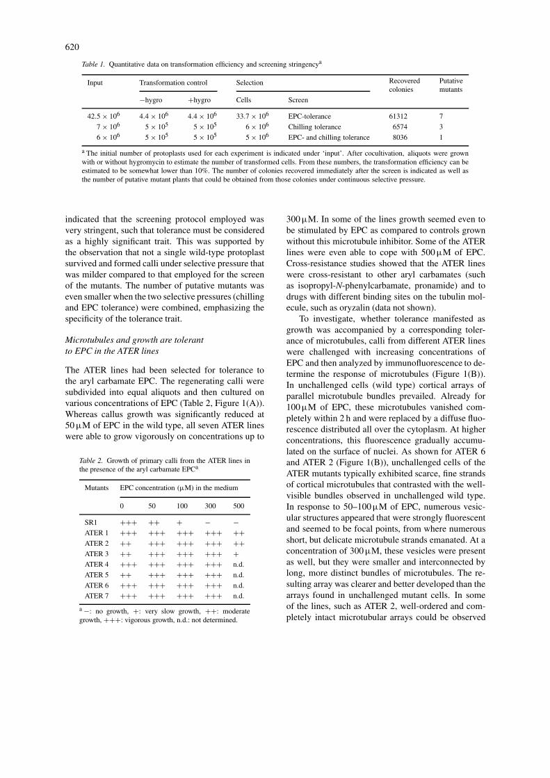
Table 1. Quantitative data on transformation efficiency and screening stringencya
Input Transformation control Selection Recovered Putativecolonies mutants
−hygro +hygro Cells Screen
42.5 × 10⁶ 4.4 × 10⁶ 4.4 × 10⁶ 33.7 × 10⁶ EPC-tolerance 61312 7
7 × 10⁶ 5 × 10⁵ 5 × 10⁵ 6 × 10⁶ Chilling tolerance 6574 3
6 × 10⁶ 5 × 10⁵ 5 × 10⁵ 5 × 10⁶ EPC- and chilling tolerance 8036 1
a The initial number of protoplasts used for each experiment is indicated under ‘input’. After cocultivation, aliquots were grownwith or without hygromycin to estimate the number of transformed cells. From these numbers, the transformation efficiency can beestimated to be somewhat lower than 10%. The number of colonies recovered immediately after the screen is indicated as well asthe number of putative mutant plants that could be obtained from those colonies under continuous selective pressure.
indicated that the screening protocol employed wasvery stringent, such that tolerance must be consideredas a highly significant trait. This was supported bythe observation that not a single wild-type protoplastsurvived and formed calli under selective pressure thatwas milder compared to that employed for the screenof the mutants. The number of putative mutants waseven smaller when the two selective pressures (chillingand EPC tolerance) were combined, emphasizing thespecificity of the tolerance trait.
Microtubules and growth are tolerantto EPC in the ATER lines
The ATER lines had been selected for tolerance tothe aryl carbamate EPC. The regenerating calli weresubdivided into equal aliquots and then cultured onvarious concentrations of EPC (Table 2, Figure 1(A)).Whereas callus growth was significantly reduced at50 µM of EPC in the wild type, all seven ATER lineswere able to grow vigorously on concentrations up to
Table 2. Growth of primary calli from the ATER lines inthe presence of the aryl carbamate EPCa
Mutants EPC concentration (µM) in the medium
0 50 100 300 500
SR1 +++ ++ + − −ATER 1 +++ +++ +++ +++ ++ATER 2 ++ +++ +++ +++ ++ATER 3 ++ +++ +++ +++ +ATER 4 +++ +++ +++ +++ n.d.
ATER 5 ++ +++ +++ +++ n.d.
ATER 6 +++ +++ +++ +++ n.d.
ATER 7 +++ +++ +++ +++ n.d.
a −: no growth, +: very slow growth, ++: moderategrowth, +++: vigorous growth, n.d.: not determined.
300 µM. In some of the lines growth seemed even tobe stimulated by EPC as compared to controls grownwithout this microtubule inhibitor. Some of the ATERlines were even able to cope with 500 µM of EPC.Cross-resistance studies showed that the ATER lineswere cross-resistant to other aryl carbamates (suchas isopropyl-N-phenylcarbamate, pronamide) and todrugs with different binding sites on the tubulin mol-ecule, such as oryzalin (data not shown).
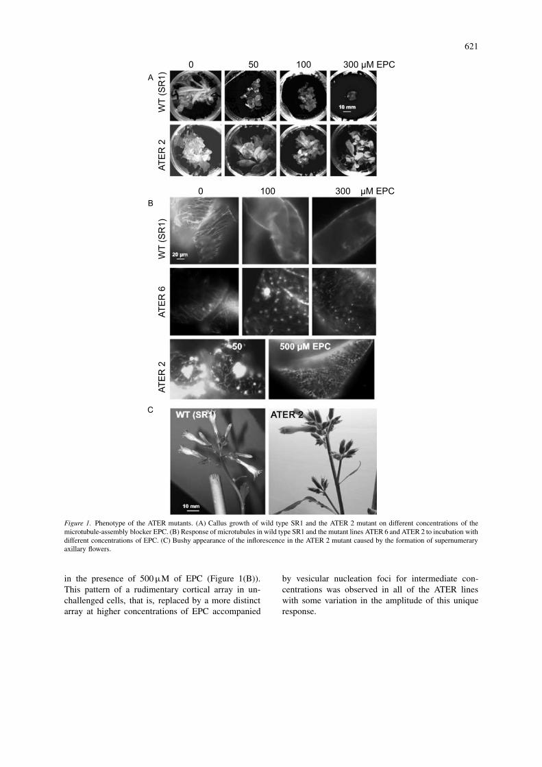
To investigate, whether tolerance manifested asgrowth was accompanied by a corresponding toler-ance of microtubules, calli from different ATER lineswere challenged with increasing concentrations ofEPC and then analyzed by immunofluorescence to de-termine the response of microtubules (Figure 1(B)).In unchallenged cells (wild type) cortical arrays ofparallel microtubule bundles prevailed. Already for100 µM of EPC, these microtubules vanished com-pletely within 2 h and were replaced by a diffuse fluo-rescence distributed all over the cytoplasm. At higherconcentrations, this fluorescence gradually accumu-lated on the surface of nuclei. As shown for ATER 6and ATER 2 (Figure 1(B)), unchallenged cells of theATER mutants typically exhibited scarce, fine strandsof cortical microtubules that contrasted with the well-visible bundles observed in unchallenged wild type.In response to 50–100µM of EPC, numerous vesic-ular structures appeared that were strongly fluorescentand seemed to be focal points, from where numerousshort, but delicate microtubule strands emanated. At aconcentration of 300 µM, these vesicles were presentas well, but they were smaller and interconnected bylong, more distinct bundles of microtubules. The re-sulting array was clearer and better developed than thearrays found in unchallenged mutant cells. In someof the lines, such as ATER 2, well-ordered and com-pletely intact microtubular arrays could be observed
621
Figure 1. Phenotype of the ATER mutants. (A) Callus growth of wild type SR1 and the ATER 2 mutant on different concentrations of themicrotubule-assembly blocker EPC. (B) Response of microtubules in wild type SR1 and the mutant lines ATER 6 and ATER 2 to incubation withdifferent concentrations of EPC. (C) Bushy appearance of the inflorescence in the ATER 2 mutant caused by the formation of supernumeraryaxillary flowers.
in the presence of 500 µM of EPC (Figure 1(B)).This pattern of a rudimentary cortical array in un-challenged cells, that is, replaced by a more distinctarray at higher concentrations of EPC accompanied
by vesicular nucleation foci for intermediate con-centrations was observed in all of the ATER lineswith some variation in the amplitude of this uniqueresponse.
622
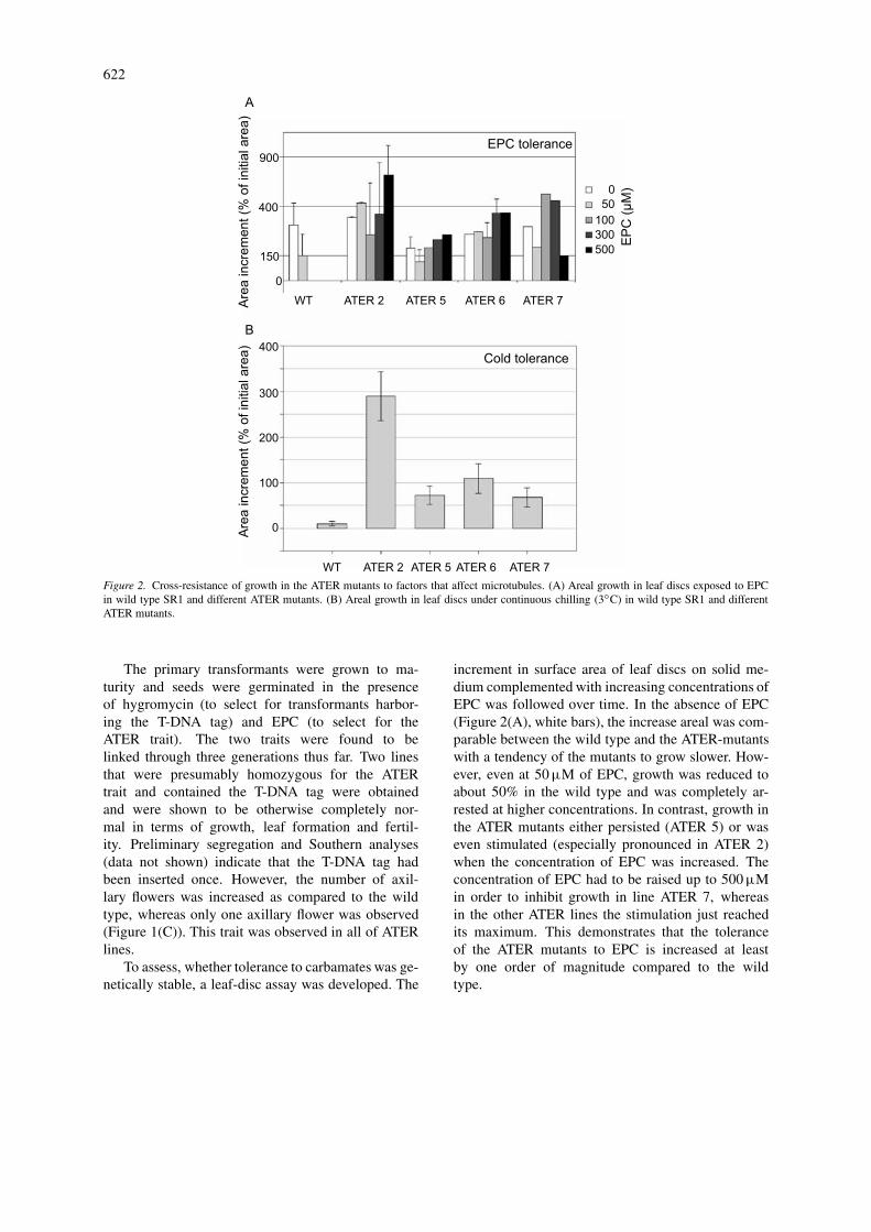
Figure 2. Cross-resistance of growth in the ATER mutants to factors that affect microtubules. (A) Areal growth in leaf discs exposed to EPCin wild type SR1 and different ATER mutants. (B) Areal growth in leaf discs under continuous chilling (3◦C) in wild type SR1 and differentATER mutants.
The primary transformants were grown to ma-turity and seeds were germinated in the presenceof hygromycin (to select for transformants harbor-ing the T-DNA tag) and EPC (to select for theATER trait). The two traits were found to belinked through three generations thus far. Two linesthat were presumably homozygous for the ATERtrait and contained the T-DNA tag were obtainedand were shown to be otherwise completely nor-mal in terms of growth, leaf formation and fertil-ity. Preliminary segregation and Southern analyses(data not shown) indicate that the T-DNA tag hadbeen inserted once. However, the number of axil-lary flowers was increased as compared to the wildtype, whereas only one axillary flower was observed(Figure 1(C)). This trait was observed in all of ATERlines.
To assess, whether tolerance to carbamates was ge-netically stable, a leaf-disc assay was developed. The
increment in surface area of leaf discs on solid me-dium complemented with increasing concentrations ofEPC was followed over time. In the absence of EPC(Figure 2(A), white bars), the increase areal was com-parable between the wild type and the ATER-mutantswith a tendency of the mutants to grow slower. How-ever, even at 50 µM of EPC, growth was reduced toabout 50% in the wild type and was completely ar-rested at higher concentrations. In contrast, growth inthe ATER mutants either persisted (ATER 5) or waseven stimulated (especially pronounced in ATER 2)when the concentration of EPC was increased. Theconcentration of EPC had to be raised up to 500 µMin order to inhibit growth in line ATER 7, whereasin the other ATER lines the stimulation just reachedits maximum. This demonstrates that the toleranceof the ATER mutants to EPC is increased at leastby one order of magnitude compared to the wildtype.
623
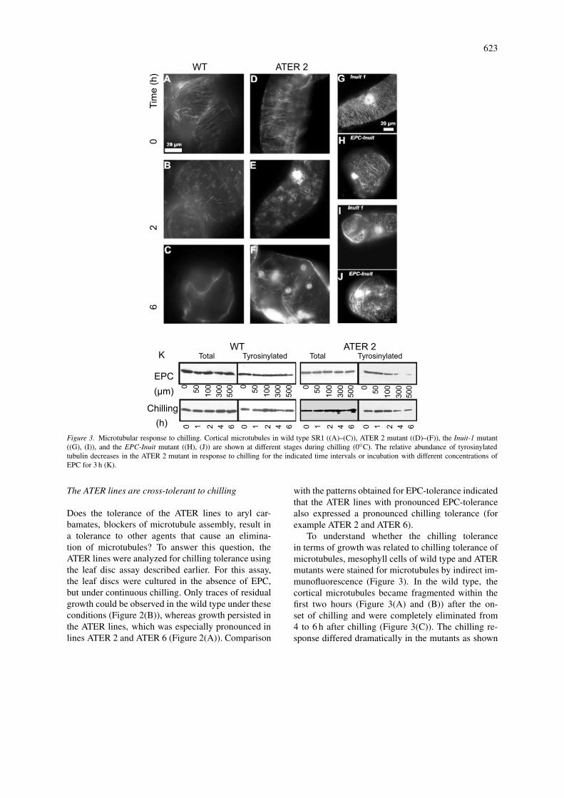
Figure 3. Microtubular response to chilling. Cortical microtubules in wild type SR1 ((A)–(C)), ATER 2 mutant ((D)–(F)), the Inuit-1 mutant((G), (I)), and the EPC-Inuit mutant ((H), (J)) are shown at different stages during chilling (0◦C). The relative abundance of tyrosinylatedtubulin decreases in the ATER 2 mutant in response to chilling for the indicated time intervals or incubation with different concentrations ofEPC for 3 h (K).
The ATER lines are cross-tolerant to chilling
Does the tolerance of the ATER lines to aryl car-bamates, blockers of microtubule assembly, result ina tolerance to other agents that cause an elimina-tion of microtubules? To answer this question, theATER lines were analyzed for chilling tolerance usingthe leaf disc assay described earlier. For this assay,the leaf discs were cultured in the absence of EPC,but under continuous chilling. Only traces of residualgrowth could be observed in the wild type under theseconditions (Figure 2(B)), whereas growth persisted inthe ATER lines, which was especially pronounced inlines ATER 2 and ATER 6 (Figure 2(A)). Comparison
with the patterns obtained for EPC-tolerance indicatedthat the ATER lines with pronounced EPC-tolerancealso expressed a pronounced chilling tolerance (forexample ATER 2 and ATER 6).
To understand whether the chilling tolerancein terms of growth was related to chilling tolerance ofmicrotubules, mesophyll cells of wild type and ATERmutants were stained for microtubules by indirect im-munofluorescence (Figure 3). In the wild type, thecortical microtubules became fragmented within thefirst two hours (Figure 3(A) and (B)) after the on-set of chilling and were completely eliminated from4 to 6 h after chilling (Figure 3(C)). The chilling re-sponse differed dramatically in the mutants as shown
624
for example in ATER 2 (Figure 3(D)–(F)). Here, theparallel bundles of cortical microtubules present priorto chilling, where replaced by vesicular structures inthe cell cortex. These vesicles were most abundantbetween 1 and 3 h after the onset of chilling. Uponprolonged chilling radial arrays of fine microtubulesemanated. This vesicle formation differed in the exacttiming and amplitude between lines but was presentin all lines (data not shown). Thus, whereas microtu-bules in the wild type are fragmented and eliminatedcompletely, in the ATER mutants transient vesicularstructures were formed that seemed to be organized ina new microtubular network.
This chilling response of microtubules seems tobe specific for the ATER mutants, since in mutantsthat had been selected directly for increased chillingtolerance (such as Inuit 1) or combined tolerance toEPC and chilling (EPC-Inuit), microtubules persist,when they are challenged by low temperature and donot show significant changes even after 6 h of chilling(Figure 3(G)–(J)), when the microtubular cytoskel-eton is already completely eliminated in the wild type(Figure 3(C)).
In the ATER mutants, factors that impair micro-tubule assembly (EPC, chilling) produced a transientstate, where vesicular structures seemed to organizenew microtubule arrays that emanate from these ves-icles (Figures 1(B) and 3(E)). The resulting microtu-bular cytoskeleton appears to be very stable againstinhibition of assembly. This might be caused by agenerally reduced dynamics of microtubules, such thatthe lifetime of individual microtubules is increased.To test this hypothesis the degree of α-tubulin ty-rosination was followed as marker for microtubuledynamics (Wiesler et al., 2002). Leaf discs fromATER 2 plants, where the formation of tubulin ves-icles was especially pronounced were analyzed forthe response to increasing concentrations of EPC orchilling (Figure 3(K)). In the wild type, the amountof total α-tubulin as well as the abundance of ty-rosinylated α-tubulin was found to be constant aftertreatment with EPC or in response to chilling. Inthe ATER 2 mutant, the amount of total α-tubulinwas found to be fairly constant as well. However,the abundance of tyrosinylated α-tubulin decreaseddramatically, when the concentration of EPC ex-ceeded 100 µM or when the chilling lasted longerthan 2 h. This suggests that the dynamics of the mi-crotubular cytoskeleton exhibit downregulation in themutant in response to factors that impair microtubuleassembly.
Chilling tolerance in the Inuit mutantsis based on different mechanisms
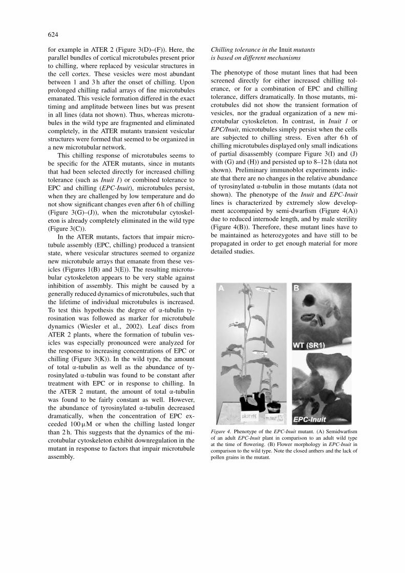
The phenotype of those mutant lines that had beenscreened directly for either increased chilling tol-erance, or for a combination of EPC and chillingtolerance, differs dramatically. In those mutants, mi-crotubules did not show the transient formation ofvesicles, nor the gradual organization of a new mi-crotubular cytoskeleton. In contrast, in Inuit 1 orEPC/Inuit, microtubules simply persist when the cellsare subjected to chilling stress. Even after 6 h ofchilling microtubules displayed only small indicationsof partial disassembly (compare Figure 3(I) and (J)with (G) and (H)) and persisted up to 8–12 h (data notshown). Preliminary immunoblot experiments indic-ate that there are no changes in the relative abundanceof tyrosinylated α-tubulin in those mutants (data notshown). The phenotype of the Inuit and EPC-Inuitlines is characterized by extremely slow develop-ment accompanied by semi-dwarfism (Figure 4(A))due to reduced internode length, and by male sterility(Figure 4(B)). Therefore, these mutant lines have tobe maintained as heterozygotes and have still to bepropagated in order to get enough material for moredetailed studies.
Figure 4. Phenotype of the EPC-Inuit mutant. (A) Semidwarfismof an adult EPC-Inuit plant in comparison to an adult wild typeat the time of flowering. (B) Flower morphology in EPC-Inuit incomparison to the wild type. Note the closed anthers and the lack ofpollen grains in the mutant.
625
Discussion
Can activation tagging yield mutantsof cytoskeletal dynamics?
So far, screens for tolerance to antimicrotubular com-pounds have recovered only few cytoskeletal mutantsin plants (for a recent review see Baird et al.,2000) that were usually affected in the affinity oftubulin for these compounds leading to increased tol-erance. Since all antimicrotubular compounds inves-tigated so far act as blockers of microtubule assembly(Mizuno & Suzaki, 1991), tolerance or sensitivityof a given microtubule will depend on its turnover.Thus, reduced microtubule dynamics should result inincreased tolerance to antimicrotubular compounds,whereas induction of microtubule dynamics is expec-ted to render microtubules more sensitive. In fact,the sensitivity of microtubules to assembly blockerscan be modulated through altering their dynamicsthrough auxin (Wiesler et al., 2002). A screen formutants with increased tolerance to blockers of mi-crotubule assembly should therefore recover, in ad-dition to mutants with reduced affinity of tubulin forthe inhibitor, mutants with reduced turnover of mi-crotubules. However, classical mutagenesis has notrecovered such mutants so far. This may be for tworeasons:
1. Selection of mutants that have been generated byclassical mutagenesis leading to point mutationswill produce a high number of mutants, wheremissense or nonsense mutations in the coding se-quence will alter the binding of the assemblyblocker to tubulin.
2. Mutants affected in factors changing the dynam-ics of microtubules will be overlooked in the firstgeneration, because the intact version of this factorwill maintain microtubule dynamics to a degreethat no selectable change of microtubule dynamicscan be observed.
A screen for mutants, where the dynamics of mi-crotubules is reduced or where the signaling towardsthis dynamics is affected, must therefore meet twoconditions:
1. The mechanism of generating mutants shouldavoid point mutations.
2. The phenotype should be screenable in the firstgeneration, that is, the phenotype should bedominant.
Both conditions are met by T-DNA activation tag-ging. Insertion of the tag into a promotor region isexpected to activate a neighboring gene. The elevatedexpression of this gene would therefore result in adominant phenotype that can be screened in the firstgeneration. Since the mutagenesis occurs by inser-tion, point mutations of the coding sequence leadingto affinity mutants of tubulin are avoided. Insertionof the tag into the coding sequence of a gene shouldresult in a knockout phenotype that in most caseswould not manifest in the first generation and thuswill not be recovered in this screen. The combina-tion of T-DNA activation tagging with selection forresistance to blockers of microtubule assembly shouldtherefore lead to the isolation of mutants with reduceddynamics of microtubules, either because associatedproteins that confer microtubule stability are inducedor because signaling components that activate suchproteins are upregulated. The screen for chilling tol-erance (Inuit) is expected to yield mutants where thechilling response of genes involved in the adaptation tolow-temperature stress is activated. These genes mayor may not be related to cytoskeletal signaling. To iso-late those genes that are part of the chilling-inducedsignaling towards the microtubules, the screens forchilling tolerance or accelerated cold acclimation werecombined with selection for increased tolerance to themicrotubule assembly blocker EPC (EPC-Inuit line).
The number of mutants recovered from thesescreens was very small indicating that the selectivepressure was very stringent. In fact, the dose–responseof leaf-disc growth (Figure 2(A)) shows that the ATERmutants can tolerate EPC-concentrations that areabout one order of magnitude higher than the dose thatis lethal for the wild type. Conversely, the Inuit andEPC/Inuit mutants, survived under continuous chillinglasting for several months. In one experiment (datanot shown), callus from the Inuit line was maintainedin the refrigerator for 8 months and upon rewarmingregenerated extremely vigorously a high number ofplantlets. This extent of perseverance is especially ex-traordinary in the background of an otherwise highlychilling-sensitive species as tobacco. Although onlyfew mutants were collected from the different screens,the phenotype of these mutants is highly significant.
To achieve a high transformation efficiency, theprotoplasts were co-cultivated with Agrobacteriumwhen most cells passed through second division. Thisperiod of optimal transformation competence wasreached about 6–8 days after the isolation of proto-plasts. Using a different tagging vector, other groups

626
reported maximal transformation efficiency for co-cultivation 4–5 days after protoplast isolation, whenthe cells entered the first cell division (Walden et al.,1995; Zubko et al., 2002). The transformation effi-ciency in our system as estimated from plating aliquotsof the co-cultivated population on hygromycin, wasin the range 5–10%, which seems to be near themaximum that can be achieved for activation tagging.Earlier reports that had claimed 15–20% of trans-formation efficiency (Walden et al., 1995) were laterretracted (Schell et al., 1999).
Summarizing, the combination of activation tag-ging with selection protocols that aimed on alterationof microtubule responses to depolymerizing factorssuch as EPC or chilling, permitted isolation of a few,but highly tolerant mutants. This shows that activationtagging is a feasible approach to generate mutationsthat affect either microtubular dynamics or the signalcontrol of such dynamics.
In ATER 2, increases of the dimer pool suppressmicrotubule dynamics
Tolerance to EPC that otherwise would cause abreakdown of the microtubular cytoskeleton could beachieved by a constitutively elevated synthesis of tu-bulin that would compensate for the loss of dimersbound by EPC. However, in ATER 2, where the EPCtolerance is particularly pronounced (Figure 2(A)), theabundance of α-tubulin is not significantly differentfrom the wild type (Figure 3(K)), discounting a tol-erance mechanism based on tubulin overexpression.This is consistent with published reports that tubulinconstitutes a highly buffered system that will com-pensate any imbalance between α- or β-tubulin or anychange in the general abundance of tubulins (Anthony& Hussey, 1998; Anthony et al., 1999). Moreover,tubulin overexpression would be a way to escape theaction of drugs that remove tubulin dimers from the as-sembly equilibrium, but not a mechanism to overcomethe effect of chilling. The ATER 2 mutant is highlycross-tolerant to chilling, however (Figure 2(B)).
This points to an alternative scenario, where thefactors that ensure the dynamic equilibrium of themicrotubular system display elevated sensitivity tochanges in the pool of unassembled tubulin. Increasesin the abundance of tubulin heterodimers are very ef-ficiently sensed in the ATER 2 mutant and trigger aresponse culminating in reduced microtubule dynam-ics. When the abundance of tyrosinylated α-tubulinis followed in response to either EPC or chilling
(Figure 3(K)), it is found to decrease in ATER 2.Since tyrosinylated α-tubulin is a marker for dynamicmicrotubules (Wiesler et al., 2002), this finding sug-gests that microtubules in this mutant compensate forthe blocked assembly by increasing the lifetime ofindividual microtubules. It will be interesting to in-vestigate, whether this is caused by a hypersensitivesensing of tubulin disassembly, a mechanism that iswell known in animal cells (for review see Cleveland,1988) or whether a step within the effector chaintriggered by this sensory mechanism is activated.
Preliminary findings with the Inuit 1 and theEPC/Inuit mutant do not give indications for alteredabundance of tyrosinylated α-tubulin, suggesting thatin these mutants chilling tolerance is based on adifferent mechanism.
Summarizing, our data suggest that in ATER 2(and possibly in the other ATER mutants as well), thestrong cross-tolerance of growth to EPC and chilling(Figure 2) is based on a strong inducible responseof microtubules that will downregulate microtubuledynamics in response to increases in the pool ofunassembled tubulin heterodimers.
Microtubule dynamics define chillingtolerance in tobacco
As other crops originating from subtropical or trop-ical regions, tobacco is a typical example of a chillingsensitive plant. This is mirrored by a rapid eliminationof microtubules (Figure 3(A)–(C)) accompanied by adrastic inhibition of growth (Figure 2(B)), when thecells are challenged by chilling. The chilling sensitiv-ity of a crop has been reported to correlate with thecold tolerance of microtubules (Jian et al., 1989) sug-gesting that it is the tolerance of microtubules that willdefine the degree of chilling tolerance of the wholeplant. Consistent with this conclusion, elimination ofmicrotubules by colchicine increased low-temperaturedamage in cotton, a classical chilling-sensitive crop(Rikin et al., 1980).
The exact mechanism, by which the microtubularnetwork breaks down in response to chilling stressis far from understood. Theoretically, chilling couldreduce the assembly of microtubules – chilling toler-ance would then be achieved by maintaining a highrate of assembly under low temperature. Alternatively,chilling could stimulate the disassembly of microtu-bules – tolerance would then be caused by suppressionof this stimulation. The exact mechanism is difficult toassess because assembly and disassembly are usually
627
balanced resulting in a more or less dynamic equilib-rium. Through a highly complex interaction betweenexpression of tubulin genes and the assembly stateof microtubules (for a recent review see Breviario &Nick, 2000), any change in the assembly rate is ex-pected to be balanced by an antagonistic response ofdisassembly (and vice versa), as long as the buffer-ing capacity of the regulatory system is not exceeded.Irrespective of this complexity, the cross-tolerance ofthe ATER mutants to chilling stress (Figures 2 and 3)allows to draw four conclusions:
1. Reduced dynamics of microtubules causes resis-tance of microtubules to chilling-induced elimin-ation.
2. Resistance of microtubules to chilling-inducedelimination causes chilling tolerance of growth.
3. Microtubule dynamics defines chilling tolerance intobacco, a chilling-sensitive crop.
4. By manipulation of microtubular dynamics itshould be possible to engineer general chillingtolerance in such crops.
Outlook: cold-induced signalingto the microtubular cytoskeleton
The signaling cascade triggering plant adaptation tocold stress is still far from being understood, butmutant studies in Arabidopsis have stimulated a model(for review see Thomashow, 2001), where cold-induced calcium influx will trigger a kinase cascade,inhibiting transcriptional repressors such as HOS1,such that constitutively expressed transcription factorssuch as ICE can activate a transcriptional cascade(initiated by the transcription factor CBF) that willculminate in the expression of adaptive proteins.This cascade somewhere merges or cross-talks withabscisic-acid induced signaling, but this might well berelatively downstream of this cascade, for instance atthe target promotors of CBF.
It is even less clear, where signaling to the mi-crotubules branches from this general cascade. How-ever, several findings indicate that calcium is a centralplayer in the cold-induced signaling towards microtu-bules. For example, the cold-induced elimination ofmicrotubules was reported to be blocked by lithium,suggesting that the phosphoinositide pathway mightbe involved (Bartolo & Carter, 1992). Through in-teraction with potential MT-associated proteins suchas the elongation factor EF-1α (Durso & Cyr, 1994)or by direct binding to tubulin (Kumagai & Nishida,
1979), calcium/calmodulin could modulate the micro-tubules (Fisher et al., 1996). Alternatively, calciumcould act on microtubule dynamics through the actincytoskeleton, since some of the calcium-dependentprotein kinases have been shown to interact withactin (Putnam-Evans et al., 1989). As expected fromthe emerging model of cold signaling (Thomashow,2001), not only calcium/calmodulin, but also kinasesignaling seems to be involved, because kinase inhib-itors such as 6-dimethylaminopurine or staurosporincan render microtubules chilling tolerant in culturedtobacco cells (Mizuno, 1992). This would favor amodel, where the bifurcation between general coldsignaling and signaling to the microtubular cytoskel-eton would occur downstream the kinase cascade thatculminates in the inhibition of HOS1.
What are the microtubular responses to this sig-naling chain that will shift the assembly/disassemblyequilibrium towards the soluble heterodimers? Thephenotype of the ATER mutants and especially theprogressive decrease of tyrosinylated α-tubulin sug-gests a rapid downregulation of tubulin dynamics. Thismight be accompanied and stabilized by changes inthe composition of tubulin isotypes as repeatedly de-scribed for cold acclimation (rye roots, Kerr & Carter,1990; Arabidopsis, Chu et al., 1993). It is worth in-vestigating whether the chilling-tolerance of the Inuitand EPC/Inuit mutants is caused by an a priori alteredpattern of isotypes.
To isolate molecular components of this signalingchain, the tagged genes will be isolated and clonedfrom the ATER mutants through plasmid rescue. Thecriterion for a true element of the signaling chainwould be a phenocopy of the mutant phenotype byoverexpression of the rescued gene in the wild typebackground. This would demonstrate that the pheno-type is caused by the activation of the tagged gene(and not by long-term activation of a neighboring non-tagged gene). A first genomic fragment isolated fromthe ATER 6 mutant contains a so-called SH3-domainthat is characteristic for a group of actin-binding pro-teins (Kioka et al., 1999) suggesting that a functionalapproach based on activation tagging will yield genesthat are novel and/or not expected from a prioriconsiderations.
Acknowledgements
We thank Czaba Koncz for the gift of tagging vector.We also thank M. Oldsen and H.T. Imam for their help
628
with the protein extraction and western blotting. Thework was supported by funds from the Volkswagen-Foundation (‘Dynamics of the plant cytoskeleton’).
References
Anthony RG and Hussey PJ (1998) Suppression of endogenous α-and β-tubulin synthesis in transgenic maize calli overexpressingα- and β-tubulins. Plant J 16: 297–304.
Anthony RG, Reichelt S and Hussey PJ (1999) Dinitroanilineherbicide-resistant transgenic tobacco plants generated by co-overexpression of a mutant α-tubulin and a β-tubulin. NatureBiotechnol 17: 712–716.
Baird V, Blume YB and Wick SM (2000) Microtubular and cyto-skeletal mutants. In: Nick P (ed.), Plant Microtubules: Potentialfor Biotechnology. (pp. 159–191) Springer, Berlin, Heidelberg.
Bartolo ME and Carter JV (1991) Effect of microtubule stabilizationon the freezing tolerance of mesophyll cells of spinach. PlantPhysiol 97: 182–187.
Bartolo ME and Carter JV (1992) Lithium decreases cold-inducedmicrotubule depolymerization in mesophyll cells of spinach.Plant Physiol 99: 1716–1718.
Bin-Bing Zh and Kirschner MW (1999) Quantitative measure-ment of the catastrophe rate of dynamic microtubules. Cell MotCytoskeleton 43: 43–51.
Breviario D and Nick P (2000) Plant tubulins: a melting pot forbasic questions and promising applications. Transgenic Res 9:383–393.
Caboche M (1980) Nutritional requirements of protoplasts derivedhaploid tobacco cells grown at low densities in liquid medium.Planta 149: 1–18.
Caudron N, Valiron O, Usson Y, Valiron P and Job D (2000) Areassessment of the factors affecting microtubule assembly anddisassembly in vitro. J Mol Biol 297: 211–220.
Chang C, Kwok SF, Bleecker AB and Meyerowitz EM (1993) Ar-abidopsis ethylene-response gene ETR1: similarity of product totwo-component regulators. Science 262: 539–544.
Chu B, Snustad DP and Carter JV (1993) Alteration of beta-tubulingene expression during low temperature exposure in leaves ofArabidopsis thaliana. Plant Physiol 103: 371–377.
Cleveland DW (1988) Autoregulated instability of tubulin mRNAs:a novel eukaryotic regulatory mechanism. Trends Biochem Sci13: 339–343.
Ding JP and Pickard BG (1993) Mechanosensory calcium-selectivecation channels in epidermal cells. Plant J 3: 83–110.
Durso NA and Cyr RJ (1994) A calmodulin-sensitive interactionbetween microtubules and a higher plant homolog of elongationfactor 1α. Plant Cell 6: 893–905.
Feldmann KA (1991) T-DNA insertional mutagenesis in Arabidop-sis: mutational spectrum. Plant J 1: 71–82.
Fisher DD, Gilory S and Cyr RJ (1996) Evidence for opposing ef-fects of calmodulin on cortical microtubules. Plant Physiol 122:1070–1087.
Jian LC, Sun LH and Liu ZP (1989) Studies on microtubule coldstability in relation to plant cold hardiness. Acta Bot Sinica 31:737–741.
Hayashi H, Czaja I, Schell J and Walden R (1992) Activation ofplant gene by T-DNA tagging: auxin independent growth in vitro.Science 258: 1350–1353.
Kakimoto T (1996) CKI1, a histidine kinase homolog implicated incytokinin signal transduction. Science 274: 982–985.
Kao RN and Michayluk MR (1975) Nutritional requirements forgrowth of Vicia hajastana cells at very low population density inliquid medium. Planta 126: 105–110.
Kerr GP and Carter JV (1990) Relationship between freezing tol-erance of root-tip cells and cold stability of microtubules in rye(Secale cereale L. cv Puma). Plant Physiol 93: 77–82.
Kioka N, Sakata S, Kawauchi T, Amachi T, Akiyama SK, OkazakiK et al. (1999) Vinexin: a novel vinculin-binding protein withmultiple SH3 domains enhances actin cytoskeletal organization.J Cell Biol 144: 59–69.
Knight MR, Campbell A, Smith SM and Trewavas AJ (1991) Trans-genic plant aequorin reports the effects of touch and cold shockand elicitors on cytoplasmic calcium. Nature 352: 524–526.
Koncz C, Martini N, Mayerhofer R, Koncz-Kalman Z, Körber H,Redei GP et al. (1989) High frequency T-DNA mediated genetagging in plants. Proc Natl Acad Sci USA 86: 8467–8471.
Koncz C, Mayerhofer R, Koncz-Kalman Z, Nawrath C, Reiss B,Redei GP et al. (1990) Isolation of a gene encoding a novelchloroplast protein by T-DNA tagging in Arabidopsis thaliana.EMBO J 9: 1337–1346.
Koncz C, Martini N, Szabados L, Hrouda M, Bachmair A and SchellJ (1994) Specialized vectors for gene tagging and expressionstudies. In: Gelvin SB and Schilperoort RA (eds), Plant Molecu-lar Biology Manual B2. (pp. 1–22) Kluwer Academic Publishers,Belgium.
Kreis TE (1987) Microtubules containing detyrosinylated tubulinare less dynamic. EMBO J 6: 2597–2606.
Kumagai H and Nishida E (1979) The interaction betweencalcium dependent regulator protein of cyclic nucleotidephosphodiesterase and microtubule proteins II, associationof calcium-dependent regulator protein with tubulin dimers.J Biochem 85: 1267–1274.
Maliga P, Sz-Breznovits A and Marton L (1973) Streptomycin-resistant plants from callus culture of haploid tobacco. Nat NewBiol 244: 29–30.
Mazars C, Thion L, Thuleau P, Graziana A, Knight MR, Moreau Met al. (1997) Organization of cytoskeleton controls the changesin cytosolic calcium of cold-shocked Nicotiana plumbaginifoliaprotoplasts. Cell Calcium 22: 413–420.
Mizuno K (1992) Induction of cold stability of microtubules inculture tobacco cells. Plant Physiol 100: 740–748.
Mizuno K and Suzaki T (1991) Effects of anti-microtubule drugson in vitro polymerization of tubulin from mung mean. Bot MagTokyo 103: 435–448.
Monroy AF, Sarhan F and Dhindsa RS (1993) Cold-inducedchanges in freezing tolerance, protein phosphorylation, and geneexpression. Plant Physiol 102: 1227–1235.
Murashige T and Skoog F (1962) A revised medium for rapidgrowth and bioassay with tobacco tissue culture. PhysiolPlantarum 15: 473–497.
Nick P (2000) Control of the response to low temperatures. In: NickP (ed.), Plant Microtubules: Potential for Biotechnology. (pp.121–135) Springer, Berlin, Heidelberg.
Nick P, Yatou O, Furuya M and Lambert AM (1994) Auxin-dependent microtubule response and seedling development areaffected in a rice mutant resistant to EPC. Plant J 6: 651–663.
Nick P, Heuing A and Ehmann B (2000) Plant chaperonins: arole in microtubule-dependent wall formation. Protoplasma 211:234–244.
Potrykus I and Shilito RD (1986) Protoplasts: isolation, culture,plant regeneration. Plant Mol Biol 118: 549–578.
Putnam-Evans C, Harmon AC, Palevitz BA, Fechheimer M andCormier MJ (1989) Calcium-dependent protein kinase is local-ized with F-actin in plant cells. Cell Motil Cytoskel 12: 12–22.

629
Rikin A, Atsmon D and Gitler C (1980) Chilling injury in cotton(Gossypium hirsutum L.): Effect of antimicrotubular drugs. PlantCell Physiol 21: 829–837.
Schell J, Bisseling T, Dülz M, Frassen H, Fritze K, John M et al.(1999) Re-evaluation of phytohormone-independent division oftobacco protoplasts-derived cells. Plant J 17(5): 461–466.
Thion L, Mazars C, Thuleau P, Graziana A, Rossignol M, Moreau Met al. (1996) Activation of plasma membrane voltage-dependentcalcium-permeable channels by disruption of microtubules incarrot cells. FEBS Lett 393: 13–18.
Thomashow MF (2001) So what’s new in the field of plant coldacclimation? Lots! Plant Physiol 125: 89–93.
Vaughn KC (2000) Anticytoskeletal herbicides. In: Nick P (ed.),Plant Microtubules: Potential for Biotechnology. (pp. 193–205)Springer, Berlin, Heidelberg.
Walden R, Fritze K and Harling H (1995) Induction of signal trans-duction pathways through promoter activation. Meth Cell Biol49: 455–469.
Watson DJ (1952) The physiological basis of variation yield. AdvAgron 4: 101–145.
Weigel D, Ahn JH, Bla’zquez MA, Borevitz JO, Christensen SK,Frankhauser C et al. (2000) Activation tagging in Arabidopsis.Plant Physiol 22: 1003–1013.
Wiesler B, Wang QY and Nick P (2002) The stability of corticalmicrotubules depends on their orientation Plant J 32(6):1023–1032.
Zubko E, Adams CJ, Machaé’kova’I, Malbeck J, Scollan C andMeyer P (2002) Activation tagging identifies a gene from Pe-tunia hybrida responsible for the production of active cytokininsin plants Plant J 29(6): 797–808.