Embed Size (px)
Citation preview

Activated charcoal fi lter prevents emphysema 217
J. Biosci. 35(2), June 2010
1. Introduction
Cigarette smoking is the world’s single most preventable
cause of disease and death. About one third of all adults in
the world are smokers (Slama 2008). Each year, over fi ve
million people throughout the world die from smoking-
related illness (IARC 2002). Cigarette smoke (CS) contains
more than 4000 compounds (Stewart and Kleihues 2003).
Among these, nicotine is the primary source of tobacco
dependence (US Department of Health and Human
Services 1988). Others are toxins and carcinogens, such
as free radicals, harmful gases, volatile organic compounds
(VOCs), aldehydes, polycyclic aromatic hydrocarbons
(PAHs) and tobacco-specifi c nitrosamines (TSNA). Most
of these chemicals are considered to be causative agents
for CS-induced life-threatening diseases, particularly
cancer of the lungs and other organs, myeloid leukaemia,
cardiovascular diseases and chronic obstructive pulmonary
disease (COPD), including bronchitis and emphysema (Shah
and Helfant 1988; Sherman 1991; Wald and Hackshaw
1996; US Department of Health and Human Services
1998; IARC 2002; World Cancer Report 2003; Harris et al.
http://www.ias.ac.in/jbiosci J. Biosci. 35(2), June 2010, 217–230, © Indian Academy of Sciences 217
Activated charcoal fi lter effectively reduces p-benzosemiquinone from
the mainstream cigarette smoke and prevents emphysema
NEEKKAN DEY, ARCHITA DAS, ARUNAVA GHOSH and INDU B CHATTERJEE*
Department of Biotechnology and Dr B C Guha Centre for Genetic Engineering and Biotechnology,
University College of Science, Kolkata 700019, India
*Corresponding author (Fax, 91-033-24614849; Email, [email protected])
In this paper, we have made a comparative evaluation of the cytotoxicity and pathophysiological effects of mainstream
smoke from cellulose acetate (CA)-fi ltered cigarettes with that of charcoal-fi ltered cigarettes developed in our
laboratory. Previously, we had demonstrated that the mainstream smoke from an Indian CA-fi ltered commercial
cigarette contains p-benzosemiquinone (p-BSQ), a major, highly toxic, long-lived water-soluble radical. Here, we
have examined 16 brands of different CA-fi ltered cigarettes including Kentucky research cigarettes, and observed
that mainstream smoke from all the cigarettes contains substantial amounts of p-BSQ (100–200 μg/cigarette). We also
show that when the CA fi lter is replaced by a charcoal fi lter, the amount of p-BSQ in the mainstream smoke is reduced
by 73–80%, which is accompanied by a reduction of carbonyl formation in bovine serum albumin to the extent of 70–
90%. The charcoal fi lter also prevented cytotoxicity in A549 cells as evidenced by MTT assay, apoptosis as evidenced
by FACS analysis, TUNEL assay, overexpression of Bax, activation of p53 and caspase 3, as well as emphysematous
lung damage in a guinea pig model as seen by histology and morphometric analysis. The results indicate that the
charcoal fi lter developed in our laboratory may protect smokers from cigarette smoke-induced cytotoxity, protein
modifi cation, apoptosis and emphysema.
[Dey N, Das A, Ghosh A and Chatterjee I B 2010 Activated charcoal fi lter effectively reduces p-benzosemiquinone from the mainstream cigarette
smoke and prevents emphysema; J. Biosci. 35 217–230] DOI 10.1007/s12038-010-0026-2
Keywords. Apoptosis; charcoal fi lter; cigarette smoke; emphysema; p-benzosemiquinone
Abbreviations used: AECS, aqueous extract of cigarette smoke; BSA, bovine serum albumin; CA-fi lter, cellulose acetate fi lter; CF, charcoal-
fi ltered; COPD, chronic obstructive pulmonary disease; CS, cigarette smoke; DAPI, 4′,6-diamidino-2-phenylindole dihydrochloride; DI:
destructive index; DMSO, dimethyl sulphoxide; ESR, electron spin resonance; H&E, haematoxylin–eosin; HPLC, high performance liquid
chromatography; HRP, horseradish peroxidase; Lm, mean linear intercept; MTT, 3-(4,5-dimethylthiazol-2-yl)-2,5-diphenyl tetrazolium
bromide; NMR, nuclear magnetic resonance; PAH, polycyclic aromatic hydrocarbon; PI, propidium iodide; PS, phosphatidyl serine; TLC,
thin-layer chromatography; TSNA, tobacco-specifi c nitrosamines; TUNEL, terminal deoxynucleotidyl transferase-mediated deoxyuridine
triphosphate nick end labelling; UV, ultraviolet; VOC, volatile organic compound
Neekkan Dey et al.218
J. Biosci. 35(2), June 2010
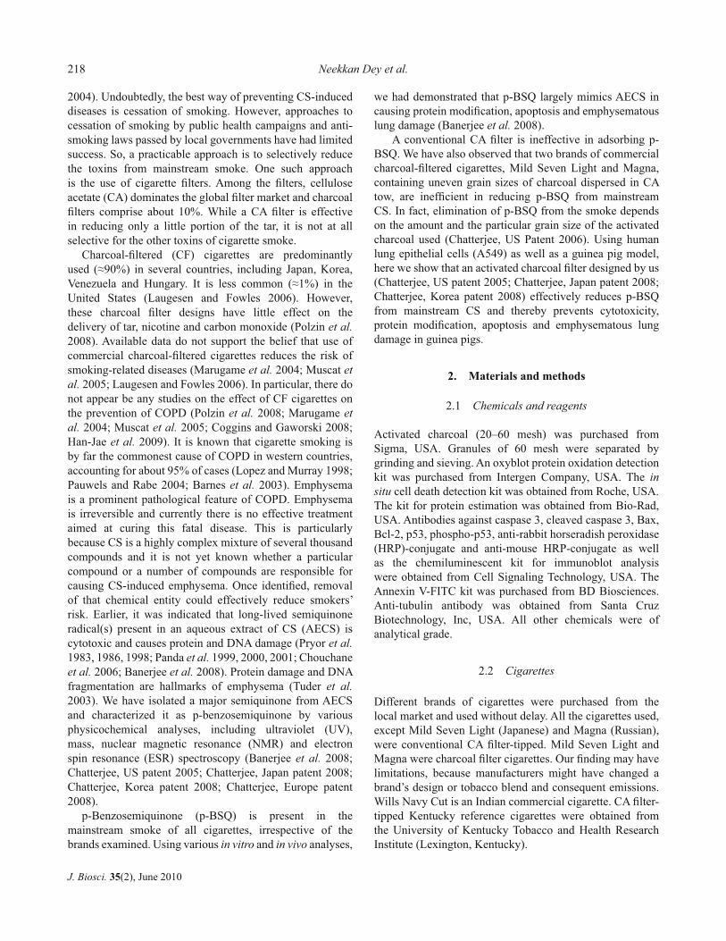
2004). Undoubtedly, the best way of preventing CS-induced
diseases is cessation of smoking. However, approaches to
cessation of smoking by public health campaigns and anti-
smoking laws passed by local governments have had limited
success. So, a practicable approach is to selectively reduce
the toxins from mainstream smoke. One such approach
is the use of cigarette fi lters. Among the fi lters, cellulose
acetate (CA) dominates the global fi lter market and charcoal
fi lters comprise about 10%. While a CA fi lter is effective
in reducing only a little portion of the tar, it is not at all
selective for the other toxins of cigarette smoke.
Charcoal-fi ltered (CF) cigarettes are predominantly
used (≈90%) in several countries, including Japan, Korea,
Venezuela and Hungary. It is less common (≈1%) in the
United States (Laugesen and Fowles 2006). However,
these charcoal fi lter designs have little effect on the
delivery of tar, nicotine and carbon monoxide (Polzin et al.
2008). Available data do not support the belief that use of
commercial charcoal-fi ltered cigarettes reduces the risk of
smoking-related diseases (Marugame et al. 2004; Muscat et
al. 2005; Laugesen and Fowles 2006). In particular, there do
not appear be any studies on the effect of CF cigarettes on
the prevention of COPD (Polzin et al. 2008; Marugame et
al. 2004; Muscat et al. 2005; Coggins and Gaworski 2008;
Han-Jae et al. 2009). It is known that cigarette smoking is
by far the commonest cause of COPD in western countries,
accounting for about 95% of cases (Lopez and Murray 1998;
Pauwels and Rabe 2004; Barnes et al. 2003). Emphysema
is a prominent pathological feature of COPD. Emphysema
is irreversible and currently there is no effective treatment
aimed at curing this fatal disease. This is particularly
because CS is a highly complex mixture of several thousand
compounds and it is not yet known whether a particular
compound or a number of compounds are responsible for
causing CS-induced emphysema. Once identifi ed, removal
of that chemical entity could effectively reduce smokers’
risk. Earlier, it was indicated that long-lived semiquinone
radical(s) present in an aqueous extract of CS (AECS) is
cytotoxic and causes protein and DNA damage (Pryor et al.
1983, 1986, 1998; Panda et al. 1999, 2000, 2001; Chouchane
et al. 2006; Banerjee et al. 2008). Protein damage and DNA
fragmentation are hallmarks of emphysema (Tuder et al.
2003). We have isolated a major semiquinone from AECS
and characterized it as p-benzosemiquinone by various
physicochemical analyses, including ultraviolet (UV),
mass, nuclear magnetic resonance (NMR) and electron
spin resonance (ESR) spectroscopy (Banerjee et al. 2008;
Chatterjee, US patent 2005; Chatterjee, Japan patent 2008;
Chatterjee, Korea patent 2008; Chatterjee, Europe patent
2008).
p-Benzosemiquinone (p-BSQ) is present in the
mainstream smoke of all cigarettes, irrespective of the
brands examined. Using various in vitro and in vivo analyses,
we had demonstrated that p-BSQ largely mimics AECS in
causing protein modifi cation, apoptosis and emphysematous
lung damage (Banerjee et al. 2008).
A conventional CA fi lter is ineffective in adsorbing p-
BSQ. We have also observed that two brands of commercial
charcoal-fi ltered cigarettes, Mild Seven Light and Magna,
containing uneven grain sizes of charcoal dispersed in CA
tow, are ineffi cient in reducing p-BSQ from mainstream
CS. In fact, elimination of p-BSQ from the smoke depends
on the amount and the particular grain size of the activated
charcoal used (Chatterjee, US Patent 2006). Using human
lung epithelial cells (A549) as well as a guinea pig model,
here we show that an activated charcoal fi lter designed by us
(Chatterjee, US patent 2005; Chatterjee, Japan patent 2008;
Chatterjee, Korea patent 2008) effectively reduces p-BSQ
from mainstream CS and thereby prevents cytotoxicity,
protein modifi cation, apoptosis and emphysematous lung
damage in guinea pigs.
2. Materials and methods
2.1 Chemicals and reagents
Activated charcoal (20–60 mesh) was purchased from
Sigma, USA. Granules of 60 mesh were separated by
grinding and sieving. An oxyblot protein oxidation detection
kit was purchased from Intergen Company, USA. The in
situ cell death detection kit was obtained from Roche, USA.
The kit for protein estimation was obtained from Bio-Rad,
USA. Antibodies against caspase 3, cleaved caspase 3, Bax,
Bcl-2, p53, phospho-p53, anti-rabbit horseradish peroxidase
(HRP)-conjugate and anti-mouse HRP-conjugate as well
as the chemiluminescent kit for immunoblot analysis
were obtained from Cell Signaling Technology, USA. The
Annexin V-FITC kit was purchased from BD Biosciences.
Anti-tubulin antibody was obtained from Santa Cruz
Biotechnology, Inc, USA. All other chemicals were of
analytical grade.
2.2 Cigarettes
Different brands of cigarettes were purchased from the
local market and used without delay. All the cigarettes used,
except Mild Seven Light (Japanese) and Magna (Russian),
were conventional CA fi lter-tipped. Mild Seven Light and
Magna were charcoal fi lter cigarettes. Our fi nding may have
limitations, because manufacturers might have changed a
brand’s design or tobacco blend and consequent emissions.
Wills Navy Cut is an Indian commercial cigarette. CA fi lter-
tipped Kentucky reference cigarettes were obtained from
the University of Kentucky Tobacco and Health Research
Institute (Lexington, Kentucky).

Activated charcoal fi lter prevents emphysema 219
J. Biosci. 35(2), June 2010
2.3 Preparation of charcoal-fi ltered cigarettes
Activated charcoal fi lter cigarettes used in the present
study were prepared from the original branded cigarettes
by replacing the conventional CA fi lter with our activated
charcoal fi lter using 150 mg charcoal granules of 60 mesh
(250 microns) (Chatterjee, US Patent 2006). Essentially,
the charcoal fi lter we developed is a cavity fi lter in which
the activated charcoal granules are placed in a void space
between two segments of CA fi lters. One portion of the CA
fi lter (≈14 mm) is the mouthpiece and other portion (≈3
mm) constitutes a barrier between the charcoal bed and the
tobacco portion. The CA mouthpiece was used to prevent
any leakage of charcoal granules in the mainstream smoke.
The various parts, i.e. the CA mouth piece, charcoal bed, thin
CA fi lter placed in between the charcoal bed and the tobacco
portion as well as the tobacco portion, were all constructed
in one single unit without any ventilation.
2.4 Preparation of aqueous extract of cigarette smoke
(AECS) solution
Smoke from one cigarette was extracted with 1 ml of 50 mM
potassium phosphate buffer, pH 7.4, fi ltered through a 0.22
μm Millipore fi lter and the pH adjusted to 7.4, as described
before in detail (Panda et al. 1999). The AECS thus obtained
was used immediately.
2.5 Isolation and characterization of p-benzosemiquinone
(p-BSQ)
p-BSQ was isolated from AECS by differential solvent
extraction, thin-layer chromatography (TLC) and high
performance liquid chromatography (HPLC) as described
earlier (Banerjee et al. 2008). p-BSQ was characterized
by various physicochemical analyses, including UV, mass,
NMR and ESR spectroscopy as reported earlier (Banerjee
et al. 2008).
2.6 Measurement of the comparative yields of p-BSQ in
smoke solution prepared from different cigarettes
p-BSQ in smoke solution was quantitatively measured by
HPLC as described earlier (Banerjee et al. 2008). About
5–10 μl of the smoke solution, fi ltered through a 0.22 μm
Millipore fi lter, was diluted about 40 times with mobile
solvent and 20 μl of this diluted solution was injected into
the HPLC (Simadzu 10A) with a UV detector set at 294
nm using a normal phase silica column (Lichrospher ®
Si60, Merck). The mobile solvent was methylene chloride:
methanol (90:10, v/v) with a fl ow rate of 0.5 ml/min.
The retention time of p-BSQ was 8.808 min. The amount of
p-BSQ present in the smoke solution was calculated from a
standard curve obtained with pure p-BSQ.
2.7 Measurement of protein damage
Protein damage was measured by carbonyl formation in
bovine serum albumin (BSA) after reaction with 2, 4-
dinitrophenyl hydrazine, similar to that done earlier in
our laboratory (Panda et al. 1999). The incubation system
contained 1 mg BSA and 50 μl of smoke solution obtained
from cigarettes with or without a charcoal fi lter in a fi nal
volume of 200 μl of 50 mM potassium phosphate buffer,
pH 7.4. After incubation for 1 h at 37°C, the protein was
precipitated with 200 μl of trichloroacetic acid solution and
the rest of the procedure followed was as described earlier
(Panda et al. 1999). The values are expressed as nmoles of
carbonyl formed per mg BSA.
2.8 Measurement of nicotine
Smoke from a lit cigarette was allowed to dissolve in 2 ml
of 50 mM potassium phosphate buffer, pH 7.4 and fi ltered
through a 0.22 μm Millipore fi lter. One millilitre of the
yellow coloured fi ltrate was extracted with 1 ml of methylene
chloride by vigorous vortexing to extract the nicotine in the
methylene chloride layer. Of the methylene chloride layer
containing the nicotine, 500 μl was then vortexed with 500 μl
of 50 mM HCl solution and the nicotine in the aqueous layer
was estimated by HPLC analysis at 254 nm (Chatterjee, US
patent 2005). About 5–10 μl of the aqueous layer was diluted
to 200 μl with the mobile solvent, and 20 μl of this diluted
solution was injected into the HPLC column. A standard
solution of nicotine was prepared in a similar manner and
analysed. The parameters used were: Instrument, Shimadzu
10A; Column, Lichrospher® 100 RP-18 endcapped (5
μm), Merck; mobile solvent: 50 mM KH2PO4 solution:
accetonitrile:methanol (78:17: 5, v/v) containing 1 mM
sodium hepatane sulphonate, pH 5.0; fl ow rate: 0.3 ml /min.
The retention time of nicotine was 4.185 min. The minimum
amount of nicotine that could be detected by the HPLC
analysis under these conditions was 10 ng.
2.9 Measurement of tar
Tar was collected from the mainstream smoke by suction (30
cm water) through a Millipore fi lter unit. The Millipore fi lter
(0.22 μm) was changed every 2 min to avoid clogging of the
fi lter. For each cigarette, 4 fi lters were used. After complete
burning of the tobacco, the fi lters were dried in a vacuum
desiccator and weighed. The difference in weight of the
fi lters before and after collecting the particulate portion was
the weight of the tar (Chatterjee, US patent 2005).

2.10 Cell culture
A549 cells were grown to 50–60% confl uence in HamF12
medium containing 10% foetal calf serum (GIBCO-BRL,
USA), 100 units /ml penicillin, 100 μg/ml streptomycin
and 4 mM glutamine/ml. The cells were grown at 37°C in a
humifi ed incubator maintained in an atmosphere of 95% air
and 5% CO2.
2.11 Cytotoxicity assay
The cytotoxicity of the aqueous extract of mainstream smoke
from CA-fi ltered and charcoal-fi ltered (CF) cigarettes was
evaluated by the 3-(4,5-dimethylthiazol-2-yl)-2,5-diphenyl
tetrazolium bromide (MTT) assay (Mosmann 1983). After
respective treatments with 50 μl/ml of the CA or CF AECS
solution for 1 h, the culture medium was replaced by medium
containing 0.5 mg/ml MTT and incubated for an additional
3 h. The blue MTT formazan was dissolved in 1 ml of
dimethyl sulphoxide (DMSO), and the absorbance values
were determined at 560 nm in a UV-VIS spectrophotometer
(Shimadzu UV-2540).
2.12 Differentiation between apoptosis and necrosis by
FACS
In the early stages of apoptosis, changes occur in the
plasma membrane. One of the cell surface alterations is
the translocation of phosphatidyl serine (PS) from the inner
surface of the cell membrane to the outer layer. Annexin V,
a Ca2+-dependent phospholipid-binding protein with high
affi nity for phosphatidyl serine, is a sensitive probe for PS
and can thereby detect early apoptotic cells in a live cell
population. Propidium iodide (PI) stains only the necrotic
cells. To distinguish between apoptosis and necrosis, A549
cells (3 x 10⁶) treated with 50 μl of CA-fi ltered or CF AECS
solution for 12 h were stained by PI and Annexin V-FITC
(Becton Dickinson) according to manufacturer’s protocols
and analysed using the FACS Calibur-Cell Quest software
(Becton Dickinson). A total of 10 000 events were acquired.
The cells were properly gated and a dual parameter dot plot
of FL2H (X-axis; PI fl uorescence, linear scale) vs FL1-H (Y-
axis; FITC-fl uorescence, logarithmic scale) was recorded.
2.13 Terminal deoxynucleotidyl transferase-mediated de-
oxyuridine triphosphate nick end labelling (TUNEL) assay
A549 cells (2 x 10⁶) treated with 50 μl of AECS were fi xed
with 4% p-formaldehyde and permeabilized with titron X-
100 (0.1%) in 0.1% Na-citrate. The cells were then washed
with phosphate buffered saline and subjected to TUNEL
assay using an in situ cell death detection kit (Roche, USA)
according to the manufacturer’s instruction. The stained cells
were counted under a fl uorescence microscope (Olympus
B). Nuclei were simultaneously counted by counterstaining
with 4′,6-diamidino-2-phenylindole dihydrochloride (DAPI)
(Santa Cruz Biotechnology, USA). The percentage of
TUNEL-positive cells was calculated (Banerjee et al. 2008).
2.14 Exposure of guinea pigs to cigarette smoke (CS)
The procedure we followed was essentially similar to that
described earlier in detail (Banerjee et al. 2007, 2008). Male
short-hair guinea pigs weighing 400–500 g were used for
all the experiments. All animal treatment procedures met
the Institutional Animal Ethics Committee guidelines. The
guinea pigs were fed a vitamin C-free diet for 7 days to
minimize the vitamin C level of tissues (Panda et al. 2000).
This is because vitamin C is a potential inhibitor of CS-
induced protein oxidation (Panda et al. 1999, 2000), which
would otherwise counteract the damaging effect of CS. The
vitamin C-free diet given to the guinea pigs was similar to that
described earlier (Misra et al. 2003). After 7 days of vitamin
C deprivation, the guinea pigs were subjected to CS exposure
(either from CA-fi ltered or CF cigarettes) from 5 cigarettes
(2 puffs per cigarette)/animal/day in a smoke chamber
(Banerjee et al. 2007), along with oral supplementation of 1
mg vitamin C/animal/day for 2 weeks (6 days a week, Sunday
excluded). Deprivation of vitamin C was discontinued to
avoid the onset of scurvy. One mg of vitamin C per day is
approximately the minimum dose needed to prevent scurvy
in the guinea pig (Banerjee et al. 2007). With this dose of
vitamin C, there was no symptom of onset of scurvy in any of
the guinea pigs during the experimental period. The smoke
chamber was similar to that of a vacuum desiccator with an
open tube at the top and a side tube fi tted with a stopcock, as
described earlier (Banerjee et al. 2007, 2008). The volume
of the chamber was 2.5 l. The cigarette placed at the top was
lit and CS was introduced into the chamber containing the
guinea pig by applying a mild suction of 4 cm water through
the side tube for 5 s. Thereafter, the vacuum was turned off
and the guinea pig was further exposed to the accumulated
smoke for another 40 s. The total duration of exposure to
smoke from one puff was thus 45 s. Altogether, 2 puffs per
cigarette were given, allowing the animal 1 min rest in a
smoke-free atmosphere to breathe air between each puff.
The gap between one cigarette and the next was 1 h. Pair-fed
sham controls were subjected to air exposure instead of CS
under similar conditions.
The guinea pigs were divided into the following
experimental groups (N = 6/group): (i) exposed to air, (ii)
exposed to CA-fi ltered CS, (iii) exposed to charcoal-fi ltered
CS. After exposure to air or CS for up to 15 days, both the
sham controls and the CS-exposed guinea pigs were deprived
of food overnight and sacrifi ced next day by diethyl ether
Neekkan Dey et al.220
J. Biosci. 35(2), June 2010
inhalation. The lungs were then excised immediately and
processed for analysis.
2.15 Histology of lung section for measurement of
emphysematous lung damage
Lung damage caused by exposure to CS was quantifi ed by
measuring the mean linear intercept (Lm) and destructive
index (DI). Lm represents the average size of alveoli,
indicating the air space, which is increased in emphysema.
It was measured by the technique originally described
by Dunnill (1962). Images randomly selected from
haematoxylin–eosin (H&E) stained lung sections were
captured in an Olympus B microscope at 20X magnifi cation
and analysed using the Dewinter Biowizard 4.1 software.
The image was resized to a fi nal magnifi cation of 70X.
Briefl y, a cross-hair grid consisting of horizontal and vertical
lines (1 cm apart) was laid over the digital image on the
computer screen. The number of times each line crossed an
alveolar wall, both horizontally and vertically, was manually
counted. Results are expressed by the formula:
Lm = L/(X×m),
where Lm = mean linear intercept; L= total length of the
lines in mm; X = magnifi cation factor and m = sum of
intercepts (the points where the horizontal and vertical
lines independently intercepted the alveolar walls). Four
independent lung sections per animal were analysed.
The degree of destruction of the alveolar walls was
quantifi ed by measuring the DI following the microscopic
manual point count method of Saetta et al. (1985) using the
Dewinter Biowizard 4.1 software, as stated above, except
that the lines of the cross-hair grid were 2 cm apart. The
spaces directly under the cross-hair points were counted as
either normal (N) or destroyed (D). The DI was computed
from the formula:
DI = D/(D+N) ×100(%),
where D indicates destroyed and N indicates normal points.
An alveolar space was considered to be normal if it was
surrounded by intact walls or by wall disrupted in only one
place. Alveolar space was considered to be destroyed when
the wall of an alveolus was disrupted in two or more places.
2.16 Western blot
For western blot, cells (3 x 10⁶) were treated with CA-
fi ltered or charcoal-fi ltered AECS solution for 1 h and then
reincubated in fresh Ham F12 medium for 16 h. Control cells
received no treatment. After that, cell extracts were prepared
by lysing the cells in lysis buffer (20 mM Tris [tris(hydroxy
methyl)aminomethane chloride], pH 7.4; 250 mM NaCl; 2
mM EDTA [ethylenediaminetetraacetic acid], pH 8.0; 0.1%
Triton-X100; 0.01 mg/ml aprotinin; 0.005 mg/ml leupeptin;
0.4 mM phenylmethanesulphonyl fl uoride [PMSF]; and 4
mM NaVO4). Lysates were then centrifuged at 20 000 g for
10 min to remove insoluble material. The supernatant (30–50
μg protein) was resolved on 12% sodium dodecyl sulphate-
polyacrylamide gel electrophoresis (SDS-PAGE) gel. After
electrophoresis, the proteins were electrotransferred to a
PVDF membrane, blocked with 5% non-fat milk (Bio-
Rad), and probed with antibodies against caspase 3, cleaved
caspase 3, Bax, Bcl-2, p53 and phospho-p53 (1:1000) for 1 h.
Thereafter, the blot was washed, exposed to HRP-conjugated
secondary antibodies for 1 h, and fi nally detected by
chemiluminescence, as done earlier (Banerjee et al. 2008).
2.17 Statistical analysis
All values are expressed as mean ± SD. Statistical
signifi cance was carried out using one-way ANOVA. The P
values were calculated using appropriate F-tests. Difference
with P values <0.05 were considered signifi cant.
3. Results
3.1 Charcoal fi lter effectively reduces p-BSQ from main-
stream smoke of different brands of cigarettes and prevents
protein carbonyl formation
Table 1 shows that mainstream smoke from different brands
of CA-fi ltered commercial cigarettes as well as Kentucky
research cigarettes contain high amounts of p-BSQ. When
the CA fi lters are replaced by charcoal fi lters, the p-BSQ
content is reduced by 73–80%. Earlier, we had reported that
p-BSQ from CS is a major toxic component that is largely
responsible for carbonyl formation in proteins (Banerjee
et al. 2008). Table 1 indicates that when the CA fi lter is
replaced by a charcoal fi lter, carbonyl formation in BSA
is markedly reduced. Table 1 further shows that tar and
nicotine delivery are also considerably reduced in smoke
from charcoal-fi ltered cigarettes. In a separate experiment,
we have observed that fortifi cation of tobacco (1 g) with 3
mg nicotine per cigarette leads to increased nicotine delivery
in the mainstream smoke to almost the original level (data
not shown). However, such fortifi cation does not cause any
increase in the p-BSQ content of the smoke, apparently
because nicotine is not a precursor of p-BSQ.
3.2 Charcoal fi lter prevents AECS-induced alteration of
morphology and loss of viability in A549 cells
In the pathogenesis of CS-induced pulmonary diseases,
injury of the alveolar epithelium is an important process.
J. Biosci. 35(2), June 2010
Activated charcoal fi lter prevents emphysema 221

Neekkan Dey et al.222
J. Biosci. 35(2), June 2010
We compared the effects of CA-fi ltered AECS with
charcoal-fi ltered AECS on the morphology and viability of
A549 cells. Examination under a phase-contrast microscope
showed that exposure of the cells to CA-fi ltered AECS
caused an increase in fl oating cells accompanied by a
decrease in the density of cells. The cells still attached
became round in shape and the gap between the cells was
enlarged (fi gure 1A, middle panel). However, when the
cells were exposed to charcoal-fi ltered AECS, both the
confl uence and the morphology were almost similar to that
of control cells (fi gure 1A, fi rst and third panels). Similar
observations were made regarding the cytotoxicity of AECS.
MTT assay revealed that compared with the normal controls,
CA-fi ltered AECS reduced the viability of the cells to about
32%. In contrast to this, the viability of charcoal-fi ltered
AECS-treated cells was about 78% (fi gure 1B).
Table 1. p-Benzosemiquinone (p-BSQ), bovine serum albumin (BSA) oxidation, nicotine delivery and tar content in aqueous extract
of cigarette smoke (AECS) prepared from different international cigarettes with and without an activated charcoal fi lter. The amount of
charcoal used in the fi lter was 150 mg of grain size 60 mesh.
S. no. Condition with
or without
charcoal fi lter
Brand of
cigarette
p-BSQ†
content (μg)
Per cent
reduction in
p-BSQ
content**
BSA* oxidation
(nmoles of
carbonyl
formed)#
Nicotine‡
delivery (mg)
Tar¶ content
(mg)
1 Without
With
Kentucky
3R4F†
100 ± 7.07
20 ± 1.79
--
80
6.0
1.5
0.75
0.40
9
4
2 Without
With
Kentucky
1R3F†
180 ± 17.90
40 ± 2.83
--
78
10.0
3.0
1.16
0.50
15
10
3 Without
With
Wills
Navy Cut§
200 ± 17.90
40 ± 3.16
--
80
10.0
2.5
1.00
0.45
20
12
4 Without
With
Winston 200 ± 20.00
45 ± 4.47
--
77
12.4
3.0
1.3
0.55
18
11
5 Without
With
Camel 180 ± 16.10
45 ± 8.94
--
75
10.6
2.8
1.3
0.52
17
10
6 Without
With
Viceroy 175 ± 13.40
40 ± 4.56
--
73
10.4
2.7
1.3
0.54
16
9
7 Without
With
Marlboro 170 ± 13.40
36 ± 3.44
--
79
10.0
2.5
1.2
0.50
16
10
8 Without
With
Benson &
Hedges
170 ± 14.30
34 ± 3.58
--
80
10.2
2.5
1.1
0.47
16
9
9 Without
With
Virginia
Slims
160 ± 14.30
34 ± 4.47
--
79
9.8
2.4
1.1
0.46
14
8
10 Without
With
Cambridge 160 ± 13.40
36 ± 4.50
--
77
10.0
2.6
1.0
0.44
15
9
11 Without
With
Kent 150 ± 9.80
30 ± 2.83
--
80
8.5
2.2
1.0
0.46
14
9
12 Without
With
Kool 155 ± 11.40
32 ± 4.56
--
79
9.0
2.0
1.0
0.44
14
9
13 Without
With
Classic 135 ± 8.94
25 ± 3.69
--
79
7.5
1.6
0.8
0.40
12
8
14 Without
With
Monte Carlo 140 ± 9.88
25 ± 2.68
--
78
7.0
1.5
0.8
0.40
12
8
15 Without
With
Mild
Seven Light*
100 ± 7.07
20 ± 2.28
--
80
6.0
1.4
0.7
0.38
8
3
16 Without
With
Magna* 110 ± 8.10
22 ± 2.83
--
80
6.5
1.5
0.6
0.36
9
4
All cigarettes are with original fi lters provided by the manufacturers; †Kentucky Research Cigarettes; §Indian commercial cigarette;
*Mild Seven Light (Japanese) and Magna (Russian) are charcoal fi lter cigarettes; #Amount of carbonyl formed in 1 mg BSA using
50 μl of aqueous extract of CS. Details of the incubation system and measurements of p-BSQ, carbonyl, nicotine and tar are given in
Materials and methods. ** Values are means of six independent determinations ± SD.

Activated charcoal fi lter prevents emphysema 223
J. Biosci. 35(2), June 2010
3.3 Charcoal fi lter prevents AECS solution-induced apop-
tosis in A549 cells as evidenced by FACS analysis using
Annexin V and PI
Flowcytometric data (fi gure 2) indicated that when A549
cells were treated with 50 μl/ml medium of CA-fi ltered
AECS solution there was a signifi cant increase (78.92%)
of Annexin V-positive cells compared with the untreated
control cells (1.49%; P<0.05). This indicates that the CA-
fi ltered AECS-treated cells were entering into the apoptotic
phase. However, when the cells were treated with 50 μl/ml
medium charcoal-fi ltered AECS solution, the percentage of
Annexin V-positive cells was only 2.18%, indicating that the
charcoal fi lter prevented AECS solution-induced apoptosis
in A549 cells. In all the cases, the percentage of PI-positive
necrotic cells were negligible.
Figure 1. Effect of charcoal fi lter on aqueous extract of cigarette smoke (AECS)-induced morphology and viability of A549 cells. (A)
Phase-contrast micrographs of control (without treatment); cellulose acetate (CA)-AECS, after 1 h treatment with CA-fi ltered AECS
solution and charcoal-fi ltered AECS, after 1 h treatment with charcoal-fi ltered AECS solution. Photographs were taken 12 h after the
respective treatments as signifi cant morphological changes were observed during this period. (B), Bar diagram showing the percentage of
viable cells compared with the control as determined by MTT cell viability assay 24 h after the respective treatments. Data are expressed
as the mean ± SD; N=6.

Neekkan Dey et al.224
J. Biosci. 35(2), June 2010
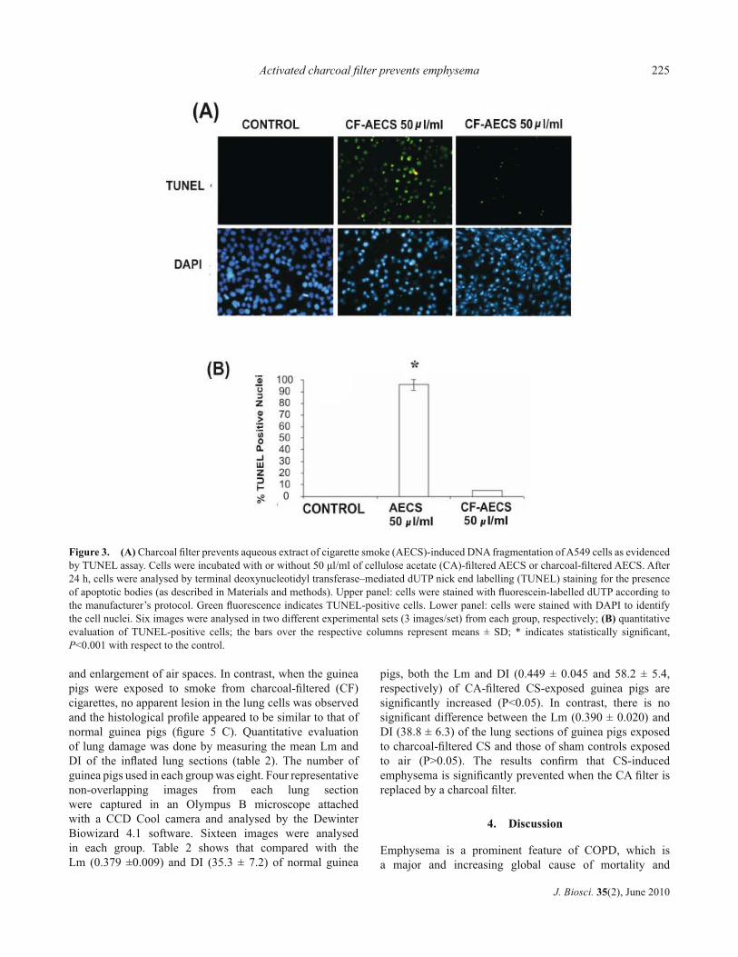
3.4 Charcoal fi lter prevents AECS-induced DNA fragmen-
tation of A549 cells as evidenced by TUNEL assay
When A549 cells were exposed to CA-fi ltered AECS (50
μl/ml medium), the percentage of TUNEL-positive nuclei
markedly increased (90 ± 5 SD), as indicated by green
fl uorescence attributable to fl uorescein-dUTP labelling
(fi gure 3 A, B, upper panel). The lower panel shows the
nuclei counterstained with DAPI. In contrast, there was
little increase (4 ± 1 SD) in TUNEL-positive cells exposed
to charcoal-fi ltered AECS (fi gure 3 A, B). This indicates
that AECS-induced oligonucleosomal fragmentation is
prevented by the charcoal fi lter.
3.5 Charcoal fi lter prevents AECS-induced apoptosis in
A549 lung epithelial cells as evidenced by immunoblotting
Apart from the Annexin V-PI staining by FACS analysis
and DNA fragmentation (TUNEL assay), apoptosis was
evidenced in A549 cells by activation of p53, increase in Bax
proteins and activation of caspase 3 (fi gure 4). The level of
p53 remained unaltered in all the groups irrespective of the
treatment they received. However, fi gure 4A (panel I, lanes
4 and 5) shows that the level of phosphorylated p53 (active
form) markedly increased when A549 cells were exposed to
CA-fi ltered AECS (30 μl/ml or 50 μl/ml medium). There was
no activation of p53 in the case of either untreated control
(lane 1) or cells treated with charcoal-fi ltered AECS (30
μl/ml or 50 μl/ml medium,.lanes 2 and 3).
It is known that one of the mechanisms of apoptosis is
overexpression of Bax, a member of the Bcl-2 family. After
treatment with CA-fi ltered AECS (30 μl/ml or 50 μl/ml
medium), the level of Bax protein increased (fi gure 4A, panel
II). In contrast to this, there was no overexpression of Bax in
the case of the untreated control cells (lane 1) or cells treated
with charcoal-fi ltered AECS (30 μl/ml or 50 μl/ml medium;
lanes 2 and 3). It is known that while Bax is proapoptotic,
Bcl-2 is anti-apoptotic. We therefore examined the level of
Bcl-2 protein. While the level of Bax protein increased in
response to CA-fi ltered AECS treatment, the level of Bcl-2
protein remained unaffected (fi gure 4A, panel III) in all the
cases. These observations suggest that the apoptotic effect of
CA-fi ltered AECS on A549 cells is caused by an increase of
activated p53 as well as an increased Bax/Bcl-2 ratio, which
is completely prevented by the charcoal fi lter.
CA-fi ltered AECS-induced apoptosis was further
supported by checking the level of cleaved caspase 3 by
western blotting of A549 cell lysate using anti-caspase
3 antibody (fi gure 4B, panel I). The level of the cleaved
product of caspase 3 (17 kDa) was markedly increased
(fi gure 4B, panel II, lanes 4 and 5) in response to CA-fi ltered
AECS treatment (30 μl/ml or 50 μl/ml medium). There
was no activation of caspase 3 in the case of the untreated
control cells (fi gure 4B, panel II, lane 1) or cells treated with
charcoal-fi ltered AECS (30 μl/ml and 50 μl/ml medium,
respectively; lanes 2 and 3).
3.6 Smoke from charcoal-fi ltered cigarettes prevents
emphysema as evidenced by air space enlargement and
parenchymal destruction
Histology profi les showed that when the guinea pigs are
exposed to CA-fi ltered CS for two weeks at an exposure rate
of 5 cigarettes (2 puffs/cigarette)/guinea pig/day, there was
marked emphysematous damage of the lung, as compared
with sham control guinea pigs exposed to air (fi gure 5 A,
B). The damage was evidenced by morphometric change
Figure 2. Charcoal fi lter prevents aqueous extract of cigarette
smoke (AECS) solution-induced apoptosis in A549 cells as
evidenced by FACS analysis using Annexin V and propidium
iodide (PI) double staining technique. After treatment with
cellulose acetate (CA)-fi ltered AECS solution and charcoal-fi ltered
AECS solution, A549 cells were labelled with PI and AnnexinV-
FITC and then analysed on a fl owcytometer. Controls were cells
without treatment with AECS, but double-stained with PI and
AnnexinV-FITC. Details are given in the Materials and methods
section 2.12. Dual parameter dot plot of FITC fl uorescence (Y-axis)
vs PI (X-axis) has been shown as fl uorescence intensity. Quadrants:
lower left, viable cells; upper left, apoptotic cells; upper right, late
apoptotic and lower right, necrotic cells.
Activated charcoal fi lter prevents emphysema 225
J. Biosci. 35(2), June 2010
and enlargement of air spaces. In contrast, when the guinea
pigs were exposed to smoke from charcoal-fi ltered (CF)
cigarettes, no apparent lesion in the lung cells was observed
and the histological profi le appeared to be similar to that of
normal guinea pigs (fi gure 5 C). Quantitative evaluation
of lung damage was done by measuring the mean Lm and
DI of the infl ated lung sections (table 2). The number of
guinea pigs used in each group was eight. Four representative
non-overlapping images from each lung section
were captured in an Olympus B microscope attached
with a CCD Cool camera and analysed by the Dewinter
Biowizard 4.1 software. Sixteen images were analysed
in each group. Table 2 shows that compared with the
Lm (0.379 ±0.009) and DI (35.3 ± 7.2) of normal guinea
pigs, both the Lm and DI (0.449 ± 0.045 and 58.2 ± 5.4,
respectively) of CA-fi ltered CS-exposed guinea pigs are
signifi cantly increased (P<0.05). In contrast, there is no
signifi cant difference between the Lm (0.390 ± 0.020) and
DI (38.8 ± 6.3) of the lung sections of guinea pigs exposed
to charcoal-fi ltered CS and those of sham controls exposed
to air (P>0.05). The results confi rm that CS-induced
emphysema is signifi cantly prevented when the CA fi lter is
replaced by a charcoal fi lter.
4. Discussion
Emphysema is a prominent feature of COPD, which is
a major and increasing global cause of mortality and
Figure 3. (A) Charcoal fi lter prevents aqueous extract of cigarette smoke (AECS)-induced DNA fragmentation of A549 cells as evidenced
by TUNEL assay. Cells were incubated with or without 50 μl/ml of cellulose acetate (CA)-fi ltered AECS or charcoal-fi ltered AECS. After
24 h, cells were analysed by terminal deoxynucleotidyl transferase–mediated dUTP nick end labelling (TUNEL) staining for the presence
of apoptotic bodies (as described in Materials and methods). Upper panel: cells were stained with fl uorescein-labelled dUTP according to
the manufacturer’s protocol. Green fl uorescence indicates TUNEL-positive cells. Lower panel: cells were stained with DAPI to identify
the cell nuclei. Six images were analysed in two different experimental sets (3 images/set) from each group, respectively; (B) quantitative
evaluation of TUNEL-positive cells; the bars over the respective columns represent means ± SD; * indicates statistically signifi cant,
P<0.001 with respect to the control.

Neekkan Dey et al.226
J. Biosci. 35(2), June 2010
morbidity (Lopez and Murray 1998; Pauwels and Rabe
2004). Cigarette smoking is by far the commonest cause of
emphysema in western countries, accounting for about 95%
of cases (Barnes et al. 2003). The cellular and molecular
mechanisms of CS-induced emphysema remain unclear,
particularly because CS contains about 4000 compounds
(Stewart and Kleihues 2003). Earlier, we had isolated p-BSQ,
a long-lived radical from an Indian commercial CA-fi ltered
CS, and demonstrated that p-BSQ largely mimics CS-
induced protein modifi cation, apoptosis and emphysematous
lung damage in a guinea pig model (Banerjee et al. 2008). In
this study, we have shown that besides the Indian cigarette,
substantial amounts of p-BSQ are present in the mainstream
smoke of all the CA-fi ltered cigarettes studied, irrespective
of the brand. We also examined Kentucky reference
research cigarettes, whose tobacco blend, nicotine content
and toxicity have been reported to be representative of other
cigarettes (Han-Jae et al. 2009; Doolittle et al. 1990; Chen
and Moldoveanu 2003).
In this paper, we have made a comparative evaluation of
the p-BSQ content, tar content, protein carbonyl formation,
toxicity, apoptosis and the extent of emphysematous lung
damage produced by CA-fi ltered cigarettes and the CF
cigarettes developed in our laboratory. The CF cigarettes
were prepared by replacing the CA fi lters with a charcoal
fi lter. The weight of tobacco, tobacco blend and the smoking
conditions remained essentially similar. Hence, the effect of
the charcoal fi lter on the biological activity of mainstream
smoke was comparable with that of the CA fi lter. We have
shown that the charcoal fi lter is far superior to the CA fi lter
in all aspects. Earlier, we had shown that p-BSQ of CA-
fi ltered AECS is largely responsible for carbonyl formation
in proteins (Banerjee et al. 2008). Here, we have presented
data to indicate that a charcoal fi lter not only reduces about
73–80% of the p-BSQ from the mainstream smoke, but
also prevents carbonyl formation in BSA to the extent of
70–79%, indicating a correlation between the reduction of
p-BSQ content and carbonyl formation. A charcoal fi lter also
reduces the tar content of CS to about 33–56% and nicotine
content to the extent of 37–58%. However, the nicotine
content of CS can be replenished almost to the original level
by fortifi cation of the tobacco with nicotine. Fortifi cation by
nicotine does not cause any increase in the p-BSQ content,
apparently because nicotine is not a precursor of p-BSQ.
Nicotine is a pharmacological component and a primary
source of tobacco dependence. The amount of nicotine
present in the CS of a cigarette does not appear to be a cause
of CS-induced toxicity and apoptosis (Ramage et al. 2006).
It has been reported that the semiquinone of CS is
cytotoxic (Chouchane et al. 2006; Pryor et al. 1998). Here
we show that a charcoal fi lter, which effectively reduces the
p-BSQ of mainstream smoke, also causes a marked reduction
in the cytotoxicity of CS. Cytotoxicity was determined by
microscopic examination of cell morphology and viability
by MTT assay.
Previous observations from our laboratory had indicated
that the initial event of exposure of lung cells to CA-fi ltered
CS is protein damage, which is followed by apoptosis
(Banerjee et al. 2008). Once protein damage is prevented,
apoptosis is also prevented (Banerjee et al. 2008). Earlier,
we had demonstrated that p-BSQ largely mimics AECS-
induced apoptosis. Here we show that reduction of p-BSQ
from mainstream smoke by a charcoal fi lter is accompanied
by prevention of apoptosis. Apoptosis is an important mode
of cell death under both physiological and pathophysiological
conditions. Several techniques are available for the study
and quantitation of apoptosis in cell culture. Two commonly
used techniques to quantify apoptosis are: (i) FACS
analysis using Annexin V and (ii) measurement of DNA
fragmentation using the TUNEL method (Rao et al. 1998;
Whiteside et al. 1998; Whiteside and Munglani 1998). Here,
we show using both the techniques that apoptosis produced
Figure 4. (A) Immunoblot of phosphorylated p53, p53, Bax and
Bcl-2 in cell lysate of A549 cells treated with charcoal-fi ltered
and cellulose-acetate (CA)-fi ltered aqueous extract of cigarette
smoke (AECS) solution. Lane 1, untreated control; lanes 2 and
3, cells treated with 30 μl and 50 μl charcoal-fi ltered AECS/ml,
respectively; lanes 4 and 5, cells treated with 30 μl and 50 μl
CA-fi ltered AECS/ml, respectively. (B) Immunoblot of caspase 3
and cleaved caspase 3 in cell lysate of A549 cells treated with CF
and CA-fi ltered AECS solution. Lane 1, untreated control; lanes
2 and 3, treated with 30 μl and 50 μl CF-AECS/ml, respectively;
lanes 4 and 5, treated with 30 μl and 50 μl CA-fi ltered AECS/ml,
respectively. α-tubulin was used as the loading control.

Activated charcoal fi lter prevents emphysema 227
J. Biosci. 35(2), June 2010
in A549 cells by AECS prepared from CA-fi ltered cigarette
smoke is prevented when the CA fi lter is replaced by a
charcoal fi lter.
Caspases are aspartate-directed cysteine proteases with a
pivotal role in apoptosis. Caspase 3 is an important caspase
in the execution of downstream events in apoptosis. Caspases
contribute to apoptosis through disassembly of cell structures
by disrupting the nuclear structure and cleaving several
cytoskeletal proteins (Schraufstatter et al. 1984; Thornberry
and Lazebnik 1998). Caspases, synthesized initially as
inactive single polypeptide chains, undergo proteolytic
cleavage to produce subunits (cleaved caspase 3, e.g. 17 kDa)
having protease activity. We have shown that while cleaved
caspase 3 is produced by using a CA fi lter, it is not produced
when the CA fi lter is replaced by a charcoal fi lter.
Apoptosis is regulated by the expression of Bax, a
member of the Bcl-2 protein family (Kluck et al. 1997;
Tsujimoto 1998). Bax is pro-apoptotic and Bcl-2 is anti-
apoptotic. Here, we show that exposure of A549 cells to CA-
fi ltered AECS results in overexpression of Bax, but no such
overexpression takes place when the CA fi lter is replaced by
a charcoal fi lter. The level of Bcl-2 protein remains unaltered
in both the cases.
It is known that the phosphorylated form of p53
increases in response to DNA damage (Banin et al. 1998).
Using TUNEL assay, we have shown that marked DNA
fragmentation occurs by exposure of A549 cells to CA-
fi ltered AECS. We have further shown that such exposure
also causes an increase in phospho-p53. No such increase
in phospho-p53 occurred when the cells were exposed to
charcoal-fi ltered AECS.
Previously, we had shown that exposure of guinea
pigs to smoke from an Indian CA-fi ltered cigarette
causes emphysematous lung damage (Banerjee et al.
2007, 2008). Emphysema is defi ned as the ‘abnormal
permanent enlargement of the airspaces distal to the
terminal bronchioles, accompanied by destruction of their
walls’ (American Thoracic Society 1995). The validity
of a potential animal model of emphysema is tested by
Figure 5. Histology of lung sections of guinea pigs exposed to cellulose-acetate (CA)-fi ltered cigarette smoke and charcoal-fi ltered
cigarette smoke stained with haematoxylin and eosin. Control, guinea pigs exposed to air; CA-fi ltered CS, guinea pigs exposed to CA-
fi ltered cigarette smoke for 14 days; charcoal-fi ltered CS, guinea pigs exposed to charcoal-fi ltered cigarette smoke for 14 days. Details of
exposure of guinea pigs are given in Materials and methods. Data on quantitative evaluation of morphometry, as measured by mean linear
intercept (Lm) and destructive index (DI), are given in table 1.
Table 2. Measurements of mean linear intercept (Lm) and
destructive index (DI) of lung sections of normal and cigarette
smoke-exposed guinea pigs
Condition of exposure Mean linear
intercept (Lm)
Mean ± SD
Destructive index
(DI) Mean ± SD
Air (normal) 0.379 ± 0.009 35.3 ± 7.2
Smoke from cellulose
acetate-fi ltered
cigarettes§
0.449 ± 0.045 58.2 ± 5.4
Smoke from charcoal-
fi ltered cigarettes§
0.390 ± 0.020 38.8 ± 6.3
§Wills Navy Cut (Indian commercial cigarette) The number of
guinea pigs used in each group was 8. The animals were exposed
to air or smoke for two weeks (6 days a week). Several 5 μm
sections from the middle lobe of both the left and right lungs
were stained with haematoxylin and eosin. Four representative
non-overlapping images from each section were captured in an
Olympus B microscope attached with a CCD Cool camera and
analysed by the Dewinter Biowizard 4.1 software. Altogether
16 images (approximately 1600 point counts) were analysed in
each group. Details are given in Materials and methods. P values
between cellulose acetate-fi ltered smoke exposed and normal (air
exposed, normal) are: for Lm, P = 0.0039; DI, P = 0.0001 (highly
signifi cant) and that between charcoal-fi ltered smoke exposed
and normal are: for Lm, P = 0.27; DI = 0.80 (not signifi cant).

Neekkan Dey et al.228
J. Biosci. 35(2), June 2010
quantitative histopathological methods measuring both
airspace enlargement and destruction of the alveolar walls.
Measurements of Lm and DI are authentic parameters for
quantitation of enlargement of airspaces and destruction
of the alveolar walls (Robbesom et al. 2003; Saetta et al.
1985; Fehrenbach 2006). We have shown that both the Lm
and DI of lung sections produced by exposure of guinea
pigs to smoke from a CA-fi ltered cigarette are signifi cantly
increased. However, when the CA fi lter is replaced by a
charcoal fi lter, the values of Lm and DI are comparable with
those of normal sham control guinea pigs, indicating that
a charcoal fi lter prevents CS-induced emphysematous lung
damage in guinea pigs.
A number of reports from other laboratories indicate
that induction of emphysematous lesions in guinea
pigs by CS requires exposure times of at least several
months (Wright and Churg 1995, 2002; Wright et al. 2002).
In contrast to this, we produced emphysema within 14
days of CS exposure. The apparent difference is because
we used vitamin C-restricted guinea pigs to minimize
the vitamin C level in the tissues (Banerjee et al. 2007).
Vitamin C is a potential inhibitor of CS-induced protein
damage (Panda et al. 1999), which is apparently an initial
event in the development of emphysema (Banerjee et al.
2008).
In conclusion, we report that irrespective of the brand
of CA-fi ltered cigarettes examined, the mainstream smoke
contains p-BSQ, a major, highly toxic, long-lived water
soluble radical. The amount of p-BSQ varies with the
tar content. We have examined 14 brands of CA-fi ltered
cigarettes from different countries of the world, including
India, England, USA, as well as Kentucky Research
Cigarettes and 2 brands of CF-fi ltered cigarettes from
Japan and Russia and observed that smoke from all the
cigarettes contains substantial amounts of p-BSQ (100–200
μg/cigarette). This indicates that p-BSQ is a prominent toxic
component of CS, irrespective of the source of the cigarette.
Previous reports from our laboratory in human lung epithelial
cells (A549) and in vivo in guinea pigs indicate that p-BSQ
from CA-fi ltered CS causes protein damage, apoptosis and
emphysematous lung lesions (Banerjee et al. 2008). Here we
show that when the CA fi lter is replaced by a charcoal fi lter
developed in our laboratory, p-BSQ is markedly reduced
from the mainstream smoke and all these pathophysiological
events are prevented.
Apparently, the results obtained with guinea pigs may be
extrapolated to human smokers. The structure of the guinea
pig lung is similar to that of human lung (Wright and Churg
2002). In addition, the guinea pig develops morphological
and pathophysiological alterations after exposure to CS in the
same pattern as humans (Wright and Churg 2002). However,
the present study has some limitations. The results obtained
with guinea pigs should be validated by epidemiological
studies. Until now, limited epidemiological data are
available to demonstrate a conclusive benefi cial effect of
commercial CF cigarettes (Coggins and Gaworski 2008). In
our study, all the guinea pigs exposed to CA-fi ltered CS had
emphysematous lesions, whereas only about 15% of smokers
develop emphysema (Snider et al. 1985). So, some genetic
predisposition and nutritional status of vitamin C might be
involved in the susceptibility of a smoker to emphysema.
Nevertheless, since there is no curative therapy available
for emphysema, a practical approach would be prevention.
If the results obtained with guinea pigs are applicable to
humans, use of a charcoal fi lter developed in our laboratory
may protect smokers from emphysema.
Acknowledgements
The authors acknowledge fi nancial support from Council of
Scientifi c and Industrial Research, New Delhi, for carrying
out this research. ND is a Juthika Research Fellow; AD is
a Phulrenu Guha Research Fellow; AG is an ICMR Senior
Research Fellow and IBC is an INSA Honorary Scientist.
References
American Thoracic Society 1995 Standards for the diagnosis and
care of patients with chronic obstructive pulmonary disease;
Am. J. Respir. Crit. Care Med. 152 S77–S121
Banerjee S, Maity P, Mukherjee S, Sil A K, Panda K, Chattopadhyay
D J and Chatterjee I B 2007 Black tea prevents cigarette smoke-
induced apoptosis and lung damage; J. Infl ammation 4 3
Banerjee S, Chattopadhyay R, Ghosh A, Koley H, Panda K, Roy
S, Chattopadhay D J and Chatterjee I B 2008 Cellular and
molecular mechanisms of cigarette smoke-induced lung damage
and prevention by vitamin C; J. Infl ammation 5 21
Banin S, Moyal L, Shieh S, Taya Y, Anderson C W, Chessa
L, Smorodinsky N I, Prives C et al. 1998 Enhanced
phosphorylation of p53 by ATM in response to DNA damage;
Science 281 1674–1677
Barnes P J, Shapiro S D and Pauwels R A 2003 Chronic obstructive
pulmonary disease: molecular and cellular mechanisms; Eur.
Respir. J. 22 672–688
Chatterjee I B 2005 Process for the isolation of a major harmful
oxidant from cigarette smoke (US Patent No. 6,929,012)
Chaterjee I B 2008 Process for the isolation of a major harmful
oxidant from cigarette smoke (Korea Patent No. 10-0868687)
Chatterjee I B 2008 Process for the isolation of a major harmful
oxidant from cigarette smoke (Japan Patent No. 4094545)
Chatterjee I B 2006 Activated charcoal fi lter for effectively
reducing p-benzosemiquinone from the mainstream cigarette
smoke (US Patent No. 7,025,067 B2)Chatterjee I B 2008 Japan
Patent No. 3966856 Activated charcoal fi lter for effectively
reducing p-benzosemiquinone from the mainstream cigarette
smoke
Chatterjee I B 2008 Europe Patent No. EP1434503 (validated in UK,
Italy, France, Spain, Germany, Portugal and Greece) Activated

Activated charcoal fi lter prevents emphysema 229
J. Biosci. 35(2), June 2010
charcoal fi lter for effectively reducing p-benzosemiquinone
from the mainstream cigarette smoke
Chen P X and Moldoveanu S C 2003 Mainstream smoke chemical
analysis for 2R4F Kentucky reference cigarette; Beitr.
Tabakforsch. 20 448–458
Chouchane S, Wooten J B, Tewes F J, Wittig A, Müller B P, Veltel
D and Diekmann J 2006 Involvement of semiquinone radicals in
the in vitro cytotoxicity of cigarette mainstream smoke; Chem.
Res. Toxicol. 19 1602–1610
Coggins C R E and Gaworski C L 2008 Could charcoal fi ltration
of cigarette smoke reduce smoking-induced diseases? A review
of the literature; Regulatory Toxicology and Pharmacology 50
359–365
Doolittle D J, Lee C K, Ivett J L, Mirsalis J C, Ricco E, Rudd C
J, Burger G T and Hayes A W 1990 Comparative studies on
the genotoxic activity of mainstream smoke condensate from
cigarettes which burn or only heat tobacco; Environ. Mol.
Mutagen. 15 93–105
Dunnill M S 1962 Quantitative methods in the study of pulmonary
pathology; Thorax 17 320–328
Fehrenbach H 2006 Animal models of pulmonary emphysema: a
stereologist’s perspective; Eur. Resp. Rev. 15 136–147
Han-Jae S, Hyung-Ok S, Jung-Ho H, Chul-Hoon P, Hyeong-Seok
L, Dong-Wook L, Keon-Joong H and Hak-Chul H 2009 Effect
of cigarette fi lters on the chemical composition and in vitro
biological activity of cigarette mainstream smoke; Food Chem.
Toxicol. 7 192–197
Harris J E, Thun M J, Mondul M L and Calle E E 2004 Cigarette
tar yields in relation to mortality from lung cancer in the caner
prevention study II prospective cohort, 1982–8; BMJ 328
72–76
International Agency for Research on Cancer 2002 IARC
monographs on the evaluation of the carcinogenic risk of
chemicals to humans (Lyon, France: IARC) p. 83
Kluck R M, Bosy-Wetzel E, Green D R and Newmeyer D D 1997
The release of cytochrome c from mytochondria: a primary site
for Bcl-2 regulation of apoptosis; Science 276 1132–1136
Laugesen M and Fowles J 2006 Marlboro UltraSmooth: a
potentially reduced exposure cigarette?; Tob. Control 15
430–435
Lopez A D and Murray C C 1998 The global burden of disease,
1990–2020; Nat. Med. 4 1241–1243
Marugame T Sobue T, Nakayama T Suzuki T, Kuniyoshi H,
Sunagawa K, Genka K, Nishizawa N et al. 2004 Filter cigarette
smoking and lung cancer risk; a hospital-based case–control
study in Japan; Br. J. Cancer 90 646–651
Misra A, Chattopadhyay R, Banerjee S, Chattopadhyay D J and
Chatterjee I B 2003 Black tea prevents cigarette smoke-induced
oxidative damage of proteins in guinea pigs; J. Nutr.133
2622–2628
Mosmann T 1983 Rapid colorimetric assay for cellular growth and
survival: application to proliferation and cytotoxicity assays; J.
Immunol. Methods 65 55–63
Muscat J E, Takezaki T, Tajima K and Stellman SD 2005 Charcoal
cigarette fi lters and lung cancer risk in Aichi Prefecture; Japan
Cancer Sci. 96 283–287
Panda K, Chattopadhyay R, Ghosh M K, Chattopadhyay D J and
Chatterjee I B 2001 Cigarette smoke-induced protein oxidation
and proteolysis is exclusively caused by its tar phase: prevention
by vitamin C; Toxicol. Lett. 123 21–23
Panda K, Chattopadhyay R, Ghosh M K, Chattopadhyay D J and
Chatterjee I B 1999
Vitamin C prevents cigarette smoke induced oxidative damage of
proteins and increased proteolysis; Free Radic. Biol. Med. 27
1064–1079
Panda K, Chattopadhyay R, Ghosh M K, Chattopadhyay D J and
Chatterjee I B 2000
Vitamin C prevents cigarette smoke induced oxidative damage in
vivo; Free Radic. Biol. Med. 29 115–124
Pauwels R A and Rabe K F 2004 Burden and clinical features of
chronic obstructive pulmonary disease (COPD); Lancet 364
613–620
Pryor W A, Deoley M M and Church D F 1986 The inactivation of
α1-proteinase inhibitor by gas-phase cigarette smoke: protection
by antioxidants and reducing species; Chem. Biol. Interact. 57
271–283
Pryor W A, Prier D G and Church D F 1983 Electron spin resonance
study of mainstream and side stream cigarette smoke: nature of
the free radicals in gas phase smoke and cigarette tar; Environ.
Health Perspect. 47 345–355
Pryor W A, Stone K, Zang L Y and Bermudez E 1998 Fractionation
of cigarette tar extracts: fractions that contain the tar radical
cause DNA damage; Chem. Res. Toxicol. 11 441–448
Polzin G M, Zhang L, Hearn B A, Tavakoli A D, Vaughan C, Ding
Y S, Ashley D L and Watson C H 2008 Effect of charcoal-
containing cigarette fi lters on gas phase volatile inorganic
compounds in mainstream cigarette smoke; Tob. Control 17
i10–i16
Ramage L, Jones A C and Whelan C J 2006 Induction of apoptosis
with tobacco smoke and related products in A549 lung epithelial
cells in vitro; J. Infl amm. 3 3
Rao J K, Letada P, Haverstick D M, Herman M M and Savory J
1998 Modifi cations to the in situ TUNEL method for detection
of apoptosis in paraffi n-embedded tissue sections; Ann. Clin.
Lab Sci. 28 131–137
Robbesom A A, Versteeg E M M, Veercamp J H, van Krieken J H
J M, Bulten H J, Smits H T J, Willems L N A, van Herwaarden
C L A et al. 2003 Morphological quantifi cation of emphysema
in small human lung specimens: comparison of methods and
relation with clinical data; Mod. Pathol. 16 1–7
Saetta M, Shiner R J, Angus G E, Kim W D, Wang N, King
M, Ghezzo H and Cosio M G 1985 Destructive index: a
measurement of lung parenchymal destruction in smokers; Am.
Rev. Respir. Dis. 131 764–769
Schraufstatter I U, Revak S D and Cochrane C G 1984 Protease
and oxidants in experimental pulmonary infl ammatory injury; J.
Clin. Invest. 73 1175–1184
Sherman C B 1991 Health effect of cigarette smoking; Clin. Chest
Med. 12 643
Shah P K and Helfant R H 1988 Smoking and coronary artery
diseases; Chest 94 449–452
Slama K 2008 Global perspective on tobacco control. Part I. The
global state of the tobacco epidemic; Int. J. Tuberc. Lung Dis.
12 3–7
Snider G L, Lucey E C and Stone P J 1985 Animal models of
emphysema; Am. Rev. Respir. Dis. 133 149–169

Neekkan Dey et al.230
J. Biosci. 35(2), June 2010
Stewart B W and Kleihues P (eds) 2003 World cancer report 2003
(Lyon, France: International Agency for Research on Cancer)
pp 21–28
Thornberry N A and Lazebnik Y 1998 Caspases enemies within;
Science 281 1312–1316
Tsujimoto Y 1998 Role of Bcl-2 family proteins in apoptosis:
apoptosomes or mitochondria? Genes Cells 3 697–707
Tuder R M, Petrache I, Elias J A, Voelkel N F and Henson P M
2003 Apoptosis and emphysema: the missing link; Am. J.
Respir. Cell Mol. Biol. 28 551–554
US Department of Health and Human Services 1988 The health
consequences of smoking: nicotine addiction: a report of the
Surgeon General (Rockville, MD: Center for Health Promotion
and Education, Offi ce on Smoking and Health) DHHS
Publication No (CDC) 88-8406
US Department of Health and Human Services 1998 Reducing the
health consequences of smoking: 25 years of progress. A report
of the Surgeon General (Rockville, MD: US Department of
Health, Centers for Disease Control, Center for Chronic Disease
Prevention and Health Promotion, Offi ce on Smoking and
Health) DHHS Publication No (CDC) 89-8411
Wald N J and Hackshaw A K 1996 Cigarette smoking: an
epidemiological overview; Br. Med. Bull. 52 3–11
Whiteside G, Cougnon N, Hunt S P and Munglani R 1998
An improved method for detection of apoptosis in
tissue sections and cell culture, using the TUNEL technique
combined with Hoechst stain; Brain Res. Brain Res. Protoc. 2
160–164
Whiteside G and Munglani R 1998 TUNEL, Hoechst and
immunohistochemistry triple-labeling: an improved method for
detection of apoptosis in tissue sections—an update; Brain Res.
Brain Res. Protoc 3 52–53
Wright J L and Churg A 1995 Smoke-induced emphysema in
guinea pigs is associated with morphometric evidence of
collagen breakdown and repair; Am. J. Physiol. Lung Cell Mol.
Physiol. 268 L17–L20
Wright J L and Churg A 2002 A model of tobacco smoke-
induced airfl ow obstruction in the guinea pig; Chest 121
188S–191S
Wright J L, Farmer S G and Churg A 2002 Synthetic serine elastase
inhibitor reduces cigarette smoke–induced emphysema in
guinea pigs; Am. J. Respir. Crit. Care Med. 166 954–960
MS received 9 December 2009; accepted 1 May 2010
ePublication: 12 May 2010
Corresponding editor: INDRANEEL MITTRA


