Embed Size (px)
Citation preview
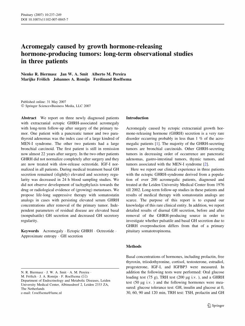
Acromegaly caused by growth hormone-releasinghormone-producing tumors: long-term observational studiesin three patients
Nienke R. Biermasz Æ Jan W. A. Smit Æ Alberto M. Pereira ÆMarijke Frolich Æ Johannes A. Romijn Æ Ferdinand Roelfsema
Published online: 31 May 2007
� Springer Science+Business Media, LLC 2007
Abstract We report on three newly diagnosed patients
with extracranial ectopic GHRH-associated acromegaly
with long-term follow-up after surgery of the primary tu-
mor. One patient with a pancreatic tumor and two para-
thyroid adenomas was the index case of a large kindred of
MEN-I syndrome. The other two patients had a large
bronchial carcinoid. The first patient is still in remission
now almost 22 years after surgery. In the two other patients
GHRH did not normalize completely after surgery and they
are now treated with slow-release octreotide. IGF-I nor-
malized in all patients. During medical treatment basal GH
secretion remained (slightly) elevated and secretory regu-
larity was decreased in 24 h blood sampling studies. We
did not observe development of tachyphylaxis towards the
drug or radiological evidence of (growing) metastases. We
propose life-long suppressive therapy with somatostatin
analogs in cases with persisting elevated serum GHRH
concentrations after removal of the primary tumor. Inde-
pendent parameters of residual disease are elevated basal
(nonpulsatile) GH secretion and decreased GH secretory
regularity.
Keywords Acromegaly � Ectopic GHRH � Octreotide �Approximate entropy � GH secretion
Introduction
Acromegaly caused by ectopic extracranial growth hor-
mone-releasing hormone (GHRH) secretion is a very rare
disorder occurring probably in less than 1 % of the acro-
megalic patients [1]. The majority of the GHRH-secreting
tumors are bronchial carcinoids. Other GHRH-secreting
tumors in decreasing order of occurrence are pancreatic
adenomas, gastro-intestinal tumors, thymic tumors, and
tumors associated with the MEN-I syndrome [2].
Here we report our clinical experience in three patients
with the ectopic GHRH-syndrome derived from a popula-
tion of over 200 acromegalic patients, diagnosed and
treated at the Leiden University Medical Center from 1976
till 2002. Long-term follow-up studies in these patients and
results of medical therapy with somatostatin analogs are
scarce. The purpose of this report is to expand our
knowledge of this rare clinical entity. In addition, we report
detailed results of diurnal GH secretion, before and after
removal of the GHRH-producing source in order to
investigate whether pulsatile and basal GH secretion due to
GHRH overproduction differs from that of a primary
pituitary somatotropinoma.
Methods
Basal concentrations of hormones, including prolactin, free
thyroxin, triiodothyronine, cortisol, testosterone, estradiol,
progesterone, IGF-I, and IGFBP3 were measured. In
addition the following tests were performed: Oral glucose
loading test (75 g), TRH test (200 lg i.v. ), and a GHRH
test (50 lg i.v. ) and the following hormones were mea-
sured: glucose tolerance test: GH, insulin and glucose at 0,
30, 60, 90 and 120 min, TRH test: TSH, prolactin and GH
N. R. Biermasz � J. W. A. Smit � A. M. Pereira �M. Frolich � J. A. Romijn � F. Roelfsema (&)
Department of Endocrinology and Metabolic Diseases, Leiden
University Medical Center, Albinusdreef 2, Leiden 2333 ZA,
The Netherlands
e-mail: [email protected]
123
Pituitary (2007) 10:237–249
DOI 10.1007/s11102-007-0045-7

at –15, 0, 15, 20, 30 45, 60, 90 and 120 min; GHRH test:
GH and prolactin at 0, 20, 30, 45, 60, and 90 min. For the
24-h GH secretion profile the patients were hospitalized,
and an indwelling i.v. cannula was inserted in a forearm
vein, and blood samples were withdrawn at 10-min inter-
vals. The patients were free to move around, but not to
sleep during daytime. Meals were served at 0800, 1230 and
1730 h. Lights were turned off between 2200 and 2400 h.
Assays
Plasma GH was measured with a sensitive time-resolved
fluoro-immunoassay (Wallac Oy, Turku, Finland). The
assay is specific for the 22 kDa GH. The standard was
biosynthetic recombinant human GH (Genotropin, Phar-
macia & Upjohn, Uppsala, Sweden), and was calibrated
against the WHO First International Reference Preparation
80/505 (to convert lg/l to mU/l multiply by 2.6). The limit
of detection of this assay (defined as the value 2 SD above
the mean value of the zero standard) was 0.01 mU/l
(0.0038 ng/ml). The intraassay coefficient of variation
varied between 1.6% and 8.4% in the range from 0.01 lg/l
to 18 lg/l and interassay coefficient of variation was 2.0–
9.0% in the same range.
Total IGF-I was determined by RIA (Incstar, Stillwater,
MN) after extraction and purification on ODS-silica col-
umns. The intraassay coefficient of variation was less than
11%. The detection limit was 1.5 nmol/l. Age-related
normal data were determined in the same laboratory. The
measurement of IGFBP3 was performed by RIA (Nichols
Institute Diagnostics, San Juan Capistrano, CA). The limit
of detection of this assay was 0.08 mg/l, and the interassay
coefficient variation was below 6.8%.
Deconvolution analysis
A multiparameter deconvolution technique was used to
estimate relevant measures of GH secretion from the 24-h
serum GH concentration profiles, as described previously
[3]. Initial estimates of basal GH secretion rate were cal-
culated to approximate the lowest 5% of all plasma GH
concentrations in the time series. Peak detection entailed
application of 95% statistical confidence intervals to two
thirds of all GH secretory peaks considered jointly and
individual 95% statistical confidence intervals to the
remaining one third smaller pulses, as validated in simu-
lations [4]. The following four secretory and clearance
measures of interest were estimated: (1) the number and
locations of secretory events; (2) the amplitudes of secre-
tory bursts; (3) the durations of randomly dispersed GH
secretory bursts; and (4) the endogenous single component
subject specific plasma half-life of GH. It was assumed the
GH distribution volume and half-life were time and
concentration invariant. The following parameters were
calculated: Half-duration of secretory bursts (duration of
the secretory burst at half-maximal amplitude), hormone
half-life, burst frequency, amplitude of the secretory burst
(maximal secretory rate attained within a burst), mass se-
creted per burst, basal secretion rate, pulsatile secretion rate
(product of burst frequency and mean burst mass) and total
secretion (sum of basal and pulsatile).
Approximate entropy
The univariate approximate entropy (ApEn) statistic was
developed to quantify the degree of irregularity, or disor-
derliness, of a time series [5]. Technically, ApEn quantifies
the summed logarithmic likelihood that templates (of length
m) of patterns in the data that are similar (within r), remain
similar (within the same tolerance r) on next incremental
comparison and has been formally defined elsewhere [6].
The ApEn calculation provides a single non-negative
number, which is an ensemble estimate of relative process
randomness, wherein larger ApEn values denote greater
irregularity, as observed for ACTH in Cushing’s disease,
GH in acromegaly, and PRL in prolactinomas [7–9]. In the
present analysis, we calculated ApEn with r = 20% of the
SD of the individual time-series and m = 1. This choice of
parameters affords sensitive, valid and statistically well-
replicated ApEn metrics for assessing hormone time-series
of this length. ApEn results are reported as absolute values
or as the ratio of the absolute value to that of the mean of
1,000 randomly shuffled data series. Ratio values that ap-
proach 1.0 thus denote mean empirical randomness.
Copulsatility
Copulsatility between the GHRH and GH time-series was
quantified by the hypergeometric (joint binomial) distri-
bution [10]. This program calculates the probability that
hormone pulses in time-series occur randomly. We used a
time-window of 20 min, with GHRH as leading hormone
series. Because details of the secretion characteristics of
GHRH are not well established, we estimated significant
pulses in both time-series with Cluster, which is largely
model-free [11].
Clinical findings at diagnosis and initial treatment
Case 1. A 50-year-old male was referred in 1982 because
of recurrent kidney stones, hypercalciuria (24 h urinary
calcium excretion between 14.5 mmol and 16.3 mmol,
normal upper value 6 mmol/24 h) and hypercalcaemia
(serum Ca between 2.86 mmol/l and 2.95 mmol/l, normal
values between 2.25 mmol/l and 2.60 mmol/l). The
238 Pituitary (2007) 10:237–249
123

referring internist suspected the patient of having mild
acromegaly, because of the coarse facial features. The
diagnosis primary hyperparathyroidism was confirmed and
the patient underwent parathyroid surgery, and two large
adenomas were removed, after which the patient became
normocalcaemic until now. The patient had noted increase
in size of his feet and hands since several years, but
otherwise he had no complaints. Glucose loading decreased
GH from 12 mU/l to below 0.5 mU/l (normal value during
glucose suppression is below 2.5 mU/l with RIA). Intra-
venous administration of 200 lg TRH, however, increased
GH from 3.5 mU/l to 58.0 mU/l and PRL increased from
7.0 lg/l to 13 lg/l. CT scanning of the pituitary gland with
the first generation CT-scanner (in 1983) did not show
abnormalities, and CT scanning of the thorax and abdomen
also failed to show the presence of a tumor. Because of
progressive complaints of fatigue after cure for hyper-
parathyroidism and strong clinical suspicion of acromeg-
aly, the patient underwent transsphenoidal pituitary
exploration. At surgery a small suspect lesion was removed
with part of the surrounding pituitary gland tissue. Histol-
ogy of the lesion was compatible with somatotrope
hyperplasia. After the patient had recovered from surgery,
CT, MRI and arteriographic studies were repeated, and
serum samples were sent to St. Bartholomew’s Hospital,
London, UK (Dr L.H. Rees) for GHRH measurement. The
abdominal CT-scan showed a 4 cm mass in the middle
section of the pancreas (Fig. 1) and the mass was also
visible with selective arteriography of the superior mes-
enterial artery (Fig. 1). The fasting GHRH concentration
amounted to 3,810 pg/ml (normal range 10–60 pg/ml).
GHRH concentration in the arterial supply to the tumor
was 12,470 pg/ml, and in the venous tumor outflow
31,120 pg/ml, while in the systemic venous system the
concentration was 8,900 pg/ml. After removal of the pan-
creatic tumor, peripheral GHRH concentration decreased to
normal values of 16–33 pg/ml. In retrospect, the first
abdominal CT-scan, 2 years before, already showed the
pancreatic tumor with a similar size.
The patient was the index case of a large kindred af-
fected by MEN-I syndrome. Later, a gene mutation was
revealed in exon 2 of chromosome 11q13. During long-
term follow-up GH and IGF-I concentrations remained
normal.
Six years later, in 1988, the patient developed diabetes
mellitus, initially treated with oral hypoglycemic drugs and
with insulin from 1992 onwards. The patient also had mild
bilateral nodular adrenal hyperplasia since 1988, with no
evidence of growth during the last recent 10 years. No
excess of adrenal (cortex and medulla) hormones or pre-
cursors was demonstrable during follow-up. During the last
3 years the plasma level of pancreatic polypeptide (PP)
increased to 350 nmol/l (normal < 100 nmol/l). Repeat CT
and MRI scanning of the upper abdomen failed to reveal
the presence of a pancreatic tumor thus far.
Random serum GH during follow-up ranged from
0.16 mU/l to 1.81 mU/l and IGF-I ranged from 12 nmol/l
to 17 nmol/l from 1994 till now (see Fig. 2). All these
values are perfectly normal for his age and gender. The last
serum GHRH measurement in 2006 was normal with
32 pg/ml.
Case 2. A 27-year-old acromegalic female patient was
referred in 1993 to our center for octreotide treatment. She
had a 5-year history of hyperhydrosis, fatigue, paraesthe-
sias, and acral enlargement. After delivery of a healthy
daughter in 1992 she had persisting amenorrhea and
galactorrhea. Physical examination revealed mild, although
characteristic features of acromegaly. Serum GH concen-
tration was elevated at 99 mU/l and decreased insuffi-
ciently to 55 mU/l following oral glucose administration.
IGF-I concentration amounted to 86 nmol/l (normal upper
level for her age 32 nmol/l) Thyroid and adrenal functions
were normal, but prolactin concentration was elevated to
20 lg/l (normal upper limit for females 12 lg/l). Other
investigations performed (TRH test, GHRH test, and the
i.v. octreotide test) are summarized in Fig. 3. TRH bolus
Fig. 1 Upper panel shows the abdominal CT at the level of the
pancreas and the lower panel the selective arteriography of the
superior mesenterial artery
Pituitary (2007) 10:237–249 239
123

injection (200 lg iv) caused a ~8-fold increase of GH and a
3.5-fold increase of PRL. Intravenous injection of 50 lg
GHRH (1-40) caused a decrease of GH while PRL con-
centrations remained unchanged. Octreotide (50 lg i.v.)
caused a 98% inhibition of serum GH but no effect on PRL.
MRI scanning of the pituitary gland showed global
enlargement without signs of an adenoma (Fig. 4). Further
investigations revealed a large tumor in the right lower lobe
of the lung (Fig. 5). The tumor and the pituitary gland were
positive on 111In-labeled-octreotide scintigraphy (Fig. 6).
The plasma GHRH concentration was increased 50-fold by
2,519 pg/ml (normal values <50 pg/ml).
Under the diagnosis of ectopic GHRH-producing lung
tumor (carcinoid) the patient underwent thoracotomy and
the tumor was resected completely. The plasma GHRH
disappearance profile after removal of the tumor is shown
in Fig. 7, but the GHRH concentration did not normalize.
GHRH concentrations in simultaneously withdrawn blood
samples from the arterial supply to the tumor, venous tu-
mor outflow and the systemic venous system were 1,890,
2,180, and 1,680 pg/ml, respectively.
Two weeks after surgery, endocrine investigations re-
vealed a persisting paradoxical increase (9-fold) of GH to
TRH, insufficient GH suppression after oral glucose load-
ing (52–16 mU/l) and a GH increase after GHRH injection.
Octreotide caused a 98% decrease of GH from 41 mU/l to
0.9 mU/l (see Fig. 3). Treatment with octreotide was
started in 1994, about 8 months after surgery, because of
persisting elevated GHRH concentration (between 360 pg/
Case 1
0 5 10 15 20 25
L/U
mH
G
0
5
10
15
20
L/lo
mnI-
FGI
0
5
10
15
20
25
30
Years
Fig. 2 Follow-up of GH and IGF-I concentration in patient 1 after
removal of the pancreatic source of GHRH. Fasting serum GH
concentrations are depicted as circles, and IGF-I as triangles
Case 2
0 20 40 60 0 20 40 60
0 20 40 60
L/U
mH
G
0
200
400
600
800
1000
1200
1400
LRP
µL /
g
0
50
100
150
200
TRH testCase 3
L/U
mH
G
0
100
200
300
400
500
600
LRP
µL/
g
0
10
20
30
40
50
0 20 40 60 80 100 120
L/U
mH
G
0
50
100
150
200
LRP
µL/
g
0
20
40
60
80
100
L/U
mH
G
0
5
10
15
20
Time (min)
0 30 60 90 120 150 180 0 30 60 90 120 150 180
L/U
mH
G
0.1
1
10
100
1000
LRP
µL/
g
0
20
40
60
80
100
Time (min)
L/U
mH
G
0.01
0.1
1
10
100
GHRH test
Octreotide test
Fig. 3 Dynamic GH (circles)
and PRL (triangles) tests in two
patients with a GHRH-secreting
lung carcinoid before (closed
symbols) and after surgery
(open symbols). Note the GH
increase after TRH (200 lg)
administration in both patients,
the decrease in serum GH
concentration in patient 2 after
GHRH (50 lg), and the
moderate GH decrease after i.v.
octreotide (50 lg)
240 Pituitary (2007) 10:237–249
123

ml and 488 pg/ml), increased GH( 31–42 mU/l), insuffi-
cient suppression by glucose loading (minimum GH
concentration 9.13 mU/l), an increased IGF-I concentration
( 40–43 nmol/l), and lesions in the liver on repeat CT
scans, suspect for metastases (one lesion in segments 2 and
8, and two lesions in segment 7).
Case 3. A 27-year-old male was referred in 1997 to our
center. He had a 9-year history of hyperhydrosis, acral
enlargement and headaches. Examination revealed moderately
advanced features of acromegaly. The circulating GH con-
centration was elevated at 110 mU/l and decreased insuffi-
ciently to 52 mU/l following glucose administration, and IGF-I
was elevated to 63 nmol/l (normal upper level at this age
32 nmol/l). Thyroid and adrenal functions were normal, but
prolactin concentration was elevated to 21 lg/l (normal upper
limit for males 6 lg/l). Other investigations performed (TRH
test and the i.v. octreotide test) are summarized in Fig. 3. TRH
Fig. 4 MRI scans of the
pituitary gland of patient 2 (left
panel) and of patient 3 (right
panel) before treatment
Fig. 5 Chest X-rays of patient
2 (left panel) and patient 3 (right
panel), showing the large
bronchial carcinoid
Fig. 6 Octreoscans of the patients 2 and 3. In the female patient (left
panel) the tumor is seen in the right lower lobe of the lung and also
the positive staining of the pituitary gland. The male patient (right
panel) had a large tumor in the left lung, without pituitary staining
Time (hours)0 20 40 60 80
lm/gp
HR
HG
101
102
103
104
105
Fig. 7 Decrease in serum GHRH concentration in two patients
before and immediately following removal of the lung tumor. Patient
2 is shown by triangles and patient 3 by circles. Note that the GHRH
concentration is shown on a logarithmic scale. The GHRH
concentration did not normalize in the patients after 2 and 3 days,
respectively. Upper normal GHRH is 50 pg/ml
Pituitary (2007) 10:237–249 241
123

bolus injection (200 lg i.v.) caused a ~8-fold increase of GH
and a 2-fold increase of PRL. Octreotide (50 lg i.v) caused a
94% inhibition of GH (Fig. 3). MRI scanning of the pituitary
gland showed a macroadenoma II AE (Hardy classification,
modified by Wilson [12, 13] see Fig. 4). He was treated for
6 months with sc octreotide (100 lg tid). The size of the ade-
noma decreased slightly, but because of the moderate clinical
response to medical treatment and failure of normalization of
GH and IGF-I, pituitary surgery was advised. One day before
surgery a chest X-ray was taken, showing a large parahilar
lesion (Fig. 5). CT scanning of this lesion was classified by the
consultant pulmonologist and radiologist as a bronchial cyst
and apparently unrelated to acromegaly. During pituitary sur-
gery the adenoma was removed completely, but the neurosur-
geon remarked that the consistency of the adenoma was firmer
than normal. Since we suspected that the lung tumor was
potentially a GHRH-producing source, postoperative investi-
gations were focused on this possibility. After surgery, MRI
scanning of the pituitary region did not show residual tumor,
GH concentrations decreased considerably and PRL concen-
trations became normal. GH dynamic tests did not completely
normalize: GH increased after TRH injection from 4.63 mU/l
to 22.1 mU/l and after glucose loading GH decreased from
4.33 mU/l to 2.56 mU/l (normal value below 1 mU/l). During
an i.v. octreotide test GH decreased from 6.21 mU/l to
0.85 mU/l, but treatment with this drug was not reinstituted. On111In-labelled-octreotide scanning the tumor was positive (see
Fig. 6).
Under the diagnosis of ectopic GHRH-producing lung
tumor (carcinoid) the patient underwent thoracotomy and
the tumor was removed completely. GHRH concentrations
in the arterial supply to the tumor were 48,290 pg/ml, in
the venous outflow 94,000 pg/ml and in the systemic ve-
nous system 49,000 pg/ml. After removal of the tumor
GHRH concentrations remained slightly elevated, as
shown in Fig. 7.
Histopathological studies
Pituitary gland
The removed part of the anterior pituitary gland of patient
1 consisted of hyperplastic cells, immunostaining posi-
tively for GH. The removed tissue of the third patient
consisted of a mixture of hyperplasia and adenoma for-
mation. The cells stained positively for both GH and PRL.
GHRH-producing tumors
Patient 1. The pancreatic tumor had a diameter of 5 cm.
Amorphous material was present between the cells, stain-
ing as amyloid. On electronmicroscopy, the cells contained
neurosecretory granules with a diameter between 100 nm
and 200 nm. The tumor stained positively, but sparsely for
somatostatin, insulin and glucagon and negatively for
cytokeratine, vimentine, neurofilaments, desmine and GH.
In the removed part of the pancreas three additional small
adenomas with identical staining characteristics were
present.
Patient 2. The diameter of the removed lung tumor was
5 cm, and contained centrally calcified material. The cells
were layered in nests, slightly polymorphic, but without
mitotic figures. The tumor cells stained positively for
keratine, vimentin, synaptophysin, SCCL (N-CAM), leu 7,
and chromogranin and negatively for calcitonin, GH,
pancreatic polypeptide, insulin, prolactin, somatostatin,
gastrin, ACTH, CEA, and neurofilaments.
Patient 3. The dimensions of the tumor were
8 · 7·7 cm3. The tumor showed clear proliferation of
neuroendocrine cells with three mitotic figures per high
power field, staining positively for NSE, CD56, and syn-
aptophysin and negatively for keratine, chromogranin,
serotonin, somatostatin, prolactin, insulin, glucagons, gas-
trin, ACTH, GH, and insulin.
Somatostatin analog therapy
After removal of the lung carcinoid, patients 2 and 3 re-
ceived long-term treatment with octreotide, because GHRH
was not normalized. Patient 2 received the medication via
chronic sc infusion (300 lg/24 h) till the end of 1998. Her
complaints quickly disappeared and the menstrual cycle
was restored. GH and IGF-I concentrations normalized and
are detailed in the left panel of Fig. 9. The size of the
pituitary gland decreased markedly (Fig. 8). PRL concen-
tration also normalized from 15.2 ± 0.2 lg/l to
6.8 ± 0.2 lg/l. From 1999 onwards the medication was
changed to octreotide long-acting repeatable (Sandostatin
LAR), 20 mg in 4-weekly i.m. injections. Growth hormone
and IGF-I concentrations remained unchanged (see Fig. 9).
During chronic treatment with the short-acting octreotide
formulation treatment was withheld several times for
GHRH measurement. Invariably, the concentration was
(slightly) elevated (range 116–363 ng/ml) so that in com-
bination with the radiological suspicion for liver metasta-
ses, treatment is continued until now. During treatment
with octreotide long-acting repeatable (Sandostatin LAR)
GHRH concentration ranged from 63 ng/ml to 108 ng/ml.
Repeat CT scanning of the liver during the successive years
demonstrated the stabilization of size and number of le-
sions until now.
After removal of the carcinoid tumor in patient 3,
GHRH, GH, and IGF-I concentrations remained elevated,
and therefore he was also treated with the long-acting
repeatable octreotide (20 mg/4 weeks). GH and IGF-I
concentrations are shown in Fig. 9, right panel, showing
242 Pituitary (2007) 10:237–249
123

clinically normal values. Repeat investigations with CT,111In-labeled octreotide and 131I-MIBG however, did not
reveal suspect (liver) metastases. At the end of 2001,
GHRH was elevated to 1,725 ng/ml, so that thereafter the
dose of Sandostatin was increased to 30 mg at 4-weekly
intervals, which normalized IGF-I concentrations. Last
year the patient stopped medication. Subsequently, IGF-I
levels increased, as shown in Fig. 9. GHRH concentration
became larger than 2,000 pg/ml. Detailed localizing stud-
ies with octreotide and CT revealed a small metastasis in
the superior anterior mediastinum for which surgery is
scheduled.
GH secretory profiles
Detailed GH secretory profiles were obtained before re-
moval of the GHRH-producing bronchial carcinoids
(Fig. 10). The secretory patterns were irregular, showing
increased burst frequency and increased basal concentra-
tions. The GH secretory parameters as estimated by mul-
tiparameter deconvolution are listed in Table 1 with
normal values obtained in healthy adults of comparable
age. The distinct and persisting abnormality in both pa-
tients after removal of the carcinoid and while on octreo-
tide treatment was the increased basal (nonpulsatile) GH
secretion.
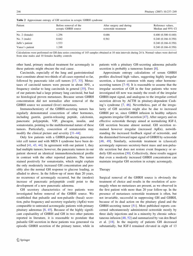
In addition, the secretory regularity was quantified with
the approximate entropy statistic, ApEn. In patient 2, ApEn
was 1.256 before removal of the carcinoid, and after sur-
gery and under somatostatin analog treatment ApEn was
still increased to 0.686 (median normal for women 0.400,
95% confidence interval 0.300–0.440). In patient 3 ApEn
also remained abnormal: preoperative 1.256 and after
surgery 0.687, median normal for males 0.240, 95% con-
fidence interval 0.160–0.350 (see Table 2). In addition, the
serum GH profiles of two patients reported in literature
were digitized and analyzed in a similar way [14, 15]. The
results of these analyses are also displayed in Table 1. In
these male patients basal GH secretion was much higher
than in our healthy controls and pulsatile secretion was
augmented via increased pulse frequency and pulse
amplitude. ApEn of GH secretion was 1.533 in Jaffe’s
patient and 1.248 in the patient reported by Vance (in-
creased SD scores by 8- and 6-fold, respectively). ApEn for
Fig. 8 MRI of the pituitary gland of patient 2 during therapy with
octreotide. These pictures were taken after 12 months treatment with
300 lg octreotide given as a continuous subcutaneous infusion
Case 2
Time (months)
0 20 40 60 80 100 120 140 160
L/U
mH
G
0
10
20
30
40
50
60
L/lo
mnI-
FG I
0
10
20
30
40
50
60Case 3
Time (months)
0 20 40 60 80 100
L/U
mH
G
0
10
20
30
40
50
60
L/lo
mnI-
FG I
0
10
20
30
40
50
60
Fig. 9 GH (circles) and IGF-I (triangles) concentrations during long-
term treatment with octreotide. Patient 3 received only the long-acting
repeatable form, but patient 2 was treated initially with chronic sc
octreotide infusion. The time of change into the slow-release
formulation is indicated by the arrow. Normal values for IGF-I for
this age: <32 nmol/l. Normal value for random GH < 5 mU/l
Pituitary (2007) 10:237–249 243
123

the serum GHRH-time series were 1.759 and 1.223,
respectively. Copulsatility of the GH and GHRH hormone
series was highly significant in both patients (P < 0.0001).
Discussion
In this study we described in detail the clinical and bio-
chemical characteristics of three patients with ectopic
extracranial GHRH secretion diagnosed in a series of about
200 acromegalics investigated and treated in our center
during the last 25 years. The incidence in the present series
agrees with that mentioned in literature [1, 16]. Faglia
summarized the clinical findings of 39 reported acromegalic
patients with proven ectopic GHRH secretion [17]. Subse-
quently, van den Bruel and colleagues [18] mentioned 52
reported patients in 1999, including their own patient and
since then 14 other patients have been reported, bringing the
total number reported to 66 patients [19–32]. From a con-
servative estimation of the total number of newly diagnosed
acromegalic patients in Europe, Japan and the USA, it is
evident that most patients with ectopic GHRH syndrome are
either not reported or remain undiagnosed.
The criteria for demonstration of ectopic extracranial
GHRH-induced acromegaly are summarized by Losa and
von Werder [2], and include the presence of high circulating
concentration GHRH by specific radioimmunoassays, the
presence of GHRH in the tumor, the presence of mRNA for
GHRH by in situ hybridization and/or a significant arterio-
venous gradient across the ectopic source. The second
requirement to be fulfilled is the reversibility of acromegaly
after complete removal of the ectopic-hormone producing
tumor. All our patients met at least two criteria, although the
tumors were not investigated for the presence of GHRH.
The clinical symptomatology in ectopic GHRH-induced
acromegaly is not different from that of the primary
pituitary adenomatous form. However, symptoms due to
the underlying neoplasm or cosecretion of other sub-
stances by the tumor might suggest the ectopic origin of
acromegaly [33]. Specific dynamical tests for GH excess
do not allow classification with certainty in either cate-
gory, although most patients with ectopic GHRH syn-
drome exhibit a paradoxical increase of GH after TRH and
glucose (i.e. >50%) and a blunted GH rise (<100%) after
exogenous GHRH injection [33]. In addition, most pa-
tients also exhibit a moderate increase in serum prolactin
concentration, which regresses after removal of the ectopic
GHRH source, supporting the notion that GHRH is di-
rectly responsible for the hyperprolactinemia, a finding
also present in hGHRH-transgenic mice [34]. The patients
reported here all exhibited GH increase after TRH, and
two had mild hyperprolactinemia. After surgery, hyperp-
rolactinemia normalized, but GH still increased after TRH
administration, suggesting that GHRH was still being
produced by tumor remnants or metastases in two patients
(nos. 2 and 3).
case 2 preoperative
9 12 15 18 21 24 3 6 9)
L/U
m(H
G0
50
100
150
200
250case 2 postoperative
Time
9 12 15 18 21 24 3 6 9
)L/
Um(
HG
0
5
10
15
20
25
case 3 preoperative
Time
9 12 15 18 21 24 3 6 9
)L/
Um(
HG
0
3
6
9
12
15case 3 postoperative
Time
9 12 15 18 21 24 3 6 9
)L/
Um(
HG
0
3
6
9
12
15
Fig. 10 Serum GH
concentrations obtained by
10 min blood sampling for 24 h.
Patient 2 was studied before
therapy and after surgical
removal of the lung tumor. Note
that GH concentration
decreased more than 10-fold
and that the secretion pattern
became more regular, but basal
GH concentration remained
slightly elevated. The left lower
panel represent the profile of
patient 3 after pituitary surgery,
but before removal of the
carcinoid tumor. Nadir values
were clearly increased. After
thoracic surgery and under
octreotide treatment GH
secretion pattern visually
normalized
244 Pituitary (2007) 10:237–249
123

The most frequent source of ectopic GHRH is the
bronchial carcinoid, followed by pancreatic islet tumors
[17], as we also found in the present evaluation of our
patient series. The pancreatic tumor in the first patient was
found by CT-scanning and MRI. The large bronchial car-
cinoids were easily seen by chest X-ray examination, and
showed uptake of radio-labeled octreotide, thus demon-
strating in vivo the presence of somatostatin receptors.
Although positive somatostatin receptor-scintigraphy is
usually found in carcinoids in general, its demonstration in
ectopic GHRH tumors is limited. Nevertheless, this
examination could be particularly useful in the diagnostic
phase of patients with suspected, but not yet proven,
ectopic GHRH secretion.
Histological investigation of surgically removed pitui-
tary tissue revealed pituitary hyperplasia in most cases (i.e.
with an intact reticulin fiber network), but in other patients
adenomatous transformation can be found [35]. Indeed, the
histological findings in hGHRH transgenic mice resemble
those in the human: in young animals diffuse hyperplasia
was found, while those of older mice showed adenomas
[34, 36]. The pathological changes in the pituitary gland
are the result of GHRH per se and not of GH, since
hyperplasia and tumor formation still occur in the absence
of GH signaling [37]. A hypothalamic GHRH-producing
tumor (gangliocytoma) is generally associated with a
pituitary adenoma rather than with pituitary hyperplasia
and is attributed to a much higher (assumed) local (pitui-
tary) concentration of GHRH or the presence of other
growth stimulating factors [38]. In addition, the combina-
tion of an intrasellar GHRH-containing gangliocytoma and
a somatotropinoma has been described in rare patients [39,
40]. In two of our patients pituitary histology was avail-
able: one patient (no. 1) showed hyperplasia in the absence
of an enlarged gland, while histology in the other patient
(no. 3) showed a mixture of hyperplasia and adenomatous
transformation.
Neuroimaging studies of the pituitary gland are variable:
in about half of the patients no tumor or a slight enlarge-
ment of the sella is detected, while in other patients in-
trasellar adenomas or macroadenomas with suprasellar
extension are found [18]. The CT-scan of patient 1 was
normal and MRI scanning in patient 2 showed diffuse
enlargement of the pituitary gland without a clear adenoma
and with diffuse uptake of gadolinium DTPA. In patient 3
asymmetric enlargement was present, suggestive of a
macroadenoma, but uptake of gadolinium DTPA through-
out the whole pituitary gland was diffuse, illustrating the
variability of imaging results. Although the pituitary MRI
studies in the ectopic GHRH syndrome are non-specific, in
the absence of a clear adenoma, ectopic GHRH secretion
should be considered and appropriate investigations in-
stalled to prevent unnecessary pituitary surgery. On theTa
ble
1D
eco
nv
olu
tio
no
fth
e2
4h
ou
rse
rum
GH
pro
file
sin
pat
ien
tsw
ith
ecto
pic
GH
RH
syn
dro
me
and
con
tro
ls
Pat
ien
t2
bef
ore
surg
.
Pat
ien
t2
afte
r
surg
.
Fem
ale
con
tro
lsP
atie
nt
3b
efo
re
surg
.
Pat
ien
t3
afte
r
surg
.
Mal
eco
ntr
ols
Jaff
e’s
pat
ien
t
Van
ce’s
pat
ien
t
Pu
lse
freq
uen
cy(n
o/2
4h
)3
01
61
7(1
4–
21
)3
02
41
2(7
–1
4)
30
21
Hal
f-li
fe(m
in)
15
.51
5.9
12
.9(1
2.0
–1
5.5
)1
5.6
15
.71
7.5
(15
.2–
19
.7)
24
.61
6.2
Pu
lse
hal
f-d
ura
tio
n(m
in)
20
.93
2.9
27
.7(2
2.7
–2
9.8
)2
5.3
23
.22
5.3
(19
.5–
34
.4)
30
.54
1.3
Pu
lse
hei
gh
t(m
U/l
/min
)4
.92
0.4
42
0.3
87
(0.1
98
–
0.8
81
)
0.2
27
0.1
60
0.1
41
(0.0
87
–
0.4
22
)
6.0
1.8
7
Pu
lse
mas
s(m
U/l
)1
01
14
.41
0.2
(7.3
–1
8.5
)5
.66
3.6
84
.44
(2.4
2–
9.5
5)
18
17
6.5
Bas
alse
cret
ion
(mU
/l/2
4h
)2
,26
13
0.2
9.1
(5.5
–1
7.6
)1
8.0
14
.93
.4(1
.4–
5.3
)3
,90
04
,60
Pu
lsat
ile
secr
etio
n(m
U/l
/
24
h)
3,0
47
23
01
73
(12
2–
31
2)
17
08
84
3.5
(17
.8–
10
3)
5,4
00
1,6
00
To
tal
secr
etio
n(m
U/l
/24
h)
5,3
09
26
01
82
(13
2–
32
5)
18
81
03
47
(19
.7–
10
7)
9,3
00
2,0
60
Blo
od
sam
ple
sw
ere
tak
enat
10
-min
inte
rval
sfo
r2
4-h
and
anal
yze
db
ym
ult
ipar
amet
erd
eco
nv
olu
tio
n.
Th
efe
mal
ep
atie
nt
(no
.2
)w
asst
ud
ied
bef
ore
surg
ical
rem
ov
alo
fth
eG
HR
H-s
ecre
tin
g
bro
nch
us
carc
ino
idan
dre
pea
tsa
mp
lin
gst
ud
yw
asd
on
eaf
ter
tho
raci
csu
rger
yu
nd
ero
ctre
oti
de
LA
R.
Th
em
ale
pat
ien
t(n
o.
3)
was
stu
die
dfi
rst
afte
rad
eno
mec
tom
yo
fth
ep
itu
itar
ytu
mo
r,b
ut
bef
ore
tho
raci
csu
rger
y.
Th
ese
con
dsa
mp
lin
gst
ud
yw
asp
erfo
rmed
afte
rre
mo
val
of
the
bro
nch
ial
carc
ino
idd
uri
ng
oct
reo
tid
e-L
AR
trea
tmen
t.T
he
seru
mp
rofi
les
of
the
pat
ien
tsre
po
rted
in
lite
ratu
rew
ere
dig
itiz
edan
dd
eco
nv
olu
ted
wit
hth
eas
say
pre
cisi
on
acco
rdin
gto
the
auth
ors
.T
he
GH
dat
aw
ere
sub
seq
uen
tly
tran
sfo
rmed
fro
mlg
-mas
su
nit
sin
tom
Uu
sin
gth
eco
nv
ersi
on
fact
or
2.0
.R
efer
ence
val
ues
wer
eo
bta
ined
inn
ine
mal
esan
d1
0fe
mal
esh
ealt
hy
con
tro
ls.
Val
ues
sho
wn
are
med
ian
san
d9
5%
con
fid
ence
inte
rval
sb
etw
een
bra
cket
s
Pituitary (2007) 10:237–249 245
123
other hand, primary medical treatment for acromegaly in
these patients might obscure the real cause.
Carcinoids, especially of the lung and gastrointestinal
tract constitute about two-thirds of all cases reported so far,
followed by pancreatic islet cell tumors [17, 33]. Metas-
tases of carcinoid tumors were present in about 30%, a
frequency similar to lung carcinoids in general [35]. Two
of our patients had a large primary lung carcinoid, but had
no histological proven metastases. Because serum GHRH
concentration did not normalize after removal of the
GHRH source we assumed (liver) metastases.
Immunochemistry of the GHRH-secreting tumors has
regularly demonstrated cosecretion of other hormones,
including gastrin, gastrin-releasing peptide, calcitonin,
pancreatic polypeptide, VIP, glucagon, insulin, and
somatostatin, pointing to the multihormonal nature of these
tumors. Particularly, cosecretion of somatostatin may
modify the clinical picture and severity [31–44].
Only few patients with a GHRH-producing pancreatic
islet cell tumor and with MEN I syndrome have been de-
scribed [41, 43, 44]. In agreement with our patient 1, they
had multiple tumors; however, the pancreatic tumors in our
patient showed an identical immunohistochemical profile
in contrast with the other reported patients. The tumor
stained positively for somatostatin, which might explain
the only moderately increased GH concentration and pos-
sibly also the normal GH response to glucose loading, as
alluded to above. In the follow-up of more than 20 years,
no recurrence of acromegaly occurred, but the (modest)
increase of pancreatic polypeptide could point to the
development of a new pancreatic adenoma.
GH secretory characteristics of two patients were
investigated before removal of the GHRH source. We
established that pulsatile and non-pulsatile (basal) secre-
tion, pulse frequency and secretory regularity (ApEn) were
comparable to untreated acromegalic patients with primary
pituitary adenomas [8, 45]. Because of the highly signifi-
cant copulsatility of GHRH and GH in two other patients
reported in literature, it is reasonable to postulate that
pulsatile GH secretion in these patients was generated via
episodic GHRH secretion of the primary tumor, while in
patients with a pituitary GH-secreting adenoma pulsatile
secretion is probably a tumorous feature [8].
Approximate entropy calculations of serum GHRH
profiles disclosed high values, suggesting highly irregular
secretion, a feature common with many other hormone-
secreting tumors [7–9]. It is reasonable to assume that the
irregular secretion of GH in the four patients who were
investigated till now was mainly the result of the irregular
GHRH-input signal, and analogous to the irregular cortisol
secretion driven by ACTH in pituitary-dependent Cush-
ing’s syndrome [7, 46]. Nevertheless, part of the irregu-
larity of GH secretion might also be due to increased
GHRH per se, since GHRH infusion in healthy subjects
augments irregular GH secretion [47]. After surgery and on
effective octreotide therapy aimed at normalizing IGF-I,
GH secretion became almost normal. GH secretion re-
mained however irregular (increased ApEn), notwith-
standing the increased feedback signal of octreotide, and
the diminished forward input signal of GHRH by octreotide
[48, 49]. Similarly, octreotide treatment in (classical)
acromegaly represses secretory-burst mass and non-pulsa-
tile secretion but does not restore event frequency or or-
derly GH secretion [50]. Collectively, these results suggest
that even a modestly increased GHRH concentration can
maintain irregular GH secretion in ectopic acromegaly.
Therapy
Surgical removal of the GHRH source is obviously the
treatment of choice and results in the resolution of acro-
megaly when no metastases are present, as we observed in
the first patient with more than 20 year follow-up. In the
presence of metastases octreotide treatment is often, but
not invariable, successful in suppressing GH and GHRH,
because of its dual action on the pituitary gland and the
GHRH-secreting tumor [51]. Most published reports con-
cerned subcutaneously administered octreotide mostly by
three daily injections and in a minority by chronic subcu-
taneous infusion [48, 52] and summarized by van den Bruel
et al. [18]. In the majority of patients GH decreased
substantially, but IGF-I remained elevated in eight of 13
Table 2 Approximate entropy of GH secretion in ectopic GHRH syndrome
Patient Before removal of the
ectopic GHRH source
After surgery and during
octreotide treatment
Reference values,
Median and 95% CI
No. 2 (female) 1.256 0.686 0.400 (0.300–0.440)
No. 3 (male) 0.842 0.561 0.240 (0.166–0.350)
Jaffe’s patient 1.533 0.240 (0.166–0.350)
Vance’s patient 1.248 0.240 (0.166–0.350)
Calculations were performed on GH data series consisting of 145 samples obtained at 10 min intervals during 24 h. Normal values were derived
from nine males and 10 females healthy controls
246 Pituitary (2007) 10:237–249
123

patients in whom IGF-I was measured. The serum con-
centration of GHRH generally remained elevated [2], as we
found in patient 3. Publications of treatment results with
the slow release formulation of octreotide and lanreotide
are scarce and follow-up short-term. We could find only
four cases in literature: three patients were treated with
lanreotide and one with octreotide-long acting repeatable
(octreotide LAR) [19, 21, 24, 25]. In the present study we
reported the long-term effect of octreotide-LAR treatment
in two patients, which resulted in suppressed GH secretion
and normalized IGF-I concentration in both patients, but
with still slightly elevated GHRH concentrations, as long
as the patients continued suppressive therapy. The latter
finding is indeed often mentioned in other reports as well,
as discussed above. Other treatment modalities applied in
single cases are the use of a GHRH-receptor antagonist and
chemotherapy in a patient with severely metastasized dis-
ease [15, 53].
During octreotide treatment no visible growth of
(eventual) metastases was noted for many years with high-
sensitivity liver CT scanning and CT scanning of the thorax
in our patients, which suggests that in selected cases
octreotide can inhibit growth of metastases from a carci-
noid tumor. In addition, pituitary hyperplasia, the likely
cause of the preoperative enlargement in patient 2, slowly
disappeared completely, as previously described in other
patients under medical therapy or after complete surgical
removal of the GHRH source [27, 54]. Nevertheless, in
patient 3 GHRH levels increased slowly, suggesting
growing metastasis under octreotide restraint. Recently, we
could localize a suspect lesion in the anterior mediastinum.
GHRH and its receptor are normally present in several
organs, including the ovary and testis, playing a role in the
regulation of steroidogenesis, and the pancreas [55–57]. It
has been found that some GHRH-producing carcinoids also
express GHRH-receptors, which may stimulate tumor
growth via an autocrine mechanism [22]. Particularly
interesting was the description of a single acromegalic
patient whose large pituitary adenoma co-expressed
GHRH-receptor and GHRH together with a 100-fold in-
creased plasma GHRH concentration. After pituitary ade-
nomectomy plasma GHRH normalized. It is likely that the
high local concentration of GHRH contributed to the
growth of the adenoma [58].
GHRH and its receptor are also expressed in cancers of
the breast, ovary, and endometrium and small-cell lung
cancer, and may function as a paracrine/autocrine growth
factor in regulating local IGF-I and/or IGF-II secretion.
Other studies have shown that GHRH receptor-antagonists
inhibit the growth of colorectal, prostatic, mammary, lung,
and pancreatic cancers, partly by direct suppression of IGF-
I and IGF-II production, or acting directly without influ-
encing these growth factors [59–63]. When more potent
GHRH-receptor antagonists will become available for
clinical application patients with advanced disease may
benefit from this new approach, because they respond at
best to somatostatin analogue therapy by suppressing GH
secretion and partly that of GHRH, but the tumor and
metastases will usually grow with fatal outcome.
References
1. Thorner MO, Frohman LA, Leong DA, Thominet J, Downs T,
Hellmann P, Chitwood J, Vaughan JM, Vale W (1984) Extra-
hypothalamic growth-hormone-releasing-factor (GRF) secretion
is a rare cause of acromegaly: plasma GRF levels in 177 acro-
megalic patients. J Clin Endocrinol Metab 59:846–849
2. Losa M, von Werder K (1997) Pathophysiology and clinical as-
pects of the ectopic GH-releasing hormone syndrome. Clin
Endocrinol 47:123–135
3. Veldhuis JD, Carlson ML, Johnson ML (1987) The pituitary
gland secretes in bursts: appraising the nature of glandular
secretory impulses by simultaneous multiple-parameter decon-
volution of plasma hormone concentrations. Proc Natl Acad Sci
USA 84:7686–7690
4. Friend K, Iranmanesh A, Veldhuis JD (1996) The orderliness of
growth hormone (GH) release process and the mean mass of GH
secreted per burst are highly conserved in individual men on
successive days. J Clin Endocrinol Metab 81:3746–3753
5. Pincus SM (1991) Approximate entropy as a measure of system
complexity. Proc Natl Acad Sci USA 88:2297–2301
6. Pincus SM, Goldberger AL (1994) Physiological time-series
analysis: what does regularity quantifies? Am J Physiol
266:H1643–H1656
7. Van den Berg, Pincus SM, Veldhuis JD, Frolich M, Roelfsema F
(1997) Greater disorderliness of ACTH and cortisol release
accompanies pituitary-dependent Cushing’s disease. Eur J
Endocrinol 136:394–400
8. Van den Berg G, Pincus SM, Frolich M, Veldhuis JD, Roelfsema
F (1998) Reduced disorderliness of growth hormone release in
biochemically inactive acromegaly after pituitary surgery. Eur J
Endocrinol 138:164–169
9. Groote Veldman R, van den Berg G, Pincus SM, Frolich M,
Veldhuis JD, Roelfsema F (1999) Increased episodic release and
disorderliness of prolactin secretion in both micro- and macro-
prolactinomas. Eur J Endocrinol 140:192–200
10. Veldhuis JD, Johnson ML (1991) Analysis of copulsatility of
anterior pituitary hormones. J Clin Endocrinol Metab 73:569–576
11. Veldhuis JD, Johnson ML (1986) Cluster analysis, a simple,
versatile, and robust algorithm for endocrine pulse detection. Am
J Physiol 250:E486–E493
12. Hardy J, Vezina JL (1976) Transsphenoidal neurosurgery of
intracranial neoplasm. In: Thompson RA, Green JR (eds) Advances
in neurosurgery, vol 15. Raven Press, New York, pp 261–275
13. Wilson CB (1979) Neurosurgical management of large and
invasive pituitary tumors. In: Tindall GT, Collins WF (eds)
Clinical management of pituitary disorders. Raven Press, New
York, pp 335–342
14. Vance ML, Kaiser DL, Evans WS, Furlanetto R, Vale W, Rivier
J, Thorner MO (1985) Pulsatile growth hormone secretion in
normal man during a continuous 14-hour infusion of growth
hormone releasing factor (1-40). J Clin Invest 75:1584–1590
15. Jaffe CA, DeMott-Friberg R, Frohman LA, Barkan AL (1997)
Suppression of growth hormone (GH) hypersecretion due to
ectopic GH-releasing hormone (GHRH) by a selective GHRH
antagonist. J Clin Endocrinol Metab 82:634–637
Pituitary (2007) 10:237–249 247
123

16. Penny ES, Penman E, Price J, Sopwith AM, Wass JA, Lytras N,
Besser GM (1984) Circulating growth hormone-releasing factor
concentrations in normal subjects and in patients with acromeg-
aly. Brit Med J 289:453–455
17. Faglia G, Arosio M, Bazzoni N (1992) Ectopic acromegaly.
Endocrinol Metab Clin North Am 21:575–595
18. Van den Bruel A, Fevery J, van Dorpe J, Hofland L, Bouillon R
(1999) Hormonal and volumetric long term control of a growth
hormone-releasing hormone-producing carcinoid tumor. J Clin
Endocrinol Metab 84:3162–3169
19. Jansson J-O, Svensson J, Bengtsson B-A, Frohman LA, Ahlman
H, Wangberg B, Nilsson O, Nilsson M (1998) Acromegaly and
Cushing’s syndrome due to ectopic production of GHRH and
ACTH by a thymic carcinoid tumour: in vitro responses to GHRH
and GHRP-6. Clin Endocrinol 48:243–250
20. Drange MR, Melmed S (1998) Long-acting lanreotide induces
clinical and biochemical remission of acromegaly caused by
disseminated growth hormone/releasing hormone/secreting car-
cinoid. J Clin Endocrinol Metab 83:3104–3109
21. Krassowski J, Zgliczynski W, Jeske W, Zgliczynski S (1999)
Comment on long-acting lanreotide inducing clinical and bio-
chemical remission of acromegaly caused by disseminated
GHRH secreting carcinoid. J Clin Endocrinol Metab 84:1761–
1762
22. Othman NH, Ezzat S, Kovacs K, Horvath E, Poulin E, Smyth HS,
Asa SL (2001) Growth hormone-releasing hormone(GHRH) and
GHRH receptor(GHRH-R) isoform expression in ectopic acro-
megaly. Clin Endocrinol 55:135–140
23. Furrer J, Hattenschwiler A, Komminoth P, Pfammatter T, Wiesli
P (2001) Carcinoid syndrome, acromegaly, and hypoglycaemia
due to an insulin-secreting neuroendocrine tumor of the liver. J
Clin Endocrinol Metab 86:2227–2230
24. Altstadt TJ, Azzarelli B, Bevering C, Edmuondson J, Nelson PB
(2002) Acromegaly caused by a growth hormone-releasing hor-
mone-secreting carcinoid: case report. Neurosurgery 50:1356–
1359
25. Boix E, Pico A, Pinedo R, Aranda I, Kovacs K (2002) Ectopic
growth hormone-releasing hormone secretion by thymic carci-
noid tumour. Clin Endocrinol 57:131–134
26. Lorcy Y, Perdu S, Sevray B, Cohen R (2002) Acromegaly due to
ectopic GHRH secretion by a bronchial carcinoid tumor: a case
report. Ann Endocrinol (Paris) 63:536–539
27. Bolanowski M, Schopol J, Marcianak M, Rseszutko M, Zatonska
K, Daroszewski J, Milewicz A, Malczewska J, Badowski R
(2002) Acromegaly due to GHRH-secreting large bronchial car-
cinoid. Complete recovery following tumor surgery. Exp Clin
Endocrinol Diabetes 110:188–192
28. Osella G, Orlandi F, Caraci P, Ventura M, Deandreis D, Papotti
M, Bongiovanni M, Angeli A, Terzolo M (2003) Acromegaly due
to ectopic secretion of GHRH by bronchial carcinoid in a patient
with empty sella. J Endocrinol Invest 26:163–169
29. Athanassiadi K, Exarchos D, Tsagarakis S, Bellenis I (2004)
Acromegaly caused by ectopic growth hormone-releasing hor-
mone secretion by a carcinoid bronchial tumor: a rare entity. J
Thorac Cardiovasc Surg 128:631–632
30. Agha FL, Downey P, Keeling P, Leen E, Sreenan S (2004)
Acromegaly secondary to growth hormone releasing hormone
secretion. Ir J Med Sci 173:215–216
31. Zatelli MC, Maffei P, Piccin D, Martini C, Rea F, Rubello D,
Margatti A, Culler MD, Sicolo N, degli Uberti EC (2005)
Somatostatin analogs in vitro effects in a growth hormone-
releasing-hormone-secreting bronchial carcinoid. J Clin Endo-
crinol Metab 90:2104–2109
32. Nasr C, Mason A, Mayberg M, Staugaitis SM, Asa SL (2006)
Acromegaly and somatotroph hyperplasia with adenomatous
transformation due to pituitary metastasis of a GHRH-secreting
pulmonary endocrine carcinoma. J Clin Endocrinol Metab
91:4776–4780
33. Losa M, Schopohl J, von Werder K (1993) Ectopic secretion of
growth hormone-releasing hormone in man. J Endocrinol Invest
16:69–81
34. Stefaneanu L, Kovacs K, Horvath E, Asa SL, Losinski LE, Bil-
lestrup N, Price J, Vale W (1989) Adenohypophyseal changes in
mice transgenic for human growth hormone-releasing hormone
factor: a histological, immunocytochemical, and electron micro-
scopic investigation. Endocrinology 125:2710–2718
35. Sano T, Asa SL, Kovacs K (1988) Growth hormone-releasing
hormone-producing tumors: clinical, biochemical and patholog-
ical manifestations. Endocr Rev 9:357–373
36. Asa SL, Kovacs K, Stefaneanu L, Horvarth E, Billestrup N,
Gonzales Manchon G, Vale W (1990) Pituitary mammosomato-
troph adenomas develop in old mice transgenic for growth hor-
mone-releasing hormone. Proc Soc Exp Biol Med 193:232–235
37. Kineman RD, Teixeira LT, Amargo GV, Coschigano KT, Ko-
pchik JJ, Frohman LA (2001) The effect of GHRH on somato-
trope hyperplasia and tumor formation in the presence and
absence of GH signalling. Endocrinology 142:3764–3773
38. Asa SL, Scheithauer BW, Bilbao JM, Horvath E, Ryan N, Kovacs
K, Randall RV, Laws ER Jr, Singer W, Linfoot JA (1984) A case
for hypothalamic acromegaly: a clinicopathological study of six
patients with hypothalamic gangliocytomas producing growth
hormone-releasing factor. J Clin Endocrinol Metab 58:796–803
39. Morikawa M, Tamaki N, Kokunai T, Imai Y (1997) Intrasellar
pituitary gangliocyto-adenoma presenting with acromegaly: a
case report. Neurosurgery 40:611–614
40. Luna V, Morales F, Luengo LM, Sanz A, Diaz J (2001) Pituitary
gangliocytoma-adenoma presenting with acromegaly: response to
treatment. Arch Intern Med 161:1010–1011
41. Berger G, Trouillas J, Bloch B, Sassolas G, Berger F, Partenski C,
Chayvialle J-A, Brazeau P, Claustrat B, Lesbros F, Girod C
(1984) Multihormonal carcinoid tumor of the pancreas secreting
growth hormone-releasing factor as a cause of acromegaly.
Cancer 54:2097–2108
42. Chadenas D, Pinsard D, Melliere D, Trouillas J, Zafrani ES,
Pradayrol L, Sassolas G, Li Y, Girod C, Aumaitre J (1985)
Tumeur pancreatique endocrine secretant de la somatostatine et
de la somatocrine. La Presse Med 14:2129–213443. Sano T, Yamasaki R, Saito H, Hirose T, Kudo E, Kameyama K,
Hiraishi K, Saito S, Hizawa K (1987) Growth hormone-releasing
hormone (GHRH)-secreting pancreatic tumor in a patient with
multiple endocrine neoplasia type I. Am J Surg Pathol 11:810–819
44. Ramsay JA, Kovacs K, Asa SL, Pike MJ, Thorner MO (1988)
Reversible sellar enlargement due to growth hormone-releasing
hormone production by pancreatic endocrine tumors in an acro-
megalic patient with multiple endocrine neoplasia type I syn-
drome. Cancer 62:445–450
45. Hartman ML, Pincus SM, Johnson ML, Matthews DH, Faunt
LM, Vance ML, Thorner MO, Veldhuis JD (1994) Enhanced
basal and disorderly growth hormone secretion distinguishes
acromegalic from normal pulsatile growth hormone release. J
Clin Invest 94:1277–1288
46. Roelfsema F, Pincus SM, Veldhuis JD (1998) Patients with
Cushing’s disease secrete adrenocorticotropin and cortisol jointly
more asynchronously than healthy subjects. J Clin Endocrinol
Metab 83:688–692
47. Evans WS, Anderson SM, Hull LT, Azimi PP, Bowers CY,
Veldhuis JD (2001) Continuous 24-hour intravenous infusion of
recombinant human growth hormone (GH)-releasing hormone-
(1-44)-amide augments pulsatile, entropic, and daily rhythmic
GH secretion in postmenopausal women equally in the estrogen-
withdrawn and estrogen-supplemented states. J Clin Endocrinol
Metab 86:700–712
248 Pituitary (2007) 10:237–249
123

48. Veldhuis JD, Straume M, Iranmanesh A, Mulligan T, Jaffe C,
Barkan A, Johnson ML, Pincus S (2001) Secretory process reg-
ularity monitors neuroendocrine feedback and feedforward sig-
nalling strength in humans. Am J Physiol Regul Integr Comp
Physiol 280:R721–R729
49. Veldhuis JD, Johnson ML, Veldhuis OL, Straume M, Pincus SM
(2001) Impact of pulsatility on the ensemble orderliness
(approximate entropy) of neurohormone secretion. Am J Physiol
Regul Integr Comp Physiol 281:R1975–R1985
50. Biermasz NR, Pereira AM, Frolich M, Romijn JA, Veldhuis JD,
Roelfsema F (2004) Octreotide represses secretory-burst mass
and nonpulsatile secretion but does not restore event frequency or
orderly GH secretion in acromegaly. Am J Physiol Endocrinol
Metab 286:E1–E6
51. Moller DE, Moses AC, Jones K, Thorner MO, Vance ML (1989)
Octreotide suppresses both growth hormone (GH) and GH
releasing-hormone (GHRH) in acromegaly due to ectopic GHRH
secretion. J Clin Endocrinol Metab 68:499–504
52. Levebre S, De Paepe L, Abs R, Rahier J, Selvais P, Maiter D
(1995) Subcutaneous octreotide treatment of a growth hormone-
releasing hormone-secreting bronchial carcinoid: superiority of
continuous versus intermittent administration to control hormonal
secretion. Eur J Endocrinol 133:320–324
53. Harris PE, Bouloux PM, Wass JA, Besser GM (1990) Successful
treatment by chemotherapy for acromegaly associated with ec-
topic growth hormone-releasing hormone secretion from a car-
cinoid tumour. Clin Endocrinol 32:315–321
54. Wilson DR, Hoffman AR (1986) Reduction of pituitary size by
the somatostatin analogue SMS 201-995 in a patient with an islet
cell tumour secreting growth hormone releasing factor. Acta
Endocrinol 113:23–28
55. Leung PC, Steele GL (1992) Intracellular signalling in the
gonads. Endocr Rev 13:476–498
56. Frohman LA, Down TR, Kashio Y, Brinster RL (1990) Tissue
distribution and molecular heterogeneity of human growth
hormone-releasing factor in the transgenic mouse. Endocrinology
127:2149–2156
57. Berry SA, Srivastava CH, Rubin LR, Phipps WR, Pescovitz OH
(1992) Growth hormone releasing hormone-like messenger
ribonucleic acid and immunoreactive peptide are present in hu-
man testis and placenta. J Clin Endocrinol Metab 75:281–284
58. Matsuno A, Katakami H, Sanno N, Ogino Y, Osamura RY,
Matsukura S, Shimizu N, Nagashima T (1999) Pituitary so-
matotroph adenoma producing growth hormone (GH)-releasing
hormone (GHRH) with an elevated plasma GHRH concentration:
a model case for autocrine and paracrine regulation of GH
secretion by GHRH. J Clin Endocrinol Metab 84:3241–3247
59. Kahan Z, Arencibia JM, Czernus VJ, Groot K, Kinema RD,
Robinson WR (1999) Expression of growth hormone-releasing
hormone (GH-RH) messenger ribonucleic acid, the presence of
biologically active GH-RH in human breast, endometrial, and
ovarian cancers. J Clin Endocrinol Metab 84:582–589
60. Doga M, Bonadonna S, Burattin A, Giustina A (2001) Ectopic
secretion of growth hormone-releasing hormone (GHRH) in
neuroendocrine tumors: relevant clinical aspects. Ann Oncology
12:S89–S94
61. Chatzistamou I, Schally AV, Pafiti A, Klaris H, Koutselini H
(2002) Expression of growth hormone releasing hormone in hu-
man primary endometrial carcinomas. Eur J Endocrinol 147:381–
386
62. Kiaris H, Schally AV, Varga JL, Groot K, Armatis P (1999)
Growth hormone-releasing hormone: an autocrine growth factor
for small cell lung carcinoma. Proc Natl Acad Sci USA
96:14894–14898
63. Kahan Z, Arencibia JM, Czernus VJ, Groot K, Kinema RD,
Robinson WR (1999) Expression of growth hormone-releasing
hormone (GH-RH) messenger ribonucleic acid and the presence
of biologically active GH-RH in human breast, endometrial, and
ovarian cancers. J Clin Endocrinol Metab 84:582–589
Pituitary (2007) 10:237–249 249
123