Embed Size (px)
Citation preview

8/11/2019 acousticneuroma-130914224034-phpapp02
http://slidepdf.com/reader/full/acousticneuroma-130914224034-phpapp02 1/62
y
DR Mohammed
Riyas

8/11/2019 acousticneuroma-130914224034-phpapp02
http://slidepdf.com/reader/full/acousticneuroma-130914224034-phpapp02 2/62
INTRODUCTION
An acoustic neuroma (also called vestibular schwannoma)
is a benign tumor arising from abnormally proliferative
shwann cells , which envelope the lateral portion of the
vestibular nerve in the internal auditory meatus

8/11/2019 acousticneuroma-130914224034-phpapp02
http://slidepdf.com/reader/full/acousticneuroma-130914224034-phpapp02 3/62
Little things aboutCP angle

8/11/2019 acousticneuroma-130914224034-phpapp02
http://slidepdf.com/reader/full/acousticneuroma-130914224034-phpapp02 4/62
CP angle tumors
Represents 10 of all intracranial tumors.
Fatal without treatment.
VS account for 78 of CPA tumors - mostly from
vestibular branch of VIIIth Nerve.
Variety of other tumors arise from this area like
meningioma , CN swannomas , dermoid tumors , arachnoid
cysts ,lipomas , metastatic tumors , vascular tumors.

8/11/2019 acousticneuroma-130914224034-phpapp02
http://slidepdf.com/reader/full/acousticneuroma-130914224034-phpapp02 5/62
Anatomy of CP angle
CPA Irregularly shaped potential space in the posterior
fossa of the brain .
Anteriorly posterior surface of temporal bone .
Posteriorly anterior surface of the cerebellum.
Medially cisterns of the pons & medulla and olive.
Superiorly inferior border of pons & cerebellar peduncle.
Inferiorly cerebellar tonsil.

8/11/2019 acousticneuroma-130914224034-phpapp02
http://slidepdf.com/reader/full/acousticneuroma-130914224034-phpapp02 6/62

8/11/2019 acousticneuroma-130914224034-phpapp02
http://slidepdf.com/reader/full/acousticneuroma-130914224034-phpapp02 7/62
8/11/2019 acousticneuroma-130914224034-phpapp02
http://slidepdf.com/reader/full/acousticneuroma-130914224034-phpapp02 8/62
Anatomy of CP angle
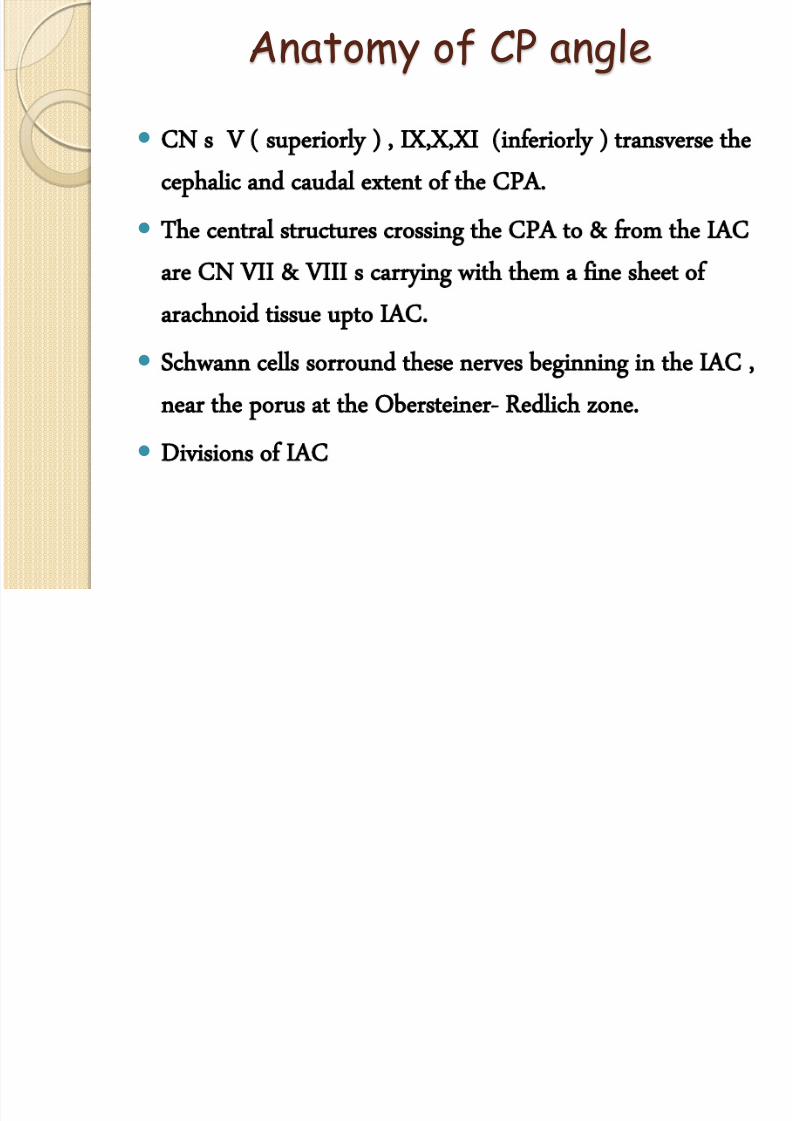
CN s V ( superiorly ) , IX,X,XI (inferiorly ) transverse the
cephalic and caudal extent of the CPA.
The central structures crossing the CPA to & from the IAC
are CN VII & VIII s carrying with them a fine sheet of
arachnoid tissue upto IAC.
Schwann cells sorround these nerves beginning in the IAC ,
near the porus at the Obersteiner- Redlich zone.
Divisions of IAC

8/11/2019 acousticneuroma-130914224034-phpapp02
http://slidepdf.com/reader/full/acousticneuroma-130914224034-phpapp02 9/62
Anatomy of CP angle
CN s V ( superiorly ) , IX,X,XI (inferiorly ) transverse the
cephalic and caudal extent of the CPA.
The central structures crossing the CPA to & from the IAC
are CN VII & VIII s carrying with them a fine sheet of
arachnoid tissue upto IAC.
Schwann cells sorround these nerves beginning in the IAC ,
near the porus at the Obersteiner- Redlich zone.
Divisions of IAC

8/11/2019 acousticneuroma-130914224034-phpapp02
http://slidepdf.com/reader/full/acousticneuroma-130914224034-phpapp02 10/62
Anatomy of CP angle
AICA is the main artery in the CPA and is the
source of the labrynthine artery .
The labrynthine artery courses via the IAC & is
an end artery for the hearing and balance organs.

8/11/2019 acousticneuroma-130914224034-phpapp02
http://slidepdf.com/reader/full/acousticneuroma-130914224034-phpapp02 11/62
Vestibular schwannoma
Nerve sheath tumors of the superior and inferior
vestibular nerves.
They arise in the medial part of the IAC or the
lateral part of the CPA and cause clinical
symptoms by displacing , distorting or
compressing adjacent structures in the CPA.

8/11/2019 acousticneuroma-130914224034-phpapp02
http://slidepdf.com/reader/full/acousticneuroma-130914224034-phpapp02 12/62
Vestibular schwannoma
Mean incidence range 9.1 tmr/yr to 13 tmr/yr( as per SB)
0.7 to 1.2 VS per lakh population/yr ( ballenger )
Types - Sporadic ( 95 ) and non sporadic ( 5 )
Age of presentation 40 to 60 yrs.
Age of presentation is less in non sporadic ( 20-30 yrs )
Mean growth rate 1.1 mm /yr.

8/11/2019 acousticneuroma-130914224034-phpapp02
http://slidepdf.com/reader/full/acousticneuroma-130914224034-phpapp02 13/62
Tumor biology
Equal frequency in sup and inf vestibular ( but recent
japanese studies suggested 85 from inf vestibular )
Arise from schwann cells within the IAC lateral to O-R
zone in the area of scarpa ganglion.
Schwannomas rarely arise from the cochlear nerve & are
rarely malignant .
8/11/2019 acousticneuroma-130914224034-phpapp02
http://slidepdf.com/reader/full/acousticneuroma-130914224034-phpapp02 14/62
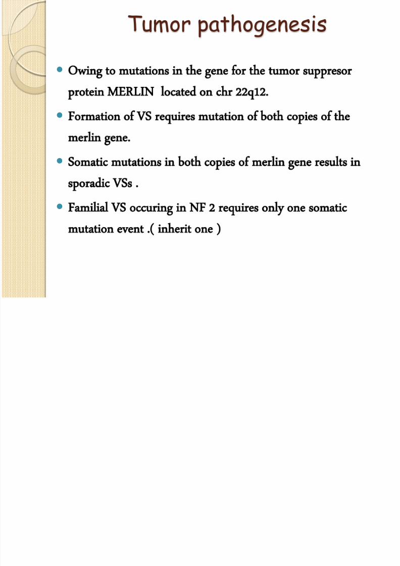
Tumor pathogenesis
Owing to mutations in the gene for the tumor suppresor
protein MERLIN located on chr 22q12.
Formation of VS requires mutation of both copies of the
merlin gene.
Somatic mutations in both copies of merlin gene results in
sporadic VSs .
Familial VS occuring in NF 2 requires only one somatic
mutation event .( inherit one )

8/11/2019 acousticneuroma-130914224034-phpapp02
http://slidepdf.com/reader/full/acousticneuroma-130914224034-phpapp02 15/62
Tumor pathogenesis
NF2 is autosomal recessive at gene level but inheritance is
autosomal dominant ( pseudodominant )
A mutation in the normal allele leads to bilateral VS by the age
of 20.
Genetic screens for the NF2 mutation have been developed and
are the basis for genetic counselling for family members of NF2
patients

8/11/2019 acousticneuroma-130914224034-phpapp02
http://slidepdf.com/reader/full/acousticneuroma-130914224034-phpapp02 16/62
Tumor pathogenesis
Biochemical factors- VS express neuregulin ,which controls
survival and proliferation of schwann cells and its receptors
erbB2 & erbB3.
FGF ,TGF B1 , PDGF & VEGF all these contribute to VS
proliferation.
VS may accelerate during pregnancy.

8/11/2019 acousticneuroma-130914224034-phpapp02
http://slidepdf.com/reader/full/acousticneuroma-130914224034-phpapp02 17/62
pathology
GROSS :
◦ VS have a smooth surface with a yellow to gray color.
◦Tumor is usually solid ,with occasional cystic components and therefore
has a firm to soft texture depending on solid to cystic components.
MICROSCOPIC :
◦Capsule 3 to 5 micrometer in thickness.
◦Two morphological tissue types Antony A & Antony B areas
◦VEROCAY bodies
◦VS sections stain S 100 immunoperoxidase.

8/11/2019 acousticneuroma-130914224034-phpapp02
http://slidepdf.com/reader/full/acousticneuroma-130914224034-phpapp02 18/62
T UMOR DEVELOPMENT
Develops in nerve sheath
Compresses rather than invading the nerve
Gradually fills all the IAC
Protrudes out of the porus
Resorption of bone sorrounding the porus

8/11/2019 acousticneuroma-130914224034-phpapp02
http://slidepdf.com/reader/full/acousticneuroma-130914224034-phpapp02 19/62
T UMOR DEVELOPMENT-extrameatally
xtrameatal expansion into the large & empty pontine cistern
Displacement and stretching of the VII & VIII th CN on the anterior
Compress cerebellum and trigeminal N
Compression & displacement of the brainstem & fourth ventricle
aspect of the tumor & of the AICA on the inferior aspect
(During this time IAM continues to become more & more widened )
which leads gradually to hydrocephalus

8/11/2019 acousticneuroma-130914224034-phpapp02
http://slidepdf.com/reader/full/acousticneuroma-130914224034-phpapp02 20/62
T UMOR DEVELOPMENT-extrameatally
umor may extent to the tentorium & can obstruct the
cochlear aqueduct
The AICA & lower cranial nerves are also displaced & become closely
Overtime , the trigeminal & abducens nerves become stretched
Adherent to the inf surface of tumor.
over the surface of the tumor and get thinned.

8/11/2019 acousticneuroma-130914224034-phpapp02
http://slidepdf.com/reader/full/acousticneuroma-130914224034-phpapp02 21/62
1. 2.
3.4.
INTRACANALICULAR CISTERNAL
COMPRESSIVE HYDROCEPHALIC

8/11/2019 acousticneuroma-130914224034-phpapp02
http://slidepdf.com/reader/full/acousticneuroma-130914224034-phpapp02 22/62
Tumor development…..
Periods of growth are intermixed b/w slow growth &
peroid of quescence.
Occasionally tumor may undergo rapid expansion owing to
cystic degeneration or hemmorhage into the tumor.
The initial intracanalicular growth effects the
vestibulocochlear nerve in the rigid IAC &
causes unilateral HL ,tinnitus and vertigo or dysequilibrium.

8/11/2019 acousticneuroma-130914224034-phpapp02
http://slidepdf.com/reader/full/acousticneuroma-130914224034-phpapp02 23/62

8/11/2019 acousticneuroma-130914224034-phpapp02
http://slidepdf.com/reader/full/acousticneuroma-130914224034-phpapp02 24/62
Stage 1
Stage 2
Stage 3

8/11/2019 acousticneuroma-130914224034-phpapp02
http://slidepdf.com/reader/full/acousticneuroma-130914224034-phpapp02 25/62

8/11/2019 acousticneuroma-130914224034-phpapp02
http://slidepdf.com/reader/full/acousticneuroma-130914224034-phpapp02 26/62
Cerebellopontine Angle:

8/11/2019 acousticneuroma-130914224034-phpapp02
http://slidepdf.com/reader/full/acousticneuroma-130914224034-phpapp02 27/62
Large Acoustic Neuroma: Tumors over 2.5 centimeters (this one is 2.6 cm) become impacted
into the brainstem and cerebellum. Complications associated with surgery and radiation are
higher. It is difficult to deliver an adequate dose of radiation to control tumor growth without
excessive dosing to the brainstem in tumors larger than this.

8/11/2019 acousticneuroma-130914224034-phpapp02
http://slidepdf.com/reader/full/acousticneuroma-130914224034-phpapp02 28/62

8/11/2019 acousticneuroma-130914224034-phpapp02
http://slidepdf.com/reader/full/acousticneuroma-130914224034-phpapp02 29/62
Symptoms & signs
Intracanalicular:
◦ Hearing loss (UL progressive ), tinnitus, vertigo
◦ Loss of speech discrimination out of propotion to HL
Cisternal:
◦ Worsened hearing and dysequilibrium
Compressive:
◦ Occasional occipital headache
◦ CN V: Midface, corneal hypesthesia
◦ CN VII : Hitzelberger’s sign, loss of taste and reduced
lacrimation on Schirmer’s test ,facial weakness ( late)
◦ CN II , IV , VI : visual acquity and diplopia

8/11/2019 acousticneuroma-130914224034-phpapp02
http://slidepdf.com/reader/full/acousticneuroma-130914224034-phpapp02 30/62
Symptoms & signs
Hydrocephalic:
◦ Fourth ventricle compressed and obstructed
◦ Headache, visual changes, altered mental status
◦ Nausea and vomiting
◦ On examination : ICP and pappiledema.
Compression of CN IX & X
◦ Dysphagia , aspiration and hoarseness
◦ Poor gag reflex and VC paralysis.
Cerebellar involvement( late )
◦ Incoordination , widely based gate , tendency to fall
owards affected side

8/11/2019 acousticneuroma-130914224034-phpapp02
http://slidepdf.com/reader/full/acousticneuroma-130914224034-phpapp02 31/62
Symptoms & signs
Brainstem involvement:• There is ataxia, weakness and numbness of arms and legs
with exaggerated tendon reflexes.

8/11/2019 acousticneuroma-130914224034-phpapp02
http://slidepdf.com/reader/full/acousticneuroma-130914224034-phpapp02 32/62
Jackler Staging System
STAGE TUMOUR SIZE
Intra canalicular Tumour confined to IAC
I (Small) <10mm
II(Medium) 11-25mm
III(Large) 25-40mm
IV(Giant) >40mm

8/11/2019 acousticneuroma-130914224034-phpapp02
http://slidepdf.com/reader/full/acousticneuroma-130914224034-phpapp02 33/62
Tumor Growth Rate

8/11/2019 acousticneuroma-130914224034-phpapp02
http://slidepdf.com/reader/full/acousticneuroma-130914224034-phpapp02 34/62
Duration of Symptoms Prior to
Diagnosis SYMPTOMS YEARS
Hearing loss 3.9
Vertigo 3.6
Tinnitus 3.4
Headache 2.2
Dysequilibrium 1.7
Trigeminal 0.9
Facial 0.6

8/11/2019 acousticneuroma-130914224034-phpapp02
http://slidepdf.com/reader/full/acousticneuroma-130914224034-phpapp02 35/62
Diagnostic evaluation
Average patient will require 4 years from theonset of symptoms to diagnosis.
Majority will present with complaints of UHL, UT,Vertigo , dysequilibrium, facial numbness ,weakness or spasm.
Initial step in evaluation includes an audiologicassessment .if it suggests a retrocochlear lesion ,
then imaging of the CPA is performed .
Vestibular testing lacks specificity in diagnosis of VS

8/11/2019 acousticneuroma-130914224034-phpapp02
http://slidepdf.com/reader/full/acousticneuroma-130914224034-phpapp02 36/62
AUDIOLOGICAL EVALUATION
Includes PTA , Speech discrimination score(SDS) , Acoustic reflex threshold & acoustic
reflex decay
PTA of patients with VS shows assymetric ,
down sloping , high frequency SNHL in almost
70% of patients

8/11/2019 acousticneuroma-130914224034-phpapp02
http://slidepdf.com/reader/full/acousticneuroma-130914224034-phpapp02 37/62
AUDIOLOGICAL EVALUATION
Retrocochlear HL causes SDS to be lower thanpredicted by the pure tone thresholds.
This out of propotion is furthur accenuatedwhen retested at a higher speech intensity
( roll over phenomenon )
Loss of acoustic reflex or acoustic reflex decay is
noted in most patients with VS

8/11/2019 acousticneuroma-130914224034-phpapp02
http://slidepdf.com/reader/full/acousticneuroma-130914224034-phpapp02 38/62
audiological tests
Cochlear Retrocochlear
a) Pure tone audiometry Sensorineural hearing loss Sensorineural hearing loss
b) Speech discriminationscore
<90% Very poor
c) Roll over phenomenon Absent Present
d) Recruitment Present Absent
e) SISI Over 70% 0-20%
f) Threshold tone decaytest
<25db >25db
g) Stapedial reflex Present Absent
i) Stapedial reflex decay
test
Normal Absent

8/11/2019 acousticneuroma-130914224034-phpapp02
http://slidepdf.com/reader/full/acousticneuroma-130914224034-phpapp02 39/62
VESTIBULAR TESTING
Not sensitive nor specific for diagnosing VS
The MC test used is ENG with caloric testing.
Shows reduced caloric response in the affected ear.
The extent of vestibular function present predicts theamount of post op vertigo.
The location of VS on the inf or sup Vestibular N mayalso be predicted.

8/11/2019 acousticneuroma-130914224034-phpapp02
http://slidepdf.com/reader/full/acousticneuroma-130914224034-phpapp02 40/62
AUDITORY BRAINSTEM RESPONSE
In patients with VS , the ABR is partially orcompletely absent , or there is a delay in
latency of wave V on the affected side.
An interaural delay of wave V greater than
0.2 ms is considered abnormal. ( 40-60 % )
Overall ABR has a sensitivity of > 90% &
specificity of > 90 % in detecting VS.

8/11/2019 acousticneuroma-130914224034-phpapp02
http://slidepdf.com/reader/full/acousticneuroma-130914224034-phpapp02 41/62
AUDITORY BRAINSTEM RESPONSE
In 20-30 % there are no identifiable
waveforms even with insignificant HL in
higher frequencies.
In 10-20 % only wave I is present.

8/11/2019 acousticneuroma-130914224034-phpapp02
http://slidepdf.com/reader/full/acousticneuroma-130914224034-phpapp02 42/62
BERA patterns in AN

8/11/2019 acousticneuroma-130914224034-phpapp02
http://slidepdf.com/reader/full/acousticneuroma-130914224034-phpapp02 43/62

8/11/2019 acousticneuroma-130914224034-phpapp02
http://slidepdf.com/reader/full/acousticneuroma-130914224034-phpapp02 44/62
Imaging studies
Intracanalicular tumors & tumors extending lessthan 5 mm into the CPA frequently are missed
with contrast enhanced CT.
Accuracy improved by air-contrast cisternography.
MRI was introduced in 1980 & has become the
GOLD standard for VS

8/11/2019 acousticneuroma-130914224034-phpapp02
http://slidepdf.com/reader/full/acousticneuroma-130914224034-phpapp02 45/62
Imaging studies MRI :
VII & VIII nerves as well as cerebellum ,brainstem , vasculature& other structures are well visualized on MRI
The addition of gadolinium furthur enhanced the diagnosticaccuracy
Typically a series of T1 weighed images in which CSF is darkand fat is bright, T1 with gadolinium contrast , T2 In whichCSF is bright is used.
A hypointense globular mass centered over the IAC on T1 With
enhancement on gadolinium.
VS are iso-to hypointense on T2.
T2 fast spin echo MRI without contrast as screening.
MRI B i

8/11/2019 acousticneuroma-130914224034-phpapp02
http://slidepdf.com/reader/full/acousticneuroma-130914224034-phpapp02 46/62
MRI Brain
Isointense to brain,
hyperintense to CSF
Hyperintense to brain,
hypointense to CSF

8/11/2019 acousticneuroma-130914224034-phpapp02
http://slidepdf.com/reader/full/acousticneuroma-130914224034-phpapp02 47/62
management options
The primary management of VSs is surgical removal.
Roles of observation and radiotherapy are currently for
the pts,who cannot tolerate a surgical procedure or have
a life span of < 5 yrs.
Surgical approaches to the CPA include:
◦
Translabrynthine◦ Retrosigmoid
◦ Middle fossa craniotomies.

8/11/2019 acousticneuroma-130914224034-phpapp02
http://slidepdf.com/reader/full/acousticneuroma-130914224034-phpapp02 48/62
management options
The appropriate approach for a particularpt. is based on the hearing status , size ofthe tumor , extent of IAC involvement andexperience of the surgeon
The approaches are either hearingpreservative or ablating.
The retrosigmoid & middle fossaapproaches are hearing preserving, whiletranslabrynthine approach is otherwise.

8/11/2019 acousticneuroma-130914224034-phpapp02
http://slidepdf.com/reader/full/acousticneuroma-130914224034-phpapp02 49/62
management options
The middle fossa approach is well suited for thepts with good hearing and tumor<2cm.
The retrosigmoid approach is well suited forthose with good hearing and tumor<4cm andnot involving the lateral part of IAC.
The translabrynthine approach causes total
hearing loss and so is appropriate for the pts withpoor hearing(PTA>30dB) or pts with goodhearing and tumors not accessible by the hearingpreserving approach.

8/11/2019 acousticneuroma-130914224034-phpapp02
http://slidepdf.com/reader/full/acousticneuroma-130914224034-phpapp02 50/62
management options
Three critical issues inherent to all the three techniques are:◦ Extent of exposure of IAC and CPA
◦ Identification and preservation of the facial nerve
◦ extent of brain retraction
These operations use electro physiologic
monitoring of CN VII and ABR in hearing
preserving approach.
S rgical Approaches

8/11/2019 acousticneuroma-130914224034-phpapp02
http://slidepdf.com/reader/full/acousticneuroma-130914224034-phpapp02 51/62
Middle Fossa Approach.
Translabyrinthine Approach
Suboccipital Approach
Surgical Approaches
Factors that influence surgical approach selection
• Age
•Hearing status
•Tumour size
•Surgeon’s preference.

8/11/2019 acousticneuroma-130914224034-phpapp02
http://slidepdf.com/reader/full/acousticneuroma-130914224034-phpapp02 52/62

8/11/2019 acousticneuroma-130914224034-phpapp02
http://slidepdf.com/reader/full/acousticneuroma-130914224034-phpapp02 53/62

8/11/2019 acousticneuroma-130914224034-phpapp02
http://slidepdf.com/reader/full/acousticneuroma-130914224034-phpapp02 54/62
Translabrynthine approach
◦ A labrynthectomy is begun by removal of bone in thesinodural angle along the horizontal scc
◦ Each SCC is then opened and followed into the vestibule,
with care taken to identify the ampulla of each SCC andthe subarcuate artery
◦ A bone is removed along the posterior fossa dura medial
to sigmoid sinus , the endolymphatic duct and sac areencountered.

8/11/2019 acousticneuroma-130914224034-phpapp02
http://slidepdf.com/reader/full/acousticneuroma-130914224034-phpapp02 55/62
Translabrynthine approach
◦ Jugular bulb location is defined by locating ampulla ofposterior canal. ( inferior extent of dissection )
◦ Bone is removed around the inferior aspect of IAC until
the cochlear aqueduct is identified
◦ Posterior aspect of the canal is skeletonized until the
superior edge of the internal canal is identified
◦ Bone is then carefully removed between th e middle
fossa dura & the IAC
8/11/2019 acousticneuroma-130914224034-phpapp02
http://slidepdf.com/reader/full/acousticneuroma-130914224034-phpapp02 56/62
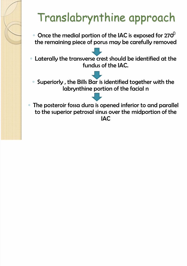
Translabrynthine approach
◦ Once the medial portion of the IAC is exposed for 270the remaining piece of porus may be carefully removed
◦ Laterally the transverse crest should be identified at the
fundus of the IAC.
◦ Superiorly , the Bills Bar is identified together with thelabrynthine portion of the facial n
◦ The posteroir fossa dura is opened inferior to and parallelto the superior petrosal sinus over the midportion of the
IAC
0

8/11/2019 acousticneuroma-130914224034-phpapp02
http://slidepdf.com/reader/full/acousticneuroma-130914224034-phpapp02 57/62
Translabrynthine approach
◦ Using the bills bar as guide , and with a fine hook , thesurgeon seperates the superior vest n from facial nerve.
◦ The capsule of the tumor is incised , & the tumor is
gutted with house urban dissector
0

8/11/2019 acousticneuroma-130914224034-phpapp02
http://slidepdf.com/reader/full/acousticneuroma-130914224034-phpapp02 58/62

8/11/2019 acousticneuroma-130914224034-phpapp02
http://slidepdf.com/reader/full/acousticneuroma-130914224034-phpapp02 59/62
Radiotherapy :-
1. Conventional radiotherapy by external beam
has no role in the treatment of Acoustic Neuromas
due to low tolerance of CNS to radiation.
2 X – or Gamma knife surgery.

8/11/2019 acousticneuroma-130914224034-phpapp02
http://slidepdf.com/reader/full/acousticneuroma-130914224034-phpapp02 60/62
THANK YOU

8/11/2019 acousticneuroma-130914224034-phpapp02
http://slidepdf.com/reader/full/acousticneuroma-130914224034-phpapp02 61/62

8/11/2019 acousticneuroma-130914224034-phpapp02
http://slidepdf.com/reader/full/acousticneuroma-130914224034-phpapp02 62/62