Embed Size (px)
Citation preview

ORIGINAL ARTICLE
Acoustic radiation force impulse (ARFI) elastographyfor detection of renal damage in children
Cemil Göya & Cihad Hamidi & Aydın Ece & Mehmet Hanifi Okur & Bekir Taşdemir &
Mehmet Güli Çetinçakmak & Salih Hattapoğlu & Memik Teke & Cahit Şahin
Received: 18 October 2013 /Revised: 28 February 2014 /Accepted: 28 May 2014# Springer-Verlag Berlin Heidelberg 2014
AbstractBackground Acoustic radiation force impulse (ARFI) imag-ing is a promising method for noninvasive evaluation of therenal parenchyma.Objective To investigate the contribution of ARFI quantita-tive US elastography for the detection of renal damage inkidneys with and without vesicoureteral reflux (VUR).Materials and methods One hundred seventy-six kidneys of88 children (46 male, 42 female) who had been referred forvoiding cystourethrography and 20 healthy controls were pro-spectively investigated. Patients were assessed according toseverity of renal damage on dimercaptosuccinic acid(DMSA) scintigraphy. Ninety-eight age- and gender-matchedhealthy children constituted the control group. Quantitativeshear wave velocity (SWV) measurements were performed inthe upper and lower poles and in the interpolar region of eachkidney. DMSA scintigraphy was performed in 62 children(124 kidneys). Comparisons of SWV values of kidneyswith and without renal damage and/or VUR were done.
Results Significantly higher SWV values were found in non-damaged kidneys. Severely damaged kidneys had the lowestSWV values (P<0.001). High-grade (grade V-IV) refluxingkidneys had the lowest SWV values, while non-refluxingkidneys had the highest values (P<0.05). Significant negativecorrelations were found between the mean quantitative USelastography values and DMSA scarring score (r=−0.788,P<0.001) and VUR grade (r=−0.634,P<0.001). SWVvaluesof the control kidneys were significantly higher than those ofdamaged kidneys (P<0.05).Conclusion Our findings suggest decreasing SWV of renalunits with increasing grades of vesicoureteric reflux, increas-ing DMSA-assessed renal damage and decreasing DMSA-assessed differential function.
Keywords ARFI elastography . Renal damage .
Vesicoureteral reflux . DMSA scintigraphy . Child
Introduction
Kidneys that are frequently exposed to urinary tractinfection (UTI) may develop renal parenchymal damagethat ultimately may lead to development of hypertensionand chronic renal failure [1, 2]. Therefore, early diag-nosis and proper management of UTI is imperative toprevent such complications.
The most frequent urinary tract abnormality that predis-poses children to pyelonephritis and scarring is high-gradevesicoureteral reflux (VUR) [1–3]. However, there are upperUTIs (pyelonephritis) in children without VUR and renaldamage is not caused by VUR alone.
Previous studies have shown that renal US has low sensi-tivity and specificity for detection of renal damage [3, 4].Dimercaptosuccinic acid (DMSA) scintigraphy is the goldstandard imaging method for detecting renal damage, visual-izing the renal cortex and determining the differential function
C. Göya :C. Hamidi (*) :M. G. Çetinçakmak : S. Hattapoğlu :M. TekeDepartment of Radiology Medical School, Dicle University,Diyarbakir, Turkeye-mail: [email protected]
A. EceDepartment of Pediatrics, Medical School, Diyarbakir University,Dicle, Turkey
M. H. OkurDepartment of Pediatric Surgery, Medical School, Dicle University,Diyarbakir, Turkey
B. TaşdemirDepartment of Nuclear Medicine Medical School, Dicle UniversityDiyarbakir, Diyarbakir, Turkey
C. ŞahinDepartment of Pediatrics, Medical School, Dicle University,Diyarbakir, Turkey
Pediatr RadiolDOI 10.1007/s00247-014-3072-3
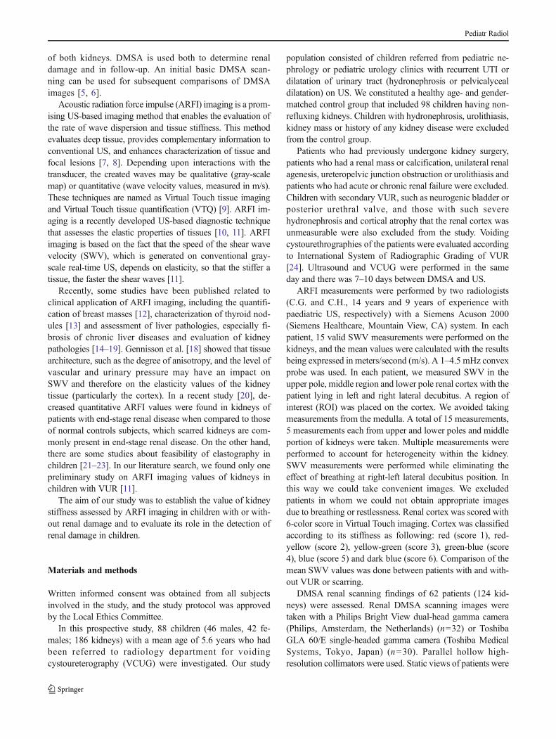
of both kidneys. DMSA is used both to determine renaldamage and in follow-up. An initial basic DMSA scan-ning can be used for subsequent comparisons of DMSAimages [5, 6].
Acoustic radiation force impulse (ARFI) imaging is a prom-ising US-based imaging method that enables the evaluation ofthe rate of wave dispersion and tissue stiffness. This methodevaluates deep tissue, provides complementary information toconventional US, and enhances characterization of tissue andfocal lesions [7, 8]. Depending upon interactions with thetransducer, the created waves may be qualitative (gray-scalemap) or quantitative (wave velocity values, measured in m/s).These techniques are named as Virtual Touch tissue imagingand Virtual Touch tissue quantification (VTQ) [9]. ARFI im-aging is a recently developed US-based diagnostic techniquethat assesses the elastic properties of tissues [10, 11]. ARFIimaging is based on the fact that the speed of the shear wavevelocity (SWV), which is generated on conventional gray-scale real-time US, depends on elasticity, so that the stiffer atissue, the faster the shear waves [11].
Recently, some studies have been published related toclinical application of ARFI imaging, including the quantifi-cation of breast masses [12], characterization of thyroid nod-ules [13] and assessment of liver pathologies, especially fi-brosis of chronic liver diseases and evaluation of kidneypathologies [14–19]. Gennisson et al. [18] showed that tissuearchitecture, such as the degree of anisotropy, and the level ofvascular and urinary pressure may have an impact onSWV and therefore on the elasticity values of the kidneytissue (particularly the cortex). In a recent study [20], de-creased quantitative ARFI values were found in kidneys ofpatients with end-stage renal disease when compared to thoseof normal controls subjects, which scarred kidneys are com-monly present in end-stage renal disease. On the other hand,there are some studies about feasibility of elastography inchildren [21–23]. In our literature search, we found only onepreliminary study on ARFI imaging values of kidneys inchildren with VUR [11].
The aim of our study was to establish the value of kidneystiffness assessed by ARFI imaging in children with or with-out renal damage and to evaluate its role in the detection ofrenal damage in children.
Materials and methods
Written informed consent was obtained from all subjectsinvolved in the study, and the study protocol was approvedby the Local Ethics Committee.
In this prospective study, 88 children (46 males, 42 fe-males; 186 kidneys) with a mean age of 5.6 years who hadbeen referred to radiology department for voidingcystoureterography (VCUG) were investigated. Our study
population consisted of children referred from pediatric ne-phrology or pediatric urology clinics with recurrent UTI ordilatation of urinary tract (hydronephrosis or pelvicalycealdilatation) on US. We constituted a healthy age- and gender-matched control group that included 98 children having non-refluxing kidneys. Children with hydronephrosis, urolithiasis,kidney mass or history of any kidney disease were excludedfrom the control group.
Patients who had previously undergone kidney surgery,patients who had a renal mass or calcification, unilateral renalagenesis, ureteropelvic junction obstruction or urolithiasis andpatients who had acute or chronic renal failure were excluded.Children with secondary VUR, such as neurogenic bladder orposterior urethral valve, and those with such severehydronephrosis and cortical atrophy that the renal cortex wasunmeasurable were also excluded from the study. Voidingcystourethrographies of the patients were evaluated accordingto International System of Radiographic Grading of VUR[24]. Ultrasound and VCUG were performed in the sameday and there was 7–10 days between DMSA and US.
ARFI measurements were performed by two radiologists(C.G. and C.H., 14 years and 9 years of experience withpaediatric US, respectively) with a Siemens Acuson 2000(Siemens Healthcare, Mountain View, CA) system. In eachpatient, 15 valid SWV measurements were performed on thekidneys, and the mean values were calculated with the resultsbeing expressed in meters/second (m/s). A 1–4.5 mHz convexprobe was used. In each patient, we measured SWV in theupper pole, middle region and lower pole renal cortex with thepatient lying in left and right lateral decubitus. A region ofinterest (ROI) was placed on the cortex. We avoided takingmeasurements from the medulla. A total of 15 measurements,5 measurements each from upper and lower poles and middleportion of kidneys were taken. Multiple measurements wereperformed to account for heterogeneity within the kidney.SWV measurements were performed while eliminating theeffect of breathing at right-left lateral decubitus position. Inthis way we could take convenient images. We excludedpatients in whom we could not obtain appropriate imagesdue to breathing or restlessness. Renal cortex was scored with6-color score in Virtual Touch imaging. Cortex was classifiedaccording to its stiffness as following: red (score 1), red-yellow (score 2), yellow-green (score 3), green-blue (score4), blue (score 5) and dark blue (score 6). Comparison of themean SWV values was done between patients with and with-out VUR or scarring.
DMSA renal scanning findings of 62 patients (124 kid-neys) were assessed. Renal DMSA scanning images weretaken with a Philips Bright View dual-head gamma camera(Philips, Amsterdam, the Netherlands) (n=32) or ToshibaGLA 60/E single-headed gamma camera (Toshiba MedicalSystems, Tokyo, Japan) (n=30). Parallel hollow high-resolution collimators were used. Static views of patients were
Pediatr Radiol

obtained from anterior, posterior, left posterior oblique andright posterior oblique positions. Gamma camera images wereevaluated by the same expert in nuclear medicine. A startingdose of 5 mCi of technetium-99 m DMSA, multiplied by anadjusted fraction based on the weight of the patient, wasinjected intravenously. Renal cortical imaging was accom-plished by delayed imaging 3–4 h after injection of the radio-isotope. A decreased radioisotope uptake area in the renalparenchyma was defined as parenchymal function loss. Wide-spread and multiple decreased radioisotope uptake areas weredefined as heterogeneous radioactivity uptake. These abovementioned kidneys were accepted as DMSA changing posi-tive, and kidneys with normal DMSA scanning images wereaccepted as DMSA negative.
DMSA scans were graded according to criteria determinedfor the Randomized Intervention for Children withVesicoureteral Reflux (RIVUR) study [25]. Briefly, the kidneyis divided into 12 segments to localize damage. If no damagewas present, it was accepted as grade 0; damage in 1–2segments as grade 1; damage in 3–4 segments as grade 2,damage in >4 segments are given grade 3 and global damagewas scored as grade 4 [25].
Kidneys with parenchymal function loss in DMSA andthose with normal DMSA scanning images were comparedto each other regarding quantitative SWV values. In 57 dam-aged kidneys, the damaged areas were localized by DMSAimages and only these areas were examined by elastography;the results were compared with the total SWV values of thesame kidneys.
Statistical analysis
All statistical analyses were performed using SPSS (StatisticalPackage for Social Sciences) version 15.0 forWindows (SPSSInc., Chicago, IL). The Kolmogorov–Smirnov test was per-formed on all continuous variables to define the distribution ofnormality. Variables are expressed as mean±standard devia-tion (SD) and categorical variables are expressed as countsand percentages. Independent samples t-test was used in com-parison of patients and the controls. Kruskal-Wallis varianceanalysis was used for comparison of more than two groups.Chi-square test was used for comparison of nominal or ordinalvariables. Pearson or Spearman correlation analyses wereused to examine relationships between numerical variables.P-value of less than 0.05 was accepted as statisticallysignificant.
Results
The study included 88 children (176 kidneys) who wereevaluated by VCUG and ARFI elastography. The mean age
of patients was 5.6±3.1 (range: 1–17) years, and themale/female ratio was 1.1:1.0. The numbers of patients inage groups were as follows: 45 (51.1%) were between 1 and5 years, 30 (34.1%) were between 6 and 9 years, and 13(14.8%) were 10 years of age or older. The mean age ofcontrol group was 6.6±5.3 (range: 1–16) years and themale/female ratio was 1.5:1.0. There were no differences inthe mean age and gender ratio between the study and thecontrol groups (P>0.05).
The mean interobserver coefficient of variance was calculat-ed as 16.7% and intraobserver coefficient of variance was 4%.
Grade I VUR was detected in 7 (4.0%), grade II in 11(6.3%), grade III in 32 (18.2%), grade IV in 41 (23.3%) andgrade V in 18 (10.2%) kidneys. VUR was not detected in 64(34.4%) kidneys. DMSA was performed in 62 patients (124kidneys). Scores of renal damage according to DMSA imageswere as follows: score 0 in 64 (50.8%), score 1 in 12 (9.5%),score 2 in 11 (8.7%), score 3 in 23 (18.3%) and score 4 in 16(12.7%) patients. Children with grades IV and V VUR hadsignificantly higher ratio of renal scarring compared withthose with VUR grades I-III (P<0.001) (Table 1). Childrenwith high-grade reflux (grades IVand V) also had significant-ly higher renal damage scores (3.14±0.81 vs. 0.82±1.0,P<0.001) and lower differential functions on DMSA (35.1±16.4 vs. 60.0±14.8, P<0.001) compared with low grade(grades I-III) VUR patients (Table 1).
No significant differences were found in the mean, lowestand highest SWV values between right and left kidneys (datanot shown, P>0.05) or between female and male patients(data not shown, P>0.05).
In healthy controls, no significant difference was observedamong different age groups in terms of SWV. In healthycontrols, the SWV value was 2.39±0.22 m/s for the rightkidney, and 2.37±0.25 m/s for the left (not significant).
The mean lowest (1.62±0.22), highest (3.02±0.32) andmean (2.39±0.23) SWV values of healthy controls were
Table 1 Low-grade (grades 1–3) and high-grade (grades 4–5)vesicoureteral reflux frequencies, mean SWV values, differential functionat dimercaptosuccinic acid scintigraphy (DMSA) between renal unitswith normal and abnormal findings at DMSA scintigraphy
DMSA abnormal(n=58)
DMSA normal(n=64)
P
Vesicoureteric reflux
Absent, n (%) 2 (4.7) 41 (95.3) <0.001
Grades I-III, n (%) 17 (44.7) 21 (55.3)
Grades IV-V, n (%) 39 (95.1) 2 (4.9)
Mean ARFI(mean±SD) m/s
1.83±0.44 2.39±0.23 <0.001
Function,%(mean±SD)
35.1±16.4 60±14.8 <0.001
SD standard deviation
Pediatr Radiol

significantly higher from those of patients with renal scar(P<0.001, for each comparison) (Table 2).
The mean SWV of damaged kidneys was significantlyhigher than that of DMSA negative kidneys (1.83±0.44 vs.2.39±0.23, P<0.001) (Table 1). There were significant differ-ences in the median, lowest and highest SWV values betweenDMSA score groups of 0, 1–2, 3 and 4 (P<0.05) (Table 2).
Parallel to increasing scores of renal damage, the mean,lowest and highest SWV values were found to be significantlydecreased (P<0.05) (Table 2). While the lowest SWV valueswere found in the most damaged kidneys (score 4), signifi-cantly higher SWV values were found in DMSA-normalkidneys (score 0), (Table 2 , Fig. 1).
When just the damaged regions of 52 abnormal kid-neys were examined by ARFI elastography, their SWVvalues were found to be significantly lower comparedwith the overall SWV values of the same kidneys (n=52, 0.99±0.36 vs. 1.82±0.45, P<0.001).
No significant differences were found in the SWV valuesbetween grades I, II and III refluxing kidneys, or betweengrades IV and V VUR kidneys (P>0.05). Therefore, grade I-III patients and grade IV-V were taken as the sole groups for
the comparisons of SWVvalues among different VUR grades.There were significant differences in SWV values betweennon-refluxing patients, patients with low-grade (grades I-III)reflux and high-grade (grades V-IV) reflux (P<0.05). High-grade refluxing kidneys had the lowest SWV values, whilenon-refluxing kidneys had the highest SWV values. Kidneyswith low-grade reflux had intermediate SWV values, lowerthan non-refluxing kidneys and higher than high-grade refluxkidneys (P<0.05) (Fig. 2).
The age of the children had no correlation with the meanSWV value (r=−0.093), DMSA damage score (r=0.095), orVUR grade (r=−0.035) (P>0.05). However, a significantpositive correlation was found between differential kidneyfunction on DMSA and the mean SWV values (r=0.599),and significant negative correlations were found between themean SWV values and DMSA severity score (r=−0.788), andVUR grade (r=−0.634) (Table 3). VUR grade was found to besignificantly positively correlated with the DMSA severityscore (r=0.800) and negatively correlated with differentialkidney function on DMSA (r=−0.646) (Table 3).
Renal cortical color score was found to be 5–6 in non-refluxing kidneys with no changes in visual touch imag-ing and DMSA (Fig. 3) Cortical renal color score was 3–4
Fig. 1 Mean shear wave velocities (SWV) of renal units bydimercaptosuccinic acid scintigraphy (DMSA) severity scores (* outliers,o extremes)
Fig. 2 Mean shear wave velocities (SWV) of renal units by ipsilateralgrade of vesicoureteral reflux (VUR) (* outliers, o extremes)
Table 2 Comparison of mean, lowest and highest readings of repeated shear wave velocity (SWV) measurements in renal units by severity of damage atdimercaptosuccinic acid scintigraphy (DMSA). (0=normal DMSA, 4=severe damage at DMSA)
SWV (mean±SD) DMSA abnormality score
Score 0 Control group (n=64) Scores 1–2 (n=23) Scores 3–4 (n=39) P1
Mean 2.39±0.23 2.18±0.23 1.51±0.34 <0.001
Lowest 1.62±0.22 1.24±0.41 0.98±0.29 <0.001
Highest 3.02±0.32 2.50±0.39 1.83±0.40 <0.001
SD standard deviation1 Kruskal-Wallis analysis of variance
Pediatr Radiol

in affected areas of kidneys with mild alterations onDMSA (Fig. 4). In kidneys with significant function lossand decreased radionuclide uptake, total DMSA colorscale was 1–2 (Fig. 5).
Discussion
In this study, we performed ARFI measurements in childrenwith or without renal damage on DMSA and detected lowerSWV values in DMSA abnormal kidneys compared withDMSA normal kidneys. Additionally, significantly lowerSWV values were found in patients with grade 4 or 5 VURcompared with those without VUR.
Increased severity of renal damage at DMSA was associ-ated with high-grade reflux (grades IV and V) in our study,similar to previous reports [26]. Differential function onDMSAwas significantly lower in damaged kidneys comparedwith non-scarred kidneys (Table 1), as expected.
The mean SWV was significantly lower in damaged kid-neys than in those with normal DMSA. The lowest SWVvalues were detected in kidneys with the highest DMSA sever-ity scores (Table 2, Fig. 2). In a study of normal organs of SWVvalues, it was reported that the mean normal renal SWV valueof the right kidney was 2.19 m/s, and that of the left kidney was
2.33 m/s [27]. We found a mean normal renal SWVof 2.4 m/s(range: 2.06-4.03 m/s) for both left and right kidneys.
Although DMSA is the reference-standard diagnostic toolfor the detection of renal damage [1, 11], low radiation expo-sure is unavoidable with scintigraphy. Therefore, a radiation-free imaging technique such as ARFI elastography may be acomplementary method to reliably identify parenchymal dam-age. Although ARFI elastography gave results considerablycorrelated with the severity of liver fibrosis in chronic liverdiseases [15, 16, 28], ARFI measurements in kidneys arecontroversial, and studies on renal masses and renal trans-plants give variable results [29–31]. These conflicting resultsmay be due to the very heterogeneous histological structure ofthe kidney tissue, which consists of the complex architectureof glomeruli, tubules and stromal components. Some technicalaspects also affect SWV measurements. Studies have foundthat applied transducer force seems to be of substantial impor-tance for SWVmeasurements in kidney transplants and SWVmeasurements [11, 30]. The "virtual touch technology" createsa pressure wave within the transducer thus overcoming limi-tations of manually applied pressure techniques as used bymany other systems. A study regarding the investigation ofchanges in the renal parenchyma has used ARFI elastographyin children with VUR [11]. One study reported 28 childrenwith VUR (≥grade III) who underwent scintigraphy andARFI, and concluded that kidneys had significantly higherSWV values compared with healthy kidneys [11]. Our studyincluded more patients to investigate ARFI elastography as acomplementary tool to DMSA scan since our SWV resultswere correlated with the renal scarring severity and consistentin itself. However, ARFI values of our patients were signifi-cantly decreased with the increasing scores of renal scarring(Table 2, Fig. 2). Our results suggest that ARFI elastographymay be used as an adjunct for estimation of the presence andseverity of renal scarring in children. Because renal scarring isnot only exclusively connected to VUR in the latest guidelinesand there are scars without VUR, detection of scars deservespriority instead of VUR [32].
Table 3 Pearson correlation coefficients among mean shear wave veloc-ity (SWV), dimercaptosuccinic acid scintigraphy (DMSA) severityscores, DMSA differential function and grades of vesicoureteric reflux(VUR) within renal units. All correlation coefficients are statisticallysignificant (P<0.001)
DMSAseverity score
DMSA differentialfunction
VURgrade
SWV m/s –0.788 0.599 –0.634
DMSA severity score –0.741 0.800
DMSA differential function –0.646
Fig. 3 Acoustic radiation forceimpulse (ARFI) imaging in an 8-year-old boy with normalappearance in a kidney withoutVUR shows normal stiffness asblue-purple
Pediatr Radiol

Considering all of these problems, we suggest the use ofARFI elastography as a noninvasive and non-radiation methodthat can detect alterations in the renal parenchyma related todamage. While comparing ARFI elastography with other non-invasive imaging methods evaluating tissue stiffness, ARFIelastography has the advantage of being integrated to conven-tional US. Therefore, ARFI elastography may be an examina-tion method with better imaging quality and lower interobserv-er variances [15]. It has been reported that ARFI technology isuseful for determination of thyroid and intra-abdominal organs,and evaluation of liver fibrosis and cirrhosis [20, 33]. In anotherstudy, the use of ARFI elastography was suggested for thediagnosis and follow-up of renal allograft rejection [30].
There were some studies on the practical use ofelastography in children [21–23]. In a study, transientelastography was found as technically applicable in all agegroups of children [21] . The upper limit of normal values wasfound to increase significantly with advanced age. Due tomovement artifacts, the measurement was found to be reliablefrom the age of 6 without sedation. In younger children, thenumber of invalid measurements increases significantly. Inour study, we did not sedate children during ARFI
measurements. The ARFI measurement was performed incrying and restless children in our study after they werecalmed down.
There are some limitations of our study. Firstly, ARFIelastography has operator-dependent characteristics and hasdifficulties in control of respirations and body movementssimilar to those during US. Another limitation of the studywas the inability to exclude congenital renal dysplasia thor-oughly. Although, there was no dehydration or overhydrationin our patients and none of our patients was critically ill duringelastography measurements, another limitation of our studymay be non-standardization of patients’ hydration status dur-ing elastography.
Conclusion
SWVis related to degree of renal damage and degree of VUR.There might therefore be a potential of ARFI elastography as anonradioactive and noninvasive method to detect renal dam-age in children.
Fig. 4 ARFI imaging in a 10-year-old girl with grade IV VURshows decreased stiffness(yellow-green-red areas) amongunaffected areas (blue-purple)
Fig. 5 ARFI imaging in a 7-year-old girl with grade V VURshows a small kidney withentirely decreased stiffness asyellow-green-red
Pediatr Radiol

Conflicts of interest None
References
1. Soylu A, Demir BK, TürkmenM et al (2008) Predictors of renal scarin children with urinary infection and vesicoureteral reflux. PediatrNephrol 23:2227–2232
2. Elder JS, Peters CA, Arant BS et al (1997) Pediatric vesicoureteralreflux guidelines panel summary report on the management of pri-mary vesicoureteral reflux in children. J Urol 157:1846–1851
3. Lee HY, Soh BH, Hong CH et al (2009) The efficacy of ultrasoundand dimercaptosuccinic acid scan in predicting vesicoureteral refluxin children below the age of 2years with their first febrile urinary tractinfection. Pediatr Nephrol 24:2009–2013
4. Zamir G, Sakran W, Horowitz Y et al (2004) Urinary tract infection:is there a need for routine renal ultrasonography? Arch Dis Child 89:466–468
5. Preda I, Jodal U, Sixt R et al (2007) Normal dimercaptosuccinic acidscintigraphy makes voiding cystourethrography unnecessary afterurinary tract infection. J Pediatr 151:581–584
6. Faust WC, Diaz M, Pohl HG (2009) Incidence of post-pyelonephriticrenal scarring: a meta-analysis of the dimercapto-succinic acid liter-ature. J Urol 181:290–297
7. Cross TJ, Mitchell JD, Cramp ME (2009) Elastrography for the non-invasive assessment of liver disease: limitations and future develop-ments. Gut 58:1171–1172
8. Friedrich-RustM,WunderK,Kriener S et al (2009) Liver fibrosis in viralhepatitis: noninvasive assessment with acoustic radiation force impulseimaging versus transient elastography. Radiology 252:595–604
9. Zhai L, Palmeri ML, Bouchard RR et al (2008) An integratedindenter-ARFI imaging system for tissue stiffness quantification.Ultrason Imaging 30:95–111
10. Fahey BJ, Nightingale KR, Nelson RC et al (2005) Acoustic radia-tion force impulse imaging of the abdomen: demonstration of feasi-bility and utility. Ultrasound Med Biol 31:1185–1198
11. Bruno C, Caliari G, Zaffanello M et al (2013) Acoustic radiationforce impulse (ARFI) in the evaluation of the renal parenchymalstiffness in paediatric patients with vesicoureteral reflux: preliminaryresults. Eur Radiol 23:3477–3484
12. Tozaki M, Isobe S, Fukuma E (2011) Preliminary study of ultraso-nographic tissue quantification of the breast using the acoustic radi-ation force impulse (ARFI) technology. Eur J Radiol 80:182–187
13. Gu J, Du L, Bai M et al (2012) Preliminary study on the diagnosticvalue of acoustic radiation force impulse technology for differentiat-ing between benign and malignant thyroid nodules. J UltrasoundMed 31:763–771
14. Hanquinet S, Rougemont AL, Courvoisier D et al (2013) Acousticradiation force impulse (ARFI) elastography for the noninvasivediagnosis of liver fibrosis in children. Pediatr Radiol 43:545–551
15. Friedrich-RustM,WunderK,Kriener S et al (2009) Liver fibrosis in viralhepatitis: noninvasive assessment with acoustic radiation force impulseimaging versus transient elastography. Radiology 252:595–604
16. Monti L, Manco M, Lo Zupone C et al (2012) Acoustic radiationforce impulse (ARFI) with Virtual Touch Tissue Quantification inliver disease associated with cystic fibrosis in children. Radiol Med117:1408–1418
17. Ternifi R, Gennisson JL, Tanter M et al (2013) Effects of storagetemperature on the mechanical properties of porcine kidney estimatedusing shear wave elastography. JMechBehavBiomedMater 28:86–93
18. Gennisson JL, Grenier N, Combe C et al (2012) supersonic shearwave elastography of in vivo pig kidney: influence of blood pressure,urinary pressure and tissue anisotropy. Ultrasound Med Biol 38:1559–1567
19. Bota S, Bob I, Spore et al (2013) Which are the kidney stiffnessvalues assessed by acoustic radiation force impulse (ARFI)elastography in subjects without known kidney pathology?Ultrasound Med Biol 39:S60
20. Guo LH, XuHX, Fu HJ et al (2013) Acoustic radiation force impulseimaging for noninvasive evaluation of renal parenchyma elasticity:preliminary findings. PLoS One 8:e68925
21. Engelmann G, Gebhardt C, Wenning D et al (2012) Feasibility studyand control values of transient elastography in healthy children. Eur JPediatr 171:353–360
22. Goldschmidt I, Streckenbach C, Dingemann C et al (2013)Application and limitations of transient liver elastography in chil-dren. J Pediatr Gastroenterol Nutr 57:109–113
23. Berko NS, Fitzgerald EF, Amaral TD et al (2014) Ultrasoundelastography in children: Establishing the normal range of muscleelasticity. Pediatr Radiol 44:158–163
24. Lebowitz RL, Olbing H, Parkkulainen KVet al (1985) Internationalsystem of radiographic grading of vesicoureteric reflux. InternationalReflux Study in Children. Pediatr Radiol 15:105–109
25. Keren R, Carpenter MA, Hoberman A et al (2008) Rationaleand design issues of the Randomized Intervention for ChildrenWith Vesicoureteral Reflux (RIVUR) study. Pediatrics 122:240–250
26. Chen MJ, Cheng HL, Chiou YY (2013) Risk factors for renalscarring and deterioration of renal function in primary vesico-ureteral reflux children: a long-term follow-up retrospective cohortstudy. PLoS One 8:57954
27. Lee MJ, Kim MJ, Han KH et al (2013) Age-related changes inliver, kidney, and spleen stiffness in healthy children measuredwith acoustic radiation force impulse imaging. Eur J Radiol 82:290–294
28. Sporea I, Sirli R, Bota S et al (2012) Comparative study concerningthe value of Acoustic Radiation Force Impulse Elastography (ARFI)in comparison with transient elastography (TE) for the assessment ofliver fibrosis in patients with chronic hepatitis B and C. UltrasoundMed Biol 38:1310–1316
29. Clevert DA, Stock K, Klein B et al (2009) Evaluation ofAcoustic Radiation Force Impulse (ARFI) imaging andcontrast-enhanced ultrasound in renal tumors of unknown etiol-ogy in comparison to histological findings. Clin HemorheolMicrocirc 43:95–107
30. Syversveen T, Midtvedt K, Berstad AE et al (2012) Tissue elasticityestimated by acoustic radiation force impulse quantification dependson the applied transducer force: an experimental study in kidneytransplant patients. Eur Radiol 22:2130–2137
31. Stock KF, Klein BS, Vo Cong MT et al (2010) ARFI-basedtissue elasticity quantification in comparison to histology forthe diagnosis of renal transplant fibrosis. Clin HemorheolMicrocirc 46:139–148
32. Subcommittee on Urinary Tract Infection, Steering Committee onQuality Improvement and Management, Roberts KB (2011) Urinarytract infection: clinical practice guideline for the diagnosis and man-agement of the initial UTI in febrile infants and children 2 to 24months. Pediatrics 128:595–610
33. Goertz RS, AmannK, Heide R et al (2011) An abdominal and thyroidstatus with Acoustic Radiation Force Impulse Elastometry a feasibil-ity study: Acoustic Radiation Force Impulse Elastometry of humanorgans. Eur J Radiol 80:226–230
Pediatr Radiol