Embed Size (px)
Citation preview

doi:10.1016/j.jacc.2006.06.076 2006;48;2141-2151; originally published online Oct 31, 2006; J. Am. Coll. Cardiol.
III, Robert L. McNamara, Manesh R. Patel, and John Spertus Mohler,Hendel, James Jollis, Eric Peterson, Jersey Chen, Frederick Masoudi, Emile
Pamela Douglas, Ami E. Iskandrian, Harlan M. Krumholz, Linda Gillam, Robert in Cardiovascular Imaging
QualityCollege of Cardiology–Duke University Medical Center Think Tank on Achieving Quality in Cardiovascular Imaging: Proceedings From the American
This information is current as of February 5, 2009
http://content.onlinejacc.org/cgi/content/full/48/10/2141located on the World Wide Web at:
The online version of this article, along with updated information and services, is
by on February 5, 2009 content.onlinejacc.orgDownloaded from
F
APMDQACHAaC
A
LR
JLRJAH
oARCTV
bHAov
Journal of the American College of Cardiology Vol. 48, No. 10, 2006© 2006 by the American College of Cardiology Foundation ISSN 0735-1097/06/$32.00P
OCUS ISSUE: CARDIAC IMAGING State-of-the-Art PaperQuality in Cardiovascular Imaging
chieving Quality in Cardiovascular Imagingroceedings From the American College of Cardiology–Duke Universityedical Center Think Tank on Quality in Cardiovascular Imagingeveloped in Collaboration With the Cardiovascular Imaging Collaborativeuality Work Group, American College of Radiology, American Heart Association,merican Society of Echocardiography, American Society of Nuclear Cardiology,oalition of Cardiovascular Organizations, Heart Failure Society of America,eart Rhythm Society, Intersocietal Accreditation Commission, Society oftherosclerosis Imaging and Prevention, Society for Cardiovascular Angiographynd Interventions, Society of Cardiovascular Computed Tomography, Society for
ublished by Elsevier Inc. doi:10.1016/j.jacc.2006.06.076
ardiovascular Magnetic Resonance, and Society for Vascular Medicine and Biology
CONFERENCE CO-DIRECTORS
Pamela Douglas, MD, MACC
STEERING COJ
WRITING G
Pamela Douglas, M
FERME
f Cardiology–Duke University Medical Center Think Tank on Quality in Cardio-ascular Imaging. J Am Coll Cardiol 2006;48:2141–51.
AMCUS
ar
a
content.onlinejacDownloaded from
mi E. Iskandrian, MD, FACC Harlan M. Krumholz, MD, FACC
MMITTEE
inda Gillam, MD, FACC ames Jollis, MD, FACC obert Hendel, MD, FACC Eric Peterson, MD, FACCROUP
D, MACC
ersey Chen, MDinda Gillam, MD, FACCobert Hendel, MD, FACC
ames Jollis, MD, FACCmi E. Iskandrian, MD, FACC
rederick Masoudi, MD, FACCmile Mohler III, MD, FACCobert L. McNamara, MD, MHS, FACCanesh R. Patel, MD
ric Peterson, MD, FACC
arlan M. Krumholz, MD, FACC John Spertus, MD, FACCThe following companies provided sponsorship for the meeting: Aetna, Inc.,stellas Pharma US, Inc., AstraZeneca Pharmaceuticals, Bristol-Myers Squibbedical Imaging, General Electric Healthcare, Pfizer, Inc., Point Biomedicalorporation, Siemens Medical Solutions, Inc., Toshiba America Medical Systems,nited Healthcare Services, Inc., Wellpoint, Inc., Anthem, and Blue Cross Bluehield.For a complete list of the ACC–Duke Think Tank participants and their
ffiliations, please see Appendix 1. For a complete list of the Writing Group’selationships with industry, please see Appendix 2.
This article is endorsed by the American College of Cardiology, American Collegef Radiology, American Heart Association, American Society of Echocardiography,merican Society of Nuclear Cardiology, Heart Failure Society of America, Hearthythm Society, Society of Atherosclerosis Imaging and Prevention, Society forardiovascular Angiography and Interventions, Society of Cardiovascular Computedomography, Society for Cardiovascular Magnetic Resonance, and Society forascular Medicine and Biology.When citing this document, the Journal requests that the following citation format
e used: Douglas P, Chen J, Gillam L, Hendel R, Jollis J, Iskandrian AE, KrumholzM, Masoudi F, Mohler E III, McNamara RL, Patel MR, Peterson E, Spertus J.chieving quality in cardiovascular imaging: proceedings from the American College
Manuscript received March 23, 2006; revised manuscript received June 26, 2006,ccepted June 26, 2006.
by on February 5, 2009 c.org
A
Cibmcaqoqmids
I
IpcspEiog
MscmScHeqAawp
Ci
piveimtte
P
TqossPpiaotPtq
ttrRuiriHalloitAfss
T
2142 Douglas et al. JACC Vol. 48, No. 10, 2006Achieving Quality in Cardiovascular Imaging November 21, 2006:2141–51
BSTRACT
ardiovascular imaging has enjoyed both rapid technolog-cal advances and sustained growth, yet less attention haseen focused on quality than in other areas of cardiovascularedicine. To address this deficit, representatives from
ardiovascular imaging societies, private payers, governmentgencies, the medical imaging industry, and experts inuality measurement met, and this report provides anverview of the discussions. A consensus definition ofuality in imaging and a convergence of opinion on qualityeasures across imaging modalities was achieved and are
ntended to be the start of a process culminating in theevelopment, dissemination, and adoption of quality mea-ures for all cardiovascular imaging modalities.
NTRODUCTION
maging has transformed cardiovascular medicine by im-roving the prevention, diagnosis, and management ofardiovascular disease. The sustained growth of imaginghows the central role that imaging plays in the care ofatients with known or suspected cardiovascular disease.nsuring a high level of quality has now become an
mportant focus for patients, physicians, and payers becausef advances in existing imaging technologies and the emer-ence of new modalities.
Quality of care has been defined by the Institute ofedicine as “the degree to which health care systems,
ervices, and supplies for individuals and populations in-rease the likelihood for desired health outcomes in aanner consistent with current professional knowledge” (1).
everal initiatives to improve quality for patients withardiovascular conditions have been implemented (2,3).owever, these programs have predominately focused on
valuating the use of evidence-based therapies (4,5), anduality in imaging has been relatively hidden from view.lthough few studies have shown marked geographic vari-
tion in imaging use (6,7), there is little information abouthere quality gaps exist and how they ultimately affectatient care and outcomes.To respond to this need, the American College of
ardiology (ACC) and Duke University convened a meet-
Abbreviations and AcronymsACC � American College of CardiologyACR � American College of RadiologyASE � American Society of EchocardiographyASNC � American Society of Nuclear CardiologyCOCATS � Core Cardiology Training SymposiumCT � computed tomographyJCAHO � Joint Commission on the Accreditation of
Healthcare OrganizationsMR � magnetic resonanceSPECT � single-photon emission computed
tomography
ng of representatives of cardiovascular imaging societies,content.onlinejacDownloaded from
rivate payers, government agencies, industry, and expertsn quality measurement in January 2006. This report pro-ides a review of the discussions and proposes efforts tostablish quality standards for cardiovascular diagnosticmaging, beginning with an emphasis on valid quality
easurement tools. The meeting achieved a consensus defini-ion of quality in imaging and a convergence of opinion towardhe development and dissemination of quality measures forach imaging modality within 18 months.
RINCIPLES OF QUALITY MEASUREMENT
he conference embraced Donabedian’s (8) methodology ofuality assessment by applying his structure-process-utcome model to cardiovascular imaging. Structure repre-ents the infrastructure through which care is delivered,uch as equipment, staff training, and laboratory protocols.rocess refers to those actions performed in delivering care toatients, and includes such concepts as patient selection,mage acquisition, interpretation, and reporting. Outcomesre the events that occur as a result of the impact of imagingn clinical decision making, and they can encompass mor-ality, morbidity, quality of life, cost, and satisfaction.erformance measures are the discrete parameters of struc-
ure, process, or outcome whose attainment defines gooduality care.Currently, quality assessment of cardiac imaging labora-
ories primarily occurs through voluntary accreditationhrough the Intersocietal Accreditation Commission and itselevant agencies (Table 1). The American College ofadiology (ACR) also provides accreditation for vascularltrasound (9) and nuclear cardiology (10) laboratories, ands developing accreditation processes for cardiac magneticesonance (MR) and cardiac computed tomography (CT)maging. The Joint Commission on the Accreditation of
ealthcare Organizations (JCAHO) provides an implicitccreditation of a facility that has a cardiac catheterizationaboratory. By remedying inconsistent adherence to pub-ished standards and guidelines, accreditation can ensure anbjective baseline level of care and provide a mechanism formplementing quality improvement initiatives. The ACC,he American Society of Nuclear Cardiology (ASNC), themerican Society of Echocardiography (ASE), the Society
or Vascular Medicine and Biology, and the ACR stronglyupport accreditation of echocardiography, vascular ultra-ound, and nuclear cardiology laboratories (11,12).
able 1. Intersocietal Commission for Accreditation Members
Intersocietal Commission for Accreditation of Vascular Laboratories(ICAVL) (33)
Intersocietal Commission for Accreditation of Nuclear Laboratories(ICANL) (36)
Intersocietal Commission for Accreditation of EchocardiographicLaboratories (ICAEL) (46)
Intersocietal Commission for Accreditation of Magnetic ResonanceLaboratories (ICAMRL)
Intersocietal Commission for Accreditation of Cardiac Computed
Tomography Laboratories (ICACCTL)—under developmentby on February 5, 2009 c.org

noiptwmdfsrcaqoa
Q
AisAmt
mdssea
iTpempwpqarrti
tcs
or eva
T
L
PI
I
R
I
2143JACC Vol. 48, No. 10, 2006 Douglas et al.November 21, 2006:2141–51 Achieving Quality in Cardiovascular Imaging
However, quality measurements beyond accreditation areeeded for the following reasons: 1) accreditation identifiesutliers who fall below baseline standards but provides lessnformation on the quality of care delivered by typicalerformers who treat the majority of patients; 2) accredita-ion typically describes conditions during a snapshot in time,hereas ongoing monitoring for continued quality improve-ent is more desirable; and 3) the value of accreditation
epends on the appropriateness of the accreditation criteria;or example, some accrediting bodies allow laboratories toelect what will be reviewed, which may provide an unrep-esentative assessment. Thus, the conference participantsoncluded that ongoing quality monitoring would be valu-ble even in accredited facilities, and should include noveluality indicators based on clear clinical evidence, validatedn suitable patient populations, and amenable to appropri-te standardization and risk adjustment.
UALITY MEASUREMENT IN IMAGING
taxonomy and model for evaluating cardiovascularmaging. The conference participants used methods de-cribed in recent reviews on creating quality measures (13).n initial step in creating quality measures is to define aodel of the dimensions of care that defines a taxonomy for
he imaging process and identifies areas for quality improve-
Figure 1. Dimensions of care framework f
able 2. Quality Goals and Action Items in the “Dimensions of
Quality Goals
aboratory structure Ensure baseline standards for equipmentand staff proficiency
atient selection Appropriatenessmage acquisition Diagnostic quality images
Patient safetymage interpretation Reproducibility
Accuracyesults communication Interpretability
ClarityDefinitivenessCompletenessTimeliness
mproved patient care(outcomes)
SatisfactionImpact on clinical managementMorbidity
Mortalitycontent.onlinejacDownloaded from
ent (Fig. 1). The proposed model consists of 4 distinctomains of process that affect clinical outcome: patientelection, image acquisition, image interpretation, and re-ults communication. Elements of laboratory structure (e.g.,quipment, staffing, protocols, and infrastructure) influencend support the 4 process domains.
The process begins with the referral for a cardiovascularmaging procedure to address one or multiple indications.he first phase of assessing quality is to ensure appropriateatient selection for a particular study on the basis ofvidence or consensus that it is reasonable, will affectedical decision making, and will lead to quantifiable
atient benefits. Next is the acquisition of images usingell-functioning equipment, proficient laboratory staff, androtocols that safely and reproducibly obtain diagnostic-uality images optimized for individual patients. The im-ges are then interpreted with goals of high accuracy andeproducibility. Finally, test results must be communicated toeferring physicians in a complete, clear, clinically relevant, andimely manner to optimize patient treatment and ultimatelymprove health outcomes.
Quality measures should be developed for each step inhis conceptual framework. General concepts of cardiovas-ular imaging quality and potential action plan items areummarized in Table 2. Because certain quality elements are
luating quality of cardiovascular imaging.
” Framework for Cardiovascular Imaging
Action Items
Mandate laboratory accreditationDevelop physician training and certification requirementsSupport technologist certificationDevelop additional laboratory accreditation processes for all modalitiesDevelop appropriateness criteria for all imaging modalitiesDefine key acquisition elements of imaging protocols and sequences
Develop standard methods for determining inter-reader andintrareader variability
Develop timeliness criteriaDevelop standards for completeness and definitivenessDefine key structured reporting data elementsCreate structured reports for all modalities
Develop standard methods for determining cross-modality correlationDevelop methods for measuring patient outcomes and impact on
medical decision making
Care
by on February 5, 2009 c.org
mmTQictimmsqf
icAcacatsaoi
acpdtroa
ptupwcpv
wsstsAirtic
I
ppiseicanpcmqrnoa
utatuc
oaanvvtp
I
pqtA(la((pBpon
gcdi
2144 Douglas et al. JACC Vol. 48, No. 10, 2006Achieving Quality in Cardiovascular Imaging November 21, 2006:2141–51
ore relevant to particular modalities, a discussion ofodality-specific quality issues follows in the text and inable 3.uality in the dimensions of care for cardiovascular
maging. PATIENT SELECTION. The growth and costs ofardiovascular imaging have focused attention on how theseests are used (14). The goal of patient selection is todentify patients who would benefit from each imaging
odality while minimizing inappropriate testing and opti-izing the opportunity for imaging to define therapeutic
trategies that improve patient outcomes. Simply stated,uality in patient selection means referring the right patientor the right test at the right time.
Appropriateness criteria can be a guide to whether anmaging procedure is a “reasonable” approach for a givenlinical circumstance. In 2005, the ACC established theppropriateness Criteria Working Group to describe indi-
ations for which imaging procedures may be consideredppropriate for generating information that has positiveonsequences for a patient’s care (15). Although manycceptable indications outside of these appropriateness cri-eria exist, measuring the degree of adherence to the clinicalituations covered by such criteria would be valuable forssessing quality of patient selection. The ACR also devel-ped appropriateness criteria for a variety of indications,ncluding chest pain, but used a different approach (16).
The conference emphasized the importance of developingppropriateness criteria for each modality. Appropriatenessriteria for myocardial perfusion imaging were recentlyublished (17) with criteria for other modalities underevelopment. The ACC and the American Heart Associa-ion in conjunction with Society for Cardiovascular Angiog-aphy and Interventions have published guidelines for cor-nary angiography that could serve as a foundation forppropriateness criteria (18).
One challenge in evaluating appropriateness is the limitedatient information available to the imaging laboratory. Al-hough the physician who supervises and interprets the study isltimately responsible for quality, the imaging test is oftenerformed solely in response to the referring physician’s requestithout engaging the imaging specialist as a consultant. Edu-
ational efforts must also include providing ordering health careroviders with the latest data regarding test performance andalue for clinical applications.
Devising and implementing measures of appropriatenessill require time-efficient methods of data collection of
tudy indications and relevant clinical history. The consen-us was that this clinical information should be provided byhe referring provider to the imaging laboratory. As a firsttep, the conference participants recommended that theCC and relevant imaging societies develop standardized
nformation about test indications to provide feedback toeferring providers about their test ordering behavior. Al-hough more work is needed, optimizing patient selection ismportant because it impacts on downstream testing, pro-
edures, and costs (19). tcontent.onlinejacDownloaded from
MAGE ACQUISITION. High-quality image acquisition de-ends on modality-specific processes, including specificrotocols and sequences that optimize the likelihood thatmages are of sufficient diagnostic quality. Adherence touch laboratory protocols could be a potential means ofvaluating quality. Modality-specific quality measures maynclude quantifying causes of inadequate studies (e.g., ex-essive patient motion, lack of adequate contrast utilization,nd so on), and use of “standard” modality-specific tech-iques (e.g., dose modulation for cardiac CT, gated single-hoton emission computed tomography [SPECT] for nu-lear cardiology, and so on). The availability and expertise ofedical physicists are also important for optimizing image
uality. Factors related to patient and staff safety such asadiation training and dosages, limiting use of potentiallyephrotoxic contrast agents, and avoidance of metallicbjects within a magnetic field can provide measurements ofcquisition safety.
Accuracy and reproducibility can be evaluated with these of standard phantoms. For example, a standard phan-om for cardiac catheterization laboratories is available tossess image quality and radiation dosimetry (20). Alterna-ively, serial examinations or standardized patients may besed to assess ultrasound, nuclear cardiology, or cardiovas-ular (CMR) studies.
The skills, training, and certification of technologists whoperate imaging equipment are also important consider-tions. Cardiovascular-specific specialty credentialing isvailable and encouraged for echocardiography (21) anduclear medicine (www.nmtcb.org). There are also ad-anced certifications for technologists in MR and CT andascular ultrasound. The percentage of studies performed byechnologists with advanced credentials in imaging is aotential example of a quality measure of staff proficiency.
MAGE INTERPRETATION. The training and expertise ofhysician readers are important standards for assessing theuality of image interpretation. Guidelines for physicianraining for each modality have been described by theCC’s Core Cardiology Training Symposium (COCATS)
22). Clinical competence statements have also been pub-ished recently for cardiac CT and MR imaging by the ACCnd the ACR (23,24) and already exist for echocardiography25), stress echocardiography (26), and nuclear cardiology26). In addition, specialized examinations of physicianroficiency are available in nuclear cardiology (Certificationoard of Nuclear Cardiology, www.cbnc.org), echocardiogra-hy (National Board of Echocardiography, www.echoboards.rg), and vascular ultrasound (American Registry for Diag-ostic Medical Sonography, www.ardms.org).However, high-quality imaging interpretation cannot be
uaranteed simply by the certification of an imaging spe-ialist. Providing objective evidence of accuracy and repro-ucibility should be a major component of quality in cardiacmaging. Mechanisms to assess accuracy include comparing
he results from one study with those from a differentby on February 5, 2009 c.org

Table 3. Examples of Quality Measures and Action Items Proposed by the Cardiovascular Imaging Modality Working Groups
Echocardiography Vascular Ultrasound Nuclear Cardiology Cardiac CT Cardiac MR Diagnostic Angiography
Laboratory structure ● Laboratory accreditation(ICAEL)
● % labs accredited*● % of studies by credentialed
sonographers*● % of sonographers with
advanced credentialing*● Interpreters with � COCATS
Level II training (trackingthose with NBE certification)*
● Laboratory accreditation(ICAVL, ACR)*
● Laboratory accreditation(ICANL, ACR)
● % with laboratory accreditation● % of studies interpreted by CBNC-
certified physicians
● Develop accreditationstandards* (ICACCTL,ACR)
● Laboratory accreditation(ICAMRL, ACR)
● % of MR technologists withadvanced credentialing
● Laboratory accreditation(JCAHO)*
Patient selection ● Develop appropriatenesscriteria*
● % studies meetingappropriateness criteria
● Appropriateness criteria● Track indications of
normal studies
● Appropriateness criteria● Develop instrument to evaluate
appropriateness*● % studies meeting appropriateness
criteria*
● Appropriateness criteria● Develop instrument to
evaluate appropriateness*● % studies meeting
appropriateness criteria
● Appropriateness criteria● % studies with indication
recorded*
● Develop instrument to evaluateappropriateness*
Image acquisition ● Define minimum criteria forexamination elements*
● Formalize guidelines for useof contrast*
● % studies performed withcontrast
● % studies uninterpretable*● Repeat studies for
reproducibility
● Compliance with existing imagingstandards
● % of nondiagnostic studies● Recording of corrective actions
● Develop standardprotocols*
● % complete studies● % studies uninterpretable● % studies using dose
modulation● % studies with incidental
findings
● Develop standard protocols*● % studies uninterpretable*
● Measure radiation dose● Annual radiation safety training● Contrast volume● Quarterly image quality
assessment● Weekly conferences
Image interpretation ● Inter-reader and intrareadervariability
● Internet-based standard imageset*
● Ongoing quality improvement
● 5% or 50 studies reviewed● Physician interpretation
examination
● Inter-reader and intrareader variability*● Internet-based standard image set● Evaluation of serial testing for
variability● Time to reading high-risk findings
● 5% overread withexamination of variability
● Internet-based standardimage set
● Concordance withcatheterization
● Inter-reader and intrareadervariability
Results communication ● Define key report dataelements*
● Develop timeliness guidelinesfor reports
● % studies with criticalparameters (e.g., LVEF)
● Define key report dataelements
● Define timelinessguidelines for reports
● Define key report data elements● Develop timeliness guidelines for
reports● % reports with complete data● �90% definitely reported as normal or
abnormal*
● Define key report dataelements*
● Define key report dataelements
● Develop timeliness guidelinesfor reports
● Develop clarity guidelines
● Define key report dataelements
Improved patient care(outcomes)
● Correlation with othermodalities*
● Cost effectiveness
● Correlation with othermodalities*
● Correlation with other modalities● Rate of coronary angiography without
CAD after abnormal perfusion study
● Rate of coronaryangiography withoutCAD after abnormal CTangiogram*
● Correlation with othermodalities
● % uninterpretable studies● Patient and physician
satisfaction survey
● Normal coronary angiographyrates*
● Data collection via ACCCathKit, NCDR*
Completed items in bold. *Highest priority items.ACR � American College of Radiology; CAD � coronary artery disease; CBNC � Certification Board of Nuclear Cardiology; COCATS � Core Cardiology Training Symposium; CT � computed tomography; ICACCTL �
Intersocietal Commission for Accreditation of Cardiac Computed Tomography Laboratories; ICAEL � Intersocietal Commission for Accreditation of Echocardiographic Laboratories; ICAMRL � Intersocietal Commission forAccreditation of Magnetic Resonance Laboratories; ICANL � Intersocietal Commission for Accreditation of Nuclear Laboratories; ICAVL � Intersocietal Commission for Accreditation of Vascular Laboratories; JCAHO � JointCommission on the Accreditation of Healthcare Organizations; LVEF � left ventricular ejection fraction; MR � magnetic resonance; NBE � National Board of Echocardiography; NCDR � National Cardiovascular Data Registry.
2145JACC
Vol.48,No.10,2006Douglas
etal.
November21,2006:2141–51
AchievingQuality
inCardiovascular
Imaging
by on February 5, 2009 content.onlinejacc.org
Dow
nloaded from

iabvfesscvtrtiamri
R
cccScfftscdscdr(qtfapi
trpTtdvm
I
pmsA
dsoRoiwro
QC
AfiqpimcElfrab(ttoAAaconamPwaEvVcVmdbt(astr
2146 Douglas et al. JACC Vol. 48, No. 10, 2006Achieving Quality in Cardiovascular Imaging November 21, 2006:2141–51
maging modality through periodic clinical conferences ornalysis of computerized databases (27). Reproducibility cane quantified by measuring intrareader and interreaderariation by selecting, on a regular basis, a master set of casesor review by each reader within a laboratory. Furthervaluation could include a periodic external review of a set oftudies by a core laboratory or other external reviewers. Atandard set of images for common diagnoses could bereated to calibrate interpretations and thereby reduceariability. Even the comparison of an older examination tohe current study may serve as means of determiningeproducibility. The effect of adding computerized quanti-ation to subjective interpretation to reduce variation andmprove accuracy should be explored. Regardless of thepproach taken, it is critical that some form of ongoingeasurement of accuracy and reproducibility be performed
outinely, and that reasonable standards for both aremplemented.
ESULTS COMMUNICATION. Reporting unambiguous con-lusions and developing image reporting standards areritical opportunities to ensure high-quality reports that areomplete and easily interpreted by referring physicians.tandardized report formats have been published for echo-ardiography (28) and nuclear cardiology (29), althoughurther delineation of report content is necessary becauseuture electronic medical records will contain uniform struc-ured data fields that span many modalities. The 17-egment model for left ventricular function is an example ofollaboration between various cardiovascular imaging mo-alities to develop a common language (30). Each modalityhould identify the minimal set of data elements thatompose a high-quality report. The ASNC has recentlyefined data elements to be used by nuclear cardiologyeports with the goal of creating a uniform national database31). Once structured reporting is in place, measures ofuality can be developed such as the percentage of reportshat contain specific data (e.g., ejection fraction in studiesor which that measure is appropriate); the percentage thatre “complete,” containing all required data elements; or theroportion of reports with definitive conclusions rather thanndeterminate results.
Imaging results must be communicated in a clear andimely fashion, and devising objective measures of timelyeporting was encouraged. High-risk imaging findings mustrompt the rapid notification of the ordering physician.imeliness standards should be developed that are specific
o the clinical situation and imaging modality. Finally,eveloping reporting mechanisms that assist referring pro-iders by indicating the significance of particular findingsay prove valuable.
MPROVED PATIENT CARE AND CLINICAL OUTCOMES. Ap-ropriate, high-quality imaging leads to improved decisionaking and patient care. However, clinical outcomes them-
elves did not emerge as a feasible initial quality measure.
lthough imaging provides abundant information regarding icontent.onlinejacDownloaded from
iagnosis and risk stratification, few randomized clinicaltudies have examined its effect on clinical decision makingr patient outcomes; this is a fertile area for further research.eferring physician satisfaction is an indirect outcome butne that could be measured. Other potential measuresnclude the rate of false-positive findings after comparisonith a gold standard, or examining the rate of false-negative
esults that subsequently led to undesirable patientutcomes.
UALITY MEASUREMENT BYARDIOVASCULAR IMAGING MODALITY
lthough many cross-modality quality themes were identi-ed, each imaging modality had its own prioritization ofuality measures during the conference (Table 2). Theseroposals are preliminary and need to be confirmed by eachmaging society, but they represent early efforts for quality
easurement development, as well as the consensus ofonference participants.chocardiography. The ASE recommends mandatory
aboratory accreditation through Intersocietal Commissionor Accreditation of Echocardiographic Laboratories as aequirement for reimbursement (11), recommends imagecquisition by credentialed sonographers and interpretationy physicians with at least COCATS level II training11,32), and supports physician certification by the Examina-ion of Special Competence in Adult Echocardiography fromhe National Board of Echocardiography (www.echoboards.rg). Appropriateness criteria are in development by anCC Foundation Working Group with participation bySE. The ASE will assist in defining key elements of image
cquisition (scan protocol), including appropriate rates ofontrast use for left ventricular opacification. Developmentf data elements and structured reporting standards areecessary, as well as identifying items critical for inclusion inll reports (e.g., left ventricular ejection fraction). Recom-endations for the timeliness of reports will be generated.roposed interpretation quality measures include the use ofeb-based case studies to assess variation of interpretation
nd for comparison against a national gold standard. Thecho Tool Kit under development by the ASE may be a
aluable tool for quality measurement and improvement.ascular ultrasound. Vascular laboratories must be ac-
redited by Intersocietal Commission for Accreditation ofascular Laboratories (ICAVL) (33) or the ACR (9) inost states to be eligible for reimbursement. A consensus
ocument on clinical competence in vascular medicine haseen published (34), and a vascular interpretation examina-ion was recently initiated for physician credentialingwww.ardms.org/examinations/pvi.htm). The appropri-te indications for vascular studies will be reviewedhortly (35). The working group recommended trackinghe number of normal studies by referring physician, theate of uninterpretable/non-diagnostic studies, and repeat-
ng a percentage of studies to determine reproducibility.by on February 5, 2009 c.org
MTdsn5vNcpnacraoathmntecscpuohcoliqihCfLsa2dtsotiescaCna
mcrCchmsicctoeatRecsDriwdetJsamp(asSXaaaiswttpCmrr
I
As
2147JACC Vol. 48, No. 10, 2006 Douglas et al.November 21, 2006:2141–51 Achieving Quality in Cardiovascular Imaging
etrics that define an adequate study need to be developed.he key data elements for a vascular laboratory report areefined by ICAVL (www.intersocietal.org/icavl/apply/tandards.htm). Also recommended are internal and exter-al review processes, including review of the lesser of 5% or0 studies annually to establish intrareader and interreaderariability.uclear cardiology. The ASNC supports mandatory ac-
reditation of laboratories and mandatory certification ofhysicians practicing nuclear cardiology by January 1, 2008;ew laboratories should be allowed 2 years to becomeccredited (12). Laboratories can be accredited via Interso-ietal Commission for Accreditation of Nuclear Laborato-ies (36); the ACR also mandates accreditation and providesn accreditation program (10). Physician certification can bebtained via the Certification Board of Nuclear Cardiology,nd physician readers should have at least COCATS level IIraining (37). Appropriateness criteria for SPECT imagingave been published (17). The working group recom-ended that an instrument for measuring the appropriate-
ess of individual procedures be developed and piloted inhe near future. The first step is to ascertain the keylements needed to measure and assess the frequency ofomplete data. The percentage of interpretable studieshould be determined in each laboratory, with a focus onorrective actions within a continuous quality improvementlan. Intrareader and interreader variability should be eval-ated by review of a standard set of studies, either internalr Internet-based. The elements for structured reportingave been defined (31); quality metrics examining theompleteness and definitiveness of reports are under devel-pment. The timeliness of reporting is critical, and time-ines will be established and monitored for compliance. Thempact of SPECT imaging may be assessed by the fre-uency with which patients with abnormal SPECT exam-nations referred for angiography are subsequently found toave normal coronary arteries.ardiac CT. Training guidelines were recently published
or cardiac CT by the ACC (23,38) and ACR (24).aboratory accreditation is under development by the Inter-
ocietal Commission and the ACR. The ACC Foundationlso published appropriateness criteria for cardiac CT in006 (39). The development of standard protocols such asose modulation is critical. Radiation dosimetry and con-rast usage are suggested metrics to monitor safety. It wasuggested that the lesser of 5% of studies or 50 studies bever-read annually to assess interpretative variability, andhat accuracy be directly evaluated by comparison withnvasive coronary angiography. Standard reporting datalements will soon be identified, with a movement toward atandardized report. As measures of outcome, impact onatheterization laboratory referrals and false-positive CTngiogram rates may serve as initial quality metrics.ardiovascular MR. The Society for Cardiovascular Mag-etic Resonance is working to provide a mechanism for
ccreditation under the Intersocietal Accreditation Com- scontent.onlinejacDownloaded from
ission umbrella. The ACR will also offer accreditation forardiac MR imaging by early 2007. Established trainingequirements for the performance and interpretation ofMR studies published by the ACC and ACR (24,40,41)
an ensure interpreter proficiency. The ACC Foundationas published appropriateness criteria for CMR (39), andethods for the evaluation of patient selection will follow,
uch as the frequency of an appropriate indication beingncluded on an ordering form. The Society for Cardiovas-ular Magnetic Resonance will create standard scan proto-ols and publish imaging guidelines. Evaluation of in-rareader and interreader variability and correlation withther modalities should be implemented and reviewed inach laboratory on a regular basis. Interreader variabilityssessments may require collaboration among multiple cen-ers or the use of an Internet-based standard image set.eporting standards are yet to be established, but key data
lements will soon be identified. As potential initial out-ome measures, patient and referring physician satisfactionhould be evaluated.
iagnostic angiography. A review of an invasive angiog-aphy laboratory is usually included within hospital accred-tation by organizations such as JCAHO. Efforts are under-ay to develop appropriateness criteria for the use ofiagnostic coronary angiography. Patient radiation exposureither by fluoroscopic time or dose area product should beracked for all cases (42), because radiation skin injury is aCAHO-reviewable sentinel event. As an additional mea-ure of safety, contrast volume, complications, and risk-djusted mortality should be assessed. Radiation exposureonitoring and annual radiation safety education should be
rovided to all employees involved with ionizing radiation42). The working group discussed the importance of imagecquisition quality assessment and dosimetry techniquesuch as the National Electrical Manufacturers Associationociety for Cardiovascular Angiography and InterventionsR-21 phantom (20). Although considered essential, usingstandardized phantom was not a formal recommendation
t this time. Intrareader and interreader variability may bessessed through conferences or by review of standardmages. Reporting of results should include key data that areubsequently incorporated into a standardized report. Theorking group is developing an outline of key elements for
he catheterization report to assist the individual labora-ory. Finally, the working group recommended partici-ation in registry programs, such as the ACC Nationalardiovascular Data Registry and use of quality improve-ent tools, such as ACC-CathKit, to provide valuable
eference data regarding quality outcomes, including theate of normal angiograms.
MPLEMENTATION ISSUES
chieving quality in cardiovascular imaging requires theustained, coordinated efforts of many stakeholders. Profes-
ional organizations can play a pivotal role by defining whatby on February 5, 2009 c.org
assmmitas
asppqlascfiIhraa
easNtp
toalItrew
tpcpr
R
Tolo
rTicpew
nisboicmocsai
nmtrstqab
nrer
TC
2148 Douglas et al. JACC Vol. 48, No. 10, 2006Achieving Quality in Cardiovascular Imaging November 21, 2006:2141–51
spects of care ought to be measured, developing datatandards and quality measures, and supplementing existingtandards and guidelines. They must also convince theirembers of the benefits of participating in quality improve-ent initiatives and develop the tools to facilitate provider
nvolvement. Subspecialty societies are encouraged to continueo establish committees whose focus is quality improvementnd should form coalitions that can commit resources toupporting data collection, analysis, and reporting.
The Cardiovascular Imaging Collaborative of the ACCnd cardiovascular imaging societies can coordinate profes-ional society efforts and liaise with other groups such asayers. Its membership should be expanded to interestedayers, regulators, credentialing/accreditation bodies, anduality experts. Both private and government payers mustook beyond cost control and actively support quality initi-tives. The Medical Directors’ Institute, an ACC-led con-ortium of payers and physicians, has already identifiedardiovascular imaging as a high priority and can establishormal lines of communication. The Cardiovascular Imag-ng Collaborative and ACC have also partnered withntegrating the Healthcare Enterprise (www.acc.org/ihe.tm) to provide a mechanism for promoting uniform dataeporting and structured report formats for each modality,s well as connectivity and cross-domain document sharingmong vendors across health information systems.
Approval and support from the provider community isssential. Although there may be reluctance from providerslready encumbered with reporting requirements, historyuggests that many are committed to quality improvement.onetheless, the initial approach must encourage participa-
ion and reward demonstrated quality and/or quality im-rovement.Collecting even the simplest measures requires expertise,
ime, and money, even if the data collection is limited to ann-site local process. External and internal reviews carrydditional costs for data transmission and review, particu-arly if a core laboratory or expert panel is involved.nformation systems that incorporate quality assuranceools, such as proper ordering information, standardizedeporting, and database construction, are also costly. How-ver, we must invest in the processes and procedures thatill improve cardiovascular care.Concerns regarding the handling of medical errors de-
ected in the quality assessment process are relevant. Thisrocess is driven by our desire to optimize patient care andreate a mechanism for correcting errors without exposing arovider participant to liability. National efforts directed ateducing medication errors can be models (43).
ESEARCH ISSUES
raditionally, imaging research has focused on pathologicalr physiological correlations, often with methodologicalimitations (44). Much less is known about the application
f imaging in practice, including variability in patient gcontent.onlinejacDownloaded from
eferrals, imaging acquisition, interpretation, and reporting.here is a paucity of research on the incremental benefits of
maging in medical decision making and few rigorousomparisons of different diagnostic strategies on meaningfulatient outcomes. Thus, existing guidelines are largely notvidence based, but rather formed by expert consensus,hich limits the development of valid quality measures (45).Future research must expand beyond the traditional
arrow focus on technology and test characteristics ofndividual modalities (Table 4). First, we need to under-tand how quality is currently measured and what are theest methods to ensure their continued effectiveness. Sec-nd, identifying specific gaps in care will be necessary todentify targets for quality improvement research. Third,omparisons of the benefits of different imaging strategiesust be conducted in representative populations to identify
ptimal approaches to diagnosis in clinical practice. Fourth,omprehensive validation of evidence-based quality mea-ures should be performed that assesses both the benefitsnd unintended consequences. Finally, future studies shouldnclude explicit considerations of cost.
Ideally, the benefits of new cardiovascular imaging tech-ologies should be proven in rigorous randomized trials, butany important questions may not be amenable to the
raditional clinical trials for ethical, cost, or feasibilityeasons. In these cases innovative investigative approacheshould be considered, such as the use of imaging registrieshat incorporate test indications, results of imaging, subse-uent patient treatments, and health outcomes. Decisionnalysis also may be a useful approach for comparing theenefits, risks, and costs of different imaging strategies (46).Regardless of the methods used, generating the data
eeded to optimize the use of cardiovascular imaging willequire substantial resources. Because a natural alignmentxists between the goals of practitioners, imaging qualityesearchers, and payers of healthcare services (whose bud-
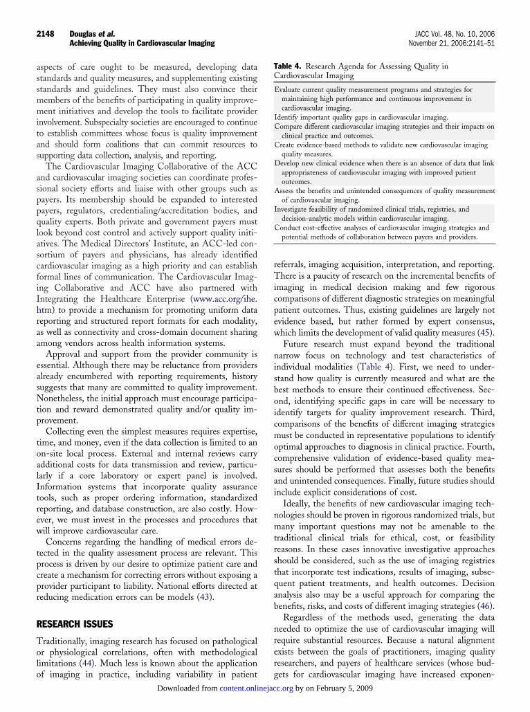
able 4. Research Agenda for Assessing Quality inardiovascular Imaging
Evaluate current quality measurement programs and strategies formaintaining high performance and continuous improvement incardiovascular imaging.
Identify important quality gaps in cardiovascular imaging.Compare different cardiovascular imaging strategies and their impacts on
clinical practice and outcomes.Create evidence-based methods to validate new cardiovascular imaging
quality measures.Develop new clinical evidence when there is an absence of data that link
appropriateness of cardiovascular imaging with improved patientoutcomes.
Assess the benefits and unintended consequences of quality measurementof cardiovascular imaging.
Investigate feasibility of randomized clinical trials, registries, anddecision-analytic models within cardiovascular imaging.
Conduct cost-effective analyses of cardiovascular imaging strategies andpotential methods of collaboration between payers and providers.
ets for cardiovascular imaging have increased exponen- by on February 5, 2009 c.org

tp
C
Ttitdfifttaam
csrtidsppesd
RDDd
R
1
1
1
1
1
1
1
1
1
1
2
2
2
2
2
2
2
2
2
2
3
3
3
3
3
2149JACC Vol. 48, No. 10, 2006 Douglas et al.November 21, 2006:2141–51 Achieving Quality in Cardiovascular Imaging
ially), productive collaboration between investigators andayers should be explored.
ONCLUSIONS
he ACC–Duke quality in imaging meeting was an ex-raordinary collaboration of stakeholders in cardiovascularmaging that accomplished multiple important steps leadingo improved quality. The consensus development of theimensions of care framework for assessing quality identi-ed common themes and concerns that lay the foundationor subsequent work for each imaging modality. It is hopedhat each subspecialty society and its members will commito move rapidly from theoretical discussions to the creationnd implementation of specific measures. We anticipate annnual series of stakeholder meetings to encourage efforts,easure progress, and ensure coordination.Strong leadership is needed to accomplish these perhaps
ostly, perhaps difficult, but necessary undertakings. To beuccessful, this effort will require commitments from a broadange of practitioners, payers, and policymakers. Commit-ed individuals should work with the ACC, cardiovascularmaging societies, payers, and industry to continue toevelop the tools and processes described. Each laboratoryhould embrace continuous quality improvement and im-lement agreed-on measures to achieve a high level oferformance. It is a professional mandate for all stakehold-rs to ensure that cardiovascular imaging is subject to theame quality considerations as more invasive or potentiallyirectly harmful treatments.
eprint requests and correspondence: Dr. Pamela S. Douglas,uke University Medical Center 3943, Duke North 7451,urham, North Carolina 27710. E-mail: pamela.douglas@
uke.edu.
EFERENCES
1. Lohr KN, Schroeder SA. A strategy for quality assurance in Medicare.N Engl J Med 1990;322:707–12.
2. Jencks SF, Huff ED, Cuerdon T. Change in the quality of caredelivered to Medicare beneficiaries, 1998–1999 to 2000–2001. JAMA2003;289:305–12.
3. Mehta RH, Montoye CK, Gallogly M, et al. Improving quality of carefor acute myocardial infarction: the Guidelines Applied in Practice(GAP) Initiative. JAMA 2002;287:1269–76.
4. Krumholz HM, Anderson JL, Brooks NH, et al. ACC/AHA clinicalperformance measures for adults with ST-elevation and non–ST-elevation myocardial infarction. J Am Coll Cardiol 2006;47:236–65.
5. Bonow RO, Bennett S, Casey DE Jr., et al. ACC/AHA clinicalperformance measures for adults with chronic heart failure. J Am CollCardiol 2005;46:1144–78.
6. Dartmouth Medical School, Center for the Evaluative Clinical Sci-ences. Dartmouth Atlas of Cardiovascular Health Care. Chicago, IL:AHA Press, 1999.
7. Lucas FL, Wennberg DE, Malenka DJ. Variation in the use ofechocardiography. Eff Clin Pract 1999;2:71–5.
8. Donabedian A. Evaluating the quality of medical care. MilbankMemorial Fund Quarterly 1966;44 Suppl:166–206.
9. American College of Radiology. Available at: www.acr.org/accreditation/Ultrasound/ultrasound.html. Accessed October 1, 2006.
0. Macfarlane CR. ACR accreditation of nuclear medicine and PETimaging departments. J Nucl Med Technol 2006;34:18–24.
content.onlinejacDownloaded from
1. American Society of Echocardiography. Proposed local coveragedetermination (LCD) language relating to laboratory accreditation andphysician and sonographer qualifications for transthoracic echocardiogra-phy. Available at: www.asecho.org/Members/Legislation_and_Regulation/mlmp.php. Accessed September 29, 2006.
2. American Society of Nuclear Cardiology. Policy Statement on Man-datory Accreditation and Certification. Available at: www.asnc.org/explore/policystatement0205.cfm. Accessed September 29, 2006.
3. Spertus JA, Eagle KA, Krumholz HM, et al. American College ofCardiology and American Heart Association methodology for theselection and creation of performance measures for quantifying thequality of cardiovascular care. Circulation 2005;111:1703–12.
4. Wolk MJ, Peterson E, Brindis R, Eagle K. President’s page: theappropriate cardiologist: responsible stewardship in a golden era ofcardiology. J Am Coll Cardiol 2004;44:933–5.
5. Patel MR, Spertus JA, Brindis RG, et al. ACCF proposed method forevaluating the appropriateness of cardiovascular imaging. J Am CollCardiol 2005;46:1606–13.
6. American College of Radiology. American College of Radiology ACRappropriateness criteria 2000. Radiology 2000;215 Suppl:1–1511.
7. Brindis RG, Douglas PS, Hendel RC, et al. ACCF/ASNC appropri-ateness criteria for single-photon emission computed tomographymyocardial perfusion imaging (SPECT MPI). J Am Coll Cardiol2005;46:1587–605.
8. Scanlon PJ, Faxon DP, Audet AM, et al. ACC/AHA guidelines forcoronary angiography. J Am Coll Cardiol 1999;33:1756–824.
9. Wennberg DE, Kellett MA, Dickens JD, Malenka DJ, Keilson LM,Keller RB. The association between local diagnostic testing intensityand invasive cardiac procedures. JAMA 1996;275:1161–4.
0. Balter S, Heupler FA, Lin PJ, Wondrow MH. A new tool forbenchmarking cardiovascular fluoroscopes. Catheter Cardiovasc Interv2001;52:67–72.
1. Ehler D, Carney DK, Dempsey AL, et al. Guidelines for cardiacsonographer education. J Am Soc Echocardiogr 2001;12:165–72.
2. Beller GA, Bonow RO, Fuster V. ACCF 2006 update for training inadult cardiovascular medicine (focused update of the 2002 COCATS2 training statement): introduction. J Am Coll Cardiol 2006;47:894–7.
3. Budoff MJ, Cohen MC, Garcia MJ, et al. ACCF/AHA clinicalcompetence statement on cardiac imaging with computed tomographyand magnetic resonance. J Am Coll Cardiol 2005;46:383–402.
4. Weinreb JC, Larson PA, Woodard PK, et al. American College ofRadiology clinical statement on noninvasive cardiac imaging. Radiol-ogy 2005;235:723–7.
5. Quinones MA, Douglas PS, Foster E, et al. ACC/AHA clinicalcompetence statement on echocardiography. J Am Coll Cardiol2003;41:687–708.
6. Rodgers GP, Ayanian JZ, Balady G, et al. American College ofCardiology/American Heart Association clinical competence state-ment on stress testing. J Am Coll Cardiol 2000;36:1441–53.
7. Berger AK, Gottdiener JS, Yohe MA, Guerrero JL. Epidemiologicalapproach to quality assessment in echocardiographic diagnosis. J AmColl Cardiol 1999;34:1831–6.
8. Kisslo J, Adams DB. Reporting of preliminary data: time to take oursonographers “off the hook.” J Am Soc Echocardiogr 1991;4:6–9.
9. Hendel RC, Wackers FJ, Berman DS, et al. American Society ofNuclear Cardiology consensus statement: reporting of radionuclidemyocardial perfusion imaging studies. J Nucl Cardiol 2003;10:705–8.
0. Cerqueira MD, Weissman NJ, Dilsizian V, et al. Standardizedmyocardial segmentation and nomenclature for tomographic imagingof the heart. Circulation 2002;105:539–42.
1. Tilkemeirer PL, Cooke CD, Ficaro EP, Glover DK, Hansen CL,MaCallister BD. Standardized reporting matrix for radionuclide myo-cardial perfusion imaging. Available at: www.asnc.org/yourpractice/standardizedreportingmatrix.pdf. Accessed September 29, 2006.
2. Stewart WJ, Aurigemma GP, Bierman FZ, et al. Guidelines fortraining in adult cardiovascular medicine. Core Cardiology TrainingSymposium (COCATS). Task Force 4: training in echocardiography.J Am Coll Cardiol 1995;25:16–9.
3. Thiele BL. Accreditation of vascular laboratories. Semin Vasc Surg1994;7:268–72.
4. Creager MA, Goldstone J, Hirshfeld JW Jr., et al. ACC/ACP/SCAI/
SVMB/SVS clinical competence statement on vascular medicine andby on February 5, 2009 c.org

3
3
3
3
3
4
4
4
4
4
4
4
A
PC
CJeBCaCRSSJCCP
DoBBbMCzDPHAlasAcSCocSKMDsPAMJDWRBtowMPMMPrMoCCRRDSBTtS
2150 Douglas et al. JACC Vol. 48, No. 10, 2006Achieving Quality in Cardiovascular Imaging November 21, 2006:2141–51
catheter-based peripheral vascular interventions. J Am Coll Cardiol2004;44:941–57.
5. Gerhard-Herman M, Gardin JM, Jaff M, Mohler E, Roman M, NaqviT. Guidelines for noninvasive vascular laboratory testing. J Am SocEchocardiogr 2006;19:955–72.
6. Wackers FJ. Blueprint of the accreditation program of the IntersocietalCommission for the Accreditation of Nuclear Medicine Laboratories.J Nucl Cardiol 1999;6:372–4.
7. Cerqueira MD, Berman DS, DiCarli MF, et al. Task Force 5: trainingin nuclear cardiology. J Am Coll Cardiol 2006;47:898–904.
8. Budoff MJ, Achenbach S, Fayad Z, et al. Task Force 12: training inadvanced cardiovascular imaging (computed tomography). J Am CollCardiol 2006;47:915–20.
9. Hendel RC, Patel MR, Kramer CM, Poon M. CCF/ACR/SCCT/SCMR/ASNC/NASCI/SCAI/SIR 2006 appropriateness criteria forcardiac computed tomography and cardiac magnetic resonance imag-ing: a report of the American College of Cardiology Foundation/American College of Radiology, Society of Cardiovascular ComputedTomography, Society for Cardiovascular Magnetic Resonance, Amer-ican Society of Nuclear Cardiology, North American Society forCardiac Imaging, Society for Cardiovascular Angiography and Inter-ventions, and Society of Interventional Radiology. J Am Coll Cardiol2006;48:1475–97.
0. Taylor AJ, Udelson JE, Fuster V, American College of CardiologyFoundation’s Cardiovascular Imaging Committee and the Cardiovas-cular Training Directors Committee. Training cardiovascular fellowsin cardiovascular magnetic resonance and vascular imaging; currentstatus following the core cardiovascular training symposium(COCATS-2) guidelines. J Am Coll Cardiol 2004;43:2108–12.
1. Pohost GM, Kim RJ, Kramer CM, Manning WJ. Task Force 12:training in advanced cardiovascular imaging (cardiovascular magneticresonance [CMR]). J Am Coll Cardiol 2006;47:910–4.
2. Hirshfeld JW Jr., Balter S, Brinker JA, et al. ACCF/AHA/HRS/SCAI clinical competence statement on physician knowledge tooptimize patient safety and image quality in fluoroscopically guidedinvasive cardiovascular procedures. Circulation 2005;111:511–32.
3. Institute of Medicine, Committee on Quality of Health Care inAmerica. To Err is Human: Building a Safer Health System.Washington, DC: National Academy Press, 2000.
4. Lijmer JG, Mol BW, Heisterkamp S, et al. Empirical evidence ofdesign-related bias in studies of diagnostic tests. JAMA 1999;282:1061–6.
5. Spertus JA, Radford MJ, Every NR, et al. Challenges and opportuni-ties in quantifying the quality of care for acute myocardial infarction.J Am Coll Cardiol 2003;41:1653–63.
6. Heidenreich PA, Masoudi FA, Maini B, et al. Echocardiography inpatients with suspected endocarditis: a cost-effectiveness analysis. Am JMed 1999;107:198–208.
PPENDIX 1. ACC–Duke Think Tank Participantsarticipants in the ACC–Duke Think Tank on Quality inardiovascular Imaging (Washington, DC, January, 2006):Brian G. Abbott, MD, American Society of Nuclear
ardiology; Joe Alexander, Jr., MD, PhD, Pfizer, Inc.;oseph Allen, MA, American College of Cardiology; Rob-rt S. Balaban, PhD, National Institutes of Health; Michaelecker, GE Healthcare; Ralph Brindis, MD, Americanollege of Cardiology; Randall Brockman, MD, U.S. Food
nd Drug Administration; John E. Brush, MD, Cardiologyonsultants Ltd.; Robert M. Califf, MD, Duke Clinicalesearch Institute; Manuel D. Cerqueira, MD, Americanociety of Nuclear Cardiology; Charles Chambers, MD,ociety for Cardiovascular Angiography and Interventions;ersey Chen, MD, MPH, Beth Israel Deaconess Medicalenter; Karen Collishaw, MP, AAmerican College ofardiology; Robert Davis, Cardiology Sales and Marketing;
amela S. Douglas, MD, Duke University Medical Center; bcontent.onlinejacDownloaded from
awn Edgerton, Certification Board of Nuclear Cardiol-gy; Kathleen Flood, American College of Cardiology;rett Fulton, Bristol-Myers Squibb; Peter Gardiner, MD,ristol-Myers Squibb Medical Imaging; Raymond J. Gib-ons, MD, American Heart Association; Linda Gillam,D, American Society of Echocardiograpy, Americanollege of Cardiology/Coalition of Cardiovascular Organi-
ations; Barbara Greenan, American College of Cardiology;avid Gundlach, MD, Anthem Blue Cross/Blue Shield;aul Heidenreich, MD, Stanford University; Robert C.endel, MD, American Society of Nuclear Cardiology,merican College of Cardiology/Coalition of Cardiovascu-
ar Organizations; Karen A. Hicks, MD, FACC, U.S. Foodnd Drug Administration; Eva Hill, Duke Clinical Re-earch Institute; Ami E. Iskandrian, MD, University oflabama at Birmingham; Neil C. Jensen, United Health-
are; James G. Jollis, MD, Duke University Medical Center;andra Katanick, RN, CAE, Intersocietal Accreditationommission; Carrie Kovar, American College of Cardiol-gy; Christopher M. Kramer, MD, Society for Cardiovas-ular Magnetic Resonance; Harlan M. Krumholz, MD,M, Yale University School of Medicine; Iacovos (Jake)yprianou, PhD, U.S. Food and Drug Administration; Lisa. Latts, MD, WellPoint, Inc.; David J. Malenka, MD,artmouth-Hitchcock Medical Center; Frederick A. Ma-
oudi, MD, Denver Health Medical Center; Rick Mather,hD, Toshiba America Medical Systems; Tilithia McBride,merican College of Cardiology; Robert McNamara, MD,HS, Yale University; Phil Mendys, PharmD, Pfizer, Inc.;
ulie M. Miller, MD, FACC, Johns Hopkins University;iane Millman, JD, Powers, Pyles, Sutter and Verville;ally Mlynarski, Siemens Medical Solutions, Inc.; Emile
. Mohler III, MD, Society for Vascular Medicine andiology; Marelle Molbert, Duke Clinical Research Insti-
ute; James W. Moser, PhD, American College of Radiol-gy; Gunnar Olsson, MD, PhD, AstraZeneca; Jerry Olsze-ski, Astellas Pharma US, Inc.; Lucinda Orsini, DPM,PH, Bristol-Myers Squibb Medical Imaging; Douglas L.
acker, MD, Heart Rhythm Society; Manesh R. Patel,D, Duke Clinical Research Institute; Eric D. Peterson,D, Duke Clinical Research Institute; Robert Phillips,
hD, U.S. Food and Drug Administration; Steve Phur-ough, MD, Centers for Medicare and Medicaid Services;
ichael H. Picard, MD, American Society of Echocardi-graphy; Michael Poon, MD, Society of Cardiovascularomputed Tomography; Wayne Powell, MPS, Society forardiovascular Angiography and Interventions; Martha J.adford, MD, New York University Medical Center; Joelaichlen, MD, AstraZeneca; John S. Rumsfeld, MD, PhD,enver VA Medical Center; Thomas Ryan, MD, American
ociety of Echocardiography; Jay Schukman, MD, Anthemlue Cross/Blue Shield; Milton D. Schwarz, MD, Aetna;homas B. Shope, PhD, U.S. Food and Drug Administra-
ion; Jack J. Slosky, PhD, Bristol-Myers Squibb; Johnpertus, MD, Mid America Heart Institute; John E. Stro-
eck, MD, PhD, Heart Failure Society of America; Allen J.by on February 5, 2009 c.org

TToASS
SAWAB
A
P
J
L
R
J
A
H
F
E
R
M
E
J
2151JACC Vol. 48, No. 10, 2006 Douglas et al.November 21, 2006:2141–51 Achieving Quality in Cardiovascular Imaging
aylor, MD, Society of Atherosclerosis Imaging; Miahomas Rosenberg, MBA, American College of Cardiol-gy; Douglas Throckmorton, MD, U.S. Food and Drugdministration; James E. Udelson, MD, Heart Failureociety of America; David Urani, MBA, Astellas Pharma;
andra Vogeler, RN, Siemens; Wm. Guy Weigold, MD, Dcontent.onlinejacDownloaded from
ociety of Cardiovascular Computed Tomography; Pamela. Wilcox, RN, American College of Radiology; Kim Allanilliams, MD, University of Chicago; Michael Wolk, MD,
merican College of Cardiology; Martin Wong , Pointiomedical Corp.; Bram Zuckerman, MD, U.S. Food and
rug Administration.PPENDIX 2. Writing Group Relationships With Industry
Committee Member Research GrantSpeakersBureau Stock Ownership
Board ofDirectors
Consultant/AdvisoryMember
amela Douglas, MD, MACC ● Medtronic None ● Millenium● Northpoint Domain
None ● GE Healthcare● Merck● Northpoint Domain
ersey Chen, MD None None None None None
inda Gillam, MD, FACC None None None None None
obert Hendel, MD, FACC None None None None None
ames Jollis, MD, FACC None None None None None
mi E. Iskandrian, MD, FACC ● CV Therapeutics● Molecular Imaging● GE● BMS● Astellas● Berlex● Pfizer
None None None ● CV Therapeutics● Astellas
arlan M. Krumholz, MD, FACC None None None None None
rederick Masoudi, MD, FACC None None None None ● UnitedHealth
mile Mohler III, MD, FACC None None None None None
obert L. McNamara, MD, MHS, FACC None None None None None
anesh R. Patel, MD None None None None None
ric Peterson, MD, FACC None None None None None
ohn Spertus, MD, FACC None None None None None
by on February 5, 2009 c.org

doi:10.1016/j.jacc.2006.06.076 2006;48;2141-2151; originally published online Oct 31, 2006; J. Am. Coll. Cardiol.
III, Robert L. McNamara, Manesh R. Patel, and John Spertus Mohler,Hendel, James Jollis, Eric Peterson, Jersey Chen, Frederick Masoudi, Emile
Pamela Douglas, Ami E. Iskandrian, Harlan M. Krumholz, Linda Gillam, Robert in Cardiovascular Imaging
QualityCollege of Cardiology–Duke University Medical Center Think Tank on Achieving Quality in Cardiovascular Imaging: Proceedings From the American
This information is current as of February 5, 2009
& ServicesUpdated Information
http://content.onlinejacc.org/cgi/content/full/48/10/2141including high-resolution figures, can be found at:
References
Lhttp://content.onlinejacc.org/cgi/content/full/48/10/2141#BIBfree at: This article cites 39 articles, 28 of which you can access for
Citations
articleshttp://content.onlinejacc.org/cgi/content/full/48/10/2141#otherThis article has been cited by 14 HighWire-hosted articles:
Rights & Permissions
http://content.onlinejacc.org/misc/permissions.dtltables) or in its entirety can be found online at: Information about reproducing this article in parts (figures,
Reprints http://content.onlinejacc.org/misc/reprints.dtl
Information about ordering reprints can be found online:
by on February 5, 2009 content.onlinejacc.orgDownloaded from