Embed Size (px)
Citation preview

Acetaminophen-mediated cardioprotection via inhibition of the mitochondrial
permeability transition pore-induced apoptotic pathway
by
Norell Melissa Hadzimichalis
A Dissertation submitted to the
Graduate School-New Brunswick
Rutgers, The State University of New Jersey
and The Graduate School of Biomedical Sciences
University of Medicine and Dentistry of New Jersey
in partial fulfillment of the requirements for the degree of
Doctor of Philosophy
Graduate program in Physiology and Integrative Biology
Written under the direction of
Gary F. Merrill, Ph.D.
And approved by
___________________________________________
___________________________________________
___________________________________________
___________________________________________
___________________________________________
New Brunswick, New Jersey
May, 2008
ABSTRACT OF THE DISSERTATION
Acetaminophen-mediated cardioprotection via inhibition of the mitochondrial
permeability transition pore-induced apoptotic pathway
by NORELL MELISSA HADZIMICHALIS
Dissertation Director:
Gary F. Merrill, Ph.D.
Historically, acetaminophen has been employed as a safe and effective
analgesic and antipyretic agent. However, our laboratory has recently reported
that acetaminophen also confers functional cardioprotection following cardiac
insult, including ischemia/reperfusion, hypoxia/reoxygenation, and exogenous
peroxynitrite and hydrogen peroxide administration. In the current study, we
examined the mechanism of acetaminophen-mediated cardioprotection following
ischemia/reperfusion injury. Langendorff-perfused guinea pig hearts were
exposed to acute treatment with acetaminophen (0.35 mM) or vehicle (Krebs-
Henseleit buffer) beginning at 15 minutes of a 30-minute baseline stabilization
period. Low-flow global myocardial ischemia was subsequently induced for 30
minutes followed by 60 minutes of reperfusion. Upon completion of reperfusion,
ii

hearts were homogenized and separated into cytosolic and mitochondrial
fractions. Mitochondrial swelling and mitochondrial cytochrome c release were
assessed and found to be significantly and completely reduced following
reperfusion in acetaminophen-treated hearts when compared to vehicle. In a
separate group of hearts, ventricular myocytes were isolated and subjected to
fluorescence-activated cell sorting. Acetaminophen-treated hearts showed a
significant decrease in late stage apoptotic myocytes when compared to vehicle-
treated hearts following injury (58±1% vs. 81±5%, respectively). These data,
together with electron micrograph analysis, suggest that acetaminophen
mediates cardioprotection, in part, by inhibiting the mitochondrial permeability
transition pore and subsequent apoptotic pathway.
iii

ACKNOWLEDGEMENTS
I would like to express my gratitude to the members of my committee: Drs. Carol
A. Bagnell, Bonnie L. Firestein, Jianjie Ma, Nansie A. McHugh, and Gary F.
Merrill for their generous donation of time and guidance in the pursuit of my
doctoral degree.
I would like to specifically thank the following individuals: Dr. Gary F. Merrill for
his mentorship and guidance throughout my graduate studies; Drs. Tyler H. Rork
and Roseli Golfetti for their generous donation of both time and advice; Sunanda
Baliga and Kathryn Jaques for their friendship and collaboration on the study;
and Carole Lewandowski for her ongoing friendship and guidance.
I wish to also thank my family and friends for their unconditional love and
support. Specifically, I would like to thank my parents Charles and Jodi Spiler for
their guidance, patience, encouragement, and love. Finally, I would like to thank
my husband, Anthony Hadzimichalis, for supporting, encouraging, and loving me.
Without these people, none of this would be possible.
iv

TABLE OF CONTENTS
ABSTRACT OF THE DISSERTATION ………………….……....………………… ii
ACKNOWLEDGEMENTS ……………………………..………………….…………. iv
LIST OF TABLES………………………….………………………………………….viii
LIST OF FIGURES………………………………………………………….…………ix
I. INTRODUCTION………………………………………………………..……………1
1. Background……………………………………………………………...……….…2
1.1 A brief history of acetaminophen…………………………………..….…….2
1.2 Acetaminophen as an analgesic antipyretic agent…………………...…..5
1.3 Acetaminophen as a cardioprotective agent………………………………6
1.4 Cytochrome c, apoptotic cell death, and myocardial
ischemia/reperfusion……………………………………………………………14
1.5 The mitochondrial permeability transition pore ..……………………......19
2. Purpose…………………………………………………………………………….22
2.1 A mechanistic elucidation of acetaminophen-mediated
cardioprotection…………………………………………………………………....22
2.2 Acetaminophen and its effects on the mitochondrial permeability transition
pore…..……………………………………………………………………………..22
2.3 Acetaminophen and its effects on mitochondrial cytochrome c
release……………………………………………………………………………...23
2.4 Acetaminophen and its effects on mitochondrial-mediated apoptosis…..23
II. MATERIALS AND METHODS………………………………………………..….25
1. Experimental preparation………………………………………………………...26
v

1.1 Animals and Langendorff preparation…………….…………...……………26
1.2 Perfusate and perfusion…………………………..………..………………...26
1.3 Statistical analysis………………………………….……………..…………..28
2. Experimental protocols…………………………………………………………...31
2.1 Myocardial homogenization and fractionation…………….....…………….31
2.2 Mitochondrial swelling………………………………..………….……………32
2.3 Miofibrillar ultrastructure…………………………………………….....……..32
2.4 Mitochondrial cytochrome c release…………………………….…………..33
2.5 Isolation of ventricular myocytes and fluorescence-activated cell
sorting……………………………………………………………………………….34
III. RESULTS………………………………………….………………………………36
1. Hemodynamic and metabolic parameters……………………………………..37
2. Acetaminophen treatment inhibits mitochondrial swelling following
myocardial ischemia/reperfusion…………………………………………...…...…40
3. Molecular consistency between vehicle-treated hearts………………………46
4. Acetaminophen treatment inhibits mitochondrial cytochrome c release
following myocardial ischemia/reperfusion…………….………………………….48
5. Acetaminophen treatment attenuates the number of late-stage apoptotic
myocytes following myocardial ischemia/reperfusion……………….…………...52
IV. DISCUSSION……………………………………………………………………..57
1. Rationale………………………………………………………….……………….58
2. The Langendorff perfusion……………………………………………………….61
2.1 Advantages and limitations of the Langendorff preparation…...….....…61
vi

2.2 The Langendorff-perfused guinea pig heart model……………..……….62
3. Acetaminophen; therapeutic dosages and experimental concentrations…..63
4. Acetaminophen-mediated inhibition of mitochondrial swelling and MPTP
opening following ischemia/reperfusion…………………………………….……..64
5. Acetaminophen-mediated inhibition of mitochondrial cytochrome c release
following ischemia/reperfusion…………………………………………….……….66
6. Acetaminophen-mediated attenuation of late stage apoptosis following
ischemia/reperfusion…………………….……………………………………..……67
7. Future directions……………………….……………………………………….…70
V. REFERENCES…………….……………..………………………………………..73
VITA…...……...……………………..………………………………………………….80
Permission to reproduce figures/tables……………………………………………..82
vii

LIST OF TABLES
Table Title Page
1 Current studies on the effect of acetaminophen during various
cardiovascular injuries.
12
2 Hemodynamic and metabolic data during myocardial
ischemia/reperfusion.
38
3 Summary table of study findings as they relate to the
mechanism of acetaminophen-mediated cardioprotection
following ischemia/reperfusion.
72
viii

LIST OF FIGURES
Figure Title Page
1 Chemical structure and space-filling model of acetaminophen
(paracetamol, APAP).
4
2 Schematic of the intrinsic/mitochondrial pathway for apoptotic
cell death.
18
3 Schematic of the minimal MPTP structure. 21
4 Schematic of modified Langendorff perfusion apparatus. 29
5 Schematic of experimental ischemia/reperfusion timeline. 30
6 Spectrophotometric analysis of mitochondrial swelling. 42
7 Electron micrograph analysis of left ventricle free wall. 44
8 Western blot analysis of cytosolic cytochrome c content
following 15 minutes of baseline perfusion.
47
9 Western blot analysis of cytosolic and mitochondrial cytochrome
c heart homogenate fractions following ischemia/reperfusion.
49
10 FACS analysis of post-ischemia/reperfused ventricular
myocytes.
54
11 Representative FACS analysis of vehicle-treated
ischemia/reperfused heart.
56
12 Schematic of proposed mechanism of action of acetaminophen
following myocardial ischemia/reperfusion.
69
ix

1
I. INTRODUCTION

2
1. Background
1.1 A brief history of acetaminophen
Historically, acetaminophen (paracetamol, APAP; Figure 1) has been
employed as an analgesic and antipyretic agent. Today it remains a key
ingredient in many popular over-the-counter medications including Tylenol,
Anacin, and Datril. Acetaminophen was originally synthesized by the reduction
of p-nitrophenol to p-aminophenol with tin and glacial acetic acid followed by
acetylation (Prescott, 2001). It was first used clinically by von Mering (1893),
however, despite potent antipyretic and analgesic properties it was determined
that the side effects were too great to recommend use. Additional studies by
Hinsberg and Treupel (1894) further detailed the antipyretic properties of
acetaminophen. They determined that a 500 mg oral dose of acetaminophen
was as effective in reducing fever as 700 mg phenacetin or 1 g antipyrine,
medically accepted drugs for fever reduction in the late 19th century. Despite
promising preliminary studies on the antipyretic and analgesic properties of
acetaminophen, other drugs such as aspirin, phenacetin, acetanilide, and
antipyrine remained more popular until the mid 20th century (Prescott, 2001).
In 1948, Brodie and Axelrod (1948a; 1948b) discovered acetaminophen to
be the major metabolite of acetanilide and phenacetin in man. This finding
renewed interest in the drug and provoked promotion of acetaminophen as a
“Triogesic” in combination with aspirin and caffeine in the United States in 1950.

3
Acetaminophen became available as a non-prescription drug in 1955 and was
subsequently marketed in the United Kingdom in 1956. Prolific investigation
found acetaminophen to be as effective as aspirin in reducing fever and pain
caused by radiant heat, cancer, dental surgery, or arthritis. Studies spanning the
following two decades confirmed the safety of this drug claiming that, unlike other
popular analgesic agents of the time, acetaminophen did not produce
gastrointestinal toxicity. Today acetaminophen remains one of the leading over-
the-counter drugs used for reducing both fever and pain (Prescott, 2001).

4
A
B
Figure 1. (A) Chemical structure and (B) space-filling model of acetaminophen
(paracetamol, APAP). The benzene ring core is substituted by one hydroxyl
group, which distinguishes this compound as a phenol.

5
1.2 Acetaminophen as an analgesic antipyretic agent
Since isolation of the constitutively expressed cyclooxygenase (COX)
enzyme in 1976 (Hemler and Lands) and discovery of its inducible COX-2
isoform in 1991 (Xie et al.), much investigative effort has focused on their roles
in both basic research and clinical environments. Non-steroidal anti-
inflammatory drugs (NSAIDs), commonly used to treat inflammation, joint pain,
headache, and fever, have been shown to produce gastrointestinal toxicity when
used chronically (Vane and Botting, 1997). The basis for these adverse effects
was thought to be related to COX-1 inhibition, while the positive antipyretic,
analgesic, and anti-inflammatory effects are thought to be associated with COX-2
inhibition (Masferrer et al., 1994; Seibert and Masferrer, 1994; Luo et al., 2005)
However, shortly after their commercial introduction, COX-2-specific inhibitors
were reported to produce unfavorable cardiovascular side effects and resulted in
voluntary manufacturer withdrawal of the compounds (Cotter and Wooltorton,
2005; Luo et al., 2005; Salzberg and Weir, 2007).
Despite widespread use as both an analgesic and antipyretic agent, the
mechanism of acetaminophen’s action in this regard is not fully clear. Unlike
NSAIDs with similar effects in reducing pain and fever, acetaminophen lacks anti-
inflammatory capabilities. Studies suggest that acetaminophen acts to inhibit
central prostaglandin synthesis by competing with arachidonic acid for the active
site on the COX enzyme (Botting, 2000). However, the exact nature of COX
enzyme inhibition is controversial. While some investigators report that
6
acetaminophen attenuates prostaglandin synthesis by inhibiting a novel COX
enzyme, COX-3, other investigators claim that COX-3 is merely a COX-1 splice
variant (Botting, 2000; Kis et al., 2005).
1.3 Acetaminophen as a cardioprotective agent
In addition to its role as an analgesic and antipyretic agent,
acetaminophen has been reported to exhibit cardioprotective efficacy when
administered during ischemia/reperfusion, hypoxia/reoxygenation, or exogenous
peroxynitrite and hydrogen peroxide administration. We have found both chronic
and acute acetaminophen treatment to be cardioprotective following
ischemia/reperfusion in the isolated perfused guinea pig myocardium (Merrill et
al., 2001; Merrill and Goldberg, 2001; Golfetti et al ., 2002; Golfetti et al., 2003).
Additional studies from our laboratory have demonstrated that acute
acetaminophen treatment also provides protection in a canine model of
myocardial infarction (Merrill et al., 2004).
Using isolated and perfused guinea pig hearts, we have established that
both chronic and acute acetaminophen administration preserve the myocardium
structurally and functionally (Merrill et al., 2001; Merrill and Goldberg, 2001;
Golfetti et al., 2002; Golfetti et al., 2003). In acute studies, acetaminophen-
treated hearts (0.35 mM) exhibited greater preservation of mechanical function
(i.e. left ventricular developed pressure, LVDP), myofibrillar ultrastructure, and
significant attenuation of reactive oxygen species when compared to vehicle-
7
treated hearts following 20 minutes of low-flow global myocardial ischemia and
40 minutes of reperfusion (Merrill and Goldberg, 2001). Additional work from
Golfetti et al. (2002) showed that creatine kinase activity (an indicator of tissue
damage) was also significantly reduced during reperfusion in acetaminophen-
treated hearts.
In chronic studies, guinea pigs were given acetaminophen-treated drinking
water (0.35 mM) ad libitum for 10 days. Hearts were subsequently extracted and
subjected to ischemia/reperfusion as described above. Golfetti et al. (2003)
established that hearts chronically treated with acetaminophen experienced
similar protection to those in the acute studies. For example, acetaminophen-
treated hearts demonstrated significantly greater retention of LVDP, attenuation
of reactive oxygen species, and preserved myofibrillar ultrastructure when
compared to vehicle-treated hearts. Taken together, these studies suggest that
the mechanical, structural, and biochemical cardioprotective efficacy of
acetaminophen during ischemia/reperfusion extends from an acute to a chronic
treatment environment.
Canine studies corroborate these findings and further demonstrate the
cardioprotective efficacy of acetaminophen following ischemia/reperfusion.
Merrill et al. (2004) examined vehicle- and acetaminophen-treated (total dose, 30
mg/kg iv) dogs exposed to 60 minutes left anterior descending coronary artery
occlusion followed by 180 minutes of reperfusion. At the completion of the
experiment, hearts were simultaneously stained with Evan’s blue dye and
triphenyltetrazolium chloride to visualize viable tissue outside and inside the area

8
at risk, respectively. Necrotic tissue remained colorless. When compared to
vehicle-treated hearts, acetaminophen-treated hearts were found to have
significantly more viable tissue, a greater ability of coronary venous effluent to
attenuate peroxynitrite, and visibly preserved myofibrillar ultrastructure. These
results demonstrate the translative capacity of ex vivo studies to the in vivo
arena.
More recent reports from Zhu et al. (2006) further support these data. In
these studies rats were treated daily with 5 mg/kg intraperitoneal injections of
acetaminophen beginning 7 days prior to surgery (permanent left coronary artery
ligation) and extending until 2 days following the surgery. Results showed that
acetaminophen-treated rats experienced a significant reduction in mortality rate
following myocardial infarction when compared to vehicle-treated rats. In
addition, electrocardiograms of treated rats 10 days post-treatment showed
noticeable reductions in ST elevation (an indicator of electrical damage following
myocardial infarction) when compared to vehicle. Triphenyltetrazolium chloride
staining was also used to show a significant reduction in left ventricular infarct
size in acetaminophen- versus vehicle-treated rats.
Based on the results of these animal studies, we conclude that
acetaminophen has a cardioprotective role during ischemia/reperfusion. It is
currently believed that the mechanism of action may involve antioxidant
properties of this drug conveyed by its phenolic structure (Figure 1). Additional
work is required in order to further delineate the pathway for this observed
protection.

9
Rork et al. (2004) have extended this work with acetaminophen to
investigate its effects in the setting of hypoxia/reoxygenation. In these studies,
our laboratory exposed isolated perfused guinea pig hearts to 6 minutes of
hypoxia followed by 36 minutes of reoxygenation and examined hemodynamic,
metabolic, mechanical, ultrastructural, and biochemical indices of function. We
found that acetaminophen-treated hearts retained significantly greater
mechanical function, preserved myofibrillar ultrastructure, and a significantly
greater ability to neutralize peroxynitrite-dependent chemiluminescence at all
recorded time periods. In addition, creatine kinase activity was significantly
decreased during both hypoxia and reoxygenation in acetaminophen- versus
vehicle-treated hearts. Thus, we concluded that the cardioprotective efficacy of
acetaminophen (0.35 mM) could be extended from an ischemia/reperfusion
environment to also include myocardial protection from hypoxia/reoxygenation
injury.
In addition to serving as a cardioprotective agent following
ischemia/reperfusion and hypoxia/reoxygenation, acetaminophen has been
shown to have protective effects in other cardiovascular injuries including
arrhythmogenesis and atherosclerosis. Work from Merrill and Goldberg (2001)
suggests that acetaminophen has the potential to attenuate sodium pentobarbital
induced ventricular arrhythmias ex vivo. Acetaminophen-treated guinea pig
hearts analyzed for ventricular salvos (VS) and ventricular premature beats
(VPB) for 90 minutes post sodium pentobarbital administration were found to be
significantly less arrhythmic when compared to vehicle. Results from this study

10
encouraged more recent in vivo investigation. Merrill et al. (2007) examined the
effects of therapeutic acetaminophen treatment on either oubain- or myocardial
infarction-induced ventricular arrhythmias in dogs. Results revealed that
acetaminophen-treated dogs experienced a significant decrease in percent
ectopy when compared to vehicle-treated dogs.
Atherosclerosis, a cardiovascular disease characterized by
myeloperoxidase-induced LDL oxidation and the development of vascular
atherosclerotic plaques, has also been shown recently to be a target of
acetaminophen administration. Nachtigal et al. (2005) investigated the role of
acetaminophen in the progression of aortic atherosclerosis. After 22 weeks of
acetaminophen treatment (1.3 mg/mouse/day), the average numbers of aortic
plaques, aneurysms, and periaortic vascular channels were significantly reduced
when compared with vehicle-treated apolipoprotein E-deficient mice. In addition,
the number of periaortic inflammatory infiltrates, in the presence of
acetaminophen, was significantly lowered. No significant differences were noted
between groups in either average food intake or average weight gain. These
data suggest that long-term treatment with acetaminophen might be effective in
reducing vascular disease. Additional evidence from hypercholesterolemic
rabbits (Rogers et al., 1999) support the idea of an anti-atherosclerotic role for
acetaminophen via a reduction of vascular fatty streaks. More recently, Chou
and Greenspan (2002) have provided conclusive evidence associating
acetaminophen treatment with a reduction in atherosclerotic plaques. These

11
studies show that 0.25 mM concentrations of acetaminophen and lower attenuate
myeloperoxidase induced LDL metabolism.
For a number of years, mechanistic evidence of acetaminophen-mediated
cardioprotection has been notably lacking. However, recently Rork et al. (2006)
have shown that acetaminophen attenuates peroxynitrite-activated matrix
metalloproteinase-2-mediated troponin I cleavage via direct inhibition of
peroxynitrite in guinea pig hearts. While this work is promising, additional
mechanistic data are essential to further characterize the role of acetaminophen
in providing myocardial protection in cases of ischemia/reperfusion and
hypoxia/reoxygenation injury. Table 1 summarizes the more recent discoveries
concerning acetaminophen treatment during cardiovascular injury.

12
Table 1. Current studies on the effect of acetaminophen during various
cardiovascular injuries.
- acute treatment, 0.3-0.6mM hydroxyl radicals and peroxynitrite). APAPalso provides functional and structuralprotection.
Merrill 2001 Basic Res Cardiol. - Langendorff perfused hearts APAP is cardioprotective against I/R viaand - 20 mins. I / 40 mins. R antioxidant mechanisms (inhibition ofGoldberg - acute treatment, 0.35mM hydroxyl radicals and peroxynitrite). APAP
attenuates ventricular arrhythmias.
Merrill 2002 AJP Heart - Langendorff perfused hearts APAP is cardioprotective against I/R via- 20 mins. I / 40 mins. R antioxidant mechanisms (inhibition of- APAP given at onset of I hydroxyl radicals, peroxynitrite, and - acute treatment, 0.35mM protein oxidation).
Golfetti et al. 2002 Exp Biol Med - Langendorff perfused hearts APAP is cardioprotective against I/R via- 20 mins. I / 40 mins. R antioxidant mechanisms (inhibition of- APAP given at onset of R peroxynitrite) when administered at the- acute treatment, 0.35mM onset of reperfusion. APAP also attenuates
CK production.Chou 2002 Biochim Biophys - culture media APAP is protective againstand Acta. - acute treatment, 0.025- atherosclerosis via inhibition ofGreenspan 0.25mM myeloperoxidase-hydrogen peroxide-nitrate
mediated modification of LDL.
Golfetti et al. 2003 Exp Biol Med - Langendorff perfused hearts APAP is cardioprotective against I/R when- 20 mins. I / 40 mins. R administered chronically. Cardioprotection- APAP given at onset of R is measured as functional and structural- chronic treatment, 0.35mM improvement, and both CK and peroxynitrite
inhibition.Merrill et al. 2004 AJP Heart - MI APAP significantly reduces infarct size.
- left coronary artery ligation- acute treatment, 30mg/kg iv
Rork et al. 2004 Exp Biol Med - Langendorff perfused hearts APAP is cardioprotective against H/ReO- 6 mins. H, 36 mins. ReO via antioxidant mechanisms (inhibition of- acute treatment, 0.35mM peroxynitrite). APAP also provides
significant functional and structuralprotection under this injury.
Rork et al. 2006 J Mol Cell Cardiol. - Langendorff perfused hearts APAP is cardioprotective via- exogenous peroxynitrite attenuation of peroxynitrite-activatedadministration matrixmetalloproteinase-2 mediated- acute treatment, 0.35mM cleavage of troponin I.
Zhu et al. 2006 AJP Heart - MI Chronic APAP treatment significantly- permanent left coronary decreases mortality rate and infarct size.artery ligation In addition, catalase and superoxide- chronic treatment dismutase activities were increased in the5mg/kg/day treated-group.
Merrill et al. 2007 Exp Biol Med - MI and oubain-induced Therapeutic administration of APAP resultsdisturbances in significant attenuation of percent ectopy- acute treatment, 30mg/kg iv and all ventricular ectopic beats (except
ventricular salvos).

13
I, ischemia; R, reperfusion; APAP, acetaminophen; CK, creatine kinase; LDL, low
density lipoproteins; MI, myocardial infarction; H, hypoxia; ReO, reoxygenation
(Merrill and Goldberg, 2001; Chou and Greenspan, 2002; Golfetti et al., 2002;
Merrill, 2002; Golfetti et al., 2003; Rork et al., 2004; Rork et al., 2006; Zhu et al.,
2006; Hadzimichalis et al., 2007).

14
1.4 Cytochrome c, apoptotic cell death, and myocardial ischemia/reperfusion
Post-ischemia reperfusion, although vital for the survival of the
myocardium, results in substantial myocardial damage including cell death
(Sutherland and Hearse, 2000). While the relative contributions of necrosis and
apoptosis to overall tissue injury is a topic of much debate, both pathways have
been clearly implicated following ischemia/reperfusion. Much recent attention
has focused on elucidating the cell death pathways active during cardiovascular
injury in an attempt to explore pathway-related therapeutics.
Apoptosis has been implicated in various cardiovascular diseases
including ischemia/reperfusion injury, myocardial infarction, atherosclerosis, and
end-stage heart failure (MacLellan and Schneider, 1997; Zidar et al., 2007).
Morphologically, apoptosis is characterized by chromatin condensation,
cytoskeletal alterations, membrane blebbing, nuclear fragmentation, and
cytoplasmic condensation. Ultimately, dying cells form apoptotic bodies which
are subsequently engulfed by other cells via phagocytosis. Unlike necrosis,
apoptosis involves an active, energy-dependent cell death, decrease in cell
volume, minimal inflammation, and preservation of membrane integrity
(MacLellan and Schneider, 1997; Haunstetter and Izumo, 1998). Consequently,
gross tissue injury is avoided. In addition, it has been shown that reperfusion
versus ischemia alone leads to a greater increase specifically in apoptotic
myocytes (Gottlieb et al., 1994; Logue et al., 2005).

15
Mechanistically, myocardial cells undergoing apoptosis follow one of two
major death pathways, both leading to aspartate-specific cysteine protease
(caspase) activation. The extrinsic cell death pathway begins extracellularly in
response to apoptotic signals. Ligands initiate apoptosis by binding to their
associated cell surface receptor. Adaptor proteins such as TRADD and FADD,
and procaspase-8 are recruited to the complex and procaspase-8 is cleaved and
subsequently activated (Zhu et al., 2006). Caspase-8 then activates downstream
effector procaspases (i.e. procaspase-3) which ultimately carry out characteristic
end-stage steps of apoptosis such as DNA fragmentation (Foo et al., 2005).
Studies in Langendorff-perfused murine hearts (Jeremias et al., 2000) implicate
this pathway in ischemia/reperfusion-induced cell death. Western blot analysis
revealed enhanced levels of the Fas death receptor ligand CD95 in the
extracellular fluid, particularly during the reperfusion phase of ischemic injury. In
addition, loss of function mutations in the Fas death receptor result in a reduction
of apoptotic cell death following myocardial ischemia/reperfusion (Jeremias et al.,
2000; Krijnen et al., 2002).
Alternatively, the intrinsic (i.e. mitochondrial) cell death pathway is initiated
in response to conditions occurring within the cell and also plays a prominent role
in apoptosis following ischemia/reperfusion injury. Following stimuli such as
oxidative stress, various proteins, including cytochrome c, are released from the
mitochondrial intermembrane space into the cytosol. Cytochrome c then binds to
the apoptosis protease activating factor-1 (Apaf-1) adapter molecule forming the
apoptosome complex, which subsequently recruits and activates procaspase-9.

16
Caspase-9 then cleaves and activates downstream executioner/effector
caspases such as caspase-3 to carry out apoptotic cell death (Green and
Kroemer, 2004; 2005). Borutaite et al. (2003) have implicated the intrinsic cell
death pathway as contributing to myocardial cell death during ischemia. They
demonstrated that following prolonged ischemia, significant amounts of
cytochrome c were present in the cytosol in addition to an increase in caspase-3
activation. More recently Vanden Hoek et al. (2003) have shown that the burst of
damaging oxidants released during reperfusion initiates the intrinsic pathways of
apoptotic cell death. Additionally, these investigators provide evidence that in the
presence of antioxidants 2-mercaptopropionylglycine (MPG) and 1,10-
phenanthroline (Phen), myocytes experience a significant reduction in overall cell
death, cytochrome c release, and nuclear condensation. A simplified schematic
illustrating this pathway can be seen in Figure 2.
Following post-ischemia reperfusion, myocytes experience calcium
overload, ATP and adenine nucleotide depletion, increased phosphate levels,
and oxidative stress. Independent from ischemia/reperfusion, these conditions
have been shown to result in cell death (Duchen, 2004; Halestrap, 2006).
Damaging oxidant release, including peroxynitrite and hydroxyl radicals, is
partially responsible for myocardial damage following ischemia/reperfusion injury
and is primarily associated with the intrinsic cell death pathway and opening of
the MPTP. Since acetaminophen is effective at attenuating post-reperfusion
production of oxidants (i.e. hydroxyl radicals and peroxynitrite), we hypothesized
that its reported cardioprotective effects were mediated, in part, via inhibition of

17
the intrinisic/mitochondrial pathway of apoptosis. (Merrill and Goldberg, 2001;
Merrill, 2002)
18
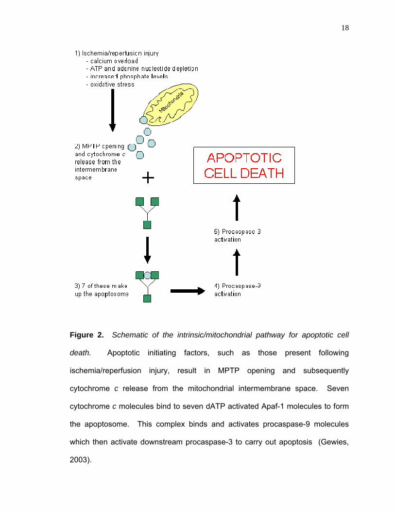
Figure 2. Schematic of the intrinsic/mitochondrial pathway for apoptotic cell
death. Apoptotic initiating factors, such as those present following
ischemia/reperfusion injury, result in MPTP opening and subsequently
cytochrome c release from the mitochondrial intermembrane space. Seven
cytochrome c molecules bind to seven dATP activated Apaf-1 molecules to form
the apoptosome. This complex binds and activates procaspase-9 molecules
which then activate downstream procaspase-3 to carry out apoptosis (Gewies,
2003).

19
1.5 The mitochondrial permeability transition pore
Since its discovery by Hunter et al. (1963), the mitochondrial permeability
transition pore (MPTP) has become the focus of much investigation. Evidence
that prolonged pore opening results in cell death suggests that this structure is an
important physiologic target in the clinical environment (Halestrap et al., 2004).
Opening of the MPTP is mediated by conditions similar to those experienced
following ischemia/reperfusion and hypoxia/reoxygenation injury and includes
calcium overload, depletion of adenine nucleotides, high phosphate levels, and
oxidative stress. As a result, the cell will experience mitochondrial ATP
hydrolysis, uncoupling of oxidative phosphorylation, cytochrome c release from
the intermembrane space, mitochondrial swelling, and ultimately cell death
(Halestrap et al., 2004).
Mitochondrial permeability transition may be either directly or indirectly
inhibited. Agents such as cyclosporin A and sanglifehrin A have been
established as direct inhibitors of the transition pore via an interaction with
cyclophilin-D, a detachable matrix element (Broekemeier et al., 1989; Halestrap
et al., 2004). Figure 3 is a schematic of the MPTP. While there is some
controversy regarding complete structural elucidation of the MPTP, it is widely
accepted that the adenine nucleotide translocase (ANT) and cyclophilin-D
element are crucial components of the MPTP (Javadov and Karmazyn, 2007).
In addition to directly targeting the pore itself, indirect inhibitors may also
be used to prevent mitochondrial permeability transition by attenuating upstream

20
mediators of pore opening, such as damaging oxidants. Numerous antioxidants
have been employed to this end. Ginkgo biloba extract, propofol, and pyruvate
have exhibited antioxidant behaviors in preventing ischemia/reperfusion-related
injury, likely leading to increased closure of permeability transition pores (Morin
et al., 2001). The mechanism of action, although not completely elucidated, is
likely attributable to the free radical scavenging properties of these compounds.
In addition to the similarity between the effects of ischemia/reperfusion
and hypoxia-reoxygenation and the causes of mitochondrial permeability
transition, the fact that there is a relationship between these two events is
suggested by electronmicrograph analysis. Merrill et al. (2001) showed
noticeably swollen mitochondria in the isolated perfused vehicle-treated guinea
pig heart as a result of ischemia/reperfusion. Similar swelling is visible as a
result of prolonged mitochondrial pore opening (Halestrap et al., 2004).
However, treatment with acetaminophen (0.35 mM) resulted in inhibition of
cellular damage, including mitochondrial swelling following ischemia/reperfusion.

21
Figure 3. Schematic of the minimal MPTP structure. In the open conformation,
the 3 nm pore diameter allows for diffusion of molecules less than 1.5 kDa.

22
2. Purpose
2.1 A mechanistic elucidation of acetaminophen-mediated cardioprotection
Previous studies from our laboratory report acetaminophen-mediated
cardioprotection following myocardial ischemia/reperfusion injury in isolated
perfused guinea pig hearts (Merrill et al., 2001; Merrill, 2002). While these data
clearly demonstrate functional preservation of the myocardium in response to
acute treatment (0.35 mM), they only suggest that protection is mediated in
response to the antioxidant nature of the compound. The purpose of the
following studies was to more definitively examine the mechanistic nature of the
compound in attenuating functional myocardial damage.
2.2 Acetaminophen and its effects on the mitochondrial permeability transition
pore
The purpose of the first part of the study was to investigate the effects of
acetaminophen treatment on one of the more upstream events in the
mitochondrial-mediated apoptotic cascade. We examined the capacity of
acetaminophen to attenuate MPTP opening following myocardial
ischemia/reperfusion injury. Previously reported acetaminophen-mediated
attenuation of both hydroxyl radicals and peroxynitrite production following
ischemia/reperfusion suggests that those biochemical pathways initiated by

23
these oxidants will also be affected in response to treatment (Merrill and
Goldberg, 2001; Merrill, 2002). Initial studies examined light scattering in
isolated mitochondrial from whole heart homogenates as an index of
mitochondrial swelling in response to pore opening. Electron micrograph
analysis was used to visually corroborate these results. Results from this part of
the study have provided the first evidence of a role for acetaminophen in the
mitochondrial-mediated pathway of apoptosis.
2.3 Acetaminophen and its effects on mitochondrial cytochrome c release
As a result of MPTP opening and ultimately outer mitochondrial
membrane rupture, cytochrome c is released into the cytosol as an apoptotic
trigger. The purpose of the second part of this study was to further delineate the
mechanism of acetaminophen-mediated cardioprotection by examining its role in
altering mitochondrial cytochrome c release. We examined both mitochondrial
and cytosolic cytochrome c content in vehicle- and acetaminophen-treated hearts
following ischemia/reperfusion injury. Results from this part of the study have
provided additional evidence of a role for acetaminophen in the mitochondrial
mediated pathway of apoptosis in addition to further delineating its mechanism.
2.4 Acetaminophen and its effects on mitochondrial-mediated apoptosis

24
Mitochondrial permeability transition pore opening and subsequent
mitochondrial cytochrome c release ultimately lead to mitochondrial-mediated
apoptosis (Javadov et al., 2000; Halestrap, 2006). If the MPTP remains open,
ATP levels will be depleted and the cell will undergo necrotic cell death.
However, transient pore opening, as is the case in less severe forms of
ischemia/reperfusion injury (i.e. low versus no flow ischemia), will result in
cytochrome c release and apoptotic cell death (Halestrap et al., 2004; Halestrap,
2006). The purpose of the third part of this study was to determine if the
observed cardioprotection resulting from acetaminophen treatment is, in part,
due to a role in apoptotic attenuation.

25
II. MATERIALS AND METHODS

26
1. Experimental preparation
1.1 Animals and Langendorff preparation
Hartley strain male guinea pigs (400 ± 25 g) were obtained from Elm Hill
Laboratories (Wilmington, MA, USA) and allowed a minimum of three days to
acclimate to their new environment. Following IACUC review and approval,
guinea pigs were anesthetized using isoflurane in accordance with National
Institutes of Health and United States Department of Agriculture guidelines.
Hearts were isolated and perfused in situ via the cannulated aorta and
subsequently extracted and attached to a Langendorff perfusion apparatus as
previously described (Bunger et al., 1975a; Bunger et al., 1975b; Merrill et al.,
2001). Pacing electrodes were placed at the base of the right ventricle to control
heart rate at approximately 200 beats per minute (model S44, Grass-Telefactor;
West Warwick, RI, USA), and physiologic heart temperature was monitored using
a thermistor probe (model BAT-12, Physitemp; Clifton, NJ, USA). Coronary
perfusion pressure was controlled hydrostatically (55 ± 5 mmHg).
1.2 Perfusate and Perfusion
Hearts were perfused with a modified Krebs-Henseleit physiological salt
solution/buffer (KHB) containing (in mM): 128.0 NaCl, 4.7 KCl, 1.5 MgSO4 •
7H2O, 2.5 CaCl2, 1.2 KH2PO4, 24.9 NaHCO3, 10.0 glucose, 2.0 pyruvate, and
200 µU/ml insulin. Perfusate was warmed to 37ºC, equilibrated with a 95% O2,

27
5% CO2 gas mixture (pH 7.40 ± 0.02), and delivered from a water-jacketed
perfusion reservoir. Flow was allowed to vary naturally and continuously
monitored ultrasonically (model T106 flow meter, Transonic Systems; Ithaca, NY,
USA). A schematic of the experimental setup is depicted in Figure 4.
Guinea pigs were randomly assigned to vehicle (KHB) or acetaminophen
(0.35 mM dissolved in KHB) treatment groups. Following extraction and
suspension from the Langendorff apparatus, all hearts remained untreated and
were perfused with KHB for the first 15 minutes of the baseline stabilization
period. Subsequently, hearts were treated with either acetaminophen (dissolved
in KHB and added to the perfusate reservoir, 0.35 mM) or vehicle for the
remainder of the 30-minute baseline period and for the duration of the
Langendorff perfusion. Low-flow global myocardial ischemia (1 ml/min) was then
induced for 30 minutes followed by 60 minutes of reperfusion (Figure 5). Animal
choice, age, and the use of low-flow ischemia were employed in order to be
consistent with previous reports from our laboratory which establish the
functional cardioprotective capacity of acetaminophen (Merrill and Goldberg,
2001; Merrill, 2002).
Monitored variables included heart rate (HR; beats/minute), coronary
perfusate flow (CPF; ml/min/g), and coronary perfusion pressure (CPP; mmHg).
A data acquisition system (model 214, iWorx/CB Sciences; Dover, NH, USA) in
series with a personal computer (Compaq Evo running LabScribe software
version 6.0) was used to record monitored variables. Metabolic data including

28
pH, PO2 (mmHg), and PCO2 (mmHg) were recorded using a standard blood-gas
analyzer (model 248, Chiron Diagnostics; Norwood, MA, USA).
1.3 Statistical analysis
Reported values are shown as mean ± SEM. Data were analyzed for
significance (p < 0.05) using ANOVA followed by Tukey’s Multiple Comparison
Test (InStat, GraphPad; San Diego, CA).

29
Figure 4. Schematic of modified Langendorff perfusion apparatus. The reservoir
on the right was only used during ventricular myocyte isolation to deliver calcium-
free KHB and subsequently calcium-free KHB with dissolved collagenase to the
heart.

30
A
15 mins. 15 mins. 30 mins. 60 mins.
stabilization KHB low-flow reperfusion
(KHB perfusion ischemia
perfusion)
B
15 mins. 15 mins. 30 mins. 60 mins.
stabilization KHB + low-flow reperfusion
(KHB 0.35 mM
APAP ischemia
perfusion) perfusion
Figure 5. Schematic of experimental ischemia/reperfusion timeline. Isolated
perfused guinea pig hearts were subjected to a 30 minute low-flow global
ischemia, 60 minute reperfusion protocol. Treatment (vehicle, A or
acetaminophen, B) was administered following 15 minutes baseline.

31
2. Experimental protocols
2.1 Myocardial homogenization and fractionation
Hearts were randomly divided into vehicle and acetaminophen treatment
groups and exposed to the ischemia/reperfusion protocol described in section
1.2. Monitored variables and metabolic data were collected just prior to perfusion
termination (i.e. at 15 minutes baseline for control hearts or the end of
reperfusion). Following termination of Langendorff perfusion, hearts were
immersed in homogenization buffer (10 ml/g) containing (in mM): 210.0
mannitol, 7.0 sucrose, and 5.0 4-morpholinopropanesulfonic acid, pH 7.4., 37ºC,
1 tablet/10 ml buffer protease inhibitor tablets (complete mini, Roche Diagnostics;
Indianapolis, IN, USA). Hearts were then homogenized using both Polytron
blade (model PT 2100, Kinematica; Littau-Lucerne, Switzerland) and Teflon
(model JR4000, Arrow Engineering; Hillside, NJ, USA) homogenizers.
Separation of cytosolic and mitochondrial fractions was modified from previously
described procedures (Tokarska-Schlattner et al., 2005; Bopassa et al., 2006).
The homogenate was centrifuged at 1000 x g for 10 minutes at 4°C and the
resulting supernatant was centrifuged at 7,000 x g for 10 minutes at 4°C. The
pellet from the second centrifugation represented the mitochondrial fraction and
was resuspended in 10 mM sodium phosphate, pH 9.0. The supernatant
represented the cytosolic fraction. An additional group of hearts was
homogenized following 15 minutes of baseline perfusion as a control.

32
2.2 Mitochondrial swelling
Mitochondrial suspensions were assayed spectrophotometrically (540 nm)
at 25°C for changes in light scattering (Hunter et al., 1963; Gadelha et al., 1997;
Bosetti et al., 2004; Rousou et al., 2004; Tokarska-Schlattner et al., 2005).
Mitochondrial fractions of heart homogenate were assessed following
ischemia/reperfusion from both vehicle- and acetaminophen-treated hearts
subsequent to a Bradford assay to determine total protein concentration. Light
absorbance values were expressed as a percentage with respect to the average
of baseline hearts.
2.3 Myofibrillar ultrastructure
A separate group of hearts was used to assess myofibrillar ultrastructure
as previously described (Golfetti et al., 2002). Hearts were randomly assigned to
one of two termination groups (15 minutes baseline or reperfusion)
corresponding to the experimental period following which the Langendorff
perfusion would be terminated. The reperfusion group was further divided into
either vehicle- or acetaminophen-treatment groups.
Hearts were perfused with Karnovsky’s fixative for two minutes at the end
of baseline or reperfusion conditions. Hearts were then submerged in fixative
and 2-3 mm3 blocks of myocardium were removed longitudinally from the anterior

33
free wall of the left ventricle midway between the left ventricular and left anterior
descending branches of the left main coronary artery, equidistant from base to
apex. Blocks were subsequently fixed using 1% osmium tetroxide and
dehydrated in graded ethanol (Golfetti et al., 2003; Rork et al., 2004). Samples
were embedded in Epon-Araldite cocktail, sectioned with a diamond knife
ultramicrotome (model LKB-2088, LKB; Bromma, Sweden), and viewed with an
electron microscope (model JEM-100CXII, JEOL USA; Peabody, MA), using
standard protocols (Bazzola and Russel, 1999).
2.4 Mitochondrial cytochrome c release
Following termination of Langendorff perfusion, hearts were freeze-
clamped in liquid nitrogen using a modified Wollenberger clamp and stored at
-80ºC until homogenization (Rork et al., 2006). A Bradford assay (Biorad Protein
Assay, Biorad; Hercules, CA, USA) was used to determine total protein
concentration. Cytosolic and mitochondrial fractions from baseline, vehicle- and
acetaminophen-treated ischemia/reperfused heart homogenates were then
loaded randomly into wells with the investigator blinded for band density
quantification. Proteins were resolved on a 15% SDS polyacrylamide gel and
transferred to a polyvinylidene difluoride (PVDF) membrane. The membrane
was then probed with a mouse monoclonal antibody to cytochrome c (clone
7H8.2C12, 1:1000; Stressgen Bioreagents; Victoria, BC, Canada). Cytosolic and
mitochondrial fractions were further probed for rabbit anti-α-actin (1:2500; Sigma-

34
Aldrich; St. Louis, MO, USA) and rabbit anti-voltage-dependent anion
channel/Porin (VDAC, 1:2500; Sigma-Aldrich; St. Louis, MO, USA), respectively,
as loading controls. Film was scanned, and quantification was carried out
through optical density analysis using imaging software (Scion Corporation;
Frederick, MD, USA).
2.5 Isolation of ventricular myocytes and fluorescence-activated cell sorting
Hearts were randomly divided into vehicle and acetaminophen treatment
groups and exposed to the ischemia/reperfusion protocol described above.
Hemodynamic and metabolic data were collected at 15 minutes baseline, 30
minutes ischemia, and 60 minutes reperfusion. Isolation of ventricular myocytes
was a modification of previously described methods (Piper and Isenberg, 1989;
Huang et al., 1996). Briefly, following ischemia/reperfusion, hearts were
perfused with calcium-free KHB for 2-3 minutes to arrest contractions. Hearts
were then perfused with 0.08% collagenase Type 2 (Worthington Biochemical
Corporation; Lakewood, NJ, USA) dissolved in calcium-free KHB in a re-
circulating mode for approximately 10-15 minutes. Subsequently, hearts were
removed from the perfusion apparatus and ventricles cut longitudinally into 6-8
slices and incubated while mildly agitated with 15 ml of KHB plus 0.08%
collagenase at 37ºC for five minutes. Cells were centrifuged at 10,000 x g for 50
seconds and washed two times in KHB. An additional group of hearts was
digested following 15 minutes of baseline perfusion as a control.

35
Following isolation, myocytes were re-suspended in annexin V binding
buffer, loaded with annexin V-fluorescein isothiocyanate (FITC) and propidium
iodide (PI), and analyzed using a fluorescence-activated cell sorter (FACS model
FC500 flow cytometer, Beckman Coulter; Fullerton, CA, USA) according to the
manufacturer’s protocol (Vybrant Apoptosis Assay Kit #3, Molecular Probes;
Carlsbad, CA, USA). Early and late stage apoptotic myocytes were
characterized as annexin V-FITC positive or both annexin V-FITC and PI
positive, respectively.

36
III. RESULTS

37
1. Hemodynamic and metabolic parameters
Hemodynamic and metabolic data were collected just prior to perfusion
termination (baseline or reperfusion) for hearts subjected to homogenization
(MPTP light scattering and cytochrome c release studies). In addition to baseline
and reperfusion collection, these data were also recorded at ischemia for hearts
subjected to myocyte isolation. There were no significant differences between
vehicle- and acetaminophen-treated hearts or between baseline and reperfused
hearts during any sample time. Expected hemodynamic differences in CPF were
observed between baseline and ischemic hearts in the myocyte isolation studies
(Table 2).

38
Table 2. Hemodynamic and metabolic data during myocardial
ischemia/reperfusion.
A
V A V ApO 2 (mmHg) 507 ± 9 535 ± 6 514 ± 19 524 ± 15
pCO 2 (mmHg) 30 ± 1 33 ± 1 31 ± 1 31 ± 1
pH 7.41 ± 0.01 7.39 ± 0.01 7.41 ± 0.01 7.41 ± 0.01
CPF (ml/min/g) 7.5 ± 1.0 6.8 ± 0.6 9.5 ± 2.5 8.2 ± 1.0
B
V A V A V ApO 2 (mmHg) 515 ± 10 525 ± 8 520 ± 10 534 ± 17 528 ± 15 515 ± 15
pCO 2 (mmHg) 31 ± 1 33 ± 1 32 ± 1 31 ± 1 30 ± 1 30 ± 1
pH 7.40 ± 0.01 7.40 ± 0.01 7.41 ± 0.01 7.41 ± 0.01 7.42 ± 0.01 7.41 ± 0.01
CPF (ml/min/g) 7.0 ± 1.0 7.2 ± 1.0 0.9 ± 0.1* 1.0 ± 0.1* 9.0 ± 2.0 8.5 ± 1.0
15 minutes baseline Ischemia Reperfusion
15 minutes baseline Reperfusion
Data are mean ± SEM. (A) Arterial samples were collected and data recorded at
the completion of baseline or ischemia/reperfusion in vehicle- and
acetaminophen-treated hearts (n=4 per group). Following perfusion termination
hearts were homogenized and separated into cytosolic and mitochondrial
fractions. (B) Arterial samples were collected and data recorded following 15
minutes baseline, ischemia, and reperfusion in vehicle- (n=4) and
acetaminophen-treated (n=4) hearts. Following perfusion termination, hearts

39
were digested with collagenase and myocytes were isolated in this study. V,
vehicle-treated hearts; A, APAP-treated hearts; pO2, partial pressure of oxygen;
pCO2, partial pressure of carbon dioxide; HR, heart rate; CPF, coronary
perfusate flow. *p<0.05, as determined by ANOVA followed by Tukey’s Multiple
Comparison Test relative to corresponding baseline value. Table used with
permission (Hadzimichalis et al., 2007).

40
2. Acetaminophen treatment inhibits mitochondrial swelling following myocardial
ischemia/reperfusion
Decreases in light absorbance are representative of increases in
mitochondrial matrix volume as a result of the opening of MPTPs, subsequent
water influx, and mitochondrial swelling (Ruiz-Meana et al., 2003; Kaasik et al.,
2007). In the present study, we examined differences in light absorbance of
isolated mitochondria following baseline and ischemia/reperfusion as an index of
MPTP opening. Mitochondrial and cytosolic fractions were each probed for
VDAC, a mitochondrial outer membrane protein, to confirm mitochondrial
membrane integrity. The lack of VDAC in the cytosolic fraction of vehicle-treated
baseline heart homogenates, compared to its presence in mitochondrial
fractions, demonstrates that mitochondrial membranes were intact (Figure 6A).
Our spectrophotometric results indicate a significant decrease in mitochondrial
light absorbance of vehicle-treated ischemia/reperfused mitochondrial fractions
(0.66±0.04) when compared to either baseline or post-reperfusion
acetaminophen-treated mitochondrial fractions (1.27±0.09). However, there
were no significant changes in light absorbance between acetaminophen-treated
mitochondrial fractions following ischemia/reperfusion and baseline values,
suggesting that acetaminophen treatment attenuates ischemia/reperfusion-
induced mitochondrial swelling via inhibition of the MPTP (Figure 6B).
To assess whether acetaminophen preserves myofibrillar ultrastructure
during cardiac ischemia/reperfusion, we examined electron micrographs in both

41
vehicle- and acetaminophen-treated hearts following injury and compared to
baseline hearts. As shown in Figure 7, myofibrillar ultrastructure from vehicle-
treated hearts displayed extensive post-reperfusion tissue damage when
compared to myocardial sections from either baseline or acetaminophen-treated
hearts. As indicated by arrows, mitochondria from left ventricular free wall
sections appear dense and intact in baseline and acetaminophen-treated
ischemia/reperfused hearts. However, mitochondria from vehicle-treated
ischemia/reperfused hearts are visually swollen and structurally more rounded,
indicating increased cellular damage. These data further support the conclusion
that acetaminophen treatment inhibits MPTP-induced mitochondrial swelling
following ischemia/reperfusion.

42
Figure 6. Spectrophotometric analysis of mitochondrial swelling. (A) Western
blot showing the lack of VDAC in the cytosolic fraction of a representative
vehicle-treated baseline heart following homogenization and centrifugation. (B)
Hearts were homogenized and mitochondria were isolated via centrifugation
following baseline and ischemia/reperfusion in vehicle- and acetaminophen-

43
treated hearts (n=4 per group). Mitochondrial swelling was measured as a
decrease in light scattering at 540 nm and expressed as a percentage of the
average corresponding baseline value (protein density approximately 1.0 µg/µl).
V, vehicle-treated hearts; A, APAP-treated hearts. *p<0.05 as determined by
ANOVA followed by Tukey’s Multiple Comparison Test compared to
corresponding baseline hearts. †p<0.05 as determined by ANOVA followed by
Tukey’s Multiple Comparison Test compared to ischemia/reperfused vehicle-
treated hearts. Figure used with permission (Hadzimichalis et al., 2007).

44
Figure 7. Electron micrograph analysis of left ventricle free wall. Representative
electron micrographs following (A) baseline, (B) vehicle ischemia/reperfusion,
and (C) acetaminophen ischemia/reperfusion (n=2 per group). Swollen
mitochondria (white arrows in B relative to A and C) imply the opening of MPTPs.
The presence of acetaminophen during ischemia/reperfusion appears to
attenuate permeability transition and consequently mitochondrial swelling. Note
the similarity in mitochondrial color and shape between (A) baseline and (C)

45
acetaminophen-treated ischemia/reperfused hearts. Figure used with permission
(Hadzimichalis et al., 2007).

46
3. Molecular consistency between vehicle-treated hearts
Previous studies from our laboratory that have examined the effects of
acetaminophen following cardiac injury have reported mostly descriptive and
functional data (Merrill et al., 2001; Golfetti et al., 2002; Merrill, 2002; Golfetti et
al., 2003; Rork et al., 2004). Drug- and vehicle-treated hearts were considered
similar if the hemodynamic and metabolic parameters collected at baseline were
not statistically different. In the current study, we further explored the molecular
consistency between discrete Langendorff preparations at baseline by comparing
cytosolic cytochrome c content. We found quantitative consistency in cytosolic
cytochrome c content between vehicle-treated hearts following 15 minutes of
baseline perfusion (Figure 8). These data show the first evidence that our
preparations are biochemically consistent.

47
Figure 8. Western blot analysis of cytosolic cytochrome c content following 15
minutes of baseline perfusion. Hearts were freeze-clamped and homogenized
immediately following 15 minutes of baseline perfusion with KHB (n=4).
Approximately 10 µg of protein from each heart was resolved on a 15% SDS
polyacrylamide gel and probed for cytochrome c and α-actin by Western blotting.
Ratio of cytosolic cytochrome c/α-actin band intensity was 0.76 ± 0.03 in baseline
hearts. Figure used with permission (Hadzimichalis et al., 2007).

48
4. Acetaminophen treatment inhibits mitochondrial cytochrome c release
following myocardial ischemia/reperfusion
Following MPTP opening, cytochrome c is released from the mitochondrial
intermembrane space into the cytosol thus triggering the intrinsic apoptotic
cascade (Weiss et al., 2003). In the current study, we analyzed cytosolic and
mitochondrial cytochrome c content following baseline and ischemia/reperfusion.
Mitochondrial cytochrome c release was defined by increases in cytosolic
cytochrome c content concomitant with decreases in mitochondrial cytochrome c
content. Our results indicate that following ischemia/reperfusion in vehicle-
treated hearts there was a significant increase in mitochondrial cytochrome c
release compared to corresponding baseline values. However, no differences
were noted between mitochondrial or cytosolic cytochrome c content of
acetaminophen-treated hearts when compared to corresponding baseline
samples (Figure 9). In addition, treatment with acetaminophen resulted in
significant attenuation of cytochrome c release post-reperfusion when compared
to vehicle-treated hearts. Cytosolic cytochrome c levels (normalized to α-actin
and baseline hearts) were 11.15±2.18 and 1.21±0.48 in vehicle- and
acetaminophen-treated hearts, respectively. These data show that
acetaminophen treatment significantly and completely inhibits the mitochondrial
cytochrome c release normally observed following myocardial
ischemia/reperfusion.

49
A
B

50
C
Figure 9. Western blot analysis of cytosolic and mitochondrial cytochrome c
heart homogenate fractions following ischemia/reperfusion. Hearts were freeze-
clamped, homogenized, and separated into mitochondrial and cytosolic fractions
following 15 minutes of baseline perfusion or following ischemia/reperfusion in
vehicle- and acetaminophen-treated hearts (n=4 per group). Approximately 10
µg of protein from each heart was resolved on a 15% SDS polyacrylamide gel
and probed for cytochrome c and the appropriate loading control (either α-actin
or VDAC) by Western blotting. (A) Representative Western blots from cytosolic
(cyt) and mitochondrial (mito) heart homogenate fractions. (B) Statistical
analysis of cytosolic cytochrome c content normalized to α-actin and the

51
corresponding baseline. (C) Statistical analysis of mitochondrial cytochrome c
arts; A, APAP-treated hearts. *p<0.05 as determined by ANOVA followed by
Tukey’s Multiple Comparison Test compared to corresponding baseline hearts.
p<0.05 as determined by ANOVA followed by Tukey’s Multiple Comparison Test
ompared to ischemia/reperfused vehicle-treated hearts. Figure used with
ermission (Hadzimichalis et al., 2007).
content normalized to VDAC and the corresponding baseline. V, vehicle-treated
he
†
c
p

52
5. Acetaminophen treatment attenuates the number of late-stage apoptotic
myocytes following myocardial ischemia/reperfusion
Ischemia/reperfusion injury can induce both necrotic and apoptotic cell
death (Honda et al., 2005). Our group has previously reported that
acetaminophen mediates attenuation of necrotic cell death following myocardial
infarction; however, acetaminophen’s specific role in myocardial apoptosis has
not yet been explored (Merrill et al., 2004). To address whether inhibition of
apoptosis plays a role in the mechanism of acetaminophen-mediated
cardioprotection, we isolated ventricular myocytes following baseline and
ischemia/reperfusion. We then loaded myocytes with annexin V-FITC and PI
and analyzed fluorescent intensity using flow cytometry. As shown in Figure 10,
the total percentage of late apoptotic cells following ischemia/reperfusion was
significantly reduced in acetaminophen- versus vehicle-treated hearts (58±1% vs.
81±5%, respectively). However, no significant differences were noted between
treatment groups during early apoptosis (17±5% vs. 17±6% for vehicle- and
acetaminophen-treated hearts, respectively). Additionally, significant increases
in late stage apoptotic myocytes were observed in both treatment groups
following reperfusion when compared to data from baseline hearts. These data
suggest that our preparation was successful at inducing late stage apoptosis and
that acetaminophen may play a cardioprotective role by attenuating the
progression of apoptosis in cardiomyocytes following ischemia/reperfusion.
Figure 11 is a representative analysis of cellular distribution (viable, early

53
apoptotic, late apoptotic, and necrotic) in a vehicle-treated heart following
ischemia/reperfusion.

54
A
B
55
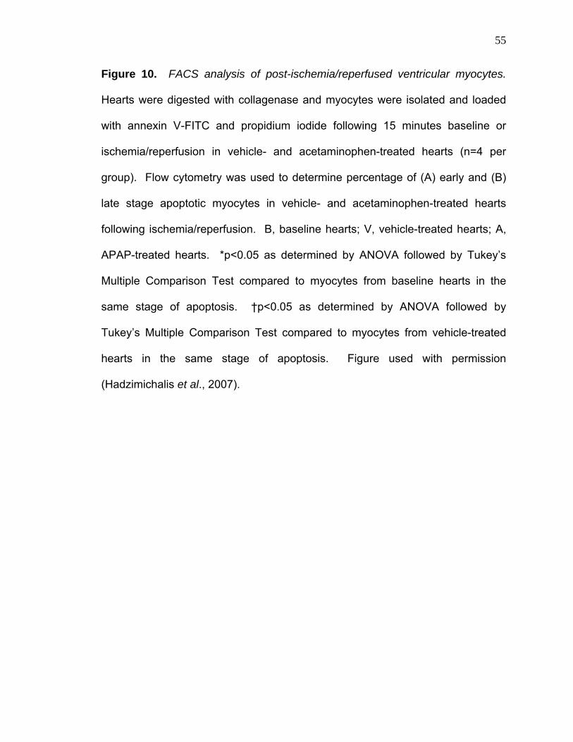
Figure 10. FACS analysis of post-ischemia/reperfused ventricular myocytes.
ed and loaded
(B)
Tukey’s
by
adzimichalis et al., 2007).
Hearts were digested with collagenase and myocytes were isolat
with annexin V-FITC and propidium iodide following 15 minutes baseline or
ischemia/reperfusion in vehicle- and acetaminophen-treated hearts (n=4 per
group). Flow cytometry was used to determine percentage of (A) early and
late stage apoptotic myocytes in vehicle- and acetaminophen-treated hearts
following ischemia/reperfusion. B, baseline hearts; V, vehicle-treated hearts; A,
APAP-treated hearts. *p<0.05 as determined by ANOVA followed by
Multiple Comparison Test compared to myocytes from baseline hearts in the
same stage of apoptosis. †p<0.05 as determined by ANOVA followed
Tukey’s Multiple Comparison Test compared to myocytes from vehicle-treated
hearts in the same stage of apoptosis. Figure used with permission
(H

56
Figure 11. Representative FACS analysis of vehicle-treated
chemia/reperfused heart. J1, necrotic cells; J2, late apoptotic cells; J3, viable
ells; J4, early apoptotic cells. Figure used with permission (Hadzimichalis et al.,
2007).
is
c

57
IV. DISCUSSION
58
1. Rationale
With the marked rise in heart disease, the need for preventative cardiac
care has become essential. Many groups have investigated the protective
capacity of a variety of compounds in inhibiting myocardial ischemia/reperfusion-
induced injury. Studies by Varga et al. (2004) investigated the effects of
pretreatment with dexamethasone, a potent glucocorticoid, on post-
ischemia/reperfusion. They reported that dexamethasone inhibits ventricular
fibrillation via attenuation of mitochondrial cytochrome c release. Kovacs et al.
(2001) administered non-specific caspase inhibitors at the onset of reperfusion to
examine their ability to maintain cardiac function and limit both infarct size and
apoptosis. Additional reports from Das et al. (2005) examined the
cardioprotective effects of pretreatment with palm tocotrienol, a vitamin E isomer,
following myocardial ischemia/reperfusion. They demonstrated that treatment
with tocotrienols, derived from a tocotrienol-rich fraction of palm oil, results in
attenuation of ischemia/reperfusion-induced damage via inhibition c-Src
phosphorylation and maintenance of proteasomal activity. However, despite
efforts to discover and/or synthesize new cardioprotective compounds, very little
effort has focused on examining the potential cardioprotective effects of
historically safe drugs, including acetaminophen (Bi et al., 2007).
In this study, we examined the mechanistic basis for acetaminophen-
mediated functional cardioprotection. Previous studies reported that in an in vivo
canine preparation of myocardial infarction, acetaminophen treatment results in a

59
significant reduction of necrotic tissue (Merrill et al., 2004). In the current study,
hat acetaminophen might also have an effect on the mitochondrial
l., 2001; Merrill
we proposed t
pathway of apoptosis following ischemia/reperfusion. Specifically, we explored
whether therapeutic concentrations of acetaminophen can attenuate MPTP
opening, cytochrome c release, and apoptotic cell death. The major finding of
our study is that following myocardial ischemia/reperfusion, acetaminophen
treatment completely blocks opening of the MPTP and mitochondrial swelling as
well as mitochondrial cytochrome c release. Furthermore, although
acetaminophen attenuates late stage apoptosis, it does not completely block it.
These results suggest that acetaminophen inhibits the MPTP-induced pathway of
apoptosis; however, other pathways leading to apoptosis may not be affected by
acetaminophen.
Acetaminophen, when taken at therapeutic concentrations, has been
established as a safe antipyretic and analgesic drug (Prescott, 2001). More
recently, this compound has also been established as an effective
cardioprotective agent during myocardial ischemia/reperfusion injury (Merrill et
al., 2001; Merrill and Goldberg, 2001; Merrill, 2002; Halestrap et al., 2004).
Mechanistically, the phenolic hydroxyl group of acetaminophen likely donates its
hydrogen atom to aid in the reported reduction of free radicals, namely
peroxynitrite and hydroxyl radicals, post-reperfusion (Merrill et a
and Goldberg, 2001; Prescott, 2001; Merrill, 2002). Ischemia/reperfusion-
induced oxidative stress is a well-known trigger for MPTP opening, mitochondrial
cytochrome c release, and downstream apoptotic cell death pathway activation

60
(Weiss et al., 2003; Halestrap et al., 2004; Gateau-Roesch et al., 2006; Orrenius
et al., 2007). We hypothesize that acetaminophen-mediated inhibition of ROS
generation results, in part, in attenuation of reperfusion-induced myocardial injury
via a reduction in MPTP opening, mitochondrial cytochrome c release, and
apoptotic cell death.

61
2. The Langendorff perfusion
2.1 Advantages and limitations of the Langendorff preparation
Since its conception over 100 years ago, the Langendorff-perfused
ammalian heart preparation still remains one of the most popular methods for
studying cardiac metabolism, hemodynamics, metabolic and pharmacological
interventions, electrical activity, and global myocardial ischemia and hypoxia
(Langendorff, 1895; Sutherland and Hearse, 2000). Modification of the
preparation in this study, including perfusate composition, temperature, pressure,
and pacing rate, was established based on previously published reports from our
laboratory (Merrill et al., 2001; Merrill and Goldberg, 2001; Merrill, 2002).
The isolated perfused Langendorff heart preparation provides an efficient
and highly reproducible means of collecting widespread physiologic data during
global myocardial ischemia and reperfusion (Hearse and Sutherland, 2000).
Denervation presents a unique opportunity to study cardiac function devoid of
sympathetic and vagal stimulation (Sutherland and Hearse, 2000). However,
while there are countless advantages, this preparation also introduces several
limitations. Myocardial extraction and ex vivo placement result in restricted
clinical application and continual tissue deterioration (i.e. 5-10% decrement in
contractile function/hour) over prolonged periods (Sutherland and Hearse, 2000).
Nevertheless, the Langendorff-perfused heart presented the most optimal
compromise between quality and quantity of data for our studies.
m

62
2.2 The Langendorff-perfused guinea pig heart model
dy was an identification of the mechanism underlying previously
idant
This stu
reported acetaminophen-mediated functional cardioprotection following
ischemia/reperfusion. As such, it was essential to employ an identical animal
model, namely the Langendorff-perfused guinea pig heart. However, certain
factors were taken into consideration when initially choosing an animal model.
Firstly, measures were taken to establish the best compromise between
clinical relevance, cost, data quality and quantity, and reproducibility (Hearse and
Sutherland, 2000). In addition, it was noted that guinea pigs, similar to humans,
are unable to synthesize ascorbic acid, an organic acid exhibiting antiox
properties. Deficiency in the enzyme required to synthesize this compound
suggests that the isolated perfused guinea pig heart would be a remarkably
valuable model to study the effects of a drug on post-reperfusion injury (Meister,
1994).

63
3. Acetaminophen; therapeutic dosages and experimental concentrations
Therapeutic concentrations of acetaminophen in human plasma samples
nd
liver
may range from 10-100 µg/ml, with hepatatoxicity occurring at concentrations
>300 µg/ml (Prescott, 2000; Spiler et al., 2005). Clinically, administration of 1000
mg of acetaminophen every 4 hours for 4 doses (50-70 kg patient), will result in
fluctuating plasma concentrations within the therapeutic range (Rumack, 2004).
In these studies acetaminophen (0.35 mM) was dissolved into the perfusate a
de ed continuously. HPLC analysis from our laboratory reveals net extraction
of acetaminophen by the myocardium, with arterial and venous concentrations
ranging from 45-50 µg/ml (Spiler et al., 2005). Hence, concentrations used in our
studies fall well within the effective therapeutic range and far below the toxic
range.

64
4. Acetaminophen-mediated inhibition of mitochondrial swelling and MPTP
pening following ischemia/reperfusion
drial fractions of whole heart homogenate.
e found a significant decrease in the light absorbance of isolated
mitochondria from vehicle-treated ischemia/reperfused hearts when compared to
either baseline, and acetaminophen treatment completely reversed this effect
(Figure 1B). These results suggest that our model of ischemia/reperfusion (30
minutes low-flow global ischemia and 60 minutes reperfusion) successfully
induced mitochondrial permeability pore opening at the completion of
reperfusion, and that the presence of acetaminophen resulted in inhibition of this
opening (Figure 6B). These data are further strengthened by electron
micrograph analysis showing preserved myofibrillar ultrastructure and intact
mitochondria in acetaminophen-treated hearts, similar to baseline controls, and
o
Reports indicate that mitochondrial swelling is indicative of MPTP opening
and ultimately results in outer mitochondrial membrane (OMM) rupture
(Halestrap et al., 2004; Di Lisa and Bernardi, 2006). Increases in mitochondrial
swelling, as assessed by decreases in light absorbance, would therefore imply
downstream cytochrome c release and activation of the mitochondrial-mediated
pathway of apoptosis (Di Lisa and Bernardi, 2006; Kaasik et al., 2007). Central
to the successful analysis of mitochondrial light scattering was the isolation of
purified intact mitochondria. Voltage dependent anion channel, an outer
mitochondrial membrane protein, was used as a loading control in isolated
mitochon
W
65
visually swollen mitochondria post-ischemia/reperfusion in vehicle-treated hearts
(Figure 7). These data suggest that acetaminophen completely attenuates pore
ening following ischemia/reperfusion in our model. op

66
5. Acetaminophen-mediated inhibition of mitochondrial cytochrome c release
following ischemia/reperfusion
Following ischemia/reperfusion-induced OMM rupture in response to
MPTP opening and mitochondrial swelling, cytochrome c is released into the
cytosol to initiate the intrinsic pathway of apoptosis (Halestrap et al., 2004). We
found a significant increase in cytosolic cytochrome c content, with a concomitant
decrease in mitochondrial cytochrome c content following ischemia/reperfusion in
vehicle-treated hearts. This suggests that our model of ischemia/reperfusion was
successful at inducing mitochondrial cytochrome c release at the completion of
reperfusion. In addition, acetaminophen treatment resulted in a significant and
complete inhibition of cytochrome c release following injury when compared to
vehicle-treated hearts. These data suggest that acetaminophen treatment
completely inhibits mitochondrial cytochrome c release following
ischemia/reperfusion in our model. We have shown (Figures 1 and 2) that the
observed inhibition of cytochrome c release is likely a response to the complete
upstream inhibition of MPTP opening; however, it is possible that acetaminophen
also exhibits functional cardioprotection via other pathways upstream to
cytochrome c release.

67
6. Acetaminophen-mediated attenuation of late stage apoptosis following
ischemia/reperfusion
In our protocol, early stage apoptotic myocytes were defined as those
cells that were stained with annexin V-FITC. This population was comprised of
myocytes that had externalized phosphatidylserine residues and active caspases
but no DNA degradation or loss of membrane integrity. Late stage apoptotic
myocytes were defined as those cells that were both annexin V-FITC and PI
positive. This myocyte population had active caspases and permeabilized cell
membranes (Schmid et al., 2007). We found that acetaminophen treatment
significantly inhibited the number of late stage apoptotic myocytes when
compared to vehicle-treated hearts at the completion of reperfusion. However,
there was also a significant increase in late stage apoptotic myocytes between
baseline and acetaminophen-treated ischemia/reperfused hearts. This
increasing index of damage following ischemia/reperfusion in acetaminophen-
treated hearts was not as apparent as mitochondrial swelling or cytochrome c
release. It is possible that the changes in apoptotic cell death noted between
treatment groups at reperfusion are due, in part, to acetaminophen-mediated
MPTP inhibition and cytochrome c release. However, these data also suggest
at while acetaminophen may abolish permeability pore transition and
cytochrome c release following ischemia/reperfusion, other pathways of
apoptosis, unaffected by acetaminophen treatment, are still active during injury.
We propose that acetaminophen-mediated cardioprotection is, at least in part,
th

68
specific to inhibition of MPTP-induced cytochrome c release and apoptosis. A
schematic of these data are shown in Figure 12.
Damaging post-reperfusion oxidants, including peroxynitrite and hydroxyl
radicals, activate the intrinsic pathway of apoptosis, and the efficacy of
acetaminophen in attenuating these compounds makes it a likely inhibitor of this
pathway (Merrill, 2002). However, it is possible that the incomplete attenuation
of apoptosis in response to treatment is the result of ischemia/reperfusion-
induced activation of other apoptotic cell death pathways, including the extrinsic
pathway of apoptosis.

69
Figure 12. Schematic of proposed mechanism of action of acetaminophen
following myocardial ischemia/reperfusion. Our studies imply that while
cetaminophen completely inhibits MPTP opening and mitochondrial cytochrome
release, apoptosis is not completely blocked. Thus, additional apoptotic
pathways are still active following insult. Figure used with permission
(Hadzimichalis et al., 2007).
a
c

70
7. Future directions
These data (Table 3) begin to explain the mechanism underlying
previously reported acetaminophen-mediated cardioprotection following
ischemia/reperfusion. They suggest that administration of acetaminophen just
prior to an ischemic attack can result in attenuation of functional damage via
inhibition of MPTP opening and cytochrome c release-induced apoptotic cell
that while acetaminophen completely inhibits
chemia/reperfusion-induced MPTP opening, mitochondrial swelling, and
ytochrome c release, it only partially attenuates apoptosis. Future studies may
also aim to employ the use of acetaminophen in conjunction with caspase
inhibitors or other anti-apoptotic drugs, in order to further prevent apoptotic cell
death post-reperfusion.
Our laboratory has already begun investigating the effects of
acetaminophen following other cardiovascular injuries caused, in part, by
damaging oxidants. Preliminary data suggest that functional cytoprotectivity may
also be observed in other organ systems, including the brain following cerebral
ischemia/repefusion. We encourage other laboratories to examine the effects of
death following ischemia/reperfusion. While these data are promising in that they
offer a historically safe alternative to preventative cardiac care, additional
pathway details must be elucidated prior to clinical application. Future studies
should examine the specific role of acetaminophen in the extrinsic versus
intrinsic apoptotic pathways.
We have reported
is
c

71
acetaminophen in an attempt to expand the spectrum of uses for this historically
afe drug. s

72
Table 3. Summary table of study findings as they relate to the mechanism of
acetaminophen-mediated cardioprotection following ischemia/reperfusion.
Acetaminophen Vehicle Acetaminophen vs. vs. vs. Baseline Baseline Vehicle MPTP opening ns * * (Spectrophotometry and electron microscopy) Mitochondrial cytochrome c release ns * * (Western blotting) Apoptosis Early ns ns ns Late * * * (Flow cytometry)
Acetaminophen completely inhibits MPTP opening and mitochondrial cytochrome
c release and partially attenuates apoptosis when compared to vehicle-treated
hearts following ischemia/reperfusion. *p<0.05; ns, no significance. Figure used
with permission (Hadzimichalis et al., 2007).

73
V. REFERENCES

74
Bazzola JJ, Russel LD (1999) Electron microscopy: principles and techniques for biologists. , 2nd Edition rtlett Publishers.
Bi W, Cai J, Liu S, Baudy-Floc'h M, Bi L (2007) Design, synthesis and cardioprotective
effect of a new class of dual-acting agents: Phenolic tetrahydro-beta-carboline RGD peptidomimetic conjugates. Bioorg Med Chem 15:6909-6919.
Bopassa JC, Vandroux D, Ovize M, Ferrera R (2006) Controlled reperfusion after
hypothermic heart preservation inhibits mitochondrial permeability transition-pore opening and enhances functional recovery. Am J Physiol Heart Circ Physiol 291:H2265-2271.
Borutaite V, Jekabsone A, Morkuniene R, Brown GC (2003) Inhibition of mitochondrial
permeability transition prevents mitochondrial dysfunction, cytochrome c release and apoptosis induced by heart ischemia. J Mol Cell Cardiol 35:357-366.
Bosetti F, Baracca A, Lenaz G, Solaini G (2004) Increased state 4 mitochondrial
respiration and swelling in early post-ischemic reperfusion of rat heart. FEBS Lett 563:161-164.
Botting RM (2000) Mechanism of action of acetaminophen: is there a cyclooxygenase 3?
Clin Infect Dis 31 Suppl 5:S202-210. Brodie BB, Axelrod J (1948a) The estimation of acetanilide and its metabolic products
aniline, N-acetyl p-aminophenol and p-aminophenol (free and total conjugated) in biological fluids and tissues. J Pharmacol Exp Ther 94:22-28.
Brodie BB, Axelrod J (1948b) The fate of acetanilide in man. J Pharmacol Exp Ther 94. Broekemeier KM, Dempsey ME, Pfeiffer DR (1989) Cyclosporin A is a potent inhibitor
of the inner membrane permeability transition in liver mitochondria. J Biol Chem 264:7826-7830.
Bunger R, Haddy FJ, Gerlach E (1975a) Coronary responses to dilating substances and
competitive inhibition by theophylline in the isolated perfused guinea pig heart. Pflugers Arch 358:213-224.
Bunger R, Haddy FJ, Querengasser A, Gerlach E (1975b) An isolated guinea pig heart
preparation with in vivo like features. Pflugers Arch 353:317-326. Chou TM, Greenspan P (2002) Effect of acetaminophen on the myeloperoxidase-
hydrogen peroxide-nitrite mediated oxidation of LDL. Biochim Biophys Acta 1581:57-63.
Cotter J, Wooltorton E (2005) New restrictions on celecoxib (Celebrex) use and the
withdrawal of valdecoxib (Bextra). Cmaj 172:1299.
. Sudbury, MA: Jones and Ba

75
Powell SR, Wang P, Divald A, Nesaretnam K, Tosaki A, Cordis GA,Das S, Maulik N, Das DK (2005) Cardioprotection with palm tocotrienol: antioxidant activity of
i Lisa F, Bernardi P (2006) Mitochondria and ischemia-reperfusion injury of the heart:
Duchen
adelha FR, Thomson L, Fagian MM, Costa AD, Radi R, Vercesi AE (1997) Ca2+-
rch Biochem Biophys 345:243-250.
70:264-273.
Golfett aminophen and the
ischemia/reperfused myocardium. Exp Biol Med (Maywood) 228:674-682.
olfetti R, VanDyke K, Rork T, Spiler N, Merrill G (2002) Acetaminophen in the post-
Gottlieb RA, Burleson KO, Kloner RA, Babior BM, Engler RL (1994) Reperfusion
injury induces apoptosis in rabbit cardiomyocytes. J Clin Invest 94:1621-1628.
Green
ition pore-induced apoptotic pathway. Am J Physiol Heart Circ Physiol.
tocotrienol is linked with its ability to stabilize proteasomes. Am J Physiol Heart Circ Physiol 289:H361-367.
Dfixing a hole. Cardiovasc Res 70:191-199.
MR (2004) Roles of mitochondria in health and disease. Diabetes 53 Suppl 1:S96-102.
Foo RS, Mani K, Kitsis RN (2005) Death begets failure in the heart. J Clin Invest
115:565-571.
Gindependent permeabilization of the inner mitochondrial membrane by peroxynitrite is mediated by membrane protein thiol cross-linking and lipid peroxidation. A
Gateau-Roesch O, Argaud L, Ovize M (2006) Mitochondrial permeability transition pore
and postconditioning. Cardiovasc Res Gewies A (2003) Introduction to Apoptosis. ApoReview:1-26.
i R, Rork T, Merrill G (2003) Chronically administered acet
G
ischemia reperfused myocardium. Exp Biol Med (Maywood) 227:1031-1037.
DR, Kroemer G (2004) The pathophysiology of mitochondrial cell death. Science 305:626-629.
Green DR, Kroemer G (2005) Pharmacological manipulation of cell death: clinical
applications in sight? J Clin Invest 115:2610-2617. Hadzimichalis NM, Baliga SS, Golfetti R, Jaques KM, Firestein BL, Merrill GF (2007)
Acetaminophen-mediated cardioprotection via inhibition of the mitochondrial permeability trans

76
Halestrap AP (2006) Calcium, mitochondria and reperfusion injury: a pore way to die.
Halestr
perfusion--a target for cardioprotection. Cardiovasc Res 61:372-385.
Haunst ic mechanisms and implications for cardiovascular disease. Circ Res 82:1111-1129.
Hearse DJ, Sutherland FJ (2000) Experimental models for the study of cardiovascular function and disease. Pharmacol Res 41:597-603.
Hemler M, Lands WE (1976) Purification of the cyclooxygenase that forms prostaglandins. Demonstration of two forms of iron in the holoenzyme. J Biol
Hinsbe
onda HM, Korge P, Weiss JN (2005) Mitochondria and ischemia/reperfusion injury.
uang XD, Horackova M, Pressler ML (1996) Changes in the expression and distribution
iomyocytes. Exp Cell Res 228:254-261.
Hunter lysis of rat liver mitochondria induced by ferrous ions. J Biol Chem 238:828-835.
Javado an endpoint to initiate cell death and as a putative target for cardioprotection. Cell
avadov SA, Lim KH, Kerr PM, Suleiman MS, Angelini GD, Halestrap AP (2000)
he mitochondrial permeability transition. Cardiovasc Res 45:360-369.
Jeremia nkel J, Boekstegers P, Debatin KM (2000) Involvement of CD95/Apo1/Fas in cell death after myocardial
Kaasik
lume. Am J Physiol Cell Physiol 292:C157-163.
Biochem Soc Trans 34:232-237.
ap AP, Clarke SJ, Javadov SA (2004) Mitochondrial permeability transition pore opening during myocardial re
etter A, Izumo S (1998) Apoptosis: bas
Chem 251:5575-5579.
rg O, Treupel G (1894) 33:216-250.
HAnn N Y Acad Sci 1047:248-258.
Hof connexin 43 in isolated cultured adult guinea pig card
FE, Jr., Gebicki JM, Hoffsten PE, Weinstein J, Scott A (1963) Swelling and
v S, Karmazyn M (2007) Mitochondrial permeability transition pore opening as
Physiol Biochem 20:1-22.
JProtection of hearts from reperfusion injury by propofol is associated with inhibition of t
s I, Kupatt C, Martin-Villalba A, Habazettl H, Sche
ischemia. Circulation 102:915-920.
A, Safiulina D, Zharkovsky A, Veksler V (2007) Regulation of mitochondrial matrix vo

77
Kis B, Snipes JA, Busija DW (2005) Acetaminophen and the cyclooxygenase-3 puzzle: sorting out facts, fictions, and uncertainties. J Pharmacol Exp Ther 315:1-7.
d ischaemic/reperfused rat hearts. Naunyn Schmiedebergs Arch Pharmacol 364:501-507.
Krijnen E, Niessen HW (2002) Apoptosis in myocardial ischaemia and infarction. J Clin Pathol 55:801-811.
Langen en Saugethierherzen. Pflugers Arch fur die Gesamte Physiologie des Menschen and der Tiere 61:291-332.
Logue l Cell Cardiol 38:21-33.
perpetrator or a protector? Selective COX-2 inhibitors remain controversial. Acta Pharmacol Sin 26:926-933.
MacLellan WR, Schneider MD (1997) Death by design. Programmed cell death in cardiovascular biology and disease. Circ Res 81:137-144.
Masfer4) Selective inhibition of inducible cyclooxygenase 2 in vivo is
antiinflammatory and nonulcerogenic. Proc Natl Acad Sci U S A 91:3228-3232.
Meiste hem 269:9397-9400.
Mering
errill GF (2002) Acetaminophen and low-flow myocardial ischemia: efficacy and
Merrill GF, Goldberg E (2001) Antioxidant properties of acetaminophen and
cardioprotection. Basic Res Cardiol 96:423-430.
Merrill ophen and myocardial infarction in dogs. Am J Physiol Heart Circ Physiol 287:H1913-1920.
Kovacs P, Bak I, Szendrei L, Vecsernyes M, Varga E, Blasig IE, Tosaki A (2001) Non-
specific caspase inhibition reduces infarct size and improves post-ischaemic recovery in isolate
PA, Nijmeijer R, Meijer CJ, Visser CA, Hack C
dorff O (1895) Untersuchungen am uberlebend
SE, Gustafsson AB, Samali A, Gottlieb RA (2005) Ischemia/reperfusion injury at the intersection with cell death. J Mo
Luo C, He ML, Bohlin L (2005) Is COX-2 a
rer JL, Zweifel BS, Manning PT, Hauser SD, Leahy KM, Smith WG, Isakson PC, Seibert K (199
r A (1994) Glutathione-ascorbic acid antioxidant system in animals. J Biol C
vJ (1893) Beitrage zur Kenntniss der Antipyretica. Therapeutishe Monatshefte 7:577-578.
Merrill G, McConnell P, Vandyke K, Powell S (2001) Coronary and myocardial effects
of acetaminophen: protection during ischemia-reperfusion. Am J Physiol Heart Circ Physiol 280:H2631-2638.
Mantioxidant mechanisms. Am J Physiol Heart Circ Physiol 282:H1341-1349.
GF, Rork TH, Spiler NM, Golfetti R (2004) Acetamin

78
Morin D, Hauet T, Spedding M, Tillement J (2001) Mitochondria as target for antiischemic drugs. Adv Drug Deliv Rev 49:151-174.
Nachtigal M, Mayer E, Ghaffar A, Greenspan P (2005) The effect of acetaminophen on
the development of aorta athersclerotic lesions in apoE deficient mice. Experimental Biology, San Diego California (abstract 60510).
rrenius S, Gogvadze V, Zhivotovsky B (2007) Mitochondrial oxidative stress:
iper H, Isenberg G (1989) Isolated adult cardiomyocytes. Boca Raton, FL: CRC Press,
rescott LF (2000) Paracetamol: past, present, and future. Am J Ther 7:143-147.
Presco raphic Review., 2nd Edition. New York: Taylor & Francis.
Rogersformation and lipid oxidation in hypercholesterolemic rabbits. In: Free Radic Biol
ork TH, Van Dyke K, Spiler NM, Merrill GF (2004) Acetaminophen in the hypoxic and
Rork T s
peroxynitrite-activated matrix metalloproteinase-2-mediated troponin I cleavage
ousou AJ, Ericsson M, Federman M, Levitsky S, McCully JD (2004) Opening of
drial matrix volume, calcium accumulation, and respiration. Am J Physiol Heart Circ Physiol 287:H1967-1976.
Ruiz-Mrial proton gradient and delays ATP depletion in
cardiomyocytes during ischemic conditions. Am J Physiol Heart Circ Physiol
umack BH (2004) Acetaminophen misconceptions. Hepatology 40:10-15.
Salzbe d cardiovascular risk. Subcell Biochem 42:159-174.
Oimplications for cell death. Annu Rev Pharmacol Toxicol 47:143-183.
PInc.
P
tt LF (2001) Paracetamol (Acetaminophen): A Critical Bibliog
L, Taylor A, Raya R, Gest A, Smith C (1999) Acetaminophen lowers fatty streak
Med, p S108.
Rreoxygenated guinea pig myocardium. Exp Biol Med (Maywood) 229:1154-1161.
H, Hadzimichalis NM, Kappil MA, Merrill GF (2006) Acetaminophen attenuate
in the isolated guinea pig myocardium. J Mol Cell Cardiol 40:553-561.
Rmitochondrial KATP channels enhances cardioprotection through the modulation of mitochon
eana M, Garcia-Dorado D, Pina P, Inserte J, Agullo L, Soler-Soler J (2003) Cariporide preserves mitochond
285:H999-1006.
R
rg DJ, Weir MR (2007) COX-2 inhibitors an

79
Schmid I, Uittenbogaart C, Jamieson BD (2007) Live-cell assay for detection of apoptosis by dual-laser flow cytometry using Hoechst 33342 and 7-amino-actinomycin D. Nat Protoc 2:187-190.
Seibert
Haematol Disord 5:419-429.
Sutherland FJ, Hearse DJ (2000) The isolated blood and perfusion fluid perfused heart. Pharmacol Res 41:613-627.
okarska-Schlattner M, Zaugg M, da Silva R, Lucchinetti E, Schaub MC, Wallimann T,
supply. Am J Physiol Heart Circ Physiol 289:H37-47.
Vandenerfusion, not simulated ischemia, initiates intrinsic apoptosis injury
in chick cardiomyocytes. Am J Physiol Heart Circ Physiol 284:H141-150.
Vane JRheum 26:2-10.
Varga orking rat
hearts: the role of cytochrome c release. Life Sci 75:2411-2423.
Weiss
ie WL, Chipman JG, Robertson DL, Erikson RL, Simmons DL (1991) Expression of a
Zhu YZ, Chong CL, Chuah SC, Huang SH, Nai HS, Tong HT, Whiteman M, Moore PK
(2006) Cardioprotective effects of nitroparacetamol and paracetamol in acute l Heart Circ
Physiol 290:H517-524.
Zidar N, Jera J, Maja J, Dusan S (2007) Caspases in myocardial infarction. Adv Clin Chem 44:1-33.
K, Masferrer JL (1994) Role of inducible cyclooxygenase (COX-2) in inflammation. Receptor 4:17-23.
Spiler NM, Rork TH, Merrill GF (2005) An old drug with a new purpose: cardiovascular
actions of acetaminophen (paracetamol). Curr Drug Targets Cardiovasc
T
Schlattner U (2005) Acute toxicity of doxorubicin on isolated perfused heart: response of kinases regulating energy
Hoek TL, Qin Y, Wojcik K, Li CQ, Shao ZH, Anderson T, Becker LB, Hamann KJ (2003) Rep
R, Botting RM (1997) Mechanism of action of aspirin-like drugs. Semin Arthritis
E, Nagy N, Lazar J, Czifra G, Bak I, Biro T, Tosaki A (2004) Inhibition of ischemia/reperfusion-induced damage by dexamethasone in isolated w
JN, Korge P, Honda HM, Ping P (2003) Role of the mitochondrial permeability transition in myocardial disease. Circ Res 93:292-301.
Xmitogen-responsive gene encoding prostaglandin synthase is regulated by mRNA splicing. Proc Natl Acad Sci U S A 88:2692-2696.
phase of myocardial infarction in experimental rats. Am J Physio

80
VITA
NORELL MELISSA HADZIMICHALIS (formerly Spiler)
EDUCATION
B.A., 2 gical Sciences, Rutgers University, New Brunswick,
New Jersey
Ph.D., nd Integrative Biology, Graduate School- New Brunswick, Rutgers University, New Brunswick, New Jersey
all 2003-2004 Teaching Assistant for Systems Physiology Letcure and
all 2004–2008 Head Teaching Assistant for Systems Physiology Lecture
PUBLICATIONS
Golfett . Acetaminophen in the post-ischemia reperfused myocardium. Exp Biol Med (Maywood) 227: 1031-
Merrill aminophen and myocardial
infarction in dogs. Am J Physiol Heart Circ Physiol 287: H1913-1920, 2004.
Rork Tand115
Ca tions of acetaminophen (paracetamol). Curr Drug Targets Cardiovasc Haematol Disord 5: 419-429, 2005.
003 Biolo
2008 Physiology a
and Graduate School of Biomedical Sciences, University of Medicine and Dentistry of New Jersey, Piscataway, New Jersey
PRINCIPAL OCCUPATIONS
FLaboratory
Fand Laboratory
i R, VanDyke K, Rork T, Spiler N, and Merrill G
1037, 2002.
GF, Rork TH, Spiler NM, and Golfetti R. Acet
H, Van Dyke K, Spiler NM, and Merrill GF. Acetaminophen in the hypoxic reoxygenated guinea pig myocardium. Exp Biol Med (Maywood) 229: 4-1161, 2004.
Spiler NM, Rork TH, and Merrill GF. An old drug with a new purpose:
rdiovascular ac

81
Rork TH, Hadzimichalis NM, Baliga SS, Golfetti R, and Merrill GF. New Perspectives on Acetaminophen. Current Cardiology Reviews 2: 131-146, 2006.
Rork TH, Hadzimichalis NM, Kappil MA, and Merrill GF. Acetaminophen
attenuates peroxynitrite-activated matrix metalloproteinase-2-mediated troponin I cleavage in the isola myocardium. J Mol Cell Cardiol 40: 553-561, 2006.
Rork TH. Antiarrhythmic properties of acetaminophen in the dog. Exp Biol Med (Maywood) 232: 1245-1252, 2007.
Hadzimichalis NM,GF. Acetaminomitochondrial pePhysiol Heart Circ Physiol, in press, 2007.
ted guinea pig
Merrill GF, Merrill JH, Golfetti R, Jaques KM, Hadzimichalis NM, Baliga SS, and
Baliga SS, Golfetti R, Jaques KM, Firestein BL, and Merrill phen-mediated cardioprotection via inhibition of the rmeability transition pore-induced apoptotic pathway. Am J

82
![Paracetamol(acetaminophen)ornon-steroidalanti ... (acetaminophen) … · [Intervention Review] Paracetamol (acetaminophen) or non-steroidal anti-inflammatory drugs, alone or combined,](https://img.dokumen.tips/doc/110x75/5f387cbd43d51a2eb45648f8/paracetamolacetaminophenornon-steroidalanti-acetaminophen-intervention.jpg)