Embed Size (px)
Citation preview

Cubital tunnel syndrome is the second most common compression neuropathy after carpal tunnel syndrome and the cubital tunnel of the elbow is the most common site of ulnar nerve compression.1) The diagnosis of cubital tunnel syndrome has traditionally depended on information ob-
tained from detailed history taking, physical examination, and electrodiagnostic study.2) However, ulnar neuropathy around the elbow has a variety of clinical features, mak-ing it difficult to make a proper treatment decision based on clinical presentation and an electrodiagnostic study. Although electrodiagnostic study is a valuable diagnostic tool for cubital tunnel syndrome, it has some limitations such as discomfort for patients, difficulty with precise localization, inability to identify structural abnormalities, possibility of false-negative results, and the time-consum-ing process.3)
Recently, high-resolution ultrasonography (HRUS) has shown the potential to alleviate diagnostic uncertainty
Accuracy of Preoperative Ultrasonography for Cubital Tunnel Syndrome: A Comparison with
Intraoperative FindingsChul-Hyun Cho, MD, Yong-Ho Lee, MD, Kwang-Soon Song, MD, Kyung-Jae Lee, MD,
Si-Wook Lee, MD, Sung-Moon Lee, MD
Department of Orthopedic Surgery, Dongsan Medical Center, Keimyung University School of Medicine, Daegu, Korea
Background: The aim of this study was to assess the consistency between preoperative ultrasonographic and intraoperative measurements of the ulnar nerve in patients with cubital tunnel syndrome. Methods: Twenty-six cases who underwent anterior transposition of the ulnar nerve for cubital tunnel syndrome were enrolled prospectively. On preoperative ultrasonography, largest cross-sectional diameters of the ulnar nerve were measured at the level of medial epicondyle (ME) and 3 cm proximal (PME) and distal (DME) to the ME on the transverse scan by a single experienced radiologist. Intraoperative direct measurements of the largest diameter at the same locations were performed by a single surgeon without knowledge of the preoperative values. The consistency between ultrasonographic and intraoperative values including the largest diameter and swelling ratio were assessed. Results: Significant differences between ultrasonographic and intraoperative values of the largest diameter were found at all levels. The mean difference was 1.29 mm for PME, 1.38 mm for ME, and 1.12 mm for DME. The mean ME-PME swelling ratio for ultrasonographic and intraoperative measurements was 1.50 and 1.39, respectively, showing significant difference. The mean ME-DME swelling ratio for ultrasonographic and intraoperative measurements was 1.53 and 1.43, respectively, showing no significant difference. Conclusions: Ultrasonographically measured largest diameters of the ulnar nerve at any levels were smaller than the real values determined intraoperatively. The ME-DME swelling ratio of the ulnar nerve measured by ultrasonography was consistent with the intraoperative measurement.Keywords: Ulnar nerve, Cubital tunnel syndrome, Ultrasonography
Original Article Clinics in Orthopedic Surgery 2018;10:352-357 • https://doi.org/10.4055/cios.2018.10.3.352
Copyright © 2018 by The Korean Orthopaedic AssociationThis is an Open Access article distributed under the terms of the Creative Commons Attribution Non-Commercial License (http://creativecommons.org/licenses/by-nc/4.0)
which permits unrestricted non-commercial use, distribution, and reproduction in any medium, provided the original work is properly cited.Clinics in Orthopedic Surgery • pISSN 2005-291X eISSN 2005-4408
Received April 30, 2018; Accepted June 6, 2018Correspondence to: Chul-Hyun Cho, MD Department of Orthopedic Surgery, Dongsan Medical Center, Keimyung University School of Medicine, 56 Dalseong-ro, Jung-gu, Daegu 41931, KoreaTel: +82-53-250-7729, Fax: +82-53-250-7205E-mail: [email protected]
353
Cho et al. Ultrasonography for Cubital Tunnel SyndromeClinics in Orthopedic Surgery • Vol. 10, No. 3, 2018 • www.ecios.org
for ulnar neuropathy around the elbow.4) It can provide much information about the space-occupying lesion or surrounding anatomy and allow for optimal visualization of the nerve running the pathway and quantitative size of the nerve itself.5) Several studies have been performed to investigate the feasibility of HRUS for cubital tunnel syn-drome.1,2,6-9)
However, the accuracy of HRUS on measurements of the ulnar nerve is still controversial.4) Several studies showed inconsistent results concerning the diagnostic sensitivity and specificity of HRUS in ulnar neuropathy around the elbow.4,10,11) The differences in reported values may be the results of methodological differences in the research design, differences in measurement techniques, measurement errors, or differences in the characteristics of the patient population.7) Furthermore, few studies have in-vestigated the accuracy of ultrasonography for cubital tun-nel syndrome compared to intraoperative measurements of the ulnar nerve.12) The aim of this study was to assess the consistency between preoperative ultrasonographic and intraoperative measurements of the ulnar nerve in patients with cubital tunnel syndrome. We hypothesized that ultrasonographically measured values may be smaller than the direct intraoperative measurement values and the swelling ratio may improve diagnostic accuracy in patients with cubital tunnel syndrome.
METHODS
Between 2011 and 2012, we prospectively enrolled 26 con-secutive cases (three bilateral and 20 unilateral) in 23 pa-tients who underwent anterior transposition of the ulnar nerve for cubital tunnel syndrome. All patients with cubi-tal tunnel syndrome were diagnosed using detailed history taking, physical examination, and electrodiagnostic study according to the criteria of the American Association of
Electrodiagnostic Medicine.13) Exclusion criteria included (1) a history of polyneuropathy; (2) acute trauma involv-ing the upper extremity; (3) a history of previous elbow surgery; or (4) brachial plexus injury. We conducted this study in compliance with the principles of the Declaration of Helsinki. The protocol of this study was reviewed and approved by the Institutional Review Board of Keimyung University Dongsan Medical Center (IRB No. 201511009). Informed consent was waived.
Ultrasonographic ExaminationPreoperatively, ultrasonographic examination was con-ducted using a 12–17 MHz linear transducer of IU 22 HRUS (Philips Medical Systems, Bothell, WA, USA) by a single experienced musculoskeletal radiologist (SML) blinded to the clinical and electrodiagnostic data.
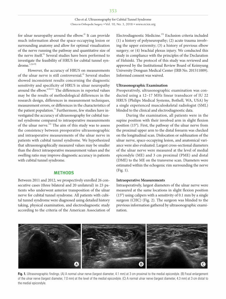
During the examination, all patients were in the supine position with their involved arm in slight flexion position (15°). First, the pathway of the ulnar nerve from the proximal upper arm to the distal forearm was checked on the longitudinal scan. Dislocation or subluxation of the ulnar nerve, space-occupying lesion, and anatomical vari-ance were also evaluated. Largest cross-sectional diameters of the ulnar nerve were measured at the level of medial epicondyle (ME) and 3 cm proximal (PME) and distal (DME) to the ME on the transverse scan. Diameters were estimated within the echogenic rim surrounding the nerve (Fig. 1).
Intraoperative MeasurementsIntraoperatively, largest diameters of the ulnar nerve were measured at the same locations in slight flexion position (15°) using calipers with a sensitivity of 0.1 mm by a single surgeon (CHC) (Fig. 2). The surgeon was blinded to the previous information gathered by ultrasonographic exami-nation.
A B C
Fig. 1. Ultrasonographic findings. (A) A normal ulnar nerve (largest diameter, 4.1 mm) at 3 cm proximal to the medial epicondyle. (B) Focal enlargement of the ulnar nerve (largest diameter, 7.0 mm) at the level of the medial epicondyle. (C) A normal ulnar nerve (largest diameter, 4.3 mm) at 3 cm distal to the medial epicondyle.

354
Cho et al. Ultrasonography for Cubital Tunnel SyndromeClinics in Orthopedic Surgery • Vol. 10, No. 3, 2018 • www.ecios.org
Swelling ratios were calculated by dividing the larg-est diameter measured at the ME by that at PME (ME-PME) and at DME (ME-DME). The consistency between ultrasonographic and intraoperative values including the largest diameter and swelling ratio were assessed.
Statistical AnalysisStatistical analyses were performed using IBM SPSS ver. 22.0 (IBM Corp., Armonk, NY, USA). The Wilcoxon signed-rank test was used to compare ultrasonographic and intraoperative values according to the locations and to compare ultrasonographic and intraoperative swelling ratios. Statistical significance was set at a p-value < 0.05.
RESULTS
The mean age of all patients was 58.8 years (range, 28 to 78 years), and there were 16 males and 10 females. The affected limbs were equally divided between the right and left sides (13 each), and the mean duration of symptoms was 17.6 months (range, 2 to 84 months). Causes of cubital tunnel syndrome included elbow contracture due to os-teoarthritis (11 cases), idiopathic causes (10 cases), cubitus valgus (three cases), ganglion (one case), and dislocation of the ulnar nerve (one case). The patient data are listed in Table 1.
On preoperative ultrasonography, enlargement of the ulnar nerve was observed at the level of the ME in all patients. The mean value of the largest diameter of the ulnar nerve at PME, ME, and DME on the axial scan was 3.90 mm, 5.72 mm, and 3.89 mm, respectively. On the intraoperative measurement, the mean value of the largest diameter of the ulnar nerve at PME, ME, and DME was 5.19 mm, 7.10 mm, and 5.01 mm, respectively. Significant
differences between ultrasonographic and intraoperative values of the largest diameters were found at all levels (all p < 0.001) (Table 2). The mean difference was 1.29 mm for PME, 1.38 mm for ME, and 1.12 mm for DME.
The mean ME-PME swelling ratio was 1.50 for ul-trasonographic measurement and 1.39 for intraoperative measurement, showing significant difference (p = 0.03). The mean ME-DME swelling ratio was 1.53 for ultraso-nographic measurement and 1.43 for intraoperative mea-surement, showing no significant difference (p = 0.17).
DISCUSSION
This study showed that the largest diameters of the ulnar nerve at three locations (PME, ME, and DME) on the ultrasonographic transverse scan were smaller than the actual values determined intraoperatively. The ME-DME swelling ratios of the ulnar nerve measured by ultraso-nography were consistent with intraoperatively measured values.
Although the accuracy of ultrasonographic exami-nation may vary depending on the experience and ability of the operator, it can show anatomical and morphological changes of the nerve, the precise compression site, osteo-phyte, space-occupying lesion, and anatomical variation around the elbow.1,11) Because of these advantages, ultraso-nographic examination has been considered as a valuable adjunctive method in the diagnosis, screening, and follow-up of cubital tunnel syndrome.1,2,11) In the present study, we could confirm morphological or dynamic changes of the ulnar nerve including enlargement, subluxation, or dislocation through ultrasonographic examination in all patients.
Several quantitative parameters including the diam-
A B
Fig. 2. Intraoperative findings. (A) Focal enlargement of the ulnar nerve. (B) Measurement of the largest diameter of the ulnar nerve at the level of the medial epicondyle using a caliper.
355
Cho et al. Ultrasonography for Cubital Tunnel SyndromeClinics in Orthopedic Surgery • Vol. 10, No. 3, 2018 • www.ecios.org
eter, cross-sectional area, and distal to proximal swelling ratio have been provided for ultrasonographic evaluation of cubital tunnel syndrome.1,2,6-9) The cross-sectional area and largest diameter on transverse scans are frequently used as quantitative parameters.4,9,14,15) Okamoto et al.16,17) reported that the mean ultrasonographic value of the largest diameter of the ulnar nerve at the level of ME in
patients with cubital tunnel syndrome (7.2 mm) was sig-nificantly larger than that in healthy men (3.1 mm). How-ever, recommendations on what and how to measure vary among publications and there is no commonly agreed “standard technique.”4) Furthermore, the consistency between ultrasonographic and intraoperative measure-ments is undetermined as of yet. Only a single study was
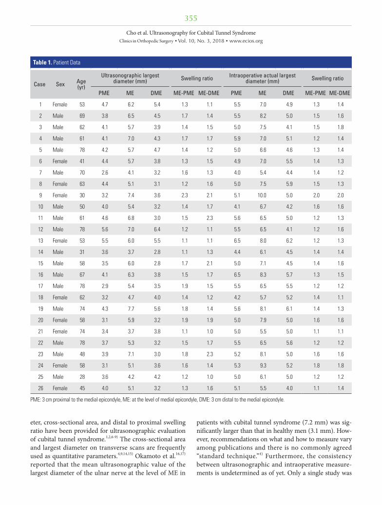
Table 1. Patient Data
Case Sex Age (yr)
Ultrasonographic largest diameter (mm) Swelling ratio Intraoperative actual largest
diameter (mm) Swelling ratio
PME ME DME ME-PME ME-DME PME ME DME ME-PME ME-DME
1 Female 53 4.7 6.2 5.4 1.3 1.1 5.5 7.0 4.9 1.3 1.4
2 Male 69 3.8 6.5 4.5 1.7 1.4 5.5 8.2 5.0 1.5 1.6
3 Male 62 4.1 5.7 3.9 1.4 1.5 5.0 7.5 4.1 1.5 1.8
4 Male 61 4.1 7.0 4.3 1.7 1.7 5.9 7.0 5.1 1.2 1.4
5 Male 78 4.2 5.7 4.7 1.4 1.2 5.0 6.6 4.6 1.3 1.4
6 Female 41 4.4 5.7 3.8 1.3 1.5 4.9 7.0 5.5 1.4 1.3
7 Male 70 2.6 4.1 3.2 1.6 1.3 4.0 5.4 4.4 1.4 1.2
8 Female 63 4.4 5.1 3.1 1.2 1.6 5.0 7.5 5.9 1.5 1.3
9 Female 30 3.2 7.4 3.6 2.3 2.1 5.1 10.0 5.0 2.0 2.0
10 Male 50 4.0 5.4 3.2 1.4 1.7 4.1 6.7 4.2 1.6 1.6
11 Male 61 4.6 6.8 3.0 1.5 2.3 5.6 6.5 5.0 1.2 1.3
12 Male 78 5.6 7.0 6.4 1.2 1.1 5.5 6.5 4.1 1.2 1.6
13 Female 53 5.5 6.0 5.5 1.1 1.1 6.5 8.0 6.2 1.2 1.3
14 Male 31 3.6 3.7 2.8 1.1 1.3 4.4 6.1 4.5 1.4 1.4
15 Male 58 3.5 6.0 2.8 1.7 2.1 5.0 7.1 4.5 1.4 1.6
16 Male 67 4.1 6.3 3.8 1.5 1.7 6.5 8.3 5.7 1.3 1.5
17 Male 78 2.9 5.4 3.5 1.9 1.5 5.5 6.5 5.5 1.2 1.2
18 Female 62 3.2 4.7 4.0 1.4 1.2 4.2 5.7 5.2 1.4 1.1
19 Male 74 4.3 7.7 5.6 1.8 1.4 5.6 8.1 6.1 1.4 1.3
20 Female 58 3.1 5.9 3.2 1.9 1.9 5.0 7.9 5.0 1.6 1.6
21 Female 74 3.4 3.7 3.8 1.1 1.0 5.0 5.5 5.0 1.1 1.1
22 Male 78 3.7 5.3 3.2 1.5 1.7 5.5 6.5 5.6 1.2 1.2
23 Male 48 3.9 7.1 3.0 1.8 2.3 5.2 8.1 5.0 1.6 1.6
24 Female 58 3.1 5.1 3.6 1.6 1.4 5.3 9.3 5.2 1.8 1.8
25 Male 28 3.6 4.2 4.2 1.2 1.0 5.0 6.1 5.0 1.2 1.2
26 Female 45 4.0 5.1 3.2 1.3 1.6 5.1 5.5 4.0 1.1 1.4
PME: 3 cm proximal to the medial epicondyle, ME: at the level of medial epicondyle, DME: 3 cm distal to the medial epicondyle.

356
Cho et al. Ultrasonography for Cubital Tunnel SyndromeClinics in Orthopedic Surgery • Vol. 10, No. 3, 2018 • www.ecios.org
performed to investigate the relationship and accuracy of ultrasonographic examination compared to intraoperative measurements of the ulnar nerve;12) based on anatomi-cal knowledge gathered during surgery, ultrasonographic measurements of the ulnar nerve were smaller than intra-operative directmeasurements.12)
Bartels et al.12) reported that the mean difference be-tween ultrasonographic and intraoperative measurement values of the largest diameter was 3.7 mm (2.3 mm vs. 6.0 mm) on the longitudinal scan and 1.5 mm (4.4 mm vs. 6.0 mm) on the transverse scan. They concluded that the di-ameter of the ulnar nerve as determined ultrasonographi-cally suggesting ulnar neuropathy did not correspond with the anatomically determined actual diameter. Because the difference on the longitudinal scan was substantial and caused by the difference in the angle of view of the sur-geon and that of the probe, they suggested that the largest diameter can be best estimated using a transverse scan.12)
In this study, as a quantitative parameter of the ulnar nerve, we used the largest diameter on the transverse scan. The mean values of the largest diameter of the ulnar nerve at all reference points on the transverse scan in ultraso-nography were smaller than the intraoperatively deter-mined values. The mean difference was 1.29 mm for PME, 1.38 mm for ME, and 1.12 mm for DME. The explanation for this phenomenon may be that the epineural fibrous tissues are not seen separately in ultrasonography. Because all measurements were made within the echogenic rim, the difference may be associated with the exclusion of the thickness of the epineurium, perineurium, and perineural fibrous tissue or the difference of the ultrasonic velocity in the tissues.10,12,16)
There is still controversy regarding the most accu-rate ultrasonographic parameter. Thoirs et al.7) reported that two cross-sectional measurements of the area and largest diameter of the ulnar nerve at the level of the ME were found to be robust discriminators between nerves
with and without ulnar neuropathy around the elbow. Be-cause of the high individual variation of ultrasonographic measurements of the nerve size and discrepancy between ultrasonographic and intraoperative measurements of the ulnar nerve, several researchers have been using the swell-ing ratio.4,9,14,15) Gruber et al.4) reported that a combination of at least partial inner fascicular masking plus a cubital-to-humeral swelling ratio for cross sectional area > 1.4 showed a positive linear coherence with cubital tunnel syndrome at a specificity > 95% and a positive predictive value > 90%. They concluded that the swelling ratio may be an effective parameter for the ultrasonographic diag-nosis of cubital tunnel syndrome. In the present study, we used the largest cross-sectional diameter and swelling ra-tios (ME-PME and ME-DME) on the transverse scan. The largest cross-sectional diameter was the only quantitative parameter that can be measured intraoperatively because cross-sectional area can only be determined by cutting the nerve to see the axial plane. According to recom-mendations by Yoon et al.,15) we chose to use both distal and proximal sites along the ulnar nerve for comparison because, albeit rare, one of these sites can be difficult to vi-sualize. In our results, no significant difference was found between ultrasonographic and intraoperative ME-DME swelling ratios (1.53 vs. 1.43), although ME-PME swell-ing ratios (1.50 vs. 1.39) were significantly different. We believe that the ME-DME swelling ratio can be a useful parameter for diagnostic confirmation of cubital tunnel syndrome. A swelling ratio of > 1.4 may be informative for detection of cubital tunnel syndrome.
There are several limitations of this study. First, we did not perform a power analysis test. Second, a control group was not included. However, this study was focused on the consistency between ultrasonographic and intra-operative measurements of the ulnar nerve in each patient with cubital tunnel syndrome. Also, it is impossible to observe an ulnar nerve intraoperatively in healthy people. Third, both the radiologist and surgeon were blinded to the previous information including clinical record, electrodiagnostic study, ultrasonographic examination, but they were aware of the existence of cubital tunnel syndrome. Fourth, we cannot rule out the possibility of measurements errors. Although we were able to observe the complete nerve in all cases in this study, it is theoreti-cally possible that the obtained values were not the largest diameters.12) Also, the effect of decompression could di-minish the intraneural pressure leading to a smaller actual diameter of the ulnar nerve. To confirm the diagnostic value of ultrasonography in cubital tunnel syndrome, fur-ther well-designed prospective studies with larger cohorts
Table 2. Comparison between Ultrasonographic and Intraoperative Values for Largest Diameter of the Ulnar Nerve
Variable Ultrasonographic value (mm)
Intraoperative value (mm) p-value
PME 3.90 ± 0.72 5.19 ± 0.61 < 0.001
ME 5.72 ± 1.09 7.10 ± 1.16 < 0.001
DME 3.89 ± 0.95 5.01 ± 0.61 < 0.001
Values are presented as mean ± standard deviation.PME: 3 cm proximal to the medial epicondyle, ME: at the level of medial epicondyle, DME: 3 cm distal to the medial epicondyle.

357
Cho et al. Ultrasonography for Cubital Tunnel SyndromeClinics in Orthopedic Surgery • Vol. 10, No. 3, 2018 • www.ecios.org
may be needed.Ultrasonographic largest diameters of the ulnar
nerve at any levels were smaller than the actual values determined intraoperatively. The ME-DME swelling ratio of the ulnar nerve measured by ultrasonography was con-sistent with intraoperatively measured values and can be used as a diagnostic tool for cubital tunnel syndrome.
CONFLICT OF INTEREST
No potential conflict of interest relevant to this article was reported.
ACKNOWLEDGEMENTS
We thank Eun-Ji Jeon, Keimyung University Dongsan Medical Center, for her support with data collection. This study was supported by a National Research Founda-tion of Korea grant funded by the Korean Government (grant no. 2017R1D1A1B03035113).
REFERENCES
1. Chiou HJ, Chou YH, Cheng SP, et al. Cubital tunnel syn-drome: diagnosis by high-resolution ultrasonography. J Ul-trasound Med. 1998;17(10):643-8.
2. Wiesler ER, Chloros GD, Cartwright MS, Shin HW, Walker FO. Ultrasound in the diagnosis of ulnar neuropathy at the cubital tunnel. J Hand Surg Am. 2006;31(7):1088-93.
3. Jeon IH, Lee SM, Choi JW, Kim PT. Dynamic morphologic study of the ulnar nerve around the elbow using ultrasonog-raphy. J Korean Shoulder Elbow Soc. 2007;10(1):99-105.
4. Gruber H, Glodny B, Peer S. The validity of ultrasono-graphic assessment in cubital tunnel syndrome: the value of a cubital-to-humeral nerve area ratio (CHR) combined with morphologic features. Ultrasound Med Biol. 2010;36(3):376-82.
5. Kim BK, Shin HD, Kim KC, Park JY. Tardy ulnar nerve pal-sy secondary to the anconeus epitrochlearis muscle: 2 case report. Clin Should Elbow. 2010;13(2):270-4.
6. Park GY, Kim JM, Lee SM. The ultrasonographic and elec-trodiagnostic findings of ulnar neuropathy at the elbow. Arch Phys Med Rehabil. 2004;85(6):1000-5.
7. Thoirs K, Williams MA, Phillips M. Ultrasonographic mea-surements of the ulnar nerve at the elbow: role of confound-ers. J Ultrasound Med. 2008;27(5):737-43.
8. Volpe A, Rossato G, Bottanelli M, et al. Ultrasound evalu-ation of ulnar neuropathy at the elbow: correlation with electrophysiological studies. Rheumatology (Oxford). 2009;48(9):1098-101.
9. Bayrak AO, Bayrak IK, Turker H, Elmali M, Nural MS. Ultrasonography in patients with ulnar neuropathy at the elbow: comparison of cross-sectional area and swell-ing ratio with electrophysiological severity. Muscle Nerve.
2010;41(5):661-6.
10. Beekman R, Visser LH, Verhagen WI. Ultrasonography in ulnar neuropathy at the elbow: a critical review. Muscle Nerve. 2011;43(5):627-35.
11. Mondelli M, Filippou G, Frediani B, Aretini A. Ultraso-nography in ulnar neuropathy at the elbow: relationships to clinical and electrophysiological findings. Neurophysiol Clin. 2008;38(4):217-26.
12. Bartels RH, Meulstee J, Verhagen WI, Luttikhuis TT. Ultra-sound imaging of the ulnar nerve: correlation of preopera-tive and intraoperative dimensions. Clin Neurol Neurosurg. 2008;110(7):687-90.
13. Practice parameter for electrodiagnostic studies in ulnar neuropathy at the elbow: summary statement. American Association of Electrodiagnostic Medicine, American Acad-emy of Neurology, American Academy of Physical Medicine and Rehabilitation. Muscle Nerve. 1999;22(3):408-11.
14. Scheidl E, Bohm J, Farbaky Z, Simo M, Bereczki D, Aranyi Z. Ultrasonography of ulnar neuropathy at the elbow: axonal involvement leads to greater nerve swelling than demyelin-ating nerve lesion. Clin Neurophysiol. 2013;124(3):619-25.
15. Yoon JS, Walker FO, Cartwright MS. Ultrasonographic swelling ratio in the diagnosis of ulnar neuropathy at the elbow. Muscle Nerve. 2008;38(4):1231-5.
16. Okamoto M, Abe M, Shirai H, Ueda N. Diagnostic ultraso-nography of the ulnar nerve in cubital tunnel syndrome. J Hand Surg Br. 2000;25(5):499-502.
17. Okamoto M, Abe M, Shirai H, Ueda N. Morphology and dynamics of the ulnar nerve in the cubital tunnel. Observa-tion by ultrasonography. J Hand Surg Br. 2000;25(1):85-9.