Embed Size (px)
Citation preview

Accommodation of IOL; true of false?
Mari Hiraoka
Koganei Eye Infirmary, Tokyo, JAPAN
Purpose: Accommodation of artificial phakic eye after IOL implantation is well known. By the classical theory of Helmholtz, it is incomprehensive and called as “false accommodation”. In the present study we used modern technique to measure the triad of near response by TriIRIS*. Among the triad, accommodative stimulus is employed as a trigger and accompanying pupillary reaction with converging eye movement are recorded simultaneously on both eyes. Continuous three trials are analyzed to calculate the “accommodative contraction rate” (ACR) of pupil. Methods: Ten subjects of each control group from 50 to 80-age decade with normal near vision, full convergence and prompt pupillary reaction are examined. Age matched IOL groups of two months or more post-operative period are compared with non-IOL ones. Maximum loading of accommodative stimuli depending on subjective response are taken to measure the ACR. The initial pupil size (T1), loading dioptor to induce the accommodation (Load Diopt) and ACR % is compared. Also differences of age, IOL-material and pre-operative refractive myopia (over –5D) are analyzed. To calculate the maximum accommodative diopter standard curve of normal workers on 50th is applied1). Results: Control Group IOL Group
n=10 T1 mm Load Diopt ACR % T1 mm Load Diopt ACR % 50 th 5.0 3.8 40 5.2 4.6 38 60 th 5.1 3.8 37 4.5 4.5 32 70 th 4.6 3.9 34 4.5 4.6 36 80 th 4.6 3.6 32 4.2 3.9 31
Conclusion: Age-dependent difference of 50th and 80th is almost 10% decrease on T1 and ACR. The artificial phakic eyes have almost equal accommodative ability to each control group measured by pupillary contraction. This finding reveals the true accommodation is occurred to IOL-implanted eyes. Actual reading diopter to maintain the near vision is almost a half of maximum power measured by this method1). The dynamic connective tissue theory of accommodation2) is given the evidence to understand this finding. Novel machine “TriIRIS” is reliable to measure the quantitative and qualitative accommodation objectively. And further development of accommodative IOL will be appraised by this method. References: 1) Hiraoka, M et al: Dynamic measurement of pupillometry with horizontal eye tracker by accommodative stimulation. J. Jap. Ophthalmol. Soc. 107:702-708, 2003. 2) Hiraoka, M et al: Immunohistochemical study of the extracellular matrices related to accommodation in monkeys. J. Jap. Ophthalmol. Soc. 106:565-573, 2002.

Blink before and after you think: Blinks occur prior to and following cognitive load indexed by pupillary responses
Greg J. Siegle1, Naho Ichikawa1,2, Stuart Steinhauer1,3
1 University of Pittsburgh School of Medicine 2 Nagoya University 3 VA Pittsburgh Healthcare System
Corresponding author: Greg Siegle, Ph.D., Western Psychiatric Institute and Clinic, 3811 O’Hara St., Pittsburgh, PA 15213, ph: 412-586-9233, fax: 412-246-5880, e-mail: [email protected] Objectives: Pupil dilation and blinks provide complementary, mutually exclusive indices of information processing. Though each index is associated with cognitive load, the occurrence of a blink precludes the measurement of pupil diameter. These indices have generally been assessed in independent literatures. As literature suggests that blinks occur before and after cognitive load while pupil dilation occurs during cognitive load, they could provide complementary information regarding the time-course of cognitive information processing. Methods: We examined the extent to which these measures are related on two cognitive tasks (Stroop and digit-sorting, in which participants put digits in numerical order) using a novel method that quantifies the proportion of trials on which blinks occur at each sample acquired during the trial. This measure allowed cross-correlation of continuous trial-related pupil-dilation and blink waveforms. Results: On both tasks, blinks occurred during early sensory processing and following sustained information processing. Pupil dilation better reflected sustained information processing. Conclusions: Together pupil dilation and blink indices provide a rich picture of the time-course of information processing, from early reactivity through sustained cognition, and after stimulus-related cognition ends. Supported by MH064159, MH55762, NARSAD, Veteran's Research Foundation, Japanese Society for Promotion of Science (JSPS)

Dynamic variation in pupil diameter as a measure of non-task-related information processing: Examples in healthy and depressed individuals
Greg Siegle 1, Neil Jones 1, Peter Franzen 1
1 University of Pittsburgh School of Medicine
Corresponding author: Greg Siegle, Ph.D., Western Psychiatric Institute and Clinic, 3811 O’Hara St., Pittsburgh, PA 15213, ph: 412-586-9233, fax: 412-246-5880, e-mail: [email protected] Objective: For decades, systematic variation in pupil diameter in response to stimuli has been used as a measure of task-related information processing on cognitive tasks. The conventional explanation for this observation, supported by brain imaging data, is that task-related pupil dilation reflects activity in brain mechanisms subserving cognitive and emotional processing. By the same logic, variation in pupil dilation which occurs during a task, but not at the frequency of task-stimuli might reflect brain activity associated with task-unrelated processes such as rumination, distraction, in addition to sleepiness, with which such oscillations have previously been associated. If this is true, both task-related and task-unrelated variation in pupil diameter should be related to performance on cognitive tasks. This type of analysis could be particularly useful for understanding information procesisng in depressed individuals, who are believed to suffer from decreased performance due to task-unrelated thought processes such as rumination. Methods: Pupillary responses were assessed during performance on the Paced Auditory Serial Attention Task (Gronwall, 1977), in 21 healthy individuals and 43 depressed individuals. On this task participants added digits to previously presented digits continuously, where digits were presented at trial frequencies presented in blocks in which the interstimulus interval varied from one digit every 2.4 seconds to one digit every 1.2 seconds. Participants also completed self-report measures of trait rumination. Data on all tasks were recorded using an ISCAN RK426 eye-tracker at 60Hz.
Pupil-diameter data were subjected to wavelet transformation to yield time-frequency representations of the pupillary motility. Pupillary responses occurring at the stimulus frequency were assumed to be task-related. Pupillary motility occurring at other low frequencies were considered to be non-task-related variation. Initial regressions were performed in which performance and trait rumination were
the dependent variables, and in which task-related and task-unrelated variation in pupil diameter were simultaneously entered as independent variables.

Results: Results suggested that the wavelet transformation method reliably quantified task-related variation in pupil diameter. Task-related pupillary motility was positively related to performance both between participants and within participants on a trial-to-trial basis. It was also positively related to rumination. Task-unrelated pupillary motility was negatively and independently related to performance and rumination above and beyond task-related pupillary motility, and was negatively related to performance within individuals on a trial-to-trial basis. The difference in task-related power to correct and error trials was related to self-reported rumination whereas the difference in task-unrelated power to fast and slow trials was related to self-reported self-punishment. Task related and unrelated variation in pupil diameter were significantly associated with performance above and beyond depression status. To rule better understand whether observed results for non-task-related variation in
pupil diameter, a different data set using sleep-deprived individuals on a comparable task was explored – group differences in task-related and non-task-related pupillary oscillatory activity were not observed in that data. Conclusions: These data suggest that on difficult, speeded tasks, irregular non-task-related dynamic variation in pupil diameter varies inversely with performance and may reflect cognitive processing not associated with the task (e.g., rumination). Supported by MH64159, NARSAD, MH30915, MH16804, and the National Sleep Foundation Pickwick Fellowship

The estimation of the spontaneous pupillary fluctuation variability using time-frequency representation and fractal dimension
Wioletta Nowak, Andrzej Hachoł
Faculty of Basic Problems of Technology, Institute of Biomedical Engineering and Instrumentation, Wroclaw University of Technology, Poland
Objective: The phenomenon of spontaneous pupillary fluctuation (SPF) represents a dynamic equilibrium of pupil size for which the sympathetic and parasymphatetic activity modulated by central nervous system is responsible [1]. The analysis of SPF signal is used in neurology and neuro-ophthalmology for monitoring the level of alertness in the clinical conditions, for diagnosing sleep disorderes, and for assessing the efficacy of therapeutic interventions [2]. The literature analysis shows that typically the classical Fourier Transform (FT) is applied to analyse SPF spectrum in range up to ~1Hz. The limitation of the spectra range results mostly from the resolution of the accesible apparatus to the SPF measurement. From our research it results that the signal SPF is nonstationary, so the usage of the classical FT in this instance seems a too large simplification. In the work, the time-frequency analysis and fractal dimension analysis are proposed to the estimation of the variability SPF signal. The SPF signal registered using the original and quick pupillometer (worked out by authors) was analysed in the range up to 20 Hz [3]. Methods and results: The signal was registered with proposed pupillometer with the frequency 90Hz and the linear resolution 0.008mm. On the basis of the KPSS and ART tests, the strong nonstationary of the SPF signal was showed. A short-time Fourier Transform (SWFT) with Hamming window of the length equal to L/4 (L-signal length) was used for time-frequency analysis. To the quantitative description of obtained characteristics were used proposed parameterers: the power instability factor, the frequency instability factor and statistical parameters (range, mean, variance) describing the characteristic power spectral density as a function of time. To calculate the fractal dimension of the signal the Higuchi algorithm was used. The variability of the fractal dimension as a function of the time using the range of the dimension, the mean value and tha standard deviation was analysed. This work presents example results obtained for 4 different subject cases. Conclusions: The respiratory, blood pulse and higher harmonics components in a range up to 20 Hz in SPF spectra were obserwed. The natura of this occurence is not well-known and demands further fundamental clinical research. Additionally, the distribution of power spectral density amplitude peaks was different for different frequencies. The detection of those irregularities would not be possible with use of the classical FT. The obtained parameteres are indeed different for examined cases. The proposed parameters can be very useful as clinical diagnostic indicators in the estimation of the SPF mechanism irregularity. However it demands further clinical reasearch. References: [1] Loewenfeld IE: The Pupil: Anatomy, Physiology, and Clinical Applications, University of Iowa Press, Iowa City, 1993 [2] O’Neill WD, Oroujeh AM, et al: Neurological pupillary noise in narcolepsy. J. Sleep Res 5, 265-271, 1996 [3] Hachoł A, Szczepanowska-Nowak W, et al: Measurement of pupil reactivity using Fast pupillometry. Physiol Meas 28, 61-72, 2007

Feasibility of Assessing Usability with Pupillary Responses
Minoru Nakayama and Makoto Katsukura
The Center for Research and Development of Educational Technology (CRADLE), Tokyo Institute of Technology, Japan Introduction: System usability is often discussed as a human-computer interaction (HCI) issue. Most methods are based on the observer's criterion, therefore, a more objective analysis is required. To develop an objective and temporal evaluation index for HCI may help to improve the HCI environment. Purpose: The relationship between pupillary change and the traditional subjective index for “usability'', pupillary responses such as pupil size and power spectrum reading of pupillary changes were investigated to assess the usability as HCI. Method: An experiment for observing system usability was conducted as an input operation task. The input devices were a Mouse, a Keyboard (KeyBD) and a Keypad (KeyPAD) (Nakayama and Katsukura, 2007). Twenty successive trials were conducted three times, once for each input device, and the duration of each trial was 5 seconds. Subjective usability for the three devices was measured using a Japanese version of the system usability score (SUS). The temporal pupil sizes were measured using a video-based eye-tracker (nac:EMR-NL8), and the sampling rate was 60 Hz. Results: Firstly, SU-scores and error rates were compared amongst the three experimental conditions. For SU-scores, Mouse scored the highest and KeyPAD the lowest. For error rates, the rate for KeyPAD is the highest, and the rate for Mouse is the lowest. Secondly, mean pupil sizes were compared amongst the four experimental conditions. The order of the pupil sizes corresponds to the results of the SU-scores and error rates. Thirdly, to examine the relationship between SU-scores and average pupil size, the correlation coefficient was extracted. As a result, there is a significant negative correlation (r=-0.72, p<0.01). Fourthly, average PSD for all three input devices for each 0.5Hz period were summarized up to 4Hz. The average PSD of pupillary changes increased with input devices in the frequency area between 0 and 2.0Hz in the order of Mouse, KeyBD and KeyPAD. This order coincided with the error rate. There were significant differences between PSDs for Mouse and KeyPAD for 0-1.5 Hz. Conclusion: During an input operational experiment, pupil size significantly correlates with subjective system usability scores, and the order of pupil size was maintained in each trial. The order of PSDs for pupillary responses coincided with an index of usability. These results provide evidence that the pupil can be used as an index of usability. Reference: M. Nakayama & M. Katsukura, Proc. of AUIC 2007, pp.15-22 (2007)

FLUID INTELLIGENCE AND NEURO-COGNITIVE EFFICIENCY
Elke E. van der Meer1, Isabell Wartenburger2
1 Department of Psychology, Humboldt University at Berlin, Germany
2 Berlin Neuroimaging Center, Charité Campus Mitte, Berlin, Germany
Objective: It has long been argued that the essence of intelligent insights primarily lies in making fluid analogies (cf., French, 2002). Individuals who score high in fluid intelligence consistently perform better than individuals who score low across a broad range of cognitive tasks. This observation has led to the proposal that individuals differ in the efficiency with which a limited set of fundamental cognitive operations or processing resources are performed (cf., Just & Carpenter, 1992; Rypma et al., 2006). Deary (2000), Haier (2003), and Rypma and D’Esposito (1999) found negative correlations between intelligence and brain activation. On the contrary, some studies have shown a positive correlation (cf., Duncan, 2003, Gray et al., 2003, Lee et al., 2005). The aim of our study was to investigate the activation-performance relations in individuals who differ in fluid intelligence in more detail. Methods: Thirty five pupils attending the 13th grade of a Berlin school specialized in natural sciences participated in the experiment (26 males, 9 females, mean age: 18,4 years, SD: 0,44). All participants were screened with regard to their fluid intelligence (Spearmans g-factor) administering the Raven Advanced Progressive Matrices. Participants were divided into a superior intelligence group (IQ/RAPM=126, SD=12) and an average intelligence group (IQ/RAPM=99, SD=10; i.e., normal controls). They performed a low-level cognitive task, namely choice reaction time, as compared to a higher-level cognitive task, namely solving two-dimensional geometric analogies which differ in relational complexity (cf., Wharton et al., 2000; van der Meer, 1996). Our strategy was to relate processing speed (RTs) and processing quality (error rates) (1) to central activation by observing pupillary responses (indexing resource consumption, cf., Beatty & Lucero-Wagoner, 2000) and (2) to specific patterns of brain activation by fMRI scanning during processing geometric analogies (cf., O’Boyle et al., 2005). Imaging was performed in a subgroup of participants (N=15 males). Results: (1) Participants with superior fluid intelligence showed significantly shorter inspection times, moderately shorter RTs and lower error rates in analogical reasoning, and significantly higher pupil dilation in processing geometric analogies compared to normal controls. (2) Processing geometric analogies resulted in activation of a fronto-parietal network. Orbitofrontal brain activation was positively correlated with IQ. When solving complex analogies a reduction of brain activation over time was seen, suggesting a more efficient processing of the task over time through learning (cf., Sayala et al., 2006). Conclusions: For processing a high-level cognitive task, that is, geometric analogies, pupillary responses support a positive activation-performance relation in individuals who differ in fluid intelligence. Compared to RTs, pupil data provided more sensitive and additional information about the quality and efficiency of information processing. FMRI-data confirm the positive activation-performance relation and provide fine-grained insight into where analogical reasoning processes occur in the brain. The identified network is known from letter string analogies (Geake and Hansen, 2005), verbal analogies (Bunge et al., 2005; Green et al., 2006), analogical mapping (Wharton et al., 2000), and nonverbal reasoning tasks (Kroger et al., 2002). The present results suggest the existence of a cognitive efficiency mechanism that mediates interindividual performance differences. A similar mechanism may mediate intraindividual learning-induced performance differences. (cf., Jung & Haier, 2006; Rypma et al., 2006). The combination of pupillary response, fMRI and behavioral data is a powerful approach to study the interplay of mind and brain in more detail (cf., Siegle et al., 2003)

Neuroinformatics Database for Pupil Research
Shiro Usui1, Yutaka Hirata2, Kazutsuna Yamaji3, Hiroyuki Sakai1, Junpei Nishiyama2
1. Laboratory for Neuroinformatics, RIKEN Brain Science Institute, 2-1 Hirosawa, Wako, Saitama, Japan 2. Department of Computer Science, Chubu University College of Engineering, 1200 Matsumonoto-cho, Kasugai, Aichi, Japan 3. Research and Development Center for Academic Networks, National Institute of Informatics, 2-1-2 Hitotsubashi, Chiyoda-ku, Tokyo, Japan
Objective: Neuroinformatics (NI) is a new field to provide informatics technology that supports neuroscience research. Recently, the International Neuroinformatics Coordinating Facility (INCF) was established and various NI projects have begun in many institutions and organizations under the INCF. In this context, we have proposed a concept of the NI database for pupil research (Pupil Platform: PP) [1], and a test site is now in operation. This presentation introduces various potential benefits emerging from the PP with some examples of content registered in the test site. Pupil Platform: The PP has been constructed using a NI database system XooNIps [2], and tentatively opened at http://pupil.brain.riken.jp. As the initial contents, we registered all the abstracts presented in the Pupil Colloquium (since 1997) with appropriate metadata such as author names, title, conference place, etc. Site visitors can search the abstracts with arbitral keywords and download their PDF. In addition, source codes for mathematical models of the pupil light reflex are available on the PP. Visitors can download the models and simulate/modify them within fair use. Moreover, if desired a visitor can also register his/her research resources after obtaining a user account on the PP. In the registration process, one can control who can download his/her registered resource as well as the license terms. Conclusion: We have launched a NI platform for pupil research using XooNIps. The PP may provide a site for information and resource sharing for pupil researchers. We hope that the PP becomes a way to support the revitalization of pupil research. The INCF Japan-node (http://www.neuroinf.jp/) will support the official operation for the PP upon the request. References: 1. Usui S, Yamaji K, Sakai H, Okumura Y. A proposed neuroinformatics platform for the pupil. Proceedings of the 4th StarkFest Conference on Vision and Movement in Men and Machines, 49-51, ERL, Univ. of California, Berkeley, 2005 2. Yamaji K, Sakai H, Okumura Y, Usui S. Customizable neuroinformatics database system: XooNIps and its application to the pupil platform. Comput Biol Med, 37 (7, Special Issue for the 4th StarkFest Conference), 1036-41, 2007

Cardiac and Respiratory Variations Expressed in Spontaneous Pupillary Oscillations
Anne Marie Petrock, Michael Rosenberg
Neuroscience Research, NJ Neuroscience Institute, JFK Medical Center, Edison, NJ, USA Objective: Thoss et al. (1999) at this meeting and Calcagnini et al. (2001) have studied the effect of external influences on the relationship between heart rate and pupil diameter and found that forced changes in cardio-respiratory and baro-receptor activity were detectable in pupil recordings. We employed wavelet analysis to further investigate the possible relationships between spontaneous pupillary oscillations and variability of the heart and respiratory systems. Methods: Pupil diameter (PD), heart rate (HR) and respiration were recorded continuously for 20 minutes on three control subjects and one patient with severely impaired autonomic function. A wavelet transform was then performed on all data sets, which calculated continuous time-frequency signals over a set of bandwidths ranging from 0.02 to 1.5 Hz. The correlations between each system (PD, HR and respiration) for each of these bandwidths were calculated. Results: Strong correlations, both positive and negative, were detected for all subjects between the heart rate variability (HRV) and respiration signals at respiration frequencies between 0.2 and 0.4 Hz. At these respiratory frequencies the correlations between respiration and PD were weak for two of the three controls, but strong for the patient. The relationship between HRV and PD was variable. At respiratory frequencies the correlation varied with a weak relationship in two subjects and strong correlations in the patient and other subject. At lower frequencies (0.02-0.04 Hz) there were strong correlations between HRV and pupil for all subjects. Conclusions: The frequencies of forced cyclic changes in the cardio-respiratory system have been previously identified in pupil recordings. Correlations were detected between respiration and both cardiac and spontaneous pupillary oscillations at respiratory frequencies of 0.2 – 0.4 Hz. As expected, the HRV and respiration were linked in a known way. It was also observed that the respiration and pupil were linked, as has been previously reported (Otshuka 1988). What is of interest is that the common frequencies of the HRV, respiration and PD were evidenced in the correlations between the pupil and HRV at respiratory frequencies. Unexpectedly, there were also strong relationships between HRV and pupil in the VLF frequency range that are not present between respiration and pupil. The origin of the VLF component of HRV remains a topic for debate (Togo 2006). Our data suggest that the cardiac and pupil control systems share a common ANS center while this and/or other centers may provide input to these systems individually. References: Calgcagnini G., Giovannelli P., Censi F., Bartolini P. and V. Barbaro. Baroreceptor-sensitive fluctuations of heart rate and pupil diameter. IEEE EMBS 23rd International Conference 2001; Istanbul, Turkey. Otshuka K., Asakyra K., Kawasaki H. and M. Sawa. Respiratory Fluctuations of the Human Pupil. Exp Brain Res 1988; 71: 215-217. Thosss F., Lutdke H. and D. Sandner. Forced parallel oscillations in the pupil system and the heart rate. Abstracts of the 23rd Pupil Colloquium 1999; Nottingham, UK. Togo F., Kiyono K., Struzik Z.R. and Y. Yamamoto. Unique very low-frequency heart rate variability during sleep in humans. IEEE Trans Biomed Eng 2006; 53(1): 28-34.

Measuring the time-course of anticipatory anxiety with the pupil
Bitsios P, Koudas V, Giakoumaki S Dept of Psychiatry and Behavioral Sciences, University of Crete, Heraklion, Greece
The time course of the inhibition of the pupillary light reflex and the pupillary
mydriasis induced by anticipation of electric shocks was measured in 12 normal
volunteers. Shocks could be administered during the last 2 s of 20-s threat
conditions but not during 20-s safe conditions. Consistent with previous data, overall
baseline pupil diameter levels were greater and overall light reflex amplitude levels
were lower during the threat than during the safe conditions. However, the light
reflex amplitude became progressively more inhibited in the threat condition as the
shock was becoming more imminent and then it abruptly returned to normal with the
onset of the safe condition. These effects of the threat of shock on light reflex
amplitude are interpreted in terms of anticipatory anxiety. Baseline pupil diameter
did not follow the same pattern. It was greater at the beginning of the threat and safe
conditions alike, becoming progressively smaller with time. There was a small
anticipatory mydriasis in the threat condition, as the shock was becoming more
imminent. These effects on baseline pupil diameter are interpreted in terms of
arousal and context. This paradigm provides a method to assess the time course of
anticipatory anxiety and response to contextual changes in humans.

Analysis of TcES effect on NAION patients -Relationship between electrical pupillary reflex and visual acuity improvement-
Kenji Matsushita1)Takashi Fujikado2)Hiroshi Shimojo1)Yoschiyuki Kitaguchi2) Yositaka Okawa1)Takeshi Morimoto2) Yasuo Tano 1
Department of Ophthalmology1,Department of Visual Science2Graduate school of Medicine, Osaka University, Yamadaoka 2-2, Suita, Osaka, Japan, 565-0871
Objective: A recent challenge has been to apply transcorneal electrical stimulation (TcES) to the treatment of NAION patients. The improvement of visual function elicited by TcES should be evaluated objectively rather than subjectively. In this study, we tested the hypothesis that pupillary reflex, rather than visual acuity, is a better objective index of improvement of visual function. Methods: Consecutive patients with NAION who were examined between March of 2003 and July of 2006 and who had been followed up for more than half a year after TcES was performed were entered into the study. Cases with bilateral involvement were excluded. The number of patients was 31. During TcES application, a biphasic current was applied to the retina through Burian-Allen contact lens electrodes (current; 600-1000μA, frequency; 20Hz, duration; 10msec). During TcES treatment an infrared pupillometer, the IRISCORDER® (HAMAMATSU, Japan), was used to measure the electrical pupillary responses (EPR) induced by TcES (1000±200μA).
Linear regression analysis was performed using out EPR and electrical current data, and we compared this with the improvement of visual acuity (VA) (≧0.3 logMAR). EPR was considered normal when the slope of this line was greater than half of that of the healthy volunteers’, and poor when it was less than half. Four groups were then created: (1) patients with improved VA and normal EPR (2) improved VA / poor EPR (3) no improved VA/normal EPR, (4) no improved VA/poor EPR. We analyzed the relationship between EPR and VA in these groups using Fisher’s exact test. Results: When all cases were analyzed, the number of group (1) was 2, (2)’s was 7, (3)’s was 10, (4)’s was 12. EPR had no significant relationship to VA(p=0.42). If only cases with best corrected VA s more than (0.1), the number of group (1) was 2, (2)’s was 7, (3)’s was 10, (4)’s was 2. In this case EPR had a significant inverse relationship to VA (p=0.03). Conclusions: The ganglion fibers involved in the pupillary reflex and those involved in the visual pathway appear to respond differently to ischemia. Analysis of EPR should take into account the consideration that moderate cases of NAION have an inverse relationship between VA and EPR. Reference: Fujikado et al: Jpn J Ophthalmol.; 50:266-273, 2006

Correlation between age and pupil constriction and myopic shift under binocular viewing conditions in patients with intermittent exotropia
Takashi Fujikado1, Sanae Asonuma2, Yoshiyuki Kitaguchi1,
Hiroshi Shimojyo2, Kenji Matsushita2
1 Department of Applied Visual Science, 2 Ophthalmology, Osaka University Medical School, Osaka, Japan
Objective: To investigate the age-related changes of the pupil constriction and myopic shift under binocular conditions in patients with intermittent exotropia (IXT). Methods: Forty-five patients with IXT; 21 were ≤9 years (children group), 11 were between 10 to 19 years (adolescent group), and 13 were between 20 to 43 years (adult group), were studied. The angle of strabismus was determined by the alternating prism cover test (APCT). The spherical refractive error and pupil diameter were measured simultaneously by infrared video retinoscopy and pupillography under monocular and binocular viewing conditions at 1m. Results: The change of the spherical refractive error (ΔR) and pupil diameter (ΔP) between binocular and monocular conditions were significantly larger in the adult group (ΔR=-1.11 ± 1.01 diopters (D), ΔP = - 0.89 ± 1.15 mm, average ± standard deviation) than in the children group (ΔR = -0.34 ± 0.34 D, ΔP = -0.06 ± 0.56 mm; p <0.05, ANOVA). The ΔR was significantly correlated with the angle of exotropia only in the adult group (R2 = 0.31, P = 0.04). After the strabismus surgery, ΔR and ΔP were reduced in the adult group (N = 3). Conclusions: Large myopic shifts and pupil constrictions were detected under binocular conditions in IXT patients older than 20-years-of-age. The degree of myopic shift is reduced after strabismus surgery. Even if functional disturbances are not observed in children or adolescent IXT patients, a myopic shift with pupil constriction can occur after 20-years-of-age. References: 1. Burian, Hermann M: Intermittent (facultative) divergent strabismus: its influence on visual acuity and the binocular visual act. Am J Ophthal 28, 525-527, 1945 2. Seaber JH: Pseudomyopia in exodeviations. Am Orthopt J 16, 67-72, 1966 3. Sasamoto K: The resting position of exophoria, a discussion of phoria myopia. Gannka Ronsho Iho (Japanese) 75, 290-292, 1981 4. Wilhelm H, Schaeffel F, et al: Die Altersabhangigkeit der pupillennahreaktion. Klin Monatsbl Augenheilkd 203, 110-116, 1993

Measurements of Pupil and Cerebral Oxygenation - Technical Introduction
Tsuyoshi Kamada, Kota Inoue, Yukio Kobayashi, Tsuyoshi Suzuki, Sumio Takasaki, Takeo Ozaki, Susumu Suzuki
Systems Division, Hamamatsu Photonics K.K.
812 Joko-cho, Higashi-ku, Hamamatsu City, 431-3196, Japan
Objective: Pupil reactions are caused by various factors and are closely related to the brain activation. Therefore the simultaneous measurement of pupil reactions and changes in cerebral oxygenation caused by the brain activation is expected to come up with new information for pupil studies. We introduce the technology of video-pupillography and non-invasive cerebral oxygenation monitor, and some measurement results of pupil and cerebral oxygenation are presented. Methods: Using healthy subjects, reactions of pupil to mental tasks and accommodative stimuli were measured by video-pupillography (Iriscorder C7364 and TriIRIS C9000, Hamamatsu Photonics, Japan), while the cerebral oxygenation was continuously monitored (NIRO-200, Hamamatsu Photonics K.K.). Results: Measurements were successfully done with little stress given by the probes of instrument to the subjects and quality of the measured data was acceptable for further analysis. Although investigations of the measured data have not been done minutely from a physiological point of view, expected reactions of pupil and cerebral oxygenation to the given stimuli were observed. An example of the measured data (a mental calculation task) is shown. Conclusions: Use of the video - pupillography with the cerebral oxygenation monitor was suggested to be useful for pupil studies.

New methods for the assessment of accommodative convergence
Ken Asakawa1, Hitoshi Ishikawa1, 2
1 Departments of Ophthalmology and Visual Science,
Doctors Program of Medical Science, Kitasato University Graduate School 2 Departments of Orthoptics and Visual Science,
School of Allied Health, Kitasato University
Purpose: To develop a new objective method for measuring vergence eye movement based on the first Purkinje image using infrared CCD cameras, and to compare the stimulus AC/A ratios as determined by standard gradient methods. Methods: There were 20 patients, ranging in age from 5 to 9 years, who had intermittent exotropia (10 eyes) and accommodative esotropia (10 eyes). Measurement of accommodative convergence in millimeters (mm) was based on the first Purkinje image, and was performed using a TriIRIS C9000. The stimulus AC/A ratio was determined using the alternate prism cover test that measures the phoria (⊿) at 5 m and 0.33 m with the subject’s full optical correction in place, followed by additional measurements using pairs of minus and plus lenses (i.e., using the far and near gradient methods). The average values of the horizontal eye movement distance (mm) and eye deviation (⊿) (a) before and (b) after an accommodative stimulus of 3 D were calculated by using the following formula: accommodative vergence eye movement (mm / D) and the stimulus AC/A ratio (⊿ / D) = (b – a) / 3. Results: The average value for the stimulus AC/A ratio using the far gradient method and the average value of the vergence eye movement were 3.9 ± 1.5 ⊿ / D and 0.5 ± 0.2 mm / D, respectively. In all subjects, the correlation analysis demonstrated that there was a strong positive correlation between these two parameters (Spearman’s rank correlation coefficient: r=0.93, p<.0001). Moreover, employing infrared CCD cameras to measure pupil constriction amplitude was useful in the determination of the ability to fixate on the target clearly. Conclusions: Our new method can be displayed as tracing results and which is repeatable. Since this technique uses infrared CCD cameras and eye monitor, this allows for rejection of results with artifacts such as eye or head movements. Thus, the new methods used in this study now make it possible to obtain objective recordings of accommodative convergence in many clinical situations when lack of subjective responses and values is a problem. . References: 1) Effert R, Barry JC et al: Self-assessment of angles of strabismus with photographic Purkinje I and IV reflection pattern evaluation. Graefe’s Arch Clin Exp Ophthalmol 233, 494-506, 1995 2) Schiavi C, Orciuolo M: Automated measurement of strabismic deviation. Curr Opin Ophthalmol 3, 731-734, 1992

Effect of the recovery in eye-strain by chiropractic treatment
Hiroshi Matsuda※1, Naotoshi Hakamata※5, Yoshifumi Arakawa※2, Yoshinori Satake ※3, Yoshiharu Nakajima ※5, Satoshi Ishikawa※4
※ 1:Masuda Chiropractic Center,Shizuoka,Japan ※ 2:Arakawa Chiropractic research , Aichi,Japan ※ 3:Satake Clinic ,Shizuoka,Japan ※ 4:Environmental Health Center Kitasato Hospital,Tokyo,Japan ※ 5:Hamamatsu Photonics ,Shizuoka ,Japan
Purpose: We made a presentation of Pupil Measurement System (TriIRIS C9000 HAMAMATSU) in Crete (2003) and showed the results that Pupil Response may express the eyestrain. In this presentation, we would like to report the effect of recovery from the eye-strain by Chiropractic treatment (Chiropractic). Chiropractic has been used as one of the healing disciplines especially on muscle-skeletal problems. We postulated chiropractic adjustments evoke a positive effect on an abnormal human nervous system possibly via autonomic nervous system. Problems have been treated in the patients with a low back pain as well as a general fatigue. In the modern working circumstance, the eye strain is an annoying problem although there are many etiologies. There may be abnormal constriction or relaxation of the tonicity of the ciliary muscle, pupillary sphincter muscle, and orbicularis oculi muscle etc... We considered a chiropractic therapy may improve those eye strains. We have employed our “Activator Method” .This method uses the instrument so that we can treat the patient with almost the same intensity. We have measured the pupil response elicited by photic stimulation (intensity:10μWatts (W) ± 3μW and with the duration of 1 second) as well as accommodative stimuli before and after the Chiropractic. Methods: 1. Chiropractic: The “activator method” uses the leg length inequality to find a possible joint dysfunctions of spine and extremities in combination with isolation tests. These tests can help examiner to find out a possible specific segments of dysfunctional joints. This method uses the specific instrument for adjusting a consistent low-force and a high-speed execution. 2. Pupil study: We measured 44 healthy volunteers with the eye strain. They did not take any pills prior to the examination. The subjects were selected from the office workers with visual display terminal users. They are 29 males and 15 females. Their age ranged 24-50 years and the mean age was 33.5 yeas, respectively. All subject followed the following protocol: A, B and C were the time when the pupil measurements and Chiropractic were made.
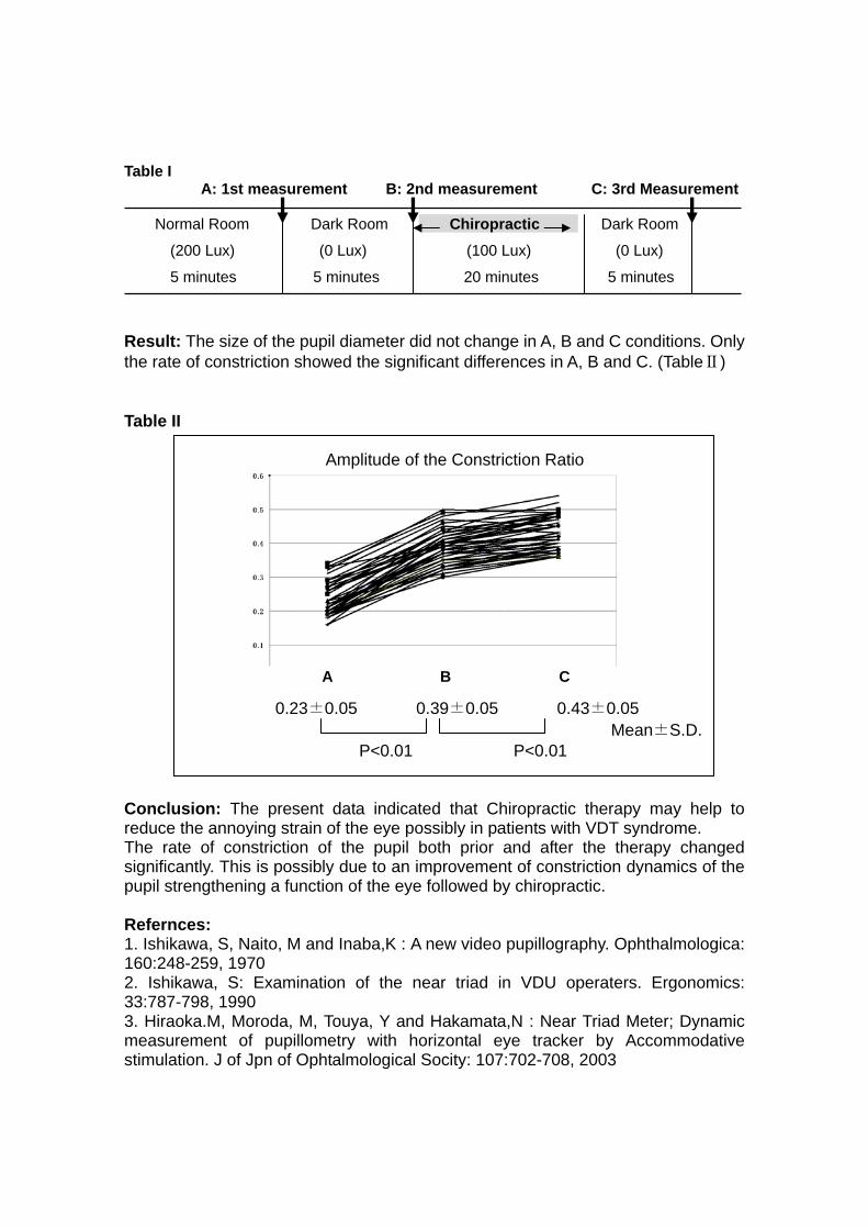
Table I A: 1st measurement B: 2nd measurement C: 3rd Measurement Normal Room Dark Room Chiropractic Dark Room
(200 Lux) (0 Lux) (100 Lux) (0 Lux)
5 minutes 5 minutes 20 minutes 5 minutes
Result: The size of the pupil diameter did not change in A, B and C conditions. Only the rate of constriction showed the significant differences in A, B and C. (TableⅡ) Table II Amplitude of the Constriction Ratio
0.23±0.05 0.39±0.05 0.43±0.05 Mean±S.D. P<0.01 P<0.01 Conclusion: The present data indicated that Chiropractic therapy may help to reduce the annoying strain of the eye possibly in patients with VDT syndrome. The rate of constriction of the pupil both prior and after the therapy changed significantly. This is possibly due to an improvement of constriction dynamics of the pupil strengthening a function of the eye followed by chiropractic. Refernces: 1. Ishikawa, S, Naito, M and Inaba,K : A new video pupillography. Ophthalmologica: 160:248-259, 1970 2. Ishikawa, S: Examination of the near triad in VDU operaters. Ergonomics: 33:787-798, 1990 3. Hiraoka.M, Moroda, M, Touya, Y and Hakamata,N : Near Triad Meter; Dynamic measurement of pupillometry with horizontal eye tracker by Accommodative stimulation. J of Jpn of Ophtalmological Socity: 107:702-708, 2003
A B C

Observation of spontaneous pupillary movement by the new high speed camera system
Jumpei Matsuda1,2, Nakamura Yoshiko3, Suzuki Kazutaka4, Haruyoshi Toyoda4, Naotoshi Hakamada4, Morio Ueno2, Satoshi Kawasaki1, Shigeru Kinoshita1
1. Department of Ophthalmology, Kyoto Prefectural University of Medicine, Kyoto, Japan 2. Department of Ophthalmology, National Center for Geriatrics and Gerontology, Obu, Japan 3. NTT WEST Kansai Health Administration Center, Osaka, Japan 4. Hamamatsu Photonics, Hamamatsu, Japan Corresponding Author: Jumpei Matsuda, M.D. Department of Ophthalmology, Kyoto Prefectural University of Medicine, 465 Kajii-cho, Hirokoji-agaru, Kawaramachi-dori, Kamigyo-ku, Kyoto 602-0842, Japan Tel: +81-75-251-5578 Fax: +81-75-251-5663 E-mail: [email protected]
Purpose: To observe slight spontaneous pupillary movement by the new high speed camera system. Subjects and Methods: We measured pupillary movement in 3 normal male using the 1kHz video camera system developed newly by Hamamatsu Photonics. The examination which continues for 30 seconds in the gloomy room is done before and 60 minutes after instillation of the parasympatholytic agent. The preservation of the 1000 pictures which are acquired in each 1 second and the representation of diameter graphic are performed automatically. Results: The graphic of spontaneous pupillary movement by the new high speed camera is smoother and more detailed than the graphic by an old camera. Pupillary unrest that was bigger movement seen about 1.5 times at 1 second and pupillary fluctuation that was smaller movement seen about 7 times at 1 second was observed. As a result of carrying out mydriasis by instillation of the parasympatholytic agent, pupil became macroscopic almost constant. Although the pupillary unrest was decreasing at that time, pupil fluctuation existed. Conclusions: The pupillary fluctuaton can be observed by this new system.This slight spontaneous movement is exist even when the autonomic nerve changed extremely.

Reduced lateralization of pupillary dynamics in patients with chronicheadache by Kampo medicine (Japanese herbal medicine)
Akino Wakasugi, Hiroshi Odaguchi, Tetsuro Oikawa, Toshihiko Hanawa
Oriental Medicine Research Center of the Kitasato Institute
Objective: Autonomic nervous imbalance is associated with in chronic headache. We investigated the effects of goshuyuto-a representative Kampo medicine for headache-on the lateralization of the pupillary autonomic nervous system by using binocular infrared video pupillography (Iriscorder C7364, Hamamatsu Photonics, Hamamatsu, Japan). Methods: Patients with chronic headache were administered with goshuyuto extract (TJ-31; Tsumura & Co., Tokyo, Japan) for 1 month (1st stage). After 1 month of discontinuation of goshuyuto, the patients were randomly divided into 2 groups: placebo and goshuyuto. In the 2nd stage, placebo or goshuyuto was administered for 3 months to the respective groups. The pupillary dynamics were measured at the beginning and at the end of the randomized, double-blinded, and placebo-controlled comparative study. Results: The subjects comprised 23 patients (male/female: 1/22; mean age, 39.0 years). Of these, 9 patients were assigned to the placebo group, 14 to the goshuyuto group. There was no significant difference in the laterality response between the 2 groups at the beginning of the 2nd stage. After 3months, the average laterality had decreased significantly in the goshuyuto group. Conclusions: Goshuyuto resulted in a significant decrease in laterality. These results suggest that goshuyuto relieves chronic headache by reducing the autonomic imbalance in the pupils. References: 1) Odaguchi, H., et al: The efficacy of goshuyuto, a typical Kampo (Japanese herbal medicine) formula, in preventing episodes of headache. Curr. Med. Res. Opin. 22: 1587-1597, 2006 2) Wakasugi, A., et al. Differentiation between Hangekobokuto and Kososan based on pupillary dynamics -Evaluation of autonomic nerve function-. J. Trad. Med. 23: 132-140, 2006 3) Wakasugi, A., et al. Effect of Kampo medicine on pupillary dynamics and heart rate variability: establishment of an objective evaluation method. Auton. Nerv. Syst. 43: 424-430, 2006

Blink Influence on Evaluation of Sleepiness using Pupillary Responses
Minoru Nakayama1, Keiko Yamamoto2 and Fumio Kobayshi2
1 The Center for Research and Development of Educational Technology (CRADLE),
Tokyo Institute of Technology, Tokyo, Japan 2 Department of Health and Psychosocial Medicine, Aichi Medical University, Aichi, Japan
Introduction: Pupillary response has been used as an index of sleepiness, but the validity of this index has also been debated. Although the index has been applied to diagnostic procedures, a series of studies of multiple sclerosis patients suggests that for healthy people, there is no significant relationship between pupillary indices and subjective sleepiness indices. A possible problem is the influence of blink, because most methods of measuring pupil size are based upon processing the image of the eye. Purpose: To examine the influence of blink on the pupillary indices, and to examine the relationship between pupil indices and subjective sleepiness scores. Method: A traditional sleepiness test, with a duration of 12 minutes, was conducted on 35 healthy people using measuring equipment (Hamamatsu photonics: C7364). Subjects had declared their own sleepiness using the Stanford Sleepiness Score. To estimate pupil size during blink, a pre-processing procedure was developed using the Support Vector Regression (SVR) technique (Nakayama 2005). Another pre-processing method is the moving average of temporal pupillary changes. As indices of pupil instability, the Pupillary Unrest Index (PUI) and Power Spectrum Desity (PSD) were extracted and analyzed. The parameters of analysis for the eye sleepiness test were modified in response to the accuracy of the measuring equipment. Results: Firstly, the values of PUI increased with experimental time or the sequence of the segment, and the values and deviations of PUI for experimental observations were larger than the ones for SVR estimation. The blink time also increased with experimental time or the progress of observation segments, but there were significant correlation relationship between the value of PUI and blink time in the first half of the experimental time. The relationship was also influenced when the blink rate was increased during the second half of the experiment. Therefore, the influence of blink changed as the experimental progressed. The mean PSD also correlated significantly with blink time, and the mean PSDs for f<0.8Hz were the same level across segments and pre-processing. The relationship between pupillary indices and the subjective sleepiness index was not significant, which is similar to results of previous experiments. In comparing the two groups for high and low subjective sleepiness subjects, blink time for the high sleepiness group increased monotonically. Conclusion: These results provide evidence that pupillary indices were significantly affected by blink, and pupillary indices extracted by ordinal processing procedures did not reflect sleepiness correctly. Acknowledgement: We would like to thank Dr. Misuzu Watanabe, Dr. Reiko Hori and Dr. Hirohito Tsuboi, Aichi Medical University, who kindly provided us with the opportunity to analyze pupillography data collected during their sleepiness tests.

FFT (Fast Fourier Transformation) analysis of fatigue wave of pupil under listening to music
Hiroshi Mochizuki1, Naoto Hara2, Nobuyuki Shoji1, Kazuo Mukuno2
1Department of Ophthalmology, Kitasato University Graduate School, Kitasato1-15-1, Sagamihara, Kanagawa, Japan 228-8555
2Department of Ophthalmology, Yokohama Dental and Medical Clinic, Kanagawa Dental College
Objectives: Pupillary oscillations are known to be originated from activities of the central nervous system1). In this study, the pupillary oscillations when listen to music, were analyzed applying the Fast Fourier Transformation (FFT) method. Methods: The subjects were 6 adults (28.0±7.0 yr.). After 10 minutes dark adaptation, the pupillary diameters were recorded for 1 minute during listening to 2 kinds of music (hard rock and classical). Measurements were done at 1, 5 and 10 minutes after beginning to listening in both music. The pupillary oscillations were recorded by an infrared pupillometer (C7364, HAMAMATSU Photonics), and were analyzed applying FFT. Results: The pupillary diameters and oscillations were similar under 2 kinds of music. In 2 subjects, the characteristic regular slow pupillary oscillations appeared. This oscillation showed clear frequently of 0.25 Hz associated with pupillary constriction. Conclusions: The 0.25 Hz pupillary oscillations were considered with “fatigue wave” reported in the past2). “Fatigue wave” was considered which appeared in physical fatigue, for example, in sleepiness. Frequency analysis of pupil in sleepiness showed accentuated whole frequency’s3). But, when observed this wave in this study, the subjects said they were not sleepy and feel relaxed. “Fatigue wave” appeared when not only be sleepy but also be relaxing and comfortable during enjoying music. We suggest that “fatigue wave” of 0.25 Hz in other words for “relax wave” has a special significance among various slow large oscillations4) References: 1. Mochizuki H et al. The Autonomic Nervous System 2007; 44: 24-28. 2. Lowenstein O et al. Invest Ophthalmol 1963; 2: 138-157. 3. Lüdtke H et al. Vision Res 1998; 38: 2889-2896. 4. Yoss RE et al. Am J Ophthalmol. 1970; 70: 935-41.

Relationship between PUI and cardiac autonomic nervous activity
Keiko Yamamoto1), Minoru Nakayama2), Reiko Hori1), Misuzu Watanabe3),
Hirohito Tsuboi4), Yasuhiro Akamatsu1), Fumio Kobayashi1)
1) Department of Health and Psychosocial Medicine, Aichi Medical University, Aichi,
Japan 2) The Center for Research and Development of Educational Technology
(CRADLE), Tokyo Institute of Technology, Tokyo, Japan 3) Institute for Occupational Health Science, Aichi Medical University, Aichi, Japan 4) Developmental and Regenerative Medicine, Genomics and Regenerative
Biology, Mie University Graduate School of Medicine, Mie, Japan Purpose: To examine a relationship between Pupillary Unrest Index (PUI) and cardiac autonomic nervous activity. Methods: Subjects were 20 healthy male participants in their annual health checkups (mean age 37.7+/-3.9 years, range 33-49). Pupil diameter and heart rate were measured for 12 minutes in a sitting position in a quiet room. Pupil diameter was measured by IRISCODER C7364 (Hamamatsu Photonics Corporation, Japan) with the sample rate of 60 Hz, and the PUI was calculated from the data of consecutive pupil diameter. The blink effect on the measured pupil diameter was eliminated using an estimation procedure with Support Vector Regression (SVR). Heart rate was measured using a wristband type pulse sensor (Denso Corporation, Japan) and heart rate variability was calculated by the three frequency bands (ULF; 0.003-0.04Hz, LF; 0.04-0.15Hz, HF; 0.15-0.4Hz). We divided the subjects into two groups based on the PUI value (high group; PUI>=10, low group; PUI<10), and compared the two groups in heart rate and heart rate variability by using student t-test. Results: The high PUI group showed a significantly lower heart rate than the low PUI group. (p<0.05) However, no significant difference was observed in the heart rate variability between the two groups. (ULF; p>0.1, LF; p>0.7, HF; p>0.2) Conclusions: Although PUI did not relate to heart rate variability, it related to heart rate itself. A possible relationship between pupil size variability and cardiac autonomic nervous activity should be clarified in further study.

An application of Eye Steam- Sheets to bilateral eyes activates autonomic nervous system and improves systemic hemodynamics
Yoshinao Nagashima1, Michihito Igaki1, Atsushi Suzuki1, Yukihiro Yada1,
Shuichi Tsuchiya1, Toshiyuki Suzuki1, Sachiko Oh-ishi2
1 Tokyo Research Laboratories, Kao Corporation,Tokyo, Japan 2 Basic Research Division, Kitasato Institute, Tokyo, Japan
Objective: Visual display terminal (VDT) syndrome, which is expressed as fatigue of the eyes and shoulder-, neck-, and lower back-pain, caused by work using electronic instruments including personal computers, has recently been problematic. As an auxiliary therapy for recovery from these symptoms, thermotherapy has been attracting attention. We previously reported that application of Eye Steam-Sheets containing exothermal powder, utilizing the oxidation reaction between oxygen in the air and iron powder, to the eye regions increased α brain waves, the HF component of R-R interval variation on electrocardiography (parasympathetic activity parameter), and the miosis rate in the pupillary light reflex, showing induction of the dominance of parasympathetic activity. In the present study, we improved the sheet, and investigated its effects on improvements in systemic hemodynamics and autonomic nervous activity. We improved the previous sheet of gravity-induced clustering of powder on one side, to make the sheet thin and flexible by replacing the exothermal powder with exothermal paper, which allowed homogenous supply of steam to the eyes. Methods: In healthy adult subjects, Eye Steam-Sheets, which produce heat and steam, warming the skin around the eyes to about 40°C, were applied to the bilateral eyes for 10 minutes, regarding application of a sham eye mask as a control. Palmar sweating was measured by the finite-difference ventilation capsule method using a rapid-responsive microheater, palmar skin blood flow was measured using a light integration laser Doppler flowmeter, the shoulder tissue hemoglobin level was measured by near infrared spectroscopy utilizing the modified Lambert Beer’s principle, R-R interval variation on electrocardiography was measured, continuous blood pressure was measured by the volume compensation method, and the pupillary light reflex was measured by the closed loop method. Results: The shoulder tissue hemoglobin level increased when the Eye Steam-Sheets were applied, suggesting that peripheral hemodynamics were improved. Palmar sweating was decreased, and skin blood flow was increased, suggesting that sympathetic tension was reduced. The diameter of the pupil before light stimulation, D1, was significantly reduced (constriction). Pupillary change after light stimulation, D1-D2, and the miosis rate, CR (D1-D2 / D1), were significantly increased. Sensitivity of the baroreceptor reflex calculated from changes in the ECG R-R interval and systolic blood pressure by the sequence method was markedly increased. These results suggest that parasympathetic activity could be elevated. Conclusions: Application of the Eye Steam-Sheets to the bilateral eyes may have improved peripheral hemodynamics, reduced sympathetic activity, and induced the dominance of parasympathetic activity. References: Nagashima, Y, et al, Auton Nerv Syst (Jpn) 43: 260-268, 2006

Influence of Higher Sound Pressure on Human Arousal Reaction during Eye-Tracking Task
-A Quantitative Analysis of Saccade-
Akinori Ueno1, Shiro Kokubun1, Yuichi Inoue2 1 Department of Electronic and Computer Engineering, Graduate School of Science and Engineering, Tokyo Denki University, Japan 2 Japan Somnology Center, Neuropsychiatric Research Institute, Japan
Objective: The objective of this research is to explore effective stimulation conditions of the beep sound at keeping the subject awake during monotonous task. Methods: We measured saccade during eye-tracking task and applied sound stimulation to the subject by using a computerized stimulator which triggers a beep sound automatically 70 ms after the generation of saccade when the ratio of peak velocity and duration (VDR) of the saccade is below a preprogrammed threshold. Since the VDR of saccade was revealed to be a sensitive vigilance index by our previous studies [1], [2], the sound was applied when the vigilance level of the subject was reduced. We compared the mean VDR before and after the sound stimulation having various sound pressures of 70dB, 80dB, 90dB and 0dB. Results: Mean VDR increased after the stimulation for each sound pressure of 70, 80, and 90dB in all 3 subjects. Mean VDR after 0dB stimulation (i.e. no stimulation) also increased in 2 of 3 subjects for each sound pressure. However, mean VDR only after 80dB stimulation were significantly larger than mean VDR after 0dB stimulation in all subjects. Conclusions: The results of the analysis suggested that it is not necessarily the case that the higher sound pressure exerts the higher arousal effect during monotonous eye-tracking task. References: [1] A. Ueno, S. Kokubun, and Y. Uchikawa: "A prototype real-time system for assessing vigilance levels and for alerting the subject with sound stimulation," International Journal of Assistive Robotics and Mechatronics, Vol.8, No.1, pp.19-27, March 2007. [2] A. Ueno, Y. Uchikawa: "An approach to quantification of human alertness using dynamics of saccadic eye movement -For an application to Human Adaptive Mechatronics-", Proceedings of 8th International Conference on Mechatronics Technology, (Hanoi, Vietnam), pp.563-568, Nov. 2004.

Characteristics of a light reflex caused by color stimulation
Keiichi Tanzawa1), Fumiatsu Maeda2), Tsuyoshi Yoneda2), Mayumi Oka1)2), Kazutaka Kani1)2), Akio Tabuchi1)2).
1) Master’s Program in Sensory Science, Graduate School of Health Science and Technology, Kawasaki University of Medical Welfare 2) Department of Sensory Science, Faculty of Health Science and Technology, Kawasaki University of Medical Welfare Objective: Recently it has been reported that retinal ganglion cells contain melanopsin which mediates the pupil light reflex, and also that there cells respond strongry to blue stimulation. The aim of our study was to clarify the characteristics of this light reflex caused by color stimulation, which changed wavelengths with the same size and luminance intensity. Methods: The methods for this study were 15 eyes (right eye) of 15 normal adult volunteers (22.5 ± 1.4 years) without ophthalmologic disease except for refractive errors. The stimulus parameters were an intensity of 100 cd/m2, a background intensity of 0.79 cd/m2, a diameter of 4 degrees, a duration of 200 msec, and the colors white ( 544 nm ), red ( 612 nm), green ( 544 nm ) and blue ( 437 nm ). Five points were on the superior temporal meridians of 45 degrees at eccentricities of 0, 5, 10, 15 and 20 degrees in the right eye stimulated. The diameter of the pupil was measured for evaluation of the light reflex, and the pupillary contraction ratio (PCR): (diameter of pre-stimulation – diameter of post-stimulation) / diameter of pre-stimulation was analyzed. Results: The average PCRs were white 13.8%, red 13.8%, green 12.0% and blue 17.6%, at an eccentricity of 0 degrees, followed by white 10.8 %, red 10.1 %, green 12.0 % and blue 17.6 % at an eccentricity of 5 degrees, and then white 9.1 %, red 7.7 %, green 11.4 %, blue 15.9 % at an eccentricity 10 degrees. At an eccentricity of 15 degrees, the rates were white 8.3 %, red 7.0 %, green 9.6 % and blue 15.2%. Finally, at an eccentricity 20 degrees, the rates were white 8.8 %, red 6.0 %, green 8.6 % were blue 14.2 %. The PCRs caused by blue stimulus were significantly greater than those resulting from a white, red and green stimuli at each eccentricity. Conclusion: The blue stimulation was able to trigger a light reflex larger than the other color stimuli. References: 1) Kardon RH: Pupil perimetry. Curr Opin Ophthalmol 3: 565-570, 1992. 2) Hong S, Narkiewicz J, Kardon RH: Comparison of pupil perimetry and visual perimetry in normal eyes: decibel sensitivity and variability. Invest Ophthalmol Vis Sci 42: 957-965, 2001. 3) Maeda F, Tanzawa K, Fukushima S, Inakagata S, Kani K, Tabuchi A: A pupil perimeter for objective visual field measurement. Proc of 2007 IEEE/ICME International Conference on Complex Medical Engineering: 1122-1125, 2007. 4) Gamlin PD, McDougal DH, Pokorny J, Smith VC, Yau KW, Dacey DM: Human and macaque pupil responses driven by melanopsin-containing retinal ganglion cells. Vision Res 47: 946-54, 2007.

pupi
l con
stric
tion
rate
(%)
5060708090
100
(1) (2) (3) (4)
Target No.
***
Pupil response during fixating the different size and distance of target
Nanae Sakakibara, Yuka Ohta, Atsufumi Hashimoto, Ayako Hattori, Akina Hamasaki, Hitoshi Isikawa
Department of Orthoptics and Visual Science, School of Allied Health, Kitasato University
Objective: To compare the pupil response during fixating the different size and distance of target. Methods: We examined 38 healthy volunteers, with the age ranging 18-22 years. Target size and distance were as in table1. Each target was gray colored circle with black fixation point in the center. The target (1) and (2), (3) and (4) were calculated so that they had the same retinal image size. After positioning a red fixation point located at 5 m from a subject (control pupil size), each target was then displayed for 15 seconds. Pupil responses were recorded by infrared pupillography (TriIRIS C9000, Hamamatsu) during these procedures. The results were expressed as % of pupil constriction, with the control pupil size defined as 100%. Results: There was no significant difference in pupil constriction rate when fixating the target (1) and (2). When comparing (3) and (4), the pupil constriction rate was significantly higher when fixating the target (3) (p<0.05). Moreover, in case of (2) and (4), the pupil constriction rate was significantly higher when fixating the target (4) (p<0.01). Conclusions: These results suggest that it is mainly the size of retinal images that induces the pupil response when a point is fixed more than a meter away (psychological effect). However, when a fixation point is less than a meter, the pupil response is induced mainly by the distance of the target. References: S. Kasthurirangan, A. Glasser. Characteristics of pupil responses during far-to-near and near-to-far accomodation.Ophthal.Physiol.Opt.2005 25:328-339 J.D. Hunter, J.G. Milton, H. Lüdtke, B. Wilhelm, H. Wilhelm. Spontaneous fluctuations in pupil size are not triggered by lens accommodation. Vis.Res.2000 40:567-573
Target No. distance size
(1) 5 m 5 cm
(2) 1 m 1 cm
(3) 20 cm 5 cm
(4) 1 m 25 cm
Fig. 1 Table 1