Embed Size (px)
Citation preview

This article has been accepted for publication and undergone full peer review but has not been through the copyediting,
typesetting, pagination and proofreading process, which may lead to differences between this version and the Version of Record. Please cite this article as doi: 10.1002/humu.22760. This article is protected by copyright. All rights reserved. 1
Humu-2014-0283
Research Article
Identification of variants in the 4q35 gene FAT1 in patients with a Facioscapulohumeral dystrophy
(FSHD)-like phenotype
Francesca Puppo 1,2,*, Eugenie Dionnet 1,2,*, Marie-Cécile Gaillard 1,2, Pascaline Gaildrat 3, Christel
Castro 1,2, Catherine Vovan 4, Bertaux Karine 4, Rafaelle Bernard 4, Shahram Attarian 1,2,5, Kanako Goto
6, Ichizo Nishino 6, Yukiko Hayashi 7, Frédérique Magdinier 1,2, Martin Krahn 1,2,4, Françoise
Helmbacher 8, Marc Bartoli 1,2,4,£,# and Nicolas Lévy 1,2,4,£
1Aix Marseille Université, GMGF, 13385, Marseille, France.
2Inserm, UMR_S 910, 13385, Marseille, France.
3Institute for Research and Innovation in Biomedecine (IRIB), Inserm, UMR 1079, University of Rouen,
Rouen, France.
4 Département de Génétique Médicale et de Biologie Cellulaire, AP-HM, Hôpital d'Enfants de la
Timone, 13385, Marseille, France.
5Department of Neurology and Neuromuscular Diseases, CHU La Timone, 13385, Marseille, France.
6NCNP, National Institute of Neuroscience, Tokyo, 187-8502, Japan.
7 Department of Neurophysiology, Tokyo Medical University, Tokyo, 160-0022, Japan.
8Aix Marseille Université, CNRS, IBDM, UMR 7288, , , 13288, Marseille, France.
*These authors should be considered co-first authors.
£The last two authors contributed equally to this article.
#Corresponding author:
Marc Bartoli
Corresponding author’s postal address: Aix Marseille Université, GMGF, UMR_S 910, Faculté de
Médecine 27, Bd Jean Moulin 13385 Marseille, France.
Corresponding author’s phone: +33 (0) 4 91 32 49 08, fax +33 (0) 4 91 80 43 19
Corresponding author’s e-mail address: [email protected]

This article is protected by copyright. All rights reserved. 2
ABSTRACT
Facioscapulohumeral muscular dystrophy (FSHD) is linked to copy number reduction (n<10) of the 4q
D4Z4 subtelomeric array, in association with DUX4-permissive haplotypes. This main form is
indicated as FSHD1. FSHD-like phenotypes may also appear in the absence of D4Z4 copy number
reduction. Variants of the SMCHD1 gene have been reported to associate with D4Z4
hypomethylation in DUX4-compatible haplotypes, thus defining FSHD2. Recently, mice carrying a
muscle-specific knock-out of the protocadherin gene Fat1 or its constitutive hypomorphic allele were
shown to develop muscular and non-muscular defects mimicking human FSHD. Here we report FAT1
variants in a group of patients presenting with neuromuscular symptoms reminiscent of FSHD. The
patients do not carry D4Z4 copy number reduction, 4q hypomethylation or SMCHD1 variants.
However, abnormal splicing of the FAT1 transcript is predicted for all identified variants. To determine
their pathogenicity, we elaborated a minigene approach coupled to an antisense oligonucleotide
(AON) assay. In vitro, four out of five selected variants induced partial or complete alteration of
splicing by creating new splice sites or modifying splicing regulators. AONs confirmed these effects.
Altered transcripts may affect FAT1 protein interactions or stability. Altogether, our data suggest that
defective FAT1 is associated with an FSHD-like phenotype.
Keywords: neuromuscular pathology, facioscapulohumeral dystrophy-FSHD; FAT1-protocadherin.

This article is protected by copyright. All rights reserved. 3
INTRODUCTION
Facioscapulohumeral muscular dystrophy (FSHD; MIM# 158900) (Justin Besancon et al. 1964;
Balatsouras et al. 2007) is the third most common human neuromuscular disease. Symptoms mainly
appear during the second decade of life and are characterized by progressive weakness of the facial,
scapular and humeral muscles, later spreading toward the lower limb muscles. Non-muscular tissues
are also frequently affected in FSHD; hearing loss manifests in 75% of patients, and retinal
telangiectasia occurs in 60% of patients (Wohlgemuth et al. 2004). Learning difficulties and epilepsy
may also occur as atypical symptoms in subsets of severely affected children (Saito et al. 2007).
Genetically, FSHD is linked to the presence of a reduced copy number (1-10 copies) of a tandemly
repeated 3.3 kb segment (D4Z4 macrosatellite) on chromosome 4q35, while in healthy individuals,
this repeat varies from 11 to 100 copies (Wijmenga et al. 1990; van Deutekom et al. 1993). This main
form of FSHD, referred to as FSHD1, accounts for approximately 95% of cases (Richards et al. 2012).
Such a heterozygous D4Z4-array reduction is associated with chromatin relaxation on specific
permissive haplotypes (Lemmers et al. 2002), which might result in stabilization of the DUX4
transcript, encoded by the retrogene located in D4Z4, and expression of this transcription factor
(Gabriëls et al. 1999; Dixit et al. 2007; Lemmers et al. 2010; Spurlock et al. 2010; Vanderplanck et al.
2011). A form of FSHD not linked to D4Z4 contraction accounts for 5% of patients (contraction-
independent FSHD) (van Overveld et al. 2003). In a majority of these patients described at present,
now referred to as FSHD2, variants of SMCHD1 (MIM# 614982, GenBank NM_015295.2) (Lemmers et
al. 2012; Larsen et al. 2014; Lemmers et al. 2014), a gene known to play a role in chromatin
relaxation and maintenance of X chromosome inactivation (Blewitt et al. 2008; Gendrel et al. 2012),
have been reported to associate with D4Z4 hypomethylation in DUX4-compatible 4q haplotypes,
thereby leading to DUX4 overexpression as well.
Nevertheless, large-scale studies performed on healthy individuals and FSHD patients have shown
that the permissive haplotype (4qA161) on FSHD-sized repeats, although rare, is present in the
healthy population, thus constituting a rare uncommon polymorphism (van Overveld et al. 2000;

This article is protected by copyright. All rights reserved. 4
Lemmers et al. 2007; Scionti et al. 2012a; Scionti et al. 2012b) and indicating that DUX4 protein
expression is not in itself sufficient to trigger FSHD symptoms. Furthermore, DUX4 RNA and protein
have been detected, although at low levels, in muscle biopsies of healthy individuals carrying DUX4-
compatible haplotypes in the absence of 4q contraction (Jones et al. 2012; Broucqsault et al. 2013).
This finding indicates that although necessary, DUX4 activation is not sufficient on its own to trigger
the appearance of symptoms (Jones et al. 2012), implying the existence of modifier genes/mutations
that contribute to this complex syndrome. As such, SMCHD1 variants were recently shown to
constitute modifier alleles of FSHD1 (Sacconi et al. 2013; Larsen et al. 2014). Therefore, evidence of
co-segregation for multiple genetic factors began to accumulate, contributing to the genotype-
phenotype correlation in a proportion of FSHD families. However, other cases of contraction-
independent FSHD are not explained by SMCHD1/DUX4 synergy, leaving room for the identification
of other genes involved in FSHD pathogenesis.
Recently, mouse models carrying a muscle-specific knock-out of the protocadherin gene Fat1 or its
constitutive hypomorphic allele have been shown to develop defects in the shape and position of
specific groups of muscles in the shoulder and face (Caruso et al. 2013). The human FAT1 gene
(MIM# 600976; GenBank NM_005245.3,) is composed of 27 exons spanning a region of 139kb and
encodes a 506kDa transmembrane protein with 34 cadherin domains followed by a laminin and 5
EGF motifs in its extracellular part. Studies on the protocadherin Fat1 demonstrated its influence on
smooth muscle cell motility (Hou and Sibinga 2009), actin accumulation at neuronal synapses
(Moeller et al. 2004), the Hippo signaling pathway (Bennett and Harvey 2006; Cho et al. 2006) and
cell polarity (Skouloudaki et al. 2009). While constitutive loss of FAT1 leads to a significant degree of
perinatal lethality in mice (Ciani et al. 2003; Caruso et al. 2013), incomplete alterations of the FAT1
gene have been identified in rare developmental 4q syndromes characterized by mild facial
dysmorphisms, abnormalities in skeletal and cardiac development, and growth and mental
retardation (Ockey et al. 1967; Bendavid et al. 2007; Kitsiou-Tzeli et al. 2008). Genetic links between
FAT1 gene polymorphisms and susceptibility to bipolar disorder or schizophrenia have also been

This article is protected by copyright. All rights reserved. 5
established (Blair et al. 2006; Abou Jamra et al. 2008; Jung and Jun 2013). Finally, somatic mutations
inducing high expression of FAT1 or its variant isoforms have been associated with both acute
lymphoblastic leukemia (de Bock et al. 2012) and breast cancer progression (Lee et al. 2012), while
other mutations (and CNV, Copy Number Variation) causing FAT1 protein loss of function are
associated with glioblastoma, colorectal or head and neck cancers (Morris et al. 2013a).
For the first time, in a study by Caruso and colleagues (Caruso et al. 2013), FAT1 was shown to
control muscle patterning by modulating the polarity of myoblast migration during embryonic
development, to be involved in regionalized muscle wasting, and to have a role in adult muscle fiber
functions. Mice also exhibit extra-muscular defects, such as retinal vasculopathy and abnormal inner
ear patterning, which possibly represents a hearing impairment (Caruso et al. 2013). Furthermore,
misregulation of FAT1 expression was observed in fetal FSHD1 muscle tissue, and a CNV mapping in a
putative regulatory enhancer of FAT1 preferentially segregated with FSHD-like patients who
presented no D4Z4 contraction (Caruso et al. 2013). Although FSHD-like symptoms only represent a
subset of FAT loss-of-function phenotypes, the map of muscles that show developmental impairment
in FAT1 mutants strongly resembles one affected by human FSHD, raising the provocative hypothesis
that FAT1 acts as a modifier gene to the disease.
Here, we sought to identify FAT1 variants in 49 Japanese cases affected by neuromuscular disease
that closely resembles FSHD, according to the diagnostic criteria defined by the European Expert
Group on FSHD (Padberg et al. 1991) and presenting no D4Z4 copy number reduction in either 4q35
or 10q26, no reciprocal rearrangements (Yamanaka et al. 2004), no hypomethylation at D4Z4, and no
SMCHD1 mutations. In 10 out of 49 cases analyzed, we identified 10 different variants of the FAT1
gene. To determine their pathogenic effect on FAT1 splicing, we elaborated a minigene approach,
coupled to an antisense oligonucleotide (AON) assay. The in vitro results showed a partial or
complete alteration of splicing either by creating a new acceptor splice site or by modifying splicing
regulators in FAT1 variants. AONs confirmed the effect of these nucleotide substitutions. Altogether,
our data suggest that a defective FAT1 protein may be associated with an FSHD-like phenotype.

This article is protected by copyright. All rights reserved. 6
METHODS
Study subjects and samples DNA preparation and PCR and Sanger Sequencing are reported in the
Supp. Methods.
Bioinformatic predictions
Amino acid substitutions were predicted for their impact on protein function by three online
algorithms: Polymorphism Phenotyping v2 (Polyphen-2), SIFT and Prophyler (see web links for
references and Supp. Table S1 for results). PolyPhen-2 version 2.2.2
(http://genetics.bwh.harvard.edu/pph2) calculates the impact of amino acid substitutions using
protein sequences from the UniProt database, structural data from PDB/DSSP and comparative
considerations from UCSC multiple alignments of 45 vertebrate genomes. For a mutation, PolyPhen-2
calculates the probability that this mutation is damaging and estimates a false positive rate (the
chance that the mutation is classified as damaging when it is in fact non-damaging). Based on the
threshold of this rate, a mutation is qualitatively defined as benign, possibly damaging, or probably
damaging. SIFT (v1.03, http://sift.jcvi.org) predictions are based on the degree of conservation of
amino acid residues in sequence alignments derived from closely related sequences. The score
ranges from 0 to 1. The amino acid substitution is predicted to be damaging if the score is <= 0.05.
The impact of variants on FAT1 splicing was predicted by Human Splicing Finder, HSF, version 2.4.1
(cut-off for donor or acceptor sites is 55%) (Desmet et al. 2009) and Alamut Visual 2.2, released in
June 2012. This software integrates 4 different algorithms for splicing prediction: SpliceSiteFinder
(http://www.umd.be/HSF/; cut-off for donor and acceptor sites 70%), MaxEntScan (no cut-off
provided), NN Splice (score for donor and acceptor ranges between 0 and 1, cut-off is 0.4) and
GeneSplicer (no cut-off provided). For HSF, as well as for Alamut, the threshold for ESEFinder are the
following: SF2/ASF, 1.956; SC35, 2.383; SRp40, 2.67; and SRp55, 2.676. Predictions were considered
strong enough to be tested in a minigene reporter assay in the following cases: nucleotide
substitutions that create or break regulatory ESE or SSE splicing sites or determine the creation of a
new splicing site stronger than the natural one. The inclusion criteria for experimental analysis of
variants also accounted for available data from dbSNP137 and included a minor allele frequency
lower than 0.005 (Table 1 and Supp. Table S1).
This article is protected by copyright. All rights reserved. 7
Methylation analysis
For sodium bisulfite sequencing, 2 μg of genomic DNA was denatured for 30 minutes at 37°C in 0.4 M
NaOH and incubated overnight in a solution of 3 M sodium bisulfite, pH5 and 10 mM hydroquinone
using a previously described protocol (Ehrlich et al. 1982). Converted DNA was then purified using
the Wizard DNA CleanUp kit (Promega) following manufacturer’s recommendation and precipitated
with ethanol. Modified DNA was amplified using the forward 5’-AAATATGTAGGGAAGGGTGTAAGTT-3’
and reverse 5’-GGAGAGAGGGTTTGGTATATTTAAG-3’ primer set designed with the MethPrimer
software (Bird 2002) to amplify 21 CpGs (275 bp) within the D4Z4 proximal region (Gaillard et al.
2014). We avoided CpGs in the primer sequence in order to amplify methylated and unmethylated
DNA with the same efficiency. Amplification was conducted using the High Fidelity Taq polymerase
(Roche) according to manufacturer’s instructions. After initial denaturation, the amplification
conditions were the following: 94°C for 20 seconds, 54°C for 30 seconds, 72°C for one minute for 10
cycles and then the elongation was increased by 30 seconds at each subsequent cycle for 25 cycles.
The PCR products were then purified using the Wizard SV gel and PCR Purification system (Promega)
and cloned using the pGEM®-T Easy Vector cloning kit (Promega). Randomly selected colonies were
grown overnight at 37°C with ampicillin selection and PCR amplified directly using T7 and SP6
primers. At least ten independent colonies were sequenced according to the Sanger method by
Eurofins MWG Operon (Ebersberg, Germany) with either T7 or SP6 primers.
Sequences were analyzed using BiQ Analyzer software, and the average methylation score was
calculated as the number of methylated CpGs for the total number of CpGs in the reference
sequence (Table 2).
Cell culture
HEK293 cells (Human Embryonic Kidney cell line) were grown in Dulbecco’s Modified Eagle Medium
(Gibco) supplemented with 15% fetal calf serum and 1% PSA (penicillin streptomycin actinomycin) in
a 5% CO2 atmosphere at 37°C.

This article is protected by copyright. All rights reserved. 8
Generation of constructs for splicing minigene reporter assay
The splicing minigene assay has been previously described (Gaildrat et al. 2010). DNA fragments
corresponding to FAT1 wild-type and mutant exons surrounded by upstream and downstream
intronic sequences were amplified from genomic DNA of patients heterozygous for the nucleotide
substitution by using exon-specific forward and reverse primers (Supp. Table S2) carrying 5’ tails with
BamHI (or BglII) and MluI restriction sites, respectively (underlined in Supp. Table S3). After digestion
with BamHI (or BglII) and MluI, the PCR products were inserted into the BamHI and MluI cloning sites
in the intron of the pCAS2 vector containing a two-exon splicing reporter minigene. The insert was
then sequenced to identify the minigene constructs containing the FAT1 exon carrying the wild-type
or substituted nucleotide and to ensure that no extra substitutions were added during amplification
or cloning.
Transfection and analysis of RT-PCR products
The wild-type and mutant minigene constructs were transiently transfected into HEK293 cells using
the promofectine transfection reagent, according to manufacturer’s instructions (PromoKine). Cells
were collected 24 hours post-transfection. Total RNA was extracted using the PureLink® RNA Mini Kit
(Ambion; Life Technologies), according to the manufacturer ’s instructions, followed by a DNase
treatment with the DNA-free Kit (Ambion; Life Technologies). The RT-PCR reactions were performed
using the SuperScript OneStep RT-PCR with Platinium Taq kit (Life Technologies), according to the
manufacturer’s instructions, with 500 ng RNA as a template in a 50 µl reaction volume. Reactions
were performed using 300 nM of forward pCAS-KO1F (5’-TGACGTCGCCGCCCATCAC-3’) and reverse
pCAS2R primers (5’-ATTGGTTGTTGAGTTGGTTGTC-3’) (Gaildrat et al. 2010). The reverse transcription
program had 1 cycle: 50°C for 30 minutes. The PCR program had 35 cycles of amplification of 96°C for
45 seconds, 50°C for 45 seconds, and 72°C for 1 minute. RT-PCR products were separated by
electrophoresis on 2% agarose 1000 (Invitrogen) gels containing ethidium bromide and visualized by
exposure to non-saturating ultraviolet light.
This article is protected by copyright. All rights reserved. 9
AON design and transfection
AONs with full-length 2-O-methyl-substituted ribose moieties and phosphorothioate internucleotide
linkages were purchased from Eurogentec, France. AONs were designed according to several criteria:
sequences targeting exon 5 (5’-U*U*U*C*C*C*G*C*U*C*A*G*G*G*A*G*U*C*U*G-3’) and exon 17
(5’-G*A*C*A*C*U*G*U*A*G*U*U*U*C*C*C*C*U*G*G*A-3’) were designed complementarily to
the wild-type sequence. In parallel, the AON sequence for exon 11 (5’-
A*U*C*A*G*U*U*G*C*U*G*U*G*A*U*A*G*U*A*A-3’) was designed complementarily to the
variant sequence.
HEK293 cells (4 × 10⁵ cells) grown in twelve-well plates to 80% confluence were transfected with 1.1
µg of AON and 0.5 µg of pCAS2 constructions using Lipofectamine 2000 reagent according to the
manufacturer’s instructions (Life Technologies). Total RNA was isolated from cultured cells 24 hours
after transfection, and RT-PCR analysis was performed as described above.

This article is protected by copyright. All rights reserved. 10
RESULTS
In this work, 49 Japanese cases affected by neuromuscular disease were selected based on the
homogeneity of muscles presenting clinical signs (Table 3) and closely resembling FSHD according to
the diagnostic criteria defined by the European Expert Group on FSHD (Padberg et al. 1991). No
patients presented with D4Z4 copy number reduction (Yamanaka et al. 2004). The age distribution of
patients with available clinical information was 60.8% of patients under the age of 30 at the time of
disease presentation. The facial, scapular and humeral muscles were simultaneously affected in
45.8% of patients, and 89.6% of them had at least 2 out of the three muscle groups affected.
Progression of the disease toward the lower limbs was present in 72.9% of patients, with most
affected at the proximal limb muscles. Other myopathies presenting with similar phenotypic
appearances but specific histological defects were ruled out in 47 out of 49 individuals by muscular
biopsy examination. Specifically, muscle from the J21 case showed scattered fibers with rimmed
vacuoles. Protein analyses showed normal expression of dystrophin, sarcoglycans, dystroglycans,
merosin, collagen 6, dysferlin, caveolin-3, calpain 3 and emerin. A sequence analysis of the GNE gene
was normal (data not shown). Moreover, J51 was diagnosed with Nemaline myopathy based on the
muscular biopsy (data not shown), while no variants in the ACTA1 gene were found (Wallefeld et al.
2006). Finally, clinical data from other members of the patients’ families were not available for any
individual included in this study.
Among these patients, we found nucleotide substitutions in the FAT1 gene with a potentially
damaging effect on transcript processing in 10 cases (Table 1 and Supp. Tables S1 and S4). In
particular, 3 of the substitutions have not been previously reported (c.4723G>A; c.4959G>A and
c.12051C>T). In all patients, the FAT1 variants were present on one allele and distributed in different
exons without a specific mutation hotspot. The transition type of nucleotide substitution was
overrepresented and corresponded to 9 out of 10 nucleotide substitutions. The age of onset was
under the age of 30 for 6 out of 10 patients carrying FAT1 nucleotide variants, and for two
individuals, the age of symptom appearance was not available. The facial, scapular and humeral

This article is protected by copyright. All rights reserved. 11
muscles were simultaneously affected in 3 out of these 10 cases, and 9 of them had at least 2 out of
the 3 muscle groups affected. Progression of the disease toward the lower limbs was present in 7 of
the patients (details in Table 1).
Several publications have reported that D4Z4 hypomethylation, in the context of a DUX4-permissive
haplotype associated with variants in the SMCHD1 gene, may cause FSHD2 and contribute to FSHD1
(Lemmers et al. 2012; Sacconi et al. 2013; Lemmers et al. 2014) with moderate but significant
differences in the methylation level between asymptomatic carriers and individuals with clinical
FSHD (Gaillard et al. 2014; Lemmers et al. 2014). To determine whether variation in the FAT1 gene
segregates with D4Z4 hypomethylation in our different cases, we used the sodium bisulfite
sequencing method to measure the level of methylation of the 21 CpGs in the proximal D4Z4 region
in 8 out of the 10 patients carrying FAT1 variants. The results, reported in Table 3, show that less than
50% of CpGs displaying D4Z4 hypomethylation associated with the appearance of the muscular
defects clinically similar to FSHD.
Furthermore, we screened and excluded SMCHD1 variants in all patients carrying nucleotide variants
for FAT1 (Table 2 and Supp. Table S5); thus, 8 out of 10 FSHD-like patients do not carry any of the
genetic features and epigenetic marks usually observed in FSHD1 and SMCHD1-linked FSHD2
patients.
Interestingly, for some of the FAT1 variants identified, the minor allele count in the East Asian and
general population relative to the total allele number is available for 63,000 exomes by the Exome
Aggregation Consortium (ExAC), Cambridge, MA (URL: http://exac.broadinstitute.org) [11/2014], as
reported in Table 1. However, these variants are not reported as SNPs in any of the Japanese SNP
databases that we could access (HAPMAP, KYUGEN JPK2 http://www.ncbi.nlm.nih.gov/projects/SNP/
and JSNP, http://snp.ims.u-tokyo.ac.jp/). The main criteria for inclusion in our functional
investigations accounted for a frequency of the minor allele in the general population lower than
0.002. The frequency of FAT1 SNPs that we found in our group of patients compared to the Japanese
and general Caucasian population is indicated in Supp. Table S3.

This article is protected by copyright. All rights reserved. 12
Six out of ten nucleotide substitutions that we observed lead to amino acid substitutions in different
extracellular cadherin domains of FAT1. For all of the substitutions, anomalies in splicing of the FAT1
transcript have been predicted using Human Splicing Finder, HSF and Alamut software (see methods
for URLs), as detailed in Supp. Table S1 (Desmet et al. 2009). The drastic loss of splicing regulator sites
or the creation of new splicing sites stronger than the natural ones was predicted in seven cases. Five
of the six amino acid substitutions were also predicted to have a deleterious impact on protein
function by several algorithms (see methods for URL references and Supp. Table S1 for detailed
results).
To evaluate the splicing effect of selected FAT1 variants, we performed a functional assay based on a
comparative analysis of the splicing pattern of wild-type and mutant sequences in the pCAS2
minigene-expressing vector (Gaildrat et al. 2010). Five variants were selected based on their
suitability for the experimental conditions of the test and the strength of in silico splicing predictions.
Fragments corresponding to the FAT1 exons studied, surrounded by 150 bp of upstream and
downstream intronic sequences, were amplified from genomic DNA of patients and subcloned into
the pCAS2 vector. After transient transfection in HEK293 cells, the splicing patterns and efficiency of
wild-type and mutant FAT1 exon incorporation in the minigene were analyzed by RT-PCR and
sequencing. As reported in Figure 1, we showed that 4 cases, c.3770G>A (pArg1257Gln), c.8963A>T
(pLys2988Ile), c.8991G>A (pThr2997Thr) and c.10331A>G (pAsn3444Ser), result in partial or
complete splicing defects. One defect may lead to nonsense mediated decay for c.8963A>T, which
implies a loss of frame of aberrant mRNA. In the other three cases, shorter half-lives of the aberrant
mRNAs or deleterious forms of the translated FAT1 protein may be produced. In contrast to in silico
predictions, no splicing effect in HEK293 cells was depicted for the c.4723G>A (p.Ala1575Thr) amino
acid substitution, (Supp. Figure S1) but in vivo tissue-specific splicing effects cannot be excluded.
Among the nucleotide substitutions with a splicing effect in the functional minigene assay, the
c.3770G>A transition (identified in patient J16) induced complete skipping of exon 5 from the mutant
transcript, as indicated in Figure 1a, suggesting an effect on splicing either by disrupting an exonic

This article is protected by copyright. All rights reserved. 13
splicing enhancer (ESE) element and/or by creating an exonic splicing silencer (ESS), as predicted by
HSF and Alamut (Supp. Table S1). Interestingly, co-transfection with the antisense oligonucleotide
(AON) carrying the wild-type sequence (c.3770G) induced a partial skipping of the exon, suggesting
that this AON masks the predicted ESE element (Figure 1a). The aberrant transcript missing exon 5
(RNA r.3643_3972del) produced by the variant allele would result, if translated, into an in-frame
deletion of 110 amino acids at the protein level (p.Val1215_Ser1324del) with partial truncation of the
cadherin 10 and 11 extracellular domains.
The second nucleotide substitution with an evident effect on splicing was the c.8963A>T
transversion, located in exon 11 and identified in patient J29. The minigene assay showed the
production of 2 transcripts: a primary one corresponding to normal exon inclusion and an aberrant
minor transcript (5% expression ratio versus the full-length transcript), which skips exon 11 (Figure
1c). These results suggest that this variant alters splicing by disrupting ESE motifs, in agreement with
the prediction (Supp. Table S1). If translated, the misspliced RNA (RNA r.8879_9075del) would result
in a frameshift with the creation of a premature stop codon (p.Gly2960Asp*9) between exons 10 and
12.
The third FAT1 variant, a c.8991G>A transition, identified in patient J2, also induced an alteration in
exon 11 splicing. Indeed, the results depicted in Figure 1b show that the exon 11 minigene construct
carrying the variant produces two transcripts, a primary one corresponding to the normal inclusion
of the full-length exon and a second minor transcript (average expression ratio of 8% versus the full-
length transcript) in which the first 114 exonic nucleotides are deleted (RNA r.8879_8992del). This
effect is in agreement with the Alamut and HSF predictions suggesting the creation of an acceptor
splice site at position c.8992/c.8993 with a score slightly higher than the natural one (Supp. Table
S1). We designed an antisense oligonucleotide (AON c.8991A) carrying the variant nucleotide to
mask the created splicing acceptor site. Co-transfection of AON c.8991A and the variant minigene
rescued normal splicing (Figure 1b) and eliminated the aberrant transcript. This RNA would be

This article is protected by copyright. All rights reserved. 14
translated in an in-frame deletion (p.Gly2960_Thr2997del) that corresponds to a truncation of
cadherin domain 27 in the FAT1 protein.
Finally, the c.10331A>G transition, found in patient J51, induced a minor skipping of exon 17 (27%
average expression ratio versus the full-length transcript) in the splicing minigene assay (Figure 1d),
and produced a transcript with 144 exonic nucleotides removed (RNA r.10207_10351del). This result
could be the consequence of the predicted loss of an ESE element (Supp. Table S1) located in exon
17, thus interfering with the inclusion of exon 17 in the altered transcript. Transfection of AON
c.10331A (WT) with the wild-type exon 17 construct led to partial skipping, while in the presence of
exon 17 carrying the nucleotide substitution, it leads to complete skipping of the exon (Figure 1d).
Finally, co-transfection of the AON specific for exon 5 (c.3770G) and a wild-type exon 17 construct did
not interfere with exon 17 splicing. Thus, these results confirm that the wild-type AON specifically
masks an element involved in exon inclusion. The main consequence of exon 17 skipping is likely the
truncation of the cadherin 32 and 33 domains (p.Thr3403_Glu3451del) without the loss of the
downstream reading frame.
Other nucleotide substitutions were also predicted to interfere with splicing, but they could not be
easily tested, as they were not suitable for the conditions required for the minigene assay. In
particular, patient J21 carries 2 different substitutions: c.2215A>G (p.Met739Val) and c.13374G>A
(p.Gln4458Gln). Testing these two variants using a minigene-based assay was not possible because of
their location in FAT1 exons 2 and 27, respectively. As present, the parents’ DNA is required to
determine whether one of the two substitutions is a de novo substitution or if both substitutions are
carried on the same allele. Both substitutions are predicted to affect splicing with equal strength, and
an analysis of their respective impact on FAT1 transcription in the patient’s biological samples, such
as a muscle biopsy, would be interesting.

This article is protected by copyright. All rights reserved. 15
DISCUSSION
In this study, we identified 10 different variants in the FAT1 gene in 10 out of 49 Japanese patients
affected by an FSHD-like neuromuscular disease. The diagnosis was based on criteria defined by the
European Expert Group on FSHD (Padberg et al. 1991), while the patients presented no D4Z4 copy
number reduction in either 4q35 or 10q26 and no reciprocal rearrangements (Yamanaka et al. 2004).
To rule out myopathies presenting similar phenotypic appearance but specific histological defects, a
muscular biopsy examination was performed in 47 out of 49 individuals. For J21, the muscle biopsy
showed non-specific scattered fibers with rimmed vacuoles, and the J51 diagnosis of Nemaline
myopathy was based on histological examination of his muscle biopsy (data not shown). In both
cases, neither mutations in known genes nor expression defects in myopathy-related proteins were
observed. Thus, mutations in new genes responsible for the neuromuscular phenotype have not
been ruled out. Interestingly, as the simultaneous presence of hypomethylation in D4Z4 regions in
the context of a DUX4-permissive haplotype and variants in the SMCHD1 gene may cause FSHD2 and
contribute to FSHD1 (Lemmers et al. 2012; Sacconi et al. 2013; Larsen et al. 2014; Lemmers et al.
2014), we measured the D4Z4 methylation level according to the methods and results recently
published by our group (Gaillard et al. 2014). In that publication, a significant reduction in the DNA
methylation level at the D4Z4 proximal region was reported for individuals with clinical FSHD but not
carrying a copy number reduction (FSHD2) by sodium bisulfite sequencing, with a global level of
methylated CpG/sequence below the threshold of 50% (Gaillard et al. 2014). In the group of patients
investigated here, the average methylation level in the proximal D4Z4 region was above 50% (72%),
meaning that in these patients, FSHD symptoms are not associated with epigenetic changes at the
4q35 region (Table 2). Furthermore, no mutations in SMCHD1 were detected in any of the patients
carrying nucleotide variants for FAT1 (Table 2). Even if the diagnosis of FSHD is based on the clinical
evaluation of symptoms, genetic testing is required to confirm FSHD1 or FSHD2. However, a small
proportion of patients with FSHD-like symptoms are not associated with D4Z4 copy number
reduction or with hypomethylated 4qA alleles and SMCHD1 variants, as reported here. In this

This article is protected by copyright. All rights reserved. 16
situation, while the diagnosis may be questioned and reevaluated for some, it remains likely that the
specific FSHD clinical signs might result from other genetic changes that affect processes also
involved in FSHD. Identifying such genetic causes of FSHD-like cases would likely teach us about the
biological mechanisms of this pathology. Thus, hypomorphic Fat1 mice presenting a FSHD-like
phenotype (Caruso et al. 2013) and the identification of FAT1 variants in FSHD-like patients raise the
challenging idea that FAT1 might be a disease gene associated with FSHD-like symptoms.
For some of the FAT1 variants depicted here, the minor allele count in the general and Japanese
populations was reported, when available, in Table 1. Thus, we propose that these variants might
represent very rare mutations that were possibly identified in presymptomatic individuals.
Nonetheless, incomplete penetrance may not be excluded at this point. Consistently, a CNV located
in a putative regulatory enhancer of FAT1 has previously been shown to preferentially segregate with
FSHD in non-contracted FSHD-like patients, hence constituting the basis of tissue-specific alterations
in FAT1 expression (Caruso et al. 2013).
The variants identified here fall into two categories that are not incompatible with each other. Six out
of 10 variants led to amino acid (a.a.) substitutions localized in different extracellular cadherin
domains of FAT1 and have been predicted to have a deleterious impact on protein function by
several algorithms. Moreover, for all nucleotide substitutions, the drastic loss of splicing regulator
sites or the creation of new splicing sites stronger than the natural ones have been suggested to give
rise to partial or complete splicing defects. These defects would lead to shorter half-lives of aberrant
mRNAs or the production of deleterious forms of the translated FAT1 protein. Thus, to demonstrate
their pathogenic effect on FAT1 transcript splicing, we elaborated a minigene approach coupled to an
antisense oligonucleotide (AON) assay. Some of these variants showed potential splicing alterations
as well as deleterious a.a. changes, suggesting that both possibilities can occur; for example, any
abnormally spliced mRNA, if translated, could result in a functionally aberrant protein. Therefore,
only further experimental validation will determine the true functional relevance of each process.
The FAT1 protein can be considered a “model protein,” allowing us to correlate predictions to

This article is protected by copyright. All rights reserved. 17
experimental findings. Moreover, the results shown here indicate that selected nucleotide
substitutions have splicing effects in in vitro minigene assays. Interestingly, splicing defects might also
depend on the chromosomal context or the presence of tissue-specific regulatory elements, which
could only be analyzed in biological samples derived from patients. Nonetheless, our group recently
showed that a minigene in vitro approach is reliable for confirming endogenous splicing defects
(Kergourlay et al. 2014), supporting the idea that an actual damaging effect can be considered for
variants showing an in vitro effect, even in the absence of in vivo confirmation.
In particular, the absence of an effect in c.4723G>A (p.Ala1575Thr) (Supp. Figure S1) does not
exclude the impact of this variant on the endogenous transcript. This nucleotide substitution is
strongly predicted to cause a deleterious amino acid substitution, as detailed in Supp. Table S1. To
further validate the reliability of prediction algorithms at the experimental level, proteomic
approaches as well as functional tests focusing on cadherin domain structure are in development. In
the case of the c.3770G>A (p.Arg1257Gln), c.8991G>A (pThr2997Thr) and c.10331A>G
(p.Asn3444Ser) transitions with a validated impact on splicing, aberrantly spliced mRNA isoforms
may have more rapid turnover and shorter half-lives, thereby affecting intracellular FAT1
concentrations. The splicing effect is also compatible with an additional issue at the protein level
because these aberrant transcripts, if translated, have been predicted to contribute to deleterious
amino acid substitutions as well. For one of these variants (c.3770G>A), we showed that it
completely prevents the in vitro inclusion of exon 5 in the transcript, thus producing an
r.3643_3972del mRNA. If translated, the corresponding protein would lack the cadherin 10 and 11
extracellular domains without loss of the downstream reading frame. Nevertheless, the effect of this
variant on splicing in vivo could be less drastic, allowing the partial integration of exon 5 carrying the
variant nucleotide in the transcript. Thus, it would be interesting to investigate the importance of the
arginine 1257 change to glutamine on FAT1 protein function as well as the potential dominant-
negative effect due to the coexistence of multiple FAT1 protein splicing isoforms in cells. Along the
same line, c.10331A>G p.Asn3444Ser may contribute to two coexisting mRNA transcripts: the

This article is protected by copyright. All rights reserved. 18
aberrant mRNA r.10207_10351del (p.Thr3403_Glu3451del) and the normal transcript, which may be
translated into a FAT1 protein with a serine replacing the aspartic acid in position 3444. Even if these
variants are compatible with FAT1 protein frame conservation, extracellular cadherin domains are
expected to be lost and likely affect the stability and/or protein-protein interactions. Similar
consequences may be predicted for c.8991G>A (pThr2997Thr) in which the aberrant mRNA
r.8879_8992del, coexisting with the normal transcript, may be translated into a FAT1 protein
truncated for cadherin domain 27. Interestingly, patient J29 carries a c.8963A>T transversion
(p.Lys2988Ile) that is predicted to disrupt the ESE motifs Tra2 and 9G8 and to create a new hnRNP
A1 site, while amino acid substitution is not expected to have deleterious consequences on the
protein. Here we show the partial production of a variant transcript missing exon 11. The
juxtaposition of exons 10 and 12 would create a frame shift in the FAT1 transcript and a premature
stop codon 23 nucleotides after the beginning of exon 12 (p.Gly2960Asp*9). This incomplete FAT1
mRNA, if translated into a truncated FAT1 protein, may exert dominant-negative activity.
Alternatively, the misspliced RNA (r.8879_9075del) may be eliminated by the nonsense mediated
mRNA decay pathway, causing haploinsufficiency reminiscent of the fat1 mouse model (Caruso et al.
2013).
Antisense oligonucleotides (AON) recognize and block special sequences in the neo-translated RNA
that are otherwise bound by splicing protein complexes (Wein et al. 2010). Based on the targeted
splicing sequence, AONs may lead to the partial or total skipping of flanking exons. In our case, the
AONs were designed to precisely recognize either the substituted or wild-type alleles, allowing us to
demonstrate the specific effect of FAT1 alterations in vitro. Next, we will apply the AONs that mimic
nucleotide substitutions to FAT1-expressing cells and animal models to characterize the functional
consequences of these substitutions on the translated proteins. Moreover, for c.8991G>A, the
second minor transcript in which the first 114 exonic nucleotides were deleted, as observed using
the minigene test, was rescued by co-transfection of AON c.8991A, which carries the variant
nucleotide to mask the created splicing acceptor site. Animal models reproducing this variant are
under development to evaluate the therapeutic consequences of AON injection and splicing rescue
in vivo.

This article is protected by copyright. All rights reserved. 19
Some recent algorithms, which have been developed for functional annotation of genetic variants
from high-throughput sequencing data, suggest that FAT1 is a dispensable human gene based on the
number of stop codons identified in the study (Wang et al. 2010). Interestingly, our study did not
identify stop codons in any of the screened individuals or in healthy controls, while the constitutive
loss of FAT1 function leads to perinatal lethality (Ciani et al. 2003; Caruso et al. 2013). Thus, in
independent functional and genetic studies, the partial preservation of FAT1 function would still be
compatible with life. Accordingly, defective forms of the protein would have tissue-specific impacts
and would be exerted at low doses by a dominant-negative effect. Hence, altered FAT1 would play a
pathogenic role by affecting only specific interactions with protein partners and only during specific
stages of development. Nevertheless, consistent with mice carrying the tissue-specific knock-out of
FAT1 or its constitutive hypomorphic allele (Caruso et al. 2013), we cannot rule out
haploinsufficiency as the pathogenic mechanism, at least for some of the variants reported here.
In addition to our present results, other germline mutations in the FAT1 gene may lead to
developmental defects in subtle pathologies, such as schizophrenia and bipolar disorder
susceptibility (Ockey et al. 1967; Blair et al. 2006; Bendavid et al. 2007; Abou Jamra et al. 2008;
Kitsiou-Tzeli et al. 2008; Jung and Jun 2013). Similarly, somatic mutations contribute to Wnt/-
catenin pathway misregulation and cancer progression in specific tissues by both overexpression and
inactivation of FAT1 protein function (de Bock et al. 2012; Lee et al. 2012; Morris et al. 2013a; Morris
et al. 2013b). Thus, aberrant FAT1 expression or incomplete or complete loss of function cause
defects in tissues unrelated to FSHD, indicating that FAT1 has pleiotropic implications that may lead
to a range of clinical consequences, with only a subset of those sharing similarities with FSHD. Thus,
we propose the existence of a heterogeneous pathological entity, named FATopathy.
In perspective, adding FAT1 to the panel of neuromuscular disease-causing genes routinely tested for
molecular diagnosis will be needed both to investigate the broader significance of FAT1 in disease
pathogenesis and to better define the implication of multiple genetic interactions in neuromuscular
disease appearance.
In conclusion, according to the evidence from the mouse model recently published and the
identification of the CNV located in a transcriptional enhancer of the human FAT1 gene, which
segregates in non-contracted FSHD-like patients, our genetic data further strengthen the link
between the FAT1 gene and FSHD-like neuromuscular diseases.
This article is protected by copyright. All rights reserved. 20
ACKNOWLEDGMENTS
The authors would like to thank all patients for their participation. We thank Christophe Pécheux,
Mohamed Mesrati and Cécile Mouradian for technical assistance. All the samples explored in this
study were prepared by the Center of Biological Resources, Department of Medical Genetics, la
Timone. We thank Vincent Meyer, Emmanuelle Salort-Campana and Pr Jean Pouget for medical and
scientific discussion. Jacques Beckmann and Isabella Ceccherini are warmly acknowledged for their
assistance in reading the manuscript and their critical comments and suggestions. Grants from the
Association Française contre les Myopathies (AFM)-Téléthon (strategical pole MNH Decrypt to NL)
and the Fondation Maladies Rares financed this work. The funders had no role in the study design,
data collection and analysis, decision to publish, or preparation of the manuscript.
REFERENCES
Abou Jamra R, Becker T, Georgi A, Feulner T, Schumacher J, Stromaier J, Schirmbeck F, Schulze TG, Propping P, Rietschel M, Nöthen MM, Cichon S. 2008. Genetic variation of the FAT gene at 4q35 is associated with bipolar affective disorder. Mol. Psychiatry 13: 277–284.
Balatsouras DG, Korres S, Manta P, Panousopoulou A, Vassilopoulos D. 2007. Cochlear function in facioscapulohumeral muscular dystrophy. Otol. Neurotol. Off. Publ. Am. Otol. Soc. Am. Neurotol. Soc. Eur. Acad. Otol. Neurotol. 28: 7–10.
Bendavid C, Pasquier L, Watrin T, Morcel K, Lucas J, Gicquel I, Dubourg C, Henry C, David V, Odent S, Levêque J, Pellerin I, et al. 2007. Phenotypic variability of a 4q34-->qter inherited deletion: MRKH syndrome in the daughter, cardiac defect and Fallopian tube cancer in the mother. Eur. J. Med. Genet. 50: 66–72.
Bennett FC, Harvey KF. 2006. Fat cadherin modulates organ size in Drosophila via the Salvador/Warts/Hippo signaling pathway. Curr. Biol. CB 16: 2101–2110.
Bird A. 2002. DNA methylation patterns and epigenetic memory. Genes Dev. 16: 6–21.
Blair IP, Chetcuti AF, Badenhop RF, Scimone A, Moses MJ, Adams LJ, Craddock N, Green E, Kirov G, Owen MJ, Kwok JBJ, Donald JA, et al. 2006. Positional cloning, association analysis and expression studies provide convergent evidence that the cadherin gene FAT contains a bipolar disorder susceptibility allele. Mol. Psychiatry 11: 372–383.
Blewitt ME, Gendrel A-V, Pang Z, Sparrow DB, Whitelaw N, Craig JM, Apedaile A, Hilton DJ, Dunwoodie SL, Brockdorff N, Kay GF, Whitelaw E. 2008. SmcHD1, containing a structural-maintenance-of-chromosomes hinge domain, has a critical role in X inactivation. Nat. Genet. 40: 663–669.

This article is protected by copyright. All rights reserved. 21
Bock CE de, Ardjmand A, Molloy TJ, Bone SM, Johnstone D, Campbell DM, Shipman KL, Yeadon TM, Holst J, Spanevello MD, Nelmes G, Catchpoole DR, et al. 2012. The Fat1 cadherin is overexpressed and an independent prognostic factor for survival in paired diagnosis-relapse samples of precursor B-cell acute lymphoblastic leukemia. Leukemia 26: 918–926.
Broucqsault N, Morere J, Gaillard M-C, Dumonceaux J, Torrents J, Salort-Campana E, Maues De Paula A, Bartoli M, Fernandez C, Chesnais AL, Ferreboeuf M, Sarda L, et al. 2013. Dysregulation of 4q35- and muscle-specific genes in fetuses with a short D4Z4 array linked to facio-scapulo-humeral dystrophy. Hum. Mol. Genet. 22: 4206–4214.
Caruso N, Herberth B, Bartoli M, Puppo F, Dumonceaux J, Zimmermann A, Denadai S, Lebossé M, Roche S, Geng L, Magdinier F, Attarian S, et al. 2013. Deregulation of the protocadherin gene FAT1 alters muscle shapes: implications for the pathogenesis of facioscapulohumeral dystrophy. PLoS Genet. 9: e1003550.
Cho E, Feng Y, Rauskolb C, Maitra S, Fehon R, Irvine KD. 2006. Delineation of a Fat tumor suppressor pathway. Nat. Genet. 38: 1142–1150.
Ciani L, Patel A, Allen ND, ffrench-Constant C. 2003. Mice lacking the giant protocadherin mFAT1 exhibit renal slit junction abnormalities and a partially penetrant cyclopia and anophthalmia phenotype. Mol. Cell. Biol. 23: 3575–3582.
Desmet F-O, Hamroun D, Lalande M, Collod-Béroud G, Claustres M, Béroud C. 2009. Human Splicing Finder: an online bioinformatics tool to predict splicing signals. Nucleic Acids Res. 37: e67.
Deutekom JC van, Wijmenga C, Tienhoven EA van, Gruter AM, Hewitt JE, Padberg GW, Ommen GJ van, Hofker MH, Frants RR. 1993. FSHD associated DNA rearrangements are due to deletions of integral copies of a 3.2 kb tandemly repeated unit. Hum. Mol. Genet. 2: 2037–2042.
Dixit M, Ansseau E, Tassin A, Winokur S, Shi R, Qian H, Sauvage S, Mattéotti C, Acker AM van, Leo O, Figlewicz D, Barro M, et al. 2007. DUX4, a candidate gene of facioscapulohumeral muscular dystrophy, encodes a transcriptional activator of PITX1. Proc. Natl. Acad. Sci. U. S. A. 104: 18157–18162.
Ehrlich M, Gama-Sosa MA, Huang LH, Midgett RM, Kuo KC, McCune RA, Gehrke C. 1982. Amount and distribution of 5-methylcytosine in human DNA from different types of tissues of cells. Nucleic Acids Res. 10: 2709–2721.
Gabriëls J, Beckers MC, Ding H, Vriese A De, Plaisance S, Maarel SM van der, Padberg GW, Frants RR, Hewitt JE, Collen D, Belayew A. 1999. Nucleotide sequence of the partially deleted D4Z4 locus in a patient with FSHD identifies a putative gene within each 3.3 kb element. Gene 236: 25–32.
Gaildrat P, Killian A, Martins A, Tournier I, Frébourg T, Tosi M. 2010. Use of splicing reporter minigene assay to evaluate the effect on splicing of unclassified genetic variants. Methods Mol. Biol. Clifton NJ 653: 249–257.
Gaillard M-C, Roche S, Dion C, Tasmadjian A, Bouget G, Salort-Campana E, Vovan C, Chaix C, Broucqsault N, Morere J, Puppo F, Bartoli M, et al. 2014. Differential DNA methylation of the D4Z4 repeat in patients with FSHD and asymptomatic carriers. Neurology 83: 733–742.
Gendrel A-V, Apedaile A, Coker H, Termanis A, Zvetkova I, Godwin J, Tang YA, Huntley D, Montana G, Taylor S, Giannoulatou E, Heard E, et al. 2012. Smchd1-dependent and -independent pathways

This article is protected by copyright. All rights reserved. 22
determine developmental dynamics of CpG island methylation on the inactive X chromosome. Dev. Cell 23: 265–279.
Hou R, Sibinga NES. 2009. Atrophin proteins interact with the Fat1 cadherin and regulate migration and orientation in vascular smooth muscle cells. J. Biol. Chem. 284: 6955–6965.
Jones TI, Chen JCJ, Rahimov F, Homma S, Arashiro P, Beermann ML, King OD, Miller JB, Kunkel LM, Emerson CP, Wagner KR, Jones PL. 2012. Facioscapulohumeral muscular dystrophy family studies of DUX4 expression: evidence for disease modifiers and a quantitative model of pathogenesis. Hum. Mol. Genet. 21: 4419–4430.
Jung Y-E, Jun T-Y. 2013. Association between FAT Gene and Schizophrenia in the Korean Population. Clin. Psychopharmacol. Neurosci. Off. Sci. J. Korean Coll. Neuropsychopharmacol. 11: 67–71.
Justin Besancon L, Pequignot H, Contamin F, Delavierre P, Rolland P. 1964. [MYOPATHY OF THE LANDOUZY-D’EJERINE TYPE. REPORT OF A HISTORICAL CASE]. Sem. Hôp. Organe Fondé Par Assoc. Enseign. Méd. Hôp. Paris 40: 2990–2999.
Kitsiou-Tzeli S, Sismani C, Koumbaris G, Ioannides M, Kanavakis E, Kolialexi A, Mavrou A, Touliatou V, Patsalis PC. 2008. Distal del(4) (q33) syndrome: detailed clinical presentation and molecular description with array-CGH. Eur. J. Med. Genet. 51: 61–67.
Larsen M, Rost S, El Hajj N, Ferbert A, Deschauer M, Walter MC, Schoser B, Tacik P, Kress W, Müller CR. 2014. Diagnostic approach for FSHD revisited: SMCHD1 mutations cause FSHD2 and act as modifiers of disease severity in FSHD1. Eur. J. Hum. Genet. EJHG.
Lee S, Stewart S, Nagtegaal I, Luo J, Wu Y, Colditz G, Medina D, Allred DC. 2012. Differentially expressed genes regulating the progression of ductal carcinoma in situ to invasive breast cancer. Cancer Res. 72: 4574–4586.
Lemmers RJLF, Goeman JJ, Vliet PJ van der, Nieuwenhuizen MP van, Balog J, Vos-Versteeg M, Camano P, Ramos Arroyo MA, Jerico I, Rogers MT, Miller DG, Upadhyaya M, et al. 2014. Inter-individual differences in CpG methylation at D4Z4 correlate with clinical variability in FSHD1 and FSHD2. Hum. Mol. Genet.
Lemmers RJLF, Kievit P de, Sandkuijl L, Padberg GW, Ommen G-JB van, Frants RR, Maarel SM van der. 2002. Facioscapulohumeral muscular dystrophy is uniquely associated with one of the two variants of the 4q subtelomere. Nat. Genet. 32: 235–236.
Lemmers RJLF, Tawil R, Petek LM, Balog J, Block GJ, Santen GWE, Amell AM, Vliet PJ van der, Almomani R, Straasheijm KR, Krom YD, Klooster R, et al. 2012. Digenic inheritance of an SMCHD1 mutation and an FSHD-permissive D4Z4 allele causes facioscapulohumeral muscular dystrophy type 2. Nat. Genet. 44: 1370–1374.
Lemmers RJLF, Vliet PJ van der, Klooster R, Sacconi S, Camaño P, Dauwerse JG, Snider L, Straasheijm KR, Ommen GJ van, Padberg GW, Miller DG, Tapscott SJ, et al. 2010. A unifying genetic model for facioscapulohumeral muscular dystrophy. Science 329: 1650–1653.
Lemmers RJLF, Wohlgemuth M, Gaag KJ van der, Vliet PJ van der, Teijlingen CMM van, Knijff P de, Padberg GW, Frants RR, Maarel SM van der. 2007. Specific sequence variations within the 4q35 region are associated with facioscapulohumeral muscular dystrophy. Am. J. Hum. Genet. 81: 884–894.

This article is protected by copyright. All rights reserved. 23
Moeller MJ, Soofi A, Braun GS, Li X, Watzl C, Kriz W, Holzman LB. 2004. Protocadherin FAT1 binds Ena/VASP proteins and is necessary for actin dynamics and cell polarization. EMBO J. 23: 3769–3779.
Morris LGT, Kaufman AM, Gong Y, Ramaswami D, Walsh LA, Turcan Ş, Eng S, Kannan K, Zou Y, Peng L, Banuchi VE, Paty P, et al. 2013a. Recurrent somatic mutation of FAT1 in multiple human cancers leads to aberrant Wnt activation. Nat. Genet. 45: 253–261.
Morris LGT, Ramaswami D, Chan TA. 2013b. The FAT epidemic: a gene family frequently mutated across multiple human cancer types. Cell Cycle Georget. Tex 12: 1011–1012.
Ockey CH, Feldman GV, Macaulay ME, Delaney MJ. 1967. A large deletion of the long arm of chromosome No. 4 in a child with limb abnormalities. Arch. Dis. Child. 42: 428–434.
Overveld PGM van, Lemmers RJFL, Sandkuijl LA, Enthoven L, Winokur ST, Bakels F, Padberg GW, Ommen G-JB van, Frants RR, Maarel SM van der. 2003. Hypomethylation of D4Z4 in 4q-linked and non-4q-linked facioscapulohumeral muscular dystrophy. Nat. Genet. 35: 315–317.
Overveld PG van, Lemmers RJ, Deidda G, Sandkuijl L, Padberg GW, Frants RR, Maarel SM van der. 2000. Interchromosomal repeat array interactions between chromosomes 4 and 10: a model for subtelomeric plasticity. Hum. Mol. Genet. 9: 2879–2884.
Padberg GW, Lunt PW, Koch M, Fardeau M. 1991. Diagnostic criteria for facioscapulohumeral muscular dystrophy. Neuromuscul. Disord. NMD 1: 231–234.
Richards M, Coppée F, Thomas N, Belayew A, Upadhyaya M. 2012. Facioscapulohumeral muscular dystrophy (FSHD): an enigma unravelled? Hum. Genet. 131: 325–340.
Sacconi S, Lemmers RJLF, Balog J, Vliet PJ van der, Lahaut P, Nieuwenhuizen MP van, Straasheijm KR, Debipersad RD, Vos-Versteeg M, Salviati L, Casarin A, Pegoraro E, et al. 2013. The FSHD2 gene SMCHD1 is a modifier of disease severity in families affected by FSHD1. Am. J. Hum. Genet. 93: 744–751.
Saito Y, Miyashita S, Yokoyama A, Komaki H, Seki A, Maegaki Y, Ohno K. 2007. Facioscapulohumeral muscular dystrophy with severe mental retardation and epilepsy. Brain Dev. 29: 231–233.
Scionti I, Fabbri G, Fiorillo C, Ricci G, Greco F, D’Amico R, Termanini A, Vercelli L, Tomelleri G, Cao M, Santoro L, Percesepe A, et al. 2012a. Facioscapulohumeral muscular dystrophy: new insights from compound heterozygotes and implication for prenatal genetic counselling. J. Med. Genet. 49: 171–178.
Scionti I, Greco F, Ricci G, Govi M, Arashiro P, Vercelli L, Berardinelli A, Angelini C, Antonini G, Cao M, Muzio A Di, Moggio M, et al. 2012b. Large-scale population analysis challenges the current criteria for the molecular diagnosis of fascioscapulohumeral muscular dystrophy. Am. J. Hum. Genet. 90: 628–635.
Skouloudaki K, Puetz M, Simons M, Courbard J-R, Boehlke C, Hartleben B, Engel C, Moeller MJ, Englert C, Bollig F, Schäfer T, Ramachandran H, et al. 2009. Scribble participates in Hippo signaling and is required for normal zebrafish pronephros development. Proc. Natl. Acad. Sci. U. S. A. 106: 8579–8584.
Spurlock G, Jim H-P, Upadhyaya M. 2010. Confirmation that the specific SSLP microsatellite allele 4qA161 segregates with fascioscapulohumeral muscular dystrophy (FSHD) in a cohort of multiplex and simplex FSHD families. Muscle Nerve 42: 820–821.

This article is protected by copyright. All rights reserved. 24
Vanderplanck C, Ansseau E, Charron S, Stricwant N, Tassin A, Laoudj-Chenivesse D, Wilton SD,
Coppée F, Belayew A. 2011. The FSHD atrophic myotube phenotype is caused by DUX4 expression.
PloS One 6: e26820.
Wallefeld W, Krause S, Nowak KJ, Dye D, Horváth R, Molnár Z, Szabó M, Hashimoto K, Reina C, Carlos
J De, Rosell J, Cabello A, et al. 2006. Severe nemaline myopathy caused by mutations of the stop
codon of the skeletal muscle alpha actin gene (ACTA1). Neuromuscul. Disord. NMD 16: 541–547.
Wang K, Li M, Hakonarson H. 2010. ANNOVAR: functional annotation of genetic variants from high-
throughput sequencing data. Nucleic Acids Res. 38: e164.
Wein N, Avril A, Bartoli M, Beley C, Chaouch S, Laforêt P, Behin A, Butler-Browne G, Mouly V, Krahn
M, Garcia L, Lévy N. 2010. Efficient bypass of mutations in dysferlin deficient patient cells by
antisense-induced exon skipping. Hum. Mutat. 31: 136–142.
Wijmenga C, Frants RR, Brouwer OF, Moerer P, Weber JL, Padberg GW. 1990. Location of
facioscapulohumeral muscular dystrophy gene on chromosome 4. Lancet 336: 651–653.
Wohlgemuth M, Kooi EL van der, Kesteren RG van, Maarel SM van der, Padberg GW. 2004. Ventilatory
support in facioscapulohumeral muscular dystrophy. Neurology 63: 176–178.
Yamanaka G, Goto K, Ishihara T, Oya Y, Miyajima T, Hoshika A, Nishino I, Hayashi YK. 2004. FSHD-like
patients without 4q35 deletion. J. Neurol. Sci. 219: 89–93.

This article is protected by copyright. All rights reserved. 25
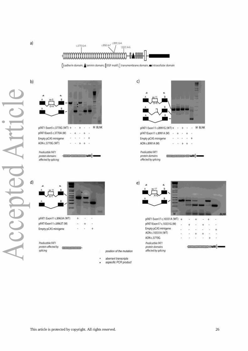
Figure 1. Abnormal splicing in FAT1-derived transcripts. (a) Diagram representing FAT1 protein
domains with the positions of the 4 variants tested by the minigene splicing assay coupled with the
AON assay. Briefly, wild-type and mutant pCAS2 minigene constructs containing different FAT1 exons
of interest were transfected into HEK293 cells alone or in combination with an AON specifically
directed against the variant or wild-type allele. At 24 hours after transfection, the splicing patterns of
the minigenes were monitored by RT-PCR. All variants are expressed as the cDNA sequence and refer
to GenBank NM_005245.3. (b) Transcripts amplified from constructs containing wild-type (pFAT1
Exon5 c.3770G) and variant (pFAT1 Exon5 c.3770A) alleles of FAT1 exon 5 in the presence or absence
of AON c.3770G and the empty pCAS2 minigene were separated on an agarose gel. The transcript
amplified from the exon 5 variant construction containing exons from the pCAS2 minigene only is
indicated by a star (*). The effect of co-transfection of AON c.3770G and the wild-type exon 5
construct is presented together with a representation of the predicted effect on the protein. (c)
Agarose gel electrophoresis of transcripts produced from minigene constructs containing no insert
(empty pCAS2 minigene), wild-type FAT1 exon 11 (pFAT1 Exon11 c.8991G) or mutant FAT1 exon 11
(pFAT1 Exon11 c.8991A) alone or co-transfected with AON c.8991A (mut). A star (*) indicates the
transcript specifically produced from the variant exon 11 minigene and corresponding to 114
nucleotides deleted from exon 11. AON c.8991A rescued the exon 11 variant allele, allowing
expression of the complete wild-type form of the exon. The potential effect on the FAT1 protein is
also represented. (d) Transcripts produced from constructs containing wild-type (pFAT1 Exon11
c.8963A) or variant (pFAT1 Exon11 c.8963T) FAT1 exon 11 and from the empty pCAS2 minigene were
analyzed by agarose gel electrophoresis. Star (*) indicates transcript amplified from exon 11 carrying
nucleotide substitutions with complete skipping of it. Potential effect on the FAT1 truncated protein
is reproduced. (e) Agarose gel electrophoresis of transcripts produced from constructs containing
wild-type (pFAT1 Exon17 c.10331A) or variant (pFAT1 Exon17 c.10331G) FAT1 exon 17 in the presence
or absence of AONs (c.10331A, c.3770G) and the pCAS2 minigene. As indicated by a star (*), the
transcript amplified from the exon 17 variant construct only contains exons from the pCAS2
minigene. The effect of co-transfection of AON c.10331A with the pFAT1 Exon17 c.10331G minigene
is also presented together with the schema of the effect on the translated FAT1 protein. AON
c.3770G from exon 5 does not have a specific splicing effect on wild-type exon 17.
This article is protected by copyright. All rights reserved. 26

This article is protected by copyright. All rights reserved. 27
Table 1. Molecular and clinical details for selected mutations
X indicates presence of mutation relative to cDNA sequence and relative to GenBank reference
NM_005245.3. Nucleotide numbering uses +1 as the A of the ATG translation initiation codon in the
reference sequence, with the initiation codon as codon 1. Black shadow indicates validated effect on
splicing while grey one stresses potentially damaging amino acid changes. Last two columns
represent minor allele count in East Asian and general healthy population respectively, as reported
by Version 0.2 ExAC (Exome Aggregation Consortium) database.
FAT1 CDS
sequencing
results: J2 J6 J7 J15 J16 J20 J21 J29 J41 J51 EXON
East Asian
Allele
count/Allele
number
General
Allele count/Allele
number
c.2215A>G
p.Met739Val X 2 1/8892 2/124670
c.3770G>A
p.Arg1257Gln X 5 12/8894 22/124752
c.4358G>A
p.Arg1453His X 8 15/8724 195/120308
c.4723G>A
p.Ala1575Thr X X 9 0 0
c.4959G>A
p.Val1653Val X 10 9/8878 9/124606
c.8963A>T
p.Lys2988Ile X 11 141/8888 155/124660
c.8991G>A
p.Thr2997Thr X 11 0 20/124668
c.10331A>G
p.Asn3444Ser X 17 9/8890 9/124578
c.12051C>T
p.Cys4017Cys X 22 0 25/124654
c.13374 G>A
p.Gln4458Gln X 27 2/8894 2/124730
age of onset
bir
th
30
-50
>50
<10
10
->2
0
20
-30
20
>30
10
>20
FSH +++ ++ + ++ +++ ++ ++ ++ +++ ++
Lower limbs ++ + + ++ ++ + +

This article is protected by copyright. All rights reserved. 28
Table 2. 5’-D4Z4 methylation percentage and SMCHD1 (GenBank NM_015295) screening results
5’-D4Z4 methylation percentage
84% 65% 71% 80% 57% 65% 74% 72%
Patients J2 J6 J7 J15 J16 J21 J29 J51
SMCHD1 exons and
SNPs identifie
d
1
HTZ rs2430853 MAF gp (G)= 46%, MAF jp (C)= 29%
HTZ rs2430853
HMZ rs2430853
HTZ rs2430853
14
HTZ rs8090967, MAF gp = 37% MAF jp = 37% HTZ rs377473058, MAF not available HTZ rs116599328, MAF gp = 2%, MAF jp = 3% HTZ rs635132, MAF gp = 40% MAF jp = 31%
HTZ rs377473058 HTZ rs635132
HTZ rs377473058 HTZ rs635132
HTZ rs377473058 HMZ rs8090967 HTZ rs8090988 MAF gp = 31% MAF jp = 36%
HTZ rs377473058 HTZ rs635132
HTZ rs377473058 HMZ rs8090967 HTZ rs8090988
HTZ rs377473058 HTZ rs635132
HTZ rs377473058 HTZ rs635132 HTZ rs8090988
18/ 19
HTZ rs28446296 MAF gp = 31% MAF jp = 22%
HMZ rs28446296
HMZ rs28446296
21
HTZ rs633422 MAF gp = 28% MAF jp = 31%
HTZ rs633422
22
HTZ rs16943716 MAF gp = 22% MAF jp = 31%
HMZ rs16943716
HMZ rs16943716
26
HTZ rs10638660 MAF gp = 34% MAF jp
HMZ rs10638660 HMZ
HTZ rs10638660 HMZ
HMZ rs10638660 HMZ
HTZ rs10638660 HMZ
HMZ rs10638660 HMZ

This article is protected by copyright. All rights reserved. 29
= 39% HMZ rs8094260 MAF gp = 35% MAF jp = 38%
rs8094260 rs8094260 rs8094260 rs8094260 rs8094260
28
PCR not available
PCR not available
PCR not available
PCR not available
PCR not available
PCR not available
PCR not available
PCR not available
31/ 32
HTZ rs2019793 MAF gp = 18% MAF jp = 31% HTZ rs2304861 MAF gp = 16% MAF jp = 31% HTZ rs2304860 MAF gp = 22% MAF jp = 31% HTZ rs2304859 MAF gp = 33% MAF jp = 38%
HMZ rs2019793 HMZ rs2304861 HMZ rs2304859
HTZ rs2019793 HTZ rs2304861 HTZ rs2304859
HMZ rs2304860
HMZ rs2019793 HMZ rs2304861 HMZ rs2304859
HMZ rs2304860
HTZ rs2019793 HTZ rs2304861 HTZ rs2304859
HMZ rs2019793 HMZ rs2304861 HMZ rs2304859
33
HTZ rs71365197 MAF gp = 33% MAF jp = 38% HTZ rs200589679 MAF gp = 0% MAF jp = 1% HMZ rs300293 MAF gp = 3% MAF jp = 0%
HMZ rs71365197 HMZ rs300293
HTZ rs71365197 HMZ rs300293
HMZ rs300293 HMZ rs7237908 MAF gp = 22% MAF jp = 31%
HMZ rs71365197 HMZ rs300293
HMZ rs300293 HMZ rs7237908
HTZ rs71365197 HMZ rs300293
HMZ rs71365197 HMZ rs300293
40
HMZ rs300291 MAF gp = 3% MAF jp = 1% HTZ rs3214732 MAF gp = 32% MAF jp = 39%
HMZ rs300291
HMZ rs300291 HTZ rs3214732
HMZ rs300291 HMZ rs3214732
HMZ rs300291
HMZ rs300291 HMZ rs3214732
HMZ rs300291 HTZ rs3214732
HMZ rs300291
42
HMZ "rs10468730" MAF gp = 18% MAF
HMZ "rs10468730"
HMZ "rs10468730"
HMZ "rs10468730"
HMZ "rs10468730"

This article is protected by copyright. All rights reserved. 30
In upper part of the table, black sections of pie charts represent percentage of proximal D4Z4
methylated CpG/sequence in 8 out of 10 patients carrying FAT1 variants. In the lower part of the
table, Minor Allele Frequencies from general population (gp) and from Asian population (jp) are
expressed as MAF for the SNPs detected each of the 8 patients analyzed. No variants were depicted
in exons not showed in this table.
jp = 32%
43/ 44
HTZ "rs3213926" MAF gp = 33% MAF jp = 39%
HMZ "rs3213926"
HTZ "rs3213926"
HMZ "rs3213926"
HTZ "rs3213926"
HMZ "rs3213926"
45
HTZ rs35853884 MAF gp = 27% MAF jp = 29%
HMZ rs35853884
HMZ rs35853884
48
HTZ rs764718 MAF gp =1% MAF jp = 2%

This article is protected by copyright. All rights reserved. 31
Table 3. Summary of neuromuscular clinical signs found in patients presented in this study
AGE at onset Clinical details
individuals % Facial Scapular Humeral Abdominal Proximal
(lower
limbs)
Distal
(lower
limbs)
Axial
Birth/First decade 11/46 23.9% 81.8% 90.9% 72.7% 27.3% 72.7% 63.6% 63.6%
Second/third decade 17/46 36.9% 58.8% 94% 88.2% 23.5% 76.5% 64.7% 47.1%
Third/fifth decade 10/46 21.7% 80% 90% 80% 20% 80% 70% 50%
Older 8/46 17.4% 50% 75% 75% 25% 50% 25% 37.5%
Total with age and diagnosis
available
46
Total with diagnosis available 48 66.7% 89.6% 77.1% 22.9% 70.8% 60.4% 54.2%
Facioscapulohumeral deficit 45.8%
2 off 3 muscles 89.6%
Progression to lower limbs 72.9%