Embed Size (px)
Citation preview

miR
NA
Product C
atalog 20
16
- 20
17
miRNA In Situ Product Catalog 2016-2017
miRNA In Situ – Dawn of a New Era Accelerating Precision Medicine. Integrated miRNA In Situ System
• Over 200 Probes
• Detection Systems
• Automation
• Cancer of Unknown Primary
• Undifferentiated Tumors
• Cancer Staging
For Clinical Research & Tumour Analysis
In the U.S., call +1 (800) 421-4149
Outside the U.S., call +91-40-27185500For USA and Canada distribution only
www.biogenex.com © 2016 BioGenex Laboratories, Inc. All Rights Reserved

Dear Customer,
We are pleased to present the BioGenex miRNA Product Catalog for 2016 - 2017. As a vertically integrated company we develop, manufacture and market highly innovative and fully automated systems for cancer diagnosis, prognosis and therapy selection.
Xmatrx® systems redefine complete automation for the molecular pathology laboratory and standardize all the steps from baking through final cover-slipping in three simple steps - Load, Click and View. Compared to any other system on the market, Xmatrx® systems offer clean intense stain(s), automate more assay steps, and enable automation of technologies for the future molecular pathology laboratory.
• Xmatrx® ELITE integrates All-in-One and All-at-Once staining of IHC, ISH, special stains and beyond • Xmatrx® Infinity is a high-performance staining platform for life sciences and translational research • Xmatrx® ULTRA is the next-generation system with new features such as touch screen and SMS intelligence • Xmatrx® NANO is a ten-slide automated system specifically designed for FISH • Xmatrx® MINI enables in situ PCR and nucleic acid hybridization with tools for building micro-chamber
We also offer a series of i6000TM systems with very high throughput; 200 slides in an 8-hour shift.
To maintain our tradition of offering superior solutions for the emerging needs of your laboratory, we offer a broad range of molecular pathology products for IHC, ISH, miRNA, multiplex and special staining of tissues including 300+ primary antibodies, molecular probes, detection systems, and ancillaries. These are offered for standardized, reliable and consistent results to support the needs of molecular pathology laboratories of today, tomorrow and beyond.
BioGenex is committed to the core values of innovation, reliability, productivity, quality and superior after sales support and service for complete customer satisfaction. These values are represented by our company’s colors that stand for “energy and innovation” (orange) and “reliability” (blue).
I invite you to learn more about our exciting products and future development through this catalog and our new website at www.biogenex.com. Should you have any suggestions for improving our products and services, I encourage you to write me directly at [email protected].
Give us an opportunity and experience the difference.
Warm Regards,Krishan Kalra, Ph.D.CEO

Dr. Krishan Kalra
Innovation
Quality
Service
Reliability
Productivity
To become a global
molecular medicine company
providing affordable solutions
for life science research and
personalized medicine
“
“

iv www.biogenex.com
miRNA Product Catalog
Table of ContentsOverview...............................................................................................v
Ordering Information..........................................................................vi
General Information..........................................................................vii
Additional Information .....................................................................vii
Technical Information........................................................................vii
MicroRNA ProbesMicroRNA Probes..............................................................................02
Hybridization Detection System.......................................................38
Substrates and Chromogens...........................................................39
Automated SystemsEAutomated Platforms for Molecular Pathology.............................41
Xmatrx® ELITE....................................................................................42
Xmatrx® ULTRA..................................................................................43
Xmatrx® NANO...................................................................................44
Xmatrx® MINI.....................................................................................45
Xmatrx® Infinity..................................................................................46
Tissue Pre-treatment & Nucleic Acid RetrievalEZ-DeWaxTM Solution.........................................................................48
Nucleic Acid Retrieval Method............................................................48
Enzymes for Tissue Digestion..........................................................49
i500 PlusTM.........................................................................................49
Ez-Retriver®........................................................................................49
Ancillary ReagentsBuffers.............................................................................................. 51
Counterstains and Mounting Media...............................................51
Microscope Slides and Accessories................................................52
Pipette tips........................................................................................52
Accessories.......................................................................................53
General Terms and Conditions........................................................54
For latest product offerings visit our website www.biogenex.com or contact our customer support: [email protected]
v1-800-421-4149 (USA)
miRNA Product Catalog
Overview
BioGenex celebrated its 33rd anniversary serving the anatomic pathology market. We take great pride in providing premier service and support while bringing new and technologically advanced products to the market.
BioGenex provides a “Total Solution” for slide-based cell and tissue analysis. Our products include a wide variety of antibodies, highly sensitive detection kits, and probes for ISH. Our automated systems streamline operations in molecular and cellular pathology laboratories, providing effective tools for the detection and diagnosis of cancer and other diseases. BioGenex continues to innovate as evidenced by the launch of the Xmatrx®
Staining System which provides complete automation “From Microtome to Microscope”.
We are committed to providing our customers and our distributors with flexible, innovative and cost-effective tools for clinical diagnostics, life science research and drug discovery.
Service We value you and your business. We want our relationship to be one of total satisfaction. Our Technical Support Specialists provide fast troubleshooting advice and technical information and they are responsive to your individual needs. Just visit our website at www.biogenex.com, send an e-mail to [email protected] or call toll free at 1-(800)-421-4149 from 7:00 AM to 4:00 PM (PST), Monday through Friday, with your request.
Quality BioGenex is committed to excellence by providing high-quality products. We offer a broad range of products which are manufactured using state-of-the-art equipment in controlled environments. They are stringently tested to ensure that they meet or exceed functional, dimensional, and environmental requirements and are compliant with federal regulations. Our automated systems are designed for high-throughput at a low cost of ownership. They provide consistent quality results with ease of use and maximum flexibility for clinical diagnostics, life science research, and drug discovery markets.
Reliability BioGenex products give consistent, reproducible and reliable results. Our automated systems are highly reliable and dependable, giving our customer peace of mind.
Innovation BioGenex has a rich history of innovation in the field of Immunohistochemistry (IHC) and In situ Hybridization (ISH). BioGenex has a strong intellectual portfolio, consisting of several US and foreign-issued patents, in the areas of
• DNA labeling and amplification • Antigen retrieval and deparaffinization • Automation of tissue and cell sample preparation • Automated IHC, and staining of nucleic Acids • Antigen retrieval and nucleic acid retrieval for tissues
Productivity BioGenex has automated cell and tissue analysis to accelerate clinical diagnostics and drug discovery development. We have developed the total walk-away, industrial scale automated systems to streamline and standardize an array of processes for cell and tissue testing in IHC, ISH/CISH, FISH, and image analysis applications. We offer a “Total Solution” automating every aspect of the histology slide preparation “From Microtome to Microscope”. These technologies significantly increase laboratory operation productivity for clinical diagnostics, drug discovery and life sciences research applications by providing high-quality staining and imaging solutions.
vi www.biogenex.com
miRNA Product Catalog
Ordering Information
BioGenex Customer ServicePlease call our Customer Service department from 07:00 A.M. to 04:00 P.M. (PST), Monday through Friday, to place an order or to inquire about an existing order.
Telephone (toll-free) 1-(800)-421-4149 (Option 1)Fax (toll-free) 1-(888)-866-2500 (orders only) Fax 1-(510)-824-1490 E-mail [email protected] Orders BioGenex Laboratories, Inc. Attention to: Customer Service 49026 Milmont Drive Fremont, CA 94538
Quote request can also be placed via our website.To expedite the order process, please include the following information on your purchase order or correspondence:
• Purchase order number• Customer number• Name, phone and fax number of person ordering• Shipping address (please do not use P.O. Box number)• Billing address (if different from above)• Name of product, catalog number, quantity, and price• Special shipping instructions• Credit card number and expiration date (for credit card payments)
International Orders To place an order from outside the US, please contact your local BioGenex channel partner/distributor. Please visit our website www.biogenex.com, for more details. For countries where BioGenex does not have any channel partners/distributors, please e-mail us at [email protected] Opening a New BioGenex Account First time orders paid by credit card (see under Payment) will be processed and shipped immediately. For other payment methods please accept a delivery time of up to five business days for credit verification purposes.
Credit TermsNet 30 days in U.S. Dollars, upon approval. Overdue accounts are subject to a finance charge of 1.5% per month (18% per annum).
Confirming OrdersTo avoid duplication of your shipment, please mark boldly “confirming order - please do not ship” on your order.
PricingAll prices are quoted in U.S. dollars, exclusive of state and county sales tax, where applicable. Prices are valid only for shipments within U.S. and are subject to change without notice. Please inquire about our standing order and quantity discount policies. ShippingShipping and handling charges are prepaid and added to the invoice. They vary with the destination, weight and content, and are available upon request at order entry and are indicated on the invoice. Reagent orders received by 2:00 P.M. (PST), Monday through Thursday, will generally be Expedited Shipping for Next Day Delivery. Early A.M. and Saturday delivery are available upon request.
PaymentAll payments must be made in U.S. dollars. The following methods of payment are accepted:
• Bank transfer (see invoice for instructions) • Check, drawn on a U.S. bank, made payable to:
“BioGenex Laboratories, Inc.” • MasterCard®
• Visa®
• American Express®
Return PolicyReagents are covered by the following Total Quality Assurance policy which states: If you are not completely satisfied with the quality of our reagents, you may return them to us for a refund or replacement, at our option. BioGenex’s liability is limited to a refund or replacement, at our option. Please obtain a Return Material Authorization (RMA) number from Customer Service prior to the return of a product. Returns, which are not caused by unsatisfactory product performance, must be made within 30 days of delivery and will be subject to a 30% restocking fee. Returns or replacements cannot be accommodated for expired products. All products sent without an RMA number will be returned to sender.

vii1-800-421-4149 (USA)
miRNA Product Catalog
Web Site For the latest information on new product releases, special offers and for placing an online quote request, please visit our new website, www.biogenex.com
Customer SupportOur technical support and customer service specialists are ready to provide fast and detailed Information for your questions and needs. Please call our toll-free number to reach us.
Technical Support USATel: 1-(800)-421-4149 (Option 2) Fax: 1-(510)-824-1490 E-mail: [email protected] Website: www.biogenex.com Customer Service USATel: 1-(800)-421-4149 (Option 1) Fax: 1-(510)-824-1490 Fax: 1-(888)-866-2500 (Orders Only)E-mail: [email protected]
Corporate OfficeBioGenex Laboratories, Inc. 49026 Milmont Drive Fremont, CA 94538 Tel: 1-(510)-824-1400 Fax: 1-(510)-824-1490
Corporate Business For general business matters not related to product orders or inquiries, please call us at 1-(510) 824-1400 or fax your correspondence to our main corporate business fax: 1-(510) 824-1490.
TrademarksThe following are trademarks of BioGenex Laboratories, Inc. USA
BioGenex® EZ-ARTM
EZ-Retriever® MultiLink®
Super SensitiveTM i6000TM
EZ-DeWaxTM GenoMx®
i500 PlusTM Xmatrx®
Power BlockTM XMountTM
AccuSlide® XVizTM
OptiPlusTM Super Mount®
InSite® XISHTM
XWashTM
Nationwide Training WorkshopsAs a service to our customers, BioGenex has developed lectures and workshops on the full range of Immunohistochemistry and in situ Hybridization techniques. Please call our Technical Support Department or Regional Account Executive for more information on how you can participate in our educational workshops. Topics include the following:
• Basic Immunohistochemistry • Cancer Panels • Microwave-Based Antigen Retrieval • ER/PR Immunostaining • Troubleshooting • Automation • in situ Hybridization • Double Staining • Multiplexing and Co-detection of Protein and Nucleic Acid Biomarkers
Free Technical LiteratureIn addition to the educational brochures produced by BioGenex, we offer other technically useful information to the histopathology specialists on our website, www.biogenex.com where you can download our data sheet, product catalog or relevant presentation that may accompany each product assay protocols, kit instruction manuals and conference posters. Please call our Technical support department to request specific items or to add your name to our mailing list.
Technology Partnering OpportunitiesWe are always interested in licensing innovative technology that will be useful to our customers. If you are a researcher and have new antibody clones or other new diagnostic technologies please think of BioGenex as a potential partner in marketing your inventions and discoveries. We have the scientific expertise and marketing experience necessary for the successful commercialization of your technical achievements. BioGenex has an active Research and Development program fully staffed with PhD and MD professionals who are experienced in immunopathology, protein chemistry, and molecular biology. For more information on technology transfer opportunities, please contact us at [email protected]
Additional InformationGeneral Information
Upgraded
Technical InformationAll BioGenex products have been listed in this catalog under easily identifiable product groups. The BioGenex miRNA Product Catalog for 2016 - 2017 is also available on our website, www.biogenex.com

MicroRNA Probes

2
miRNA Product Catalog
www.biogenex.com
*Analyte Specific Reagent. Analytical and performance characteristics are not established.
MicroRNA Probes
MicroRNA ProbesMicroRNAs (miRNAs) are endogenous, non-coding RNAs known to regulate gene expression by translational repression or RNA cleavage. Since miRNA has been observed to deregulate during progression of different cancer stages from normal to malignant and metastasis, the expression profile as a result of this deregulation can be exploited as a potential biomarker for cancer characterization.
Automated and manual protocols• Optimized for automated ISH staining by Xmatrx ELITE • Ready-to-use reagents for FFPE tissues • ISH Detection System and ancillaries
Highly Specific and Sensitive Probes• Proprietary technology for clean intense stains• in situ context of tissue morphology• Positive control tissue slides
Examples of BioGenex miRNA stainingFor additional images and information, please vising www.biogenex.com
Hsa-miR-1
miR-1 plays a key role in the development and differentiation of smooth and skeletal muscles. The fluorescinated hsa-miR-1 probe is designed to localize this miRNA in FFPE tissue by in situ hybridization.
Ready-to-use (Manual): PR026-100 (ASR*)
Specificity: miR-1
Recommended Positive Control:
TS-HM001
Hsa-miR-1 detected in FFPE tissue stained with DAB
Hsa-miR-let-7a
miR-let-7a has been shown to directly alter cell cycle progression and proinflammatory cytokine production. The fluorescinated hsa-miR-let-7a probe is designed to localize this miRNA in FFPE tissue by in situ hybridization.
Ready-to-use (Manual): HM007A-100 (ASR*)
Specificity: let-7a
Recommended Positive Control:
TS-HM007A
Hsa-miR-let-7a detected in FFPE tissue stained with DAB
Hsa-miR-let-7b
Let-7 family gene was first discovered in the nematode as a key developmental regulator. The expression of let-7 family has been reported to be lower in multiple tumor tissue than in normal tissue. The fluorescinated hsa-miR-let-7b probe is designed to localize this miRNA in FFPE tissue by in situ hybridization.
Ready-to-use (Manual): HM007B-100 (ASR*)
Specificity: let-7b
Recommended Positive Control:
TS-HM007B
Hsa-miR-let-7b detected in FFPE tissue stained with DAB
Hsa-miR-let-7c
Data suggest that miR-let-7c suppresses androgen receptor expression and activity via regulation of myc expression. The fluorescinated hsa-miR-let-7c probe is designed to localize this miRNA in FFPE tissue by in situ hybridization.
Ready-to-use (Manual): HM007C-100 (ASR*)
Specificity: let-7c
Recommended Positive Control:
TS-HM007C
Hsa-miR-let-7c detected in FFPE tissue stained with DAB

3
miRNA Product Catalog
1-800-421-4149 (USA)
*Analyte Specific Reagent. Analytical and performance characteristics are not established.
MicroRNA Probes
Hsa-miR-let-7d
Let-7 family gene was first discovered in the nematode as a key developmental regulator. The expression of let-7 family has been reported to be lower in multiple tumor tissue than in normal tissue. The fluorescinated hsa-miR-let-7d probe is designed to localize this miRNA in FFPE tissue by in situ hybridization.
Ready-to-use (Manual): HM007D-100 (ASR*)
Specificity: let-7d
Recommended Positive Control:
TS-HM007D
Hsa-miR-let-7d detected in FFPE tissue stained with DAB
Hsa-miR-let-7e
miR-let-7e plays a pivotal role in stem cell differentiation and its loss results in reversion of embryogenesis and dedifferentiation. The fluorescinated hsa-miR-let-7e probe is designed to localize this miRNA in FFPE tissue by in situ hybridization.
Ready-to-use (Manual): HM007E-100 (ASR*)
Specificity: let-7e
Recommended Positive Control:
TS-HM007E
Hsa-miR-let-7e detected in FFPE tissue stained with DAB
Hsa-miR-let-7g
Let-7 family gene was first discovered in the nematode as a key developmental regulator. The expression of let-7 family has been reported to be lower in multiple tumor tissue than in normal tissue. The fluorescinated hsa-miR-let-7g probe is designed to localize this miRNA in FFPE tissue by in situ hybridization.
Ready-to-use (Manual): HM007G-100 (ASR*)
Specificity: let-7g
Recommended Positive Control:
TS-HM007G
Hsa-miR-let-7g detected in FFPE tissue stained with DAB
Hsa-miR-9
A series of miR-9 targets, such as nuclear factor (NF)-κB1, caudal type homeobox 2 (CDX2), chromobox protein homolog 7 (CBX7), and methenyltetrahydrofolate cyclohydrolase (MTHFD2), were associated with tumorigenesisr. The fluorescinated hsa-miR-9 probe is designed to localize this miRNA in FFPE tissue by in situ hybridization.
Ready-to-use (Manual): HM009-100 (ASR*)
Specificity: miR-9
Recommended Positive Control:
TS-HM009
Hsa-miR-9 detected in FFPE tissue stained with DAB
Hsa-miR-10b
miR-10b has been identified as a target gene of transforming growth factor-β (TGF-β1) which is a multifunctional cytokine that induces EMT in multiple cell types. The fluorescinated hsa-miR-10b probe is designed to localize this miRNA in FFPE tissue by in situ hybridization.
Ready-to-use (Manual): HM010B-100 (ASR*)
Specificity: miR-10b
Recommended Positive Control:
TS-HM010B
Hsa-miR-10b detected in FFPE tissue stained with DAB
Hsa-miR-15a
miR-15a might function as a tumor suppressor in the disease, and its expression has been reported to be lower in multiple tumor tissue than in normal tissue. The fluorescinated hsa-miR-15a probe is designed to localize this miRNA in FFPE tissue by in situ hybridization.
Ready-to-use (Manual): HM015A-100 (ASR*)
Specificity: miR-15a
Recommended Positive Control:
TS-HM015A
Hsa-miR-15a detected in FFPE tissue stained with DAB
4
miRNA Product Catalog
www.biogenex.com
*Analyte Specific Reagent. Analytical and performance characteristics are not established.
MicroRNA Probes
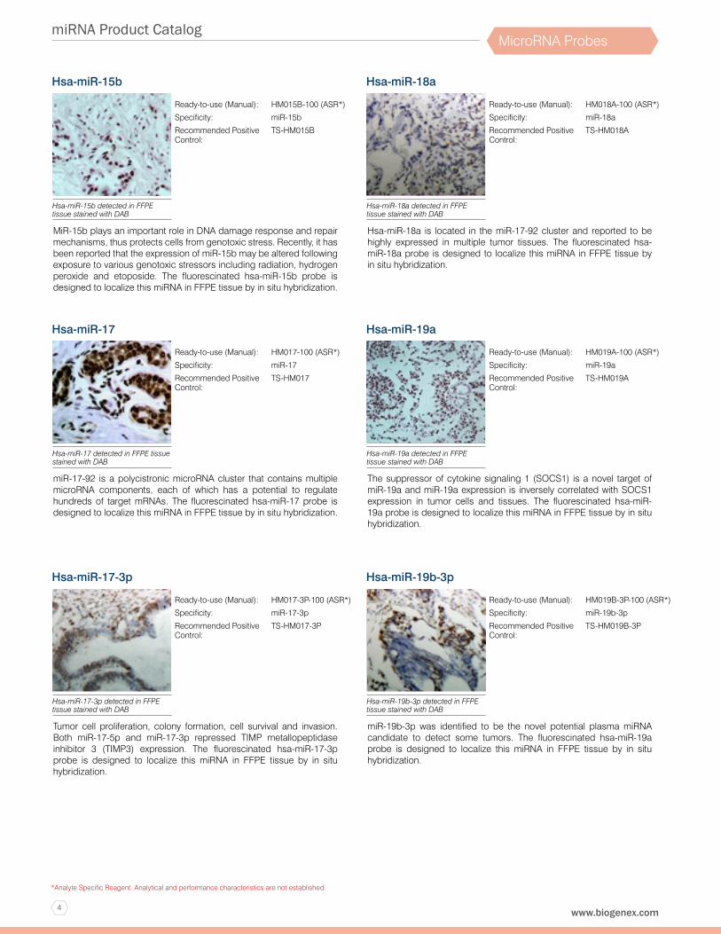
Hsa-miR-15b
MiR-15b plays an important role in DNA damage response and repair mechanisms, thus protects cells from genotoxic stress. Recently, it has been reported that the expression of miR-15b may be altered following exposure to various genotoxic stressors including radiation, hydrogen peroxide and etoposide. The fluorescinated hsa-miR-15b probe is designed to localize this miRNA in FFPE tissue by in situ hybridization.
Ready-to-use (Manual): HM015B-100 (ASR*)
Specificity: miR-15b
Recommended Positive Control:
TS-HM015B
Hsa-miR-15b detected in FFPE tissue stained with DAB
Hsa-miR-17
miR-17-92 is a polycistronic microRNA cluster that contains multiple microRNA components, each of which has a potential to regulate hundreds of target mRNAs. The fluorescinated hsa-miR-17 probe is designed to localize this miRNA in FFPE tissue by in situ hybridization.
Ready-to-use (Manual): HM017-100 (ASR*)
Specificity: miR-17
Recommended Positive Control:
TS-HM017
Hsa-miR-17 detected in FFPE tissue stained with DAB
Hsa-miR-17-3p
Tumor cell proliferation, colony formation, cell survival and invasion. Both miR-17-5p and miR-17-3p repressed TIMP metallopeptidase inhibitor 3 (TIMP3) expression. The fluorescinated hsa-miR-17-3p probe is designed to localize this miRNA in FFPE tissue by in situ hybridization.
Ready-to-use (Manual): HM017-3P-100 (ASR*)
Specificity: miR-17-3p
Recommended Positive Control:
TS-HM017-3P
Hsa-miR-17-3p detected in FFPE tissue stained with DAB
Hsa-miR-18a
Hsa-miR-18a is located in the miR-17-92 cluster and reported to be highly expressed in multiple tumor tissues. The fluorescinated hsa-miR-18a probe is designed to localize this miRNA in FFPE tissue by in situ hybridization.
Ready-to-use (Manual): HM018A-100 (ASR*)
Specificity: miR-18a
Recommended Positive Control:
TS-HM018A
Hsa-miR-18a detected in FFPE tissue stained with DAB
Hsa-miR-19a
The suppressor of cytokine signaling 1 (SOCS1) is a novel target of miR-19a and miR-19a expression is inversely correlated with SOCS1 expression in tumor cells and tissues. The fluorescinated hsa-miR-19a probe is designed to localize this miRNA in FFPE tissue by in situ hybridization.
Ready-to-use (Manual): HM019A-100 (ASR*)
Specificity: miR-19a
Recommended Positive Control:
TS-HM019A
Hsa-miR-19a detected in FFPE tissue stained with DAB
Hsa-miR-19b-3p
miR-19b-3p was identified to be the novel potential plasma miRNA candidate to detect some tumors. The fluorescinated hsa-miR-19a probe is designed to localize this miRNA in FFPE tissue by in situ hybridization.
Ready-to-use (Manual): HM019B-3P-100 (ASR*)
Specificity: miR-19b-3p
Recommended Positive Control:
TS-HM019B-3P
Hsa-miR-19b-3p detected in FFPE tissue stained with DAB
5
miRNA Product Catalog
1-800-421-4149 (USA)
*Analyte Specific Reagent. Analytical and performance characteristics are not established.
MicroRNA Probes
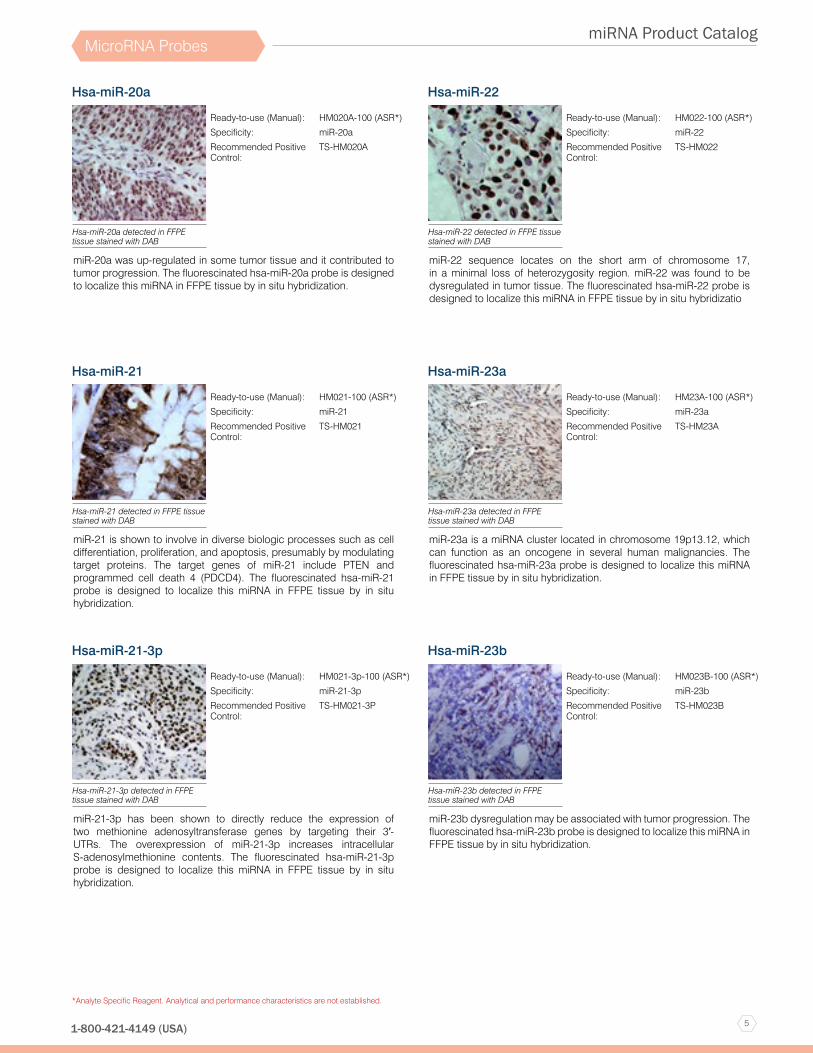
Hsa-miR-20a
miR-20a was up-regulated in some tumor tissue and it contributed to tumor progression. The fluorescinated hsa-miR-20a probe is designed to localize this miRNA in FFPE tissue by in situ hybridization.
Ready-to-use (Manual): HM020A-100 (ASR*)
Specificity: miR-20a
Recommended Positive Control:
TS-HM020A
Hsa-miR-20a detected in FFPE tissue stained with DAB
Hsa-miR-21
miR-21 is shown to involve in diverse biologic processes such as cell differentiation, proliferation, and apoptosis, presumably by modulating target proteins. The target genes of miR-21 include PTEN and programmed cell death 4 (PDCD4). The fluorescinated hsa-miR-21 probe is designed to localize this miRNA in FFPE tissue by in situ hybridization.
Ready-to-use (Manual): HM021-100 (ASR*)
Specificity: miR-21
Recommended Positive Control:
TS-HM021
Hsa-miR-21 detected in FFPE tissue stained with DAB
Hsa-miR-21-3p
miR-21-3p has been shown to directly reduce the expression of two methionine adenosyltransferase genes by targeting their 3′-UTRs. The overexpression of miR-21-3p increases intracellular S-adenosylmethionine contents. The fluorescinated hsa-miR-21-3p probe is designed to localize this miRNA in FFPE tissue by in situ hybridization.
Ready-to-use (Manual): HM021-3p-100 (ASR*)
Specificity: miR-21-3p
Recommended Positive Control:
TS-HM021-3P
Hsa-miR-21-3p detected in FFPE tissue stained with DAB
Hsa-miR-22
miR-22 sequence locates on the short arm of chromosome 17, in a minimal loss of heterozygosity region. miR-22 was found to be dysregulated in tumor tissue. The fluorescinated hsa-miR-22 probe is designed to localize this miRNA in FFPE tissue by in situ hybridizatio
Ready-to-use (Manual): HM022-100 (ASR*)
Specificity: miR-22
Recommended Positive Control:
TS-HM022
Hsa-miR-22 detected in FFPE tissue stained with DAB
Hsa-miR-23a
miR-23a is a miRNA cluster located in chromosome 19p13.12, which can function as an oncogene in several human malignancies. The fluorescinated hsa-miR-23a probe is designed to localize this miRNA in FFPE tissue by in situ hybridization.
Ready-to-use (Manual): HM23A-100 (ASR*)
Specificity: miR-23a
Recommended Positive Control:
TS-HM23A
Hsa-miR-23a detected in FFPE tissue stained with DAB
Hsa-miR-23b
miR-23b dysregulation may be associated with tumor progression. The fluorescinated hsa-miR-23b probe is designed to localize this miRNA in FFPE tissue by in situ hybridization.
Ready-to-use (Manual): HM023B-100 (ASR*)
Specificity: miR-23b
Recommended Positive Control:
TS-HM023B
Hsa-miR-23b detected in FFPE tissue stained with DAB

6
miRNA Product Catalog
www.biogenex.com
*Analyte Specific Reagent. Analytical and performance characteristics are not established.
MicroRNA Probes
Hsa-miR-24-2-5p
miR-24 governs cellular development and proliferation, acting as a tumor suppressor or oncogene in a cell type-specific manner. Multiple studies have demonstrated that miR-24 regulates the cell cycle both positively and negatively. The fluorescinated hsa-miR-24-2-5p probe is designed to localize this miRNA in FFPE tissue by in situ hybridization.
Ready-to-use (Manual): HM24-2-5P-100 (ASR*)
Specificity: miR-24-2-5p
Recommended Positive Control:
TS-HM24-2-5P
Hsa-miR-24-2-5p detected in FFPE tissue stained with DAB
Hsa-miR-24-3p
Recently, it has been shown that overexpression of miR-24-3p could alter T-cell proliferation and affect cellular gene expression through downregulation of mitogen activated protein kinase (MAPK) pathway. Thus imply the clinical relevance and prognostic value of tumor-derived exosomal miR-24-3p in T-cell dysfunction. The fluorescinated hsa-miR-24-3p probe is designed to localize this miRNA in FFPE tissue by in situ hybridization.
Ready-to-use (Manual): HM024-3P-100 (ASR*)
Specificity: miR-24-3p
Recommended Positive Control:
TS-HM024-3P
Hsa-miR-24-3p detected in FFPE tissue stained with DAB
Hsa-miR-25
miR-25 levels increase in human heart failure, and treatment with an anti-sense RNA molecule was recently reported to halt disease progression and improves cardiac function. The fluorescinated hsa-miR-25 probe is designed to localize this miRNA in FFPE tissue by in situ hybridization.
Ready-to-use (Manual): HM25-100 (ASR*)
Specificity: miR-25
Recommended Positive Control:
TS-HM25
Hsa-miR-25 detected in FFPE tissue stained with DAB
Hsa-miR-26a
miR-26 expression is induced in response to hypoxia and upregulated during smooth muscle cell (SMC) differentiation and neurogenesis. Moreover, miR-26 is consistently down-regulated in a wide range of malignant tumors. The fluorescinated hsa-miR-26a probe is designed to localize this miRNA in FFPE tissue by in situ hybridization.
Ready-to-use (Manual): HM026A-100 (ASR*)
Specificity: miR-26a
Recommended Positive Control:
TS-HM026A
Hsa-miR-26A detected in FFPE tissue stained with DAB
Hsa-miR-27a
Data suggested that miR-27a suppresses ZBTB10/RINZF expression, and this novel zinc finger protein inhibits Sp1-dependent activation of the gastrin gene promoter. The fluorescinated hsa-miR-27a probe is designed to localize this miRNA in FFPE tissue by in situ hybridization.
Ready-to-use (Manual): HM027A-100 (ASR*)
Specificity: miR-27a
Recommended Positive Control:
TS-HM027A
Hsa-miR-27A detected in FFPE tissue stained with DAB
Hsa-miR-27b
miR-27b has been identified as an oncogenic microRNA and is highly expressed in tumor cells. Inhibition of miR-27 by antisense molecules decreases cell proliferation. The fluorescinated hsa-miR-27b probe is designed to localize this miRNA in FFPE tissue by in situ hybridization.
Ready-to-use (Manual): Hsa-miR-27b (ASR*)
Specificity: miR-27b
Recommended Positive Control:
TS-HM027B
Hsa-miR-27b detected in FFPE tissue stained with DAB
7
miRNA Product Catalog
1-800-421-4149 (USA)
*Analyte Specific Reagent. Analytical and performance characteristics are not established.
MicroRNA Probes
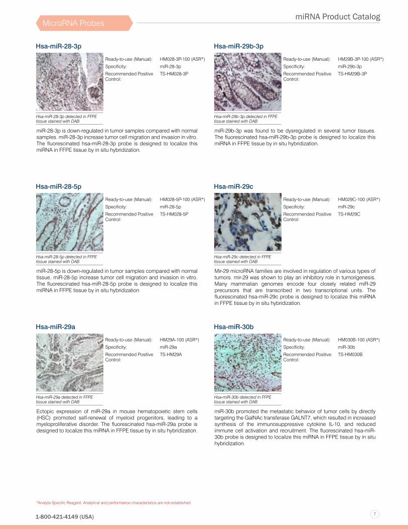
Hsa-miR-28-3p
miR-28-3p is down-regulated in tumor samples compared with normal samples. miR-28-3p increase tumor cell migration and invasion in vitro. The fluorescinated hsa-miR-28-3p probe is designed to localize this miRNA in FFPE tissue by in situ hybridization.
Ready-to-use (Manual): HM028-3P-100 (ASR*)
Specificity: miR-28-3p
Recommended Positive Control:
TS-HM028-3P
Hsa-miR-28-3p detected in FFPE tissue stained with DAB
Hsa-miR-28-5p
miR-28-5p is down-regulated in tumor samples compared with normal tissue. miR-28-5p increase tumor cell migration and invasion in vitro. The fluorescinated hsa-miR-28-5p probe is designed to localize this miRNA in FFPE tissue by in situ hybridization
Ready-to-use (Manual): HM028-5P-100 (ASR*)
Specificity: miR-28-5p
Recommended Positive Control:
TS-HM028-5P
Hsa-miR-28-5p detected in FFPE tissue stained with DAB
Hsa-miR-29a
Ectopic expression of miR-29a in mouse hematopoietic stem cells (HSC) promoted self-renewal of myeloid progenitors, leading to a myeloproliferative disorder. The fluorescinated hsa-miR-29a probe is designed to localize this miRNA in FFPE tissue by in situ hybridization.
Ready-to-use (Manual): HM29A-100 (ASR*)
Specificity: miR-29a
Recommended Positive Control:
TS-HM29A
Hsa-miR-29a detected in FFPE tissue stained with DAB
Hsa-miR-29b-3p
miR-29b-3p was found to be dysregulated in several tumor tissues. The fluorescinated hsa-miR-29b-3p probe is designed to localize this miRNA in FFPE tissue by in situ hybridization.
Ready-to-use (Manual): HM29B-3P-100 (ASR*)
Specificity: miR-29b-3p
Recommended Positive Control:
TS-HM29B-3P
Hsa-miR-29b-3p detected in FFPE tissue stained with DAB
Hsa-miR-29c
Mir-29 microRNA families are involved in regulation of various types of tumors. mir-29 was shown to play an inhibitory role in tumorigenesis. Many mammalian genomes encode four closely related miR-29 precursors that are transcribed in two transcriptional units. The fluorescinated hsa-miR-29c probe is designed to localize this miRNA in FFPE tissue by in situ hybridization.
Ready-to-use (Manual): HM029C-100 (ASR*)
Specificity: miR-29c
Recommended Positive Control:
TS-HM29C
Hsa-miR-29c detected in FFPE tissue stained with DAB
Hsa-miR-30b
miR-30b promoted the metastatic behavior of tumor cells by directly targeting the GalNAc transferase GALNT7, which resulted in increased synthesis of the immunosuppressive cytokine IL-10, and reduced immune cell activation and recruitment. The fluorescinated hsa-miR-30b probe is designed to localize this miRNA in FFPE tissue by in situ hybridization.
Ready-to-use (Manual): HM030B-100 (ASR*)
Specificity: miR-30b
Recommended Positive Control:
TS-HM030B
Hsa-miR-30b detected in FFPE tissue stained with DAB

8
miRNA Product Catalog
www.biogenex.com
*Analyte Specific Reagent. Analytical and performance characteristics are not established.
MicroRNA Probes
Hsa-miR-30c
miR-30c involved in regulating a number of tumor associated genes. It has been shown that the integrin ITGB3 and the ubiquitin conjugating E2 enzyme (UBC9) are downregulated by miR-30. It has also been suggested that the TP53 protein may be a target of miR-30c and miR-30e. Members of the miR-30 family have been found to be highly expressed in heart cells. The fluorescinated hsa-miR-30c probe is designed to localize this miRNA in FFPE tissue by in situ hybridization.
Ready-to-use (Manual): HM030C-100 (ASR*)
Specificity: miR-30c
Recommended Positive Control:
TS-HM030C
Hsa-miR-30c detected in FFPE tissue stained with DAB
Hsa-miR-30e
miR-30e involved in regulating a number of tumor associated genes. It has been shown that the integrin ITGB3 and the ubiquitin conjugating E2 enzyme (UBC9) are downregulated by miR-30. It has also been suggested that the TP53 protein may be a target of miR-30c and miR-30e. Members of the miR-30 family have been found to be highly expressed in heart cells. The fluorescinated hsa-miR-30e probe is designed to localize this miRNA in FFPE tissue by in situ hybridization.
Ready-to-use (Manual): HM030E-100 (ASR*)
Specificity: miR-30e
Recommended Positive Control:
TS-HM030E
Hsa-miR-30e detected in FFPE tissue stained with DAB
Hsa-miR-31
miR-31 is known as a tumor suppressor miRNA. miR-31 is frequently deleted and is the most underexpressed microRNA in certain tumors. It has been shown to affect the levels of tumor suppressor protein p53. The fluorescinated hsa-miR-31 probe is designed to localize this miRNA in FFPE tissue by in situ hybridization.
Ready-to-use (Manual): HM031-100 (ASR*)
Specificity: miR-31
Recommended Positive Control:
TS-HM031
Hsa-miR-31 detected in FFPE tissue stained with DAB
Hsa-miR-34a
The human miR-34a precursor is transcribed from chromosome 1. miR-34a itself is a transcriptional target of p53, suggesting a positive feedback loop between p53 and miR-34a. Thus, miR-34a functions as a tumor suppressor, in part, through a SIRT1-p53 pathway. miR-34 dysregulation is involved in the development of some tumors. The fluorescinated hsa-miR-34a probe is designed to localize this miRNA in FFPE tissue by in situ hybridization.
Ready-to-use (Manual): HM034A-100 (ASR*)
Specificity: miR-34a
Recommended Positive Control:
TS-HM034A
Hsa-miR-34a detected in FFPE tissue stained with DAB
Hsa-miR-34c
miR-34c has also been reported to be downregulated in several tumor types. Moreover, dysregulation of miR-34c has been proven to regulate tumor cell proliferation, apoptosis, senescence, migration and invasion. The fluorescinated hsa-miR-34c probe is designed to localize this miRNA in FFPE tissue by in situ hybridization.
Ready-to-use (Manual): HM34C-100 (ASR*)
Specificity: miR-34c
Recommended Positive Control:
TS-HM34C
Hsa-miR-34c detected in FFPE tissue stained with DAB
Hsa-miR-92a
miR-92a is highly expressed in some tumors. The fluorescinated hsa-miR-92a probe is designed to localize this miRNA in FFPE tissue by in situ hybridization.
Ready-to-use (Manual): HM092A-100 (ASR*)
Specificity: miR-92a
Recommended Positive Control:
TS-HM092A
Hsa-miR-92a detected in FFPE tissue stained with DAB

9
miRNA Product Catalog
1-800-421-4149 (USA)
*Analyte Specific Reagent. Analytical and performance characteristics are not established.
MicroRNA Probes
Hsa-miR-95
miR-95 expression was up-regulated in some tumors miR-95 increased proliferation by directly targeting SNX1. miR-95 expression levels correlated inversely with SNX1 protein levels. The fluorescinated hsa-miR-95 probe is designed to localize this miRNA in FFPE tissue by in situ hybridization.
Ready-to-use (Manual): HM095-100 (ASR*)
Specificity: miR-95
Recommended Positive Control:
TS-HM095
Hsa-miR-95 detected in FFPE tissue stained with DAB
Hsa-miR-96
miR-96 expression decreases the transcript and protein levels of FOXO1 by binding to one of two predicted binding sites in the FOXO1 3’-UTR sequence. The fluorescinated hsa-miR-96 probe is designed to localize this miRNA in FFPE tissue by in situ hybridization.
Ready-to-use (Manual): HM096-100 (ASR*)
Specificity: miR-96
Recommended Positive Control:
TS-HM096
Hsa-miR-96 detected in FFPE tissue stained with DAB
Hsa-miR-98
The ectopic expression of miR-98 inhibited tumor cell proliferation, invasion, and angiogenesis through repressing ALK4 and MMP11 expression. The fluorescinated hsa-miR-98 probe is designed to localize this miRNA in FFPE tissue by in situ hybridization.
Ready-to-use (Manual): HM098-100 (ASR*)
Specificity: miR-98
Recommended Positive Control:
TS-HM098
Hsa-miR-98 detected in FFPE tissue stained with DAB
Hsa-miR-99a
miR-99 family members miR-99a, -99b, and -100 were downregulated in tumor cell lines relative to the parental cell lines. miR-99 family members were also downregulated in human tumor tissue compared with normal tissue. The fluorescinated hsa-miR-99a probe is designed to localize this miRNA in FFPE tissue by in situ hybridization.
Ready-to-use (Manual): HM099A-100 (ASR*)
Specificity: miR-99a
Recommended Positive Control:
TS-HM099A
Hsa-miR-99a detected in FFPE tissue stained with DAB
Hsa-miR-99b
miR-99 family members miR-99a, -99b, and -100 were downregulated in tumor cell lines relative to the parental cell lines. miR-99 family members were also downregulated in human tumor tissue compared with normal tissue. The fluorescinated hsa-miR-99b probe is designed to localize this miRNA in FFPE tissue by in situ hybridization.
Ready-to-use (Manual): HM099B-100 (ASR*)
Specificity: miR-99b
Recommended Positive Control:
TS-HM099B
Hsa-miR-99b detected in FFPE tissue stained with DAB
Hsa-miR-100
m miR-100 is lost in many tumors and have potential function as a tumor suppressor. miR-100 inhibits the tumorigenicity, motility and invasiveness of tumor cells, and is commonly downregulated in human tumors due to hypermethylation. The fluorescinated hsa-miR-100 probe is designed to localize this miRNA in FFPE tissue by in situ hybridization.
Ready-to-use (Manual): HM100-100 (ASR*)
Specificity: miR-100
Recommended Positive Control:
TS-HM100
Hsa-miR-100 detected in FFPE tissue stained with DAB

10
miRNA Product Catalog
www.biogenex.com
*Analyte Specific Reagent. Analytical and performance characteristics are not established.
MicroRNA Probes
Hsa-miR-101-3p
NDY1 up-regulation is shown to trigger the binding of EZH2 and NDY1 to the miR-101 locus. Activation of this pathway is essential for the epigenetic regulation of gene expression elicited by FGF-2. The fluorescinated hsa-miR-101-3p probe is designed to localize this miRNA in FFPE tissue by in situ hybridization.
Ready-to-use (Manual): HM101-3P-100 (ASR*)
Specificity: miR-101-3p
Recommended Positive Control:
TS-HM101-3P
Hsa-miR-101-3p detected in FFPE tissue stained with DAB
Hsa-miR-106a
Sp1 and Egr1 are found to have an important role in miR-106a transcription and thus indirectly regulate the expression of IL-10 post-transcriptionally. The fluorescinated hsa-miR-106a probe is designed to localize this miRNA in FFPE tissue by in situ hybridization.
Ready-to-use (Manual): HM106A-100 (ASR*)
Specificity: miR-106a
Recommended Positive Control:
TS-HM106A
Hsa-miR-106a detected in FFPE tissue stained with DAB
Hsa-miR-107
miR-107 is a microRNA expressed by human tumor specimens and regulated by p53. miR-107 decreases hypoxia signaling by suppressing expression of hypoxia inducible factor-1β (HIF-1β). miR-107 may have a tumor suppressor function by directly targeting CDK6 to inhibit the proliferation and invasion activities. The fluorescinated hsa-miR-107 probe is designed to localize this miRNA in FFPE tissue by in situ hybridization.
Ready-to-use (Manual): HM107-100 (ASR*)
Specificity: miR-107
Recommended Positive Control:
TS-HM107
Hsa-miR-107 detected in FFPE tissue stained with DAB
Hsa-miR-122
miR-122 is specifically repressed in a subset of primary tumors that are characterized by poor prognosis. The loss of miR-122 results in an increase of cell migration and invasion and that restoration of miR-122 reverses this phenotype. miR-122 is a marker of hepatocyte-specific differentiation and an important determinant in the control of cell migration and invasion. The fluorescinated hsa-miR-122 probe is designed to localize this miRNA in FFPE tissue by in situ hybridization.
Ready-to-use (Manual): HM122-100 (ASR*)
Specificity: miR-122
Recommended Positive Control:
TS-HM122
Hsa-miR-122 detected in FFPE tissue stained with DAB
Hsa-miR-124
The mature miR-124 sequence is processed from 3 separate premature sequences, located at chromosomes 8p23.1 (miR-124-1), 8q12.3 (miR-124-2) and 20q13.33 (miR-124-3). The fluorescinated hsa-miR-124 probe is designed to localize this miRNA in FFPE tissue by in situ hybridization.
Ready-to-use (Manual): HM124-100 (ASR*)
Specificity: miR-124
Recommended Positive Control:
TS-HM124
Hsa-miR-124 detected in FFPE tissue stained with DAB
Hsa-miR-125a
miR-125 family has been reported to be implicated in a variety of tumors and other diseases as either repressors or promoters. miR-125 family play crucial roles in many different cellular processes like cell differentiation, proliferation and apoptosis by targeting many different transcription factors ,matrix-metalloprotease, and growth factors. The fluorescinated hsa-miR-125a probe is designed to localize this miRNA in FFPE tissue by in situ hybridization.
Ready-to-use (Manual): HM125A-100 (ASR*)
Specificity: miR-125a
Recommended Positive Control:
TS-HM125A
Hsa-miR-125a detected in FFPE tissue stained with DAB
11
miRNA Product Catalog
1-800-421-4149 (USA)
*Analyte Specific Reagent. Analytical and performance characteristics are not established.
MicroRNA Probes
Hsa-miR-125b
Enforced miR-125b expression in mammary cells is shown to decrease cell proliferation by inducing G2/M cell cycle arrest and reduced anchorage-independent cell growth of cells of mammary origin. The fluorescinated hsa-miR-125b probe is designed to localize this miRNA in FFPE tissue by in situ hybridization.
Ready-to-use (Manual): HM125B-100 (ASR*)
Specificity: miR-125b
Recommended Positive Control:
TS-HM125B
Hsa-miR-125b detected in FFPE tissue stained with DAB
Hsa-miR-126
miR-126 is a microRNA expressed predominately by endothelial cells and controls angiogenesis. The fluorescinated hsa-miR-126 probe is designed to localize this miRNA in FFPE tissue by in situ hybridization.
Ready-to-use (Manual): HM126-100 (ASR*)
Specificity: miR-126
Recommended Positive Control:
TS-HM126
Hsa-miR-126 detected in FFPE tissue stained with DAB
Hsa-miR-127-3p
miR-127 is highly expressed in normal prostate and bladder tissues. miR-127 functions to regulate the expression levels of genes involved in lung development, placental formation and apoptosis. The fluorescinated hsa-miR-127-3p probe is designed to localize this miRNA in FFPE tissue by in situ hybridization.
Ready-to-use (Manual): HM127-3P-100 (ASR*)
Specificity: miR-127-3p
Recommended Positive Control:
TS-HM127-3P
Hsa-miR-127-3p detected in FFPE tissue stained with DAB
Hsa-miR-129
miR-129-5p expression is down-regulated in several tumor types. miR-129-5p promotes apoptosis and enhances chemosensitivity, while decreased miR-129-5p expression, as a result of hypermethylation of the miR-129 promoter, is associated with poor clinicopathological factors, such as clinical stage and progression in several tumors. The fluorescinated hsa-miR-129 probe is designed to localize this miRNA in FFPE tissue by in situ hybridization.
Ready-to-use (Manual): HM129-100 (ASR*)
Specificity: miR-129
Recommended Positive Control:
TS-HM129
Hsa-miR-129 detected in FFPE tissue stained with DAB
Hsa-miR-130b
MiR-130b, located at the 22q11 locus, plays an oncogenic or suppressive role in several tumors. The fluorescinated hsa-miR-130b probe is designed to localize this miRNA in FFPE tissue by in situ hybridization.
Ready-to-use (Manual): HM130B-100 (ASR*)
Specificity: miR-130b
Recommended Positive Control:
TS-HM130B
Hsa-miR-130b detected in FFPE tissue stained with DAB
Hsa-miR-132
miR-132, transcribed from an intergenic region on human chromosome 17, is aberrantly expressed in many tumor types. The fluorescinated hsa-miR-132 probe is designed to localize this miRNA in FFPE tissue by in situ hybridization.
Ready-to-use (Manual): HM132-100 (ASR*)
Specificity: miR-132
Recommended Positive Control:
TS-HM132
Hsa-miR-132 detected in FFPE tissue stained with DAB

12
miRNA Product Catalog
www.biogenex.com
*Analyte Specific Reagent. Analytical and performance characteristics are not established.
MicroRNA Probes
Hsa-miR-133a
miR-133a is downregulated in some tumor types. The fluorescinated hsa-miR-133a probe is designed to localize this miRNA in FFPE tissue by in situ hybridization.
Ready-to-use (Manual): HM133A-100 (ASR*)
Specificity: miR-133a
Recommended Positive Control:
TS-HM133A
Hsa-miR-133a detected in FFPE tissue stained with DAB
Hsa-miR-133b
miR-133b is significantly downregulated in many tumor types. The fluorescinated hsa-miR-133b probe is designed to localize this miRNA in FFPE tissue by in situ hybridization.
Ready-to-use (Manual): HM133B-100 (ASR*)
Specificity: miR-133b
Recommended Positive Control:
TS-HM133B
Hsa-miR-133b detected in FFPE tissue stained with DAB
Hsa-miR-135a
miR-135a is significantly downregulated in the tumor cell lines and plays a tumor-suppressive role. miR-135a expression is downregulated in the majority of human tumor tissues compared with pair-matched adjacent non-tumor tissues. The fluorescinated hsa-miR-135a probe is designed to localize this miRNA in FFPE tissue by in situ hybridization.
Ready-to-use (Manual): HM135A-100 (ASR*)
Specificity: miR-135a
Recommended Positive Control:
TS-HM135A
Hsa-miR-135a detected in FFPE tissue stained with DAB
Hsa-miR-135b
miR-135b is involved in the progression of several types of tumors and it is frequently dysregulated in tumor tissue. The fluorescinated hsa-miR-135b probe is designed to localize this miRNA in FFPE tissue by in situ hybridization.
Ready-to-use (Manual): HM135B-100 (ASR*)
Specificity: miR-135b
Recommended Positive Control:
TS-HM135B
Hsa-miR-135b detected in FFPE tissue stained with DAB
Hsa-miR-136
miR-136 was significantly downregulated in tumor specimens. The low-level expression of miR-136 is significantly associated with a more aggressive and/or poor prognostic phenotype. The fluorescinated hsa-miR-136 probe is designed to localize this miRNA in FFPE tissue by in situ hybridization.
Ready-to-use (Manual): HM136-100 (ASR*)
Specificity: miR-136
Recommended Positive Control:
TS-HM136
Hsa-miR-136 detected in FFPE tissue stained with DAB
Hsa-miR-137
Recently studies revealed that miR-137 play essential roles in tumorigenesis. miR-137 modulates tumor cell growth, invasion and sensitivity. miR-137 was significantly down-regulated in tumors and inhibited proliferation of tumor cells by targeting PAK2. The fluorescinated hsa-miR-137 probe is designed to localize this miRNA in FFPE tissue by in situ hybridization.
Ready-to-use (Manual): HM137-100 (ASR*)
Specificity: miR-137
Recommended Positive Control:
TS-HM137
Hsa-miR-137 detected in FFPE tissue stained with DAB
13
miRNA Product Catalog
1-800-421-4149 (USA)
*Analyte Specific Reagent. Analytical and performance characteristics are not established.
MicroRNA Probes
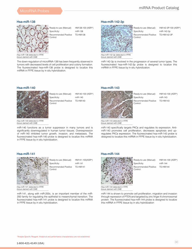
Hsa-miR-138
The down-regulation of microRNA-138 has been frequently observed in tumors with decreased levels of cell proliferation and colony formation. The fluorescinated hsa-miR-138 probe is designed to localize this miRNA in FFPE tissue by in situ hybridization.
Ready-to-use (Manual): HM138-100 (ASR*)
Specificity: miR-138
Recommended Positive Control:
TS-HM138
Hsa-miR-138 detected in FFPE tissue stained with DAB
Hsa-miR-140
miR-140 functions as a tumor suppressor in many tumors and is significantly downregulated in human tumor tissues. Overexpression of miR-140 inhibited tumor growth, invasion, and metastasis. The fluorescinated hsa-miR-140 probe is designed to localize this miRNA in FFPE tissue by in situ hybridization.
Ready-to-use (Manual): HM140-100 (ASR*)
Specificity: miR-140
Recommended Positive Control:
TS-HM140
Hsa-miR-140 detected in FFPE tissue stained with DAB
Hsa-miR-141
miR-141, along with miR-200c, is an important member of the miR-200 family for regulating the epithelial to mesenchymal transition. The fluorescinated hsa-miR-141 probe is designed to localize this miRNA in FFPE tissue by in situ hybridization.
Ready-to-use (Manual): HM141-100(ASR*)
Specificity: miR-141
Recommended Positive Control:
TS-HM141
Hsa-miR-141 detected in FFPE tissue stained with DAB
Hsa-miR-142-3p
miR-142-3p is involved in the progression of several tumor types. The fluorescinated hsa-miR-142-3p probe is designed to localize this miRNA in FFPE tissue by in situ hybridization.
Ready-to-use (Manual): HM142-3P-100 (ASR*)
Specificity: miR-142-3p
Recommended Positive Control:
TS-HM142-3P
Hsa-miR-142-3p detected in FFPE tissue stained with DAB
Hsa-miR-143
miR-143 specifically targets PKCε and regulates its expression. Anti-miR-143 promotes cell proliferation, decreases apoptosis and up-regulates PKCε expression. The fluorescinated hsa-miR-143 probe is designed to localize this miRNA in FFPE tissue by in situ hybridization.
Ready-to-use (Manual): HM143-100 (ASR*)
Specificity: miR-143
Recommended Positive Control:
TS-HM143
Hsa-miR-143 detected in FFPE tissue stained with DAB
Hsa-miR-144
miR-144 is shown to promote cell proliferation, migration and invasion through repression of PTEN and targeted by zinc finger X-chromosomal protein. The fluorescinated hsa-miR-144 probe is designed to localize this miRNA in FFPE tissue by in situ hybridization
Ready-to-use (Manual): HM144-100 (ASR*)
Specificity: miR-144
Recommended Positive Control:
TS-HM144
Hsa-miR-144 detected in FFPE tissue stained with DAB
14
miRNA Product Catalog
www.biogenex.com
*Analyte Specific Reagent. Analytical and performance characteristics are not established.
MicroRNA Probes
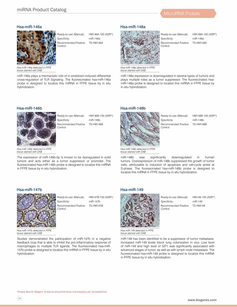
Hsa-miR-146a
miR-146a plays a mechanistic role of in endotoxin-induced differential cross-regulation of TLR Signaling. The fluorescinated hsa-miR-146a probe is designed to localize this miRNA in FFPE tissue by in situ hybridization.
Ready-to-use (Manual): HM146A-100 (ASR*)
Specificity: miR-146a
Recommended Positive Control:
TS-HM146A
Hsa-miR-146a detected in FFPE tissue stained with DAB
Hsa-miR-146b
The expression of miR-146b-5p is known to be dysregulated in solid tumors and acts either as a tumor suppressor or promoter. The fluorescinated hsa-miR-146b probe is designed to localize this miRNA in FFPE tissue by in situ hybridization.
Ready-to-use (Manual): HM146B-100 (ASR*)
Specificity: miR-146b
Recommended Positive Control:
TS-HM146B
Hsa-miR-146b detected in FFPE tissue stained with DAB
Hsa-miR-147b
Studies demonstrated the participation of miR-147b in a negative feedback loop that is able to inhibit the pro-inflammatory response of macrophages to multiple TLR ligands. The fluorescinated hsa-miR-147b probe is designed to localize this miRNA in FFPE tissue by in situ hybridization.
Ready-to-use (Manual): HM147B-100 (ASR*)
Specificity: miR-147b
Recommended Positive Control:
TS-HM147B
Hsa-miR-147b detected in FFPE tissue stained with DAB
Hsa-miR-148a
miR-148a expression is downregulated in several types of tumors and plays multiple roles as a tumor suppressor. The fluorescinated hsa-miR-148a probe is designed to localize this miRNA in FFPE tissue by in situ hybridization.
Ready-to-use (Manual): HM148A-100 (ASR*)
Specificity: miR-148a
Recommended Positive Control:
TS-HM148A
Hsa-miR-148a detected in FFPE tissue stained with DAB
Hsa-miR-148b
miR-148b was significantly downregulated in human tumors. Overexpression of miR-148b suppressed the growth of tumor cells, attributable to induction of apoptosis and cell-cycle arrest at S-phase. The fluorescinated hsa-miR-148b probe is designed to localize this miRNA in FFPE tissue by in situ hybridization.
Ready-to-use (Manual): HM148B-100 (ASR*)
Specificity: miR-148b
Recommended Positive Control:
TS-HM148B
Hsa-miR-148b detected in FFPE tissue stained with DAB
Hsa-miR-149
miR-149 has been identified to be a suppressor of tumor metastasis. Increased miR-149 levels block lung colonization in vivo. Low level of miR-149 and high level of GIT1 was significantly associated with advanced stages of tumor, as well as with lymph node metastasis. The fluorescinated hsa-miR-149 probe is designed to localize this miRNA in FFPE tissue by in situ hybridization.
Ready-to-use (Manual): HM149-100 (ASR*)
Specificity: miR-149
Recommended Positive Control:
TS-HM149
Hsa-miR-149 detected in FFPE tissue stained with DAB
15
miRNA Product Catalog
1-800-421-4149 (USA)
*Analyte Specific Reagent. Analytical and performance characteristics are not established.
MicroRNA Probes
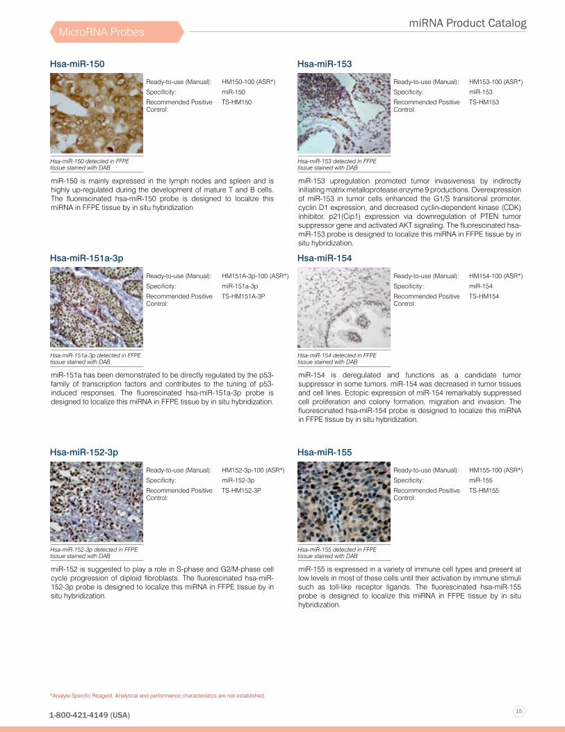
Hsa-miR-150
miR-150 is mainly expressed in the lymph nodes and spleen and is highly up-regulated during the development of mature T and B cells. The fluorescinated hsa-miR-150 probe is designed to localize this miRNA in FFPE tissue by in situ hybridization
Ready-to-use (Manual): HM150-100 (ASR*)
Specificity: miR-150
Recommended Positive Control:
TS-HM150
Hsa-miR-150 detected in FFPE tissue stained with DAB
Hsa-miR-151a-3p
miR-151a has been demonstrated to be directly regulated by the p53-family of transcription factors and contributes to the tuning of p53-induced responses. The fluorescinated hsa-miR-151a-3p probe is designed to localize this miRNA in FFPE tissue by in situ hybridization.
Ready-to-use (Manual): HM151A-3p-100 (ASR*)
Specificity: miR-151a-3p
Recommended Positive Control:
TS-HM151A-3P
Hsa-miR-151a-3p detected in FFPE tissue stained with DAB
Hsa-miR-152-3p
miR-152 is suggested to play a role in S-phase and G2/M-phase cell cycle progression of diploid fibroblasts. The fluorescinated hsa-miR-152-3p probe is designed to localize this miRNA in FFPE tissue by in situ hybridization.
Ready-to-use (Manual): HM152-3p-100 (ASR*)
Specificity: miR-152-3p
Recommended Positive Control:
TS-HM152-3P
Hsa-miR-152-3p detected in FFPE tissue stained with DAB
Hsa-miR-153
miR-153 upregulation promoted tumor invasiveness by indirectly initiating matrix metalloprotease enzyme 9 productions. Overexpression of miR-153 in tumor cells enhanced the G1/S transitional promoter, cyclin D1 expression, and decreased cyclin-dependent kinase (CDK) inhibitor, p21(Cip1) expression via downregulation of PTEN tumor suppressor gene and activated AKT signaling. The fluorescinated hsa-miR-153 probe is designed to localize this miRNA in FFPE tissue by in situ hybridization.
Ready-to-use (Manual): HM153-100 (ASR*)
Specificity: miR-153
Recommended Positive Control:
TS-HM153
Hsa-miR-153 detected in FFPE tissue stained with DAB
Hsa-miR-154
miR-154 is deregulated and functions as a candidate tumor suppressor in some tumors. miR-154 was decreased in tumor tissues and cell lines. Ectopic expression of miR-154 remarkably suppressed cell proliferation and colony formation, migration and invasion. The fluorescinated hsa-miR-154 probe is designed to localize this miRNA in FFPE tissue by in situ hybridization.
Ready-to-use (Manual): HM154-100 (ASR*)
Specificity: miR-154
Recommended Positive Control:
TS-HM154
Hsa-miR-154 detected in FFPE tissue stained with DAB
Hsa-miR-155
miR-155 is expressed in a variety of immune cell types and present at low levels in most of these cells until their activation by immune stimuli such as toll-like receptor ligands. The fluorescinated hsa-miR-155 probe is designed to localize this miRNA in FFPE tissue by in situ hybridization.
Ready-to-use (Manual): HM155-100 (ASR*)
Specificity: miR-155
Recommended Positive Control:
TS-HM155
Hsa-miR-155 detected in FFPE tissue stained with DAB
16
miRNA Product Catalog
www.biogenex.com
*Analyte Specific Reagent. Analytical and performance characteristics are not established.
MicroRNA Probes
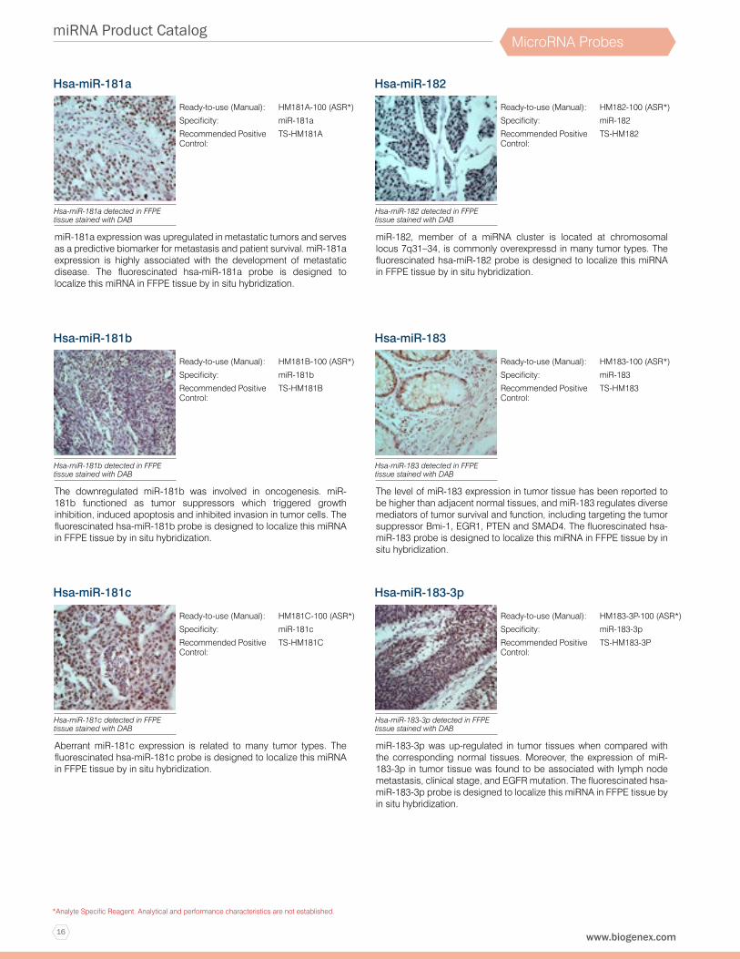
Hsa-miR-181a
miR-181a expression was upregulated in metastatic tumors and serves as a predictive biomarker for metastasis and patient survival. miR-181a expression is highly associated with the development of metastatic disease. The fluorescinated hsa-miR-181a probe is designed to localize this miRNA in FFPE tissue by in situ hybridization.
Ready-to-use (Manual): HM181A-100 (ASR*)
Specificity: miR-181a
Recommended Positive Control:
TS-HM181A
Hsa-miR-181a detected in FFPE tissue stained with DAB
Hsa-miR-181b
The downregulated miR-181b was involved in oncogenesis. miR-181b functioned as tumor suppressors which triggered growth inhibition, induced apoptosis and inhibited invasion in tumor cells. The fluorescinated hsa-miR-181b probe is designed to localize this miRNA in FFPE tissue by in situ hybridization.
Ready-to-use (Manual): HM181B-100 (ASR*)
Specificity: miR-181b
Recommended Positive Control:
TS-HM181B
Hsa-miR-181b detected in FFPE tissue stained with DAB
Hsa-miR-181c
Aberrant miR-181c expression is related to many tumor types. The fluorescinated hsa-miR-181c probe is designed to localize this miRNA in FFPE tissue by in situ hybridization.
Ready-to-use (Manual): HM181C-100 (ASR*)
Specificity: miR-181c
Recommended Positive Control:
TS-HM181C
Hsa-miR-181c detected in FFPE tissue stained with DAB
Hsa-miR-182
miR-182, member of a miRNA cluster is located at chromosomal locus 7q31–34, is commonly overexpressd in many tumor types. The fluorescinated hsa-miR-182 probe is designed to localize this miRNA in FFPE tissue by in situ hybridization.
Ready-to-use (Manual): HM182-100 (ASR*)
Specificity: miR-182
Recommended Positive Control:
TS-HM182
Hsa-miR-182 detected in FFPE tissue stained with DAB
Hsa-miR-183
The level of miR-183 expression in tumor tissue has been reported to be higher than adjacent normal tissues, and miR-183 regulates diverse mediators of tumor survival and function, including targeting the tumor suppressor Bmi-1, EGR1, PTEN and SMAD4. The fluorescinated hsa-miR-183 probe is designed to localize this miRNA in FFPE tissue by in situ hybridization.
Ready-to-use (Manual): HM183-100 (ASR*)
Specificity: miR-183
Recommended Positive Control:
TS-HM183
Hsa-miR-183 detected in FFPE tissue stained with DAB
Hsa-miR-183-3p
miR-183-3p was up-regulated in tumor tissues when compared with the corresponding normal tissues. Moreover, the expression of miR-183-3p in tumor tissue was found to be associated with lymph node metastasis, clinical stage, and EGFR mutation. The fluorescinated hsa-miR-183-3p probe is designed to localize this miRNA in FFPE tissue by in situ hybridization.
Ready-to-use (Manual): HM183-3P-100 (ASR*)
Specificity: miR-183-3p
Recommended Positive Control:
TS-HM183-3P
Hsa-miR-183-3p detected in FFPE tissue stained with DAB
17
miRNA Product Catalog
1-800-421-4149 (USA)
*Analyte Specific Reagent. Analytical and performance characteristics are not established.
MicroRNA Probes
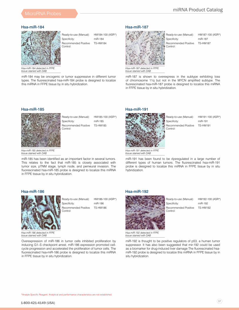
Hsa-miR-184
miR-184 may be oncogenic or tumor suppressive in different tumor types. The fluorescinated hsa-miR-184 probe is designed to localize this miRNA in FFPE tissue by in situ hybridization.
Ready-to-use (Manual): HM184-100 (ASR*)
Specificity: miR-184
Recommended Positive Control:
TS-HM184
Hsa-miR-184 detected in FFPE tissue stained with DAB
Hsa-miR-185
miR-185 has been identified as an important factor in several tumors. This relates to the fact that miR-185 is closely associated with tumor size, pTNM stage, lymph node, and perneural invasion. The fluorescinated hsa-miR-185 probe is designed to localize this miRNA in FFPE tissue by in situ hybridization.
Ready-to-use (Manual): HM185-100 (ASR*)
Specificity: miR-185
Recommended Positive Control:
TS-HM185
Hsa-miR-185 detected in FFPE tissue stained with DAB
Hsa-miR-186
Overexpression of miR-186 in tumor cells inhibited proliferation by inducing G1–S checkpoint arrest. miR-186 expression promoted cell-cycle progression and accelerated the proliferation of tumor cells. The fluorescinated hsa-miR-186 probe is designed to localize this miRNA in FFPE tissue by in situ hybridization.
Ready-to-use (Manual): HM186-100 (ASR*)
Specificity: miR-186
Recommended Positive Control:
TS-HM186
Hsa-miR-186 detected in FFPE tissue stained with DAB
Hsa-miR-187
miR-187 is shown to overexpress in the subtype exhibiting loss of chromosome 11q but not in the MYCN amplified subtype. The fluorescinated hsa-miR-187 probe is designed to localize this miRNA in FFPE tissue by in situ hybridization.
Ready-to-use (Manual): HM187-100 (ASR*)
Specificity: miR-187
Recommended Positive Control:
TS-HM187
Hsa-miR-187 detected in FFPE tissue stained with DAB
Hsa-miR-191
miR-191 has been found to be dysregulated in a large number of different types of human tumors. The fluorescinated hsa-miR-191 probe is designed to localize this miRNA in FFPE tissue by in situ hybridization.
Ready-to-use (Manual): HM191-100 (ASR*)
Specificity: miR-191
Recommended Positive Control:
TS-HM191
Hsa-miR-191 detected in FFPE tissue stained with DAB
Hsa-miR-192
miR-192 is thought to be positive regulators of p53, a human tumor suppressor. It has also been suggested that mir-192 could be used as a biomarker for drug-induced liver damage The fluorescinated hsa-miR-192 probe is designed to localize this miRNA in FFPE tissue by in situ hybridization.
Ready-to-use (Manual): HM192-100 (ASR*)
Specificity: miR-192
Recommended Positive Control:
TS-HM192
Hsa-miR-192 detected in FFPE tissue stained with DAB

18
miRNA Product Catalog
www.biogenex.com
*Analyte Specific Reagent. Analytical and performance characteristics are not established.
MicroRNA Probes
Hsa-miR-193a-3p
miR-193a-3p induces the accumulation of intracellular reactive oxygen species, DNA damage in tumor cells. Furthermore, miR-193a-3p directly recognizes the 3′-UTR of the ERBB4 transcript and regulates ERBB4 expression, one of four ErbB receptor tyrosine kinase family members. The fluorescinated hsa-miR-193a-3p probe is designed to localize this miRNA in FFPE tissue by in situ hybridization.
Ready-to-use (Manual): HM193A-3P-100 (ASR*)
Specificity: miR-193a-3p
Recommended Positive Control:
TS-HM193A-3P
Hsa-miR-193a-3p detected in FFPE tissue stained with DAB
Hsa-miR-193b
Aberrant expression of miR-193b is frequently observed in tumor tissuer and it acts as a tumor suppressor in many types of tumors. miR-193b is down-regulated in tumor tissue and can promote tumorigenesis by inhibiting stathmin 1 and urokinase-type plasminogen activator (uPA). The fluorescinated hsa-miR-193b probe is designed to localize this miRNA in FFPE tissue by in situ hybridization.
Ready-to-use (Manual): HM193B-100 (ASR*)
Specificity: miR-193b
Recommended Positive Control:
TS-HM193B
Hsa-miR-193b detected in FFPE tissue stained with DAB
Hsa-miR-194
miR-194 is expressed in liver parenchymal cells, and in human gastrointestinal tract. miR-194 plays a role in the activation of stellate cells during liver fibrogenesis. miR-194 expression varies in human organs and in different status of hepatocyte differentiation. miR-194 is an epithelial cell-specific marker in the liver. The fluorescinated hsa-miR-194 probe is designed to localize this miRNA in FFPE tissue by in situ hybridization.
Ready-to-use (Manual): HM194-100 (ASR*)
Specificity: miR-194
Recommended Positive Control:
TS-HM194
Hsa-miR-194 detected in FFPE tissue stained with DAB
Hsa-miR-195
miR-195 is aberrantly expressed in multiple types of disease. miR-195 was significantly downregulated in tumors. The fluorescinated hsa-miR-195 probe is designed to localize this miRNA in FFPE tissue by in situ hybridization.
Ready-to-use (Manual): HM195-100 (ASR*)
Specificity: miR-195
Recommended Positive Control:
TS-HM195
Hsa-miR-195 detected in FFPE tissue stained with DAB
Hsa-miR-196a
miR-196a is a microRNA that suppresses the expression of specific homeobox genes that are of high relevance for the development of human embryos. The fluorescinated hsa-miR-196a probe is designed to localize this miRNA in FFPE tissue by in situ hybridization.
Ready-to-use (Manual): HM196A-100 (ASR*)
Specificity: miR-196a
Recommended Positive Control:
TS-HM196A
Hsa-miR-196a detected in FFPE tissue stained with DAB
Hsa-miR-197
miR-197 is an onco-miR which functions as a key repressor of p53-dependent apoptotic cascade in tumor cells. The fluorescinated hsa-miR-197 probe is designed to localize this miRNA in FFPE tissue by in situ hybridization.
Ready-to-use (Manual): HM197-100 (ASR*)
Specificity: miR-197
Recommended Positive Control:
TS-HM197
Hsa-miR-197 detected in FFPE tissue stained with DAB
19
miRNA Product Catalog
1-800-421-4149 (USA)
*Analyte Specific Reagent. Analytical and performance characteristics are not established.
MicroRNA Probes
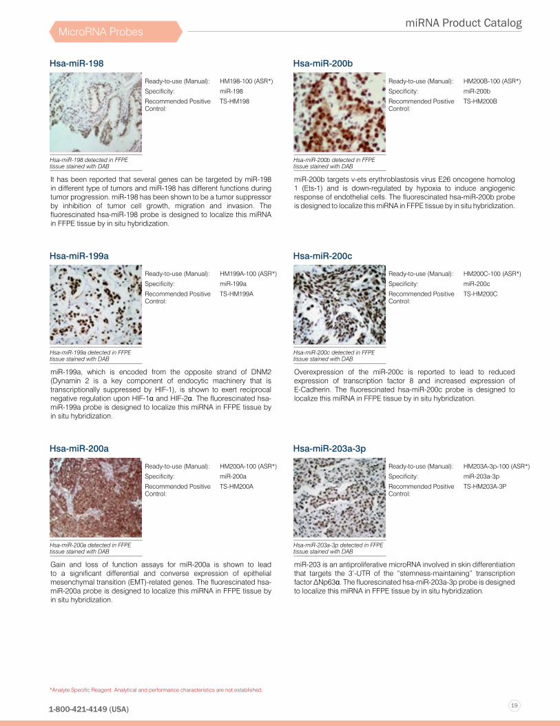
Hsa-miR-198
It has been reported that several genes can be targeted by miR-198 in different type of tumors and miR-198 has different functions during tumor progression. miR-198 has been shown to be a tumor suppressor by inhibition of tumor cell growth, migration and invasion. The fluorescinated hsa-miR-198 probe is designed to localize this miRNA in FFPE tissue by in situ hybridization.
Ready-to-use (Manual): HM198-100 (ASR*)
Specificity: miR-198
Recommended Positive Control:
TS-HM198
Hsa-miR-198 detected in FFPE tissue stained with DAB
Hsa-miR-199a
miR-199a, which is encoded from the opposite strand of DNM2 (Dynamin 2 is a key component of endocytic machinery that is transcriptionally suppressed by HIF-1), is shown to exert reciprocal negative regulation upon HIF-1α and HIF-2α. The fluorescinated hsa-miR-199a probe is designed to localize this miRNA in FFPE tissue by in situ hybridization.
Ready-to-use (Manual): HM199A-100 (ASR*)
Specificity: miR-199a
Recommended Positive Control:
TS-HM199A
Hsa-miR-199a detected in FFPE tissue stained with DAB
Hsa-miR-200a
Gain and loss of function assays for miR-200a is shown to lead to a significant differential and converse expression of epithelial mesenchymal transition (EMT)-related genes. The fluorescinated hsa-miR-200a probe is designed to localize this miRNA in FFPE tissue by in situ hybridization.
Ready-to-use (Manual): HM200A-100 (ASR*)
Specificity: miR-200a
Recommended Positive Control:
TS-HM200A
Hsa-miR-200a detected in FFPE tissue stained with DAB
Hsa-miR-200b
miR-200b targets v-ets erythroblastosis virus E26 oncogene homolog 1 (Ets-1) and is down-regulated by hypoxia to induce angiogenic response of endothelial cells. The fluorescinated hsa-miR-200b probe is designed to localize this miRNA in FFPE tissue by in situ hybridization.
Ready-to-use (Manual): HM200B-100 (ASR*)
Specificity: miR-200b
Recommended Positive Control:
TS-HM200B
Hsa-miR-200b detected in FFPE tissue stained with DAB
Hsa-miR-200c
Overexpression of the miR-200c is reported to lead to reduced expression of transcription factor 8 and increased expression of E-Cadherin. The fluorescinated hsa-miR-200c probe is designed to localize this miRNA in FFPE tissue by in situ hybridization.
Ready-to-use (Manual): HM200C-100 (ASR*)
Specificity: miR-200c
Recommended Positive Control:
TS-HM200C
Hsa-miR-200c detected in FFPE tissue stained with DAB
Hsa-miR-203a-3p
miR-203 is an antiproliferative microRNA involved in skin differentiation that targets the 3’-UTR of the “stemness-maintaining” transcription factor ΔNp63α. The fluorescinated hsa-miR-203a-3p probe is designed to localize this miRNA in FFPE tissue by in situ hybridization.
Ready-to-use (Manual): HM203A-3p-100 (ASR*)
Specificity: miR-203a-3p
Recommended Positive Control:
TS-HM203A-3P
Hsa-miR-203a-3p detected in FFPE tissue stained with DAB
20
miRNA Product Catalog
www.biogenex.com
*Analyte Specific Reagent. Analytical and performance characteristics are not established.
MicroRNA Probes
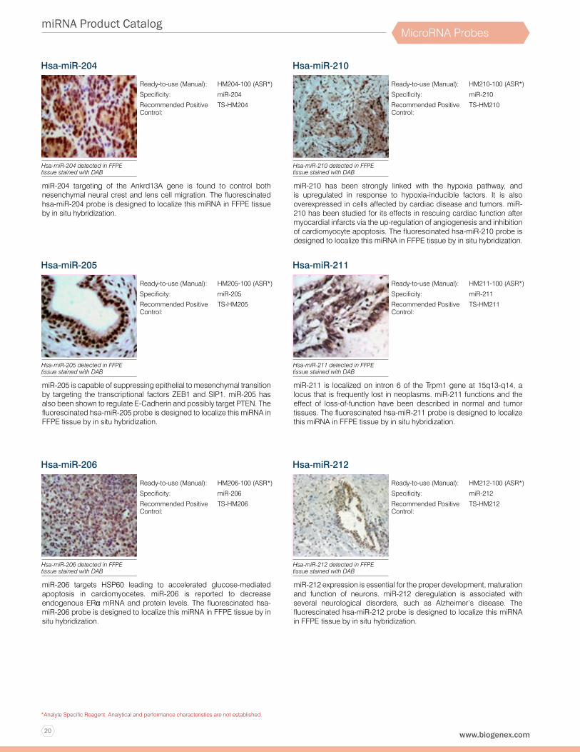
Hsa-miR-204
miR-204 targeting of the Ankrd13A gene is found to control both nesenchymal neural crest and lens cell migration. The fluorescinated hsa-miR-204 probe is designed to localize this miRNA in FFPE tissue by in situ hybridization.
Ready-to-use (Manual): HM204-100 (ASR*)
Specificity: miR-204
Recommended Positive Control:
TS-HM204
Hsa-miR-204 detected in FFPE tissue stained with DAB
Hsa-miR-205
miR-205 is capable of suppressing epithelial to mesenchymal transition by targeting the transcriptional factors ZEB1 and SIP1. miR-205 has also been shown to regulate E-Cadherin and possibly target PTEN. The fluorescinated hsa-miR-205 probe is designed to localize this miRNA in FFPE tissue by in situ hybridization.
Ready-to-use (Manual): HM205-100 (ASR*)
Specificity: miR-205
Recommended Positive Control:
TS-HM205
Hsa-miR-205 detected in FFPE tissue stained with DAB
Hsa-miR-206
miR-206 targets HSP60 leading to accelerated glucose-mediated apoptosis in cardiomyocetes. miR-206 is reported to decrease endogenous ERα mRNA and protein levels. The fluorescinated hsa-miR-206 probe is designed to localize this miRNA in FFPE tissue by in situ hybridization.
Ready-to-use (Manual): HM206-100 (ASR*)
Specificity: miR-206
Recommended Positive Control:
TS-HM206
Hsa-miR-206 detected in FFPE tissue stained with DAB
Hsa-miR-210
miR-210 has been strongly linked with the hypoxia pathway, and is upregulated in response to hypoxia-inducible factors. It is also overexpressed in cells affected by cardiac disease and tumors. miR-210 has been studied for its effects in rescuing cardiac function after myocardial infarcts via the up-regulation of angiogenesis and inhibition of cardiomyocyte apoptosis. The fluorescinated hsa-miR-210 probe is designed to localize this miRNA in FFPE tissue by in situ hybridization.
Ready-to-use (Manual): HM210-100 (ASR*)
Specificity: miR-210
Recommended Positive Control:
TS-HM210
Hsa-miR-210 detected in FFPE tissue stained with DAB
Hsa-miR-211
miR-211 is localized on intron 6 of the Trpm1 gene at 15q13-q14, a locus that is frequently lost in neoplasms. miR-211 functions and the effect of loss-of-function have been described in normal and tumor tissues. The fluorescinated hsa-miR-211 probe is designed to localize this miRNA in FFPE tissue by in situ hybridization.
Ready-to-use (Manual): HM211-100 (ASR*)
Specificity: miR-211
Recommended Positive Control:
TS-HM211
Hsa-miR-211 detected in FFPE tissue stained with DAB
Hsa-miR-212
miR-212 expression is essential for the proper development, maturation and function of neurons. miR-212 deregulation is associated with several neurological disorders, such as Alzheimer’s disease. The fluorescinated hsa-miR-212 probe is designed to localize this miRNA in FFPE tissue by in situ hybridization.
Ready-to-use (Manual): HM212-100 (ASR*)
Specificity: miR-212
Recommended Positive Control:
TS-HM212
Hsa-miR-212 detected in FFPE tissue stained with DAB

21
miRNA Product Catalog
1-800-421-4149 (USA)
*Analyte Specific Reagent. Analytical and performance characteristics are not established.
MicroRNA Probes
Hsa-miR-214
miR-214 expression level is associated with metastasis and invasion of tumors. miR-214 could inhibit the proliferation capacity, migration and invasion ability of HeLa cells. Plexin-B1, a target of miR-214, may function as an oncogene in human tumor HeLa cells. The fluorescinated hsa-miR-214 probe is designed to localize this miRNA in FFPE tissue by in situ hybridization.
Ready-to-use (Manual): HM214-100 (ASR*)
Specificity: miR-214
Recommended Positive Control:
TS-HM214
Hsa-miR-214 detected in FFPE tissue stained with DAB
Hsa-miR-215
miR-215 identified from the microRNA cluster site at chromosome 1q41, has been reported to function as a tumor suppressor in a variety of human tumors by positive regulate p53. miR-215 suppressed the expression of key targets such as thymidylate synthase (TS), dihydrofolate reductase, and denticleless protein homolog (DTL). The fluorescinated hsa-miR-215 probe is designed to localize this miRNA in FFPE tissue by in situ hybridization.
Ready-to-use (Manual): HM215-100 (ASR*)
Specificity: miR-215
Recommended Positive Control:
TS-HM215
Hsa-miR-215 detected in FFPE tissue stained with DAB
Hsa-miR-216a
It was shown that TGF-β activates Akt in glomerular mesangial cells by inducing the miR-216a and miR-217, both of which target PTEN, an inhibitor of Akt activation. The fluorescinated hsa-miR-216a probe is designed to localize this miRNA in FFPE tissue by in situ hybridization.
Ready-to-use (Manual): HM216A-100 (ASR*)
Specificity: miR-216a
Recommended Positive Control:
TS-HM216A
Hsa-miR-216a detected in FFPE tissue stained with DAB
Hsa-miR-216b
miR-216b was identified as a tumor suppressor in many tumors. Forced expression of miR-216b in RInk-1 cells inhibits cell proliferation and colony formation, which is correlated with reduced expression levels of epidermal growth factor receptor and matrix metalloproteinase-14 (MT1-MMP). Furthermore, miR-216b is dysregulated in bone marrow mesenchymal stem cells, and is associated with nonalcoholic fatty liver disease. The fluorescinated hsa-miR-216b probe is designed to localize this miRNA in FFPE tissue by in situ hybridization.
Ready-to-use (Manual): HM216B-100 (ASR*)
Specificity: miR-216b
Recommended Positive Control:
TS-HM216B
Hsa-miR-216b detected in FFPE tissue stained with DAB
Hsa-miR-217
miR-217 targets oncogenes or tumor suppressor genes such as KRAS/WASF3 in different cell types by inhibiting tumor cell growth and anchorage-independent colony formation. The fluorescinated hsa-miR-217 probe is designed to localize this miRNA in FFPE tissue by in situ hybridization.
Ready-to-use (Manual): HM217-100 (ASR*)
Specificity: miR-217
Recommended Positive Control:
TS-HM217
Hsa-miR-217 detected in FFPE tissue stained with DAB
Hsa-miR-218
miR-218 is reported to be part of a regulatory circuit involving the Slit-Robo1 pathway. Decreased miR-218 levels eliminate Robo1 repression which activates the pathway through the interaction between Robo1 and Slit2. The fluorescinated hsa-miR-218 probe is designed to localize this miRNA in FFPE tissue by in situ hybridization.
Ready-to-use (Manual): HM218-100 (ASR*)
Specificity: miR-218
Recommended Positive Control:
TS-HM218
Hsa-miR-218 detected in FFPE tissue stained with DAB
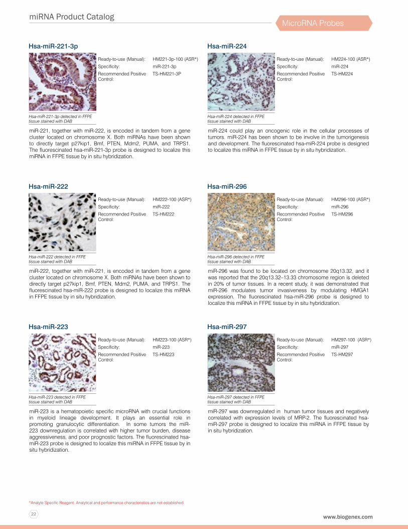
22
miRNA Product Catalog
www.biogenex.com
*Analyte Specific Reagent. Analytical and performance characteristics are not established.
MicroRNA Probes
Hsa-miR-221-3p
miR-221, together with miR-222, is encoded in tandem from a gene cluster located on chromosome X. Both miRNAs have been shown to directly target p27kip1, Bmf, PTEN, Mdm2, PUMA, and TRPS1. The fluorescinated hsa-miR-221-3p probe is designed to localize this miRNA in FFPE tissue by in situ hybridization.
Ready-to-use (Manual): HM221-3p-100 (ASR*)
Specificity: miR-221-3p
Recommended Positive Control:
TS-HM221-3P
Hsa-miR-221-3p detected in FFPE tissue stained with DAB
Hsa-miR-222
miR-222, together with miR-221, is encoded in tandem from a gene cluster located on chromosome X. Both miRNAs have been shown to directly target p27kip1, Bmf, PTEN, Mdm2, PUMA, and TRPS1. The fluorescinated hsa-miR-222 probe is designed to localize this miRNA in FFPE tissue by in situ hybridization.
Ready-to-use (Manual): HM222-100 (ASR*)
Specificity: miR-222
Recommended Positive Control:
TS-HM222
Hsa-miR-222 detected in FFPE tissue stained with DAB
Hsa-miR-223
miR-223 is a hematopoietic specific microRNA with crucial functions in myeloid lineage development. It plays an essential role in promoting granulocytic differentiation. In some tumors the miR-223 downregulation is correlated with higher tumor burden, disease aggressiveness, and poor prognostic factors. The fluorescinated hsa-miR-223 probe is designed to localize this miRNA in FFPE tissue by in situ hybridization.
Ready-to-use (Manual): HM223-100 (ASR*)
Specificity: miR-223
Recommended Positive Control:
TS-HM223
Hsa-miR-223 detected in FFPE tissue stained with DAB
Hsa-miR-224
miR-224 could play an oncogenic role in the cellular processes of tumors. miR-224 has been shown to be involve in the tumorigenesis and development. The fluorescinated hsa-miR-224 probe is designed to localize this miRNA in FFPE tissue by in situ hybridization.
Ready-to-use (Manual): HM224-100 (ASR*)
Specificity: miR-224
Recommended Positive Control:
TS-HM224
Hsa-miR-224 detected in FFPE tissue stained with DAB
Hsa-miR-296
miR-296 was found to be located on chromosome 20q13.32, and it was reported that the 20q13.32–13.33 chromosome region is deleted in 20% of tumor tissues. In a recent study, it was demonstrated that miR-296 modulates tumor invasiveness by modulating HMGA1 expression. The fluorescinated hsa-miR-296 probe is designed to localize this miRNA in FFPE tissue by in situ hybridization.
Ready-to-use (Manual): HM296-100 (ASR*)
Specificity: miR-296
Recommended Positive Control:
TS-HM296
Hsa-miR-296 detected in FFPE tissue stained with DAB
Hsa-miR-297
miR-297 was downregulated in human tumor tissues and negatively correlated with expression levels of MRP-2. The fluorescinated hsa-miR-297 probe is designed to localize this miRNA in FFPE tissue by in situ hybridization.
Ready-to-use (Manual): HM297-100 (ASR*)
Specificity: miR-297
Recommended Positive Control:
TS-HM297
Hsa-miR-297 detected in FFPE tissue stained with DAB

23
miRNA Product Catalog
1-800-421-4149 (USA)
*Analyte Specific Reagent. Analytical and performance characteristics are not established.
MicroRNA Probes
Hsa-miR-300
miR-300 was upregulated in several tumor types. miR-300 inhibits epithelial to mesenchymal transition and metastasis by targeting Twist. The fluorescinated hsa-miR-300 probe is designed to localize this miRNA in FFPE tissue by in situ hybridization.
Ready-to-use (Manual): HM300-100 (ASR*)
Specificity: miR-300
Recommended Positive Control:
TS-HM300
Hsa-miR-300 detected in FFPE tissue stained with DAB
Hsa-miR-328
A study shows that miR-328 regulates zonation morphogenesis by targeting expression of hyaluronan receptor CD44. The fluorescinated hsa-miR-328 probe is designed to localize this miRNA in FFPE tissue by in situ hybridization.
Ready-to-use (Manual): HM328-100 (ASR*)
Specificity: miR-328
Recommended Positive Control:
TS-HM328
Hsa-miR-328 detected in FFPE tissue stained with DAB
Hsa-miR-329
miR-329 functions as a tumor suppressor in some malignancies. miR-329 was decreased in metastatic tumor tissues compared with primary tumor tissues. Overexpression of miR-329 substantially suppressed cell proliferation, colony formation, migration and invasion of tumor cells. The fluorescinated hsa-miR-329 probe is designed to localize this miRNA in FFPE tissue by in situ hybridization.
Ready-to-use (Manual): HM329-100 (ASR*)
Specificity: miR-329
Recommended Positive Control:
TS-HM329
Hsa-miR-329 detected in FFPE tissue stained with DAB
Hsa-miR-330
The expression of miR-330 in tumor cells enhanced cellular proliferation, promoted cell migration and invasion, and dampened cell apoptosis. The fluorescinated hsa-miR-330 probe is designed to localize this miRNA in FFPE tissue by in situ hybridization.
Ready-to-use (Manual): HM330-100 (ASR*)
Specificity: miR-330
Recommended Positive Control:
TS-HM330
Hsa-miR-330 detected in FFPE tissue stained with DAB
Hsa-miR-331-3p
miR-331-3p expression is decreased in tumor tissue comparing to normal tissue. The fluorescinated hsa-miR-331-3p probe is designed to localize this miRNA in FFPE tissue by in situ hybridization.
Ready-to-use (Manual): HM331-3P-100 (ASR*)
Specificity: miR-331-3p
Recommended Positive Control:
TS-HM331-3P
Hsa-miR-331-3p detected in FFPE tissue stained with DAB
Hsa-miR-335
Differential microRNA expression analyses reveal that miR-335 is significantly down-regulated upon differentiation of human mesenchymal stem cells. The fluorescinated hsa-miR-335 probe is designed to localize this miRNA in FFPE tissue by in situ hybridization.
Ready-to-use (Manual): HM335-100 (ASR*)
Specificity: miR-335
Recommended Positive Control:
TS-HM335
Hsa-miR-335 detected in FFPE tissue stained with DAB

24
miRNA Product Catalog
www.biogenex.com
*Analyte Specific Reagent. Analytical and performance characteristics are not established.
MicroRNA Probes
Hsa-miR-338-3p
miR-338-3p was transcribed from the intron 8 of apoptosis-associated tyrosine kinase (AATK) gene, located on chromosome 17q25, playing a critical role in promoting cell death, neuronal differentiation and neurite extension. miR-338-3p could act as a tumor suppressor in several tumor types. The fluorescinated hsa-miR-338-3p probe is designed to localize this miRNA in FFPE tissue by in situ hybridization.
Ready-to-use (Manual): HM338-3P-100 (ASR*)
Specificity: miR-338-3p
Recommended Positive Control:
TS-HM338-3P
Hsa-miR-338-3p detected in FFPE tissue stained with DAB
Hsa-miR-339-5p
miR-339-5p targets BCL-6 and dramatically inhibited tumor cell migration and invasion in vitro. In addition, it has been reported that Dicer-regulated miR-339-5p promotes resistance of tumor cells to cytotoxic T-lymphocytes by down-regulation of ICAM-1. The fluorescinated hsa-miR-339-5p probe is designed to localize this miRNA in FFPE tissue by in situ hybridization.
Ready-to-use (Manual): HM339-5P-100 (ASR*)
Specificity: miR-339-5p
Recommended Positive Control:
TS-HM339-5P
Hsa-miR-339-5p detected in FFPE tissue stained with DAB
Hsa-miR-342-3p
The level of miR-342-3p was significantly increased in tumor, and was inversely associated with the prognosis of patients. The fluorescinated hsa-miR-342-3p probe is designed to localize this miRNA in FFPE tissue by in situ hybridization.
Ready-to-use (Manual): HM342-3P-100 (ASR*)
Specificity: miR-342-3p
Recommended Positive Control:
TS-HM342-3P
Hsa-miR-342-3p detected in FFPE tissue stained with DAB
Hsa-miR-361
miR-361 was significantly downregulated in serum of tumor patients. The level of miR-361 was lower in tumor than in benign disease and healthy individuals. The fluorescinated hsa-miR-361 probe is designed to localize this miRNA in FFPE tissue by in situ hybridization.
Ready-to-use (Manual): HM361-100 (ASR*)
Specificity: miR-361
Recommended Positive Control:
TS-HM361
Hsa-miR-361 detected in FFPE tissue stained with DAB
Hsa-miR-362
miR-362 is significantly up-regulated in tumor and associated with tumor progression. Inhibition of miR-362 in tumor cells dramatically decrease the cell proliferation, clonogenicity, migration and invasion in vitro as well as tumor growth and metastasis in vivo. The fluorescinated hsa-miR-362 probe is designed to localize this miRNA in FFPE tissue by in situ hybridization.
Ready-to-use (Manual): HM362-100 (ASR*)
Specificity: miR-362
Recommended Positive Control:
TS-HM362
Hsa-miR-362 detected in FFPE tissue stained with DAB
Hsa-miR-365a-3p
miR-365 is a direct negative regulator of IL-6. Ectopic expression of a miR-365 inhibitor elevated IL-6 expression. The negative regulation of miR-365 was strictly dependent on a microRNA binding element in the 3’-UTR of IL-6 mRNA. The fluorescinated hsa-miR-365a-3p probe is designed to localize this miRNA in FFPE tissue by in situ hybridization.
Ready-to-use (Manual): HM365A-3P-100 (ASR*)
Specificity: miR-365a-3p
Recommended Positive Control:
TS-HM365A-3P
Hsa-miR-365a-3p detected in FFPE tissue stained with DAB
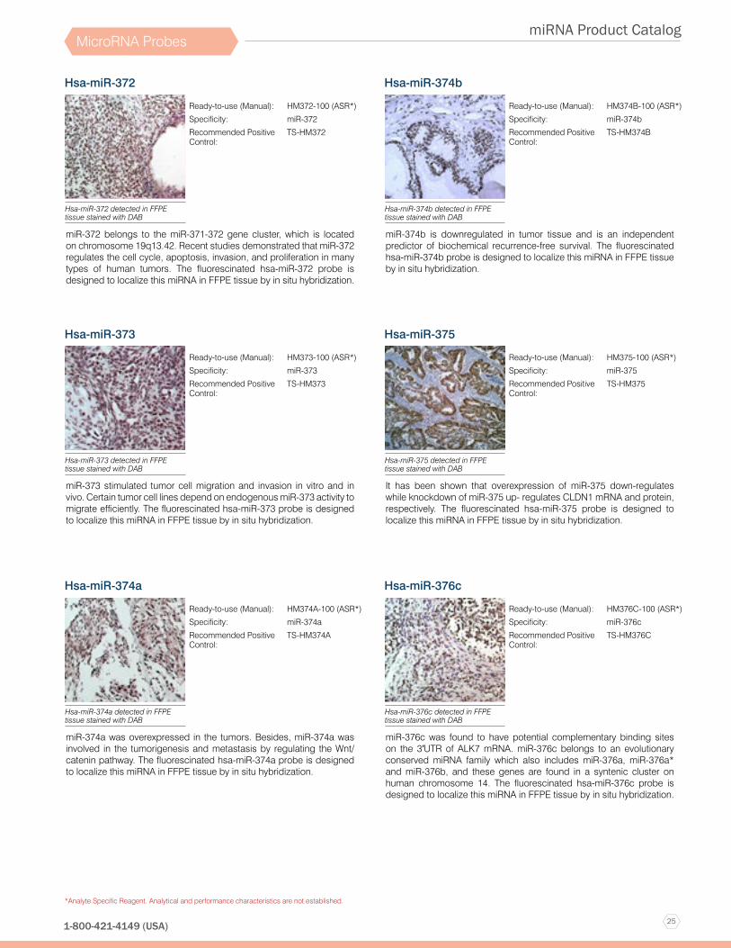
25
miRNA Product Catalog
1-800-421-4149 (USA)
*Analyte Specific Reagent. Analytical and performance characteristics are not established.
MicroRNA Probes
Hsa-miR-372
miR-372 belongs to the miR-371-372 gene cluster, which is located on chromosome 19q13.42. Recent studies demonstrated that miR-372 regulates the cell cycle, apoptosis, invasion, and proliferation in many types of human tumors. The fluorescinated hsa-miR-372 probe is designed to localize this miRNA in FFPE tissue by in situ hybridization.
Ready-to-use (Manual): HM372-100 (ASR*)
Specificity: miR-372
Recommended Positive Control:
TS-HM372
Hsa-miR-372 detected in FFPE tissue stained with DAB
Hsa-miR-373
miR-373 stimulated tumor cell migration and invasion in vitro and in vivo. Certain tumor cell lines depend on endogenous miR-373 activity to migrate efficiently. The fluorescinated hsa-miR-373 probe is designed to localize this miRNA in FFPE tissue by in situ hybridization.
Ready-to-use (Manual): HM373-100 (ASR*)
Specificity: miR-373
Recommended Positive Control:
TS-HM373
Hsa-miR-373 detected in FFPE tissue stained with DAB
Hsa-miR-374a
miR-374a was overexpressed in the tumors. Besides, miR-374a was involved in the tumorigenesis and metastasis by regulating the Wnt/catenin pathway. The fluorescinated hsa-miR-374a probe is designed to localize this miRNA in FFPE tissue by in situ hybridization.
Ready-to-use (Manual): HM374A-100 (ASR*)
Specificity: miR-374a
Recommended Positive Control:
TS-HM374A
Hsa-miR-374a detected in FFPE tissue stained with DAB
Hsa-miR-374b
miR-374b is downregulated in tumor tissue and is an independent predictor of biochemical recurrence-free survival. The fluorescinated hsa-miR-374b probe is designed to localize this miRNA in FFPE tissue by in situ hybridization.
Ready-to-use (Manual): HM374B-100 (ASR*)
Specificity: miR-374b
Recommended Positive Control:
TS-HM374B
Hsa-miR-374b detected in FFPE tissue stained with DAB
Hsa-miR-375
It has been shown that overexpression of miR-375 down-regulates while knockdown of miR-375 up- regulates CLDN1 mRNA and protein, respectively. The fluorescinated hsa-miR-375 probe is designed to localize this miRNA in FFPE tissue by in situ hybridization.
Ready-to-use (Manual): HM375-100 (ASR*)
Specificity: miR-375
Recommended Positive Control:
TS-HM375
Hsa-miR-375 detected in FFPE tissue stained with DAB
Hsa-miR-376c
miR-376c was found to have potential complementary binding sites on the 3′UTR of ALK7 mRNA. miR-376c belongs to an evolutionary conserved miRNA family which also includes miR-376a, miR-376a* and miR-376b, and these genes are found in a syntenic cluster on human chromosome 14. The fluorescinated hsa-miR-376c probe is designed to localize this miRNA in FFPE tissue by in situ hybridization.
Ready-to-use (Manual): HM376C-100 (ASR*)
Specificity: miR-376c
Recommended Positive Control:
TS-HM376C
Hsa-miR-376c detected in FFPE tissue stained with DAB

26
miRNA Product Catalog
www.biogenex.com
*Analyte Specific Reagent. Analytical and performance characteristics are not established.
MicroRNA Probes
Hsa-miR-378a
miRNA-378 promotes cell survival and angiogenesis by targeting SuFu and Fus-1 expression. The fluorescinated hsa-miR-378a probe is designed to localize this miRNA in FFPE tissue by in situ hybridization
Ready-to-use (Manual): HM378A-100 (ASR*)
Specificity: miR-378a
Recommended Positive Control:
TS-HM378A
Hsa-miR-378a detected in FFPE tissue stained with DAB
Hsa-miR-379
miR-379, is located on chromosome 14q32, 31. In the context of tumor, miR-379 regulates interleukin-11 (IL-11) production and decreased in tumor. The fluorescinated hsa-miR-379 probe is designed to localize this miRNA in FFPE tissue by in situ hybridization.
Ready-to-use (Manual): HM379-100 (ASR*)
Specificity: miR-379
Recommended Positive Control:
TS-HM379
Hsa-miR-379 detected in FFPE tissue stained with DAB
Hsa-miR-381
Recent functional studies have demonstrated that miR-381 serves as a tumor suppressor and is associated with radio-sensitivity in tumor cells. Overexpression of miRNA-381 confers increased radio-sensitivity of tumor cells, promotes nonaggressive phenotype, and growth inhibition. miRNA-381 exerts its biological functions through the regulation of various target genes, such as MITF, LRRC4, ID1, MDR1, BRD7, and WEE1. The fluorescinated hsa-miR-381 probe is designed to localize this miRNA in FFPE tissue by in situ hybridization.
Ready-to-use (Manual): HM381-100 (ASR*)
Specificity: miR-381
Recommended Positive Control:
TS-HM381
Hsa-miR-381 detected in FFPE tissue stained with DAB
Hsa-miR-383
Downregulation of miR-383 promotes tumor cell invasion by targeting IGF1R. miR-383 promoted the expression of miR-320 and enhanced miR-320-mediated suppression of granulosa cell (GC) proliferation. miR-383 was up-regulated in the follicular fluid of polycystic ovarian syndrome (PCOS) patients, while the expression of E2F1 and SF-1 was down-regulated in GCs. The fluorescinated hsa-miR-383 probe is designed to localize this miRNA in FFPE tissue by in situ hybridization.
Ready-to-use (Manual): HM383-100 (ASR*)
Specificity: miR-383
Recommended Positive Control:
TS-HM383
Hsa-miR-383 detected in FFPE tissue stained with DAB
Hsa-miR-409-3p
miR-409-3p was significantly downregulated in tumor cell lines and tissues. Overexpression of miR-409-3p in SGC-7901 tumor cells dramatically suppressed cell proliferation and induced cell apoptosis both in vitro and in vivo. The transcriptional regulator PHF10 was a target of miR-409-3p. The fluorescinated hsa-miR-409-3p probe is designed to localize this miRNA in FFPE tissue by in situ hybridization.
Ready-to-use (Manual): HM409-3P-100 (ASR*)
Specificity: miR-409-3p
Recommended Positive Control:
TS-HM409-3P
Hsa-miR-409-3p detected in FFPE tissue stained with DAB
Hsa-miR-410
miR-410 was significantly downregulated in the tumor tissue. The expression of miR-410 was inversely associated with MET in human tumor tissues. Restoring expression of miR-410 led to proliferation inhibition and reduced invasive capability . miR-410 plays an important role in regulating MET-induced AKT signal transduction. The fluorescinated hsa-miR-410 probe is designed to localize this miRNA in FFPE tissue by in situ hybridization.
Ready-to-use (Manual): HM410-100 (ASR*)
Specificity: miR-410
Recommended Positive Control:
TS-HM410
Hsa-miR-410 detected in FFPE tissue stained with DAB

27
miRNA Product Catalog
1-800-421-4149 (USA)
*Analyte Specific Reagent. Analytical and performance characteristics are not established.
MicroRNA Probes
Hsa-miR-412
miR-412 was observed to be upregulated in the tumor tissues compared with normal tissues. mRNA bound to the AGO2 complex (RIP-Chip) identified a set of miR-412 target genes that are involved in neuronal cell death processes. The fluorescinated hsa-miR-412 probe is designed to localize this miRNA in FFPE tissue by in situ hybridization.
Ready-to-use (Manual): HM412-100 (ASR*)
Specificity: miR-412
Recommended Positive Control:
TS-HM412
Hsa-miR-412 detected in FFPE tissue stained with DAB
Hsa-miR-422a
miR-422a plays a protective role in tumors where significantly reduced expression has been observed in tumors when compared to the normal tissue counterparts. miR-422a also inhibits pathways that stimulate tumor cell proliferation. The fluorescinated hsa-miR-422a probe is designed to localize this miRNA in FFPE tissue by in situ hybridization.
Ready-to-use (Manual): HM422A-100 (ASR*)
Specificity: miR-422a
Recommended Positive Control:
TS-HM422A
Hsa-miR-422a detected in FFPE tissue stained with DAB
Hsa-miR-423-3p
miR-423 is located on chromosome 17 and lies within the first intron of the gene nuclear speckle splicing regulatory protein (NSRP1) which is involved in alternate splicing of mRNAs. The fluorescinated hsa-miR-423-3p probe is designed to localize this miRNA in FFPE tissue by in situ hybridization.
Ready-to-use (Manual): HM423-3P-100 (ASR*)
Specificity: miR-423-3p
Recommended Positive Control:
TS-HM423-3P
Hsa-miR-423-3p detected in FFPE tissue stained with DAB
Hsa-miR-424
Hypoxia induces miR-424 expression and that miR-424 in turn suppresses the level of PDCD4 protein, a tumor suppressor .The inhibition of miR-424 enhanced apoptosis and increased the sensitivity of tumor cells. miR-424 levels are inversely correlated with PDCD4 expression in clinical tumor samples. The fluorescinated hsa-miR-424 probe is designed to localize this miRNA in FFPE tissue by in situ hybridization.
Ready-to-use (Manual): HM424-100 (ASR*)
Specificity: miR-424
Recommended Positive Control:
TS-HM424
Hsa-miR-424 detected in FFPE tissue stained with DAB
Hsa-miR-425
miR-425 has been identified as a potential biomarker in many types of tumors. miR-425 has been reported to promote tumorigenicity and aggressiveness in tumors. The fluorescinated hsa-miR-425 probe is designed to localize this miRNA in FFPE tissue by in situ hybridization.
Ready-to-use (Manual): HM425-100 (ASR*)
Specificity: miR-425
Recommended Positive Control:
TS-HM425
Hsa-miR-425 detected in FFPE tissue stained with DAB
Hsa-miR-429
miR-429, a member of the miR-200 family of microRNAs, was significantly downregulated in tumor tissues and cell lines. miR-429 inhibited the proliferation and growth of tumor cells in vitro and in vivo. Downregulation of miR-429 may contribute to carcinogenesis and the initiation of epithelial–mesenchymal transition (EMT) by targeting Onecut2. The fluorescinated hsa-miR-429 probe is designed to localize this miRNA in FFPE tissue by in situ hybridization.
Ready-to-use (Manual): HM429-100 (ASR*)
Specificity: miR-429
Recommended Positive Control:
TS-HM429
Hsa-miR-429 detected in FFPE tissue stained with DAB

28
miRNA Product Catalog
www.biogenex.com
*Analyte Specific Reagent. Analytical and performance characteristics are not established.
MicroRNA Probes
Hsa-miR-449a
miR-449a is downregulated in human tumor tissue and possesses potential tumor suppressor function. miR-449a-mediated growth arrest in tumor cells is dependent on the Rb protein. The fluorescinated hsa-miR-449a probe is designed to localize this miRNA in FFPE tissue by in situ hybridization.
Ready-to-use (Manual): HM449A-100 (ASR*)
Specificity: miR-449a
Recommended Positive Control:
TS-HM449A
Hsa-miR-449a detected in FFPE tissue stained with DAB
Hsa-miR-450b-3p
miR-450b-3p inhibits HER3 expression and represses the downstream signal transductions of HER family. Overexpression of miR-450b-3p inhibits tumor cells clonogenic potential and enhances their sensitivity to trastuzumab, a monoclonal antibody that binds to the HER2 receptor, or doxorubicin through repressing proliferative signal pathways mediated by HER3/HER2/PI3K/AKT. The fluorescinated hsa-miR-450b-3p probe is designed to localize this miRNA in FFPE tissue by in situ hybridization.
Ready-to-use (Manual): HM450B-3P-100 (ASR*)
Specificity: miR-450b-3p
Recommended Positive Control:
TS-HM450B-3P
Hsa-miR-450b-3p detected in FFPE tissue stained with DAB
Hsa-miR-451
miR-451 gene is located on chromosome 17 at 17q11.2. miR-451 regulates the drug-transporter protein P-glycoprotein, potentially promoting resistance to the chemotherapy drug Paclitaxel. miRNA-451 is widely dysregulated in human tumors and plays a critical role in tumorigenesis and tumor progression. The fluorescinated hsa-miR-451 probe is designed to localize this miRNA in FFPE tissue by in situ hybridization.
Ready-to-use (Manual): HM451-100 (ASR*)
Specificity: miR-451
Recommended Positive Control:
TS-HM451
Hsa-miR-451 detected in FFPE tissue stained with DAB
Hsa-miR-483
miR-483 is located within intron 2 of the IGF2 locus. The expression level of miR-483 alone can accurately diagnose a tumor as benign or malignant. miR-483 also highly expressed in colon, breast and liver tumor. The fluorescinated hsa-miR-483 probe is designed to localize this miRNA in FFPE tissue by in situ hybridization.
Ready-to-use (Manual): HM483-100 (ASR*)
Specificity: miR-483
Recommended Positive Control:
TS-HM483
Hsa-miR-483 detected in FFPE tissue stained with DAB
Hsa-miR-486
miR-486 plays a tumor-suppressor role. miR-486 is located at Chromosome 8p11, a region of frequent genomic loss in multiple tumors. miR-486 is significantly downregulated in tumor. miR-486 inactivation is required for the expression of several pro-oncogenic traits, and that this is likely mediated through miR-486 targeting the OLFM4 antiapoptotic factor. The fluorescinated hsa-miR-486 probe is designed to localize this miRNA in FFPE tissue by in situ hybridization.
Ready-to-use (Manual): HM486-100 (ASR*)
Specificity: miR-486
Recommended Positive Control:
TS-HM486
Hsa-miR-486 detected in FFPE tissue stained with DAB
Hsa-miR-486-3p
miR-486-3p dysregulation was observed in several tumors. Overexpression of miR-486-3p resulted in a moderate decrease of mature erythroid cells, indicating a possible inhibitory effect on erythropoiesis. The fluorescinated hsa-miR-486-3p probe is designed to localize this miRNA in FFPE tissue by in situ hybridization.
Ready-to-use (Manual): HM486-3P-100 (ASR*)
Specificity: miR-486-3p
Recommended Positive Control:
TS-HM486-3P
Hsa-miR-486-3p detected in FFPE tissue stained with DAB

29
miRNA Product Catalog
1-800-421-4149 (USA)
*Analyte Specific Reagent. Analytical and performance characteristics are not established.
MicroRNA Probes
Hsa-miR-494
miR-494 regulates the expression of phosphatase and tensin homolog (PTEN) post-transcriptionally and functions as a micro-oncogene in carcinogenesis induced by anti-BPDE. The fluorescinated hsa-miR-494 probe is designed to localize this miRNA in FFPE tissue by in situ hybridization.
Ready-to-use (Manual): HM494-100 (ASR*)
Specificity: miR-494
Recommended Positive Control:
TS-HM494
Hsa-miR-494 detected in FFPE tissue stained with DAB
Hsa-miR-495
miR-495 was dramatically decreased in tumor cell lines and ectopic expression of miR-495 drastically retarded the proliferation and tumorigenicity in in vitro and in vivo assays, suggesting that downregulation of miR-495 may associate with features of tumor and that it functions as an antimir. The fluorescinated hsa-miR-495 probe is designed to localize this miRNA in FFPE tissue by in situ hybridization.
Ready-to-use (Manual): HM495-100 (ASR*)
Specificity: miR-495
Recommended Positive Control:
TS-HM495
Hsa-miR-495 detected in FFPE tissue stained with DAB
Hsa-miR-497
miR-497 locates at 17p13.1, and is frequently deleted in human tumors. miR-497 showed significant growth-suppressive activity with induction of G1 arrest. miR-497 overexpression led to the aberrant cell proliferation in hepatocarcinogenesis. The fluorescinated hsa-miR-497 probe is designed to localize this miRNA in FFPE tissue by in situ hybridization.
Ready-to-use (Manual): HM497-100 (ASR*)
Specificity: miR-497
Recommended Positive Control:
TS-HM497
Hsa-miR-497 detected in FFPE tissue stained with DAB
Hsa-miR-502-5p
The expression of miR-502 was downregulated in tumor patient specimens compared with the paired normal control samples. The fluorescinated hsa-miR-502-5p probe is designed to localize this miRNA in FFPE tissue by in situ hybridization.
Ready-to-use (Manual): HM502-5P-100 (ASR*)
Specificity: miR-502-5p
Recommended Positive Control:
TS-HM502-5P
Hsa-miR-502-5p detected in FFPE tissue stained with DAB
Hsa-miR-505
miR-505 functions as a tumor suppressive microRNA. FGF18, a proangiogenic factor, is directly regulated by miR-505. miR-505 inhibits cell proliferation by inducing apoptosis. The fluorescinated hsa-miR-505 probe is designed to localize this miRNA in FFPE tissue by in situ hybridization.
Ready-to-use (Manual): HM505-100 (ASR*)
Specificity: miR-505
Recommended Positive Control:
TS-HM505
Hsa-miR-505 detected in FFPE tissue stained with DAB
Hsa-miR-508-3p
miR-508-3p (member of the miR-506 family) is located on Xq27.3, which is a fragile site of the human X chromosome. The very limited reports about miR-508-3p are controversial according to different tumor types. The fluorescinated hsa-miR-508-3p probe is designed to localize this miRNA in FFPE tissue by in situ hybridization.
Ready-to-use (Manual): HM508-3P-100 (ASR*)
Specificity: miR-508-3p
Recommended Positive Control:
TS-HM508-3p
Hsa-miR-508-3p detected in FFPE tissue stained with DAB

30
miRNA Product Catalog
www.biogenex.com
*Analyte Specific Reagent. Analytical and performance characteristics are not established.
MicroRNA Probes
Hsa-miR-509-3p
It was reported that miR-509-3p may function as a tumor suppressor. The expression level of miR-509-3p is lower in tumor than in the adjacent normal tissues and ectopic expression of miR-509-3p inhibits renal cell growth and migration. The fluorescinated hsa-miR-509-3p probe is designed to localize this miRNA in FFPE tissue by in situ hybridization.
Ready-to-use (Manual): HM509-3P-100 (ASR*)
Specificity: miR-509-3p
Recommended Positive Control:
TS-HM509-3P
Hsa-miR-509-3p detected in FFPE tissue stained with DAB
Hsa-miR-510
miR-510, is elevated in tumor samples while absent in the matched non-tumor tissue samples. The fluorescinated hsa-miR-510 probe is designed to localize this miRNA in FFPE tissue by in situ hybridization.
Ready-to-use (Manual): HM510-100 (ASR*)
Specificity: miR-510
Recommended Positive Control:
TS-HM510
Hsa-miR-510 detected in FFPE tissue stained with DAB
Hsa-miR-511
3′-UTRs of TLR4 I and TLR4 II were miR-511 target sites and that miR-511 knockdown enhanced TLR4 protein levels in differentiating dendritic cells. Downregulation of miR-511 expression was found in ovarian tumor tissues. The fluorescinated hsa-miR-511 probe is designed to localize this miRNA in FFPE tissue by in situ hybridization.
Ready-to-use (Manual): HM511-100 (ASR*)
Specificity: miR-511
Recommended Positive Control:
TS-HM511
Hsa-miR-511 detected in FFPE tissue stained with DAB
Hsa-miR-517a-3p
miR-517a-3p was differentially expressed in tumor 95D and 95C cell lines that have different metastatic potential. Manipulation of miR-517a-3p expression changed tumor cell proliferation, migration and invasion capacity. MiR-517a-3p directly regulated FOXJ3 expression by binding to FOXJ3 promoter. The fluorescinated hsa-miR-517a-3p probe is designed to localize this miRNA in FFPE tissue by in situ hybridization.
Ready-to-use (Manual): HM517A-3P-100 (ASR*)
Specificity: miR-517a-3p
Recommended Positive Control:
TS-HM517A-3P
Hsa-miR-517a-3p detected in FFPE tissue stained with DAB
Hsa-miR-520C
miR-520c is an important miRNA and has been characterized as oncogenes. In tumor cells, miR-520c stimulated tumor cell migration and invasion by suppressing the expression of CD44. The fluorescinated hsa-miR-520c probe is designed to localize this miRNA in FFPE tissue by in situ hybridization.
Ready-to-use (Manual): HM520C-100 (ASR*)
Specificity: miR-520c
Recommended Positive Control:
TS-HM520C
Hsa-miR-520c detected in FFPE tissue stained with DAB
Hsa-miR-532-5p
miR-532-5p was differentially expressed in tumor 95D and 95C cell lines that have different metastatic potential. Manipulation of miR-532-5p expression changed tumor cell proliferation, migration and invasion capacity. MiR-532-5p directly regulated FOXJ3 expression by binding to FOXJ3 promoter. The fluorescinated hsa-miR-532-5p probe is designed to localize this miRNA in FFPE tissue by in situ hybridization.
Ready-to-use (Manual): HM532-5P-100 (ASR*)
Specificity: miR-532-5p
Recommended Positive Control:
TS-HM532-5P
Hsa-miR-532-5p detected in FFPE tissue stained with DAB

31
miRNA Product Catalog
1-800-421-4149 (USA)
*Analyte Specific Reagent. Analytical and performance characteristics are not established.
MicroRNA Probes
Hsa-miR-541
miR-541 directly regulates HER2 expression in tumor. The fluorescinated hsa-miR-541 probe is designed to localize this miRNA in FFPE tissue by in situ hybridization.
Ready-to-use (Manual): HM541-100 (ASR*)
Specificity: miR-541
Recommended Positive Control:
TS-HM541
Hsa-miR-541 detected in FFPE tissue stained with DAB
Hsa-miR-544
miR-544 exhibited a progression-associated downregulation in tumors. The fluorescinated hsa-miR-544 probe is designed to localize this miRNA in FFPE tissue by in situ hybridization.
Ready-to-use (Manual): HM544-100 (ASR*)
Specificity: miR-544
Recommended Positive Control:
TS-HM544
Hsa-miR-544 detected in FFPE tissue stained with DAB
Hsa-miR-545-5p
Low miR-545 levels in tumors promote tumor cells growth, and are associated with reduced survival in patients. miR-545 inhibits the proliferation of tumor cells both in vitro and in vivo. The fluorescinated hsa-miR-545-5p probe is designed to localize this miRNA in FFPE tissue by in situ hybridization.
Ready-to-use (Manual): HM545-5p-100 (ASR*)
Specificity: miR-545-5p
Recommended Positive Control:
TS-HM545-5P
Hsa-miR-545-5p detected in FFPE tissue stained with DAB
Hsa-miR-573
miR-573 has been reported to act as a tumor suppressor gene. The fluorescinated hsa-miR-573 probe is designed to localize this miRNA in FFPE tissue by in situ hybridization.
Ready-to-use (Manual): HM573-100 (ASR*)
Specificity: miR-573
Recommended Positive Control:
TS-HM573
Hsa-miR-573 detected in FFPE tissue stained with DAB
Hsa-miR-574-3p
miR-574-3p was downregulated in clinical tumor tissues, and knockdown of endogenous miR-574-3p abrogated the tamoxifen-mediated growth suppression of MCF-7 cells. The fluorescinated hsa-miR-574-3p probe is designed to localize this miRNA in FFPE tissue by in situ hybridization.
Ready-to-use (Manual): HM574-3P-100 (ASR*)
Specificity: miR-574-3p
Recommended Positive Control:
TS-HM574-3p
Hsa-miR-574-3p detected in FFPE tissue stained with DAB
Hsa-miR-590
Downregulation of miR-590 by nicotine has been found to play a key part in the generation of atrial fibrosis by atrial structural remodeling. Expression of miR-590 was downregulated in a number of tumor cell lines. The down-regulation of miR-590-5P may result in the dysregulation of its target genes. The fluorescinated hsa-miR-590 probe is designed to localize this miRNA in FFPE tissue by in situ hybridization.
Ready-to-use (Manual): HM590-100 (ASR*)
Specificity: miR-590
Recommended Positive Control:
TS-HM590
Hsa-miR-590 detected in FFPE tissue stained with DAB
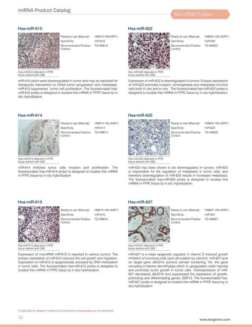
32
miRNA Product Catalog
www.biogenex.com
*Analyte Specific Reagent. Analytical and performance characteristics are not established.
MicroRNA Probes
Hsa-miR-610
miR-610 which were downregulated in tumor and may be exploited for therapeutic intervention to inhibit tumor progression and metastasis. miR-610 suppresses tumor cell proliferation. The fluorescinated hsa-miR-610 probe is designed to localize this miRNA in FFPE tissue by in situ hybridization.
Ready-to-use (Manual): HM610-100(ASR*)
Specificity: miR-610
Recommended Positive Control:
TS-HM610
Hsa-miR-610 detected in FFPE tissue stained with DAB
Hsa-miR-614
miR-614 inhibited tumor cells invasion and proliferation. The fluorescinated hsa-miR-614 probe is designed to localize this miRNA in FFPE tissue by in situ hybridization
Ready-to-use (Manual): HM614-100 (ASR*)
Specificity: miR-614
Recommended Positive Control:
TS-HM614
Hsa-miR-614 detected in FFPE tissue stained with DAB
Hsa-miR-615
Expression of microRNA miR-615 is reported in various tumors. The ectopic expression of miR-615 reduced the cell growth and migration. Expression of miR-615 is epigenetically activated by DNA methylation in tumor cells. The fluorescinated hsa-miR-615 probe is designed to localize this miRNA in FFPE tissue by in situ hybridization.
Ready-to-use (Manual): HM615-100 (ASR*)
Specificity: miR-615
Recommended Positive Control:
TS-HM615
Hsa-miR-615 detected in FFPE tissue stained with DAB
Hsa-miR-622
Expression of miR-622 is downregulated in tumors. Ectopic expression of miR-622 promotes invasion, tumorigenesis and metastasis of tumor cells both in vitro and in vivo. The fluorescinated hsa-miR-622 probe is designed to localize this miRNA in FFPE tissue by in situ hybridization.
Ready-to-use (Manual): HM622-100 (ASR*)
Specificity: miR-622
Recommended Positive Control:
TS-HM622
Hsa-miR-622 detected in FFPE tissue stained with DAB
Hsa-miR-625
miR-625 has been shown to be downregulated in tumors. miR-625 is responsible for the regulation of metastasis in tumor cells, and therefore downregulation of miR-625 results in increased metastasis. The fluorescinated hsa-miR-625 probe is designed to localize this miRNA in FFPE tissue by in situ hybridization.
Ready-to-use (Manual): HM625-100 (ASR*)
Specificity: miR-625
Recommended Positive Control:
TS-HM625
Hsa-miR-625 detected in FFPE tissue stained with DAB
Hsa-miR-627
miR-627 is a major epigenetic regulator in vitamin D induced growth inhibition of tumorous cells upon stimulation by calcitriol. miR-627 acts on target gene JMJD1A (jumonji domain containing 1A), the gene encoding a histone demethylase which is upregulated under hypoxia and promotes tumor growth in tumor cells. Overexpression of miR-627 decreased JMJD1A and suppressed the expression of growth-promoting and differentiating genes, GDF15. The fluorescinated hsa-miR-627 probe is designed to localize this miRNA in FFPE tissue by in situ hybridization.
Ready-to-use (Manual): HM627-100 (ASR*)
Specificity: miR-627
Recommended Positive Control:
TS-HM627
Hsa-miR-627 detected in FFPE tissue stained with DAB

33
miRNA Product Catalog
1-800-421-4149 (USA)
*Analyte Specific Reagent. Analytical and performance characteristics are not established.
MicroRNA Probes
Hsa-miR-628
miR-628 was significantly downregulated in tumors when compared with normal ones. The fluorescinated hsa-miR-628 probe is designed to localize this miRNA in FFPE tissue by in situ hybridization.
Ready-to-use (Manual): HM628-100 (ASR*)
Specificity: miR-628
Recommended Positive Control:
TS-HM628
Hsa-miR-628 detected in FFPE tissue stained with DAB
Hsa-miR-629
miR-629 is upregulated in many tumor tissues. miR-629 activates IL-6–JAK–STAT3 signaling in tumor cells, which in turn upregulates miR-629 expression. The fluorescinated hsa-miR-629 probe is designed to localize this miRNA in FFPE tissue by in situ hybridization.
Ready-to-use (Manual): HM629-100 (ASR*)
Specificity: miR-629
Recommended Positive Control:
TS-HM629
Hsa-miR-629 detected in FFPE tissue stained with DAB
Hsa-miR-630
miR-630 has recently been identified to be implicated in many critical processes in human malignancies. miR-630 expression was significantly increased in tumor specimens compared with that in adjacent normal specimens. The fluorescinated hsa-miR-630 probe is designed to localize this miRNA in FFPE tissue by in situ hybridization.
Ready-to-use (Manual): HM630-100 (ASR*)
Specificity: miR-630
Recommended Positive Control:
TS-HM630
Hsa-miR-630 detected in FFPE tissue stained with DAB
Hsa-miR-638
miR-638 has been reported to be downregulated in several types of tumor, and may therefore function as a tumor suppressor gene. The fluorescinated hsa-miR-638 probe is designed to localize this miRNA in FFPE tissue by in situ hybridization.
Ready-to-use (Manual): HM638-100 (ASR*)
Specificity: miR-638
Recommended Positive Control:
TS-HM638
Hsa-miR-638 detected in FFPE tissue stained with DAB
Hsa-miR-641
miR-641 is an uncharacterized microRNA located at intron-1 of the AKT2 gene and is reported to co-regulate and cooperate with AKT. The fluorescinated hsa-miR-641 probe is designed to localize this miRNA in FFPE tissue by in situ hybridization.
Ready-to-use (Manual): HM641-100 (ASR*)
Specificity: miR-641
Recommended Positive Control:
TS-HM641
Hsa-miR-641 detected in FFPE tissue stained with DAB
Hsa-miR-642a-5p
miR-642a-5p targets Toll-like Receptor 4 in monocytes. The fluorescinated hsa-miR-642a-5p probe is designed to localize this miRNA in FFPE tissue by in situ hybridization.
Ready-to-use (Manual): HM642A-5P-100 (ASR*)
Specificity: miR-642a-5p
Recommended Positive Control:
TS-HM642A-5P
Hsa-miR-642a-5p detected in FFPE tissue stained with DAB
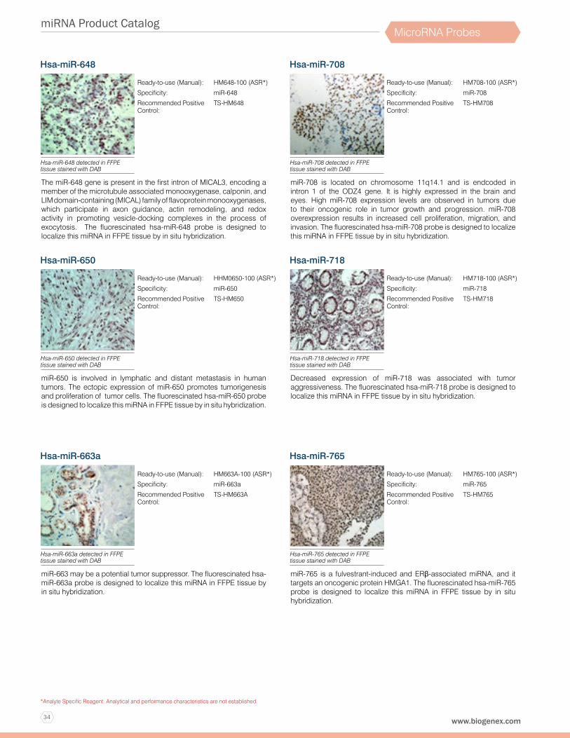
34
miRNA Product Catalog
www.biogenex.com
*Analyte Specific Reagent. Analytical and performance characteristics are not established.
MicroRNA Probes
Hsa-miR-648
The miR-648 gene is present in the first intron of MICAL3, encoding a member of the microtubule associated monooxygenase, calponin, and LIM domain-containing (MICAL) family of flavoprotein monooxygenases, which participate in axon guidance, actin remodeling, and redox activity in promoting vesicle-docking complexes in the process of exocytosis. The fluorescinated hsa-miR-648 probe is designed to localize this miRNA in FFPE tissue by in situ hybridization.
Ready-to-use (Manual): HM648-100 (ASR*)
Specificity: miR-648
Recommended Positive Control:
TS-HM648
Hsa-miR-648 detected in FFPE tissue stained with DAB
Hsa-miR-650
miR-650 is involved in lymphatic and distant metastasis in human tumors. The ectopic expression of miR-650 promotes tumorigenesis and proliferation of tumor cells. The fluorescinated hsa-miR-650 probe is designed to localize this miRNA in FFPE tissue by in situ hybridization.
Ready-to-use (Manual): HHM0650-100 (ASR*)
Specificity: miR-650
Recommended Positive Control:
TS-HM650
Hsa-miR-650 detected in FFPE tissue stained with DAB
Hsa-miR-663a
miR-663 may be a potential tumor suppressor. The fluorescinated hsa-miR-663a probe is designed to localize this miRNA in FFPE tissue by in situ hybridization.
Ready-to-use (Manual): HM663A-100 (ASR*)
Specificity: miR-663a
Recommended Positive Control:
TS-HM663A
Hsa-miR-663a detected in FFPE tissue stained with DAB
Hsa-miR-708
miR-708 is located on chromosome 11q14.1 and is endcoded in intron 1 of the ODZ4 gene. It is highly expressed in the brain and eyes. High miR-708 expression levels are observed in tumors due to their oncogenic role in tumor growth and progression. miR-708 overexpression results in increased cell proliferation, migration, and invasion. The fluorescinated hsa-miR-708 probe is designed to localize this miRNA in FFPE tissue by in situ hybridization.
Ready-to-use (Manual): HM708-100 (ASR*)
Specificity: miR-708
Recommended Positive Control:
TS-HM708
Hsa-miR-708 detected in FFPE tissue stained with DAB
Hsa-miR-718
Decreased expression of miR-718 was associated with tumor aggressiveness. The fluorescinated hsa-miR-718 probe is designed to localize this miRNA in FFPE tissue by in situ hybridization.
Ready-to-use (Manual): HM718-100 (ASR*)
Specificity: miR-718
Recommended Positive Control:
TS-HM718
Hsa-miR-718 detected in FFPE tissue stained with DAB
Hsa-miR-765
miR-765 is a fulvestrant-induced and ERβ-associated miRNA, and it targets an oncogenic protein HMGA1. The fluorescinated hsa-miR-765 probe is designed to localize this miRNA in FFPE tissue by in situ hybridization.
Ready-to-use (Manual): HM765-100 (ASR*)
Specificity: miR-765
Recommended Positive Control:
TS-HM765
Hsa-miR-765 detected in FFPE tissue stained with DAB
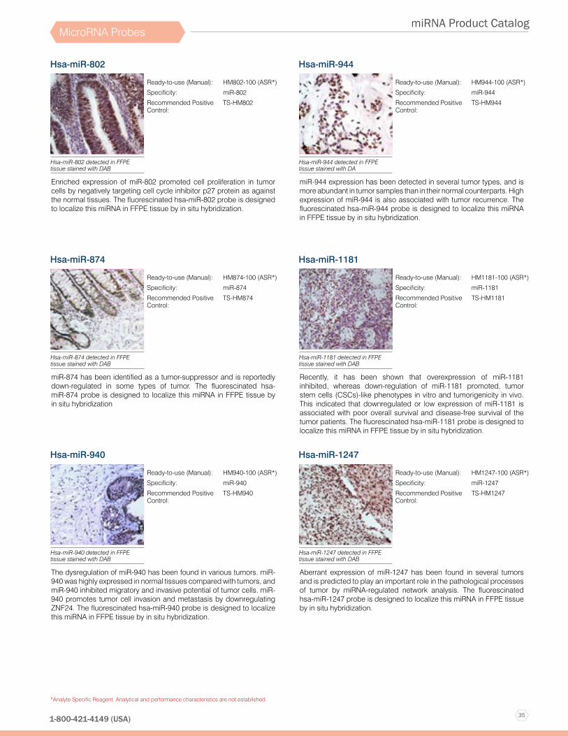
35
miRNA Product Catalog
1-800-421-4149 (USA)
*Analyte Specific Reagent. Analytical and performance characteristics are not established.
MicroRNA Probes
Hsa-miR-802
Enriched expression of miR-802 promoted cell proliferation in tumor cells by negatively targeting cell cycle inhibitor p27 protein as against the normal tissues. The fluorescinated hsa-miR-802 probe is designed to localize this miRNA in FFPE tissue by in situ hybridization.
Ready-to-use (Manual): HM802-100 (ASR*)
Specificity: miR-802
Recommended Positive Control:
TS-HM802
Hsa-miR-802 detected in FFPE tissue stained with DAB
Hsa-miR-874
miR-874 has been identified as a tumor-suppressor and is reportedly down-regulated in some types of tumor. The fluorescinated hsa-miR-874 probe is designed to localize this miRNA in FFPE tissue by in situ hybridization
Ready-to-use (Manual): HM874-100 (ASR*)
Specificity: miR-874
Recommended Positive Control:
TS-HM874
Hsa-miR-874 detected in FFPE tissue stained with DAB
Hsa-miR-940
The dysregulation of miR-940 has been found in various tumors. miR-940 was highly expressed in normal tissues compared with tumors, and miR-940 inhibited migratory and invasive potential of tumor cells. miR-940 promotes tumor cell invasion and metastasis by downregulating ZNF24. The fluorescinated hsa-miR-940 probe is designed to localize this miRNA in FFPE tissue by in situ hybridization.
Ready-to-use (Manual): HM940-100 (ASR*)
Specificity: miR-940
Recommended Positive Control:
TS-HM940
Hsa-miR-940 detected in FFPE tissue stained with DAB
Hsa-miR-944
miR-944 expression has been detected in several tumor types, and is more abundant in tumor samples than in their normal counterparts. High expression of miR-944 is also associated with tumor recurrence. The fluorescinated hsa-miR-944 probe is designed to localize this miRNA in FFPE tissue by in situ hybridization.
Ready-to-use (Manual): HM944-100 (ASR*)
Specificity: miR-944
Recommended Positive Control:
TS-HM944
Hsa-miR-944 detected in FFPE tissue stained with DA
Hsa-miR-1181
Recently, it has been shown that overexpression of miR-1181 inhibited, whereas down-regulation of miR-1181 promoted, tumor stem cells (CSCs)-like phenotypes in vitro and tumorigenicity in vivo. This indicated that downregulated or low expression of miR-1181 is associated with poor overall survival and disease-free survival of the tumor patients. The fluorescinated hsa-miR-1181 probe is designed to localize this miRNA in FFPE tissue by in situ hybridization.
Ready-to-use (Manual): HM1181-100 (ASR*)
Specificity: miR-1181
Recommended Positive Control:
TS-HM1181
Hsa-miR-1181 detected in FFPE tissue stained with DAB
Hsa-miR-1247
Aberrant expression of miR-1247 has been found in several tumors and is predicted to play an important role in the pathological processes of tumor by miRNA-regulated network analysis. The fluorescinated hsa-miR-1247 probe is designed to localize this miRNA in FFPE tissue by in situ hybridization.
Ready-to-use (Manual): HM1247-100 (ASR*)
Specificity: miR-1247
Recommended Positive Control:
TS-HM1247
Hsa-miR-1247 detected in FFPE tissue stained with DAB
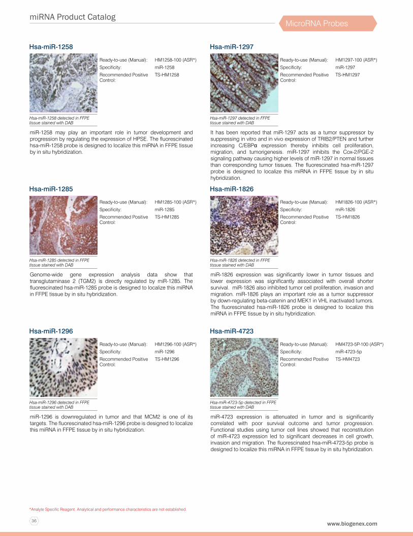
36
miRNA Product Catalog
www.biogenex.com
*Analyte Specific Reagent. Analytical and performance characteristics are not established.
MicroRNA Probes
Hsa-miR-1258
miR-1258 may play an important role in tumor development and progression by regulating the expression of HPSE. The fluorescinated hsa-miR-1258 probe is designed to localize this miRNA in FFPE tissue by in situ hybridization.
Ready-to-use (Manual): HM1258-100 (ASR*)
Specificity: miR-1258
Recommended Positive Control:
TS-HM1258
Hsa-miR-1258 detected in FFPE tissue stained with DAB
Hsa-miR-1285
Genome-wide gene expression analysis data show that transglutaminase 2 (TGM2) is directly regulated by miR-1285. The fluorescinated hsa-miR-1285 probe is designed to localize this miRNA in FFPE tissue by in situ hybridization.
Ready-to-use (Manual): HM1285-100 (ASR*)
Specificity: miR-1285
Recommended Positive Control:
TS-HM1285
Hsa-miR-1285 detected in FFPE tissue stained with DAB
Hsa-miR-1296
miR-1296 is downregulated in tumor and that MCM2 is one of its targets. The fluorescinated hsa-miR-1296 probe is designed to localize this miRNA in FFPE tissue by in situ hybridization.
Ready-to-use (Manual): HM1296-100 (ASR*)
Specificity: miR-1296
Recommended Positive Control:
TS-HM1296
Hsa-miR-1296 detected in FFPE tissue stained with DAB
Hsa-miR-1297
It has been reported that miR-1297 acts as a tumor suppressor by suppressing in vitro and in vivo expression of TRIB2/PTEN and further increasing C/EBPα expression thereby inhibits cell proliferation, migration, and tumorigenesis. miR-1297 inhibits the Cox-2/PGE-2 signaling pathway causing higher levels of miR-1297 in normal tissues than corresponding tumor tissues. The fluorescinated hsa-miR-1297 probe is designed to localize this miRNA in FFPE tissue by in situ hybridization.
Ready-to-use (Manual): HM1297-100 (ASR*)
Specificity: miR-1297
Recommended Positive Control:
TS-HM1297
Hsa-miR-1297 detected in FFPE tissue stained with DAB
Hsa-miR-1826
miR-1826 expression was significantly lower in tumor tissues and lower expression was significantly associated with overall shorter survival. miR-1826 also inhibited tumor cell proliferation, invasion and migration. miR-1826 plays an important role as a tumor suppressor by down-regulating beta-catenin and MEK1 in VHL inactivated tumors. The fluorescinated hsa-miR-1826 probe is designed to localize this miRNA in FFPE tissue by in situ hybridization.
Ready-to-use (Manual): HM1826-100 (ASR*)
Specificity: miR-1826
Recommended Positive Control:
TS-HM1826
Hsa-miR-1826 detected in FFPE tissue stained with DAB
Hsa-miR-4723
miR-4723 expression is attenuated in tumor and is significantly correlated with poor survival outcome and tumor progression. Functional studies using tumor cell lines showed that reconstitution of miR-4723 expression led to significant decreases in cell growth, invasion and migration. The fluorescinated hsa-miR-4723-5p probe is designed to localize this miRNA in FFPE tissue by in situ hybridization.
Ready-to-use (Manual): HM4723-5P-100 (ASR*)
Specificity: miR-4723-5p
Recommended Positive Control:
TS-HM4723
Hsa-miR-4723-5p detected in FFPE tissue stained with DAB

37
miRNA Product Catalog
1-800-421-4149 (USA)
*Analyte Specific Reagent. Analytical and performance characteristics are not established.
MicroRNA Probes
Hsa-miR-9500
The expression levels of miR-9500 were reduced in tumor cells and tumor tissues compared with normal tissues. Overexpression of miR-9500 impeded cell migration in human tumor cells. The fluorescinated hsa-miR-9500 probe is designed to localize this miRNA in FFPE tissue by in situ hybridization.
Ready-to-use (Manual): HM9500-100 (ASR*)
Specificity: miR-9500
Recommended Positive Control:
TS-HM9500
Hsa-miR-9500 detected in FFPE tissue stained with DAB
U6
The U6 probe identifies a small nuclear RNA U6 sequence in human FFPE and freshly prepared frozen tissues. The probe sequence does not share homology with miRNA sequences available in the miRBase database.
Ready-to-use (Manual): PR031-100 (ASR*)
Specificity: U6
Recommended Positive Control:
TS-PR031
U6 detected in FFPE tissue stained with DAB
Scramble
The scramble probe does not identify any miRNA sequences in human FFPE and freshly prepared frozen tissues. The probe sequence does not share homology with miRNA sequences available in the miRBase database.
Ready-to-use (Manual): PR032-100 (ASR*)
Specificity: Scramble
Recommended Positive Control:
TS-PR032
Nagative staining of scramble probe in FFPE tissu

38
miRNA Product Catalog
www.biogenex.com
*Analyte Specific Reagent. Analytical and performance characteristics are not established.
MicroRNA Probes
Hybridization Detection Systemin situ Hybridization (ISH) is a powerful technique for detecting and localizing specific nucleic acid sequences within cells or tissues. This is achieved by the hybridization of a labeled probe to the specific RNA/DNA sequence within the cell and subsequent detection of the bound probe. ISH technique enables the semi-quantification of mRNA expression and helps determine the temporal and spatial patterns of gene expression in cells, tissue and whole animals. ISH technique can also be used for detection of intracellular pathogens with a very high degree of sensitivity.
Super SensitiveTM (Manual) & XISH (Xmatrx) One-Step Polymer-HRP Detection SystemThis is a novel detection system using a non-biotin polymeric technology that makes use of Poly-HRP reagent. As the system is not based on the Biotin-Avidin System, problems associated with endogenous biotin are completely eliminated. The technology allows excellent cell penetration ability for intense staining, compared with other polymer HRPs.
Features & Benefits:• Clean Stain without endogenous biotin background• High signal to noise ratio for intense stain• Universal system for all fluorescein labeled probes• Available in RFID tagged (XISH kit) for Automation or in dropper bottles (Super Sensitive kit) for manual staining
SKU Size α Fluor. Polymer
HRP
DAB buffer DAB Chromo.
Peroxide block
Power block
Hematox Prot. K Hybrid. buffer
NAR-1 Washes A,B,E,F
DF400-25KE 25 test 2 ml 2 ml 5 ml 2 ml 3 ml 3 ml 3 ml 3 ml 6 ml 2 ml 10 ml
DF400-50KE 50 test 3 ml 3 ml 10 ml 2 ml 5 ml 5 ml 5 ml 5 ml 6 ml 3 ml 20 ml
DF400-YADEXmatrx-Elite
100 test 5 ml 5 ml 4x5 ml + 5 RFID vials
7 ml 10 ml 10 ml 10ml 5 ml NA 5 ml 2x10 ml
ISH Detection Systems Composition

39
miRNA Product Catalog
1-800-421-4149 (USA)
*Analyte Specific Reagent. Analytical and performance characteristics are not established.
MicroRNA Probes
Substrates and ChromogensBioGenex offers complete Substrate Packs for immunohistochemical staining with alkaline phosphatase and peroxidase labels. The kits are designed to reduce substrate preparation time and minimize exposure to chemical hazards. The chart below summarizes the substrates offered, indicating enzyme and standard mounting media compatibility.
Features & Benefits:• High Resolution AEC and Liquid DAB• Rapid Development Time• Ready-to-use Solutions• Long-Term Stability
Chromogen Stain Color Enzyme used Solubility in Alcohol/Xylene Compatible with Hematoxylin Compatible Mounting Media
AEC Brick Red HRP Yes Yes Aqueous or Super Mount
DAB Brown HRP No Yes Aqueous, Super Mount or Xmount
Elegance Red Red AP No Yes Aqueous, Super Mount or Xmount
Fast Red Red AP Yes Yes Aqueous or Super Mount
New Fuchsin Red AP Yes Yes Aqueous or Super Mount
The chart below summarizes the compatibility of mounting medium, chromogens and counterstains

Automation
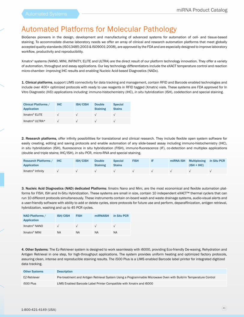
411-800-421-4149 (USA)
miRNA Product CatalogAutomated Systems
Automated Platforms for Molecular Pathology
1. Clinical platforms, support LIMS connectivity for data tracking and management, contain RFID and Barcode enabled technologies and include over 400+ optimized protocols with ready to use reagents in RFID tagged (Xmatrx) vials. These systems are FDA approved for In Vitro Diagnostic (IVD) applications including: immuno-histochemistry (IHC), in situ hybridization (ISH), codetection and special staining.
BioGenex pioneers in the design, development and manufacturing of advanced systems for automation of cell- and tissue-based staining. To accommodate diverse laboratory needs we offer an array of clinical and research automation platforms that meet globally accepted quality standards (ISO13485:2003 & ISO9001:2008), are approved by the FDA and are especially designed to improve laboratory workflow, productivity and reproducibility.
Xmatrx® systems (NANO, MINI, INFINITY, ELITE and ULTRA) are the direct result of our platform technology innovation. They offer a variety of automation, throughput and assay applications. Our key technology differentiators include the eXACT temperature control and reaction micro-chamber- improving IHC results and enabling Nucleic Acid-based Diagnostics (NADx).
2. Research platforms, offer infinity possibilities for translational and clinical research. They include flexible open system software for easily creating, editing and saving protocols and enable automation of any slide-based assay including immuno-histochemistry (IHC), in situ hybridization (ISH), fluorescence in situ hybridization (FISH), immuno-fluorescence (IF), co-detection and multiplex applications (double and triple stains; IHC/ISH), in situ PCR, micro-RNA and special staining.
3. Nucleic Acid Diagnostics (NAD) dedicated Platforms: Xmatrx Nano and Mini, are the most economical and flexible automation plat-forms for FISH, ISH and In-Situ Hybridization. These systems are small in size, contain 10 independent eXACT™ thermal cyclers that can run 10 different protocols simultaneously. These instruments contain on-board wash and waste drainage systems, audio-visual alerts and a user-friendly software with ability to add or delete cycles, store protocols for future use and perform, deparaffinization, antigen retrieval, hybridization, washing and up to 45 PCR cycles.
4. Other Systems: The Ez-Retriever system is designed to work seamlessly with i6000, providing Eco-friendly De-waxing, Rehydration and Antigen Retrieval in one step, for high-throughput applications. The system provides uniform heating and optimized factory protocols, assuring clean, intense and reproducible staining results. The i500 Plus is a LIMS enabled Barcode label printer for integrated digitizeddata tracking.
Clinical Platforms /Application
IHC ISH/CISH Double Staining
Special Stains
Xmatrx® ELITE √ √ √ √
Xmatrx® ULTRA* √ √ √ √
Research Platforms /Application
IHC ISH/CISH Double Staining
Special Stains
FISH IF miRNA ISH Multiplexing (ISH + IHC)
In Situ PCR
Xmatrx® Infinity √ √ √ √ √ √ √ √ √
NAD Platforms /Application
ISH/CISH FISH miRNAISH In Situ PCR
Xmatrx® NANO √ √ √ √
Xmatrx® MINI NA NA NA NA
Other Systems Description
EZ-Retriever Pre-treatment and Antigen Retrieval System Using a Programmable Microwave Oven with Build-In Temperature Control
i500 Plus LIMS Enabled Barcode Label Printer Compatible with Xmatrx and i6000

42 www.biogenex.com
Automated SystemsmiRNA Product Catalog
Microtome to Microscope
* optional software
Fully Automated System from Microtome to Microscope...For the Molecular Pathology Laboratory of Today, Tomorrow and Beyond
• All-in-One All-at-Once – IHC, ISH, SS, Multiplexing and Co-detection
• FISH*
• Standardized process from baking through final glass cover slip in three simple steps: Load – Click – View slides
• Automates technologies of today…IHC, ISH, SS, FISH*, Multiplexing and Co-detection of Nucleic Acid and Protein Biomarkers
• Designed to automate the technologies of tomorrow and beyond…Gene Expression Profiling, in situ PCR, miRNA… and more
• Meets global regulatory requirements and safety standards

431-800-421-4149 (USA)
miRNA Product CatalogAutomated Systems
Next Generation Fully Automated Staining System
• Next generation fully-automated slide staining system
• BioGenex’s proprietary coverslip mechanism
• Easy waste disposal system
• Intelligent SMS system
• Auto-DAB enabled – On-board automated mixing of chromogen and buffer
• High throughput - 100 slides per day, 60 slides in eight-hour shift, and 40 slides in delayed overnight run
All-in-One All-at-Once - IHC, ISH, SS, Multiplex and Co-detection

44 www.biogenex.com
Automated SystemsmiRNA Product Catalog
eFISHiency System for FISH Automation
• Add micro-reagents manually to save cost
• Intelligent SMS information for alerts
• Economical and affordable
• Touch Panel PC as user interface
• Flexible Open System Software - create, edit and save protocols for future use
• Run 10 different protocols at the same time
All-in-One - FISH, in situ PCR and ISH

451-800-421-4149 (USA)
miRNA Product CatalogAutomated Systems
• High performance in situ PCR, ISH and FISH
• Hybridizer with eXACT™ temperatures
• 10 independent thermal cyclers
• Built-in touch screen display for easy operations
• Facility of on-board wash with effective waste drainage system
• Audio-visual alerts and on screen color-coded error alerts
• User-friendly software with ability to add/delete cycles, store protocols for future use and perform up to 45 PCR cycles
eFISHiency Workstation
All-in-One - FISH, in situ PCR and ISH

46 www.biogenex.com
Automated SystemsmiRNA Product Catalog
Infinite Possibilities... ...For Translational and Clinical Research
• Intelligent and flexible system offering infinite possibilities – IHC, ISH, FISH, CISH, IF, Multiplexing and Co-detection
• Simultaneous optimization of up to 40 parameters in single run
• Reaction micro-chamber reduces micro-reagent consumption by up to 90%
• 40 independent thermocyclable (PCR) workstations
• Intuitive software designed for ease of use and flexibility
• Reports for inventory management and regulatory compliance
• Multiple slide processing options – Random, Continuous and STAT
All-in-One - IHC, IF, ISH, CISH, FISH, in situ PCR and miRNA...

Tissue Pre-treatment & Nucleic Acid Retrieval

481-800-421-4149 (USA)
miRNA Product CatalogTissue Pre-treatment & Nucleic Acid Retrieval
De-Waxing Solutions
BioGenex deparaffinization solutions are “one-step” products that simultaneously enables the removal of paraffin and allows rehydration of the tissue with a single reagent. In the past, formalin-fixed, paraffin-embedded tissue sections were traditionally deparaffinized with highly toxic, noxious chemicals (i.e. xylene, equivalents). BioGenex, a pioneer in the Immunohistochemistry technology, offers xylene-free products that removes the paraffin from mounted tissue slides easily and rapidly.
Product 1000 ml (RTU) 500 ml (2x) 1 Gallon ml (2x)
X-DeWax (Xmatrx®) HX015-XAK HX016-XAK HX016-XEK
EZ-DeWax (Manual/i6000TM) HK585-5k HK584-5k NA
One-Step DeWaxing and Rehydration Reagent
1. EZ-DeWaxSol. – For all BioGenex manual methods.
2. X-Dewax Sol. – Optimized for Xmatrx automation.
• Effectively removes paraffin and allow rehydration of the tissue in one step.• Reduces deparaffinization time from 45 minutes to 10 minutes. • Eliminates use of toxic solvents (Xylene) and minimizes hazardous waste. • Ready to use (RTU) or 2x solutions (to be diluted 1:1 with ethanol) are available.
Features & Benefits
Nucleic Acid Retrieval MethodBioGenex is the inventor of Nucleic Acid Retrieval enabling technology. This technology is an effective way of unmasking DNA in formalin-fixed, paraffin-embedded tissue sections using microwave heating. The Nucleic Acid Retrieval technique breaks the formalin induced cross-linking bonds between DNA and proteins, as well as protein-protein cross-linking thereby allowing better penetration of probes and accessibility of DNA for binding. Nucleic Acid Retrieval (NAR-1) is recommended instead of Proteinase K when DNA targeting probes are used.
• Reduces time for probe incubation
• Consistent and reliable staining quality
• Eliminates false-negative staining results
• Easy to use - Can be used in both microwave or Xmatrx Automation protocols
• Non-hazardous, non-flammable and odorless – Safe and Eco-friendly
Advantages of the method:
Product Method Features & Recommended Use
NAR-1 Microwave, 95-100oc Excellent for DNA targeting probes

491-800-421-4149 (USA)
miRNA Product CatalogTissue Pre-treatment & Nucleic Acid Retrieval
Enzymes for Tissue DigestionSome tissues require the use of enzymatic pre-treatment before staining to achieve standardized results depending on the antibodies and their different incubation and pre-treatment requirements. Each kit contains three or four vials of lyophilized enzyme powder and 15 ml of reconstitution buffer, enabling you to make fresh enzyme solutions as needed.
1. Proteinase K in a ready to use (RTU), RNase-free solution and is recommended for use with RNA targeting probes.
2. The Trypsin and Pepsin kits contain well-established enzymes suitable for routine pre-treatment at 37°C. Pepsin is recommended as pretreatment for FISH applications.
3. Protease XXIV kits contain a universal digestive agent that allows for fast and effective pre-treatment at room temperature.
LIS Enabled Barcode Label Printer
• For printing chemical resistant barcode labels
• Compatible with Xmatrx® and i6000TM
• User-friendly software
• Synchronization of protocol information
• Efficient system
• Eliminates human error
• Helps reduce operating cost
• Fast turn-around
Integrated Digitized Data Tracking System
• DeWax, re-hydration and antigen retrieval in one step
• Optimized factory protocols
• User-defined protocols
• High throughput - 96 slides in 20 minutes
• Microwavable containers
• Programmable time and temperature controls
• Built-in probe measures solution temperature in real time
• Time saving and uniform heating
• Eco-friendly solutions
Pre-treatment and Antigen Retrieval System

Ancillary Reagents

511-800-421-4149 (USA)
miRNA Product CatalogAncillary Reagents
BuffersBuffers and diluents are available for Immunohistochemistry,in situ Hybridization Special Stains and most other applications.
• General buffers, such as PBS(PH 7.6) and TBS(PH 7.6, 0.1M) can be used for washing/rinsing of slides.
• Super SensitiveTMWash Buffer is phosphate buffered saline (PH 7.4) with surfactant and isused to ensure optimal staining with even spreading of antibodies and other reagents to avoid inconsistent results.
Product Name 500 ml (20x)
Phosphate Buffered saline HK091-9K
Super Sensitive Wash Buffer HK583-5K
Tris Buffer (Wash Buffer) 3/Pack (dries powder to make 3L)
HK098-5K
Buffers - Manual & Automation
Counterstains and Mounting MediaBioGenex offers the following counterstains for use in Immunohistochemistry,in situ Hybridization and other applications with either manual or automated staining systems.
• Mayer’s hematoxylinis a blue stain that does not contain alcohol and therefore is compatible with both alcohol soluble non-permanent chromogens (AEC, Fast Red& New Fuchsin) and alcohol-insoluble chromogens (DAB & Elegance Red). It is alcohol and xylene insoluble and therefore compatible with most clearing agents and mounting media.
Product Name 1 ml (RTU) 6 ml (RTU) 250 ml (RTU)
Hematoxylin, Mayer’s (IHC, ISH) NA HK100-5K HK100-9K
Mounting of all stained biological specimens is an essential step before their microscopic evaluation. Mounting also enables the slides to be archived for long periods of time. The mounting medium may be used to attach a coverslip or may itself serve as a coverslip substitute. The choice of mounting medium depends on whether long-term or short-term preservation is desired, and whether the mounting procedure is chemically compatible with the chromogen and the counterstain.
• SuperMount®Permanent Mounting Medium is a polymer based aqueous mounting media that does not require the use of a coverslip. This innovative, patented mounting medium (BioGenex’s U.S. Patent No. 5,492,837) is designed to preserve biological specimens for long-term storage. SuperMount® medium is compatible with most aqueous and organic-soluble dyes and chromogens including AEC, DAB, Elegance Red, Fast Red, New Fuchsin, BCIP/NBT, Rhodamine, Fluorescein, Texas Red, Phycoerythrin, Phycocyanin, and Fat Stain (Oil Red O). The refractive index of SuperMount® yields greater transparency and clarity of specimens to be examined under the microscope. SuperMount® can be used for the mounting of all biological specimens, including stained tissue sections, Cytospin preparations, and blood smears.
• Aqueous Mounting Mediumis glycerol-based mounting medium that require the use of a coverslip. It is intended for short-term specimen storage and is compatible with most chromogens and counterstains.
• XMountTM Mounting Medium is a permanent mounting medium that has been optimized for use with BioGenexTM instrument for all BioGenex detection systems for immunohistochemistry (IHC), In Situ Hybridization (ISH) and special stains. XMountTM is intended for use with alcohol and xylene insoluble chromogens, such as DAB (for peroxidase systems) and Elegance Red (for alkaline phosphatase systems). XMountTM dries clear with an ideal refractive index similar to high quality glass and tissue elements. Mounted slides can be viewed with high magnification oil immersion lenses. Also, when mounting preparations stained with the BCIP/NBT substrate, crystal formation that may occur when using other media is minimized.

52 www.biogenex.com
Ancillary ReagentsmiRNA Product Catalog
Mounting Medium
Product Name 15 ml (RTU) 50 ml (RTU)
Aqueous Mounting Medium - Manual HK099-5K NA
SuperMount Permanent Mounting Medium - Manual HK079-5K HK079-7K
Xmount Mounting Media (200 tests) – RFID HX035-YCD NA
Xmount Mounting Media (200 tests) – Xmatrx Infinity HX035-10X NA
OptiPlusTM Positive-Charged Microscope Slides provide a strong adhesive surface fortissues and cells to prevent tissue displacement during harsh pre-treatments such asenzymatic digestion and the microwave Antigen Retrieval method. These slides are idealfor automated systems. Additionally, each slide has a frosted end for easy labeling.The OptiPlusTMPositive-Charged Barrier Slides have all the advantages of our regular OptiPlusTMslides, but also contain hydrophobic barriers that allow the quantity of reagents per slide to be tailored to the size of the specimen. These slides eliminate reagent waste without the need to use a PAP pen, thereby reducing set-up time in manual assays as well as in automated systems. The permanent hydrophobic barriers are compatible with dewaxing solutions and other reagents. The slides are suitable for use with frozen tissue sections, formalin-fixed paraffin sections, and cytology preparations.
Microscope Slides & Coverslips
1. i6000 Elite Automated Staining SystemsOptiPlusTM Barrier slides for i6000 come in three different configurations to accommodate different tissue sizes or multiple tissues per slide:
1. A single, full-size test area of 25 x 40 mm
2. A single 2/3-size test area of 25 x 30 mm
3. Three 1/3-size test areas per slide, each measuring 25 x 15 mm
2. Xmatrx Automated Staining Systems OptiPlusTM Barrier Slides for Xmatrx (U.S. & Foreign Equivalent Patents Pending) contain a double hydrophobic barriers that allows formation of an oil seal to prevent evaporation of microreagents during high temperature steps and prolonged incubations. Four different configurations are available:
1. A single test area of 25 x 40 mm (>80 µl of reagent recommended)
2. A single test area of 25 x 25 mm (>40 µl of reagent recommended)
3. A single test area of 18 x 18 mm (>10 µl of reagent recommended)
4. Two test area per slide, each measuring 18 x 18 mm
Coverslips are optimized for use on Xmatrx staining systems and come is three configurations to accommodate the different barrier slides.
BioGenex pipette tips are made of high-quality polypropylene and are RNase and heavy metals-free when untampered. Inner surface is extremely smooth and requires minimum wetting. 1 ml pipette tips are optimized for use on BioGenexXmatrx® andi6000™ Staining Systems, while 200 µl tips are optimized for Xmatrx® staining systems.
Pipette tips
Consumables kits for Xmatrx®
Item SKU Size Barrier Slides
25 x40 mm
Barrier Slides
25 x40 mm
Coverslips
25 x 40 mm
Coverslips
25 x 40 mm
1 ml
Pipette Tips
200 µl
Pipette Tips
ISH kit XT144-YAD 100 test NA 104 NA 900 384 960

531-800-421-4149 (USA)
miRNA Product CatalogAncillary Reagents
Accessories1. Nucleaic Acid Retrieval Accessories Kits The Nucleaic Acid Retrieval Accessory Kit consists of slide holders and slide baths that make it convenient and compatible with any of the several Nucleaic Acid Retrieval solutions. To accommodate microwave heating, the slide baths and slide holders are made of heat-stable thermoplastic polyolefin and hydrocarbon polymers of acetal resins. These accessories may be used in a microwave or a pressure cooker.
Item SKU Slide Bath + Lid Slide Holder
24- Slide Accessory kit MW001-SU 1 1 (24- slide capacity)
72- Slide Accessory kit MW001-HB 3 3 (72- slide capacity)
2. NordicWare® Microwave Pressure CookerTPlacing the NordicWare®Microwave Pressure Cookerwithin a microwave is an effective method for enhancing staining with the Nucleaic Acid Retrieval technique. The heat produced under enhanced pressure can reduce the build up of gas bubbles on the surface of tissues. This improves the intensity of staining, accompanied by preservation of tissue and cell morphology. This pressure cooker is also optimized for use with various BioGenex Nucleaic Acid Retrieval solutions. BioGenex Catalog number: NW001-PC.
3. PAP Pen for Tissue StainingThe PAP Pen is a useful pen-like tool for immunohistochemical staining methods. It is designed to prevent the waste of valuable reagents by forming a water-repellent barrier around the specimen. This barrier creates the proper surface tension to hold an antibody solution or detection reagents within the target area on the slide. The surface tension provided by the PAP Pen circle ensures that only the amount of antibody solution needed for sufficient reaction will be applied. Since over-flooding of the slide is eliminated, wiping of excess fluid around the specimen can be avoided. The PAP Pen can be used for immunostaining of paraffin sections, frozen sections, and for fluorescent antibody methods. The PAP Pen contains a special formulation, which is water repellent. It can be removed, if desired, with xylene or xylene substitutes after the staining procedure is completed.BioGenex Catalog Number: XT001-PP, sufficient for use on 500-1000 slides.
54 www.biogenex.com
Ancillary ReagentsmiRNA Product Catalog
1. Order Information• Credit Terms: BioGenex will review the customer credit
application and finalize the terms (Credit Limit and Net Days) based on inputs provided and credit rating.
• Order Confirmation: To avoid shipment duplication, please indicate in bold “CONFIRMING ORDER - PLEASE DO NOT SHIP” on your order.
2. Conditions of Sale• All prices are quoted in U.S. dollars, exclusive of Sales tax
(State and County) , as applicable.• If an order is not taxable, a tax exemption certificate must
be provided. • Products and prices are subject to change without any
prior notice.• Discounts: Please inquire about BioGenex quantity
discount policies at 1-800-421-4149.• Payment: All payments must be made in U.S. dollars. You
may choose any mode of payment (Note: Online payment systems are not implemented).
3. Return and Refund Policy BioGenex reagents are covered by Quality Assurance
(QA) policy:• Returns will only be accepted with BioGenex Return
Material Authorization (RMA). Please contact customer service for further assistance.
• BioGenex has a limited liability for a refund or replacement. The same is solely under the discretion of BioGenex management.
• A full refund will be provided when a product cannot perform according to data specifications.
• If client makes an error in ordering a product, a refund may be provided along with a 30% restocking fee.
• Express Delivery: Express delivery options are also available on request at an extra cost.
• BioGenex customer service for assistance: Tel: 1-800-421-4149, Monday through Friday
7 AM – 4 PM PST or E-mail at: [email protected]
4. Other Terms and Conditions• BioGenex is committed to quality, innovation, service,
and support. We believe that the high degree of quality control performed on all our products will help you with consistent and reproducible results.
• All orders are subject to acceptance by BioGenex and product availability.
• Delivery dates are estimates and BioGenex shall have no liability for any delays.
• There are no expressed, implied or statutory warranties,
General Terms and Conditions
including without limitation, the implied warranties of merchantability, fitness for a particular purpose and non-infringement of third party rights.
• Freight charges are prepaid and added to the invoice.• BioGenex shall not be liable for any incidental, indirect,
special or consequential damages, even if it is aware of the possibility of such damages. BioGenex’s total liability for any order shall not exceed the amount paid by customer under such order.
• These terms and conditions constitute the entire agreement between the parties with respect to the products purchased hereunder.
• Any additional, different or inconsistent terms and conditions in a purchase order form or like forms used by customer to purchase, change, accept or otherwise process the orders are objected to and not binding on BioGenex.
• This agreement between the parties shall be governed by the laws of the State of California without regard to its conflicts of laws.
• Any dispute arising out of or related to this Agreement shall be resolved solely in the U.S. District Court for the Northern District of California or in San Francisco County, and in no other courts, and Customer hereby consents to the jurisdiction of, venue in and service of process from the aforementioned courts.

551-800-421-4149 (USA)
miRNA Product CatalogAncillary Reagents
miR
NA
Product C
atalog 20
16
- 20
17
miRNA In Situ Product Catalog 2016-2017
miRNA In Situ – Dawn of a New Era Accelerating Precision Medicine. Integrated miRNA In Situ System
• Over 200 Probes
• Detection Systems
• Automation
• Cancer of Unknown Primary
• Undifferentiated Tumors
• Cancer Staging
For Clinical Research & Tumour Analysis
In the U.S., call +1 (800) 421-4149
Outside the U.S., call +91-40-27185500For USA and Canada distribution only
www.biogenex.com © 2016 BioGenex Laboratories, Inc. All Rights Reserved