Embed Size (px)
Citation preview

ACC/AHA 2005 Practice Guidelines for the Management ofPts With Peripheral Arterial Disease
(Lower Extremity)

peripheral arterial disease
• Encompasses a large series of disorders affecting arterial beds exclusive of the coronary arteries

LOWER EXTREMITY PAD-Risk Factors

Cigarette smoking
• 2 to 3 times more likely to cause lower extremity PAD than CAD
• Increases the risk of lower extremity PAD by 2- to 6-fold and the risk of intermittent claudication by 3- to 10-fold
• More than 80% of patients with lower extremity PAD are current or former smokers

Diabetes mellitus
• increases the risk of lower extremity PAD by 2- to 4-fold
• present in 12% to 20% of persons with lower extremity PAD

classic IC
• pain, ache, tightening, cramping, or sense of fatigue in one or more of the lower extremity muscle groups
• triggered by ambulation & relieved by rest• have sufficient blood flow so that limb ischemic
symptoms are absent at rest.• site of claudication is distal to the diseased arterial
segment• buttock, hip, and thigh claudication are seen with
aortoiliac disease and calf claudication with femoral-popliteal disease

critical limb ischemia
• rest pain, cold, or numbness of the feet, with or without tissue loss (nonhealing ulcers or gangrene).
• Rest pain usually occurs at night (because of the horizontal position, which deprives the patient of the effect of gravity on blood flow through the tight lesions)
• improves when the legs are in the dependent position. • superimposed edema of the affected leg(s)
occasionally may be seen in those who tend to dangle their legs overnight.

Foot Physical Examination and Differential Diagnosis of Neuropathic and Neuroischemic Ulcers

Clinical Presentation of Peripheral Arterial Disease
Most cases are asymptomatic.
•In symptomatic patients: Most have atypical exertional leg pain.
•Only 10% to 30% present with classic intermittent claudication.
•Minority progress to rest pain or ischemic ulcers (critical limb ischemia).


Risk of Cardiovascular Events
• 20% to 60% increased risk for MI • 2- to 6-fold increased risk of death due to CAD• Risk of stroke is increased by approximately
40%• Men with LL-PAD -4 to 5 times more likely to
have a stroke / TIA

Ankle-Brachial Index Testing
overt clinical PAD presenting with claudication or more severe limb ischemic symptom
Age ≥ 70 years or older
Age = 50-69 years with history of diabetes or smoking
Age < 49 years with diabetes and one additional risk factor (smoking, hypertension, or elevated cholesterol levels)
Abnormal lower extremity pulse examination
Known atherosclerotic disease elsewhere (coronary, carotid, or renal arteries)
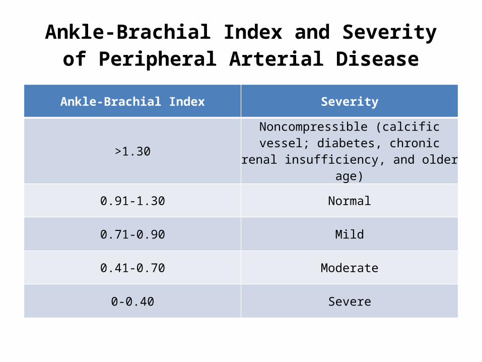
Ankle-Brachial Index and Severity of Peripheral Arterial Disease
Ankle-Brachial Index Severity
>1.30 Noncompressible (calcific vessel; diabetes, chronic renal insufficiency, and older age)
0.91-1.30 Normal
0.71-0.90 Mild
0.41-0.70 Moderate
0-0.40 Severe


Acute limb ischemia
• rapid or sudden decrease in limb perfusion • threatens tissue viability• form of CLI, may be 1stmanifestation of arterial disease in
asymptomatic pt • form of CLI, may occur as an acute event causing
symptomatic deterioration in a pt with antecedent LL-PAD and IC
• Progression of PAD from IC to CLI – gradual (may also reflect cumulative effect of multiple acute local thrombotic events that progressively increase the intensity of ischemia)

Paresthesia and paralysis imply irreversible ischemia, and muscle rigidity is a sign of a nonsalvageable limb.
Signs and Symptoms of Acute Limb Ischemia
PainPallor
PulselessnessPoikilothermia
ParalysisParesthesia

Society for VascularSurgery/International Society for Cardiac Vascular Surgery
(SVS/ISCVS)

Magnetic Resonance Angiography
• RECOMMENDATIONSClass I• 1. MRA of LL -diagnose anatomic location and degree of stenosis of
PAD ( A)• 2. MRA of LL should be performed with gadolinium enhancement ( B)• 3. MRA of LL -useful in selecting pts with LL-PAD as candidates for
endovascular intervn ( A)Class IIb• 1. MRA of LL -may be considered to select pts with LL-PAD as
candidates for surgical bypass and to select the sites of surgical anastomosis. (B)
• 2. MRA of LL -may be considered for postrevascularization surveillance in pts with LL-PAD. (B)

MRA limitations• Tends to overestimate degree of stenosis because of
turbulence• May overestimate occlusions owing to loss of signal
from retrograde collateral flow• Metal clips can cause artifacts that mimic vessel
occlusions• Some metal stents will obscure vascular flow• Pts with PPI and ICD and some cerebral aneurysm clips
cannot be scanned safely • MRA performed with gadolinium has on rare occasions
been associated with renal toxicity in patients with elevated creatinine levels

Computed Tomographic Angiography
• RECOMMENDATIONSClass IIb• 1. CTA of LL may be considered -anatomic
location and presence of signi stenosis in pts with LL-PAD (B)
• 2. CTA of LL may be considered as a substitute for MRA for pts with CI to MRA(B)

CTA has potential advantages over MRA • Pts with PPI or ICD may be imaged safely with CTA• Metal clips, stents, and prostheses usually do not cause
significant CTA artifacts • Has higher resolution• Can provide images of calcification in the vessel wall• Scan times are significantly faster with CTA than with
MRA• Claustrophobia not a problemCTA also has potential disadvantages compared with MRA• Requires iodinated contrast, which may be nephrotoxic • Requires ionizing radiation

PAG-RECOMMENDATIONSClass I• 1. Recommended for evaluation of patients with
LL-PAD when revascularization is contemplated. (B)• 2. Signi of an obstructive lesion is ambiguous →
trans-stenotic P-gradients & supplementary angulated views to be obtained. (B)
• 3. Pts with baseline renal insufficiency should receive prior hydration. (B)
• 4. Follow-up clinical evaluation (physical Ex & RFT)-recommended ≤2 weeks after contrast angio to detect presence of delayed adverse effects, such as atheroembolism, ↓ in RFT, or access site injury (C)

Class IIa• 1. Noninvasive imaging modalities, including
MRA, CTA, and color flow duplex imaging, may be used in advance of invasive imaging to develop an individualized diagnostic strategic plan, including – assistance in selection of access sites, – identification of significant lesions, and– determination of the need for invasive evaluation
• 2. Treatment with n-acetylcysteine in advance of contrast angiography is suggested for pts with baseline renal insufficiency (Cr>2.0 mg/Dl)

CILOSTAZOL-RECOMMENDATIONS
Class I• 1. Cilostazol (100 mg BD) is indicated to
improve symptoms & ↑walking distance in pts with LL-PAD & IC (in the absence of CCF). (A)
• 2. A therapeutic trial of cilostazol should be considered in all patients with lifestyle-limiting claudication (in the absence of CCF). (A)

PENTOXIFYLLINE-RECOMMENDATIONS
Class IIb• 1. Pentoxifylline (400 mg TID) 2-line
alternative therapy to cilostazol to improve walking distance in patients with IC (A)
• 2. The clinical effectiveness of pentoxifylline as therapy for IC is marginal & not well established.(C)

Lipid-Lowering Drugs-RECOMMENDATIONS
Class I• Statins indicated for all pts with PAD to
achieve a target LDL < 100 mg/DlClass IIa• 1. Statins to achieve a target LDL <70 mg/dL is
reasonable for pts with LL-PAD at very high risk of ischemic events
• 2. Fibrates can be useful for pts with PAD and low HDL , normal LDL, & elevated TGL

Antiplatelet and Antithrombotic Drugs RECOMMENDATIONS
Class I• 1. Antiplatelets indicated to ↓ risk of MI, stroke, or
vascular death in pts with atherosclr LL-PAD• 2. Aspirin ( 75 - 325 mg)- safe & effective antiplatelet
therapy • 3. Clopidogrel (75 mg ) -effective alternative
antiplatelet therapy to aspirin Class III• Oral anticoagulation therapy with warfarin is not
indicated

Indications for Revascularization in IC
Before a pt with IC is offered any invasive therapy,the following considerations must be taken into account:
• a predicted or observed lack of adequate response to lifestyle therapy and pharmacotherapies
• the presence of a severe disability• absence of other disease that would limit exercise
even if IC improved• the morphology of the lesion (appropriate
intervention would have ↓risk & a ↑ probability of initial & long-term success)

Morphological Stratification of Iliac Lesions
TASC type A iliac lesions:• 1. Single stenosis <3 cm of the CIA or EIA (unilateral/bilateral)TASC type B iliac lesions:• 2. Single stenosis 3 to 10 cm in length, not extending into the CFA• 3. Total 2 stenoses <5 cm in CIA and/or EIA & not extending into CFA• 4. U/L CIA occlusionTASC type C iliac lesions:• 5. B/L 5-10 cm-long stenosis of CIA and/or EIA, not extending into CFA• 6. U/L EIA occlusion not extending into the CFA• 7. U/L EIA stenosis extending into the CFA• 8. B/L CIA occlusionTASC type D iliac lesions:• 9. Diffuse, multiple U/L stenoses of CIA, EIA, & CFA (usually >10 cm)• 10. U/L occlusion involving both the CIA and EIA• 11. B/L EIA occlusions• 12. Diffuse disease involving Ao& both iliac arteries• 13. Iliac stenoses with an abd Ao aneu/other lesion requiring Ao/iliac sx

Morphological Stratification of Femoropopliteal Lesions
TASC type A femoropopliteal lesions:• 1. Single stenosis <3 cm SFA or popliteal ATASC type B femoropopliteal lesions:• 2. Single stenosis 3 - 10 cm, not involving distal popliteal A• 3. Heavily calcified stenoses ≤3 cm in length• 4. Multiple lesions, each <3 cm (stenoses or occlusions)TASC type C femoropopliteal lesions:• 6. Single stenosis or occlusion >5 cm• 7. Multi stenoses or occlusions, each 3-5 cm, with or without
heavy calcificationTASC type D femoropopliteal lesions:• 8. Complete CFA or SFA occlusions or complete popliteal and
proximal trifurcation occlusions

Endovascular Treatment for IC- RECOMMENDATIONS
Class I• Preferred revascularization technique for TASC type A
iliac & femoropopliteal lesions• Translesional P gradients (with and without vasodilation)
should be obtained to evaluate signi of angiographic iliac stenoses of 50%-75% D before intervn
• Provisional stentng indicated for use in iliacs as salvage therapy for a suboptimal/failed result from POBA
Class IIa• Stents can be useful in femoral, popliteal, & tibial arteries
as salvage for a suboptimal or failed result from POBA

Summary of preferred options in
interventional management of iliac
lesions

Summary of
preferred
options for
interventional treatment of
femoropoplite
al lesions

Thrombolysis for A/c & C/C LI -RECOMMENDATIONS
Class I• Catheter-based thrombolysis is indicated for
pts with a/c limb ischemia of ≤14 days (A)Class IIa• Mechanical thrombectomy devices can be
used as adjunctive therapy for a/c LI due to peripheral arterial occlusion


Thanks…


ACC/AHA 2005 Practice Guidelines for the Management ofPts With Peripheral Arterial Disease
(Renal artery)

Clinical clues to RAS
• Onset of HTN < 30 yrs (I)• Severe HTN >55 yrs (I)• Accelerated, resistant or malignant HTN (I)• New azotemia/wors RFT aft adm of ACEI/ARB (I)• Unexpl atrophic kidney/size discrep >1.5 cm (I)• Sudden unexpl Pulm Edema (I)• Unexpl ↓RFT, inc pts on RRT (IIa)• Multi-vessel CAD (IIb)• Unexplained CCF (IIb)• Refractory angina (IIb)

Prevalence in gen population (RA duplex)• ≥65 yrs (834)• →6.8% (Women-5.5% , Men-9.1%)
Screening RA angio at CAG • RA disease →30%• Significant RAS→11%-18%• Significant RAS →22%-59% pts with PAD
ESRD in HD >20-years (683pts)• 12% had documented RAS as a cause of ESRD.

• Renal function in pts with athero RAS on medical therapy (28/12 follow up)
• 46%→ ↑S.Cr• 29%→ 25%-50% ↓GFR• 37%→↓kidney size by > 10%

Median survival for • ESRD with renovascular disease- 25/12• ESRD due to malignant HTN- 55/12• ESRD due to polycystic KD- 133/12

Clinical Clues to the Diagnosis of RAS –RECOMMENDATIONSPerformance of diagnostic studies
Class I• 1. Onset of HTN <30 yrs• 2. Onset of severe HTN >55 yrs• 3. Characteristics:
– (a) acc HTN (sudden & persistent worsening of prev controlled HTN) – (b) resistant HTN– (c)malign HTN (A/C decomp CCF, A/C visual/neuro disturb &/ adv retinopathy)
• 4. New azotemia /Worsening RFT aft an ACEI or an ARB • 5. Unexp atrophic kidney/Discrepancy in size of >1.5 cm• 6. Sudden, unexp pulm edema (esp azotemic pts)Class IIa• 1.Unexp RF, including pts starting RRT

• Class IIb• 1. The performance of arteriography to
identify signi RAS - reasonable in pts with multivessel CAD & none of the clinical clues or PAD at the time of arteriography.(B)
• 2. Diagnostic studies -reasonable in pts with unexpl CCF or refractory angina(C)

Atherosclerosis
• 90% of all renovascular stenotic lesions• Most often affects the aorto-ostial segment,
including the proximal 1 cm of main RA

Fibromuscular Dysplasia
• nonatherosclerotic, noninflammatory disease• HTN in a young woman (can occur in both genders at
any age)• Middle & distal ⅔ of main RA• May involve RA branches (25%- segmental arteries)• Tends to occur in 25 - 50 yr old women • Often involves both RA (B/L in 60% )• Characteristic - “string of beads” appearance• Medial fibroplasia ≈ 80% of FMD• Intimal fibroplasia - relatively rare (thin, discrete web)

• Also affects other arteries, inclu Carotid & Vertebral, & less commonly, Iliac & Mesenteric
• Appears to be an asso betw Carotid & Vertebral FMD and intracranial aneurysmal disease
• MRA of head should be performed in all patients with cervicocranial FMD

Classification of Fibromuscular Dysplasia
Classification Frequency Pathology Angiographic Appearance
Medial dysplasia
1.Medial fibroplasia
80% Alternating thin media & thick ridges
“String of beads” "beading“> D
2.Perimedial fibroplasia
10% to 15% Ext collagen depots in outer media
“Beading” “beads”<D
3.Medial hyperplasia
1% to 2% True SM hyper-plasia ,No fibrosis
Concentric smooth stenosis
Intimal fibroplasia < 10% Deposition of collagen in intima
Concentric focal band
Adventitial (periarterial) fibroplasia
< 1% Dense collagen replaces fibrous tissue of adventitia
So rare, classic angiographic findings -not known

• Renovascular HTN may also be caused by renal artery aneurysms
• Aneurysm rupture is of greatest concern with noncalcified aneurysms >2 cm D, particularly in premenopausal women

Other causes of renovascular disease
• Takayasu’s arteritis• Atheroemboli• Thromboemboli• William’s syndrome• Neurofibromatosis• Spontaneous renal artery dissection• Arteriovenous malformations or fistulas• Trauma (e.g.,lithotripsy, direct injury, or surgery)• Prior abdominal radiation therapy• Retroperitoneal fibrosis

Diagnostic Methods
RECOMMENDATIONSClass I• 1. Duplex USG recommended screening test to establish ∆
of RAS (sens 84%-98% and speci 62%-99%). (B)• 2. CTA (pts with N-RFT ) recommended screening test to
establish ∆ of RAS. (B) (sens 59%-96% and speci 82%-99%)• 3. MRA recommended screening test to establish ∆ of RAS.
(B) (sensi 90%-100% and speci 76%-94%)• 4. Clinical index of suspicion high & Results of noninvasive tests inconclusive Angiography recommended as a diagnostic test (B)

Duplex Ultrasound
• Excellent- to monitor RA patency aft endovascular/surgical revascularization
• Limitations – absolute dependence on operator skill– diminished ability to visualize accessory RA– Difficulty to image obese/pts with intervening
bowel gas

Computed Tomographic Angiography
• Not ideal screening for pts with renal insufficiency
• Metal stents may be imaged & in-stent restenosis detected
• Higher spatial resolution than MRA

Magnetic Resonance Angiography

Catheter Angiography
• Pts -clinical clues- definitive diagnostic noninvasive images cannot be obtained
• Pts -prespecified clinical indications & in whom concomitant angiographic access has been obtained for PAG/CAG

Clinical clues to RAS
• Onset of HTN < 30 yrs (I)• Severe HTN >55 yrs (I)• Accelerated, resistant or malignant HTN (I)• New azotemia/wors RFT aft adm of ACEI/ARB (I)• Unexpl atrophic kidney/size discrep >1.5 cm (I)• Sudden unexpl Pulm Edema (I)• Unexpl ↓RFT, inc pts on RRT (IIa)• Multi-vessel CAD (IIb)• Unexplained CCF (IIb)• Refractory angina (IIb)

Hemodynamically signi Asympto(incidental)RAS
Defined as RAS in the absence of • end organ dysfunction (e.g., idiopathic pulmonary
edema, stroke,visual loss, hypertension, or refractory angina)
But in the presence of • (a) ≥50%-70% D stenosis by visual estimation + peak
trans-lesional gradient ≥ 20 mm Hg/mean gradient ≥10 mm Hg OR
• (b) any stenosis ≥ 70% D stenosis OR• (c) ≥ 70% D stenosis by IVUS

Asymptomatic Stenosis
RECOMMENDATIONSClass IIb• 1. PTA may be considered – B/L or solitary
viable kidney with a hemodynamically signi RAS (C)
• 2. PTA in U/L hemodynamically signi RAS in a viable kidney- Not well established, Clinically unproven (C)


Hypertension
RECOMMENDATIONSClass IIaPTA- reasonable if hemodyn signi RAS & • Acc HTN, resis HTN , malign HTN, • HTN with unexpl unilateral small kidney• HTN with intolerance to medication (B)

Preservation of Renal Function
RECOMMENDATIONSClass IIa• PTA- reasonable – RAS & progressive CKD
with B/L RAS or a RAS to a solitary functioning kidney (B)

sudden-onset/“flash” pulm edema
Pts with hemodyn sign B/L or solitary RAS• Volume-overload state- lack N renal function
to respond to P natriuresis • ↑LV afterload secondary to angiotensin-
mediated vasoconstriction • Contribute to unstable coronary syndromes-
sudden ↑ in myocardial O2 demand in CAD pts secondary to peripheral vasoconstriction

CCF and UA
RECOMMENDATIONSClass I• PTA indicated in hemodyn signi RAS &
recurrent, unexpl CCF or sudden, unexpl pulm edema(B)
Class IIa• PTA reasonable in hemodyn signi RAS &
UA(B)

Potential physiological benefits
Reperfusion of ischemic kidney(s)• ↓stimulus to renin production,• ↓angiotensin & aldosterone production,• ↓peripheral arterial vasoconstriction• ↓tendency to ↑ECF VolumeImprovement in renal perfusion • ↑glomerular filtration • ↑natriuresisFinally, in patients with a solitary kidney or bilateral RAS,
ability of pt to tolerate long-term adm of angiotensin antagonist medications may be facilitated by relief of a hemodynamic renal artery obstruction.

INTERVENTION
• Class I• 1. Renal stent placement for ostial
atherosclerotic RAS lesions meeting the clinical criteria for intervention (B)
• 2. Balloon angioplasty with bailout stent placement for FMD lesions (B)

Surgery for RAS
Class I• 1. Fibromuscular dysplastic RAS with clinical
indications for interventions (same as for PTA) – complex disease that extends into segmental arteries – macroaneurysms
• 2. Atherosclerotic RAS & clinical indications for intervention – multiple small renal arteries – early primary branching of main RA– in combination with pararenal aortic reconstructions

Indications for renal revascularization.


ACC/AHA 2005 Practice Guidelines for the Management ofPts With Peripheral Arterial Disease
(Mesentry)

Acute intestinal ischemia (AIO)• most frequently occlusive but also non-
occlusive (low flow states) • Occlusive AIO without treatment is nearly
always fatal • Exceptions -ischemic injury confined to
mucosal layer, gradual development of collateral circulation
Chronic intestinal ischemia (CIO)• always the result of arterial obstruction

Occlusive Acute Intestinal Ischemia
• ⅔ women, median age 70 yrs,most have h/o preexisting CAD
DiagnosisRECOMMENDATIONSClass I• 1. A/C abd pain out of proportion to physical findings & h/o
CAD- Suspect AIO• 2. A/C abd pain aft arterial interventions in which catheters
traverse visceral Ao or any prox arteries or who have arrhythmias or recent MI - Suspect AIO
Class III• In contrast to CIO, duplex sonography of abd- not an
appropriate diagnostic tool for suspected AIO

ARTERIOGRAPHY
• Diagnostic & can differentiate occlusive V/S nonocclusive• Catheter-directed therapy - intra-arterial vasodilators,
thrombolysis, or mechanical thrombectomy• Knowledge of extent & nature of intestinal A lesions, if
surgery reqd
Decision for arteriography - best individualized
• Very A/C presentation, High likelihood of arterial obstr, Susp bowel infarction→ Immediate laparotomy -Best approach
• More delayed presentation/ high likelihood of nonocclusive ischemia→ Arteriography indicated

Endovascular TreatmentRECOMMENDATIONClass IIb• Percutaneous interventions (including
transcatheter lytic therapy, balloon angioplasty, and stenting) -appropriate in selected patients with occlusive AIO. Patients so treated may still require laparotomy

Chronic Intestinal Ischemia
• most often female (70%) • severe abd pain induced by eating• Majority –h/o CAD• 30%-50%- previous operations for
atherosclerotic disease (mostly coronary & lower extremity bypass)

Chronic Intestinal Ischemia
DiagnosisRECOMMENDATIONS
Class I• 1. Abdominal pain and weight loss without
explanation, esp with CAD- Suspect CIO• 2. Duplex ultrasound, CTA, and MRA- useful initial
tests for CIO• 3. Diagnostic angiography, including lateral
aortography, in pts suspected of having CIO for whom noninvasive imaging is unavailable/indeterminate

Arteriography
• Definitive diagnosis • Lateral aortography- best suited for typical
origin lesions• Presence of an enlarged “arc of Riolan” -
arteriographic sign of proximal mesenteric A obstruction

Interventional TreatmentRECOMMENDATIONClass I
• Percutaneous endovascular treatment of intestinal arterial stenosis is indicated in patients with chronic intestinal ischemia (B)

Thanks…


ANEURYSMS OF THE ABDOMINALAORTA
• Generally, however, an AAA is• considered to be present when the minimum
anteroposterior• diameter of the aorta reaches 3.0 cm. The size of the aorta• can be measured in any plane that is perpendicular to the
vessel• axis, but in practice, the anteroposterior diameter is
measured• most easily and reproducibly. Accordingly, most screening• studies define AAA in this manner (

Symptomatic Aortic or Iliac Aneurysms
• RECOMMENDATIONS• Class I• 1. In patients with the clinical triad of abdominal and/or• back pain, a pulsatile abdominal mass, and hypotension,• immediate surgical evaluation is indicated.• (Level of Evidence: B)• 2. In patients with symptomatic aortic aneurysms,• repair is indicated regardless of diameter. (Level of• Evidence: C)

Endovascular Aortic Aneurysm Repair
• In an attempt to overcome the risk of distal migration and
• proximal attachment failure, a growing number of new
• devices now incorporate barbed hooks that are sufficiently
• long to secure the metallic frame of the stent graft to the visceral
• segment of the aorta above the renal arteries

• 5.2.7.1.1. ANATOMIC LIMITATIONS. Even with suprarenal fixation• of its metallic exoskeleton, the fabric component of an• endograft obviously cannot be permitted to overlap the origins• of the renal arteries. Accordingly, at least 1 cm of proximal• aortic cuff (1.5 cm for commercially available grafts)• presently is optimal for elective endograft repair below the• renal arteries. For devices without a suprarenal fixation• device, the optimum infrarenal aortic diameter at the time of• this writing is 25 mm or less, and for devices with a• suprarenal fixation component, it is 28 mm or less. Because• of the inflexibility of externally supported grafts, this segment• of the aorta must not be severely angulated. This• requirement may impose a gender bias in patient selection,• because in addition to the fact that their small external iliac• arteries often present problems with respect to vascular• access, women also appear to have a higher prevalence of• short, angulated aneurysm necks than men (1138,1139

Management OverviewRECOMMENDATIONS
• Class I• 1. Open repair of infrarenal AAAs and/or common iliac• aneurysms is indicated in patients who are good or• average surgical candidates. (Level of Evidence: B)• 2. Periodic long-term surveillance imaging should be• performed to monitor for an endoleak, to document• shrinkage or stability of the excluded aneurysm sac,• and to determine the need for further intervention in• patients who have undergone endovascular repair of• infrarenal aortic and/or iliac aneurysms. (Level of• Evidence: B)

• Class IIa• Endovascular repair of infrarenal aortic and/or common• iliac aneurysms is reasonable in patients at high• risk of complications from open operations because of• cardiopulmonary or other associated diseases. (Level• of Evidence: B)• Class IIb• Endovascular repair of infrarenal aortic and/or common• iliac aneurysms may be considered in patients at• low or average surgical risk. (Level of Evidence: B)