Embed Size (px)
Citation preview
Research Article
FORMULATION DEVELOPMENT EX-VIVO AND IN-VIVO EVALUATION OF NANOEMULSION FOR TRANSDERMAL DELIVERY OF GLIBENCLAMIDE
MOHAMMAD WAIS*1,3, ABDUS SAMAD2, IRAM NAZISH4, ANUBHA KHALE3, MOHD AQIL4 AND MOHIB KHAN5
1Ph.D.Scholar, Department of Pharmaceutical Science, NIMS Institute of Pharmacy, NIMS University, Shobha Nagar Jaipur-303121, Rajasthan, India, 2Department of Clinical Research, Panacea Botec Ltd, R&D Sampann, Lalru, Punjab, India, 3Maharashtra Educational
Society, Humera Khan College of Pharmacy, Jogeshwari (W) Mumbai, India 400102, 4Department of Pharmaceutics, Faculty of Pharmacy, Jamia Hamdard, New Delhi-62, India, 5Oriental Education Society, Oriental College of Pharmacy, Sanpada, Mumbai, India 400102.
Email: [email protected]
Received: 30 July 2013, Revised and Accepted: 18 Sep 2013
ABSTRACT
Introduction: Transdermal administration of glibenclamide (GLBD) might offer some advantages over oral route in subject with the treatment of hyperglycemia in non-insulin dependent diabetes mellitus (NIDDM).
Objective: The objective of the present study was to develop the nanoemulsion gel for transdermal delivery of glibenclamide (GLBD) and to evaluate with respect to various Ex-vivo and in-vivo parameters. Nanoemulsions do not need the chemical enhancers; they are advantageous over the conventional transdermal drug delivery systems.
Materials and methods: The nanoemulsion formulation consisted of Labrafac and Triacetin (1:1, ratio) as an internal oil phase in external aqueous phase, Tween 80 as surfactant and diethylene glycol monoethyl ether as cosurfactant. Nine nanoemulsion formulations were selected for the Ex-vivo And In-vivo Evaluation. Ex-vivo drug release studies and permeation studies through wistar rat skin. The blood glucose reducing hypoglycemic activity of the systems was studied in both normal and diabetic wistar rat. The histopathological studies, skin irritation tests and In-vivo evaluation were carried out in wistar rat.
Result :The optimized formulation NE-B2, which contained 15 % labrafac and tricetin, 45 % Smix (tween 80 and diethylene glycol monoethyl ether) and 40 % water showed significant increase (P < 0.01) in the transdermal flux (95.90 μg cm-2 h-1) .The in vivo studies revealed a 3.92 fold increase in relative bioavailability through transdermal application of NE-B2 formulation compared to oral drug suspension.
Conclusion: In conclusion, the present study shows that the nanoemulsion gel for transdermal systems of glibenclamide exhibited better control of hyperglycemia and more effectively reversed the diabetes mellitus complications than oral glibenclamide administration in wistar rat.
Keywords: Nanoemulsions; Transdermal Delivery; Ex-vivo And In-vivo Evaluation; GLBD.
INTRODUCTION
Nano emulsions are clear mixtures consisting of oil, surfactant, cosurfactant, and aqueous phase, which are single optically isotropic and thermodynamically stable with a droplet size typically in the range of 10–100 nm [1, 2]. Nanoemulsion prepared by spontaneous emulsification method offer several advantages over other drug carriers like lower preparation cost, long shelf life, absence of residual organic solvents, and high drug loading for both hydro- and lipophilic drugs [3-5].Numerous studies had been conducted in the recent past that showed the significance of these systems for dermal and transdermal delivery both in vitro[6-16], and in vivo[17-22]. They had been amply demonstrated to improve the transdermal permeation over conventional topical preparations such as emulsions [23-25], gels[25-28], creams[29-31], and ointments[32,33]. Diabetes mellitus is a group of metabolic diseases characterized by high blood sugar (glucose) levels, which result from defects in insulin secretion, or action, or both. A therapeutic classification includes two major types of diabetes mellitus. Type I diabetes (Insulin-Dependent
Diabetes Mellitus; IDDM) is a severe form associated with ketosis in the untreated state. Type II (Noninsulin-Dependent Diabetes Mellitus; NIDDM) represents a heterogeneous group comprising a milder form of diabetes that occurs predominately in adults. Circulatory endogenous insulin is sufficient to prevent ketoacidosis, but is often subnormal or relatively inadequate because of tissue insensitivity [34, 35]. The vast majority of diabetic patients have non-insulin dependent diabetes mellitus (NIDDM). The prevalence of diabetes for all age-groups worldwide was estimated to be 2.8% in 2000 and 4.4% in 2030. In the United States, about 90% of all diabetic patients have NIDDM. The incidence rates of NIDDM increase with age, with a mean rate of about 440 per 100,000 per year by the sixth decade in males in the United States[35]. The total number of people with diabetes is projected to rise from 171 million
in 2000 to 366 million in 2030[36]. India and China are, and will remain, the leading countries in terms of the number of people with diabetes mellitus in the year 2025. Diabetic disease is increasing rapidly and vast amounts of resources are spent in all countries.
Antidiabetic drug distinctly lower the blood glucose level by both defects of NIDDM, by stimulating pancreatic beta cells to produce more insulin and induced increased activity of peripheral insulin intra cellular receptor. Many sulfonylureas like glibenclamide (GLBD) and glipizide have been associated with severe and sometimes fatal hypoglycemia by stimulating release of insulin from pancreatic beta cells and by increasing the sensitivity of peripheral tissue to insulin and gastric disturbances like nausea, vomiting, heartburn, anorexia, and increased appetite after oral therapy. GLBD is best avoided in the elderly and in patients with even mild renal impairment because of the risk of hypoglycemia[37,38]. Owing to its high portion of hepatic first pass metabolism (~50%)[39], its low molecular weight (494 Da), its moderate lipophilicity (Log P, 4.8) as well as its clinical effectiveness in low doses[40,41] (5mg to 15mg), the percutaneous application of GLBD provides, therefore, a preferred alternative to the oral dosage form.
Transdermal administration of GLBD might offer some advantages over oral route in subject with the treatment of hyperglycemia in non-insulin dependent diabetes mellitus (NIDDM), but has been associated with severe and sometimes fatal hypoglycemic reactions and gastric disturbances like heartburn, nausea, vomiting, anorexia and increase appetite after oral therapy because of high inter individual variation [42]. Since these drugs are usually intended to be taken for a long period, patient compliance is also very important.
Transdermal drug delivery system (TDDS) provides a means to sustain drug release as well as reduce the intensity of action and thus reduce the side effects associated with its oral therapy. The NE system is a promising vehicle due to powerful ability to deliver drug through skins [43]. The stable NE system consisted of oils as
International Journal of Pharmacy and Pharmaceutical Sciences
ISSN- 0975-1491 Vol 5, Suppl 4, 2013
AAccaaddeemmiicc SScciieenncceess
Wais et al. Int J Pharm Pharm Sci, Vol 5, Suppl 4,747-754
748
combination of labrafac and triacetin (1:1), commonly used nonionic surfactant (Tween 80), non-irritant cosurfactant (diethylene glycol monoethyl ether) and water was prepared, its transdermal permeation ability and pharmacokinetics studies of GLBD were evaluated.
MATERIALS AND METHODS
Materials
GLBD was a gift sample from Cipla (Mumbai, India). Oleoyl macrogol-6 glycerides / glycerides (labrafil 1944 CS), Propylene glycol dicaprylate/dicaprate (labrafac PG), PEG-8 caprylic/capric glycerides (labrasol), Propylene glycol monocaprylate (capryol PGMC), diethylene glycol monoethylether (transcutol P) were gift samples from Gattefosse SAS (France). Castor oil and olive oil were purchased from genuine chemicals (Mumbai, India). Triacetin (glycerin triacetate), tween 80, tween 20 and polyethylene glycol 200 (PEG-200) were purchased from Ozone chemicals (Mumbai, India). Polyethylene glycol 400(PEG-400), propylene glycol (PG) and n-butanol were purchased from E-Merck (Mumbai, India).Isopropyl myristatae (IPM) was purchased from S.D. Fine chemicals (Mumbai, India). High-performance liquid chromatography (HPLC) grade methanol and acetonitrile (ACN) were purchased from Finar chemical (Ahmedabad, India). Water was obtained from Milli Q water purification system (Miliipore, MA). All other chemicals and solvents used in the study were of analytical grade.
Selection of Formulations from Phase Diagrams
Following criteria were chosen for the selection of formulations. GLBD was added to the mixture of oil, surfactant and cosurfactant with varying component ratio as described in Table -1,
and then appropriate amount of water was added to the mixture drop by drop and the nanoemulsion containing GLBD was obtained by vortexing the mixture at ambient temperature 30 °C. Formulations with Smix ratio 1:0, 1:2 and 1:3 were not selected because the formulations were unstable and showed phase separation in earlier publication glibenclamide44.
Preparation of Rat Skin
The male albino wistar rats (7-9 weeks old, 200-250 g) were sacrified by aspiration of ethyl ether and the abdominal skin was carefully excised from the underlying connecting tissue using scalpel. The hairs remaining on the skin were trimmed away. The subcutaneous tissue was removed surgically and the dermis side was wiped with isopropyl alcohol to remove the adhering fat. The cleaned skin was washed with distilled water and stored at 21°C until further use.
Ex-vivo Permeation Studies
The ex-vivo skin permeation studies were performed using Franz diffusion cell apparatus The diffusion area was 0.75 cm2 and receptor volume was 5.0 mL. The skin was brought to room temperature, cut and trimmed to appropriate size and mounted between the donor and receptor compartments of the diffusion cell with the stratum corneum side facing upwards and then the donor chambers were clamped in place. The receptor compartment was filled with phosphate buffer saline (pH 7.4). The receptor fluid was stirred with a magnetic rotor at a speed of 600 rpm and the temperature was maintained at 37 ± 1°C. After stabilization of the skin, one mL of NE formulations, was placed into the donor compartment and sealed with paraffin film to provide occlusive conditions. Samples were withdrawn at regular intervals (1, 2, 3, 4, 5, 6, 7, 8, 10, 12, 24 and 48 h), filtered through 0.45-µm membrane filter and analyzed for drug content by UHPLC/ESI-qTOF/MS method as described in earlier pulication[44]. The receptor phase was immediately replaced with equal volume of fresh receptor fluid. The same study was also performed for control formulations. Each set of experiment was performed with 3 diffusion cells (n=3).
Preparation of Nanoemulsion Gel
The nanoemulsion was insufficiently viscous and could be therefore quickly removed from the skin. Therefore, the optimized
nanoemulsion (NE-B2) was converted into nanoemulsion gel. Nanoemulsion gel was prepared by dispersing 0.75% w/w of Carbopol 934 in sufficient quantity of distilled water [22]. The dispersion was kept in dark for 24 h for complete swelling of carbopol [45].Then 0.005% w/w of GLBD nanoemulsion was added slowly to carbopol dispersion. Triethanolamine (0.5% w/w) was added in this mixture to neutralize carbopol. Then remaining quantity of distilled water was added to get the final preparation 100% w/w.
Preparation of Conventional GLBD Gel
Hydrogel with GLBD was used a classical dermal formulation for comparison of GLBD action incorporated in nanoemulsion gel. Carbopol 934 (0.75% w/w) was dispersed in sufficient quantity of distilled water. The dispersion was kept in dark for 24 h for complete swelling of Carbopol 934. Then GLBD was mixed with the carbopol dispersion, and 0.5% w/w of triethanolamine was added in this mixture to neutralize carbopol, and the remaining water was added to give homogeneously dispersed GLBD in hydrogel [46, 47].
Calculation of Permeation Parameters
The cumulative amount of GLBD permeated per unit of rat skin surface area, Qt/S (S=0.75 cm2) was plotted as a function of time (t, h). The permeation rate of GLBD at steady-state (Jss, µg/cm2/h) through rat skin was calculated by linear regression interpolation of the cumulative amount permeated through rat skin per unit area vs. time:
Jss = Δ Qt / S. Δ t (Eq. 1)
The permeability coefficient (Kp, cm/h) was calculated according
to the equation:
Kp = Jss / Cd (Eq. 2)
Where Cd = concentration of drug in donor compartment (5.0mg/mL), and is assumed that under sink conditions the drug concentration in the receiver compartment is negligible compared to that in the donor compartment.
The enhancement ratio (ER) was calculated according to the equation:
ER = Flux from formulation / flux from formulation E. (Eq.3)
All skin permeation experiments were repeated three times and data were expressed as mean of three experiments ± standard deviation (S.D).
In-vivo Permeation Study
The animal protocol to carry out in-vivo study and skin penetration study was reviewed and approved by Institutional Animal Ethics Committee, Oriental college of pharmacy Sanpada (approval no: OCP/CPCSEA/IAEC/2012/005) and their guidelines were followed for the studies. Albino rats, 7-9 weeks old and weighing 150–220g was used for the study The animals were kept under standard laboratory conditions (temperature: 25 ± 2 °C; relative humidity: 55 ± 5%). The animals were housed in polypropylene cages, with free access to standard laboratory diet (Lipton feed, Mumbai, India) and water ad libitum.
Skin Irritation Test
The animals were kept under standard laboratory conditions, with temperature of 25±1°C and relative humidity of 55±5%. They were housed in polypropylene cages with free excess to a standard laboratory diet (Lipton feed, Mumbai, India) and water ad libitum. The hairs on the abdominal side of albino Wistar rats were removed by clipping 1 day before the experiment [48]. The skin irritation study was performed using eight rats. A single dose of 10 μL of the formulation was applied to the left ear of the rat and the right was considered as a control. The development of erythema was monitored for six days using the method, as reported by Abbe et al. [49].
Induction of Diabetes mellitus:
The overnight fasted Wistar rats weighing 150–220g were made diabetic by a single intravenously injection of streptozotocin (60

Wais et al. Int J Pharm Pharm Sci, Vol 5, Suppl 4,747-754
749
mg/kg) dissolved in citrate buffer (3 mM; pH 4.5) [50]. After the dose of 60 mg/kg intravenously injection, symptoms occur already after 24–48 h with hyperglycemia up to 800 mg %, glucosuria and ketonemia. Histologically, the beta-cells are degranulated or even necrotic. A steady state is reached after 10–14 days allowing using the animals for pharmacological tests [51]. The blood was taken from the tail vein at predetermined time intervals up to 48 hours. Blood glucose level was determined by Accu-Check Glucometer (Roche Diagnostics, Germany).
Antidiabetic activity
The antidiabetic activity of the developed nanoemulsion gel was estimated by taking blood from tail vein of rats. Twenty four wistar rat were taken and they were divided into four groups (n=6). Group I, was taken as control. Diabetes was induced in the remaining groups (Group II, III, and IV). The wistar rat was treated as following:
Group I (Control) –
Group II- Applied with 2.00 cm2 transdermal system prepared with nanoemulsion gel, without drug in 0.5% carbopol gel.
Group III – Glibenclamide (5 mg/kg) the oral doses were given using a round tipped stainless steel needle attached to 1 ml syringe and the dose of 5 mg/kg was selected by conducting a series of experiments with graded doses ranging between 1 to 10 mg/kg.
Group IV - Applied with 2.00 cm2 transdermal system prepared with nanoemulsion gel, containing 5 mg of drug in 0.75% carbopol gel.
At time intervals between 2-48 h after treatment (acute study), blood was collected from tail vein; blood glucose levels were determined using Accutrend Alpha Glucometer (Roche Diagnostics, Germany).
In vivo Permeation Study
Overnight fasted wistar rat, whose hair was previously removed, were divided into 2 groups (n=6) and treated as follows.
Group I - To control group, one mL of 0.5 % GLBD suspension (5.0 mg/mL; prepared by suspending Daonil ® 5mg tablet in aqueous media) was administered orally using oral feeding sonde.
Group II - . Test group, the hair on abdominal skin was trimmed off and the abdomen was washed gently with distilled water. The drug loaded (5.0 mg/mL) optimized NE formulation was then applied to the skin surface (2.0 cm2) in open containers glued to the skin by a silicon rubber. The blood samples (0.2 mL) of both control and test groups were collected at predetermined time intervals through the tail vein of rat in vacutainer tubes, mixed and centrifuged at 5000 rpm for 20 min. The plasma was separated and stored at -21 °C until
drug analysis was carried out using UHPLC/ESI-qTOF/MS method. Pharmacokinetic parameters (PK)/ In vivo were calculated by noncompartmental analysis also called as model independent analysis using WinNonLin version 5.1 (Pharsight Corp., Mountain View, CA). Peak plasma concentration (Cmax) and time of its occurrence (tmax) were read directly from the individual plasma concentration–time profiles. Area under concentration time curve (AUC0-t) was calculated according to linear trapezoidal method whereas mean residence time (MRT) was calculated by dividing the AUMC0→t by AUC0→t.
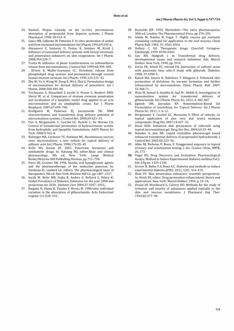
Histopathological examination of skin specimens
Abdominal skin of wistar rats were treated with optimized nanoemulsion gel. After 48 hr, rat was sacrificed and the skin samples from treated and untreated (control) area were taken. Each specimen was stored in 10% formalin solution in phosphate buffer saline (pH 7.4). The specimen was cut into section vertically. Each section was dehydrated using ethanol, embedded in paraffin for fixing and stained with hematoxylin and eosin. These samples were then observed under light microscope (Motic, Japan) and compared with control sample. In each skin sample, three different sites were scanned and evaluated for elucidation of mechanism of penetration enhancement.
Statistical Analysis
Data of in vivo analysis was expressed as mean of eight animal ± S.D. The data was compared for statistical significance by the one-way analysis of variance (ANOVA) followed by Tukey– Kramer multiple comparisons test using GraphPad Instat software (GraphPad Software Inc., CA, and USA). The data were considered to be significant at p<0.05.
RESULT AND DISCUSSION
Selection of Formulations from Phase Diagrams
Following criteria were chosen for the selection of formulations. GLBD was added to the mixture of oil, surfactant and cosurfactant with varying component ratio as described in Table-1,
and then appropriate amount of water was added to the mixture drop by drop and the nanoemulsion containing GLBD was obtained by vortexing the mixture at ambient temperature300C. Formulations with Smix ratio 1:0, 1:2 and 1:3 were not selected because the formulations were unstable and showed phase separation. Based on the phase diagrams, three Smix ratios 1:1 (NE-A), 2:1 (NE-B) and 3:1 (NE-C) were optimized. From the selected Smix ratios, NE compositions with 33 % (NE-A1, NE-B1, and NE-C1), 45 % (NE-A2, NE-B2, NE-C2) and 55 % (NE-A3, NE-B3, NE-C3) Smix ratios were selected from the region of existence pseudoternary phase diagram (Table-1)
Table 1: Composition of Selected Nanoemulsion Formulations
aSmix ratio Formulation Codeb Percent w/w of Components in Formulation Oil (%) Water (%) Smix (S + CoS) (%)
Formulation NE-A aSmix ratio = 1:1
NE-A1 10 57 33 NE-A2 15 40 45 NE-A3 18 27 55
Formulation NE-B Smix ratio = 2:1
NE-B1 10 57 33 NE-B2 15 40 45 NE-B3 18 27 55
Formulation NE-C Smix ratio = 3:1
NE-C1 10 57 33 NE-C2 15 40 45 NE-C3 18 27 55
a Surfactant/cosurfactant ratio;
bNE represents nanoemulsion; A, B and C represents Smix ratio 1:1, 2:1 and 3:1, respectively; Suffix 1,2 and 3 represents Smix concentration 33, 45 and 55 %, respectively
Ex-vivo Permeation Studies
The permeation ability of the selected NE formulations was evaluated by the ex-vivo permeation experiments. The permeation parameters of the tested NE formulations are presented in Figure 1.
The results demonstrate that the permeation rate and permeation coefficient of all the NE formulations through rat skin are significantly higher (P < 0.001) in comparison to control formulations D and E. The values of transdermal flux for different NE formulations were observed between 30.74 to 95.90 μg cm-2 h-1. This
Wais et al. Int J Pharm Pharm Sci, Vol 5, Suppl 4,747-754
750
value is approximately seven fold greater than the formulation D (12.88 ± μg cm-2 h-1), nearly nine fold higher than the formulation E (10.65μg cm-2 h-1). Although, all the formulations contain equal drug amount, low permeation rate as observed for formulations D and E, provided that concentration gradient is not a single factor affecting the rate of permeation. Enhanced permeation with NEs could be explained on basis of the several mechanisms:
Nano droplets settle down to close contact with the skin and a large amount of inner labrafac and triacetin carrying drug might penetrate into the skin.
Permeation of drug carrying nano-sized droplets through stratum corneum without NE fusion and subsequent drug release.
Continuous and spontaneous fluctuating stable interfaces of NE enable high drug mobility and might enhance the drug diffusion process.
High solubilization of GLBD in NE resulting in high thermodynamic activity of the drug providing significant driving force for its release and permeation.
Hydration of the stratum corneum due to external water phase of the NE results in high diffusivity of lipophilic drug as droplet size approaches to molecular dispersion.
The content of Smix in the nanoemulsion formulation was found to affect the skin permeation rate of GLBD directly. As the content of Smix increased from 33% to 55% (NE1 to NE3), the skin permeation increased from 45% then decreased up to 55 % Smix concentration and the overall flux enhancements were observed for Smix (2:1) NE-B formulations with the maximum flux (95.90 μg cm-2 h-1) for NE-B2, at 45 % Smix (tween 80 and diethylene glycol monoethyl ether) NE-B2, which contained 15 % labrafac and tricetin(1:1) and 40 % water (Fig 1). This might be due to a decreased thermodynamic activity of the drug in the nanoemulsion at the higher content of surfactant [52]. The thermodynamic activity of a drug in the formulation is a significant driving force for the release and penetration of the drug into the skin. In summary the different formulations can be ordered according to their descending flux values as follow: NE-B2 > NE-B3 > NE-B1 > NE-A1 > NE-A2 > NE-A3 > NE-C1 > NE-C2 > NE-C3 > formulation D > formulation E. Comparison of the permeation of the NE formulations with corresponding droplet size and viscosity indicated that NE-B2 with smallest droplet size and lowest viscosity had highest transdermal flux and therefore selected for in vivo permeation study for converting nanoemulsion gel.
Fig. 1: Permeation profiles of GLBD through excised rat skin from nanoemulsions formulated with Smix 1:1,2:1 , 3:1and control, surfactant, conventional gel and NEG (mean ± SD, n=3)
Skin Irritancy Test
Rat skin irritation experiments were, therefore, conducted in order to assess the potential irritant effects of the developed nanoemulsion formulation. The transdermal nanoemulsion formulation showed a skin irritation score (erythema and edema) of less than 2. According to Draize et al., compounds producing scores of 2 or less are considered negative (no skin irritation) 52. Hence, from the results of the preliminary skin irritation study, the optimized nanoemulsion formulation appeared to be safe for transdermal delivery.
Induction of Diabetes mellitus:
The overnight fasted Wistar rats weighing 150–220g were made diabetic by a single intravenously injection of streptozotocin (60 mg/kg) dissolved in citrate buffer (3 mM; pH 4.5) . After the dose of 60 mg/kg intravenously injection, symptoms occur already after 24–48 h with hyperglycemia up to 800 mg %, glucosuria and ketonemia.. A steady state is reached after 10–14 days allowing using the animals for pharmacological tests. The blood was taken from the tail vein at predetermined time intervals up to 48 hours. Blood glucose level was determined by Accutrend Alpha Glucometer (Roche Diagnostics, Germany).
Antidiabetic Activity
The optimized formulation which yielded satisfactory skin permeation result was subjected to in-vivo studies. The results of the
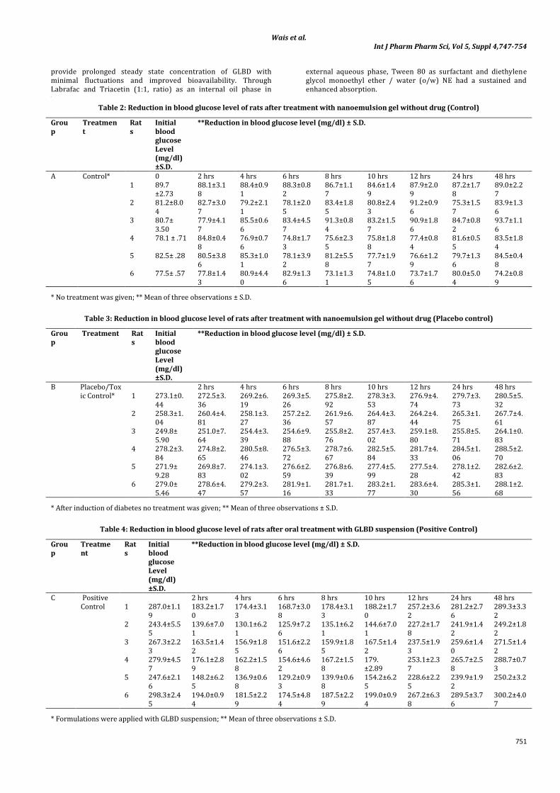
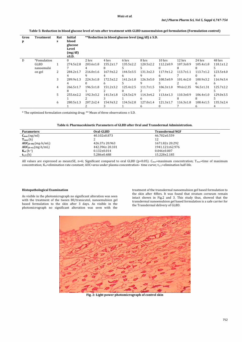
antidiabetic activity of transdermal nanoemulsion gel in comparison to placebo control, diabetic control, and control is shown in Table 2-6. Before the starting of experiment the average initial blood glucose levels of different groups were; control 81.61 mg/dl, toxic control 268.38 mg/dl, placebo control 270.58 mg/dl, formulation control 275.2 mg/dl.
The hypoglycemic effect was significant (P<0.01) in nanoemulsion gel treated animals in comparison to the control group animals. As the GLBD is an antidiabetic drug and the most common side effect of this is severe hypoglycemic reaction when administered by oral route. But here we can see in the case of transdermal treatment the reduction in blood glucose level was gradual and in initial hours of the treatment no severe hypoglycemic reaction was produced. There was no significance difference found in the blood glucose level of control, toxic control, placebo control group of animals during 48 hrs of study. But on the other side significant reduction in blood glucose level (P<0.05 compared to toxic control) was observed gradually in the transdermal formulation treated group of animals (group D). The maximum hypoglycemic response was observed at 6th hour of study (51.1% reduction in blood glucose level, P<0.05 in comparison to toxic control) and it remained stable up to 48 hrs of study. In initial hours of study there was not much but optimum reduction in blood glucose level was observed (22.85% at 2nd hour of study), but after that it increases gradually and from the 6th hour of the study it remains somewhat stable upto 48 hours. Whereas regarding the placebo control there was no reduction in blood glucose level found this shows that other ingredients of the formulation don’t have any type of blood glucose lowering property. Where as animals subjected to medicated transdermal nanoemulsion gel shows gradual decrease in blood glucose level in initial 6 hours (at 2nd hour 25.15%, at 4th hour 41.26%, and at 6th hour 51.1% decrease). This is clear from the observed data that in initial hour no severe hypoglycemic reactions were produced as it was most commonly observed in oral therapy of GLBD. From 6th hours unto 48th hour the reduction in blood glucose level was almost stable (55-61% decrease in blood glucose level). This shows that the transdermal formulation of GLBD avoids the side effects associated with its oral therapy, and on the other hand as diabetes is a chronic and long lasting manageable (not curable) disease, the formulation is effective for the 48 hours of time, so it increases the patient compliance by reducing the frequency of administration and also provides ease in application.
In Vivo Permeation Study
Pharmacokinetic study was carried out on rats to judge the efficacy of the developed formulation against the oral dosage form. The data so obtained was subjected to pharmacokinetic analysis shown in Fig.1. The mean Tmax of GLBD was 2 hrs for oral treatment and 12 hrs for transdermal treatment. A significant difference in Tmax value was observed between oral and transdermal treatment. A shift in the Tmax value towards higher side from transdermal treatment indicated the controlled release behavior of the formulation. The mean Cmax of GLBD was 40.102 ng/ml for oral treatment and 46.7 ng/ml for transdermal treatment. The Cmax values for both were almost similar, however in case of oral treatment the peak and valley pattern was quiet evident with the fluctuation in the plasma concentration whereas in transdermal treatment steady state plasma concentration level was maintained. The pharmacokinetic parameters were calculated from the plasma concentrations of the drug and recorded in Table.6 The mean AUC0-t values after oral treatment was 426.37 ng hr/ml and after transdermal treatment was 1671.82 ng.hr/ml. The significantly (p<0.05) high AUC0-t values observed with transdermal nanoemulsion gel also indicate increased bioavailability of the drug from nanoemulsion gel compared to oral administration. The difference in the AUC0-t
values clearly reflects that comparatively lesser amount of drug was available by oral administration to rat body because of high first pass metabolism of GLBD. Thus the transdermal nanoemulsion gel formulation in the present study was found to enhance the bioavailability of GLBD by 3.92 times with reference to an oral delivery of drug. The increased bioavailability might be due to elimination of hepatic first pass metabolism in transdermal delivery. Thus the transdermal formulation NGF was found to
Wais et al. Int J Pharm Pharm Sci, Vol 5, Suppl 4,747-754
751
provide prolonged steady state concentration of GLBD with minimal fluctuations and improved bioavailability. Through Labrafac and Triacetin (1:1, ratio) as an internal oil phase in
external aqueous phase, Tween 80 as surfactant and diethylene glycol monoethyl ether / water (o/w) NE had a sustained and enhanced absorption.
[
Table 2: Reduction in blood glucose level of rats after treatment with nanoemulsion gel without drug (Control)
Group
Treatment
Rats
Initial blood glucose Level (mg/dl) ±S.D.
**Reduction in blood glucose level (mg/dl) ± S.D.
A Control* 0 2 hrs 4 hrs 6 hrs 8 hrs 10 hrs 12 hrs 24 hrs 48 hrs 1 89.7
±2.73 88.1±3.18
88.4±0.91
88.3±0.82
86.7±1.17
84.6±1.49
87.9±2.09
87.2±1.78
89.0±2.27
2 81.2±8.04
82.7±3.07
79.2±2.11
78.1±2.05
83.4±1.85
80.8±2.43
91.2±0.96
75.3±1.57
83.9±1.36
3 80.7± 3.50
77.9±4.17
85.5±0.66
83.4±4.57
91.3±0.84
83.2±1.57
90.9±1.86
84.7±0.82
93.7±1.16
4 78.1 ± .71 84.8±0.48
76.9±0.76
74.8±1.73
75.6±2.35
75.8±1.88
77.4±0.84
81.6±0.55
83.5±1.84
5 82.5± .28 80.5±3.86
85.3±1.01
78.1±3.92
81.2±5.58
77.7±1.97
76.6±1.29
79.7±1.36
84.5±0.48
6 77.5± .57 77.8±1.43
80.9±4.40
82.9±1.36
73.1±1.31
74.8±1.05
73.7±1.76
80.0±5.04
74.2±0.89
* No treatment was given; ** Mean of three observations ± S.D.
Table 3: Reduction in blood glucose level of rats after treatment with nanoemulsion gel without drug (Placebo control)
Group
Treatment Rats
Initial blood glucose Level (mg/dl) ±S.D.
**Reduction in blood glucose level (mg/dl) ± S.D.
B Placebo/Toxic Control*
2 hrs 4 hrs 6 hrs 8 hrs 10 hrs 12 hrs 24 hrs 48 hrs 1 273.1±0.
44 272.5±3.36
269.2±6.19
269.3±5.26
275.8±2.92
278.3±3.53
276.9±4.74
279.7±3.73
280.5±5.32
2 258.3±1.04
260.4±4.81
258.1±3.27
257.2±2.36
261.9±6.57
264.4±3.87
264.2±4.44
265.3±1.75
267.7±4.61
3 249.8± 5.90
251.0±7.64
254.4±3.39
254.6±9.88
255.8±2.76
257.4±3.02
259.1±8.80
255.8±5.71
264.1±0.83
4 278.2±3.84
274.8±2.65
280.5±8.46
276.5±3.72
278.7±6.67
282.5±5.84
281.7±4.33
284.5±1.06
288.5±2.70
5 271.9± 9.28
269.8±7.83
274.1±3.02
276.6±2.59
276.8±6.39
277.4±5.99
277.5±4.28
278.1±2.42
282.6±2.83
6 279.0± 5.46
278.6±4.47
279.2±3.57
281.9±1.16
281.7±1.33
283.2±1.77
283.6±4.30
285.3±1.56
288.1±2.68
* After induction of diabetes no treatment was given; ** Mean of three observations ± S.D.
Table 4: Reduction in blood glucose level of rats after oral treatment with GLBD suspension (Positive Control)
Group
Treatment
Rats
Initial blood glucose Level (mg/dl) ±S.D.
**Reduction in blood glucose level (mg/dl) ± S.D.
C Positive Control
2 hrs 4 hrs 6 hrs 8 hrs 10 hrs 12 hrs 24 hrs 48 hrs 1 287.0±1.1
9 183.2±1.70
174.4±3.13
168.7±3.08
178.4±3.13
188.2±1.70
257.2±3.62
281.2±2.76
289.3±3.32
2 243.4±5.55
139.6±7.01
130.1±6.21
125.9±7.26
135.1±6.21
144.6±7.01
227.2±1.78
241.9±1.42
249.2±1.82
3 267.3±2.23
163.5±1.42
156.9±1.85
151.6±2.26
159.9±1.85
167.5±1.42
237.5±1.93
259.6±1.40
271.5±1.42
4 279.9±4.57
176.1±2.89
162.2±1.58
154.6±4.62
167.2±1.58
179. ±2.89
253.1±2.37
265.7±2.58
288.7±0.73
5 247.6±2.16
148.2±6.25
136.9±0.68
129.2±0.93
139.9±0.68
154.2±6.25
228.6±2.25
239.9±1.92
250.2±3.2
6 298.3±2.45
194.0±0.94
181.5±2.29
174.5±4.84
187.5±2.29
199.0±0.94
267.2±6.38
289.5±3.76
300.2±4.07
* Formulations were applied with GLBD suspension; ** Mean of three observations ± S.D.
Wais et al. Int J Pharm Pharm Sci, Vol 5, Suppl 4,747-754
752
Table 5: Reduction in blood glucose level of rats after treatment with GLBD nanoemulsion gel formulation (Formulation control)
Group
Treatment Rats
Initial blood glucose Level (mg/dl) ±S.D.
**Reduction in blood glucose level (mg/dl) ± S.D.
D *Fomulation GLBD nanoemulsion gel
0 2 hrs 4 hrs 6 hrs 8 hrs 10 hrs 12 hrs 24 hrs 48 hrs 1 274.5±2.8
7 203.6±1.84
155.2±1.78
135.5±2.25
120.5±2.25
112.2±0.90
107.3±0.98
105.4±1.88
118.1±1.25
2 284.2±1.74
216.0±1.69
167.9±2.24
144.5±3.56
131.3±2.39
117.9±1.26
113.7±1.17
113.7±1.20
123.5±4.04
3 289.9±1.34
224.3±1.80
172.5±2.26
141.2±1.85
126.3±5.09
108.5±0.95
101.4±2.02
100.9±3.26
116.9±3.46
4 266.5±1.78
196.5±1.81
151.2±3.22
125.4±2.50
111.7±1.52
106.3±1.07
99.6±2.35 96.5±1.31 125.7±2.25
5 255.6±2.21
192.3±3.22
141.5±1.83
124.5±2.91
114.3±4.26
113.6±1.32
110.3±0.98
106.4±1.08
129.0±3.55
6 280.5±1.31
207.2±2.42
154.9±3.23
134.5±2.81
127.0±1.46
121.3±1.70
116.3±1.87
108.4±1.56
135.3±2.44
* The optimized formulation containing drug; ** Mean of three observations ± S.D.
Table 6: Pharmacokinetic Parameters of GLBD after Oral and Transdermal Administration.
Parameters Oral-GLBD Transdermal NGF Cmax (ng/ml) 40.102±0.873 46.702±0.559 Tmax (h) 2 12 AUC(0-48) (mg-h/mL) 426.37± 20.963 1671.82± 20.292 AUC(0-∞) (mg-h/mL) 442.396± 20.101 1941.121±62.976 Kel (h-1) 0.132±0.014 0.046±0.007 t1/2 (h) 5.286±0.488 15.228±2.185
All values are expressed as mean±SE, n=6; Significant compared to oral GLBD (p<0.05). Cmax=maximum concentration; Tmax=time of maximum concentration; Kel=elimination rate constant; AUC=area under plasma concentration– time curve; t1/2=elimination half-life.
Fig. 1: Plasma concentration–time profile of Thus transdermal administration of GLBD
Histopathological Examination
As visible in the photomicrograph no significant alteration was seen with the treatment of the tween 80/transcutol, nanoemulsion gel based formulation to the skin after 3 days. As visible in the photomicrograph no significant alteration was seen with the
treatment of the transdermal nanoemulsion gel based formulation to the skin after 48hrs. It was found that stratum corneum remain intact shown in Fig.2 and 3. This study thus, showed that the transdermal nanoemulsion gel based formulation is a safe carrier for the Transdermal delivery of GLBD.
Fig. 2: Light power photomicrograph of control skin

Wais et al. Int J Pharm Pharm Sci, Vol 5, Suppl 4,747-754
753
Fig. 3: Light power photomicrograph of treated skin
CONCLUSION
An appropriate combination of the oil, surfactant, cosurfactant, and water is a major formulation consideration in nanoemulsion preparation for the transdermal drug delivery The GLBD loaded thermodynamically stable o/w NE system were prepared and various formulation factors were evaluated to find optimized formulation which shows desirable efficacy both ex-vivo and in vivo. The optimized formulation NE-B2, which contained labrafac and triacetin (1:1), (15 % w/w), tween 80 (30 % w/w), diethylene glycol monoethyl ether (15 % w/w) and water (30 % w/w) showed significant increase (P < 0.001) in the steady state flux (Jss) and permeability coefficient (Kp) compared to control or drug loaded neat components. The in vivo studies revealed a 3.92 fold increased relative bioavailability. From ex-vivo and in vivo data it can be concluded that the developed nanoemulsion systems are potential vehicles for transdermal delivery of GLBD for prolonged periods.
REFERENCES
1. Kim BS, Won M, Lee KM, Kim S. In vitro permeation studies of nanoemulsions containing ketoprofen as a model drug. Drug Del. 2008;15:465–9.
2. Shafiq S, Shakeel F, Talegaonkar S, Ahmad FJ, Khar RK, Ali M. Design and development of oral oil in water ramipril nanoemulsion formulation: in vitro and in vivo assessment. J Biomed Nanotech. 2007;3:1–17
3. Azeem A, Khan ZI, Aqil M, Ahmad FJ, Khar RK, Talegaonkar S. Microemulsions as a surrogate carrier for dermal drug delivery. Drug Dev Ind Pharm. 2009;35:525–47.
4. Azeem A, Rizwan M, Ahmad FJ, Khan ZI, Khar RK, Aqil M, et al. Emerging role of microemulsions in cosmetics. Recent Pat Drug Deliv Formul. 2008;2:275–89.
5. Heuschkel S, Goebel A, Neubert RHH. Microemulsions—modern colloidal carrier for dermal and transdermal drug delivery. J Pharm Sci. 2008;97:603–31.
6. El Maghraby GM. Transdermal delivery of hydrocortisone from eucalyptus oil microemulsion: effects of cosurfactants. Int J Pharm. 2008;355:285–92.
7. Shevachman M, Garti N, Shani A, Sintov AC. Enhanced percutaneous permeability of diclofenac using a new U-type dilutable microemulsion. Drug Dev Ind Pharm. 2008;34:403–12.
8. Huang YB, Lin YH, Lu TM, Wang RJ, Tsai YH, Wu PC. Transdermal delivery of capsaicin derivative-sodium nonivamide acetate using microemulsions as vehicles. Int J Pharm. 2008; 349:206–11.
9. Yuan JS, Ansari M, Samaan M, Acosta EJ. Linker-based lecithin microemulsions for transdermal delivery of lidocaine. Int J Pharm. 2008;349:130–43.
10. Biruss B, Kählig H, Valenta C. Evaluation of an eucalyptus oil containing topical drug delivery system for selected steroid hormones. Int J Pharm. 2007;328:142–51.
11. Biruss B, Valenta C. The advantage of polymer addition to a non-ionic oil in water microemulsion for the dermal delivery of progesterone. Int J Pharm. 2008;349:269–73.
12. Kamal MA, Iimura N, Nabekura T, Kitagawa S. Enhanced skin permeation of diclofenac by ion-pair formation and further enhancement by microemulsion. Chem Pharm Bull. 2007;55: 368–71.
13. Kantarci G, Ozgüney I, Karasulu HY, Arzi S, Güneri T. Comparison of different water/oil microemulsions containing diclofenac sodium: preparation, characterization, release rate, and skin irritation studies. AAPS Pharm Sci Tech. 2007;8:E91.
14. Yuan Y, Li SM, Mo FK, Zhong DF. Investigation of microemulsion system for transdermal delivery of meloxicam. Int J Pharm. 2006;321:117–23.
15. Lee PJ, Langer R, Shastri VP. Novel microemulsion enhancer formulation for simultaneous transdermal delivery of hydrophilic and hydrophobic drugs. Pharm Res. 2003;20:264–9.
16. Junyaprasert VB, Boonsaner P, Leatwimonlak S, Boonme P. Enhancement of the skin permeation of clindamycin phosphate by Aerosol OT/1-butanol microemulsions. Drug Dev Ind Pharm. 2007;33:874–80.
17. Talegaonkar S, Akhter S, Jain GK, Ahmad FJ, Khar RK, Jain N, et al. Investigation of nanoemulsion system for transdermal delivery of domperidone: ex-vivo and in-vivo studies. Curr Nanosci. 2008;4:381–90.
18. Ambade KW, Jadhav SL, Gambhire MN, Kurmi SD, Kadam VJ, Jadhav KR. Formulation and evaluation of flurbiprofen microemulsion.Curr Drug Deliv. 2008; 5:32–41.
19. Shakeel F, Baboota S, Ahuja A, Ali J, Aqil M, Shafiq S. Nanoemulsions as vehicles for transdermal delivery of aceclofenac. AAPS PharmSciTech. 2007;8:E104.
20. Zhao X, Liu JP, Zhang X, Li Y. Enhancement of transdermal delivery of theophylline using microemulsion vehicle. Int J Pharm. 2006;327:58–64.
21. Paolino D, Ventura CA, Nisticò S, Puglisi G, Fresta M. Lecithin microemulsions for the topical administration of ketoprofen: percutaneous adsorption through human skin and in vivo human skin tolerability. Int J Pharm. 2002;244:21–31.
22. Shakeel F, Baboota S, Ahuja A, Ali J, Shafiq S. Skin permeation mechanism and bioavailability enhancement of celecoxib from transdermally applied nanoemulsion. J Nanobiotech. 2008; 6:8.
23. Bolzinger MA, Briancon S, Pelletier J, Fessi H, Chevalier Y. Percutaneous release of caffeine from microemulsion, emulsion and gel dosage forms. Eur J Pharm Biopharm. 2008;68:446–51.
Wais et al. Int J Pharm Pharm Sci, Vol 5, Suppl 4,747-754
754
24. KtistisG, Niopas I.Astudy on the in-vitro percutaneous absorption of propranolol from disperse systems. J Pharm Pharmacol. 1998; 50:413–9.
25. Gasco MR, Gallarate M, Pattarino F. In vitro permeation of azelaic acid from viscosized microemulsions. Int J Pharm. 1991;69:193–6.
26. Abramović Z, Sustarsic U, Teskac K, Sentjurc M, Kristl J. Influence of nanosized delivery systems with benzyl nicotinate and penetration enhancers on skin oxygenation. Int J Pharm. 2008;359:220–7.
27. Trotta M. Influence of phase transformation on indomethacin release from microemulsions. J Control Rel. 1999;60:399–405.
28. . Kriwet K, Muller-Goymann CC. Diclofenac release from phospholipid drug systems and permeation through excised human stratum corneum. Int J Pharm. 1995;125:231–42.
29. Zhu W, Yu A, Wang W, Dong R, Wu J, Zhai G. Formulation design of microemulsion for dermal delivery of penciclovir. Int J Pharm. 2008;360:184–90.
30. Teichmann A, Heuschkel S, Jacobi U, Presse G, Neubert RHH, Sterry W, et al. Comparison of stratum corneum penetration and localization of a lipophilic model drug applied in an o/w microemulsion and an amphiphilic cream. Eur J Pharm Biopharm. 2007;67:699–706.
31. Kreilgaard M, Pedersen EJ, Jaroszewski JW. NMR characterization and transdermal drug delivery potential of microemulsion systems. J Control Rel. 2000;69:421–33.
32. Fini A, Bergamante V, Ceschel GC, Ronchi C, De Moraes CA. Control of transdermal permeation of hydrocortisone acetate from hydrophilic and lipophilic formulations. AAPS Pharm Sci Tech. 2008;9:762–8.
33. Bolzinger MA, Carduner TC, Poelman MC. Bicontinuous sucrose ester microemulsion: a new vehicle for topical delivery of niflumic acid. Int J Pharm. 1998;176:39–45.
34. Nolte MS, Karam JH. 2001. Pancreatic hormones and antidiabetic drugs. In: Katzung BG, editor.Basic and clinical pharmacology, 8th ed. New York: Lange Medical Books/McGraw-Hill Publishing Division, pp 711–734.
35. Davis SN, Granner DK. 1996. Insulin, oral hypoglycemic agents, and the pharmacotherapy of the endocrine pancreas. In: Hardman JG, Limbird LE, editors. The pharmacological basis of therapeutics, 9th ed. New York: McGraw-Hill Co., pp 1487–1517.
36. Sarah W, Bchir MB, Gojka R, Anders G, Richard S, Hilary K; Global Prevalence of Diabetes, Estimates for the year 2000 and projections for 2030 , Diabetes Care 2004;27:1047–1053,.
37. Ikegami H, Shima K, Tanaka Y, Hirota M. 1986.Inter individual variation in the absorption of glibenclamide. Acta Endocrinol Copehn 111:528–532.
38. Reynolds JEF. 1993. Martindale—The extra pharmacopoeia, 30th ed. London: The Pharmaceutical Press, pp 276–291.
39. Ishida M, Nambu N, Vagai T. Highly viscous gel ointment containing carbopol for application to the oral mucosa. Chem Pharm Bull. 1983; 31: 4561-4564.
40. Dollery C. Ed. Therapeutic drugs, Churchill Livington: Edinburgh; 1999. D196-D201.
41. Guy RH, Hadgraft J. In Transdermal drug delivery, developmental issues and research initiatives. Eds; Marcel Dekker: New York, 1990; pp. 59-8.
42. Gorus FK, Schuit FC, Intveld PA. Interaction of sulfonyl ureas with pancreatic beta cells-A study with gliburide. Diabetes. 1988; 37:1090-5.
43. Kamal MA, Iimura N, Nabekura T, Kitagawa S. Enhanced skin permeation of diclofenac by ion-pair formation and further enhancement by microemulsion. Chem. Pharm. Bull. 2007; 55:368-71.
44. Wais M, Samad A, Anubha K, Aqil M , Mohib K. Investigation of nanoemulsion system for transdermal delivery of glibenclamide .Int J Pharm Pharm Sci. 2012; 4: 482-487.
45. Jignesh DM, Jayvadan KP. Nanoemulsion-Based Gel Formulation of Aceclofenac for Topical Delivery. Int J Pharm Pharm Sci. 2011; 1: 6-12.
46. Bergamante V, Ceschel GC, Marazzita S. Effect of vehicles on topical application of aloe vera and arnica montana components. Drug Del. 2007;14:427–32.
47. Desai KGH. Enhanced skin permeation of rofecoxib using topical microemulsion gel. Drug Dev Res. 2004;63:33–40.
48. Namdeo A, Jain NK. Liquid crystalline pharmacogel based enhanced transdermal delivery of propranolol hydrochloride. J Control Rel. 2002;82:223–36.
49. Abbe NJ, Nicholas P, Boon, E. Exaggerated exposure in topical irritancy and sensitization testing. J. Soc. Cosmet. Chem., 1975, 26, 173.
50. Vogel HG. Drug Discovery and Evaluation: Pharmacological Assays; Method to Induce Experimental Diabetes mellitus.Vol.2, Edi-3,Pg.no. 1329-1330.
51. Grover N, Bafna P.A, Rana A.C. Diabetes and methods to induce experimental diabetes.IJPBS,: 2011, 1(4) : 414-419.
52. Shah VP. Skin penetration enhancers: scientific perspectives. In: Hsieh DS, editor. Drug permeation enhancement; theory and applications. New York: Marcel Dekker; 1994. p. 19–24.
53. Draize JH, Woodward G, Calvery HO. Methods for the study of irritation and toxicity of substances applied topically to the skin and mucous membranes. J Pharmacol Exp Ther. 1944;82:377–90.


![Acaadeemmiicc SSccii eenncess International Journal of ... · Drug displacement in the used bases was first determined [18] and the amount of IBU required was calculated.The prepared](https://img.dokumen.tips/doc/110x75/5ebfa06db13b693d1431c575/acaadeemmiicc-ssccii-eenncess-international-journal-of-drug-displacement-in.jpg)