Embed Size (px)
Citation preview

ABSTRACT
Title of Dissertation: IDENTIFICATION, LIFE HISTORY, AND ECOLOGY
OF PERITRICH CILIATES AS EPIBIONTS ON
CALANOID COPEPODS IN THE CHESAPEAKE BAY
Laura Roberta Pinto Utz, Doctor of Philosophy, 2003
Dissertation Directed by: Professor Eugene B. Small Department of Biology Adjunct Professor D. Wayne Coats Department of Biology and Smithsonian Environmental Research Center
Epibiotic relationships are a widespread phenomenon in marine, estuarine and
freshwater environments, and include diverse epibiont organisms such as bacteria,
protists, rotifers, and barnacles. Despite its wide occurrence, epibiosis is still poorly
known regarding its consequences, advantages, and disadvantages for host and
epibiont. Most studies performed about epibiotic communities have focused on the
epibionts’ effects on host fitness, with few studies emphasizing on the epibiont itself.
The present work investigates species composition, spatial and temporal
fluctuations, and aspects of the life cycle and attachment preferences of Peritrich
epibionts on calanoid copepods in Chesapeake Bay, USA. Two species of Peritrich

ciliates (Zoothamnium intermedium Precht, 1935, and Epistylis sp.) were identified to
live as epibionts on the two most abundant copepod species (Acartia tonsa and
Eurytemora affinis) during spring and summer months in Chesapeake Bay. Infestation
prevalence was not significantly correlated with environmental variables or
phytoplankton abundance, but displayed a trend following host abundance.
Investigation of the life cycle of Z. intermedium suggested that it is an obligate
epibiont, being unable to attach to non-living substrates in the laboratory or in the
field. Formation of free-swimming stages (telotrochs) occurs as a result of binary
fission, as observed for other peritrichs, and is also triggered by death or molt of the
crustacean host. Attachment success of dispersal stages decreased as telotroch age
increased, suggesting that colonization rates in nature may be strongly dependent on
intense production of telotrochs by the epibiont ciliates.
Laboratory experiments demonstrated that Z. intermedium colonizes equally
adult and copepodite stages of A. tonsa and E. affinis. The epibiont is also able to
colonize barnacle nauplii and a harpacticoid copepod, when these were the only living
host available, but fails to colonize non-crustacean hosts, such as the rotifer
Brachionus calyciflorus or polychaete larvae. When the epibiont could choose
between adults of A. tonsa and alternate hosts from the zooplankton community, it
always colonized preferentially its primary host, with only a few telotrochs attaching
to other crustaceans (barnacle nauplii and harpacticoid copepod), and to rotifer eggs,
suggesting that specific cues may be involved in host selection by this epibiotic
species.
IDENTIFICATION, LIFE HISTORY, AND ECOLOGY OF PERITRICH CILIATES AS EPIBIONTS ON CALANOID COPEPODS
IN THE CHESAPEAKE BAY
by
Laura Roberta Pinto Utz
Dissertation submitted to the Faculty of the Graduate School of the University of Maryland, College Park in partial fulfillment
of the requirements for the degree of Doctor of Philosophy
2003
Advisory Committee:
Professor Eugene B. Small, Co-Chair, Advisor Adjunct Professor D. Wayne Coats, Co-Advisor Dr. Marie H. Bundy Dr. Darcy J. Lonsdale Professor Michael R. Roman Professor Diane K. Stoecker Professor Stephen Wolniak

DEDICATION
To my husband Eduardo,
for his love, support, and advice during
the development of this project.
ii
ACKNOWLEDGMENTS
I would like to thank the following people and institutions for their help during
the development of this project, and also in other related areas of my life.
Dr. Wayne Coats, for the opportunity to develop this research at the
Smithsonian Environmental Research Center, for the advice, support and friendship,
and for the encouragement to continue pursuing my scientific interests with
enthusiasm.
Dr. Eugene Small, for the advice, support, and friendship throughout this
project, and for stimulating my interest in ciliate systematics. I also thank my other
Committee members Dr. Marie Bundy, Dr. Darcy Lonsdale, Dr. Michael Roman, Dr.
Diane Stoecker, and Dr. Stephen Wolniak for constructive suggestions, helpful advice
and encouragement to pursue this research.
My husband Eduardo Eizirik, for his love and constant support during this
project and over the years, and particularly for the great advice and help in
proofreading this dissertation, as well in the formatting of some of the included figures
and reference list.
iii

My parents, Hermilo and Stela, and my sister Silvia, for their love and support
throughout my life, which have been of fundamental importance for me to become
who I am.
My parents-in-law Cláudio and Marisa, and my sister-in-law Mariana for their
love and support during the development of this project, and in the last 10 years of my
life.
All my other family members, for the support and encouragement over the
years.
My friends and past and current colleagues at the Protistan Ecology
Laboratory, for their help and advice during the development of this research. In
particular, I thank Tamieka Armstrong, Yvan Bettarel, Sean Cooney, Michael
Goodison, Sarah Jardeleza, John Miller, Myung Gil Park, Suzanna Ribblett, and
Gabriela Smalley for helpful discussions related to this project.
Tim Maugel and Sarah Jardeleza for their help with the Scanning Microscopy.
Dr. John Clamp for his help with the identification of the epibiont and for
interesting discussions about Peritrich taxonomy.
Deborah Morrin-Norlund, and Lois Reid, for their friendship and
administrative help at several moments during the development of this research.
My friends Gila Bar-Gal, Marina Carreiro e Silva, Michele Diez, Anne Innis,
Tonya Rawlings, Emma Teeling, and Karen Yee for their help and support during the
development of this project.
I thank Yvan Bettarel, Eduardo Eizirik, Anne Innis, John Miller, and Tonya
Rawlings for critically reading and providing helpful suggestions to different portions
iv

of this dissertation. I am also grateful to Tonya Rawlings, Darrick Sparks, and Karen
Yee for helping me with sample collection.
The first four years of this work were supported by a fellowship from the
Coordenação de Aperfeiçoamento de Pessoal de Nível Superior (CAPES), Brazil.
Additional support was provided by the Smithsonian Institution, and the Department
of Biology, University of Maryland, College Park. Support to attend scientific
meetings during the development of this project was provided by the University of
Maryland, College Park, and the Smithsonian Environmental Research Center.
v

TABLE OF CONTENTS
List of Tables …………………………………………………………………. viii List of Figures………………………………………………………………… ix Chapter 1. Introduction to Dissertation 1
Overview of epibiosis…………………………………………... 2Implications of an epibiotic relationship………………………... 2Epibiosis at an ecosystem level…………………………………. 6Epibiosis in the Chesapeake Bay……………………………….. 7Techniques applied in peritrich identification………………….. 8The genus Zoothamnium……………………………………….. 11Scope of the research………………………………………….. 13
Chapter 2. Morphological characterization of Zoothamnium intermedium Precht, 1935 and Epistylis sp. (Ciliophora, Peritrichia) attached to calanoid copepods in the Chesapeake Bay………………………………....... 16
Abstract…………………………………………………….…… 17Introduction……………………………………………….…….. 18Materials and Methods………………………………………….. 21Results…………………………………………………………... 24Discussion………………………………………………………. 30Tables…………………………………………………………… 36Figures………………………………………………………….. 43
Chapter 3. Spatial and temporal patterns in the occurrence of peritrich ciliates as epibionts on calanoid copepods in the Chesapeake Bay………… 50
Abstract…………………………………………………….….… 51Introduction……………………………………………….…….. 52Materials and Methods………………………………………….. 54Results ………………………………………………………….. 56Discussion………………………………………………………. 61Tables…………………………………………………………… 68Figures…………………………………………………………... 71
Chapter 4.Telotroch formation, survivorship, attachment success, and growth of the epibiotic peritrich Zoothamnium intermedium Precht, 1935.. 80
Abstract…………………………………………………….… …… 81Introduction……………………………………………….…….. 83
vi

Materials and Methods………………………………………….. 86Results…………………….………………………………….… 96Discussion……………………………………………………... 101Figures………………………………………………………….. 111
Chapter 5. Attachment patterns of the epibiotic peritrich Zoothamnium intermedium Precht, 1935…………………………………………………….. 122
Abstract………………...……………………………………….. 123Introduction…………………………….……………………….. 125Materials and Methods…………………...……………………... 126Results…………………………………………………………... 133Discussion…………………...………………………………….. 136Figures…………………………………………………………... 142
Chapter 6. General Discussion………………………………………………. 149Glossary……………………………………………………………………….. 159List of References……………………………………………………………... 162
vii

LIST OF TABLES
Page
2.1. Measurements of field, Protargol stained colonies of Zoothamnium intermedium attached to Acartia tonsa and Eurytemora affinis ………………… 362.2. Number of annular ridges on the membrane of Zoothamnium intermedium attached to Acartia tonsa and Eurytemora affinis….………………………………………………………..……………… 372.3. Measurements of cultured live colonies of Zoothamnium intermedium attached to Acartia tonsa and Eurytemora affinis………………………..…………………...……………………………….. 382.4. Measurements of cultured, Protargol stained colonies of Zoothamnium intermedium attached to Acartia tonsa and Eurytemora affinis…....………………………………………………………….………….. 392.5. Measurements of live telotrochs from cultured Zoothamnium intermedium attached to Acartia tonsa and Eurytemora affinis ………...…….. 402.6. Measurements of field, Protargol stained colonies of Epistylis sp. attached to Acartia tonsa…....…………………………………………………………... 412.7. Morphological comparison between Zoothamnium intermedium and other similar species in the genus Zoothamnium……………………………………... 423.1. Latitude and longitude of stations where host abundance (Chesapeake Bay Program) and infestation prevalence were assessed……………………… 683.2. Total number of A. tonsa and E. affinis infested by Z. intermedium and A. tonsa infested by Epistylis sp. during the study period……………………… 693.3. Correlation coefficient and p values obtained from Spearman Rank Correlation analyses for host abundance, environmental variables and infestation prevalence of Zoothamnium intermedium attached to Acartia tonsa and Eurytemora affinis and Epistylis sp. attached to A. tonsa between 1994-1996 and 1999-2000 in the Chesapeake Bay…………………………………... 70
viii
LIST OF FIGURES
Page 2.1. Photomicrographs of Zoothamnium intermedium attached to A. tonsa and E. affinis from life and after Protargol staining ……………...………………... 462.2. Morphology of Zoothamnium intermedium epibiont on Acartia tonsa and Eurytemora affinis from life and after Protargol staining……………………... 472.3. Scanning Electron Micrographs of Zoothamnium intermedium attached to Acartia tonsa and Eurytemora affinis………………………………………….. 482.4. Photomicrographs after Protargol and morphology of Epistylis attached to A. tonsa………………………………………………………………… ……... 493.1. Map of Chesapeake Bay showing stations where infestation prevalence was estimated from, and routine stations sampled by the Chesapeake Bay Program used to estimate host abundance…………..…………………………. 733.2. Infestation prevalence of A. tonsa and E. affinis relative to salinity and temperature profiles……………….…………………………………………… 743.3. Infestation Prevalence of Z. intermedium and Epistylis sp. attached to A. tonsa and E. affinis in a temporal and spatial distribution……………………………………………………………………... 753.4. Mean infestation prevalence of Zoothamnium intermedium attached to A. tonsa and E. affinis, and mean abundance of A. tonsa and E. affinis between 1994-1996 and 1999-2000 along the main axis of the Chesapeake Bay.…………………………………………………………………………….. 763.5. Mean infestation prevalence of Z. intermedium and Epistylis sp. at different categories of A. tonsa abundance………………………………….…. 773.6. Infestation density, mean number of zooids per colony, and infestation load of Z. intermedium and Epistylis sp. attached to adults and copepodites of A. tonsa and E. affinis.…………………………………………………………. 783.7. Infestation density, mean number of zooids per colony, and infestation load of Z. intermedium and Epistylis sp. attached to cephalothorax, abdomen, antenna, and swimming legs of A. tonsa and E. affinis.…………………………………………………………………..……… 794.1. Telotroch formation of Zoothamnium intermedium following the death of the host..…………….………………………………………………………….. 1144.2. Telotroch formation of Z. intermedium attached to tethered Acartia tonsa compared to telotroch formation on dead A. tonsa…...………………………... 1154.3. Summary of the results obtained for all experiments inducing telotroch formation on the peritrich epibiont Zoothamnium intemrmedium attached to Acartia tonsa. ……………………………………………………………………………….. 1164.4. Survivorship of telotrochs of Zoothamnium intermedium in absence of living host...………………….………………………………………………… 1174.5. Attachment of peritrich ciliates to cover slips and copepods in the field…………………………………………………………………………….. 118
ix

4.6. Attachment success of telotrochs of Zoothamnium intermedium to Acartia tonsa……………………………………………………………………………. 1194.7. Growth, colonization and proliferation rates of Zoothamnium intermedium attached to Acartia tonsa……………….………………………... 1204.8. Concentration of Isochrysis galbana observed throughout the growth rate experiment…………………………………………………………………...… 1215.1. Number of zooids of Zoothamnium intermedium per unit of area of primary hosts Acartia tonsa and Eurytemora affinis……………...…………… 1445.2. Growth of Zoothamnium intermedium attached to Acartia tonsa and Eurytemora affinis……………………………………………………………... 1455.3. Ability of Zoothamnium intermedium telotrochs to colonize Acartia tonsa, Eurytemora affinis and alternate hosts from the zooplankton community…...….……………………………………………………………... 1465.4. Number of zooids of Zoothamnium intermedium per unit area of alternate and primary hosts……………………..………………………………………... 1475.5. Percentage of zooids of Zoothamnium intermedium lost, and attached to a host during the experiment of preferential attachment to Acartia tonsa and alternate hosts from the zooplankton community……………………………………………………………………... 148
x
CHAPTER 1
GENERAL INTRODUCTION
1

Overview of Epibiosis
Epibiotic relationships involving planktonic crustacea are a widespread
phenomenon in marine, estuarine and freshwater environments, and encompass a
variety of epibiont organisms including protists, bacteria, hydrozoa, barnacles, and
rotifers (Fenchel, 1965; Green, 1974; Fenchel and Finlay, 1989; Abelló and
Macpherson, 1992; Iyer and Rao, 1995; Hanamura, 2000; Gilbert and Schröder, 2003;
Song, Al-Rasheid, and Hu, 2003). In spite of its wide occurrence, epibiosis remains
poorly understood with respect to its ecological implications for both the host and the
epibiont. Most studies of crustacean epibiosis have been performed in freshwater
systems, with very few focusing on marine and estuarine environments (Carman and
Dobbs, 1997). In general, these studies have stressed aspects of the ecology and life
history of the host, with less emphasis on the epibiont. Examination of species
composition, abundance, and life history of epibionts in aquatic environments is
recognized as an important tool to better understand seasonal occurrence of epibiosis,
host-epibiont population dynamics, and epibiont substrate specificity (Fenchel, 1965;
Wahl, 1989; Threlkeld, Chiavelli, and Willey, 1993; Carman and Dobbs, 1997;
Hanamura, 2000). However, most prior reports of crustacean epibiosis have not
considered the relationship from the perspective of the epibiont.
Implications of an epibiotic relationship
Historically, epibiosis has been viewed as a commensal relationship between
two or more organisms; however, several studies have suggested that epibionts can
2

have a deleterious effect on the host. For example, Kankaala and Eloranta (1987)
found that the cladoceran Daphnia longispina and its epibiont Vorticella sp., a
peritrich ciliate, grazed on particles within the same size-range and suggested that the
epibiont and host competed for food. Since Vorticella sp. had 50-80% higher feeding
rates than D. longispina, the epibiont seemed to have a competitive advantage that
might reduce host fitness. Similarly, Xu and Burns (1991) reported that, under food
limiting conditions, the peritrich ciliate epibiont Epistylis daphniae reduced
reproduction, growth, and survivorship of its host, the freshwater copepod Boeckella
triarticulata. The observed effects may reflect an additional energy cost for the host,
which was more pronounced in a food-depleted environment.
Weissman, Lonsdale, and Yen (1993) recorded slower sinking rates for the
copepod Acartia hudsonica when infested by the solitary peritrich Rhabdostyla sp.,
suggesting that epibiont burden may increase drag forces, thereby hindering
locomotion and increasing energy expended by the host. In addition to reducing host
swimming speed, epibionts may increase host visibility and thus susceptibility to
predation. For example, Willey, Cantrell, and Threlkeld (1990) observed that
cladocerans and copepods carrying the euglenoid epibiont Colacium calvum were
selectively preyed upon by visually-oriented fish (Menidia beryelina) and pump-filter
feeders (Dorosoma cepediarum and Tilapia aurea). In a similar study, Chiavelli,
Mills, and Threlkeld. (1993) reported that planktivory by young fish (Perca flavescens
and Dorosoma cepedianum) was associated with a decline of Daphnia populations
(including D. pulex and D. galeata mendotae) infested by the epibionts Colacium
calvum, Colacium vesiculosum and Synedra cyclopum.
3

Detailed information about epibionts can also provide important insights into
the biology and ecology of host organisms. For example, Abelló and McPherson
(1992) used the age of epibiont hydrozoan colonies (Stegopoma plicatile) and
individual barnacles (Poecilasma kaempferi) to show that adult males of the anomuran
crab Lithodes ferox have a shorter intermolt period than females. In a similar study,
Gili, Abelló, and Villanueva (1993) used growth rates of the epibiont barnacle
Poecilasma kaempferi, and the hydroid Stegopoma plicatile to determine carapace age
and intermolt duration of the crab Bathynectes piperitus.
When colonizing living substrates, epibionts have to adapt to morphological
fluctuations of the host such as growth, and molting, and be able to form motile stages
and seek new substrates when these changes occur (Wahl, 1989). In the case of
crustacean hosts, epibionts must be able to leave their host and re-colonize the same or
another substrate, after each molt. Also, reproduction of the epibiont often occurs
while it is attached to the host’s carapace, resulting in a positive burden increase
associated to host’s intermolt time. Willey and Threlkeld (1995) demonstrated that the
epibiotic burden of Colacium calvum and Vorticella campanula on Daphnia galeata
mendotae and Daphnia pulex, respectively, increased linearly and could provide
useful information about the intermolt time of the host. In addition, they observed that
V. campanula produced free-swimming stages immediately before its host started the
molting process, and suggested that this behavior allows the epibiont to re-colonize the
same substrate right after the host’s molt. The same pattern was not observed in C.
calvum, which exhibited a lag time between host’s molt, formation of free-swimming
stages, and re-colonization. The behavior observed in V. campanula could be
4

attributed to possible chemical stimulus released by the host before the molt and
detected by the epibiont as a signal to form telotrochs.
As mentioned previously, few studies have focused on the epibiont aspect of
the epibiotic relationship, yet available information does provide glimpses of
interactions that can influence epibiont populations. When more than one species of
epibiont colonize a host, competition for space and/or resources can occur. This
appears to be the case for caprellid amphipods (Caprella andreae) and barnacles
(Chelonibia testudinaria) associated with loggerhead sea turtles (Caine 1986). On
hosts with high densities of epibionts, amphipods appeared to have a negative impact
on the barnacles, preying upon newly settled barnacle spats. Competition among
conspecific epibionts may also occur, as suggested by Reynoldson’s (1950)
observation that the average cell size of peritrich epibionts (Urceolaria mitra)
decreased when hosts (the flatworm Polycelis tenuis) were heavily infested. In other
cases, epibiosis may provide the epibiont with a refuge from predation, as
demonstrated for the rotifer Brachionus rubens. When living on Daphnia carinata, B.
rubens is less susceptible to predation by the carnivorous rotifer Asplanchna
intermedia (Iyer and Rao 1995). Other biotic and abiotic factors may also affect
epibiont success. For example, fluctuations in rainfall and bacterial concentrations are
reflected by parallel changes in populations of Urceolaria mitra associated with
Polycelis tenuis (Reynoldson 1955).
Several authors have raised the issue of host specificity when considering
epibiosis. In some instances, epibionts appear specific to only one or two host species
and sometimes fail to attach to non-living substrates, as reported for Lagenophrys
5

lunatus attached to shrimps in the genus Palemonetes (Clamp, 1973), for Vorticella
microstoma colonizing Scapholeberis kingi (Henebry and Ridgeway, 1979), and for
Epistlylis pygmaeum colonizing Brachionus calyciflorus and Brachionus angularis
(Gilbert and Schröder, 2003). In contrast, other epibionts (e.g. Lagenophrys denisi)
are able to colonize living host species (e.g. the crayfish Cambarellus patzcuarensis),
as well as non-living substrates (Mayén-Estrada and Aladro-Lubel, 2000). The
assessment of host specificity may help in understanding the seasonal occurrence of
epibiosis in aquatic environments, encouraging further studies about the epibiont’s life
cycle, such as occurrence of encystment when the specific host is not available.
Epibiosis at an ecosystem level
Epibiont populations may play relevant roles at the ecosystem level. For
example, Baldock (1986) estimated that the biomass of the peritrichs Opercularia
spp., Epistylis breviramosa, E. racemosa and Vorticella campanula associated with
Trichoptera larvae (Brachycentrus subnubilus) exceeded that of free-living ciliates in
similar environments. When abundant, epibionts may even help fuel higher trophic
levels. Interestingly, the algal epibiont Korshicoviella gracilipes on Daphnia
pulicaria forms free-swimming, dispersal, and unattached overwintering stages that
can represent an important food source for the host when more typical planktonic prey
is at low abundance levels (Barea-Arco, Perez-Martinez, and Morales-Barquero 2001).
Henebry and Ridgeway (1979) reported that the prevalence of peritrich and
suctorian epibionts on crustacean zooplankton reflected the eutrophic condition of
their study site, raising the possibility that protistan epibiosis might be used as a
6

bioindicator for water quality. Commensalism between ciliates and invertebrate hosts
has been suggested before as an indicator of water pollution. For example, Antipa
(1977) demonstrated that the obligatory commensal ciliates Conchophtirus curtus and
Heterocinetopsis uniondarum could disappear from their bivalve hosts Lampsilis
ventricosa and Anodonta grandis when exposed to sewage contamination. In spite of
this, the occurrence of epibiosis has not so far been included in the assessment of
water pollution or in the establishment of water quality indices.
Epibiosis in the Chesapeake Bay
The only account of ciliate epibionts associated with zooplankton of
Chesapeake Bay is that of Herman and Mihursky (1964), who reported Zoothamnium
sp., a stalked, colonial peritrich, as a common epibiont on the most abundant calanoid
copepod, Acartia tonsa, in the Patuxent River (a subestuary of the Chesapeake Bay).
Infestation rates on A. tonsa reached 100% from early March to mid-April, and
apparently only this copepod species, among the zooplankton community, was found
carrying epibionts. In addition, partial examination of samples collected in the
previous year in the same months (March and April) revealed that Zoothamnium was
present on A. tonsa, but infestation rates were not quantified, and no other details were
provided. Even for the focal year of the study, the analysis was limited to only one
station in the Patuxent River on three sampling dates.
Although it was observed in that study that Zoothamnium only infested A.
tonsa even when this was not the most abundant species in the zooplankton
community, no further investigation about host specificity or ability to colonize other
7
substrates was performed. Also, the authors mentioned that most of the epibionts were
attached to the appendages, but no quantitative approach was taken to verify density of
colonies on different body parts or preferential colonization of a determined site. In
addition, no attempt was made to identify the epibiont to species level, or to survey the
distribution of epibiosis throughout the Chesapeake Bay and across life history stages
of the copepod. Higher sinking rates for dead infested vs. non-infested fixed copepods
were recorded by Hermann and Mihursky (1964), suggesting that the peritrichs might
put hosts at a disadvantage in the natural environment, however, no further
experiments to identify other possible deleterious effect of epibionts on copepods were
carried out. The lack of detailed information about identity, biology and ecology of
the epibiont, and potential deleterious effects on the host, limits the conclusions that
can be drawn from this initial study about occurrence of epibiosis in the Chesapeake
Bay, as well as any comparison with the occurrence of epibiosis in other aquatic
systems.
Techniques applied in peritrich identification
An important consideration when working with peritrich epibionts is careful
identification of species. Since identification of peritrich species can be difficult and
time consuming, epibionts are often characterized only to the genus level, as is the
case for the peritrich associated with copepods in Chesapeake Bay (Herman and
Mihursky, 1964). In some instances, even generic identifications are questionable,
due to insufficient or inappropriate examination of specimens.
8

Identification of peritrich species is typically based on light microscopic
examination of living specimens (e.g. Kahl, 1935; Precht, 1935; Fauré-Fremiet, 1930).
However, some recent studies of free living and epibiont peritrichs have demonstrated
that techniques such as cytological staining and scanning electron microscopy are also
valuable approaches (e.g. Bauer-Nebelsick, Bardele, and Ott, 1996; Green and Shiel,
2000). Cytological techniques like Protargol silver staining reveal cortical characters
such as kinetosomes, ciliary and cortical microtubules, nuclei and mitochondria
(Zagon and Small, 1970; Montagnes and Lynn, 1987). In peritrichs, unlike most other
ciliates, body ciliature is highly reduced and cannot be used to differentiate genera
and/or species. On the other hand, oral structure has proven to be conserved within
species, thus being useful for identification. For example, Zagon and Small (1970)
showed with Protargol staining that the oral structure of Carchesium polypinum was
consistent within the population of a polluted stream in the United States and was the
same as a population of the same species found in Europe, suggesting that this
morphological character may be suitable for identification of peritrichs in general, as
has been suggested by Lom (1964). Gross (1986) was able to distinguish four
different species in the genus Zoothamnium based on the structure of the oral
apparatus stained with Protargol combined with morphological characteristics of the
living and stained colony, demonstrating the importance of the oral structure for
peritrich identification.
Topography of the cell, shape of the peristome and features of the scopula are
characteristics that have also been used for peritrich species descriptions and can be
better observed by utilizing scanning electron microscopy (SEM). Even though the
9

majority of peritrich species has been described using morphological characters from
living cells, a growing number of recent studies have used SEM as an additional tool
to observe such characteristics in the sessile and motile stages of peritrichs. These
observations may be useful to when comparing different stages in the life cycle and
identifying peritrichs to species level. For instance, Valbonesi (1989) used SEM to
study morphological characteristics of the peritrich Zoothamnium intermedium, an
epibiont on Acartia clausi, and could distinguish two different free-swimming
morphotypes, which he tentatively identified as microgamont and telotroch forms.
Even though it is not clear from this study whether the two observed forms are
different morphotypes or only stages of telotroch formation, the use of SEM was
shown to provide relevant information for detailed morphological studies of peritrichs.
Despite the use of light microscopy, staining techniques and/or scanning
microscopy, the majority of studies of epibiotic relationships involving crustacea have
failed to identify the precise species of ciliate epibionts, especially peritrichs (e.g.
Foster, Sarphie, and Hawkings, 1978; Nagasawa, 1986). Identification of epibionts to
species level allow detailed studies from the epibiont’s perspective, including species
composition, temporal and spatial distributions of epibionts and dynamics of their
natural populations. Moreover, it would facilitate comparison of epibiont species
among different aquatic environments, as well as the investigation of the occurrence of
the same species as free-living versus epibiont forms in different systems.
10

The genus Zoothamnium
The peritrich genus Zoothamnium was first described by Bory de St. Vincent in
1826. There are approximately 50 described species belonging to this genus (Corliss,
1979), which is characterized by a colonial form and arboroid appearance. In general,
the colony possesses a continuous myoneme that runs from the basal stalk to the
branches, enabling the organism to contract as a single unit. Due to this diagnostic
characteristic, and to the presence of a continuous membrane connecting all the cells
in the colony, the genus Zoothamnium is thus recognized to be a coenobium and not a
simple colony like other colonial genera in the order Sessilina, and is now placed in a
separate Family (Zoothamnidae) within the SubClass Peritrichia (Lynn and Small,
2000). Even though the cellular organization of the species in the genus Zoothamnium
would be more accurately described as a coenobium, to conform to the prevalent
usage in the current and old literature, the terms “coenobium” and “colony” will be
used as synonyms throughout this dissertation when referring to this genus.
Two kinds of coenobial organization can be identified in species within the
genus Zoothamnium: (i) coenobia where all zooids are capable of metamorphosing
into free-swimming stages (telotrochs), and (ii) colonies where zooids have different
sizes (macro- and microzooids) and one type of zooid (macrozooids, or, at times,
specialized microzooids) is capable of forming dispersal stages (telotrochs). This
second type is called heteromorphic. The coenobial development of a heteromorphic
species in the genus Zoothamnium was first comprehensively described by Fauré-
Fremiet, (1930) who investigated Z. alternans. He observed that the colony is
composed by axial macrozooids and several lateral microzooids. The axial
11
macrozooids would be responsible for the formation of free-swimming stages, that he
called “ciliospores”, and the microzooids would be involved in the nutrition of the
colony and in the formation of sexual stages (microgamonts). A few years later,
Summers (1938) also investigated the development of several colonies of Z. alternans
and suggested that there are at least four types of zooids in a single colony: (i) a single
apical macrozooid, which is characterized by the bigger size and position within the
colony; (ii) several microzooids that are involved in the nutrition of the colony; (iii)
terminal microzooids at the tip of each branch that could give rise to microgamonts;
and (iv) axial microzooids that can foster telotroch stages, which can transform into a
new colony.
In species in the genus Zoothamnium where all zooids have the potential to
form telotrochs, no size difference is observed in zooids within the colony, and the
coenobial development is similar to other colonial genera such as Carchesium and
some species in the genus Epistylis. The only difference is the division of the shared
branched myoneme, as well as zooid extension into the stalk, which occurs in the
genus Zoothamnium but is not observed in the contractile colonial genus Carchesium
where the myoneme is not continuous throughout the branches. Nagasawa (1987)
described the coenobial development of an unidentified species in the genus
Zoothamnium attached to a calanoid copepod. Despite the lack of species level
identification, she was able to observe that all the zooids in the coenobium were
approximately of the same size and potentially capable of forming telotrochs. The
development of a new coenobium starts with the settlement of a telotroch that attaches
to the substrate, starts to secrete a stalk, transforms into an adult zooid, and undergoes
12
binary fission (Succhard, 1979). After the division is completed the new daughter
cells will remain in the same stalk and will elongate until reaching the normal
vegetative size and shape of the organism. Only after that the common stalk and
myoneme begin to divide forming a new colony. Examples of species that possess
this kind of colony development include Zoothamnium intermedium Precht, 1935; and
Zoothamnium arbuscula. For this latter species see especially Succhard, 1979.
Scope of the Present Research
Most of the prior work on ciliate epibionts of planktonic crustacea has been limited to
short-term observations of natural populations (with few exceptions, e.g. Green,
1974; Chiavelli, Mills, and Threlkeld, 1993) and has largely emphasized possible
effects of epibiosis on host organisms. Few studies have considered important
aspects of epibiont biology, such as species composition, abundance, life history,
morphology, and attachment mechanisms. In an attempt to address these issues, the
present study aims to: (i) identify and characterize species of peritrich epibionts
living on Chesapeake Bay copepods using live observations, Protargol staining, and
SEM techniques; (ii) determine the prevalence and distribution of peritrich
epibionts on planktonic crustacea along the main axis of the Chesapeake Bay; (iii)
examine relationships among epibiont prevalence, host abundance and
environmental variables; (iv) characterize the biology and life history of Z.
intermedium; and (v) assess substrate specificity of Z. intermedium.
In Chapter 2, I characterize morphologically (using live observations, Protargol
staining and Scanning Electron Microscopy) Zoothamnium intermedium, a peritrich
13

ciliate attached to populations of Acartia tonsa and Eurytemora affinis from the
Chesapeake Bay. I also provide a morphological characterization (using Protargol
staining) of Epistylis sp., another peritrich attached to A. tonsa. I present a
comprehensive redescription of Z. intermedium and comparisons with other
described species in the genus Zoothamnium.
In Chapter 3, I investigate the spatial and temporal distribution of Z. intermedium and
Epistylis sp. as epibionts on calanoid copepods in the Chesapeake Bay, and perform
correlation analyses between infestation prevalence and host abundance relative to
environmental variables. I also provide data on the distribution of epibiont colonies
on different body parts and life history stages of the hosts.
In Chapter 4, I explore different aspects of the life cycle of Z. intermedium including
(i) formation, survivorship and attachment success of telotroch stages, (ii) growth
and development of the coenobium, and (iii) the ability of Z. intermedium to attach
to non-living substrates in the field. I also provide comparisons of biological
aspects observed in the epibiont with other free-living peritrichs.
In Chapter 5, I assess the ability of Z. intermedium to colonize alternate hosts from the
zooplankton community, as well as test for preferential attachment to different life
history stages of its primary hosts (A. tonsa and E. affinis), and for preferential
attachment to A. tonsa when exposed to alternate hosts. In addition, I explore
predatory relationships between hosts and free-swimming stages of Z. intermedium,
and compare my results with those found for other epibiont species.
14

In Chapter 6, I review the main results described in chapters 2, 3, 4, and 5, and discuss
the potential relevance and impacts of these findings for the study of epibiosis in
aquatic environments, particularly those addressing estuarine ecosystems.
15

CHAPTER 2
MORPHOLOGICAL CHARACTERIZATION OF ZOOTHAMNIUM
INTERMEDIUM PRECHT, 1935 AND EPISTYLIS SP. (CILIOPHORA,
PERITRICHIA) ATTACHED TO CALANOID COPEPODS IN THE
CHESAPEAKE BAY, USA
16

ABSTRACT
I have more completely characterized morphologically Zoothamnium
intermedium Precht, 1935, a peritrich epibiont on the copepods Acartia tonsa and
Eurytemora affinis in the Chesapeake Bay, and Epistylis sp colonizing populations of
A. tonsa in the same ecosystem. The colonial sessile Z. intermedium possesses a bell
shaped zooid that ranges in size in vivo from 31.2 to 54.7 µm x 16.7 to 31.3 µm. The
cell body has a single contractile vacuole that empties its contents into the
infundibulum. A “C shaped” macronucleus lies in the upper half of the body.
Colonies of Z. intermedium can have up to 30 zooids per main stem stalk, but most of
them have 2-4 zooids. The oral ciliature, revealed by the Protargol method, consists of
an outer haplokinety and an inner polykinety that performs about 1 ½ turns around the
peristomial disk before entering the infundibulum. Three infundibular polykineties,
each possesses three rows of kinetosomes, were identified. Scanning electron
microscopy shows an annular pattern of ridges and, between ridges, scattered
irregularly distributed pores on the zooid cell membrane. Epistylis sp. has a “C
shaped” macronucleus that lies close to the peristomial opening. The oral ciliature is
similar to Z. intermedium, but the haplo- and polykineties perform 1 ¼ turns before
entering the infundibulum. Three oral polykinetids were identified each consisting of
three rows of kinetosomes. Morphological comparisons between specimens of Z.
intermedium attached to A. tonsa and E. affinis, are presented along with comparisons
with other described species in this genus.
17

INTRODUCTION
Sessile peritrich ciliates are commonly found attached to a variety of non-
living and living substrates in fresh, brackish and marine habitats. Living hosts (i.e.
basibionts) often constitute a suitable attachment site for peritrichs and other sessile
organisms since the movement of the substrate organism can supply the epibionts
with nutrients and facilitate removal of waste material (Felgenhauer and Schram,
1978; Wahl, 1989). Crustacea are the most common basibionts for various species
of peritrichs, and several studies have been focused on this relationship involving
planktonic or benthic hosts (e.g. Fenchel, 1965; Clamp, 1973; 1988; 1994; Green,
1974; Foster, Sarphie, and Hawkings, 1978; Nagasawa, 1986; Green and Shiel,
2000)
Despite the well-documented occurrence of peritrichs as epibionts on
planktonic crustacea in a variety of aquatic environments (Foster, Sarphie, and
Hawkings, 1978; Nagasawa, 1986; Willey and Trelkeld, 1993; Weissmann,
Lonsdale and Yen, 1993; Hanamura, 2000), only a few studies have identified
epibionts to species level (Henebry and Ridgeway, 1979; Valbonesi and Guglielmo,
1988; Xu and Burns, 1991; Xu, 1992; 1993). Species identification of peritrich
ciliates can be difficult and time consuming because of superficial original
descriptions or incomplete redescriptions, as pointed out by Leitner and Foissner
(1997). Also, most of the available species descriptions of non-loricate peritrichs
are based on live observations only, although studies have shown that characters
like oral infraciliature are diagnostic across genus/species boundaries, and are
useful for identification (Zagon and Small, 1970; Zagon, 1971; Gross, 1986).
18

When working with peritrich epibionts, precise identification of species would be
valuable to studies emphasizing species composition, host specificity and seasonal
occurrence of infestation because it allows comparisons of temporal and spatial
distribution of epibiotic species in different aquatic environments.
The peritrich genus Zoothamnium Bory de St. Vincent, 1826 contains
approximately 50 species (Corliss, 1979), which are mostly described, based on the
examination of living forms, that emphasize aspects of the colony such as
branching pattern, size of basal and lateral stalks, and length and width of the zooid
(Kahl, 1935; Precht, 1935). Despite the wide use of staining techniques and
electron microscopy in taxonomy of various species of ciliates (e.g. Foissner,
Berger, and Schaumburg, 1999; Foissner, Agatha, and Berger, 2002), only a few
studies have reported morphological aspects of species in the genus Zoothamnium
using cytological stains and scanning or transmission electron microscopy (Laval,
1968; Couch, 1978; Succhard, 1979; Gross, 1986; Valbonesi, 1989, Bauer-
Nebelsick, Bardele, and Ott, 1996; Hu and Song, 2001; Song, Al-Rasheid and Hu,
2002), and even in some of these reports detailed information about morphology or
species identification is lacking.
Zoothamnium intermedium Precht, 1935 was first described as an epibiont on
amphipods and isopods in the Kiel Bay, Germany (Precht, 1935); the original
description was based on live specimens only, emphasizing gross morphological
characters such as size of the zooid and branching pattern of the colony. A more
detailed description of this species was given by Valbonesi and Guglielmo (1988)
and Valbonesi (1989) who provided morphological observations using light and
19

scanning electron microscopy of the trophont (sessile) and telotroch (free-
swimming) stages collected from the Lagoon of Venice in Italy. However, all
morphological data from light microscopy were based only on formalin-fixed
organisms with no comparison to live specimens, and no use of any staining
technique.
The genus Epistylis Ehrenberg, 1830 contains approximately 100 described
species (Corliss, 1979), most of which are believed to live as epibionts on plants,
crustaceans, insects, and rotifers in freshwater or marine environments (Sládecek,
1986). Similar to other peritrichs, most species in the genus Epistylis are described
based on live observations of morphological characters, with only a few studies
including the use of staining techniques (e.g Lom, 1964; Fernández-Galiano and
Carrascosa, 1989).
In this study, I characterize morphologically two species of peritrich epibionts
(Zoothamnium intermedium, and Epistylis sp.) attached to calanoid copepods
(Acartia tonsa and Eurytemora affinis) in Chesapeake Bay, USA, and provide a
redescription of Z. intermedium using live observations from cultured specimens,
Protargol staining from specimens from cultures and the field, and scanning
electron microscopy from cultures. I also compare the morphology of colonies of
Z. intermedium attached to A. tonsa and E. affinis, in order to assess whether or not
the same species of epibiont colonizes both host species.
20

MATERIALS AND METHODS
Collection of Field Specimens and Staining: A. tonsa and E. affinis were collected at
nine stations along the main axis of the Chesapeake Bay, USA. Stations were sampled
monthly during spring and summer of 1994-1996 and 1999-2000, using a 35 µm
plankton net (30 cm in diameter) to obtain an integrated sample from the upper 10-
15m in the water column at day time. A 250-ml subsample of each net tow was
preserved with modified Bouin’s fixative (Coats and Heinbokel, 1982) at a final
concentration of 5%. Copepods were examined using a dissecting microscope and
infested individuals were removed from the samples and stained using the Protargol
technique (Montagnes and Lynn, 1993). Due to the large size of copepods, filters
were omitted from the preparation and infested individuals were placed in small
baskets (approximately 10 copepods/basket) made out of screw cap Polypropylene
tubes with a removed bottom. The center of the cap was also removed and replaced
by a screen with a 253µm-mesh net. The entire staining procedure was run using the
baskets, except the final steps (Xylene and Xylene + Permount) where copepods
were removed from the baskets with a glass Pasteur pipette and placed inside screw
cap glass jars. To mount permanent slides, copepods were placed on a glass slide in a
drop of mounting medium (Permount), and a cover slip was pressed gently on top of
the specimen.
Cultures of Acartia tonsa, Eurytemora affinis and Z. intermedium: A. tonsa and E.
affinis were collected from the Patuxent River (a tributary of the Chesapeake Bay)
using a plankton net with a mesh size of 35 µm. Live copepods were brought to the
21

laboratory and examined for the presence of ciliate epibionts. Copepod cultures were
initiated by placing 30 non-infested males and 30 non-infested females in 4-liter
beakers (3 cultures for each species) containing 2 liters of 15 psu (practical salinity
units) Patuxent River water that had been screened through a 20µm-mesh Nitex
screen. As food for the copepods, Thalassiosira weissflogii and Isochrysis galbana
grown in f/2 medium (Nerad and Daggett, 1992) were added to the beakers at final
concentrations of 1X10³ cells/ml and 3X10³ cells/ml, respectively. Algal prey and
copepod cultures were maintained at 20oC, with cool white fluorescent lamps
providing a 14 h light, 10 h dark cycle. Copepod cultures were transferred once a
week to a fresh medium with algal prey, by concentration in a 20µm-mesh net.
Cultures of ciliate epibionts (Zoothamnium intermedium) were initiated by
placing five infested copepods, A. tonsa or E. affinis into two established cultures of
corresponding host species. Z. intermedium-copepod cultures were kept in the same
growth conditions described above and transferred to a fresh medium once a week.
Morphology of living and stained epibionts: Live peritrichs (a total of 32 coenobia
of Z. intermedium attached to each host) obtained from laboratory cultures were
observed and photographed using a Axioscope microscope equipped with an Axiocam
digital camera (Zeiss Inc.). Images were captured electronically and measurements of
the peritrichs were performed using Axiovision software (Zeiss Inc. version 2.0.5).
Telotrochs (free swimming stages) were also obtained from the cultures (telotroch
formation was induced by killing infested copepods, see Chapter 4) for morphological
measurements. To facilitate videotaping and measurements, a relaxing solution that
22

chelates calcium (Clamp and Coats, 2000) was added to the water until telotroch
motility was slowed down but cell shape was preserved. A total of 30 telotrochs
obtained from colonies attached to each host species were videotaped using a ZVS
3C75DE camera attached to an Axioscope microscope (Zeiss Inc.).
Stained specimens in the trophont stage (a total of 70 Z. intermedium, 40
attached to A. tonsa and 30 attached to E. affinis; and 30 Epistylis sp. (attached to A.
tonsa from field samples) were observed using an Axioscope microscope (Zeiss Inc.)
and measured with a filar micrometer. Drawings of living specimens were based on
micrographs, and those of stained specimens were made using a drawing tube
mounted to a microscope.
Scanning Electron Microscopy: Epibionts and hosts were obtained from cultures,
washed five times in < 0.45 µm 15 psu water, and fixed in 1% Osmium Tetraoxide
(OsO4) for one hour. Copepods and epibionts were washed in double-distilled water
to remove the fixative, dehydrated in a graded ethanol series, critical-point dried in
liquid carbon dioxide using a DCP-1 Critical Point Dryer (Denton Company), and
coated with a gold-palladium alloy, using a DV-503 Vacuum Evaporator. Specimens
were visualized using an AMRAY 1820D Scanning Microscope (AMRAY Corp.),
and the number of ridges on the pellicle, was assessed in 30 zooids from colonies
attached to each copepod host.
Data Analyses: As data were normally distributed and passed the test of homogeneity
of variance, Two-Way Analysis of Variance was performed to assess differences in
23

morphological measurements between Zoothamnium intermedium attached to A. tonsa
and E. affinis. Measured characters were analyzed as factor A, and host species factor
B. Due to a positive interaction (AXB), pairwise multiple comparisons were
performed using the Tukey test. The analysis was performed using Sigma Stat
Version 2.0 (SPSS, Inc.) with data reported in the text as mean + standard deviation of
the mean (SD), or ranges when appropriate.
RESULTS
Morphological analyses revealed that one species of coenobial peritrich
belonging to the genus Zoothamnium Bory de St. Vincent, 1826 colonized populations
of Acartia tonsa and Eurytemora affinis in the Chesapeake Bay, while a colonial
peritrich genus, Epistylis Ehremberg, 1838, colonized only A. tonsa. The species
belonging to the genus Zoothamnium was identified as Zoothamnium intermedium
Precht, 1935 based on a comparison of the morphology of the coenobium, morphology
and size of the zooids, and epibiotic way of life. The characters that could be
compared with the original description of Z. intermedium are based on live specimens
only, and below I provide a redescription of this species based on live observations,
Protargol staining and scanning electron microscopy (Figures 2.1 to 2.3 and Tables
2.1, 2.2, 2.3 and 2.4). Epistylis, on the other hand, could not be identified to species
level since it was not possible to make live observations. Protargol staining
observations had been previously published for only 5 species in the genus Epistylis
(Lom, 1964; Fernández-Galiano and Carrascosa, 1989) but none of them matched the
24

morphology observed for the specimens of this genus attached to A. tonsa from the
Chesapeake Bay. Therefore, below I present morphological characters observed for
Epistylis sp. from Protargol staining (Figure 2.4 and Table 5), without assigning a
species name.
Morphological comparisons between Z. intermedium attached to A. tonsa and E.
affinis: Two Way ANOVA comparisons of morphological characters measured from
silver stained Z. intermedium attached to A. tonsa and those on E. affinis did not reveal
any significant difference in the measured characters (Table 2.1). The number of
pellicle ridges above and below the telotroch band revealed by scanning microscopy
also did not show any statistically significant difference (One Way ANOVA) between
Z. intermedium attached to the two host species (Table 2.2). On the other hand,
comparisons between live specimens attached to A. tonsa and E. affinis showed
significant difference (p<0.05) in four characters (Table 2.3): (i) length of the zooid
from peristomial lip, (ii) length of basal stalk, (iii) length of the first order branch, and
(iv) size of the gap present between the attachment site and the beginning of the
secreted of the myoneme. Since there was no difference in fine structure detectable by
Protargol staining or scanning electron microscopy, and since Protargol staining of the
specimens from cultures attached to A. tonsa and E. affinis did not show any statistical
significant difference between cultured and specimens from the field (Table 2.4), size
of zooid and length of colony branches, in vivo, do not provide enough support to
consider the Chesapeake Bay Zoothamnium sp. a different species in the genus
Zoothamnium. Therefore the redescription of Zoothamnium intermedium presented
25

below will include range of measurements taken from colonies attached to both host
species.
Family Zoothamniidae Sommer, 1951 Genus Zoothamnium Bory de St. Vincent, 1826
Zoothamnium intermedium Precht, 1935
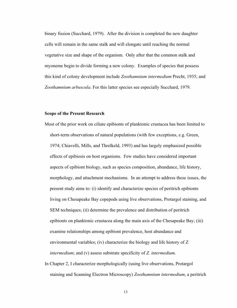
Morphology of Live specimens. The coenobium is dichotomously branched, with
alternate branches terminating at different levels. The basal stalk is the longest in the
colony and had a smooth surface without ridges or protuberances (Figs. 2.1 A, B, and
E, 2.2 A, 2.3 A ). The length of the basal stalk and first order branches was highly
variable and a statistically significant difference in stalk length was observed between
specimens attached to A. tonsa and E. affinis (Table 2.3). The diameter of the basal
stalk and first order branches is similar, and no significant difference from specimens
attached to both host species was observed (Table 2.3). A continuous thin myoneme
extended from the basal stalk to the branches and to each zooid, allowing the
coenobium to contract as an entire unit in a zigzag fashion, typical of the genus
Zoothamnium is identified (Figs. 2.1 B, and E, 2.2 A). The attachment disk and the
most basal part of the main stalk were the only two structures in the colony that lacked
myonemes (Fig. 2.1 B). The space between the attachment site and the tip of the
myoneme was highly variable (Table 2.3), and the mean size of this gap is
significantly different between specimens attached to A. tonsa (24.0 µm + 22.0 µm)
and E. affinis (13.0 µm + 5.7 µm). Coenobia could have up to 30 zooids, but most of
them had 2 or 4 zooids (mean: 2.7 + 1.0, Table 2.3) that were similar in size and
capable of forming telotrochs. Zooids are inverted bell-shaped (Figs. 2.1 A, and C,
26

2.2 A, 2.3 B) and ranged in length from 31.2 to 54.7 µm (total length including all
specimens attached to A. tonsa and E. affinis, see Table 2.3) and in width from 16.7 to
31.1 µm (width of the body at midpoint). The peristomial disk had a diameter similar
to the body width (Table 2.3), and it possessed a moderately thick peristomial lip
(Figs. 2.1 C, 2.2 A) that hung over the body when the cell was fully extended. The
epistomial disk is slightly elevated (Fig. 2.1 C) and is round on its surface, with a
width that was ~ ¾ the width of the peristomial disk. A single contractile vacuole is
located below the peristomial lip and discharges its contents into the infundibulum
(Figs. 2.1 D, 2.2 A). A transverse pattern of ridges on the pellicle was visible in the
living specimen (Figs. 2.1 C; 2.2 A), as well as a “C-shaped” macronucleus that lies in
the upper half of the cell close to the peristomial lip (Fig. 2.2 A).
Free-swimming stages (telotrochs) were round in shape and possess a single
row of cilia located near the aboral end (Fig. 2.3 E). Telotrochs range in size, in vivo,
from 26.7 to 31.6 µm in length and from 25.8 to 35.7 µm in width (Table 2.5).
Protargol stained telotrochs were observed in field samples from colonies attached to
A. tonsa and E. affinis, but no measurements were performed since the presence of the
dispersal stage was not observed in a high frequency. No statistically significant
difference was observed in size of telotrochs originating from colonies attached to A.
tonsa and E. affinis (Two-Way ANOVA; p > 0.05).
Morphology of fixed and stained specimens. Infraciliary and nuclear characteristics
of Z. intermedium are well recognizable in Protargol stained specimens. The
micronucleus possesses a round shape (Figs. 2.1 F, 2.2 A, and B) and always lies close
27

to the macronucleus (Figs. 2.2 A and B). The macronucleus has the same
configuration (“C-shaped”) observed in live specimens, and is always located in the
upper half of the zooid, close to the peristomial lip (Figs. 2.1 E and F, 2.2 C). A total
of 21 somatic myonemes that extended from the scopula to the epistomial disk are
present in the cell body (Figs 2.1 H, 2.2 B). The oral infraciliature is typical for the
genus Zoothamnium and sessiline peritrichs, in general, with an outer haplokinety and
an inner polykinety (PK1) that performes ca 1 ½ turns around the peristomial disk
before entering the infundibulum through the oral opening. Inside the infundibulum,
the haplo- and polykineties run together for a short distance with the former separating
at a 90o angle from the latter and running parallel to the lower end of the cell (Fig. 2.2
C). In addition to PK1, the infundibular polykineties PK2 and PK3 were identified
(Figs. 2.1 G, 2.2 C and D): PK1 consists of three rows of kinetosomes that are equal in
length and continue as the peristomial polykinety at the oral end. PK2 also consisted
of three rows of kinetosomes, that were shorter in length in comparison to PK1. The
central row of kinetosomes in PK2 is shorter than the two adjacent rows at the oral
end, with the third row slightly diverging obliquely form the other two. The PK2 rows
do not merge with the oral peristomial kinety and a gap between PK1 and PK2 could
be observed at the oral end. All PK2 rows terminate at the curvature of PK1 leaving a
space that ranged in size between 1.2 and 3.0 µm (Table 2.2). PK3 consists of three
short rows of kinetosomes of equal size that terminate at the aboral end of PK2
extending about ¼ of the length PK2 towards the oral end of the cell (Figs. 2.2 C and
D).
28

Scanning electron microscopy revealed that pellicular pores are irregularly
distributed over the cell surface (Fig. 2.3 C). A mean of 40 parallel pellicular
ridges (+ 3.5; range 36 - 50) are present between the peristomial lip and trochal
band and 20 (+ 1.7; range 17 - 25) are observed between the telotroch band and
scopula (Figs 2.3 B and C). The trochal band consists of three closely spaced
ridges that encircle the zooid near the aboral end. Scanning electron microscopy
also revealed several bacteria colonizing the attachment disk and the stalk of Z.
intermedium as can be seen in Figure 2.3 D.
Family Epistylididae Kahl, 1933
Genus Epistylis Ehrenberg, 1838
Epistylis sp.
Morphology of stained specimens. Colony with alternate branches terminating at
different levels are supported by a long, thin, non-contractile basal stalk (28.7 µm +
18.4 µm X 4.3 µm + 0.9 µm; Table 2.5). Colonies could have up to 15, inverted bell-
shaped zooids that are similar in size (30.7 µm + 5.1 µm X 18.5 µm + 3.2 µm; Table
2.6). Nuclear characteristics and buccal infraciliature are easily recognizable in
Protargol-stained specimens. The macronucleus is “C-shaped” and is located in the
upper half of the cell, close to the peristomial opening (Figs. 2.4 A and C). The
micronucleus is round shaped and lies closed to the macronucleus (Fig. 2.4 C). The
general features of the oral infraciliature is typical of sessiline peritrichs with an outer
haplokinety and an inner polykinety that perform about 1 ¼ turns before entering the
29

infundibulum. Inside the oral opening the haplokinety run parallel to the polykinety
for a short distance and completes one turn before ending at the wall of the
infundibulum (Fig. 2.4 D). Three infundibular polikineties are present inside the
infundibulum (PK1, PK2, and PK3). PK1 is formed by three rows of kinetosomes that
are equal in length and merge with the peristomial kinety at the oral end (Figs. 2.4 D
and E). PK2 consists of three rows of kinetosomes equal in length to each other, but
shorter than PK1. These rows do not merge at the oral end, leaving a gap between
PK1 and PK2 (Figs. 2.4 D and E). PK2 terminates at the curvature of PK1. PK3 is
formed by three short rows of kinetosomes equal in size that terminate at the aboral
end of PK2. PK3 extends about 1/3 of PK2 towards the oral end of the cell (Figs. 2.4
D and E). Only one row of kinetosomes is present in the trochal band (Figs 2.4 A, B,
and C). The pellicle presented a reticulated pattern of striations (Fig. 2.4 B) and
approximately 28 horizontal ridges (+ 2.4; Table 2.6) are observed between the oral
end of the cell and trochal band, and 18 (+ 2.0; Table 2.6) are present between the
trochal band and scopula.
DISCUSSION
The genus Zoothamnium Bory de St. Vincent, 1826 is characterized by the
presence of a continuous myoneme that extends from the basal stalk to the zooids,
enabling the entire coenobium to contract as a single unit (Corliss, 1979; Curds, Gates,
and Roberts, 1983). After being compared with original descriptions of several
species in the genus Zoothamnium, the species investigated in the present study was
identified as Zoothamnium intermedium. Although the original description of Z.
30

intermedium (Precht, 1935) is very brief and based only on the measurements of few
characters, almost all of them fall in the same range of size observed in the
investigated species. The only exception is the size of the gap present between the
attachment site and the beginning of the secreted myoneme, which in the present
species is highly variable, but in the original description provided by Precht (1935) is
cited as being always constant (~12 µm). Since there is no reference to sample
number in the original description and this character proved to be quite variable
among colonies (encompassing the value originally described), it is a conservative
position to assume that the species investigated in the present study is the same as that
originally described as Z. intermedium. Other species in the genus Zoothamnium also
present a similar coenobium shape and size, but other morphological features were not
compatible with the species of Zoothamnium observed in the present study (see Table
2.7). For example, Zoothamnium parasiticum Stein, 1859 has the same overall colony
shape and branching pattern observed in Z. intermedium, but the heavy peristomial lip
and the size of the zooid (~ 70 µm; Kahl, 1935) are considerably different from the
species investigated here. Zoothamnium carcini Kent, 1881 also presents a similar
colony shape, but the zooids are approximately 100 µm in size when fixed (Kahl,
1935). Zoothamnium rigidum Precht, 1935 and Zoothamnium hiketes Prech, 1935 are
also similar to Z. intermedium, differing only on the size of the zooids, with both
former species being larger than the latter (Prech, 1935).
Gross (1986) characterized morphologically, by live observations and
Protargol staining, four species in the genus Zoothamnium collected on glass slides
from Chesapeake Bay. One of these species was identified as Zoothamnium alternans
31

Claparède and Lachmann, 1858, and differs from Z. intermedium basically by the
presence of micro- and macrozooids, overall shape of the colony, and arrangement of
the oral polykinetids. The other three morphospecies of Zoothamnium were not
named, but differ from Z. intermedium in the size and overall shape of the colonies,
size of the zooids, and arrangement of oral polykinetids.
A more recent description of Zoothamnium intermedium was provided by
Valbonesi and Guglielmo (1988) and Valbonesi, (1989) who found the species
attached to copepods and shrimp zoea in a Lagoon in Italy. Although the species
description was based on formalin-fixed organisms, the measurements obtained for
those specimens fall approximately in the same range recorded for stained specimens
in the present study. Also, overall shape of the colony and of individual zooids, shape
and position of macro- and micronucleus, and the epibiotic way of life are the same
observed for Z. intermedium attached to copepods in Chesapeake Bay. Scanning
electron microscopy observations also revealed an annular pattern of ridges and a
scattered pattern of pores distributed on cell the membrane (Valbonesi, 1989) as was
observed in the present study for Z. intermedium. Investigations of the motile stage of
Z. intermedium by Valbonesi (1989) revealed two different morphotypes defined on
the basis of the length of the cilia in the trochal band as telotroch (with short cilia in
the trochal band) and microgamont (with long cilia in the trochal band). Interestingly,
in the present study I was able to identify the telotroch stage of Z. intermedium as
round cells with cilia in the trochal band measuring on average 4 µm in length (range
2.5-6.4 µm, see Table 2.5), approximately the same size reported by Valbonesi (1989)
for what he classified as microgamont. The free-swimming cells observed and
32
measured in vivo in the present study were ready to attach as soon as they contacted a
suitable host (personal observation), indicating that they were the dispersal stage and
not a microconjugant (microgamont). No other motile form was observed in the
present study. Therefore, I suggest that the telotroch stage of Z. intermedium has a
round shape and a single row of long cilia in the trochal band. The motile stage
presenting short cilia as described by Valbonesi (1989) might be the microconjugant
(which was not observed in the present study) or cells detached at an early stage of
telotroch formation (due to specimen preparation), with not fully developed cilia in the
trochal band.
The genus Epistylis Ehrenberg, 1830 is a colonial peritrich characterized by a
non-contractile stalk. In the colony, only the zooids are able to contract due to the
presence of a somatic myoneme (Corliss, 1979). Since I was not able to observe
Epistylis from the Chesapeake Bay “in vivo”, no comparisons with original species
descriptions based on live specimens could be made. Therefore, I compared the
arrangement of oral polykinetids in the investigated species to those of five other
species of Epistylis characterized with Protargol preparations, but none of which
corresponded to pattern observed here. For example, the rows of kinetosomes of PK1
and PK2 in Epistylis digitalis Ehremberg, 1838 do not fuse together at the oral end ,
and PK1 and PK3 terminate at the same level in the infundibulum, with PK2 ending at
the curvature of PK1 (Lom, 1964), This arrangement is different from the one
observed for Epistylis in the present study, in which PK2 and PK3 terminate at the
same level and at the curvature of PK1. The oral architecture of Epistylis lwofii
investigated with Protargol preparations revealed that the three oral polykinetids
33

terminate at the same level inside the infundibulum (Lom, 1964) differing from
Epistylis observed in the present study. Oral architecture of two other species of
Epistylis epibionts (Epistylis lacustris Imhoff, 1884 and an unidentified species) was
also investigated by Lom (1964) based on Protargol stains. In both Epistylis lacustris
and Epistylis sp., PK3 was not observed, a character that was easily recognizable in
Epistylis attached to A. tonsa in this study. Also, E. lacustris presents a belt shaped
structure highly impregnable by Protargol, which is absent in the Epistylis from
Chesapeake Bay. Epistylis plicatilis, Ehrenberg, 1830 also differs from the Epistylis
investigated in the present study by the presence of only two rows of kinetosomes in
P3 (Fernández-Galiano and Carrascosa, 1989), instead of three.
In summary, this study provided a redescription of a species in the genus
Zoothamnim epibiont on copepods in Chesapeake Bay, which was recognized as
Zoothamnium intermedium. Since the original description is based on very few
characters and no sample number is available in that publication, direct and detailed
comparisons of Z. intermedium from the Kiel Bay (location type) with the
Zoothamnium examined here would be required to confirm definitively whether or not
they are the same species. Also, molecular analyses comparing Z. intermedium
attached to the two copepod hosts may be performed in the future to investigate if they
indeed comprise a single genetic unit, as suggested by the morphological results
presented here. Epistylis sp. attached to A. tonsa in Chesapeake Bay may be an
undescribed species; detailed comparisons to other members of the genus (including in
vivo observations, as well as measurements of morphological characters using live
34

specimens, Protargol staining and scanning electron microscopy) are necessary to
either assign it a new name or to classify it as one of the currently known species.
35

Table 2.1. Measurements of field, Protargol stained colonies of Zoothamnium
intermedium attached to Acartia tonsa (At) and Eurytemora affinis (Ea). A total
number of 40 colonies attached to A. tonsa and 30 colonies attached to E. affinis were
measured from the months with highest incidence (July 1994, March, April, May and
August 1996). No statistically significant difference was found between the species for
the measured characters.
Character Species Mean (µm) SD (µm) Mode CV (%) Range
At 32.3 + 4.5 25.0 13.9 22.8 – 41.2 Total length of the body from epistomial disk to aboral end Ea 30.6 + 4.6 28.9 15.0 23.9 – 43.5
At 22.6 + 2.5 21.2 11.1 17.8 – 28.5 Width of the body below the peristomial lip Ea 21.5 + 2.5 21.4 11.6 16.9 – 28.3
At 22.5 + 3.2 22.0 14.2 16.6 – 30.3 Width of the body at midpoint between oral and aboral ends Ea 20.1 + 3.8 22.5 18.9 12.9 – 30.8
At 8.1 + 2.3 7.4 28.3 4.5 – 14.6 Distance between trochal band and scopula Ea 7.8 + 1.5 7.2 19.2 5.5 – 12.4
At 38.6 + 20.3 29.5 52.6 7.4 – 83.9 Length of basal stalk Ea 34.4 + 25.4 26.2 73.8 7.4 – 86.5 At 7.1 + 2.4 5.3 33.8 4.8 – 17.3 Width of basal stalk Ea 6.8 + 1.8 6.0 26.5 3.6 – 9.9 At 20.4 + 13.8 20.0 67.6 1.2 – 50.0 Distance of myoneme from
attachment point Ea 20.8 + 18.6 10.2 89.4 1.9 – 73.2 At 2.5 + 0.4 2.5 16.0 1.5 – 3.4 Length of micronucleus Ea 2.3 + 0.4 2.0 17.3 1.6 – 3.4 At 2.4 + 0.4 2.3 16.7 1.7 – 3.0 Width of micronucleus Ea 2.3 + 0.5 2.2 21.7 2.7 – 6.9 At 4.2 + 1.0 3.0 23.8 2.3 – 6.5 Width of macronucleus at
midpoint Ea 4.8 + 1.1 4.3 22.9 2.7 – 6.9 At 2.0 + 0.4 1.7 20 1.2 – 3.0 Distance between ends of
OPK1 and OPK2 Ea 2.0 + 0.3 1.8 15.0 1.4 – 2.5 At 5.5 + 2.8 2.0 50.9 2.0 – 13.0 Number of zooids in the
colony Ea 6.2 + 2.8 4.0 45.2 2.0 – 14.0
36

Table 2.2. Number of annular ridges on the membrane of Zoothamnium intermedium
attached to Acartia tonsa (At) and Eurytemora affinis (Ea). A total of 30 zooids
attached to each host species were observed.
Character Species Mean SD Mode CV (%) Range
At 39.4 2.1 37 5.4 37 – 45 Number of Ridges from the peristome to
the trochal band Ea 40.1 3.5 38 8.7 36 – 50
At 20.0 1.7 19 9.0 17 – 25 Number of Ridges from the trochal band
to the scopula Ea 20.0 1.5 20 7.0 18 – 25
37

Table 2.3. Measurements of cultured live colonies of Zoothamnium sp. attached to
Acartia tonsa (At) and Eurytemora affinis (Ea). A total number of 32 colonies were
measured for each character in each copepod species. Asterisks indicate characters
where there is a statistically significant difference between the two host species
(p<0.05)
Character Species Mean (µm) SD (µm) Mode CV (%) Range
At 41.0 + 5.5 43.0 13.4 31.2 - 53.0 Total length of the body from epistomial disk to aboral end Ea 44.9 + 4.8 43.6 10.7 35.8 – 54.7
At 34.0 + 5.9 38.0 17.3 20.7 - 44.3 Length of body from peristomial lip to aboral end * Ea 38.7 + 5.2 38.2 13.4 26.8 – 49.9
At 23.0 + 2.2 23.8 9.6 18.2 – 28.8 Width of the body below the peristomial lip Ea 23.1 + 2.4 21.9 10.4 18.2 – 28.1
At 21.2 + 2.8 22.8 13.2 16.7 – 27.4 Width of the body at midpoint between oral and aboral ends Ea 23.2 + 3.1 21.4 13.4 16.7 – 31.3
At 25.8 + 2.7 25.2 10.5 18.3 – 31.5 Width of peristomial lip Ea 26.5 + 2.4 29.1 9.0 21.5 – 30.8 At 4.1 + 1.2 3.6 29.7 2.5 – 7.7 Thickness of peristomial lip Ea 3.6 + 0.6 4.1 16.7 2.5 – 4.4 At 18.0 + 2.2 16.8 12.2 11.9 – 23.6 Width of epistomial disk Ea 18.6 + 2.1 19.0 11.3 13.5 – 22.8 At 7.3 + 2.0 5.1 27.4 4.3 – 11.8 Width of scopula Ea 8.1 + 1.6 5.8 19.7 4.8 – 8.2 At 43.3 + 21.8 12.6 50.3 13.0 – 105.4 Length of basal stalk * Ea 28.9 + 12.2 20.0 42.4 10.3 – 60.5 At 10.2 + 1.5 10.0 14.7 7.3 – 14.6 Width of basal stalk Ea 10.1 + 1.8 10.2 17.8 4.7 – 13.4 At 38.0 + 27.2 22.0 71.6 5.8 – 115.1 Length of lateral stalk * Ea 25.2 + 14.9 10.7 59.1 6.7 – 62.9 At 9.3 + 2.1 11.6 22.6 0.9 – 11.6 Width of lateral stalk Ea 9.3 + 1.8 10.6 19.3 6.3 – 13.9 At 24.7 + 22.0 16.8 89.1 3.7 – 115.1 Distance of myoneme from
attachment point * Ea 13.7 + 5.7 15.0 41.6 4.1 – 23.6 At 2.0 + 0.4 1.8 20.0 1.3 – 3.0 Thickness of myoneme Ea 2.3 + 0.5 2.5 21.7 1.6 – 3.7 At 2.7 + 0.9 2.0 33.0 2.0 – 6.0 Total number of zooids in the
colony Ea 2.7 + 1.0 2.0 37.0 2.0 – 6.0 At 2.1 + 1.0 2.0 47.6 1.0 – 5.0 Total number of branches in the
colony Ea 2.5 + 1.4 1.0 56.0 1.0 – 6.0
38
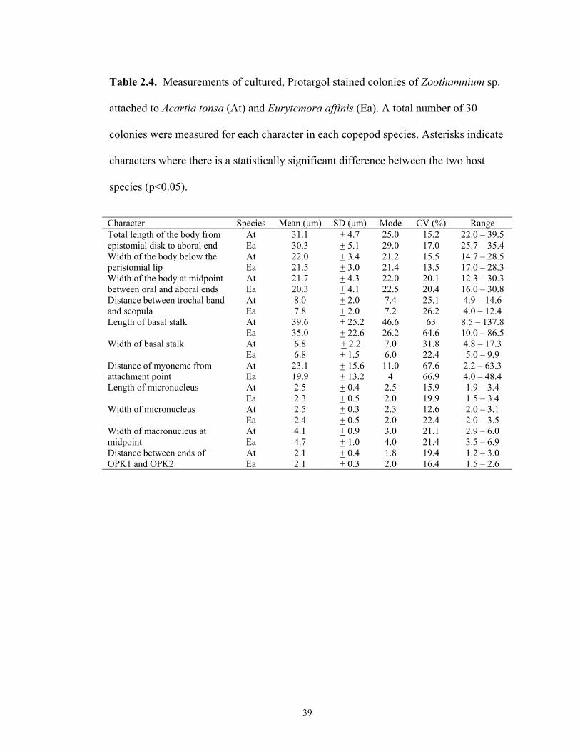
Table 2.4. Measurements of cultured, Protargol stained colonies of Zoothamnium sp.
attached to Acartia tonsa (At) and Eurytemora affinis (Ea). A total number of 30
colonies were measured for each character in each copepod species. Asterisks indicate
characters where there is a statistically significant difference between the two host
species (p<0.05).
Character Species Mean (µm) SD (µm) Mode CV (%) Range
At 31.1 + 4.7 25.0 15.2 22.0 – 39.5 Total length of the body from epistomial disk to aboral end Ea 30.3 + 5.1 29.0 17.0 25.7 – 35.4
At 22.0 + 3.4 21.2 15.5 14.7 – 28.5 Width of the body below the peristomial lip Ea 21.5 + 3.0 21.4 13.5 17.0 – 28.3
At 21.7 + 4.3 22.0 20.1 12.3 – 30.3 Width of the body at midpoint between oral and aboral ends Ea 20.3 + 4.1 22.5 20.4 16.0 – 30.8
At 8.0 + 2.0 7.4 25.1 4.9 – 14.6 Distance between trochal band and scopula Ea 7.8 + 2.0 7.2 26.2 4.0 – 12.4
At 39.6 + 25.2 46.6 63 8.5 – 137.8 Length of basal stalk Ea 35.0 + 22.6 26.2 64.6 10.0 – 86.5 At 6.8 + 2.2 7.0 31.8 4.8 – 17.3 Width of basal stalk Ea 6.8 + 1.5 6.0 22.4 5.0 – 9.9 At 23.1 + 15.6 11.0 67.6 2.2 – 63.3 Distance of myoneme from
attachment point Ea 19.9 + 13.2 4 66.9 4.0 – 48.4 At 2.5 + 0.4 2.5 15.9 1.9 – 3.4 Length of micronucleus Ea 2.3 + 0.5 2.0 19.9 1.5 – 3.4 At 2.5 + 0.3 2.3 12.6 2.0 – 3.1 Width of micronucleus Ea 2.4 + 0.5 2.0 22.4 2.0 – 3.5 At 4.1 + 0.9 3.0 21.1 2.9 – 6.0 Width of macronucleus at
midpoint Ea 4.7 + 1.0 4.0 21.4 3.5 – 6.9 At 2.1 + 0.4 1.8 19.4 1.2 – 3.0 Distance between ends of
OPK1 and OPK2 Ea 2.1 + 0.3 2.0 16.4 1.5 – 2.6
39

Table 2.5. Measurements of live telotrochs from cultured Zoothamnium intermedium
attached to Acartia tonsa (At) and Eurytemora affinis (Ea). A total of 30 telotrochs
originating form colonies attached to each host species was measure for each
character.
Character Species Mean (µm) SD (µm) Mode CV (%) Range
At 27.7 2.2 26.8 8.1 23.7 – 31.5 Length of the body Ea 28.2 2.3 26.3 8.2 24.2 – 31.6 At 28.6 1.9 26.3 6.7 26.3 – 32.1 Width of the body Ea 28.9 2.2 29 7.7 25.8 – 33.7 At 4.0 + 0.8 4.6 20.6 2.5 – 6.2 Length of the cilia* Ea 4.0 + 0.9 4.0 21.7 2.5 – 6.4
*Length of cilia in the trochal band was measured from Lugol’s fixed cells
40

Table 2.6. Measurements of field, Protargol stained colonies of Epistylis sp. attached
to Acartia tonsa. A total number of 30 colonies were measured from the months with
highest infestation prevalence (August and September 1994, and September 1996).
Character Mean (µm) SD (µm) Mode CV (%) Range Total length of the body from epistomial disk to aboral end
30.7 + 5.1 29.2 16.7 20.0 – 45.7
17.0 + 2.5 16.4 14.7 13.4 – 22.5 Width of the body below the peristomial lip Width of the body at midpoint between oral and aboral ends
18.5 + 3.2 20.4 17.4 12.2 – 24.3
Distance between trochal band and scopula
11.2 + 2.3 12.5 20.2 6.7 – 14.9
Length of basal stalk 28.7 + 18.4 20.0 64.0 10.0 – 44.9 Width of basal stalk 4.3 + 0.9 4.0 21.4 2.8 – 6.4 Length of micronucleus 2.1 + 0.3 2.0 13.0 1.5 – 2.7 Width of micronucleus 2.2 + 0.3 2.2 12.7 1.8 – 2.6 Width of macronucleus at midpoint
3.3 + 0.6 3.2 18.9 2.4 – 4.5
Number of zooids in the colony
7.9 + 3.2 10.0 41.2 2 - 15
41

Table 2.7. Morphological comparison between Zoothamnium intermedium and other similar species in the genus Zoothamnium.
Character Z.
intermediuma Z. parasiticumb
Z. carcinic
Z. rigidumd
Z. hyketese
Z. alternansf
Z. sp1g
Z. sp2h
Size of the zooid (µm)
45 70 100 95 55 53 58 70
Branching pattern alternate alternate
alternate alternate alternate alternate alternate dichotomous
Presence of macrozooids
No No No No No Yes No No
Shape of macronucleus
“C” shaped Horse-shoe shaped
Not indicated
“C” shaped “C” shaped
“C” shaped Horse-shoe
shaped
Horse-shoe shaped
Position of contractile vacuole
Below the peristomial lip
In the upper half of the cell
Not indicated
In the upper half of the
cell
In the upper half of the cell
Not indicated
Not indicated
Not indicated
Distance of myoneme from attachment point
Highly variable (2.2 – 63.3 µm)
Not indicated Not indicated
Not indicated
Constant (15 µm)
Not indicated
Not indicated
Not indicated
a: this study b and c: Kahl, 1935 d and e: Precht, 1935 f, g and h: Gross 1986
42

FIGURE LEGENDS:
Figure 2.1 A -2.1 H: Photomicrographs of Zoothamnium intermedium attached to A.
tonsa and E. affinis from life (2.1 A-2.1 D; Differential Interference Contrast) and
after Protargol staining (2.1 E-2.1 H; Bright Field). 2.1 A. Colonies at low
magnification attached to the abdomen of the host. 2.1 B. Colony with two zooids
attached to the head of the host. Arrow shows the continuous myoneme in the basal
stalk. 2.1 C. Detailed zooid: arrow shows the peristomial lip, arrowhead shows the
infundibulum, and double arrowheads show the elevated epistomial disk. 2.1 D.
Detailed zooid showing the contractile vacuole close to the peritomial lip. 2.1 E.
Colony after Protargol staining. Arrow shows the myoneme in the basal stalk and
branches; arrowhead shows basal bodies in the telotroch band. 2.1 F. Detailed zooid
after Protargol staining showing the macronucleus (arrow) and the micronucleus
(arrowhead). 2.1 G. Zooid at high magnification showing the structure of the oral
polykinetids (P) 1, 2, and 3. 2.1 H. Zooids showing stained sphincter myoneme
delimiting the peristomial opening (arrow). Arrowhead shows somatic myonemes
close to the peristome. Bars = 50 µm (2.1 A); 20 µm (2.1 B, 2.1 C, 2.1 D, 2.1 E); 10
µm (2.1 F, 2.1 G, 2.1 H)
Figure 2.2 A-2.2 D: Morphology of Zoothamnium intermedium epibiont on Acartia
tonsa and Eurytemora affinis from life (Fig. 2.2 A) and after Protargol staining (Fig.
2.2 B-2.2 C). 2.2 A. General aspect of the colony. 2.2 B. Detail of the zooid showing
somatic myonemes (SMY), macronucleus (MAC) and micronucleus (MIC). 2.2 C.
43

Zooid at high magnification showing arrangement of the oral polykinetids (PK1, PK2,
PK3). 2.2 D. Detail of the oral polykinetids at high magnification. CV, contractile
vacuole; FV, food vacuole; g, germinal row; INF, infundibulum; MAC, macronucleus;
MIC, micronucleus; MY, myoneme; ODK, oral dikinetid; OPK, oral polykinetid;
SCP, scopula; SMY, somatic myoneme; STK, stalk; STR, striae; TB, telotroch band.
Bars = 20 µm (2.2 A); 10 µm (2.2 B, 2.2 C); 1 µm (2.2 D)
Figure 2.3 A-2.3 E: Scanning Electron Micrographs of Zoothamnium intermedium
attached to Acartia tonsa and Eurytemora affinis. 2.3 A. Colonies at low
magnification (730X), arrow shows the telotroch band. 2.3 B. Detail of the zooid
showing the parallel striae pattern on the pellicle (arrow) and attached bacteria
(arrowhead). 2.3 C. Zooid at very high magnification (13.900X) showing the
scattered pattern of pores on the pellicle (arrowheads) and the parallel striae. 2.3 D.
Stalk showing the attachment disk on the surface of the host (arrow) and bacteria. 2.3
E. Telotroch of Z. intermedium showing cilia in the telotroch band (arrow). Bars =
100 µm (2.3 A); 10 µm (2.3 B, 2.3 C, 2.3 D); 1 µm (2.3 E)
Figure 2.4 A-2.4 E: Photomicrographs after Protargol (2.4 A, 2.4 B) and
morphology (2.1 C-2.1 E) of Epistylis sp. attached to Acartia tonsa. 2.4 A. Colony at
low magnification attached to the abdomen of the host. 2.4 B. Detail of the zooid
showing a reticulate pattern of striae on the pellicle (arrowhead) and telotroch band
(arrow). 2.4 C. Zooid showing somatic myonemes (SMY), macronucleus (MAC) and
micronucleus (MIC). 2.4 D. Zooids at high magnification showing the arrangement of
44

the oral structure. 2.4 E. Detail of the oral polykinetids (PK1, PK2, PK3) at high
magnification. g, germinal row; MAC, macronucleus; MIC, micronucleus; ODK, oral
dikinetid; OPK, oral polykinetid; SMY, somatic myoneme; STK, stalk; STR, striae;
TB, telotroch band. Bars = 20 µm (2.4 A); 10 µm (2.4 B, 2.4 C, 2.4 D); 1 µm (2.4 E).
45
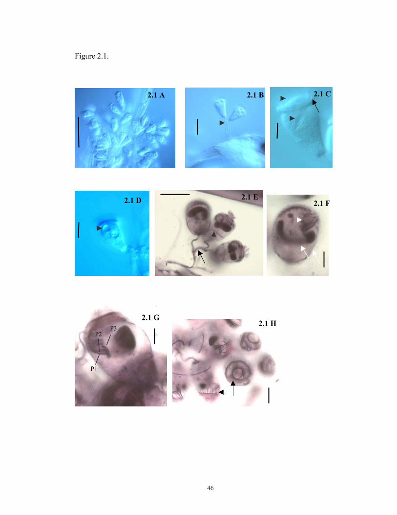
Figure 2.1.
2.1 C2.1 A 2.1 B
2.1 E2.1 D 2.1 F
2.1 G 2.1 H
P3 P2
P1
46

Figure 2.2
SMY CV MIC
MAC
MACINF
FV
OPK
PK
2.2 A
2.2 C
STR
MICMY TB
STK
PK3g
PK1
ODK
2 TB
SCP
MY
2.2 B
MY
P1
P2
47
P3
2.2 D
Figure 2.3
2.3 A 2.3 B
2.3 C 2.3 D
2.3 E
48

Figure 2.4
2.4 B 2.4 A
SMY
STR
TB
SCP
MIC
MACg ODK
PK2OPK
PK3
PK2
2.4 C 2.4 D
STK
49
PK1
PK32.4 E
PK1

CHAPTER 3
SPATIAL AND TEMPORAL PATTERNS IN THE OCCURRENCE OF
PERITRICH CILIATES AS EPIBIONTS ON CALANOID COPEPODS IN THE
CHESAPEAKE BAY, USA
50

ABSTRACT
I investigated temporal and spatial patterns of distribution in two peritrich
ciliates (Zoothamnium intermedium and Epistylis sp.) living as epibionts on calanoid
copepods (Acartia tonsa and Eurytemora affinis) in Chesapeake Bay. Net tow
samples collected along the main axis of the Bay were analyzed to estimate occurrence
of epibionts on copepods and to explore relationships among infestation prevalence,
host abundance and environmental variables. Z. intermedium and Epistylis sp.
colonized populations of A. tonsa during spring and summer months, while only Z.
intermedium colonized E. affinis during spring. Occurrence of epibionts on copepods
showed a high interannual variation, marked seasonality, and geographic
heterogeneity. Extensive statistical analyses rejected simple scenarios of interactions
between epibiosis, environmental variables, and host density, suggesting a more
complex dynamics for the system. Overall, epibiont infestation density (colonies/host
area) and load (zooids/host area) were higher on copepodites than on adults for both
host species suggesting a preferential attachment to juveniles or a higher predation
pressure on adult stages. Infestation density and load of both epibiont species were
higher on cephalothorax and abdomen of A. tonsa and E. affinis in comparison to
antenna and swimming legs, suggesting that ciliates can more easily colonize less
active parts.
51

INTRODUCTION
Epibiotic relationships involving planktonic crustacea as basibionts (i.e. hosts)
are widespread in marine environments and encompass a variety of organisms as
epibionts, including bacteria, algae, and ciliates (Gibson 1978; Hanamura, 2000;
Nagasawa, Simidu, and Nemoto 1985; Turner, Postek, and Collard 1979).
Historically, epibiosis has been viewed as a commensal relationship, however several
studies have suggested that epibionts can have deleterious effects on the host. These
effects include decreased reproduction, growth, and survivorship (Xu and Burns
1991), increased susceptibility to predation (Chiavelli, Mills, and Threlkeld 1993;
Weissman, Lonsdale, and Yen 1993; Willey, Cantrel, and Threlkeld 1990), and
starvation due to competition with the epibionts (Kankaala and Eloranta 1987). In
other cases, the epibiont itself may serve as food for the host. For example, Daphnia
spp. readily prey on free-swimming stages of their epibionts (Al-Daheri and Willey
1996; Barea-Arco, Pérez-Martinez, and Morales-Barquero 2001). Thus, from an
ecosystem perspective, epibiosis may interfere with fitness of the host organism, or
fuel higher trophic levels. In addition, epibionts often occur on planktonic crustacea
under eutrophic conditions, suggesting that the prevalence of epibionts on zooplankton
might be used as an index of water quality (Henebry and Ridgeway 1979).
Few studies have addressed temporal and spatial patterns in the occurrence of
epibionts that infest planktonic crustacea. Most of these are from freshwater
environments and have considered algal or ciliate epibionts on relatively small
temporal (annual or subannual) and spatial (from few meters to a couple
kilometers) scales (Chiavelli, Mills, and Threlkeld 1993; Green 1974; Strinadel and
52

Ebert 1997; Xu 1992). While numerous accounts exist for ciliates as epibionts of
marine zooplankton, little information is available on the prevalence of epibionts in
zooplankton populations (Weissman, Lonsdale, and Yen 1993), and data on
seasonality and geographic distribution of epibionts is almost lacking.
The only record of ciliate epibionts associated with zooplankton of Chesapeake
Bay, the largest estuary in the United States, is that of Herman and Mihursky (1964).
They reported Zoothamnium sp., a stalked, colonial peritrich, as a common epibiont on
the most abundant calanoid copepod, Acartia tonsa, in the Patuxent River, a sub-
estuary of the Chesapeake Bay. Samples collected on three dates between early
March and mid-April revealed 100% infestation of A. tonsa, but other zooplankton
species encountered during the study were not infested. Despite several studies have
focused on the ecology of Chesapeake Bay zooplankton, no data are available on the
seasonal, interannual, or spatial distribution of ciliate epibionts associated with Acartia
tonsa or other zooplankton species.
Here I describe seasonal occurrence of peritrich ciliates as epibionts on
Chesapeake Bay populations of Acartia tonsa and Eurytemora affinis by analyzing net
tow samples taken over a five-year period. I also report epibiont density and load on
copepod life-history stages and body parts by estimating number ofcolonies and
zooids distributed over the host and on different body parts. Finally, I consider
relationships among epibiont prevalence, host abundance, and environmental
variables.
53

MATERIALS AND METHODS
Field Sampling. The occurrence of peritrich epibionts on Acartia tonsa and
Eurytemora affinis was determined from samples collected at nine stations (one
sample per station) along the main axis of the Chesapeake Bay (Fig. 3.1; filled circles;
Table 3.1). Stations were sampled monthly in spring and summer of 1994-1996 and
1999-2000, using a 35-µm-mesh plankton net (30-cm diameter) to obtain a vertically
integrated sample over the upper 10-15 m of the water column (a total of 21
samples/station during the study period). A 250-ml subsample of each net tow was
fixed with modified Bouin’s fluid (Coats and Heinbokel 1982) at a final concentration
of 5% and examined using a dissecting microscope (Bausch and Lomb; 30X
magnification) to determine the presence of ciliate epibionts on host organisms.
Vertical CTD-O2 (conductivity, temperature, depth, and dissolved oxygen) profiles
were taken at each station using shipboard instrumentation (R/V Cape Henlopen; CTD
Type Seabird 911 Plus).
Epibiont, Host, and Phytoplankton Population Assessments. Infestation
prevalence (i.e., percent hosts with epibionts) was obtained for adults and copepodites
of each host species by scoring all individuals in the sample as either infested or non-
infested. Epibionts present on each host were tentatively identified to genus based on
key morphological features characterized by Lynn and Small (2000). Infestation
density (i.e., number of peritrich colonies/host area) and infestation load (i.e., number
of peritrich zooids/host area) were then determined for samples where epibiont
prevalence was > 5% for E. affinis and > 10% for A. tonsa. For these data, 30 adults
and 30 copepodites were selected from each sample and examined using a Zeiss RA
54

microscope (100X) to enumerate colonies/host and zooids/colony. The position of
colonies on host organisms (i.e., antennae, cephalothorax, abdomen, and swimming
legs) was recorded, and the dimensions (length and width) of host body parts were
measured using a calibrated ocular micrometer. The area of host body parts was
calculated from mean length and width measurements assuming a cylindrical shape
(i.e., A= (2πW)*L), as recommended by Mauchline (1998). To confirm epibiont
identification, 2-3 representative specimens were taken from each sample and silver-
stained using the Protargol technique (Montagnes and Lynn, 1993). Stained
specimens were identified to genus or species using criteria described in Chapter 2.
Since net tows used to assess epibiont prevalence were not suitable for
determining copepod densities, estimates of host abundances were obtained from the
Chesapeake Bay Monitoring Program (CBMP) Database
(http://www.chesapeakebay.net). Nine CBMP stations (Fig. 3.1; filled triangles; Table
3.1), one close to each location sampled for presence of epibionts, were selected for
analysis involving basibiont abundance. Data on phytoplankton abundance (cells/ml
in surface water) were also obtained for the nine CBMP stations.
Data Analyses. Multivariate Correlation analyses (Spearman-Rho correlation) were
performed to evaluate inter- and intra-annual trends in epibiont prevalence relative to
host abundance, phytoplankton abundance, and environmental variables (i.e.,
temperature, salinity, and dissolved oxygen). Phytoplankton abundance used in these
analyses was based on cells smaller than 10 µm in diameter, as the ciliate epibionts
encountered in this study typically ingested bacteria to nanoflagellate-sized prey
(Corliss, 1979). Kruskal-Wallis ANOVA on ranks was performed to discern
55
differences in epibiont density (colonies/mm2) and load (zooids/mm2) among host
body parts, with Dunn’s test used for pairwise comparisons of means. Correlation
analyses were performed using JMPIN Version 4.04 (SAS Institute), Analyses of
Variance were run using SigmaStat Version 2.0 (SPSS Inc.) and data are reported in
the text as mean + standard error of the mean (SE).
RESULTS
The coenobial and colonial peritrichs, Zoothamnium intermedium Precht, 1935
and Epistylis sp., respectively, colonized Chesapeake Bay populations of Acartia tonsa
(adults and copepodites) during spring and summer months of 1994-2000. Adults and
copepodites of Eurytemora affinis were also infested by Z. intermedium, but not by
Epistylis sp. Other copepod species (e.g. Cyclops spp., Oithona spp., Diaptomus
spp.), rotifers (e.g. Brachionus spp.,Synchaeta spp., Trichocerca spp.), cladocerans
(e.g. Evadne spp., Podon polyphemoides), and barnacle nauplii (Balanidae) were also
present in the samples, but did not harbor ciliate epibionts.
Infection prevalence for Z. intermedium on A. tonsa reached appreciable levels
(> 5%) at mid to high salinity (10 to 25 psu), regardless of temperature (Fig. 3.2A).
Infestations of Epistylis sp. on A. tonsa also exceeded 5% at higher salinities (16-26
psu), but only when water temperature was above 23oC (Fig. 3.2B). Interestingly, Z.
intermedium and Epistylis sp. did not co-occurr in any sample. By contrast, Z.
intermedium on E. affinis showed high prevalence (>5%) over a broad range of
temperatures (8-23oC) and salinities (7 to 24 psu; Fig. 3.2C).
56

Occurrence of peritrich epibionts on Chesapeake Bay copepods exhibited high
interannual variation, with yearly mean prevalence ranging from a low of 0.1% + 0.3
(median = 0.2; n = 27) to a high of 5.2% + 1.4 (median = 1.7; n = 63) for Z.
intermedium on A. tonsa, 0.1 + 0.1 (median = 0.1; n = 45) to 0.8 + 0.3 (median = 0.9;
n = 45) for Epistylis sp. on A. tonsa, and 0.2 + 0.1 (median = 0.7; n = 45) to 1.5 + 0.4
(median = 0.2; n=63) for Z. intermedium on E. affinis (Fig. 3.3A). Host taxa also
showed considerable variation in annual mean abundance across the study period, but
there was no clear relationship between epibiont prevalence and host abundance (cf.
Fig. 3.3A and B). In addition, epibionts exhibited marked seasonality with peak
prevalence of Z. intermedium occurring in early spring and late summer on A. tonsa,
but only in spring on E. affinis (Fig. 3.3 C). Infestations of Epistylis sp. on A. tonsa
were only present in late summer and early fall. Seasonal fluctuations in epibionts
were not clearly related to host abundance, with high prevalence of Z. intermedium on
A. tonsa occurring when host density was either high (13.2 + 2.1, n=45; August) or
low (3.2 + 1.4, n=9; March). The spring maximum in prevalence of Z. intermedium
on E. affinis, however, did coincide with the maximum monthly abundance for that
host (cf. Fig. 3.3C and D).
Epibionts also showed geographic heterogeneity along the main axis of the
Bay, with mean prevalence for Z. intermedium on A. tonsa being highest in the lower
Bay (4.3 + 1.9 to 5.8 + 3.1 for stations 707-744; Fig. 3.3E) and decreasing abruptly at
station 804 northward (range: 0.3 + 0.1 to 1.8 + 0.7). Epistylis sp. infestations of A.
tonsa were less common than Z. intermedium on A. tonsa, but followed the same
pattern of decreasing prevalence from south to north (range: 0.1 + 0.1 to 1.4 + 0.8;
57

Fig. 3.3E). By contrast, Z. intermedium was most prevalent on E. affinis in the upper
Bay (0.9 + 0.5 and 1.1 + 0.7 for stations 908 and 858, respectively; Fig. 3.3E), with
consistently lower values (range: 0.2 + 0.2 to 0.4 + 0.) south of station 845. Host
density appeared to have little influence on the spatial distribution of A. tonsa
epibionts, as mean abundance for this copepod was relatively stable along the axis of
the Bay (range: 4.5 + 1.4 to 10.9 + 2.5; Fig. 3.3F). E. affinis, however, exhibited peak
concentrations in the upper Bay (6.2 + 1.5 L-1 at station 908) that coincided with
maximum epibiont prevalence (1.2 + 0.7; Fig. 3.3F).
Contour plots for mean occurrence of peritrich epibionts (Fig. 3.4) showed
infestation foci for Z. intermedium on A. tonsa in the lower Bay during late summer
and on E. affinis in the upper Bay during spring. Those foci coincided with temporal-
spatial regions of elevated host abundance, suggesting a density dependent epibiont-
host interaction (cf. Fig. 3.4 A and B for A. tonsa and C and D for E. affinis). High
prevalence of Z. intermedium on A. tonsa in early spring (late March and April),
however, did not coincide with high host densities, nor did areas of high host
abundance in late spring (May and early June) support increased levels of epibiosis
(Fig. 3.4A). A density dependent interaction was also indicated for Z. intermedium,
but not for Epistylis sp., when data for epibiont prevalence on A. tonsa were grouped
into categories of low (<5 L-1), medium (>5-15 L-1), and high (> 15 L-1) host
abundance (Fig. 3.5). Correlation analysis, however, failed to show a significant
relationship between infestation prevalence and copepod abundance when data for
stations was pooled across years for each epibiont-host pair (p > 0.05; Table 3.1).
58
Also, no significant correlation was observed between environmental variables,
phytoplankton abundance, and occurrence of epibiosis (p > 0.05).
Infestation density and load: Estimates of epibiont density (colonies/host) and load
(zooids/colony) were obtained from three stations in 1994 (724 and 707 in May; 724
in September), eight stations in 1996 (908, 858, 845 in March; 858 in April; 744, 724,
707 in August; 724 in September), and one station in 2000 (724 in September).
Ciliates colonized all major body parts of A. tonsa and E. affinis, and a
statistically significant difference was observed in infestation density of Z.
intermedium between adults and copepodites of the two host species (ANOVA
Kruskal-Wallis, p<0.05; Fig. 3.6 A) with higher numbers of colonies per body area
occurring on copepodite stages. The same pattern was observed for Epistylis sp.
where colony density was significantly higher on copepodites than on adults of A.
tonsa (ANOVA Kruskal-Wallis; p<0.05; Fig 3.6 A). However when mean number of
zooids per epibiont colony was considered no difference was observed for Z.
intermedium colonizing adults and copepodites of A. tonsa (ANOVA Kruskal-Wallis;
p>0.05; Fig 3.6 B). On the other hand, the number of zooids/mm2 was higher on
adults than on juveniles of E.affinis (ANOVA Kruskal-Wallis; p<0.05; Fig 3.6 B)
displaying the same pattern observed for infestation density in this host species. Mean
number of zooids per Epistylis sp. colony was not significantly different for adults and
copepodites of A.tonsa in a similar pattern observed for Z. intermedium colonizing the
same host species. Infestation load of Z. intermedium was significantly higher on
copepodites than on adults of A. tonsa (ANOVA Kruskal-Wallis; p<0.05; Fig 3.6 C),
59
but no significant difference was observed in infestation load of Z. intermedium on
adults and copepodites of E. affinis (ANOVA Kruskal-Wallis; p>0.05; Fig 3.6 C).
Likewise, load of Epistylis sp. was not significantly different between adults and
copepodites of A. tonsa (ANOVA Kruskal-Wallis; p>0.05; Fig 3.6 C).
When epibiont colonization was analyzed across host body parts, higher
infestation density and load were observed on the cephalothorax and abdomen in
comparison to more mobile parts (antenna and swimming legs). For example,
infestation density and load of Z. intermedium were significantly higher on the
cephalothorax and abdomen of A. tonsa in comparison to antenna and swimming legs
(ANOVA Kruskal-Wallis, p<0.05; Fig. 3.7 A,C). Also, a significantly higher density
of Z. intermedium colonies was observed on the abdomen of E. affinis when compared
to the other body parts (ANOVA Kruskal-Wallis, p<0.05; Fig. 3.8 A), with an
infestation load significantly higher on both cepahlothorax and abdomen of when
compared to antenna and swimming legs (ANOVA Kruskal-Wallis, p<0.05; Fig. 3.7
C). A higher density of Epistylis sp. was recorded on the abdomen of A. tonsa
(ANOVA Kruskal-Wallis, p<0.05; Fig. 3.8 A) in comparison to the other body parts,
while infestation load was significantly higher on cephalothorax and abdomen when
compared to antenna and swimming legs (ANOVA Kruskal-Wallis, p<0.05; Fig. 3.8
C). When the mean number of zooids per epibiont colony was considered, a
significant difference was observed on the cephalothorax and abdomen of the two
copepod species in comparison to antenna and swimming legs (ANOVA Kruskal-
Wallis, p<0.05; Fig. 3.7 B) for both epibiont species. Analyses of epibiont infestation
60

density, load, and mean number of zooids per colony by year, station, and host life
stage revealed the same pattern observed for the combined data.
DISCUSSION
Zoothamnium intermedium and Epistylis sp. were the only species of peritrich
ciliates colonizing populations of Acartia tonsa and Eurytemora affinis in the
Chesapeake Bay during the study period. Z. intermedium was originally described as
an epibiont on amphipods and isopods from the Kiel Bay (Precht, 1935), and later was
found attached to copepods and barnacle nauplii in the Lagoon of Venice, Italy
(Valbonesi and Guglielmo, 1989). The genus Epistylis is also a common peritrich
found as an epibiont on crustaceans in freshwater (Willey and Threlkeld, 1993) and
estuarine environments (Turner, Postek, and Collard, 1979), with several species
having been originally described from crustacean hosts (Kahl, 1935; Precht 1935).
Epibionts in the present study colonized only adults and copepodites of A.
tonsa (both Z. intermedium and Epistylis sp.) and E. affinis (Z. intermedium only), not
being observed attached to other zooplankton species present in the samples. The lack
of colonization of other substrates may be due to host specificity, high preference for
these two host species, or to a host-epibiont density dependent relationship, since A.
tonsa and E. affinis are the dominant copepod species in the Bay (Brownlee and
Jacobs, 1987). Interestingly, Valbonesi and Guglielmo (1988) reported Z.
intermedium colonizing several species of planktonic crustacea (e.g. Acartia latisetosa
Calanipeda aquadulcis, and the zoea stage of the shrimp Hippolyte longirostris) from
Lagoon of Venice, Italy, but high infestation prevalence was observed only on the
61

most abundant copepod species (Acartia clausi), also supporting a host-epibiont
density dependent relationship. In addition, other studies demonstrated that these
species, which were considered to be specific of one host, could colonize different
substrates in different systems. For example, Henebry and Ridgeway (1979) in an
assessment of the epibiont community attached to planktonic crustacea from a lake in
Illinois observed that Vorticella microstoma infested only the cladoceran
Scapholeberis kingii, failing to colonize other hosts and non-living substrates. By
contrast, the same species of Vorticella has been reported heavily colonizing the
cyanobacteria Microcystis aeruginosa in a lake (Pratt and Rosen, 1983) and also being
able to attach to non-living surfaces in a river in Spain (Garcia et al., 1989).
High epibiont prevalence has generally been observed when host species are
more abundant (Mohlemberg and Kaas, 1990; Xu, 1992; Chiavelli, Mills, and
Threlkeld, 1993), however, some studies failed to show a clear relationship between
epibiont prevalence and substrate density (Lopez et al., 1998; Hanamura, 2000). In
Chesapeake Bay, prevalence of Z. intermedium and Epistylis sp. was not directly
correlated to host abundance, although an increase in prevalence with increasing
substrate abundance was observed for Z. intermedium attached to A. tonsa when host
densities were categorized (Fig. 3.5). One possible explanation for the absence of
strong correlation is that Z. intermedium and Epistylis sp. are obligatory epibionts on
A. tonsa and E. affinis, and factors like fluctuations in phytoplankton and bacteria
populations, influx of freshwater in the system and pollution in addition to host
abundance could be governing the colonization of these two copepod species by
epibionts. In this case, infestation prevalence will be higher when ideal conditions are
62

met, but epibionts will always be present in very low (sometimes undetectable)
numbers. The lack or very low infestation prevalence of Z. intermedium and Epistylis
sp. observed during the study period could support this hypothesis.
In epibiotic communities, strong positive host density dependence suggests high
specificity of the epibiont species to the host (Threlkeld, Chiavelli, and Willey, 1993).
On the other hand, correlation between host abundance and epibiont prevalence for
some substrates but not for others can indicate that some organisms may function as
habitat source and others may act as sink habitats (Threlkeld, Chiavelli, and Willey,
1993). Pulliam (1988) defined sink habitats as patches where mortality rates exceed
reproductive rates and source habitats as places where reproduction exceeds mortality.
In his model, he predicts that an organism migrates from an overpopulated source
habitat to an empty sink habitat because it is more advantageous to colonize a poorer
habitat than not being able to find a substrate at all. Also, if the number of migratory
organisms from the source habitat is large and the reproductive deficit in the sink
habitat is small, the majority of the population will occur in the sink habitat.
Z. intermedium and Epistylis sp. colonized only populations of A. tonsa and E.
affinis not being observed on any other zooplankton. In this case, if these two copepod
species act as habitat sink, the source habitat could be an organism from the benthic
community, which was not assessed in the present study. The interannual variation
observed in this study could also support the idea of copepods as sink habitats, since
migration from source habitat presents high fluctuation and is only seen when
population density in the source habitat is very high (Pulliam, 1988).
63

The absence of Epistylis sp. from most samples, or its restriction to specific
times of the year, suggest the possibility of a competition with Z. intermedium, since
these two epibiont species never co-occurred. Ebert, Hottinger, and Pajunen (2001)
observed a competition between Epistylis helenae and Vorticella octava attached to
Daphnia spp., mainly because V. octava disturbed the surrounding environment and
required more space especially due to its ability to contract. Like V. octava, Z.
intermedium has a contractile stalk, being able to contract the entire colony at once,
while Epistylis sp. is a non-contractile peritrich. This morphological difference might
have led to an interespecific competition for space between Z. intermedium and
Epistylis sp. favoring the former. The hypothesis that Z. intermedium is a better
competitor is based on the observation that Epistylis sp. abundance was restricted to
late summer and early fall and that this species was absent when Z. intermedium was
abundant at these times of the year. On the other hand, the temporal and spatial
segregation observed for the two epibiont species might not be only associated with
competition for space. Biotic and abiotic factors could also be influencing the
relationship, favoring one species in detriment of the other.
Z. intermedium appears to be euryhaline, and eurythermal since the species
colonized Chesapeake Bay copepods at salinities that ranged from 3 psu to 22 psu
over a wide range of temperature (4oC to 26oC). A similar result was reported for an
unidentified species of Zoothamnium colonizing crayfishes in an estuary in India,
where it attached to hosts at salinities ranging from 0 to 34 psu (Jayasree, Jankiran,
Madahavi, 2001). Although this species of Zoothamnium was found in a broad range
of salinity, high infestation prevalence was observed only at salinities lower than 16
64

psu. Epibiont species may suffer environmental stress depending on the capability of
their hosts to live in or migrate to freshwater and marine environments (Wahl, 1989).
Thus, epibionts that survive in a broad range of salinity and temperature, for example,
will be able to maintain a wide geographical distribution as observed for Z.
intermedium in the Chesapeake Bay. By contrast, Epistylis sp. exhibited a more
restricted range of salinity (11 to 27 psu) and temperature (22oC to 25oC), colonizing
only A. tonsa during the studied period. This restricted distribution of Epistylis sp.,
however, could also be due to competition with Z. intermedium and not only to
tolerance to salinity or temperature.
Z. intermedium and Epistylis sp. attached to the entire body of their copepod
hosts, but overall infestation density and load of Z. intermedium and Epistylis sp.
was significantly higher on copepodites than on adults for both A. tonsa and E.
affinis. This result differs from what was found for other systems where high
infestation was observed on the adult stages of planktonic crustacea (Xu, 1992;
Willey and Threlkeld, 1993). Higher density of colonies and zooids on copepodite
stages is a counterintuitive result because colonies are shed with the cast skin every
time copepodites molt, forcing epibionts to find another substrate. Accordingly,
adult copepods should constitute a more stable substrate for epibionts because they
do not molt and can accumulate a higher density of colonies.
One possible explanation for the difference in infestation density observed
between adults and copepodites in the Chesapeake Bay, is that heavily infested
adults could have a higher predation risk than juvenile stages, since epibionts would
increase the apparent size of adults and make them more visible to fish that prey
65
visually. Willey, Cantrel and Threlkeld (1990) demonstrated that prevalence of
Colacium vesiculosum on Daphnia spp. was significantly lower in experimental
tanks containing fish than in control tanks, suggesting that epibionts made the host
more susceptible to predation by increasing size and contrast. Moreover, epibionts
may affect swimming behavior and escape reaction by increasing drag forces, and
making hosts also more susceptible to fishes that are pump filter feeders (Willey,
Cantrel, and Threlkeld, 1990; Chiavelly, Mills and Threlkeld, 1993).
An alternate hypothesis is that juveniles are more susceptible to infestation due
to differences in swimming behavior or speed in comparison to adults, or perhaps free-
swimming stages of epibionts are less vulnerable to copepodite predation when
compared to adults. Experiments are necessary to discern if epibionts colonize
preferentially copepodite stages of A. tonsa and E. affinis or if the difference observed
from field samples just reflects the predation pressure that infested adults suffer in
nature.
When presence of epibionts was analyzed across host body parts, infestation
density and load were higher on the cephalothorax and abdomen of both host species
when compared to antennae or swimming legs. Epibionts have been reported to attach
to sites of the host where chances of gathering food are high. For example, Evans,
Sicko-Goad, and Omair (1979) found that the suctorian Tokophrya quadripartita
attached especially to the head of Limnocalanus macrurus, and attributed this
preference to the carnivorous habits of this ciliate. Also, Colacium vesiculosum
colonized the post abdomen of Daphnia spp. suggesting that the epibiont was able to
take up dissolved nutrients eliminated by its host. The preferential attachment
66

observed for Z. intermedium and Epistylis sp., however, is not apparently related to
availability of food, but might be related to the mobility of body parts, since copepods
use antennae and legs for swimming and food gathering. The presence of epibiont
colonies on antennae and swimming legs demonstrates that once epibionts attach to
these body parts they can grow, but the significantly lower colony density suggests
that these mobile parts are harder to colonize than cephalothorax and abdomen.
In the present study, I documented the spatial and temporal patterns of
occurrence of peritrich epibionts on copepods in Chesapeake Bay and showed that
peaks of infestation prevalence occur during early spring and late summer. Statistical
analyses rejected any simple scenario of interaction between infestation prevalence,
host abundance and environmental variables, suggesting that complex interactions
govern this epibiotic system. Interestingly, copepodite stages had higher density of
epibiont colonies when compared to adults implying a preferential colonization on the
juvenile stages or a high predation pressure on the adult stages in nature. In addition,
epibiont colonies were denser on the cephalothorax and abdomen of copepod hosts
when compared to antenna and swimming legs, demonstrating that more mobile parts
are harder to colonize. Future research will explore preferential epibiont attachment to
copepodite stages of A. tonsa and E. affinis.
67

Table 3.1. Latitude and longitude of stations where host abundance (Chesapeake Bay
Program) and infestation prevalence were assessed.
Abundance Infestation Prevalence
39’26N 76’22W 39’07N 76’20W
39’20N 76’25W 38’57N 76’23W
38’59N 76’20W 38’44N 76’26W
38’33N 76’18W 38’34N 76’25W
38’08N 76’24W 38’18N 76’17W
37’35N 76’15W 38’03N 76’13W
37’14N 76’12W 37’43N 76’10W
37’13N 76’08W 37’23N 76’05W
36’59N 76’09W 37’07N 76’06W
68
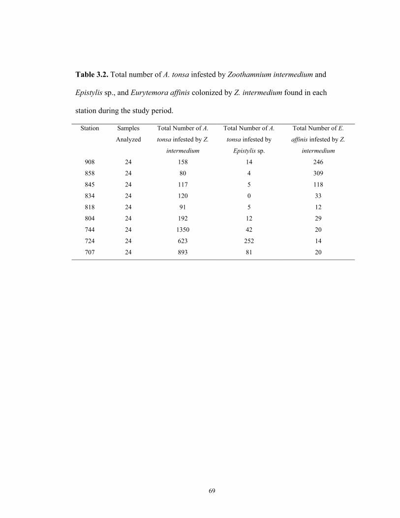
Table 3.2. Total number of A. tonsa infested by Zoothamnium intermedium and
Epistylis sp., and Eurytemora affinis colonized by Z. intermedium found in each
station during the study period.
Station Samples
Analyzed
Total Number of A.
tonsa infested by Z.
intermedium
Total Number of A.
tonsa infested by
Epistylis sp.
Total Number of E.
affinis infested by Z.
intermedium
908 24 158 14 246
858 24 80 4 309
845 24 117 5 118
834 24 120 0 33
818 24 91 5 12
804 24 192 12 29
744 24 1350 42 20
724 24 623 252 14
707 24 893 81 20
69

Table 3.3. Correlation coefficient and p values obtained from Spearman Rank
Correlation analyses for host abundance, environmental variables and infestation
prevalence of Zoothamnium intermedium attached to Acartia tonsa and Eurytemora
affinis and Epistylis sp. attached to A. tonsa in 1994-1996 and 1999-2000 in the
Chesapeake Bay. C = correlation coefficient (r2); p = p value; n=number of samples
VARIABLES INFESTATION PREVALENCE Z. intermedium attached
to A. tonsa Z. intermedium attached
to E. affinis Epistylis sp. attached to
A. tonsa C = 0.04 C = -0.1 C = 0.09 p = 0.5 p = 0.1 p = 0.1
Abundance of A. tonsa
n= 216 n = 216 n = 216 C = 0.1 C = 0.5 C = -0.02 p = 0.1 p = 0.1 p = 0.2
Abundance of E. affinis
n= 216 n = 216 n = 216 C = -0.08 C = -0.3 C = 0.1 p = 0.2 p = 0.1 p = 0.06
Salinity
n= 216 n = 216 n = 216 C = -0.3 C = -0.3 C = 0.1 p = 0.1 p = 0.1 p = 0.1
Temperature
n= 216 n = 216 n = 216 C = 0.2 C = 0.02 C = 0.2 p = 0.1 p = 0.6 p = 0.1
Dissolved Oxygen
n= 216 n = 216 n = 216 C = 0.2 C = -0.07 C = 0.2 p = 0.1 p = 0.2 p = 0.1
Chlorophyll a
n= 216 n = 216 n = 216 C = -0.1 C = -0.03 C = -0.03 p = 0.06 p = 0.6 p = 0.6
Abundance of Non-diatom < 10 µm
n= 216 n = 216 n = 216 C = 0.009 C = 0.2 C = -0.06
p = 0.8 p = 0.1 p = 0.3 Abundance of Non-
diatom > 10 µm n= 216 n = 216 n = 216
70

FIGURE LEGENDS Figure 3.1. Map of Chesapeake Bay showing stations where infestation prevalence
was estimated from ( ) and routine stations of the Chesapeake Bay Monitoring
Program that were used to determine host abundance ( ).
Figure 3.2. Infestation Prevalence (%) relative to salinity (psu) and temperature (oC)
profiles: (A) Z. intermedium attached to A. tonsa; (B) Epistylis sp. attached to A.
tonsa; (C) Z. intermedium attached to E. affinis. (.) for infestation prevalence <5%, ( )
for infestation prevalence >5<10%,( ) for infestation prevalence >10%
Figure 3.3. Infestation Prevalence (%) of Zoothamnium intermedium and Epistylis
sp.(A, C, E), and abundance (copepod/L) of Acartia tonsa and Eurytemora affinis (B,
D, F) in a temporal and spatial distribution.
Figure 3.4. Mean infestation prevalence (%) of Zoothamnium intermedium attached to
A. tonsa (A) and E. affinis (C), and mean abundance of A. tonsa (B) and E. affinis (D)
(copepod/L) between 1994-1996 and 1999-2000 along the main axis of the
Chesapeake Bay.
Figure 3.5. Mean infestation (%) of Z. intermedium and Epistylis sp. at different
categories of A. tonsa abundance (copepod/L).
71

Figure 3.6. Infestation density (colonies/mm2), mean number of zooids per colony,
and infestation load (zooids/mm2) of Z. intermedium and Epistylis sp. attached to
adults and copepodites of A. tonsa and E. affinis. Asterisks indicate a statistically
significant difference (p < 0.05).
Figure 3.7. Infestation density (colonies/mm2), mean number of zooids per colony,
and infestation load (zooids/mm2) of Z. intermedium and Epistylis sp. attached to
cephalothorax, abdomen, antenna, and swimming legs of A. tonsa and E. affinis.
Asterisks indicate a statistically significant difference (p < 0.05).
72

Figure 3.1
73

Figure 3.2
Temperature (0C)
0 5 10 15 20 25 30
Salin
ity (p
su)
0
5
10
15
20
25
30
Temperature (0C)
0 5 10 15 20 25 30
Sal
inity
(psu
)
5
10
15
20
25
30
Temperature (0C)0 5 10 15 20 25 30
Salin
ity (p
su)
0
5
10
15
20
25
A
B
C
74

Figure 3.3
Distance from the Mouth of the Bay (Km)
0 50 100 150 200 250
Infe
stat
ion
Pre
vale
nce
(%)
0
2
4
6
8
10
Distance from the Mouth of the Bay (Km)
0 50 100 150 200 250
Abu
ndan
ce (c
opep
od/L
)
0
2
4
6
8
10
12
14
16E FZ. intermedium on A. tonsa
Z. intermedium on E. affinisEpistylis sp. on A. tonsa
A. tonsa
E. affinis
MonthsMAR APR MAY JUN JUL AUG SEP
Infe
stat
ion
Prev
alen
ce (%
)
0
2
4
6
8
10C Z. intermedium on A. tonsa
Z. intermedium on E. affinisEpistylis sp. on A. tonsa
MonthsMAR APR MAY JUN JUL AUG SEP
Abu
ndan
ce (c
opep
od/L
)
0
2
4
6
8
10
12
14
16
18D A. tonsa
E. affinis
Years1994 1995 1996 1999 2000
Infe
stat
ion
Pre
vale
nce
(%)
0
1
2
3
4
5
6
7Z. intermedium on A. tonsaZ. intermedium on E. affinisEpistylis sp. on A. tonsa
A
Years1994 1995 1996 1999 2000
Abu
ndan
ce (c
opep
od/L
)
0
2
4
6
8
10
12
14
16
18B A. tonsa
E. affinis
75

Figure 3.4
Months
Mar Apr May Jun Jul Aug Sep
50
100
150
200
250
6
6
68
6
6
68 81210 10
8
5
1510
15
10
205
5
5
5
10
5
10
10
10
10
520 15
5
5
C
Dis
tanc
e fro
m th
e M
outh
of t
he B
ay (K
m)
50
100
150
200
250
2
2
22
2 10
8
86 4
1210 10
88
8
6
6
6 4
44
4
2
2
2
2
2
44
4
4
10
108
88
6
6
6
2
108
12
4 A
Mar Apr May Jun Jul Aug Sep
5 55
151010
B
D
76

Figure 3.5
Host Abundance (copepod/L)0-5 6-15 >16
Mea
n P
reva
lenc
e (%
)
0
2
4
6
8
10
11265
39
Z. intermediumEpistylis sp.
77
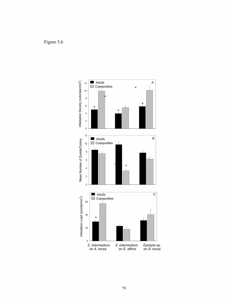
Figure 3.6
Infe
stat
ion
Den
sity
(col
onie
s/m
m2 )
0
2
4
6
8
10
12In
fest
atio
n Lo
ad (z
ooid
s/m
m2 )
0
20
40
60
AduCop
AdultsCopepodites
Z. interme on A. ton
Mea
n N
umbe
r of Z
ooid
s/C
olon
y
0
2
4
6
8
10
12
AdulCope
A
*
*
*
ltsepodites
dium Z. intermedisa on E. affin
tspodites
*
*
78
*
B
C
*
*
*
um Epistylis sp.is on A. tonsa

Figure 3.7
Body Part
Ceph Abd Ant Legs
Infe
stat
ion
Den
sity
(col
onie
s/m
m2 )
0
5
10
15
20
25Z. intermedium on A. tonsaZ. intermedium on E. affinisEpistylis sp. on A. tonsa
Ceph Abd Ant Legs
Mea
n N
umbe
r of Z
ooid
s/C
olon
y
0
1
2
3
4
5
6
Ceph Abd Ant Legs
Infe
stat
ion
Load
(zoo
ids/
mm
2 )
0
10
20
30
40
50
60
70
Z. intermedium on A. tonsaZ. intermedium on E. affinisEpistylis sp. on A. tonsa
Z. intermedium on A. tonsaZ. intermedium on E. affinisEpistylis sp. on A. tonsa
A
B
C
*
*
* **
*
**
** *
*
* **
**
*
79

CHAPTER 4
TELOTROCH FORMATION, SURVIVORSHIP, ATTACHMENT SUCCESS,
AND GROWTH OF THE EPIBIOTIC PERITRICH Zoothamnium intermedium
PRECHT, 1935
80

ABSTRACT
In the present study I investigated aspects of the life cycle of the peritrich
Zoothamnium intermedium, an epibiont on calanoid copepods in the Chesapeake Bay.
Controlled laboratory experiments were designed to characterize induction of
formation, survivorship, and attachment of free-swimming stages (telotrochs), as well
as growth rates of the trophont stage attached to its primary host (the copepod Acartia
tonsa). Results showed that formation of telotroch stages started two hours after the
death of the host, and within seven hours more than 90% of the zooids had formed
dispersal stages and left the substrate. Induction of telotroch formation occurred only
following the death of the copepod to which the epibiont was attached, suggesting that
a combination of mechanical and chemical cues is involved in triggering formation of
dispersal stages in Z. intermedium.
Telotrochs of Z. intermedium were able to survive for 14 hours in the absence
of their primary copepod hosts (A. tonsa and Eurytemora affinis) independent of the
copepod species from which the telotrochs had dispersed. In addition, no attachment
to non-living substrates was observed in either the laboratory or the field, strongly
indicating that this species of peritrich is an obligate epibiont. Attachment success of
dispersal stages decreased as their age increased; the highest attachment frequency
was attained by three-hour old telotrochs, suggesting that colonization rates in nature
may strongly depend on the production of free-swimming stages by the ciliate.
Maximum growth rates of Z. intermedium were 0.8 day-1 with a bacterial or
algal primarily based diet. Colonization rates (rate of change of colonies/host) were
81

lower than growth rates (0.4 day-1) and reached a plateau at high concentrations of
food. The rate of change in zooids per colony (proliferation rate) did not reach a
plateau in the algal primarily based diet study, offering a support to the theory that
epibiont abundance and frequency increases with increased environmental
eutrophication.
The experiments and results presented here may serve as a tool for the design
of studies assessing epibiont host specificity, as well as a model for the investigation
of the life cycle of free-living and epibiont peritrichs in other systems.
82

INTRODUCTION
The occurrence of peritrich ciliates colonizing planktonic crustacea is well
documented (Hermann and Mihursky, 1964; Green, 1974; Henebry and Ridgeway,
1979; López et al., 1998), with the majority of the reports focusing specially on the
effects of these epibionts on host survivorship and reproduction (Kankaala and
Eloranta, 1987; Xu and Burns, 1991; Xu, 1992; Weissman, Lonsdale and Yen, 1993).
However, studies emphasizing aspects of the peritrich life cycle such as formation and
survivorship of free-swimming dispersal stages, ability to colonize multiple hosts or
non-living substrates, or growth rates of these ciliates are still lacking.
The life cycle of sessile peritrichs is composed, generally, by a stalked feeding
stage, the trophont, which is attached to a substrate, and a free-swimming dispersal
stage, called telotroch. Telotroch formation in coenobial, colonial or solitary
peritrichs can be a result of asexual reproduction or a direct transformation of the
zooids, which frequently occurs in response to unfavorable environmental conditions
such as low concentration of oxygen or direct disturbance of the organism. In the case
of peritrichs living as epibionts, telotroch formation could also be triggered by the
death or molt of the host substrate.
Induction of telotroch formation in laboratory cultures of peritrichs has been
performed by briefly exposing cells to air (Nakajima, Hashimoto, and Asai, 1986), by
changing the culture medium (Rose and Finley, 1976), by disturbing mechanically the
colonies (Succhard, 1979), or by treating the trophont stage with a combination of
chemicals (de Baufer et al., 1999). However, such experiments were all performed
83

with peritrichs attached to non-living substrates and no data on formation of dispersal
stages are available for those ciliates colonizing living organisms.
Studies emphasizing epibiont host specificity have pointed out that some
peritrichs are able to attach to only one or two host species, failing to colonize non-
living substrates (Clamp, 1973; Henebry and Ridgeway, 1979; Gilbert and Schröder,
2003). By contrast, some authors demonstrated that some epibionts are opportunistic,
being able to attach to living or non-living substrates (Mayén-Estrada and Aladro-
Lubel, 2000). However, only a few studies have investigated the ability of a peritrich
epibiont to attach to non-living substrates in the laboratory and/or in the field
(Henebry and Ridgeway, 1979; Mayén-Estrada and Aladro-Lubel, 2000).
Assessment of community composition and growth rates of free-living
peritrichs have been the subject of only a few studies in freshwater environments, such
as streams and activated sludge plants (Curds and Vandyke, 1966; Taylor, 1983;
Kusuoka and Watanabe, 1987; Harmsworth, Sleigh, and Baker, 1992; Harmsworth
and Sleigh, 1993). One of the most comprehensive studies of growth in situ of
peritrichs in the field was performed by Kusuoka and Watanabe (1987) in an urban
stream in Japan. They sampled colonies of Carchesium polypinum and individuals of
Vorticella microstoma attached to transparent plastic films and found growth rates of
1.37 day-1 and 1.01 day-1 for the two peritrichs respectively. Regarding peritrichs as
epibionts, only one study so far performed by Gilbert and Schröder (2003) has dealt
with growth rates of peritrichs attached to living substrates in the laboratory. The
authors observed an exponential growth of colonies of Epistylis pygmaeum, from a
84

single zooid, attached to the rotifer Brachionus angularis , but no actual growth or
colonization rates were calculated for the trophont stage of this species.
In the present study, I have investigated induction of telotroch formation,
survivorship, attachment success, growth rate, and the ability of Zoothamnium
intermedium, a peritrich epibiont attached to Acartia tonsa and Eurytemora affinis
from the Chesapeake Bay, USA, to colonize non-living substrates in the field. To
determine if telotroch formation in Z. intermedium is associated with chemical and/or
mechanical cues released by the host in which the peritrich is attached, or to some
cues present in the environment, or to impairment or reduced swimming of the host, a
series of experiments was performed, where infected copepods were killed, injured, or
tethered or exposed live to dead non-infested hosts. To investigate the survivorship of
telotrochs and the ability of them to attach to non-living substrates in absence of their
primary hosts, laboratory experiments where the abundance and attachment of
telotrochs were monitored over time were carried out. The ability of Z. intermedium
to attach to non-living substrates in the field was also investigated by sampling the
aufwuchs community of a tributary of the Chesapeake Bay over a period of seven
weeks. To determine if attachment success decreases as age increases, telotrochs of
different ages were exposed to their primary host A. tonsa and attachment was
monitored over time. Finally, to follow the sequence of telotroch formation,
survivorship, and attachment success, growth rates of Z. intermedium colonizing A.
tonsa were carried out with two diets, bacteria only and algae as the main sources of
food.
85

MATERIALS AND METHODS
Cultures of Acartia tonsa, Eurytemora affinis and their epibionts: A. tonsa and E.
affinis were collected from the Patuxent River (a tributary of the Chesapeake Bay)
brought live to the laboratory and examined for the presence of ciliate epibionts.
Copepods were transferred to 4-liter beakers (3 cultures for each species) containing 2
liters of < 0.45 µm filtered seawater with salinity adjusted for 15 psu, and maintained
as described in Chapter 2. Cultures of Zoothamnium intermedium were also initiated
and maintained following the methods described in Chapter 2.
1.0. Telotroch formation following death of the host: The formation of free-
swimming stages (telotrochs) in the peritrich epibiont Zoothamnium intermedium
following host substrate death was investigated by performing a series of three
identical experiments. Infested A. tonsa and E. affinis, obtained from laboratory
cultures were placed on a glass slide and a cover slip was gently pressed on top of the
copepod. This procedure killed the host, but the epibionts remained alive. Dead hosts
and attached epibionts were placed in plastic Petri dishes (in each of the three runs
containing 5 replicates with one copepod each) filled with 10 ml of < 0.45µm 15psu
water, and the number of zooids/host was assessed using a dissecting microscope.
Dishes were placed in an incubator at 20oC and the number of zooids attached to the
host was assessed hourly. The experiment was terminated when more than 90% of the
zooids had formed telotrochs and left the host.
86

1.1. Telotroch formation induced by dead copepods: To determine if telotroch
formation of the epibiont Zoothamnium intermedium can be induced by the exposure
of an infested copepod to epibiont-free dead copepods, a series of experiments was
conducted. Non-infested A. tonsa obtained from laboratory cultures were killed as
described above. Dead copepods were placed in plastic containers filled with <
0.45µm 15psu water with a final concentration of 1, 2 and 10 dead, clean hosts per ml.
The experiment with 10 dead hosts/ml was carried out in a multi-well culture
chamber, with a final volume of water of 1 ml per replicate. The experiments
conducted with 1 and 2 dead hosts/ml were carried out in Petri dishes with a final
volume of water of 10 ml per replicate. Epibiont colonies attached to A. tonsa
obtained from laboratory cultures were enumerated, and infested hosts were placed in
chambers containing dead copepods at a final concentration of one infested host per
experimental unit. Experiments for each dead host concentration were run three times
with five replicates each. Experimental containers were placed in an incubator at 20oC
and the total number of attached zooids was assessed hourly for 7 hours (see Results
for rationale on selecting time).
1.2 Telotroch formation triggered by inability of host to swim: To assess if
telotroch formation in Z. intermedium can be induced by slowing down the movement
of the host, infested A. tonsa from laboratory cultures were tethered with a cat hair
(approximately 1 cm in length). One infested A. tonsa was placed in a drop of water
on a glass slide, the number of zooids per epibiont colony was assessed, and the excess
of water was removed. Both tips of the cat hair were dipped into an ethyl
87
cyanoacrilate-based glue (commercially known as “crazy glue”), and one of them was
attached to the copepod (to a region where no epibiont colonies were present). The
tethered copepod was immediately transferred to a Petri dish filled with 10 ml of <
0.45µm 15psu water with a final concentration of one infested host per experimental
unit, and the second tip of the hair was glued to the container’s wall above the water
line (while the copepod remained submerged). As a control for telotroch formation, a
tethered infested A. tonsa, with a known number of attached zooids, was killed by
pressing gently a Pasteur pipette in the middle of the host’s cephalothorax (in a place
where no epibiont colonies were present). Control and treatment units had five
replicates each and the experiment was run three times. Experimental containers were
placed in an incubator at 20oC and number of attached zooids in each replicate was
assessed hourly for 7 hours.
1.3. Telotroch formation induced by host injury: To determine if telotroch
formation in Z. intermedium is induced by impaired swimming of the host and/or
exudates present in host wounds, one swimming leg and one antenna were removed
with a forceps from infested A. tonsa obtained from laboratory cultures. After limb
removal, the number of zooids per epibiont colony was assessed for each host, and
infested copepods were placed in Petri dishes filled with 10 ml of < 0.45µm 15psu
water, with a final concentration of one host per experimental container. The
experiment was run three times with five replicates each. Dishes were placed in
incubator at 20oC and the number of attached zooids was assessed hourly for 7 hours.
88

1.4. Telotroch formation induced by inability of the host to swim and by the
presence of non-infested dead hosts: To assess if telotroch formation can be induced
by a combination of reduced swimming by the host and a chemical cue released by
dead copepods in the environment, infested A. tonsa obtained from laboratory cultures
were tethered with a cat hair, as described above, the number of zooids per copepod
was assessed, and hosts were placed in a multi-well chamber filled with 1 ml of <
0.45µm 15psu water at a final concentration of one tethered host per well. Non-
infested A. tonsa also obtained from laboratory cultures were killed with a cover slip
as described above and placed in the wells containing a tethered infested host at a final
concentration of 10 dead hosts per experimental unit. Chambers were kept in the same
conditions described for the other experiments and number of zooids per host was
assessed hourly for 7 hours.
2.0. Survivorship of Z. intermedium in the absence of live hosts: To determine how
long the free-swimming stages of Zoothamnium intermedium are able to survive in the
absence of the two host species, and also if they were able to attach to the bottom and
walls of the experimental chamber, 30 infested A. tonsa and E. affinis (with a load of
about 100 zooids each) obtained from laboratory cultures were killed as described
above. Dead hosts were placed in glass Petri dishes (3 replicates with 10 copepods
each) filled with 30 ml of < 0.45µm 15psu water. Dishes were placed in an incubator
at 20oC, and after 3 hours (see Results for rationale on selecting time), dead copepods
with their remaining attached zooids were removed from the chambers using a glass
Pasteur pipette. Abundance of telotrochs in the water was assessed over time by
89

taking 2-ml samples at two-hour intervals, preserving them with one drop of acid
Lugol’s, and counting the number of cells using a Sedgewick-Rafter chamber (one
entire chamber was counted for each replicate). The experiment was terminated when
the average concentration of cells/ml in the three replicates was < 2. Attachment of Z.
intermedium to the bottom and walls of the experimental dish was verified by
scanning the dish (all replicates) in a dissecting microscope.
3.0 Attachment of Z. intermedium to coverslips in the field: To extend the previous
experiment regarding the ability of Z. intermedium to attach to non-living substrates,
and to determine if this species is a common member of the benthic community,
cover-slip traps were sampled from the field and analyzed for the presence of Z.
intermedium during a period of seven weeks. Small rectangular boxes (6 cm long X
3.5 cm wide) made with Plexiglas®, with open sides, and six grooves in the internal
sides, each capable of holding 2 cover slips (22 mm X 22 mm), were used as traps.
Inside each box, 12 cover slips were placed back to back along the grooves and a
rubber band was crossed over the open sides to assure that the cover slips would stay
in place. Two sets of traps were suspended in the Rhode River (a tributary of the
Chesapeake Bay) from the dockside at the Smithsonian Environmental Research
Center (38o46’N and 76o52’W), Edgewater, Maryland, on April 15th, 21st, 28th, May
12th, 23rd, and June 3rd 2003. On the first three sampling dates, traps were left in the
field for a period of seven days to allow enough time for colonization, which is
dependent of water temperature. For all other sampling dates, traps were left in the
90

field for a period of four days. At the time of recovery, traps were directly placed in a
beaker filled with water from the site and taken to the laboratory.
Two cover slips from each set of traps were preserved with modified Bouin’s
fixative at a final concentration of 5% (Coats and Heinbokel, 1982), and four cover
slips from each set were placed in small Petri dishes filled with water from the
collection site for observation of live organisms with an inverted microscope
(Invertscope, 25X magnification; Zeiss Corp.). Five fields of view in each cover slip
were inspected for the presence of colonies of Zoothamnium. The total number of
colonies and zooids in each field of view was recorded and the abundance of
Zoothamnium per area was calculated. The Protargol staining technique (Zagon and
Small, 1970) was applied to the preserved material to confirm the identification of the
Zoothamnium species that were observed alive and attached to the cover slips. Filters
were omitted from the preparation and cover slips were run throughout the procedure
and mounted on glass slides. Protargol slides were observed using light microscopy,
and morphological characteristics such as organization of the oral apparatus were
observed for each Zoothamnium colony and compared to the described morphology of
Z. intermedium attached to copepods (see Chapter 2).
To assess the occurrence of Z. intermedium on copepods, plankton samples
were also collected from each site at the time of cover-slip recovery. Horizontal net
tows were taken using a plankton net with 202-µm mesh size and 50-cm in diameter.
A 125 ml sub-sample from the net tow was fixed with modified Bouin’s fluid (Coats
and Heinbokel, 1982) at a final concentration of 5 %, and examined using a dissecting
microscope (Bausch and Lomb; 30X magnification) to determine the presence of Z.
91

intermedium on adults and copepodites of Acartia tonsa and Eurytemora affinis.
Identification of Z. intermedium was confirmed by staining infested copepods
randomly selected from the samples (~5 copepods/sample). The Protargol staining
technique (Zagon and Small, 1970) was run following the procedure described in
Chapter 2 and diagnostic characteristics of Z. intermedium (the same as those
described in Chapter 2) were observed using light microscopy. Infestation prevalence
(percentage of copepods carrying epibionts) was calculated for adults and copepodites
of both host species.
4.0 Attachment success of Z. intermedium to A. tonsa: To determine if telotroch
attachment success decreases as its age increases, infested A. tonsa (60 copepods with
a load of about 50 zooids each) were obtained from laboratory cultures and were killed
as described in experiment 1.0. Dead hosts were placed in a glass Petri dish with 30
ml of 15 psu filtered water (< 0.45µm). Petri dishes were placed in an incubator at
20oC, and after three hours, dead copepods with their remaining zooids were removed
from the container with a glass Pasteur pipette. A 2 ml sample was taken from the
Petri dish, fixed with acid Lugol’s and counted using a Sedgewick-Rafter chamber to
assess the initial concentration of telotrochs/ml. A final concentration of 1 telotroch/ml
was added to each of 15 scintillation vials filled with 24 ml of 15 psu filtered water (<
0.45µm). Non-infested, adult A. tonsa obtained from laboratory cultures were added to
the experimental vials (one copepod/vial). Scintillation vials were placed randomly in
a plankton wheel (rotating at two rpm) and three replicates were fixed with acid
Lugol’s every two hours for 10 hours. To assess attachment rate, preserved copepods
92
were removed from the sample with a Pasteur pipette and observed with light
microscope and the number of zooids attached to the host was recorded. The rest of
the sample was settled for at least 24 hours and after that it was reduced to a volume of
10 ml, transferred to a settling chamber and settled for another 24 hours. The number
of telotrochs that did not attach to the available host was counted using an inverted
microscope (Invertscope; 100X magnification; Zeiss Corp.). The assessment of
telotrochs that did not attach in addition to the attached zooids would allow an
estimate of predation rates of copepods on free-swimming stages of their epibionts.
The same procedure was repeated in separate experiments where telotrochs entered the
experiments after 5, 7, and 9 hours after the death of the original hosts.
5.0. Growth rates of Z. intermedium feeding on bacteria: To assess growth rates of
Z. intermedium feeding on bacteria alone, the development of clonal ceonobia was
observed over a period of 96 hours. Adult females of A. tonsa were exposed to
telotrochs of Z. intermedium, and hosts carrying a single zooid were transferred to
glass Petri dishes filled with 150 ml of <0.45µm 15 psu water, enriched with varying
amounts of Cerophyl® to reach different bacterial concentrations. As food for the
copepods, the diatom Thalassiosira weisflogii was added at a final concentration of
1X10³ cells/ml. The experiment had a total of five treatments (bacterial concentration
of 104, 105, 106, 107, and 108) with three replicates per treatment and one infested host
per replicate. Experimental chambers were kept in an incubator at 20oC with
photoperiod of 14:10 light:dark hours. Copepods were observed every 24 hours for a
period of four days and the total number of attached zooids; number of coenobia per
93

host and mean number of zooids per coenobia were recorded. Growth rates (rate of
change in number of epibiont zooids) were calculated using the following formula:
ln(Nt) = ln(N0) + rt, where, Nt is the number of zooids at time t; N0 is the number of
zooids at time 0; t is the period of time in days; and r is the growth rate. Colonization
rates (rate of change in number of epibiont coenobia) were calculated using the same
formulae as suggested by Kusuoka and Watanabe (1987): ln(Nt’) = ln(N0
’) + r’ t,
where, Nt’ is the number of epibiont coenobia at time t; N0
’ is the number of epibiont
coenobia at time 0, t is the period of time in days; and r’ is the colonization rate.
Proliferation rates (rate of change in the mean number of epibiont zooids/coenobium)
were calculated using the same formulae: ln(Nt”) = ln(N0
”) + r” t; where Nt” is the mean
number of epibiont zooids/coenobium at time t; N0” is the mean number of epibiont
zooids/coenobium at time 0; t is the time in days; and r” is the proliferation rate.
A water sample (5 ml) from each replicate was taken at the beginning (T0) and
at the end of the experiment (T4), and preserved with formaldehyde for enumeration of
bacteria using the DAPI staining technique (Porter and Feig, 1980). Counts were
performed using an Axioscope equipped with optics epifluorescence (Zeiss Corp.).
For each replicate, duplicate counts were carried out up to a total of 200 cells/count.
Enumeration of background bacteria was performed by replicate counts of blank filters
(for each concentration) and the result was included in the calculation of bacterial
concentration.
5.1. Growth rates of Z. intermedium feeding on Isochrysis galbana based diet: To
determine growth rates of Z. intermedium feeding primarily on the haptophyte algae
94

Isochrysis galbana, the development of coenobia originated from a single zooid was
observed over a period of 96 hours. The experimental set up used in this experiment
was the same used to assess growth rates of the epibiont feeding on bacteria. The
experiment had a total of six concentrations of I. galbana (101, 102, 103, 104, 105, and
106 cells/ml) with three replicates per treatment and one infested A. tonsa per replicate.
The number of zooids and coenobia per host and mean number of zooids per coenobia
were recorded every 24 hours for 96 hours. A water sample (5 ml) from each replicate
was taken at every observation point and fixed with acid Lugol’s for enumeration of I.
galbana. Concentration of algal food was assessed by direct counts using a Palmer-
Maloney counting chamber. Triplicate counts were performed for each replicate to a
total of 100 cells/count.
Data analyses: As the data passed normality and homogeneity of variance tests,
regression analyses to determine the best-fit model and slopes of the curves were run
in the telotroch formation, survivorship and attachment success experiments. Time
was used as independent variable and percentage of zooids in the colonies, telotrochs
in the water, or zooids attached were the dependent variables. Analyses of Variance
were performed on the slope of the curves to discern differences in telotroch formation
and survivorship between the two host species and in attachment success among
different telotroch ages. Statistical Analyses were performed using SPSS for
Windows Version 11.0 (SPSS Inc.) and Sigma Stat Version 2.0 (SPSS, Inc.). Data is
plotted as means + SE (standard error of the mean), and for all statistical tests the level
of alpha used was 0.05.
95
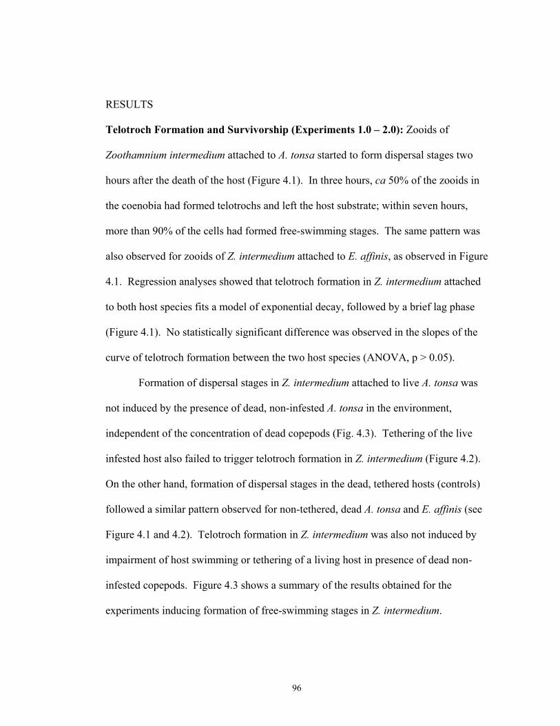
RESULTS
Telotroch Formation and Survivorship (Experiments 1.0 – 2.0): Zooids of
Zoothamnium intermedium attached to A. tonsa started to form dispersal stages two
hours after the death of the host (Figure 4.1). In three hours, ca 50% of the zooids in
the coenobia had formed telotrochs and left the host substrate; within seven hours,
more than 90% of the cells had formed free-swimming stages. The same pattern was
also observed for zooids of Z. intermedium attached to E. affinis, as observed in Figure
4.1. Regression analyses showed that telotroch formation in Z. intermedium attached
to both host species fits a model of exponential decay, followed by a brief lag phase
(Figure 4.1). No statistically significant difference was observed in the slopes of the
curve of telotroch formation between the two host species (ANOVA, p > 0.05).
Formation of dispersal stages in Z. intermedium attached to live A. tonsa was
not induced by the presence of dead, non-infested A. tonsa in the environment,
independent of the concentration of dead copepods (Fig. 4.3). Tethering of the live
infested host also failed to trigger telotroch formation in Z. intermedium (Figure 4.2).
On the other hand, formation of dispersal stages in the dead, tethered hosts (controls)
followed a similar pattern observed for non-tethered, dead A. tonsa and E. affinis (see
Figure 4.1 and 4.2). Telotroch formation in Z. intermedium was also not induced by
impairment of host swimming or tethering of a living host in presence of dead non-
infested copepods. Figure 4.3 shows a summary of the results obtained for the
experiments inducing formation of free-swimming stages in Z. intermedium.
96

Telotrochs of Z. intermedium were able to survive a short period of time in the
absence of their primary hosts A. tonsa and E. affinis. After seven hours following the
death of the host (i.e. 7-hour old telotrochs), 80 % of the telotroch population was still
present in the water (Figure 4.4). This number decreased to 50 % in 9-hour-old
telotrochs and reached 20 % in 11-hour-old telotrochs. After 14 hours following the
death of the host (14 hour-old telotrochs), the concentration of telotrochs in the water
was < 2 cells/ml (Figure 4.4). No attachment was observed to the walls or bottom of
the experimental container in the experiments with telotrochs originated from
coenobia attached to both A. tonsa or E. affinis. Similarly to telotroch formation,
regression analyses indicated that survivorship curves of Z. intermedium telotrochs fit
in an exponential decay model (R2=0.93) followed by a lag phase. No statistically
significant difference was observed for the slopes of the curves of telotroch
survivorship for A. tonsa and E. affinis (ANOVA, p > 0.05).
Attachment of Z. intermedium to non-living substrates in the field (Experiment
3.0): Live observations and Protargol staining revealed that four species in the genus
Zoothamnium (two of them tentatively identified as Z. alternans and Z. arbuscula)
were present in the benthic community of the Rhode River during a period of seven
weeks between April and June 2003, but none of them was identified as the same
epibiont species (Zoothamnium intermedium) that colonizes populations of A. tonsa
and E. affinis in the Chesapeake Bay and its tributaries. Assessment of the number of
coenobia and zooids of Zoothamnium per substrate area showed that this genus was
the dominant Peritrich from April 15th to April 21st, but was outnumbered by
97
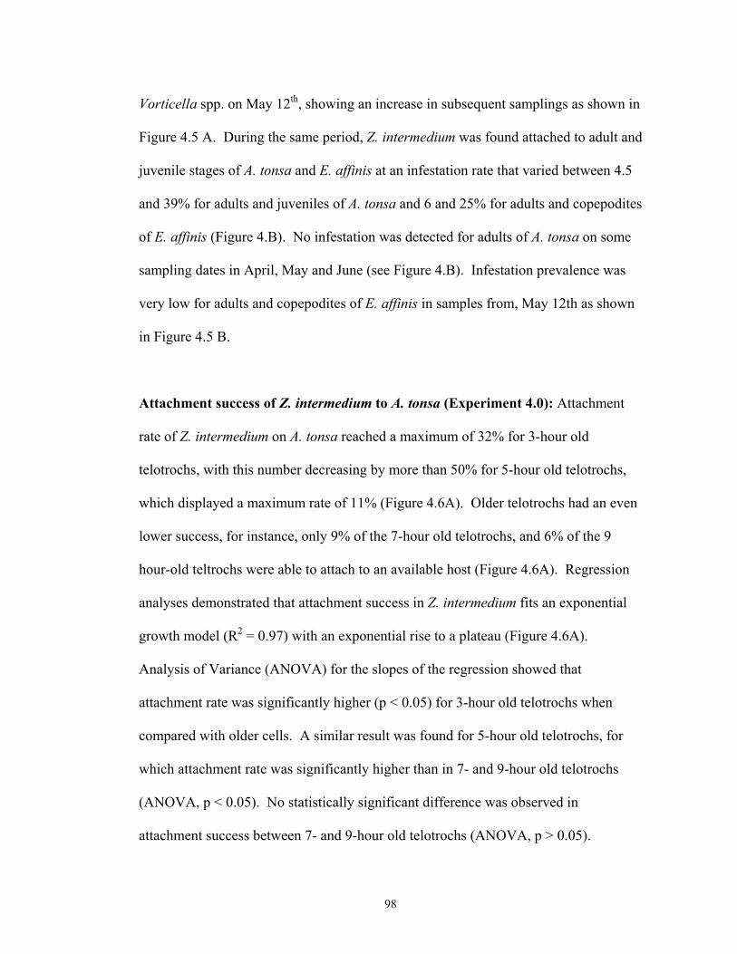
Vorticella spp. on May 12th, showing an increase in subsequent samplings as shown in
Figure 4.5 A. During the same period, Z. intermedium was found attached to adult and
juvenile stages of A. tonsa and E. affinis at an infestation rate that varied between 4.5
and 39% for adults and juveniles of A. tonsa and 6 and 25% for adults and copepodites
of E. affinis (Figure 4.B). No infestation was detected for adults of A. tonsa on some
sampling dates in April, May and June (see Figure 4.B). Infestation prevalence was
very low for adults and copepodites of E. affinis in samples from, May 12th as shown
in Figure 4.5 B.
Attachment success of Z. intermedium to A. tonsa (Experiment 4.0): Attachment
rate of Z. intermedium on A. tonsa reached a maximum of 32% for 3-hour old
telotrochs, with this number decreasing by more than 50% for 5-hour old telotrochs,
which displayed a maximum rate of 11% (Figure 4.6A). Older telotrochs had an even
lower success, for instance, only 9% of the 7-hour old telotrochs, and 6% of the 9
hour-old teltrochs were able to attach to an available host (Figure 4.6A). Regression
analyses demonstrated that attachment success in Z. intermedium fits an exponential
growth model (R2 = 0.97) with an exponential rise to a plateau (Figure 4.6A).
Analysis of Variance (ANOVA) for the slopes of the regression showed that
attachment rate was significantly higher (p < 0.05) for 3-hour old telotrochs when
compared with older cells. A similar result was found for 5-hour old telotrochs, for
which attachment rate was significantly higher than in 7- and 9-hour old telotrochs
(ANOVA, p < 0.05). No statistically significant difference was observed in
attachment success between 7- and 9-hour old telotrochs (ANOVA, p > 0.05).
98
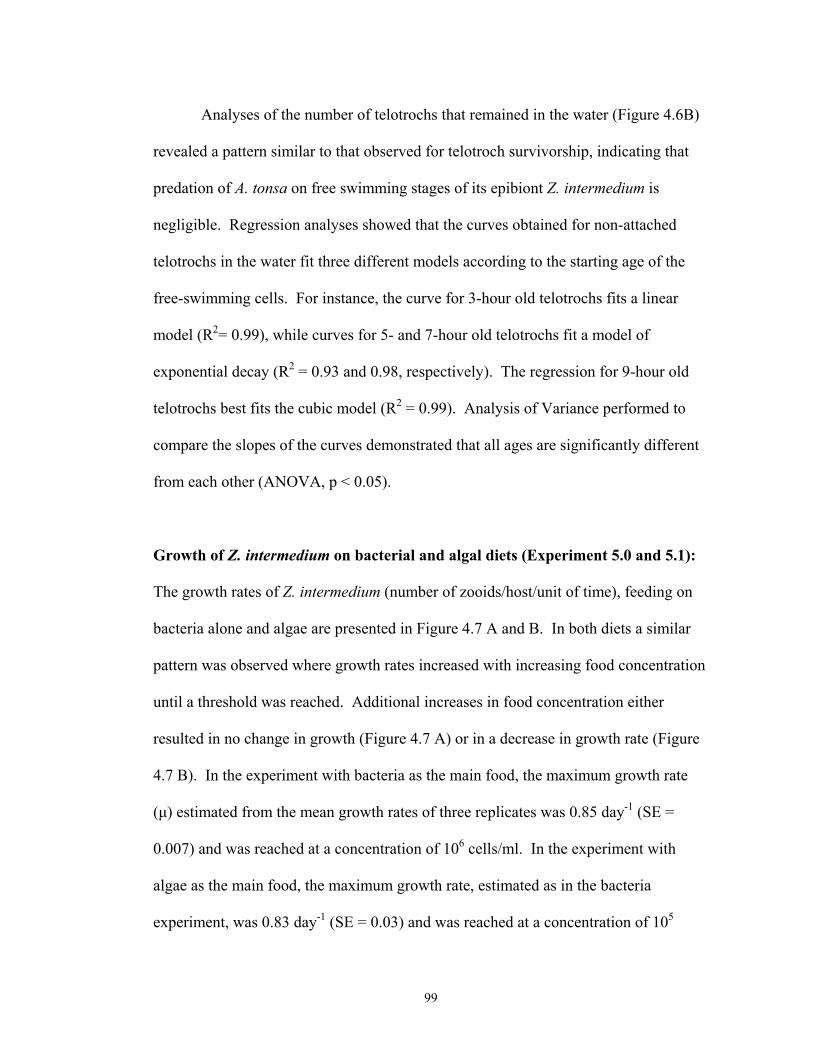
Analyses of the number of telotrochs that remained in the water (Figure 4.6B)
revealed a pattern similar to that observed for telotroch survivorship, indicating that
predation of A. tonsa on free swimming stages of its epibiont Z. intermedium is
negligible. Regression analyses showed that the curves obtained for non-attached
telotrochs in the water fit three different models according to the starting age of the
free-swimming cells. For instance, the curve for 3-hour old telotrochs fits a linear
model (R2= 0.99), while curves for 5- and 7-hour old telotrochs fit a model of
exponential decay (R2 = 0.93 and 0.98, respectively). The regression for 9-hour old
telotrochs best fits the cubic model (R2 = 0.99). Analysis of Variance performed to
compare the slopes of the curves demonstrated that all ages are significantly different
from each other (ANOVA, p < 0.05).
Growth of Z. intermedium on bacterial and algal diets (Experiment 5.0 and 5.1):
The growth rates of Z. intermedium (number of zooids/host/unit of time), feeding on
bacteria alone and algae are presented in Figure 4.7 A and B. In both diets a similar
pattern was observed where growth rates increased with increasing food concentration
until a threshold was reached. Additional increases in food concentration either
resulted in no change in growth (Figure 4.7 A) or in a decrease in growth rate (Figure
4.7 B). In the experiment with bacteria as the main food, the maximum growth rate
(µ) estimated from the mean growth rates of three replicates was 0.85 day-1 (SE =
0.007) and was reached at a concentration of 106 cells/ml. In the experiment with
algae as the main food, the maximum growth rate, estimated as in the bacteria
experiment, was 0.83 day-1 (SE = 0.03) and was reached at a concentration of 105
99

cells/ml. The maximum doubling rate of Z. intermedium was 1.20 and 1.23 per day in
the experiments with algae and bacteria respectively.
Figure 4.7 C and D show the colonization rates (increasing of colonies/host) of
Z. intermedium on A. tonsa feeding on bacteria and algae respectively. With both
diets, colonization rates increased with an increasing food concentration until a
maximum was reached. Further increases in food concentration, in both cases,
resulted in a decrease in colonization rates after reaching this maximum (Figure 4.7 C
and D). In the experiment with bacteria, the maximum colonization rate was 0.50 day-
1 (SE = 0.04) and was observed at a concentration of 107 cells/ml, one order of
magnitude higher than the observed growth rates. Colonization rates in an algal-based
diet were similar to those observed for bacteria alone. The maximum rate was 0.51
day-1 (SE = 0.01) and was observed at the same concentration for which maximum
growth rates were achieved (105 cells/ml).
The mean number of zooids per coenobium (proliferation rate) of Z.
intermedium was also similar for bacterial and algal diets (Figure 4.7 E and F). With a
diet based on bacteria, the maximum proliferation rate was 0.44 day-1 (SE = 0.04) and
was reached at a concentration of 106 cells/ml (Figure 4.7 E). Differently from the
bacterial diet and from growth and colonization rates, proliferation rate on an algal
diet did not reach a plateau, and the highest rate of 0.42 (SE = 0.038) was recorded at
106 cells/ml (Figure 4.7 F). Algae concentration remained fairly constant throughout
the study, as can be seen in figures 4.8 A and B.
100

DISCUSSION
The present study investigated induction of telotroch formation, survivorship,
attachment success, ability to colonize non-living substrates, and growth rates of the
peritrich epibiont Zoothamnium intermedium attached to calanoid copepods in the
Chesapeake Bay, USA. This is the first comprehensive study focusing on aspects of
the life cycle of an obligate peritrich epibiont, some of these aspects have been
investigated for free-living species of peritrichs.
Formation of free swimming stages in the epibiont peritrich Zoothamnium
intermedium was induced by killing the infested host, with no telotroch formation
observed when the host was tethered, injured, or when clean dead hosts were present
in the environment. Telotrochs were also formed in this species as a result of binary
fission, as was indicated by the appearance of new colonies in the growth rate
experiment (Figure 4.7). Transformation of trophonts into dispersal stages in peritrich
epibionts has been demonstrated to follow the death of or molt of the substrate
(Clamp, 1973), and as in free-living peritrichs, to form as a product of binary fission
during asexual reproduction (Nagasawa, 1986). In addition to a result of binary
fission, telotroch formation in peritrichs attached to Crustacea can occur before or
after the host sheds its molt, as was reported by some authors who observed this aspect
of the epibiont life cycle. For example, Fenchel (1965) observed that all species of the
ciliates chonotrich and peritrich ciliates (including species of Epistylis and
Zoothamnium) attached to the amphipod Gammarus spp. formed free-swimming cells
prior to the host’s molt, colonizing the same substrate immediately after the molt.
Green (1974) observed that the population density of Epistylis helenae attached to the
101

cladoceran Daphnia pulex decreased before the end of the host’s instar. He also
demonstrated that most of the stalks in the colony were empty before the molting
process started, suggesting that some cue could be detected by the epibiont, which in
turn would form dispersal stages and be available for recolonization of the same host
individual when its cast skin was shed. Willey and Threlkeld (1995), also observed
that Vorticella campanula reached a positive burden immediately after its cladoceran
host, Daphnia pulex casted its carapace. By contrast, Clamp (1973) reported that
Lagenophrys lunatus attached to species of shrimp in the genus Palemonetes started
the process of forming telotrochs after the host has cast its molt. The time was not
quantified but several hours were needed to complete telotroch formation and
evacuation of the old carapace. In the present work, observations of infested molts
demonstrated that Zoothamnium intermedium started forming telotrochs after the old
carapace had been shed by the copepod hosts, and not beforehand as was
demonstrated for other peritrichs including species in the genus Zoothamnium
(Fenchel, 1965). Although the time taken to form telotrochs from the shed carapace
was not quantified in the present study, observation of old carapaces and
quantification of time of telotroch formation following the death of the host suggest
that, as described for Lagenophrys lunata (Clamp, 1973), several hours would be
required for Z. intermedium to form dispersal stages and leave the host’s old
exoskeleton.
Despite these observations, the nature of the mechanisms involved in triggering
telotroch formation following the death or the molt of the host is still not well
understood. The results obtained for Z. intermedium in this study suggest that a
102

combination of chemical and mechanical stimuli coming from the individual host the
epibiont is attached to, is responsible for the triggering of telotroch formation in this
species of peritrich (Figure 4.3). Interestingly, production of dispersal stages in free-
living peritrichs is sometimes associated with changes in environmental conditions
such as low oxygen concentration, or high carbon dioxide tension (Bick, 1972). It was
demonstrated for Vorticella spp. that when the oxygen concentration falls below 2
mg/l these organisms form free-swimming stages and leave their stalks behind to find
a more suitable substrate. Formation of telotroch stages of Z. intermedium, in the
present study, seemed to be linked to cues coming from the host and not to changes or
even signals released in the environment. At same time, since change in pH, bacterial
concentration, and dissolved oxygen concentration were not measured in the
performed experiments, these environmental parameters might also be involved in the
whole process of telotroch formation in this epibiont species.
Although the results obtained from the induction of telotroch formation
experiments were not conclusive with respect to the exact stimulus received by the
epibiont, if any, this study demonstrated an efficient and reproducible method of
massive production of telotrochs that can be used to investigate morphological
developmental, and ecological aspects of Z. intermedium. The usefulness of a similar
approach was pointed out by Vachianno et al. (1992) who developed a method of
telotroch formation for Vorticella convallaria attached to non-living substrates.
Interestingly, in their experiment with V. convallaria a very high concentration of
telotrochs (about 1000 cells/ml) was formed two hours after the cells were rinsed in a
particular medium, while zooids of Z. intermedium started forming telotroch two hours
103

after the death of the host, reaching a maximum within seven hours following
copepod’s death (Figure 4.1 A and 4.1 B). This suggests that free-living peritrichs
show a faster response to changes in their surrounding environment, while some
epibionts may need to receive a mechanical, chemical or even electrical signal from
their substrate organism to start forming dispersal stages.
Trophont stages of peritrich ciliates undergo morphological changes, such as
disintegration of the oral cilia, production of a ciliary wreath at the aboral pole of the
cell, and modification of the cell shape, to transform into telotrochs (Succhard, 1979).
The dispersal stage does not feed, and after being released from the stalk the cells have
to find a suitable substrate for attachment in a period that, generally, goes from hours
to a few days (as inferred from Fenchel, 1965; Clamp, 1973). In peritrichs that can
colonize a variety of substrates, it is not a difficult task to find a suitable site for
attachment in such restricted time, but for those that display host or substrate
specificity this limited period can be critical. Vacchiano et al. (1992) observed that
approximately 70% of a population of Vorticella convallaria telotrochs introduced to
an experimental container with appropriate medium attached and transformed into
stalked zooids within two hours. By contrast, some telotrochs from peritrich
epibionts, when isolated from their original host, and not presented with a new one,
are not able to transform into trophont stages and die after a few days, as observed by
Fenchel (1965) when studying epibiont fauna in Gammarid amphipods. In the present
study, telotrochs of Z. intermedium lived for 14 hours in the absence of their primary
hosts, independent of the copepod species from which telotrochs originated. The lack
of telotroch attachment to the walls or bottom of the experimental container suggests
104
that Z. intermedium is an obligate epibiont that needs a living substrate to start
metamorphosing into the trophont stage. In the Chesapeake Bay, this species of
epibiont was found colonizing only Acartia tonsa and Eurytemora affinis even when
other planktonic species were also abundant (see Chapter 3), but whether Z.
intermedium specifically targets these two copepod species, or living hosts in general,
could only be determined by experimentally exposing telotrochs to other potential
hosts from the zooplankton community.
Selection of suitable substrates for settlement has been the subject of several
studies including invertebrate larvae, especially barnacle species. In general,
invertebrate larvae show a pattern of extensive initial exploration of the substrate and
finally they settle where they encounter more individuals of their own species (Knight-
Jones and Crisp, 1953). This searching and settlement behavior would help avoid
unfavorable environments where the adult will have a lower chance of survival, since
early post-settlement mortality sometimes can be extremely high (Gosselin and Qian,
1996). This similar pattern of settling next to conspecifics displayed by different
species of larvae was suggested to be convergent (Knight-Jones and Crisp, 1953), and
might be the same utilized by telotrochs of peritrich ciliates (Langlois, 1975).
Peritrich epibionts can choose their host based on availability of space and access to
resources (facultative), or based on chemical attraction to a specific host or group of
hosts, not being able to attach to non-living substrates (obligate). Zoothamnium
intermedium found as epibiont on Chesapeake Bay copepods is an example of obligate
epibiont, since it failed to attach to glass surfaces in the laboratory and to glass cover-
slips in the field, as was demonstrated by live and Protargol staining observations of
105

peritrichs attached to cover-slips sampled from nature. At the same time, Z.
intermedium was colonizing copepods, demonstrating that free-swimming stages were
available for potential colonization of non-living substrates if possible. Although Z.
intermedium was not found attached to cover slips, four other species in the genus
Zoothamnium (including Z. arbuscula and Z. alternans) were found to be very
abundant from April to the beginning of May, when they started to be outnumbered by
Vorticella spp. Gross (1986) in a study characterizing the morphology by Protargol
staining of Zoothamnium species from the Patuxent River, a subestuary of the
Chesapeake Bay, also found four species of this genus colonizing slides, but none of
them presented morphological characteristics similar to those observed for Z.
intermedium attached to copepods.
Although Green (1974) had suggested that the specificity among epibionts is
much more related to the site of attachment on the host than to a host species, more
recent studies have shown that this is not always the case for peritrichs and other
sessile ciliates. For example, Henebry and Ridgeway (1979), investigating the
epibionts attached to zooplankton in an eutrophic lake, observed that Vorticella
microstoma was able to attach to one species of cladoceran and was never found on
cover slips, suggesting that it constitutes an obligate epibiont. Nolting and Rustige
(1998), sampling the ciliate fauna attached to amphipods in two rivers in Germany,
found that 12 out of 22 species of epibionts were able to attach to cover slips. Among
the 10 species that were obligate epibionts, they found two species of peritrichs in the
genus Lagenophrys (L. nassa and L. ampulla), again demonstrating substrate
specificity of members of this group.
106

Studies focusing on host-parasite assemblages have demonstrated that the age
of the infective stage of the parasite and sometimes the age of the host are important
factors determining parasite infestation success (Theron, Rognon, and Pages, 1998;
Cho et al., 1999). Although epibionts are not true parasites, the existence of an
optimum infestation period would be compatible with the fact that in several
planktonic assemblages epibiont prevalence has a strong correlation with host
abundance (Threlkeld, Chiavelli, and Willey, 1993). Experiments of attachment
success performed in the present study showed that the ability of telotrochs of
Zoothamnium intermedium to find and attach to a host decreases as telotroch age
increases. The maximum success was attained by three-hour-old telotrochs, which
displayed an attachment frequency significantly higher than older telotrochs (Figure
4.7 A). These results indicate that high infestation rates observed in the field at certain
times of the year (see Chapter 3) would not be dependent only on the abundance of the
host species, but also on the production of dispersal stages by the epibiont, since the
amount of time to form telotrochs, and for them to find, and to attach to a host is very
limited. Attachment success of free-living peritrichs was investigated in Vorticella
convallaria and it was demonstrated that approximately 70% of the telotrochs settled
at the bottom of experimental dishes within two hours of exposure, with almost 100%
of settlement occurring within 12 hours (Vacchiano et al., 1992). Telotrochs of Z.
intermedium, however, showed a maximum attachment success within six hours of
exposure to a preferred host, and this rate remained constant up to 10 hours of
exposure. Interestingly, the attachment rate observed for Z. intermedium was much
lower than that observed for V. convalaria and could be attributed to predation
107

pressure on the telotrochs. However, examination of the contents of the experimental
containers demonstrated that predation was negligible, so that the curves obtained for
telotrochs that did not attach (i.e. remaining in the water) were similar to the ones
obtained for telotroch survivorship (Figures 4.4 and 4.7 B). Predation on free-
swimming stages of epibiont was reported for the algae Korshikoviella gracilipes
attached to Daphnia pulicaria, and was regarded as a major source of phytoplankton
for the host, since in cold months the abundance of phytoplankton is low (Barea-Arco,
Pérez-Martínez, and Morales-Barquero, 2001). Even though telotrochs of Z.
intermedium are in the range of particle size grazed by copepods, some adaptation to
the epibiotic life perhaps makes them less vulnerable to predation by the host.
Studies focusing on growth rates of peritrichs in the laboratory or in the field
are scarce (Curds and Vandyke, 1966; Sudo and Aiba, 1971; Kusuoka and
Watanabe,1987) however, these ciliates are important consumers of bacteria in aquatic
environments, especially eutrophic habitats, due to their high metabolic rates (Fenchel,
1987). Z. intermedium, in the present study, had a maximum population growth rate
of approximately 0.8 day-1 (Figure 4.7 A and B) when exposed to diets based
primarily on bacteria or Isochrysis galbana, demonstrating that this peritrich is able to
grow at approximately the same rate when the main available food is bacteria or small
algae. No other study assessing growth rates of a species in the genus Zoothamnium is
yet available, but, when compared to another peritrich, the growth rates of Z.
intermedium are lower than those found for Carchesium polypinum in the field (1.4
day-1; Kusuoka and Watanabe, 1987), or in the laboratory (1.8 day-1; Sudo and Aiba,
1971). On the other hand, the growth rates found for Z. intermedium were similar to
108

those reported for other species of ciliates. For example, Jack and Gilbert (1993)
found maximum growth rates of 0.87 day-1 for the oligotrich ciliate Strobilidium
gyrans, and 0.86 day-1 for Euplotes eurystomus feeding on a cryptomonad algae at a
temperature of 20oC.
The only other record of growth rates estimated in the laboratory for a peritrich
epibiont is that of Gilbert and Scröder (2003), who observed colony development of
Epistylis pygmaeum attached to Brachionus angularis. They reported an exponential
increase in zooids and colonies originated from a single attached zooid, but the actual
growth rates were not calculated for the trophont stage.
The maximum rate at which Z. intermedium on A. tonsa formed new colonies
(colonization rates) was approximately 0.4 day-1 at a concentration of 107 bacteria
cells/ml or 106 algal cells/ml (Figure 4.7 C and D), and was much lower than the
actual growth rate (zooids/host) for this species. Interestingly, Kusuoka and Watanabe
(1987) found colonization rates of C. polypinum higher than the actual growth rate
estimated for the same species, due to migration of telotrochs from other sites, since
their experiment was performed in situ. By contrast, in the present study no other
population of Z. intermedium was present and new colonies were formed from
telotrochs originated by binary fission from the original zooid, or in later stages by
direct transformation of the trophont stage.
Proliferation rates (i.e. number of zooids/colony) of Z. intermedium reached a
plateau when these peritrichs had bacteria as the main food (Figure 4.7 E). On the
other hand, in the algal-based diet the number of zooids per colony continued to
increase with higher food concentration (Figure 4.7 F). This observation is in
109
agreement with the fact that abundance of certain species of epibionts varies with the
degree of eutrophication of aquatic environments (Henebry and Ridgeway, 1979;
Mannesmann and Rustige, 1994). Mannesmann and Rustige (1994) found a strong
correlation between increase in epibiont abundance on the amphipod Gammarus pulex
and the increase in organic load. Although they only tested polluted environments, the
correlation between load and eutrophication observed by them in the field and
suggested by laboratory experiment in the present study, could provide some insights
about the relationship between epibiont load and organic pollution.
In summary, this study described several aspects of the life cycle of the
peritrich epibiont Zoothamnium intermedium, and the results presented here may serve
as tools for the design of detailed laboratory experiments focusing on ecological
aspects of this relationship such as host specificity. Moreover, the experiments
described here could be used as models for studying aspects of the life-cycle of free-
living and epibiont peritrichs in other systems.
110

FIGURE LEGENDS:
Figure 4.1:Telotroch formation of Zoothamnium intermedium following the death of
the host. Symbols represent means of three experiment runs with five replicates each.
Error bars represent the Standard Error of the Mean.
Figure 4.2:Telotroch formation of Z. intermedium attached to tethered Acartia tonsa
(open circles) compared to telotroch formation on dead A. tonsa (filled circles).
Symbols represent means of three experiment runs with five replicates each. Error
bars represent the Standard Error of the Mean.
Figure 4.3:Summary of the results obtained for all experiments inducing telotroch
formation on the peritrich epibiont Zoothamnium intemrmedium attached to Acartia
tonsa. Symbols represent means of three experiment runs with five replicates each.
Error bars represent the Standard Error of the Mean.
Figure 4.4:Survivorship of telotrochs of Zoothamnium intermedium in absence of
living host. Circles represent a telotroch population originated form Acartia tonsa and
squares represent a telotroch population originated from Eurytemora affinis. Symbols
represent means of three replicates and error bars represent the Standard Error of the
Mean.
111

Figure 4.5:Attachment of peritrich ciliates to cover slips and copepods in the field.
(A) Number of peritrich zooids/ mm2 of cover-slip recovered in six sampling dates
(between April 15th and June 3rd 2003) from the Rhode River, a tributary of the
Chesapeake Bay. Bars are mean numbers of zooids calculated from eight replicates
and error bars are the Standard Error of the Mean. (B) Infestation Prevalence (%) of
Z. intermedium on adults and copepodites of A. tonsa and E. affinis collected from the
Rhode River in six sampling dates between April 15th and June 3rd 2003.
Figure 4.6: Attachment success of telotrochs (at different ages) of Zoothamnium
intermedium to Acartia tonsa. (A) Percentage of attached zooids over time. Symbols
represent means of three replicates and error bars are the Standard Error of the Mean.
(B) Percentage of telotrochs of Z. intermedium that remained in the water after
termination of the experiment. Symbols represent means of three replicates and error
bars are the Standard Error of the Mean.
Figure 4.7: Growth, colonization and proliferation rates of Zoothamnium intermedium
attached to Acartia tonsa. Panels A, C, E show results obtained with a diet based
primarily on bacteria, and panels B, D, F represent a diet based primarily on Isochrysis
galbana. Symbols are the means of three replicates and error bars are the Standard
Error of the Mean.
112

Figure 4.8: Concentration of Isochrysis galbana observed throughout the growth rate
experiment. Panel A shows fluctuation of I. galbana at concentrations of 101, 102, and
103 cells/ml, and panel B shows concentrations of 104, 105, and 106.
113

Figure 4.1
Time (hours)
0 1 2 3 4 5 6 7
Zooi
ds A
ttach
ed (%
)
0
20
40
60
80
100
Acartia tonsaEurytemora affinis
114

Figure 4.2
Time (hours)
0 1 2 3 4 5 6 7
Zooi
ds A
ttach
ed (%
)
0
20
40
60
80
100
Control Treatment
115

Figure 4.3
Time (hours)
0 1 2 3 4 5 6 7
Zooi
ds a
ttach
ed (%
)
0
20
40
60
80
100
A. tonsa deadA. tonsa tetheredA. tonsa and dead hostsA. tonsa injured
116

Figure 4.4
Time (hours)
3 5 7 9 11 13 15
Telo
troch
s in
the
wat
er (%
)
0
20
40
60
80
100
Z.intermedium on A. tonsa
Z. intermedium on E. affinis
117

Figure 4.5
Sampling Dates
15 April 21 April 28 April 12 May 23 May 03 June
Infe
stat
ion
Pre
vale
nce
(%)
0
5
10
15
20
25
30
35
40B A. tonsa adults
A. tonsa copepoditesE. affinis adultsE. affinis copepodites
Sampling Dates
15 April 21 April 28 April 12 May 23 May 03 June
Num
ber o
f Zoo
ids/
mm
2
0.0
0.1
0.2
0.3
0.4Zoothamnium spp.Vorticella spp.A
118

Figure 4.6
Time after adding host (hours)
0 2 4 6 8 10
Telo
troch
s in
the
wat
er (%
)
0
20
40
60
80
100Age of telotrochs B
Time after adding host (hours)2 4 6 8 10
Zooi
ds A
ttach
ed (%
)
0
10
20
30
40
3 hours5 hours7 hours9 hours
Age of Telotrochs
3 hours5 hours7 hours9 hours
A
12
119

Figure 4.7
Gro
wth
rate
day
-1
0.4
0.5
0.6
0.7
0.8
0.9
Treatments (cells/ml)
Gro
wth
rate
day
-1
0.4
0.5
0.6
0.7
0.8
0.9A B
Treatments (cells/ml)
Col
oniz
atio
n ra
te d
ay-1
0.2
0.3
0.4
0.5
0.6
Col
oniz
atio
n ra
te d
ay-1
0.1
0.2
0.3
0.4
0.5
0.6C D
Prol
ifera
tion
rate
day
-1
0.2
0.3
0.4
0.5
Pro
lifer
atio
n ra
te d
ay-1
0.2
0.3
0.4
0.5E F
104 105 106 107 108
104 105 106 107 108
104 105 106 107 108 101 102 103 104 105 106
101 102 103 104 105 106
101 102 103 104 105 106
120

Figure 4.8
Time (days)
0 1 2 3 4 5
Alg
al c
ells
/ml
1.00e+4
2.10e+5
4.10e+5
6.10e+5
8.10e+5
1.01e+6
10^4 cells/ml10^5 cells/ml10^6 cells/ml
Time (days)
0 1 2 3 4 5
Alga
l cel
ls/m
l
-90
10
110
210
310
410
510
610
710
810
910
1010
1110
1210
10^1 cells/ml10^2 cells/ml10^3 cells/ml
A
B
121

CHAPTER 5
ATTACHMENT PATTERNS OF THE EPIBIOTIC PERITRICH Zoothamnium
intermedium PRECHT, 1935
122

ABSTRACT
In the present study I investigated the occurrence of preferential attachment of
the epibiont peritrich Zoothamnium intermedium on calanoid copepods in the
Chesapeake Bay. Controlled laboratory experiments tested preferential colonization
of the epibiont on adult versus juvenile stages of its primary hosts Acartia tonsa and
Eurytemora affinis, as well as the epibiont’s ability to colonize other hosts from the
zooplankton community (the rotifer Brachionus plicatilis, barnacle nauplii, polychaete
larvae, and a harpacticoid copepod) in presence or absence of A. tonsa. Results
demonstrated that there is no preferential colonization by Z. intermedium of juvenile
or adult stages of A. tonsa or E. affinis. Also, no selective attachment was observed
when adults or copepodites were compared between the two host species, but growth
rates of the epibiont were significantly higher on adults of the rather pelagic A. tonsa
when compared to the epibenthic E. affinis. Z. intermedium was able to colonize
barnacle nauplii and the harpacticoid copepod in the absence of A. tonsa or E. affinis,
but no attachment was observed to B. plicatilis or polychaete larvae. Although the
epibiont was able to attach to alternate hosts, the frequency of attachment was
significantly lower for these species (15% for barnacle nauplii and 7% for the
harpacticoid copepod) when compared to A. tonsa and E. affinis. Z. intermedium
colonized preferentially A. tonsa when simultaneously exposed to this species and an
alternate host. Although no attachment of Z. intermedium was observed in the
experiment with B. plicatilis, in the presence of A. tonsa the epibiont was able to
attach to the eggs of the rotifer. These results suggested that chemical, behavioral, and
123

possibly mechanical cues are involved in the recognition of the host by the peritrich
epibiont Z. intermedium.
124

INTRODUCTION
Stalked peritrich ciliates are known for their ability to colonize the surface of
zooplankton in marine, estuarine and freshwater habitats (e.g. Nagasawa, 1987;
Weissmann, Lonsdale, and Yen, 1993; Gilbert and Schröder, 2003). The implications
of this epibiotic relationship regarding effects on the host, and specific colonization by
the ciliate in the field have received considerable attention. For example, Hermann
and Mihursky (1964) reported that 100% of the population of Acartia tonsa was
infested by the colonial peritrich Zoothamnium in the Patuxent River, USA, while only
a few individuals of A. clausii were observed carrying epibionts. On the other hand,
Green (1974), studying the epibiotic community of freshwater cladocera, observed
that epibionts had preference for specific sites in different hosts, but in general they
were able to colonize a variety of host species. A similar observation was made by
López et al. (1998), who found Epistylis sp. attached to several zooplankton crustacea
in a tropical lake. Although these reports suggest that host specificity in epibiotic
assemblages varies across different systems, studies involving controlled experiments
testing the ability or preference of peritrich epibionts to colonize different planktonic
are very limited.
The only study focusing on host specificity of a peritrich ciliate using
laboratory experiments is that of Gilbert and Schröder (2003). They investigated
preferential attachment of Epistylis pigmaeum to rotifers and cladocerans, and found
that the epibiont can colonize a variety of hosts, but would not attach to non-living
substrates. In that study, the initial concentration of dispersal stages of the epibiont
125

employed in each host combination was not determined, which makes it difficult to
compare across experiments since colonization could be affected by encounter rates
between epibiont and host.
In the present study I investigated the ability of Zoothamnium intermedium (a
colonial peritrich epibiont colonizing populations of Acartia tonsa and Eurytemora
affinis in the Chesapeake Bay, USA) to attach to other potential hosts from the
zooplankton community. I also assessed preferential attachment of the epibiont to
juvenile and adult stages of the two primary host species, and tested for preferential
attachment to A. tonsa in comparison to other hosts. The hypotheses to be tested by
this study were: (i) Z. intermedium will attach preferentially to copepodites of A. tonsa
relative to adult stages (see Chapter 3); (ii) when exposed to potential hosts, it will be
able to attach to crustacea only; and (iii) it will colonize preferentially A. tonsa when
having a choice between this primary host and other species from the zooplankton
community.
MATERIALS AND METHODS
Cultures of Acartia tonsa, Eurytemora affinis, and Zoothamnium intermedium:
Cultures of A. tonsa and E. affinis were initiated with 60 adults each (30 males and 30
females) collected from the Patuxent River (a tributary of the Chesapeake Bay).
Copepods were placed in 4-liter beakers filled with < 0.45 µm filtered seawater with
salinity adjusted to 15 psu, and maintained as described in Chapter 2. Cultures of Z.
intermedium and its hosts were also initiated and kept as described in Chapter 2.
126

Culture of Alternate Hosts: A culture of the rotifer Brachionus plicatilis was
initiated with 10 adult females collected from the Patuxent River. Rotifers were
placed in 600 ml beakers filled with 15 psu filtered seawater (< 0.45 µm) and
maintained in the same conditions as the infested and non-infested cultures of
copepods. As food for the rotifers, Isochrysis galbana grown in medium F/2 (Nerad
and Daggett, 1992) was added to the culture at a final concentration of 1X10⁵ cells/ml.
The organisms were kept in the laboratory for at least one week before they were used
in the experiments. The species of harpacticoid copepod that was used in the
experiments was also collected from the Patuxent River. Cultures were initiated with
10 males and 10 females that were placed in 1-liter beakers filled with filtered 15 psu
seawater (< 0.45 µm). Harpacticoid copepods were maintained in the laboratory in the
same conditions and with the same food concentration used for the other copepod
hosts (see Chapter 2). Barnacle nauplii and polychaete larvae were collected from the
Patuxent River and maintained in the laboratory in 250 ml beakers in the same
conditions described for the other alternate hosts. Since these organisms are larval
stages, animals were kept in laboratory cultures for two days before they were used in
the experiments. Barnacle nauplii were kept in a culture enriched with a concentration
of 3X10⁵ cells/ml of Thalassiosira weissflogii grown in F/2 medium (Nerad and
Daggett, 1992); the culture of polychaete larvae was also enriched with T. weissflogii,
but at a final concentration of 1X10³ cells/ml.
Preferential attachment of Z. intermedium to adults versus copepodites of A. tonsa
and E. affinis: To test if Z. intermedium colonizes preferentially adults or copepodites
127

of its primary hosts (A. tonsa and E. affinis), infested A. tonsa (60 copepods with a
load of about 60 zooids each) were obtained from the laboratory cultures and killed as
described in Chapter 4. Dead copepods with attached zooids were placed in a glass
beaker filled with 25 ml of < 0.45 µm 15 psu water. Beakers were placed in an
incubator at 20oC, and after three hours, copepods with their remaining attached
zooids were removed from the beaker with a glass Pasteur pipette. A 2-ml sample was
taken from the beaker, fixed with acid Lugol’s and enumerated in a Sedgwick-Rafter
counting chamber to assess the concentration of telotrochs/ml. A final concentration
of 2 telotrochs per ml was added to 12 scintillation vials (12 treatment replicates)
filled with 24 ml of 15 psu filtered water (< 0.45 µm). Non-infested adults and
copepodites (Stages IV and V) of A tonsa obtained from laboratory cultures were
added to the experimental vials (1 adult and 1 copepodite/vial), which were then
closed and placed in a plankton wheel rotating at 2 rpm. After six hours, vials were
removed from the wheel and contents were fixed with 8 drops of acid Lugol’s. To
assess infestation load (number of zooids/mm2), preserved hosts were removed from
the vials with a Pasteur pipette, observed with light microscope, and the number of
zooids attached to each host was recorded. Dimensions of the host’s body parts
(length and width of cephalothorax, abdomen, antenna, and swimming legs) were
measured using a calibrated ocular micrometer, and the area was calculated assuming
a cilindrical shape as described in Chapter 3. The rest of the sample was settled for at
least 24 hours and after that the sample was reduced to 10 ml and settled for an
additional 24 hours. The same experimental set up was repeated for adults versus
copepodites of E. affinis.
128

Preferential attachment to A. tonsa versus E. affinis: To investigate if Z.
intermedium attaches preferentially to A. tonsa or E. affinis, infested A. tonsa (60
adults with a load of ~ 50 zooids each) obtained from the cultures were killed and
epibiont telotrochs produced as described above, and added to the experimental units
at a final concentration of 2/ml. Non-infested adults of A. tonsa and E. affinis
obtained from the cultures were added to the scintillation vials, that were then placed
on a plankton wheel (rotating at two rpm) in an incubator at 20oC. The experiment
was terminated in six hours and the contents of the vials were fixed with acid Lugol’s
(10 drops/ vial). Infestation load was assessed as described above and samples from
T0 were reduced to 10 ml and settled for 24 hours, and the number of inoculated
telotrochs was counted with an inverted microscope. The same experimental set up
was repeated to compare copepodite stages of A. tonsa and E. affinis.
Growth rates of Z. intermedium on A. tonsa and E. affinis: To assess if growth rates
of Z. intermedium vary according to the host it is attached to, adult females of A. tonsa
and E. affinis were exposed to telotrochs of the epibiont (produced as described
above), after which females carrying a single zooid were separated for the experiment.
The experimental design was the same used to assess growth rates of Z. intermedium
(see Chapter 4) and will be described here briefly. Females (three from each host
species) carrying a single zooid of Z. intermedium were transferred to six Petri dishes
filled with 150 ml of < 0.45 µm 15 psu water enriched with Isochrysis galbana at a
final concentration of 105 cells/ml (see Results of Chapter 4). Experimental containers
129

were placed in an incubator at 20oC with a photoperiod of 14:10 hours light:dark.
Growth, colonization and proliferation rates (see Chapter 4 for details) were assessed
every day for 4 days and a 5 ml water sample was taken and preserved with Lugol’s
for algal enumeration. Concentration of algal food was assessed by direct counts
using a Palmer-Maloney counting chamber. Triplicate counts were performed for
each replicate to a total of 100 cells/count.
Attachment to alternate hosts: To test if Z. intermedium is able to colonize
additional members of the zooplankton community in absence of its primary copepod
hosts, a series of experiments was carried out, in which telotrochs were exposed
separately to four different species: the rotifer Brachionus plicatilis, barnacle nauplii,
polychaete larvae, and a harpacticoid copepod. In each experimental set, infested A.
tonsa (60 copepods with a load of approximately 50 zooids/host) from laboratory
cultures were killed, telotrochs were obtained, and the number of telotrochs/ml was
assessed as described in the experiment above. A final concentration of 1 telotroch/ml
was added to six scintillation vials (3 T0 and 3 treatment replicates) filled with 24 ml
of 15 psu filtered water (< 0.45 µm). Alternate hosts obtained from laboratory
cultures were added to the vials at a final concentration of 1 host/ml, with exception of
the experiment with B. plicatilis, which had a final concentration of 3 hosts/ml.
Scintillation vials were placed on a plankton wheel (rotating at two rpm) in an
incubator at 20oC, and the experiment was terminated after six hours. As a control for
telotroch competence (ability to attach), experiments with A. tonsa or E. affinis were
run at the same time with identical conditions to those used for attachment to alternate
130

hosts. The contents of the vials were fixed with 10 drops of acid Lugol’s and
infestation load was assessed as described above. Samples were settled and telotrochs
that did not attach to the hosts were counted using an inverted microscope. The
following calculation was made to assess number of telotrochs lost during the
experiment: IC –FC –TA, where: IC is the initial concentration of telotrochs (obtained
from enumeration of T0); FC is the final concentration of telotrochs in the water
(obtained by enumeration of telotrochs in the settled sample); and AT is the number of
telotrochs that attached to a host (obtained by direct enumeration of attached zooids).
Preferential attachment of Z. intermedium when exposed to A. tonsa and alternate
hosts: To investigate if Z. intermedium colonizes preferentially A. tonsa when exposed
to alternate hosts from the zooplankton community, a series of experiments was
performed in which telotrochs were exposed to adults of A. tonsa and to the same four
host species tested in isolation in the experiment described above (B. plicatilis,
barnacle nauplii, polychaete larvae, and a Harpacticoid copepod). In each
experimental set, Z. intermedium telotrochs were produced from infested A. tonsa (60
copepods with a load of ~ 60 zooids each), and their concentration (cells/ml) assessed,
as described above. A final concentration of 2 telotrochs/ml was added to 15
scintillation vials (3 T0 and 12 replicates) filled with 24 ml of 15 psu filtered water (<
0.45 µm). Non-infested A. tonsa and alternate hosts were added to the scintillation
vials at a final concentration of 1 A. tonsa and 1 alternate host/ml, with exception of
the experiment with B. plicatilis in which 10 individuals were added to each
experimental unit due to possible predation of A. tonsa on this rotifer species. Vials
131

were placed on a plankton wheel (rotating at 2 rpm) in an incubator at 20oC, and the
experiment was terminated after six hours. Contents of the vials were fixed with acid
Lugol’s (10 drops/vial) and infestation load was assessed as described in the
experiments with adults and copepodites of A. tonsa and E. affinis. Dimensions of the
host body parts (length and width of the body and legs for barnacle nauplii; length and
width of cephalothorax, abdomen, and swimming legs for harpacticoid copepod;
length and width of the body and area of the eggs for B. plicatilis) were measured for
each individual with a calibrated ocular micrometer, and the area available for
attachment was calculated assuming a triangular, cylindrical, and rectangular
/spherical shape for barnacle nauplii, harpacticoid copepod, and B. plicatilis body/eggs
respectively. Samples were settled and telotrochs that did not attach to the hosts were
counted using an inverted microscope. Number of telotrochs lost during the
experiment was calculated in the same way described above. In the experiment with
B. plicatilis, the final number of individuals was also assessed from the settled samples
to evaluate predation of A. tonsa on this rotifer species.
Data analyses: As data passed tests for normality and homogeneity of variance,
Student’s t tests were performed to assess any preferential attachment of Z.
intermedium to copepodite and adult stages of A. tonsa and E. affinis, and preferential
colonization of A. tonsa in comparison to alternate hosts. Analyses of Variance
(ANOVA) were carried out to test differences in colonization rates of telotrochs of Z.
intermedium when exposed to A. tonsa, E. affinis and alternate hosts from the
zooplankton community, as well as to assess differences in telotroch loss during the
132

experiment. When a significant difference among the treatments was observed,
pairwise comparisons were perfomed using Tukey test. Data in the text is reported as
mean + SE (standard error of the mean). The analyses were performed using Sigma
Stat Version 2.03 (SPSS Inc).
RESULTS
Preferential colonization and growth rates of Z. intermedium attached to A. tonsa
and E. affinis: As had been observed from field samples (see Chapter 3) Z.
intermedium was able to colonize the whole body of A. tonsa and E. affinis, including
antennae and swimming legs. Cephalothorax and abdomen were the most colonized
sites, with almost all copepods having zooids attached to these two body parts. No
statistically significant difference was observed in the mean number of zooids/mm2 of
Z. intermedium attached to adult versus copepodite stages of A. tonsa or E. affinis (t
Test; p > 0.05), as shown in Figure 5.1 A and B. Telotrochs of Z. intermedium also
colonized equally adults and juvenile stages when the two host species were
compared, as can be observed in Figures 5.1 C and D. However, when comparing
growth rates of Z. intermedium attached to adults of both host species, a statistically
significant difference was observed between A. tonsa and E. affinis. Growth rates of
0.8 day-1 were observed for Z. intermedium attached to A. tonsa, while the epibiont
attached to E. affinis grew at a rate of 0.6 day-1 (t Test p < 0.05; Figure 5.2 A).
Colonization rates were also significantly higher for Z. intermedium attached to A.
tonsa (0.4 day-1; Figure 5.2 B) than to E. affinis (0.3 day-1; t Test p < 0.05).
133
Interestingly, proliferation rates did not show a statistically significant difference
between hosts (Figure 5.2 C; t Test; p> 0.05).
Attachment to alternate hosts: Telotrochs of Z. intermedium were able to attach to
barnacle nauplii and to a harpacticoid copepod in absence of their primary hosts
(Figure 5.3 A). By contrast, no attachment was observed to non-crustacean hosts
(polychaete larvae and the rotifer Brachionus plicatilis) when they were the only
substrate available (Figure 5.3 A). Z. intermedium colonized only the dorsal surface
of the barnacle nauplii, with no zooids found attached to the legs or ventral portion.
Differently from A.tonsa and E. affinis, zooids of Z. intermedium attached only to the
cephalothorax of the harpacticoid copepod, with no attachment observed on the
abdomen, antennae or swimming legs. Although Z. intermedium was able to colonize
barnacle nauplii and a Harpacticoid copepod, the percentage of zooids attached to
these hosts (15.4% and 8% respectively) was significantly lower that the percentage
observed for A. tonsa (39%) or E. affinis (36%; ANOVA; p < 0.05). No significant
difference in zooid attachment was observed between A. tonsa and E. affinis
(ANOVA; p > 0.05). The percentage of telotrochs lost during the experiment was
significantly higher in the experiments with alternate hosts (ANOVA, p < 0.05; Figure
5.3 B) in comparison to the experiments with A. tonsa and E. affinis. The values
obtained in the experiment with barnacle nauplii (64 % + 1.28; Figure 5.3 B) and the
harpacticoid copepod (57 % + 2.6; Figure 5.3 B) were higher than those obtained for
telotroch survivorship (see Chapter 4, Figure 4.4), suggesting that these two host
species may be preying on free-living stages of Z.intermedium. No statistically
134

significant difference was observed in the percentage of telotrochs lost in the
experiments with A. tonsa and E. affinis (Figure 5.3 B), and the values were similar to
those observed in the experiment with A. tonsa presented in Chapter 4 (Figure 4.6 B).
Preferential attachment of Z. intermedium when exposed to A. tonsa versus other
potential hosts: Zoothamnium intermedium attached preferentially to adults of A.
tonsa when given the choice between this primary and alternate hosts. Similar to the
observed in the experiments with alternate hosts alone, Z. intermedium was able to
colonize barnacle nauplii and the harpacticoid copepod, but in both cases the number
of zooids per mm2 was significantly lower than that observed for A. tonsa (t Test; p <
0.05; Figure 5.4 A and B). No attachment was observed to polychaete larvae in the
presence of A. tonsa (Figure 5.4 C). Interestingly, telotrochs of Z. intermedium were
able to colonize Brachionus plicatilis (0.5 zooids/mm2) in the presence of A. tonsa
(Figure 5.4 D). Zooids of Z. intermedium were observed exclusively on the eggs of B.
plicatilis, with no colonization recorded on the body. Even though attachment to this
rotifer species was observed in this experiment, the number of zooids per mm2 was
significantly higher on A. tonsa (t Test; p < 0.05). Enumeration of telotrochs lost
during the experiment revealed no statistically significant difference among the
treatments (ANOVA, p > 0.05; Figure 5.5) and similar patterns to those observed for
telotroch survivorship (see Chapter 4; Figure 4.4). This suggests that predation
pressure on telotrochs by alternate hosts may be negligible in presence of A. tonsa. No
predation of A. tonsa on B. plicatilis was observed during the course of the
135

experiment, since all 10 individuals from each replicate could be recovered at the end
of the incubation.
DISCUSSION
The results of the present study demonstrated that Z. intermedium colonizes
equally adults and copepodites stages of its primary hosts, A. tonsa and E. affinis, but
that growth rates and colonization rates were significantly higher when the epibiont
attached to A. tonsa. This ciliate also was shown to be able to attach to other
crustaceans commonly found in the zooplankton community of the Chesapeake Bay.
However, when offered an option to attach to A. tonsa or alternate hosts, Z.
intermedium always colonized preferentially the former.
Laboratory studies testing preferential attachment of epibionts to different life
history stages of the host are lacking; however, several observations from the field
suggested that adults are always more heavily colonized than juveniles (Green, 1974;
Chiavelli, Mills and Threlkeld, 1993; Gilbert and Schröder, 2003). This fact is
attributed not to preferential epibiont colonization, but to a larger size of the adults
and, in the case of crustacean hosts, to a high molt frequency, which decreases or is
completely absent in the adult stages. For example, Xu (1993) observed that Epistylis
daphniae was able to colonize mature and immature stages of Moina macrocopa in a
polluted stream, but old specimens were frequently saturated with epibionts. This
difference was compared and attributed to the length of the intermolt duration in this
cladoceran, which is much longer in old than in young adults or juveniles. Similarly,
Green and Shiel (2000) reported higher infestations of Trichodina sp. on adults of two
136

species of calanoid copepods in comparison to young stages. In fact, early copepodite
stages (CI to CII) were never found carrying epibionts in that study, which was
attributed to their smaller size and high molting rates. The most intuitive explanation
for higher epibiont load on adults in comparison to juveniles of crustacea is the
duration of the intermolt stage, but other factors such as swimming behavior,
characteristics of the exoskeleton or secretion of chemicals could be different between
young and adult stages, and may influence the behavior of certain species of epibionts.
Moreover, those studies just compared epibiont load between adult and young stages
instead of density of epibionts per unit of area, which could yield a completely
different result. Interestingly, observation of field samples revealed higher density of
Z. intermedium on copepodite than on adult stages of A. tonsa and E. affinis from
Chesapeake Bay (Chapter 3), implying a preferential attachment to the juvenile stages.
However, laboratory experiments presented here demonstrated that Z. intermedium
colonizes equally adults and copepodites of both host species, suggesting that adults
might be suffering a higher predation pressure in the field than young stages (see
Chapter 3). Field studies investigating susceptibility to predation of zooplankton
infested by epibionts, suggested that fish selectively prey upon heavily infested
organisms (Willey, Cantrel and Threlkeld, 1990) due to their larger size or to slower
escape responses in comparison to non-infested or lightly colonized ones. Another
possible explanation for the difference observed in Chesapeake Bay copepods is a
higher attachment rate to copepodites in the field, or even higher growth and
colonization rates of Z. intermedium when attached to young stages of its primary
host.
137

Data from field studies have indicated preferential colonization of certain hosts
by some species of epibiont, or complete avoidance of others. For instance, López et
al. (1998) reported that the ciliate Epistylis sp. was able to colonize a variety of
zooplankton hosts, but completely avoided calanoid copepods. Likewise, Chiavelli,
Mills, and Threlkeld (1993) found an exclusive selection by the epibionts Colacium
calvum and Synedra cyclopum towards species in the genus Daphnia in comparison to
other zooplankton present in Oneida Lake. Laboratory experiments performed by
Gilbert and Schröder (2003) also suggested that Epistylis pigmaeum, primarily
attached to the rotifer Brachionus angularis, was able to colonize a variety of hosts,
but demonstrated a strong preference for rotifers, some species of cladocerans and a
copepod, with no complete avoidance detected for any tested species. Although field
samples of Z. intermedium colonizing copepod populations in the Chesapeake Bay
revealed higher infestation prevalence of the epibiont on A. tonsa in comparison to E.
affinis (see Chapter 3), thus implying a preferential attachment to this copepod
species, laboratory experiments demonstrated that Z. intermedium colonizes equally
both host species with no apparent preference for either of them.
Interestingly, when growth and colonization rates of Z. intermedium attached
to A. tonsa and E. affinis were assessed, a significant difference was recorded for the
two host species, with higher rates observed for the epibionts attached to A. tonsa.
Growth rates in ciliates are influenced by temperature, salinity, and by the
concentration and availability of food in the environment (Fenchel, 1987). Peritrich
epibionts have a better chance of encountering adequate food concentrations due to
constant movement of the host, than peritrichs attached to non-living substrates that
138

are dependent on the food availability passively surrounding them. On the other hand,
the habits of the host may directly influence the growth of the epibionts by different
access to more or less suitable food. For example, the copepod Eurytemora affinis can
be considered an epibenthic organism (Jones et al., 1990), spending most of its time in
bottom waters, while Acartia tonsa, usually swims freely in the water column. In
Chesapeake Bay, E. affinis is the most abundant copepod in the estuarine turbidity
maximum zone (ETM), and its retention in that zone is possible due to its feeding,
behavioral, and reproductive characteristics (Roman, Holliday, and Sanford, 2001).
On the other hand, A. tonsa, the most abundant zooplankton in Chesapeake Bay
(Brownlee and Jacobs, 1987), is not retained in this region especially due to its
reproductive strategies (Roman, Holliday, and Sanford, 2001). This difference in
behavior between E. affinis (inhabiting bottom waters) and A. tonsa (spending more
time in the water column), was also observed in laboratory cultures, and could have
contributed for the difference in growth rates of the epibiont observed in the
experiments comparing these two host species.
Z. intermedium was able to attach to other crustacean hosts in the absence of A.
tonsa or E. affinis. The attachment success, however, was much lower than that
observed for its primary hosts (Figure 5.3). By contrast, no attachment to non-
crustaceans host was observed in the absence of the A. tonsa. Z. intermedium also
preferred A. tonsa when offered the choice between this host and other members of the
zooplankton community. Interestingly, the epibiont was able to attach to the eggs of
B. plicatilis in presence of A. tonsa, but the reason why this was observed is not
known. One possible explanation is the presence of chitin in the shell of the eggs of
139

some rotifers (including Brachionus species) and not in the integument, which consists
primarily of keratin (Bender and Kleinow, 1988). The presence of chitin may be one
of the required cues for settlement and, since it is present only in the eggs, the epibiont
would be attracted to this attachment site. A similar result was reported by Gilbert and
Schröder (2003) for Epistylis pigmnaeum attached to rotifers. They observed that the
epibiont had a strong preference for colonizing the eggs, attaching to the body only
when the eggs were overcrowded. However, this does not explain why Z.
intermedium in this study did not attach to the eggs of the females when A. tonsa was
not present. Another possibility is that A. tonsa releases some chemical in the
environment that cues settlement and telotrochs could have mistaken the host due to
the spread of the cue in the experimental container, or the ciliates need both chitin and
other chemical cue from the copepod host to be able to attach to other hosts.
The nature of the preference that some epibionts display for the outer covering
of certain species of crustaceans is unknown, but a theory proposed by Shomay (cited
by Clamp, 1973) suggests that some epibionts are restricted to certain crustacean
groups due to the affinity of the carapace for water. A peritrich colonizer would be
attracted by a hydrophilic carapace while it would be repelled by a hydrophobic one.
This theory offers an explanation only about the absence of peritrich epibionts from
certain groups of crustacea, but it does not address the fact that some peritrichs are
specific to one species, genus or family. Moreover, the mechanisms involved in the
selection of hosts are probably much more complex than a simple attraction or
repellence caused by the amount of water present in the exoskeleton. Chemistry of the
140

substrate as well as chemical cues released by the host may be playing an important
role in the selection of an attachment site by peritrichs and other groups of epibionts.
Z. intermedium in the present study demonstrated no attachment preference
when exposed to its two primary copepod hosts from the field. In contrast, the
peritrich always colonized preferentially A. tonsa when exposed to this copepod
species versus other potential hosts. While Z. intermedium attached preferentially to
A. tonsa, it demonstrated to be able to colonize other species from the zooplankton
community as well, showing that although some chemical, morphological or
mechanical characteristics are more attractive in A. tonsa, they are not completely
specific to this copepod species. Further studies addressing the release of chemical
cues by the host and biochemical aspects of A. tonsa and E. affinis exoskeleton are
needed to shed more light on the mechanisms mediating host selection in Z.
intermedium.
141

FIGURE LEGENDS
Figure 5.1: Number of zooids of Zoothamnium intermedium per unit of area of
primary hosts Acartia tonsa and Eurytemora affinis: (A): Preferential attachment to
adult versus copepodite stages of A. tonsa; (B): Preferential colonization of adult
versus juvenile stages of E. affinis; (C): Preferential attachment to adults of A. tonsa
versus E. affinis; (D): Preferential attachment to copepodites of A. tonsa versus E.
affinis. Bars represent means of 12 replicates and error bars are the Standard Error of
the Mean.
Figure 5.2: Growth of Zoothamnium intermedium attached to Acartia tonsa and
Eurytemora affinis. (A): Growth rates (zooids/host); (B): Colonization rates
(colonies/host); (C): Proliferation rates (mean number of zooids/colony). Bars
represent means of 3 replicates and error bars are the Standard Error of the Mean.
Asterisks indicate a statistically significant difference (p < 0.05).
Figure 5.3: Ability of Zoothamnium intermedium telotrochs to colonize Acartia tonsa,
Eurytemora affinis and alternate hosts from the zooplankton community. (A):
Percentage of zooids attached to A. tonsa, E. affinis, Barnacle nauplii, Harpacticoid
copepods, Polychaete larvae, and Brachionus plicatilis; (B): Percentage of zooids lost
during the experiment. Bars represent means of 12 replicates and error bars are the
Standard Error of the Mean. Asterisks indicate a statistically significant difference (p
< 0.05).
142

Figure 5.4: Number of zooids of Zoothamnium intermedium per unit area of the host.
(A): Preferential attachment of Z. intermedium to A. tonsa versus Barnacle nauplii;
(B): Preferential colonization of Z. intermedium on A. tonsa versus a Harpacticoid
copepod; (C): Preferential attachment of Z. intermedium to A. tonsa versus Polychaete
larvae; (D): Preferential attachment of Z. intermedium to A. tonsa versus Brachionus
plicatilis. Bars represent means of 12 replicates and error bars are the Standard Error
of the Mean. Asterisks indicate a statistically significant difference (p < 0.05).
Figure 5.5: Percentage of zooids of Zoothamnium intermedium lost, and attached to a
host during the experiment of preferential attachment to Acartia tonsa and alternate
hosts from the zooplankton community.
143

Figure 5.1
Host
Mea
n nu
mbe
r of z
ooid
s/m
m2
0
2
4
6
8
10
12
14
16
18
20
A. tonsa A. tonsacopepodites adults
Host
Mea
n nu
mbe
r of z
ooid
s/m
m2
0
2
4
6
8
10
A. tonsa E. affiniscopepodite copepodite
Host
Mea
n nu
mbe
r of z
ooid
s/m
m2
0
2
4
6
8
10
12
14
16
A.tonsa E.affinisadults adults
A
C D
HostM
ean
num
ber o
f zoo
ids/
mm
2
0
2
4
6
8
10
12
14
16
E. affinis E. affiniscopepodites adults
B
144

Figure 5.2
A. tonsa E. affinis
Gro
wth
rate
0.0
0.2
0.4
0.6
0.8
1.0
A. tonsa E. affinis
Col
oniz
atio
n ra
te
0.0
0.1
0.2
0.3
0.4
0.5
A
B
HostA. tonsa E. affinis
Pro
lifer
atio
n ra
te
0.0
0.1
0.2
0.3
0.4
0.5C
*
*
145
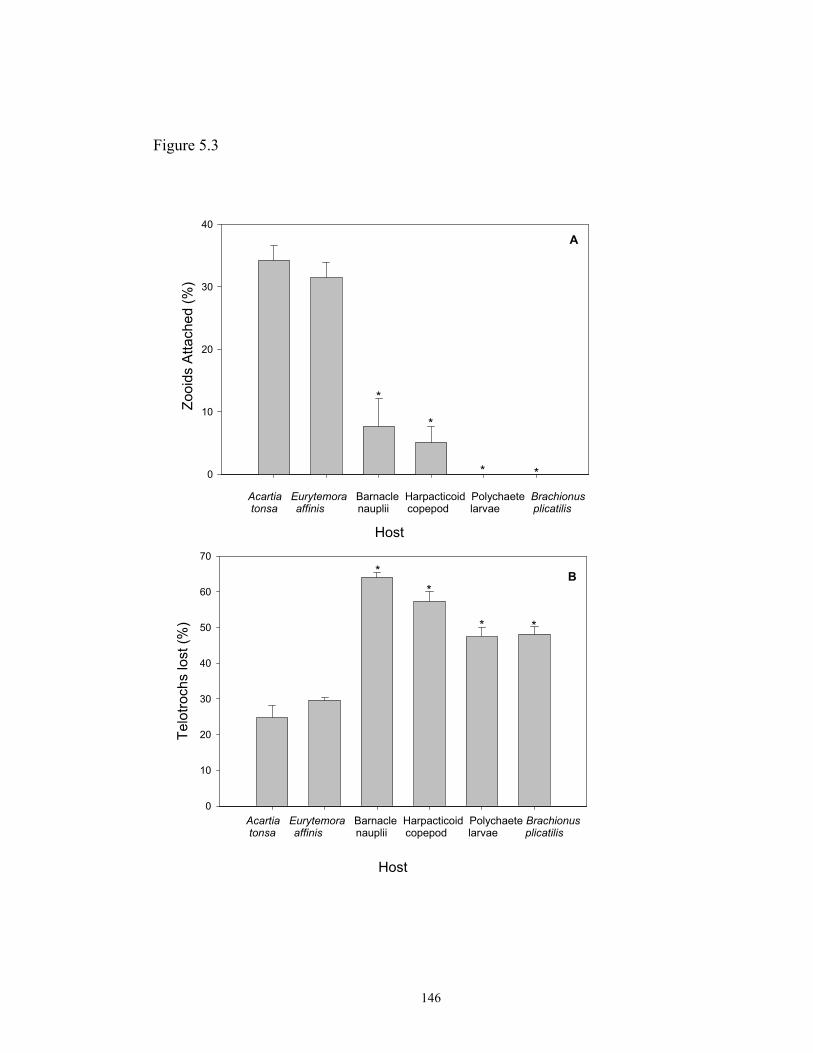
Figure 5.3
Host
Zooi
ds A
ttach
ed (%
)
0
10
20
30
40
Acartia Eurytemora Barnacle Harpacticoid Polychaete Brachionus tonsa affinis nauplii copepod larvae plicatilis
A
Telo
troch
s lo
st (%
)
0
10
20
30
40
50
60
70
Acartia Eurytemora Barnacle Harpacticoid Polych tonsa affinis nauplii copepod larvae
Host
B
*
*
* *
**
*
146
aete Brachionus plicatilis
*

Figure 5.4
Host
Mea
n nu
mbe
r of z
ooid
s/m
m2
0
2
4
6
8
10
12
14
16
Acartia Barnacletonsa nauplii
Host
Mea
n nu
mbe
r of z
ooid
s/m
m2
0
1
2
3
4
5
6
Acartia Harpacticoidtonsa copepod
Host
Mea
n nu
mbe
r of z
ooid
s/m
m2
0
2
4
6
8
10
Acartia Brachionustonsa plicatilis
Host
Mea
n nu
mbe
r of z
ooid
s/m
m2
0
2
4
6
8
10
Acartia Polychaetetonsa larvae
A B
C D
* *
*
*
* *
147

Figure 5.5
A. tonsa- A. tonsa- A. tonsa- A. tonsa-Barnacle Harpacticoid B. plicatilis Polychaetenauplii copepod larvae
Host
Per
cent
age
of T
elot
roch
s
0
10
20
30
40
50
60
70Telotrochs Lost during ExperimentTelotroch attached to hosts
148

CHAPTER 6
GENERAL DISCUSSION
149

Epibiotic relationships in aquatic environments have been known for more than
a century. Ryder (1883), in a report published in Science magazine focusing on protist
parasites in oysters, mentioned the existence of colonies of an organism identified as
Zoothamnium attached to the back and legs of the oyster-crab Pinnotheres osterum.
The author suggested that the relationship would be advantageous for the host, since it
could feed on zooids that detached from the colony. Since then, reports and
investigations of epibiotic relationships emphasizing seasonal occurrence of epibionts,
issues about host specificity and possible deleterious effects that epibionts may have
on the host have tried to shed light on the nature and consequences of this ecological
association. Although the occurrence of epibiotic relationships has been documented
frequently, especially in freshwater environments, some aspects such as life history
stages of the epibiont, mechanisms of attachment, and assessment of host specificity
are still not well explored (Carman and Dobbs, 1997).
The initial goals of the present study were to characterize morphologically the
ciliate epibiont community on calanoid copepod populations from the Chesapeake
Bay, USA, and to assess patterns of spatial and temporal occurrence of epibiosis in the
same environment. The results presented in Chapters 2 and 3 revealed that two
species of peritrich ciliates, identified as Zoothamnium intermedium and Epistylis sp.,
are common epibionts on Acartia tonsa and Eurytemora affinis populations during the
spring and summer months of the year. The most abundant species of peritrich
attached to copepods in the Bay was identified as Zoothamnium intermedium Precht,
1935. As pointed out by Carman and Dobbs (1997), the identification of epibiont
species and comprehensive studies about species composition in epibiotic
150

communities are still lacking especially in marine environments. This is also the case
in estuarine systems such as the Chesapeake Bay. As discussed in Chapter 2, species
identification of peritrichs can be difficult and time consuming due to incomplete
original descriptions and redescriptions. The application of cytological techniques
such as Protargol staining, Scanning Electron Microscopy in peritrich identification
(e.g. Zagon and Small, 1970; Gross, 1986; Leitner and Foissner, 1997; Song, 1997;
Hu and Song 2001) has provided useful knowledge on relevant diagnostic
characteristics that may help with species comparison from different habitats in the
world. At the same time, it is important to provide diagnostic features and
measurements of reliable morphological characters observed in live specimens, so that
researchers who are not trained to apply staining techniques or electron microscopy
would be able to correctly identify peritrichs to species level (Leitner and Foissner,
1997). In addition, characters from live specimens are required for comparisons with
original descriptions.
Occurrence of epibionts on organisms from zooplankton communities has been
widely reported in the literature (e.g. Sherman and Schaner, 1965; Green, 1974;
Willey, Cantrel, and Threlkeld, 1990; Chiavelli, Mills, and Threlkeld, 1993;
Weissman, Lonsdale, and Yen, 1993; Gilbert and Schröder, 2003), but only few
studies investigated seasonal occurrence of epibiosis over a period of one year (e.g.
Green, 1974; Xu, 1992; Chiavelli, Mills, and Threlkeld, 1993; Willey and Threlkeld,
1993; Barea-Arco, Pérez-Martinez, and Morales-Barquero, 2001). Most of these
studies observed a strong correlation between epibiont prevalence and host abundance,
while others reported a neutral or negative relationship with substrate availability.
151
Threlkeld, Chiavelli and Willey (1993) suggested that the existence of a positive or
negative correlation between epibiont prevalence and host abundance could be
explained by the existence of source and sink habitats (Pulliam, 1988). A negative
correlation with a given host species would suggest that substrate is a sink habitat for
that particular epibiont. In other words, the epibiont is able to colonize that given
substrate, but reproduction and migration is very reduced or does not occur at all,
while in a source, or preferred habitat, the epibiont is capable of completing its life
cycle and to migrate and colonize other hosts. The authors pointed out that some
exceptions to this pattern could be due to selective predation on infested hosts or to
poor sampling.
Infestation prevalence of Zoothamnium intermedium from Chesapeake Bay did
not show any significant correlation with abundance of its copepod hosts, Acartia
tonsa and Eurytemora affinis (see Chapter 3), but indicated a trend of infestation peaks
following high densities of the host. From the point of view suggested by Threlkeld,
Chiavelli, and Willey (1993), the copepod hosts might be hypothesized to be a sink
habitat for Z. intermedium with perhaps benthic living or non-living substrates serving
as source habitat. However, laboratory experiments and observations in this study
revealed that Z. intermedium could reproduce and disperse when attached to A. tonsa
(see Chapter 4, growth rates experiments) and E. affinis (observation from laboratory
cultures), demonstrating that copepods are not sink habitats in the studied system.
Moreover, Z. intermedium was not able to attach to non-living substrates in the
laboratory nor in the field (see Chapter 4), suggesting that this epibiont has an obligate
relationship with living hosts.
152

The dynamics of infestation prevalence could be dependent on other abiotic and
biotic factors in addition to host abundance. One of the factors that could help explain
patterns of infestation observed in the field is epibiont production. Temperature,
salinity, dissolved oxygen, availability of food, and strategies of dispersal may be
determinant factors for the increase or decrease in epibiont prevalence. For example,
dispersal stages of sessile peritrichs are unable to feed, therefore surviving from only a
few hours to a few days (Fenchel, 1965; Clamp, 1973) if they do not find a suitable
substrate for attachment. The survival time for telotrochs of Z. intermedium was
shown to be approximately 14 hours at 200 C(see Fig. 4.4), implying the the epibont
has to find a new substrate in a very short period for the epibiont to be able to find
another substrate. Moreover, it was demonstrated that the attachment success of
telotrochs of Z. intermedium decreases as telotroch age increases (see Chapter 4, Fig.
4.6 A ), suggesting that it is extremely important to find a host immediately after the
formation of the dispersal stage. Despite the short-lived telotroch and a highest
attachment frequency recorded in the laboratory of approximately 37%, infestation
prevalence of Z. intermedium in the field was found to be as high as 70% on A. tonsa
(see Chapter 3). This high prevalence observed in the field might occur as a
combination of high host abundance and high epibiont production fueled by different
biotic and abiotic factors, whose complexity could not be fully characterized with the
extent of sampling and available variables analyzed in this work (see Chapter 3).
Therefore, I suggest that Z. intermedium is an obligate epibiont of living substrates and
its abundance is dependent on the availability of the host substrate, as well as on the
153

production of dispersal stages, which may be directly related to salinity, temperature
and availability of food.
Host specificity is another subject commonly discussed in studies of epibiotic
relationships in zooplankton communities, especially because epibionts often colonize
one or more groups or species of hosts in a given environment. Wahl and Mark
(1999) suggested that most epibionts exhibited a facultative relationship with their
living host based on a survey of the literature and on field experiments with artificial
microhabitats carried out in the Baltic Sea. The majority of the epibiont species
surveyed were invertebrates, but unidentified species of Vorticella and Zoothamnium
were also found in the field study, and were reported to colonize a variety of living
(especially macrophytes) and non-living substrates. Green (1974) also reported that
the majority of epibionts he found in freshwater cladocerans were not specific to one
host, with exception of Colacium vesiculosum that, in the studied environment, was
found colonizing only Daphnia magna. A similar situation was reported by Henebry
and Ridgeway (1979), who found Vorticella microstoma only attached to
Scapholeberis kingii in a polluted lake in Illinois, and by Hermann and Mihursky
(1964) who found Zoothamnium sp. colonizing only Acartia tonsa in the Patuxent
River, even when it was not the most abundant species. Field samples analyzed in the
present study revealed that Z. intermedium was only attached to A. tonsa and E. affinis
(see Chapter 3) even when other zooplankton species were abundant. On the other
hand, laboratory experiments demonstrated that the ciliate epibiont is able to attach to
additional potential hosts from the zooplankton community (e.g. barnacle nauplii, and
Harpacticoid copepod; see Chapter 5) in presence or absence of its primary host A.
154

tonsa. Moreover, observation of live samples collected from the field showed that,
when infestation prevalence on A. tonsa and E. affinis was ca. 100%, Z. intermedium
was able to attach to barnacle nauplii and rotifer eggs in the field. A similar
observation was pointed out by Green (1974), who suggested that heavy infestation of
a preferred host species could lead to transient association with other living organisms
present in the environment. From the results obtained in the analyses of field samples
(Chapter 3), laboratory experiments (Chapters 4 and 5) and field study (Chapter 4), Z.
intermedium seems to be an obligate epibiont of living substrates, perhaps with
preference for crustacean hosts. The fact that the epibiont was able to attach to eggs of
B. plicatilis in the presence of A. tonsa can be due to the release of chemical cues by
the copepod, the presence of chitin in the shell of the eggs, and/or overcrowding of
the copepod with temporary attachment to the rotifer (see discussion in Chapter 5).
The lack of attachment of Z. intermedium to other zooplankton species observed in
samples from the field (Chapter 3) could be due to non-saturation (i.e. no
overcrowding) of the primary hosts in the surveyed period, or to insufficient sampling.
Epibionts can also display preferential attachment to specific sites on the host’s
body, and in most cases, these preferred sites are related to advantageous uptake of
food. For example, Evans, Sicko-Goad, and Omair (1979) observed that the suctorian
Tokophrya quadripartita attached preferentially to the head of its host, the copepod
Limnocalanus macrurus, an ideal site to come to contact with prey. Clamp (1973)
also observed that the peritrich Lagenophrys lunatus exhibited strong selection for
some areas of the body (especially the uropods) of its shrimp host that received more
flux of water. Colacium calvum also attached preferentially to the anal region of its
155

host, the cladoceran Daphnia spp,. possibly to take advantage of the material excreted
by the substrate (Chiavelli, Mills, and Threlked, 1993). Preferential attachment of
epibionts to males or females was pointed out by Sherman and Schaner (1965) who
observed only slight to moderate infestation of Paracineta sp. on males of the copepod
Meritridia lucens. Females, in contrast, were always heavily infested by the suctorian
epibiont. Xu and Burns (1991) also reported that the peritrich epibiont Epistylis
daphniae attached preferentially to females of the copepod Boeckella triarticulata,
with very low infestation rates in males. Weissman, Lonsdale and Yen (1993) did not
find any preference of the peritrich Rhabdostyla sp. for age, stage or site in the body of
the copepod Acartia hudsonica. In the present work, Z. intermedium was also able to
attach to the cephalothorax, abdomen, antennae and swimming legs of its copepod
hosts, but density of colonies and zooids were higher on the abdomen of both host
species (see Chapter 3), and there was no apparent preference for males or females.
Interestingly, a high density of epibiont colonies was found on copepodite stages of
the hosts, suggesting a preferential attachment to juvenile stages (Chapter 3). This
observation is unique (especially for copepods that have a terminal molt), and
surprising since studies that compared infestation prevalence throughout life stages
reported higher epibiont infestation on adults of crustacean hosts than on juvenile
forms (Sherman and Schaner, 1965; Mohlemberg and Kaas, 1990; Xu, 1993;
Chiavelli, Mills, and Threlkeld, 1993). However, subsequent laboratory experiments
demonstrated that Z. intermedium does not colonize preferentially copepodite or adult
stages of either host species (see Chapter 4), suggesting that the pattern observed in
field samples reflects other interactions that may be occurring in nature. One possible
156

explanation is the selective predation on heavily infested adults, since a large number
of colonies could make the individuals more visible for fish that prey by sight, and
also hamper locomotion, thus affecting their swimming escape reaction. Surveys and
manipulations in the field demonstrated that the prevalence of euglenoid epibionts on
cladocerans and copepods was significantly reduced in the presence of pump-filter
feeders or fish that prey by sight, in comparison to sites without zooplankton predators
(Willey, Cantrel and Threlkeld, 1990; Willey, Willey, and Threlkeld, 1993). In the
case of small euglenoids, the attraction to predators possibly comes from the bright
green color of the chloroplast inside the epibiont cell. Z. intermedium would not have
on copepods the color effect that epibionts like Colacium spp. may have on their host,
but the high number of colonies may lead the host to appear larger for predators, and
potentially easy to capture.
Frequent molts, death, and sometimes desiccation of the substrate organism are
some of the unstable conditions that epibionts have to face (Wahl, 1989). To be
successful, the epibiont must be able to form dispersal stages, leave the old substrate
and find another suitable host to start another cycle. For example, some peritrich
epibionts are able to start forming free-swimming stages prior to the completion of the
molt cycle of their crustacean hosts (Fenchel, 1965; Green, 1994; Willey and
Threlkeld, 1995), while others remain attached to the molt and only after it is shed do
they produce dispersal stages (Clamp, 1973). In the present study, results of
experiments stimulating telotroch formation in Z. intermedium, suggested that a
combination of chemical, mechanical and perhaps electrical cues coming from the
host to which the epibiont is attached is probably needed to trigger telotroch formation
157
in this peritrich (Chapter 4). Observations of copepod exuvia from laboratory cultures
revealed that Z. intermedium, differently from other peritrichs attached to crustaceans,
starts forming telotroch stages after the host has cast the old skin. The mechanisms by
which epibionts recognize that the host is about to molt or has molted are still
unknown, but perhaps biochemical changes in the host’s body fluids or hormonal
fluctuation play an important role (Clamp, 1973)
The inferences and conclusions obtained in this work may provide some insights
about the complexity of epibiotic relationships. The experiments presented here may
serve as models that can be employed in other systems to assess aspects of the life-
cycle and host specificity of epibionts or free-living peritrichs. Suggestions for future
studies of the Z. intermedium-copepod association in Chesapeake Bay include: (i) the
assessment of potential deleterious effects that the epibiont might have on the fitness
of the host, including effects on reproduction and survivorship; (ii) the analysis of
potential biochemical recognition and affinity of the epibiont by elements present on
the surface of the host; and (iii) investigation of possible demographic impacts that
this epibiotic relationship might have on copepod populations and its consequence for
the ecosystem as a whole.
158

GLOSSARY
Some of the technical terms used in this dissertation are defined below:
Basibiont – substrate organism which is host to the epibiont.
Coenobium – a type of colonization in which all the colonial cell-like units are
interconnected a continuous, common cell membrane.
Colonization rate – increase in the number of colonies/host per unit of time.
Dikinetid – organellar complex in ciliates composed of two approximately adjacent
kinetosomes, associated cilia, and infraciliary organelles. See kinetid.
Epibiont – organism that grows attached to a living surface.
Germinal row – line of non-ciliated kinetosomes associated with the terminal portion
of the infraciliary base of the haplokinety. It plays a productive role in stomatogenesis
in Peritrichs.
Growth rate – change in the number of zooids/host per unit of time. It is a function of
the colonization and proliferation rates.
Haplokinety – a double row of kinetosomes associated with the oral ciliature and
exhibiting a zigzag pattern.
Infestation density – number of epibiont colonies/unit of area of the host.
Infestation load – number of epibiont zooids/unit of area of the host.
Infestation prevalence – percentage of host population carrying epibionts in a given
time.
Infundibulum – section of the oral cavity in peritrich ciliates. It is usually funnel-
shaped, and contains the oral infraciliature.
159

Kinetid – elementary organizational unit of the ciliate cortex (region underneath the
membrane that contains the ciliary organelles). Consists of one or more kinetosomes
and associated structures.
Kinetosome – basal body. Cylindrical organelle consisting of 9 peripheral
microtubular triplets and 2 inner microtubules. Basal termination of cilia (and
flagella).
Kinety – longitudinally oriented row of a single, paired, or multiple kinetosomes, their
cilia, and other associated cortical organelles.
Oral dikinetid (ODK) – Dikinetid located to the right side of the cytostome in the
oral region of ciliates. See Dikinetid.
Oral polykinetid (OPK) – Polykinetid located to the left side of the cytostome in the
oral region of ciliates. See Polykinetid.
Peristome – circular region of the zooid apical surface which opens to the
infundibulum in peritrich ciliates.
Polykinetid - organellar complex in ciliates composed of more than two kinetosomes,
associated cilia, and infraciliary organelles.
Proliferation rate – change in the mean number of epibiont zooids/colony attached to
a host per unit of time.
Scopula – compound organelle located at the aboral pole of sessiline peritrich ciliate
zooids that may function as a holdfast organelle, or as a source of the secreted stalk.
Telotroch – free-swimming, dispersal stage found in sessiline peritrichs.
Telotroch band – synonym of trochal band. Ring of cilia and kinetosomes near the
posterior (aboral) end of a telotroch.
160
Trophont – mature, vegetative, sessile feeding adult stage in the peritrich life cycle.
Zooid – individual cell or cell like unit in a peritrich colony or coenobium.
161

LIST OF REFERENCES Abelló, P.; McPherson, E. (1992). Epibiosis and rhizocephalan infestation patterns in
relation to the reproductive biology of Lithodes ferox (Filhol, 1885)
(Anomura:Lithodidae). Journal of Crustacean Biology 12(4): 561-570.
Al-Dhaheri, R.S.; Willey, R.L. (1996) Colonization and reproduction of the epibiotic
flagellate Colacium vesiculosum (Euglenophyceae) on Daphnia pulex. J. of
Phycology 32: 770-774
Antipa, G. A. (1977). Use of commensal protozoa as biological indicators of water
quality and pollution. Transactions of the American. Microscopy Society
96(4): 482-489.
Baldock, B. M. (1986). Peritrich ciliates epizoic on larvae of Brachycentrus
subnubilus (Trichoptera): importance in relation to the total protozoan
population in streams. Hydrobiologia 132: 125-131.
Barea-Arco, J.; Pérez-Martinez, C.; Morales-Baquero, R. (2001). Evidence of a
mutualistic relationship between an algal epibiont and its host Daphnia
pulicaria. Limnology and Oceanography 46(4): 871-881.
Bauer-Nebelsick, M.; Bardele, C.F.; Ott, J.A. (1996). Redescription of Zoothamnium
niveum (Hemprich & Ehrenberg, 1831) Ehrenberg, 1838 (Oligohymenophora,
Peritrichida), a ciliate with ectosymbiotic, chemoautotrophic bacteria.
European Journal of Protistology 32: 18-30.
Bender, K.; Kleinow, W. (1988) Chemical properties of the lorica and related parts
from the integument of Brachionus plicatilis Comparative Biochemical
Phisiology 3: 483-487
162

Bick, H. (1972) Ciliated Protozoa - An Ilustrated Guide to the species used as
biological indicators in freshwater biology. World Health Organization 1st
edition 198p.
Brownlee, D.C.; Jacobs, F. (1987) Mesozooplankton and microzooplankton in the
Chesapeake Bay. In Majumdar, L.W.; Austin, H.M. (eds) Contaminant
Problems and Management of Living Chesapeake Bay Resources. The
Pensylvania Academy of Science 217-269
Caine, E. A. (1986). Carapace epibionts of nesting loggerhead sea turtles: Atlantic
coast of USA Journal of Experimental Marine Biology and Ecology 95: 15-26.
Carman, K. R.; Dobbs, F.C. (1997). Epibiotic microorganisms on copepods and other
marine crustaceans. Microscopy Research and Technique 37: 116-135.
Chiavelli, D. A.; Mills, E.; Threlkeld, S.T. (1993). Host preference, seasonality, and
community interactions of zooplankton epibionts. Limnology and
Oceanography 38(3): 574-583.
Cho, S. Y.; Lee, N. S.; Shin, M. H.; Kong, Y., (1999) Age-dependent infectivity of
orally transferred juvenile Fasciola hepatica. Journal of Parasitology 85: 739-
742
Clamp, J. C. (1973). Observations on the host-symbiont relationships of Lagenophrys
lunatus Imamura. Journal of Protozoology 20(5): 558-561.
Clamp, J.C. (1988) A new species of Lagenophrys (Ciliophora: Peritricha:
Lagenophryidae) ectocommensal on the wood-boring isopod Limnoria
(Flabellifera:Limnoridae). Trans. Am. Microsc. Soc. 107: 12-16
163

Clamp, J.C. (1994) New species of Lagenophrys (Ciliophora, Peritrichia) from New
Zealand and Australia. Journal of Eukariotic Microbiology 41: 343-349
Clamp, J.C.; Coats, D.W. (2000) Planeticovorticella finleyi n.g., n.sp. (Peritrichia,
Vorticellidae) a planktonic ciliate with a polymorphic life cycle. Invertebrate
Biology 119: 1-16
Coats, D.W.; Heibokel, J. (1982)A study of reproduction and other life cycle
phenomena in planktonic protists using acridine orange floresence technique.
Marine Biology 64: 17-19
Corliss, J.O. (1979) The ciliated Protozoa. 2nd ed. Pergamon Press New York 455p
Couch, J.A. (1978) Diseases, parasites, and toxic responses of commercial penaeid
shrimps of the Gulf of Mexico and South Atlantic Coasts of North America.
Fishery Bulletin 76: 1-44
Curds, C.R.; Gates, M.A.; Roberts, D.M. (1983) British and other Freshwater Ciliated
Protozoa Part II Ciliophora Oligohymenophora and Polyhymenophora.
Synopses of the British Fauna No. 23. Cambrige University Press 474 p.
Curds C. R.; Vandyke, J.M. (1966) The feeding habits and growth rates of some fresh-
water ciliates found in activated-sludge plants. Journal of Applied Ecology 3:
127-137
de Baufer, P.J.; Amin, A.A.; Pak, S.C.; Buhse Jr., H.E. (1999) A method for the
synchronous induction of large numbers of telotrochs in Vorticella convalaria
by monocalcium phosphate at low pH. J. Euk. Microbiol. 46: 12-16
Ebert D., Hottinger, J.W.; Pajunen V. I. (2001) Temporal and spatial dynamics of
parasitre richness in a Daphnia metapopulation. Ecology 82: 3417-3434
164
Evans, M.S.; Sicko-Goad, L.; Omair, M. (1979) Seasonal occurrence of Tokophrya
quadripartita (Suctoria) as epibionts on adult Limnocalanus macrurus
(Copepoda:Calanoida) in Southeastern Lake Michigan. Trans. Am. Microsc.
Soc. 98: 102-109
Fauré-Fremiet, E. (1930). Growth and differentiation of the colonies of Zoothamnium
alternans (Clap. and Lachm.). Biological Bulletin 58: 28-51.
Felgenhauer, B.E.; Schram, F.R.(1978) Differential epibiont fouling in relation to
grooming behavior in Palaemonetes kadiakensis. Fieldiana Zoology 72: 83-99
Fenchel, T. (1965). On the ciliate fauna associated with the marine species of the
amphipod Gammarus J.G. Frabricius. Ophelia 2(2): 281-303.
Fenchel, T. (1987) Ecology of Protozoa: The biology of free-living phagotrophic
protists. 1st ed. Science Tech. Publishers, Madison, Wisconsin. 192 p
Fenchel, T. Finlay, B.J. (1989). Kentrophoros: a mouthless ciliate with a symbiotic
kitchen garden. Ophelia 30(2): 73-95.
Fernandez-Galiano D; Carrascosa, A. (1989) Morfología general, desarollo de la larva
y crescimiento de la colonia en Epistylis plicatilis Ehrenberg, 1830
(Ciliophora, Peritrichida). Boletin de la Real Sociedad Española de Historia
Natural (Seccion Biologica) 84: 219-234
Foissner, W.; Berger, H.; Schaumburg, J. (1999) Identification and Ecology of
Limnetic Plankton Ciliates. Bayerisches Landesamt für Wasserwirtschaft Heft
3/99 Munich 793 p.
165

Foissner, W.; Agatha, S.; Berger H. (2002) Soil ciliates (Protozoa, Ciliophora) from
Namibia (Sothwest Africa), with emphasis on two contrasting environments, the
Etosha Region and the Namibia Desert. Denisia 05 1st vol. 1066 p
Foster, C. A.; Sarphie, T.G.; Hawkins, W.E. (1978). Fine structure of the peritrichous
ectocommensal Zoothamnium sp. with emphasis on its mode of attachment to
penaeid shrimp. Journal of Fish Diseases 1: 321-335.
Garcia, M.P.; Castelon, C.; Igual, J.; Sunyer, R. (1989) Ciliated protozoan
communities in a fluvial ecosystem Hydrobiologia 183: 11-31
Gibson (1978) Pseudohimantidium-pacificum new-record. An epizoic diatom new to
the Florida Current Western North Atlantic Ocean. Journal of Phycology 14:
371-373
Gilbert, J.J.; Schröder, T. (2003). The ciliate epibiont Epistylis pygmaeum: selection
for zooplankton hosts, reproduction and effect on two rotifers. Freshwater
Biology 48: 878-893
Gili, J. M.; Abelló, P.; Villanueva, R. (1993). Epibionts and intermoult duration in the
crab Bathynectes piperitus. Marine Ecology Progress Series 98: 107-113.
Gosselin, L.A.; Qian, P.Y. (1996) Early post-settlement mortality of an intertidal
barnacle: a critical period of survival. Marine Ecology Progress Series 135: 69-
75
Green, J. (1974). Parasites and epibionts of cladocera. Transactions of the Zoological
Society of London 32: 417-515.
Green, J. D.; Shiel, R.J. (2000). Mobiline peritrich riders on Australian calanoid
copepods. Hydrobiologia 437: 203-212.
166

Gross, M.E. (1986) Protargol silver staining as a method for the comparative study of
five species in the genus Zoothamnium Boryde St. Vincent, 1826 (Ciliophora,
Peritrichia). Master's Thesis, University of Maryland at College Park, USA,
122 p.
Hanamura, Y. (2000). Seasonality and infestation pattern of epibiosis in the beach
mysid Archaeomysis. Hydrobiologia 427: 121-127.
Harmsworth, G.C.; Sleigh, M.A.; Baker, J.H. (1992) The abundance of different
peritrich ciliates on stone surfaces in contrasting lowland streams throughout
the year. J. Protozoology 39: 58-65
Harmsworth, G. C. Sleigh, M.A. (1993). Colonization of non-living surfaces in
streams by peritrich ciliates. European Journal of Protistology 29: 294-301.
Henebry, M. S.; Ridgeway, B.T. (1979). Epizoic ciliated protozoa of planktonic
copepods and cladocerans and their possible use as indicators of organic
pollution. Transactions of the American Microscopical Society 98(4): 495-508.
Herman, S. S.; Mihursky, J.A. (1964). Infestation of the copepod Acartia tonsa with
the stalked ciliate Zoothamnium. Science 146: 543-544.
Hu, X.; Song, W. (2001). Description of Zoothamnium chlamydis sp. n.
(Protozoa:Ciliophora:Peritrichida), an ectocommensal peritrichous ciliate from
cultures scallop in North China. Acta Protozoologica 40: 215-220
Iyer, N.; Rao, T.R. (1995). Epizoic mode of life in Brachionus rubens Ehrenberg as a
deterrent against predation by Asplanchna intermedia Hudson. Hydrobiologia
313/314: 377-380.
167
Jack, J.D.; Gilbert, J.J. (1993) Effects of metazoan predators on ciliates in freshwater
plankton communities. J. Euk. Microbiol. 44: 194-199
Jayasree, L.; Janakiran, P.; Madhavi, R. (2001) Epibionts and parasites of
Macrobrachium rosembergii and Metapenaeus dobsoni from Gosthani
estuary. Journal of Natural History 35: 157-167
Jones, K.K.; Simenstad, C.A.; Higley, D.L.; Bottom, D.L. (1990) Community
structure, distribution and standing stock of benthos, epibenthos and plankton
in Columbia River estuary. Progress in Oceanography 25: 211-241
Kahl, A. (1935). Urtiere oder Protozoa I: Wimpertiere oder Ciliata (Infusoria), 4.
Peritricha und Chonotricha, In Dahl, F., ed., Die Tierwelt Deutschlands, G.
Fischer, Jena, pt. 30, pp. 651-805.
Kankaala, P.; Eloranta, P. (1987). Epizooic ciliates (Vorticella sp.) compete for food
with their host Daphnia longispina in a small polyhumic lake. Oecologia 73:
203-206.
Knight-Jones, E. W.; Crisp D.J. (1953) Gregariousness in barnacles in relation to the
fouling of ships and to anti-fouling research. Nature 171: 1109-10
Kusuoka, Y.; Watanabe, Y. (1987) Growth and survival of peritrichs ciliates in an
urban stream. Oecologia 73:16-20
Langlois, G.A. (1975) Effect of algal exudates on substratum selection by motile
telotrochs of the marine peritrich ciliate Vorticella marina. J. Protozoology 22:
115-123
Laval, M. (1968) Zoothamnium pelagicum Du Pleiss. Cilié péritriche plactonique:
morphologie, croissance et comportement. Protistologica 4: 333-367
168

Leitner, A.R.; Foissner, W. (1997) Taxonomic characterization of Epicarchesium
granulatum (Kellicott, 1887) Jankowski, 1985 and Pseudovorticella elongata
(Fromentel, 1876) nov. comb., two peritrichs (Protozoa, Ciliophora) from
activated sludge. European Journal of Protistology 33: 13-29
Lom, J. (1964) The morphology and morphogenesis of the buccal ciliary organelles in
some peritrichous ciliates. Arch. Protistenk. 107:131-162
López, C.; Ochoa, E.; Páez, R.; Theis, S. (1998) Epizoans on a tropical freshwater
crustacean assemblage. Mar. Fresh. Res. 49: 271-276
Lynn, D.; Small, E.B. (2000) Phylum Ciliophora In: Lee, J.J; Leedale, G.F.; Bradbury,
P. (eds) The Illustrated Guide to the Protozoa. Society of Protozoologists Allen
Press 2nd edition Vol. I 689 p.
Mannesmann, R.; Rustige, K.H. (1994) Correlation of epizoic ciliates of Gammarus
pulex and freshwater quality. Zeitscrift fuer Angewandte Zoologie 80: 486-499
Mauchline, J. (1998) The Biology of Calanoid Copepods. Advances in Marine
Biology Vol. 33Academic Press 710 p.
Mayén-Estrada, R.; Aladro-Lubel, M.A (2000) First record of Lagenoprys dennisi
(Ciliophora: Peritrichia) on the exoskeleton of crayfish Cambarellus
patzcuarensis. Journal of Eukariotic Microbiology 47: 57-61
Mayén-Estrada, R.; Aladro-Lubel, M.A. (2001). Epibiont peritrichids
(Ciliophora:Peritrichida:Epistylididae) on the crayfish Cambarellus
patzcurensis in Lake Pátzcuaro, Michocán, Mexico. Journal of Crustacean
Biology 21(2): 426-434.
169

Mohlemberg, F.; Kaas, H. (1990). Colacium vesiculosum Ehrenberg (Euglenophycea),
infestation of planktonic copepods in the Western Baltic. Ophelia 31: 125-132
Montagnes, D. J. S.; Lynn, D.H. (1987). A quantitative Protargol stain (QPS) for
ciliates: method description and test of its qualitative nature. Marine Microbial
Food Webs 2(2): 83-93.
Montagnes, D. J. S.; Lynn, D.H. (1993) A quantitative Protargol Stain (QPS) for
ciliates and other protists. In: Kemp, P.F.; Sherr, B.F.; Sherr, E.B.; Cole, J.J.
Handbook of Methods in Aquatic Microbial Ecology Lewis Publishers: 229-
240
Nagasawa, S. (1986). The peritrich ciliate Zoothamnium attached to the copepod
Centropages abdominalis in Tokyo Bay waters. Bulletin of Marine Science
38(3): 533-558.
Nagasawa, S. (1987) Asexual reproduction of the stalked ciliate Zoothamnium
attached to the copepod Centropages abdominalis. Proc. Japan Acad. 63: 101-
103
Nagasawa, S; Simidu, U.; Nemoto, T. (1985) Scanning electron microscopy
investigation of bacterial colonization of the marine copepod Acartia clausii.
Marine Biology 87: 61-66
Nakajima, Y.; Hashimoto, K.; Asai, H. (1986) Behaviour and function of scopula
organelles during differentiation of a sessile Carchesium polypinum into a free
swimming telotroch. Cytobios 47: 179-185
170

Nerad, T.; Daggett, P. (1992) Cultivation of selected photosynthetic cryptomonads. In:
Lee, J.J.; Soldo, A. (eds) Protocols in Protozoology. Allen Press, Kansas Vol.1
pp: A-6.1 -A6.5
Nolting, E.; Rustige, K.H. (1998) Investigations on ciliate settlement in the Rivers
Werra and Weser (Germany). Limnologica 28: 255-262
Porter, K. G.; Feig, Y. S. (1980) The use of DAPI for identifying and counting aquatic
microflora. Limnology and Oceanography 25: 943-948
Pratt, J.R.; Rosen, B.H. (1983) Associations of species of Vorticella (Peritrichida) and
planktonic algae. Trans. Am. Microsc. Soc. 102: 48-54
Precht, H. (1935). Epizoen der Kieler Bucht. Nova Acta Leopoldina 3(15): 405-474.
Pulliam, H.R. (1988) Sources, sinks, and population regulation. The American
Naturalist 132: 652-661
Reynoldson, T. B. (1950). Natural population fluctuations of Urceolaria mitra
(Protozoa, Peritricha) epizioic on flatworms. Journal of Animal Ecology 19:
106-118.
Reynoldson, T. B. (1955). Factors influencing population fluctuations of Urceolaria
mitra (Peritricha) epizoic on freshwater triclads. Journal of Animal Ecology
24: 57-83.
Roman, M.R.; Holliday, D.V.; Sanford, L.P. (2001) Temporal and spatial patterns of
zooplankton in the Chesapeake Bay turbidity maximum. Marine Ecology
Progress Series 213: 215-227
Rose, C. R.; Finley, H. E. (1976) A method for mass cultivation of sessile peritrich
protozoa. Trans. of the Am. Microsc. Soc. 95: 541-544
171

Ryder, J.A. (1883) The protozoan parasites of the oyster. Science 1: 567-568
Sherman, K.; Schaner, E.G. (1965) Paracineta sp., an epizoic suctorian found on Gulf
of Maine copepods. J. Protozoology 12: 618-625
Sládecek, V. (1986) Indicator value of the genus Epistylis (Ciliata) Arch. Hydrobiol.
107: 119-124
Song, W. (1997). A new genus and two new species of marine peritrichous ciliates
(Protozoa, Ciliophora, Peritrichida) from Qingdao, China. Ophelia 47: 203-214
Song, W.; Al-Rasheid, K.A.S.; Hu, X. (2002) Notes on the poorly-known marine
peritrichous ciliate Zoothamnium plumula Kahl, 1933 (Protozoa: Ciliophora)
an ectocommensal organism from cultured scallops in Qingdao, China. Acta
Protozoologica 41: 163-168
Stirnadel, H.A.; Ebert, D. (1997) Prevalence, host specificity and impact on host
fecundity of microparasites and epibionts in three sympatric. Daphnia species
Journal of Animal Ecology 66: 212-222
Succhard, S.J. (1979) An analysis of the morphogenetic events leading to telotroch
formation, settlement, and stalk development in the peritrich ciliate
Zoothamnium arbuscula. Ph.D. Dissertation, University of Maryland at
College Park
Sudo R.; Aiba, S. (1971) Growth rate of Vorticellidae isolated from activated sludge.
Japanese Journal of Ecology 21: 70-76
Summers, F.M. (1938) Some aspects of normal development colonial ciliate
Zoothamnium alternans. Biological Bulletin 74: 117-129
172

Taylor, W.D. (1983) A comparative study of the sessile, filter-feeding ciliates of
several small streams. Hydrobiologia 98: 125-133
Theron, A.; Rognon, A.; Pages, J. R. (1998) Host choice by larval parasites: A study
of Biomphalaria glabrata snails and Schistosoma mansoni miracidia related to
host size. Parasitology Research 84: 727-732
Threlkeld, S. T. Chiavelli, D.A.; Willey, R.L. (1993). The organization of zooplankton
epibiont communities. Trends in Ecology and Evolution 8(9): 317-321.
Turner, J.T.; Postek, M.T.; Collard, S.B. (1979) Infestation of the estuarine copepod
Acartia tonsa with the ciliate Epistylis. Trans. Am. Microsc. Soc. 98: 136-138
Vacchiano, E.J.; Dreisbach, A.; Locascio, D.; Castaneda, L.; Vivian, T.; Buhse Jr,
H.E. (1992) Morphogenetic transitions and cytoskeletal elements of the stalked
zooid and the telotroch stages in the peritrich ciliate Vorticella convalaria. J.
Protozoology 39: 101-106
Valbonesi, A. (1989). Scanning electron microscope observations on Zoothamnium
intermedium (Ciliophora:Peritrichia). Bolletino di Zoologia 56: 131-136.
Valbonesi, A.; Guglielmo, L. (1988) Infestation of a lagoon zooplanktonic community
with the epizoic peritrich Zoothamnium intermedium Precht (Peritrichia,
Zoothamnidae). Boll. Zool. 3: 179-183
Wahl, M. (1989). Marine epibiosis: I. Fouling and antifouling some basic aspects.
Marine Ecology Progress Series 58: 175-189.
Wahl, M. Mark, O. (1999) The predominantly facultative nature of epibiosis:
experimental and observational evidence. Marine Ecology Progress Series
187: 59-66
173

Weissman, P.; Lonsdale, D.J.; Yen, J. (1993). The effect of peritrich ciliates on the
production of Acartia hudsonica in Long Island Sound. Limnology and
Oceanography 38: 613-622
Willey, R.L.; Cantrell, P.A.; Threlkeld, S.T. (1990) Epibiotic euglenoid flagellates
increase the susceptibility of some zooplankton to fish predation. Limnology
and Oceanography 35: 952-959
Willey, R.L.; Threlkeld, S.T. (1993). Organization of crustacean epizoan communities
in a chain of subalpine ponds. Limnology and Oceanography 38: 623-627
Willey, R.L.; Willey, R.B.; Threlkeld, S.T. (1993). Planktivore effects on zooplankton
epibiont communities: epibiont pigmentation effects. Limnology and
Oceanography 38: 1818-1822
Willey, R.L.; Threlkeld, S.T. (1995). Changes in epibiotic burden during the intermolt
of Daphnia determined by hypodermal retraction stages. Hydrobiologia 307:
213-219
Xu, Z. (1992) The abundance of epizoic ciliate Epistylis daphniae related to their host
Moina macrocopa in an urban stream. Journal of Invertebrate Pathology 60:
197-200
Xu, Z.; Burns, C.W. (1991) Effects of the epizoic ciliate Epistylis daphinae, on
growth, reproduction and mortality of Boeckella triarticulata (Thompson)
(Copepoda:Calanoida). Hydrobiologia 209: 183-189
Xu, Z. (1993) The infection of Moina macrocopa by a colonial peritrich Epistylis
daphniae, and its effects on the host. Freshwater Biology 30: 181-186
174
Zagon, I. S.; Small, E.B. (1970). Carchesium polypinum: somatic and buccal structure
analysis after protargol staining. Transactions of the American Microscopical
Society 89(3): 443-449.
Zagon, I.S. (1971) Scanning electron microscope observations on some life history
stages of Carchesium polypinum (Ciliata, Peritrichia). J. Protozoology 18: 317-
328
175