Embed Size (px)
Citation preview

Available online at www.sciencedirect.com
7) 296–308www.elsevier.com/locate/yviro
Virology 368 (200
Absence of E protein arrests transmissible gastroenteritis coronavirusmaturation in the secretory pathway
Javier Ortego a,1, Juan E. Ceriani b, Cristina Patiño c, Juan Plana b, Luis Enjuanes a,⁎
a Centro Nacional de Biotecnología, CSIC, Department of Molecular and Cell Biology, Campus Universidad Autónoma,Darwin 3, Cantoblanco, 28049 Madrid, Spain
b Fort-Dodge Veterinaria, Department of Research and Development, Girona, Spainc Centro Nacional de Biotecnología, CSIC, Macromolecular Structure, Campus Universidad Autónoma, Darwin 3, Cantoblanco, 28049 Madrid, Spain
Received 12 March 2007; returned to author for revision 17 April 2007; accepted 14 May 2007Available online 10 August 2007
Abstract
A recombinant transmissible gastroenteritis coronavirus (rTGEV) in which E gene was deleted (rTGEV-ΔE) has been engineered. This deletionmutant only grows in cells expressing E protein (E+ cells) indicating that E was an essential gene for TGEV replication. Electron microscopy studiesof rTGEV-ΔE infected BHK-pAPN-E− cells showed that only immature intracellular virions were assembled. These virions were non-infectious andnot secreted to the extracellular medium in BHK-pAPN-E− cells. RNA and protein composition analysis by RNase-gold and immunoelectronmicroscopy showed that rTGEV-ΔE virions contained RNA and also all the structural TGEV proteins, except the deleted E protein. Nevertheless, fullvirion maturation was blocked. Studies of the rTGEV-ΔE subcellular localization by confocal and immunoelectron microscopy in infected E− cellsshowed that in the absence of E protein virus trafficking was arrested in the intermediate compartment. Therefore, the absence of E protein in TGEVresulted in two actions, a blockade of virus trafficking in the membranes of the secretory pathway, and prevention of full virus maturation.© 2007 Elsevier Inc. All rights reserved.
Keywords: Coronavirus; Nidovirus; E protein; TGEV; Virus maturation
Introduction
Since the identification of a novel coronavirus associated withsevere acute respiratory syndrome (SARS), and the discovery ofnew human coronaviruses such as HCoV-NL63 and HCoV-NH,associated with respiratory illnesses, the interest on corona-viruses has clearly increased (Esper et al., 2005a, 2005b; Kuikenet al., 2003; van der Hoek et al., 2004). The design of antiviraldrugs interfering with coronavirus replication (Hertzig et al.,2004) and the development of replication-competent propaga-tion-deficient virus vectors (Ortego et al., 2002) are potentialpowerful tools to prevent and control coronavirus infections.
Transmissible gastroenteritis coronavirus (TGEV) is a mem-ber of the Coronaviridae family within the Nidovirales order
⁎ Corresponding author. Fax: +34 915854915.E-mail address: [email protected] (L. Enjuanes).
1 Present address: Centro de Investigación en Sanidad Animal, C.I.S.A.-I.N.I.A. Carretera Algete-El Casar, Km 8,100. Valdeolmos 28130. Madrid, Spain.
0042-6822/$ - see front matter © 2007 Elsevier Inc. All rights reserved.doi:10.1016/j.virol.2007.05.032
(Enjuanes et al., 2000b). TGEV is an enveloped virus with asingle-stranded, positive-sense RNA genome of 28.5 kb. Abouttwo-thirds of the genome encode the replicase gene, whichcomprises open reading frames 1a and 1b, the last one beingexpressed by ribosomal frameshifting (Brierley et al., 1989;Penzes et al., 2001). The 3′ one-third of the genome includesstructural and nonstructural genes, in the order 5′-S-3a-3b-E-M-N-7-3′ (Enjuanes et al., 2000a).
In the Coronaviridae family, the viral envelope contains atleast three structural proteins. The most abundant is themembrane (M) protein, spanning the membrane three or fourtimes and interacting with the nucleocapsid (N) and spike (S)proteins during assembly (Escors et al., 2001; Rottier, 1995).The second most abundant is the S protein, a large type I-transmembrane glycoprotein that forms peplomers and isresponsible for cell receptor binding and membrane fusion(Lewicki and Gallagher, 2002; Sui et al., 2004; Suñé et al., 1990;Suñé et al., 1991). The third is the small envelope (E) protein, atransmembrane protein detected as aminor structural component

297J. Ortego et al. / Virology 368 (2007) 296–308
in TGEV, mouse hepatitis virus (MHV), SARS-CoV, and avianinfectious bronchitis virus (IBV) (Godet et al., 1992; Liu andInglis, 1991; Shen et al., 2003; Yu et al., 1994). Another essentialconstituent of the virion is the N protein, an internalphosphoprotein that interacts with the genomic RNA to formthe viral nucleocapsid (Escors et al., 2001; Kapke and Brian,1986; Narayanan and Makino, 2001).
Coronavirus maturation takes place at the cis-Golgi networkalso known as endoplasmic reticulum-Golgi intermediatecompartment (ERGIC) (Salanueva et al., 1999; Tooze et al.,1984, 1987). The E protein plays an important role duringvirus budding and transiently resides in a pre-Golgi compart-ment before progressing to the Golgi apparatus (Corse andMachamer, 2000; Maeda et al., 1999; Raamsman et al., 2000).It has been proposed that the E protein induces virus envelopecurvature in pre-Golgi membranes during MHV and SARS-CoV infections (Arbely et al., 2004; Kuo et al., 2002). Studieson the assembly of coronavirus structural proteins byheterologous mammalian expression systems have shownthat coexpression of E and M proteins from bovine coronavirus(BCoV), MHV, TGEV, IBV, and SARS-CoV results in theformation of virus like-particles (VLPs) that are morphologi-cally identical to spikeless virions (Baudoux et al., 1998; Corseand Machamer, 2000; Hsieh et al., 2005; Kuo et al., 2007;Mortola and Roy, 2004; Vennema et al., 1996). In addition, ithas been described that both the MHV and IBV E proteins aresufficient for the generation of VLPs (Corse and Machamer,2000; Maeda et al., 1999). These observations suggested thatneither the N nor the S proteins are needed for viral budding.In contrast, recent studies (Huang et al., 2004) described thatM and N proteins are necessary and sufficient for theformation of SARS-CoV pseudoparticles. Therefore, the roleof E protein in coronavirus morphogenesis requires additionalstudies.
Fig. 1. Lack of rTGEV-ΔE propagation in E− cell lines. Analysis of virus propagrespectively, by plaque assay and immunofluorescence microscopy at 16 h p.i. usin
The construction of coronavirus full length cDNA clones(Almazán et al., 2000; Casais et al., 2001; Thiel et al., 2001;Yount et al., 2000, 2003) or strategies of targeted recombination(Koetzner et al., 1992; Kuo et al., 2000; Masters, 1999) haveallowed the manipulation of viral genomes to study coronavirusmorphogenesis. Recently, a recombinant MHV virus with theentire gene E deleted was constructed (Kuo and Masters, 2003).This virus replicates with a low infectious titer, indicating that Eprotein is critical, but not essential for MHV replication in vitro.At variance, our laboratory has described an essential role forthe E protein during TGEV replication. rTGEV-ΔE virus wasrescued by complementation within E+ packaging cell lines(Ortego et al., 2002).
In this article, we confirm that E protein is essential forTGEV replication in different cell lines, and report the firstevidence that TGEV virions containing RNA are generated inabsence of E protein, although virus maturation was arrested inthe budding compartment, and immature virions were accumu-lated between the rough endoplasmic reticulum and cis-Golgi.These results provide evidence of an essential role of the Eprotein during TGEV morphogenesis and in the intracellularvirus trafficking through the secretory pathway.
Results
Requirement of E protein for the generation of infectiverTGEV-ΔE virus
A replication-competent propagation-deficient rTGEV-ΔEvirus has been developed. This virus was rescued by com-plementation within E+ packaging cell lines (Ortego et al.,2002). To determine whether TGEV propagation deficiencywas dependent on the infected cell line, a collection of E− celllines permissive for TGEV replication (ST, CRFK, BHK-
ation in rTGEV-wt and rTGEV-ΔE infected ST-E− cells at 24 and 96 h p.i.,g a TGEV specific serum. Mock, non-infected cells.

298 J. Ortego et al. / Virology 368 (2007) 296–308
pAPN, and LLC-PK1) were infected with rTGEV-ΔE. Virusproduction was undetectable in infected BHK-pAPN, LLC-PK1, ST, and CRFK E− cell lines either after 48 h post-infection or during four consecutive cell passages. In contrast,infection with rTGEV-ΔE of E+ cell lines produced infectivevirus, with titers up to 107 pfu/ml in BHK-pAPN-E+ and 106
pfu/ml in LLC-PK1-E+. These data indicated that the lack ofinfectious rTGEV-ΔE virus production was independent of thecell line.
To confirm the propagation deficiency of rTGEV-ΔE, ST-E− cells were infected with this virus and the propagationanalyzed by plaque assay and immunofluorescence micro-scopy. Lysis plaques were observed in monolayers of ST-E−
cells infected with rTGEV-wt at 24 h post-infection, whereasno plaques were generated by rTGEV-ΔE in these cells up to96 h post-infection (Fig. 1). Immunofluorescence microscopyof ST-E− cells infected at low m.o.i. (0.05) showed groups ofcells infected by rTGEV-wt at 16 h post-infection. In contrast,single infected ST-E− cells were observed in monolayersinfected with rTGEV-ΔE virus (Fig. 1) at the same time post-
Fig. 2. Characterization of intracellular and extracellular rTGEV-ΔE virus in BHK-pAPN-E+ and E− cells by electron microscopy. Panels show sections from rTGEV-Δpresence of TGEV proteins in supernatants was analyzed by SDS-PAGE using 5 to 2the positions of TGEV structural proteins. (C) Electron micrographs of virions from200-fold, and stained with 2% uranyl acetate are shown. Scale bar, 100 nm.
infection and, later in the infection, the cells died and thefluorescence disappear (data not shown), suggesting that ΔEvirions did not spread to neighboring uninfected cells andconfirming that rTGEV-ΔE was a replication-competent pro-pagation-deficient virus.
Formation of TGEV virions in the absence of E protein
BHK-pAPN-E+ and E− cells infected with rTGEV-ΔE viruswere analyzed by electron microscopy to determine theformation of TGEVvirions.Mature forms of the virions showinga dense core (60-nm diameter), preferentially located insidesecretory vesicles, were abundant in infected BHK-pAPN-E+
cells at 24 h post-infection (Figs. 2A, E+), whereas large virus-like particles, exhibiting an electron dense periphery and atranslucent central zone, were unexpectedly observed in infectedBHK-pAPN-E− cells at the same time post-infection (Figs. 2A,E−). These large virus-like particles resembled the TGEVimmature virions previously reported (Risco et al., 1998) butwith a higher diameter (larger than 80-nm). Mature intracellular
pAPN-E+ and E− cells. (A) Assembly characterization of rTGEV-ΔE in BHK-E infected BHK-pAPN-E+ and E− cells at 24 h p.i. Scale bars, 300 nm. (B) The0% gradient gels and silver staining. The arrows to the right of the panel indicatesupernatants of rTGEV-ΔE infected BHK-pAPN-E+ and E− cells concentrated

299J. Ortego et al. / Virology 368 (2007) 296–308
virions were not observed in rTGEV-ΔE infected BHK-pAPN-E− cells.
To determine whether mature or immature virions weresecreted from rTGEV-ΔE infected BHK-pAPN-E− cells, thepresence of virions in the supernatants of infected BHK-pAPN-E+ and E− cells was analyzed after virion purification by silver-staining SDS-PAGE and negative-staining electron microscopy(Fig. 2). No structural TGEV proteins were detected in the
Fig. 3. Characterization of rTGEV-ΔE virions by immunoelectron microscopy. rTGshown. MAbs specific for the TGEV structural proteins S, M, N, and E (A) and aprocessed by freeze-substitution in methanol. Panel B shows samples embedded in Etreated negative controls (−). Colloidal gold conjugates 10 nm in diameter were usedendoplasmic reticulum.
supernatants of BHK-pAPN-E− cells infected with rTGEV-ΔEat 48 h post-infection, whereas S, M, and N proteins weredetected in the supernatants of infected cells expressing Eprotein (Fig. 2B). The absence of secreted virions in BHK-pAPN-E− cells infected with rTGEV-ΔE was confirmed bynegative-staining electron microscopy on 200-fold concentratedsupernatants, whereas virions were observed in supernatants ofBHK-pAPN-E+ infected cells (Fig. 2C). These observations
EV-wt (WT) or rTGEV-ΔE (ΔE) infected BHK-pAPN-E− cells at 24 h p.i. arecomplex of RNase and colloidal gold (B) were used. Panel A shows samplesML-812. The specificity of RNase-gold labeling (+) was assessed with RNase-for panels A and B. Scale bars, 100 nm. Mock, non-infected cells. RER, rugose

Fig. 4. Detection of TGEV structural proteins in rTGEV-ΔE infected BHK-pAPN-E+ and E− cells by immunofluorescence. TGEV structural proteins weredetected by immunofluorescence using MAbs specific for S, M, N, and Eproteins in rTGEV-ΔE infected BHK-pAPN-E+ and E− cells at 16 h p.i.
300 J. Ortego et al. / Virology 368 (2007) 296–308
demonstrated that rTGEV-ΔE infected BHK-pAPN-E− cellsdid not secrete mature or immature virions.
Immunochemical characterization of rTGEV-ΔE virions
The composition of the large immature viral particlesassembled in BHK-pAPN-E− cells was determined byimmunogold detection in sections of freeze-substitutedsamples. MAbs specific for S, M, and N proteins provided apositive signal on the rTGEV-wt and rTGEV-ΔE virionsassembled on BHK-pAPN-E− cells, whereas only the rTGEV-wt virions bound the MAb specific for E protein, as expected(Fig. 3A).
To study whether rTGEV-ΔE virions incorporated RNA,BHK-pAPN-E− cells infected with rTGEV-ΔE or rTGEV-wtwere analyzed by RNase-gold electron microscopic cytoche-mistry. RNase-gold complex showed similar binding levels tovirions assembled on BHK-pAPN-E− cells infected withrTGEV-ΔE or rTGEV-wt (Fig. 3B(+)) indicating that therTGEV-ΔE virions incorporated RNA. Binding of RNase-gold to ribosomes of the rough endoplasmic reticulum (RER)was also clearly observed (Fig. 3B(+)), whereas the RNase-treated negative controls lacked any labeling either on ribo-somes or on viral particles (Fig. 3B(−)), confirming bindingspecificity.
Intracellular localization of TGEV structural proteins inBHK-pAPN-E− cells infected with rTGEV-ΔE
The intracellular localization of TGEV structural proteinswas studied in BHK-pAPN-E− and E+ cells infected with therTGEV-ΔE by indirect immunofluorescence using MAbsspecific for TGEV S, M, N, and E proteins. The immunofluor-escent signal associated with the TGEV structural proteinsM, N,and S was similar in BHK-pAPN-E− and E+ cells (Fig. 4) withsome significative differences. Thus, proteins S and M wereaccumulated in the perinuclear region in infected BHK-pAPN-E− cells, whereas specific signals for these two proteins wereextended to the cytoplasm in infected BHK-pAPN-E+ cells.Immunofluorescence specific for N protein was observed in thecytoplasm of BHK-pAPN-E− and E+ cells but, in addition, apunctuate immunofluorescence pattern that corresponded to theaccumulation of secretory vesicles in late infection stages wasalso observed in BHK-pAPN-E+ cells (Salanueva et al., 1999).The changes observed in the subcellular localization of theTGEV structural proteins, in the absence of E protein, suggestedan arrest in TGEV intracellular transport, whereas in BHK-pAPN-E+ cells rTGEV-ΔE virions progressed through the Golgito release from the cell by exocytosis.
Previous studies (Salanueva et al., 1999) suggested thatTGEV morphological transformation from large immatureparticles into more condensed mature virus particles takesplace during its transport along the exocytic pathway. Todetermine the effect of E gene deletion on TGEV infection ofBHK-pAPN-E− cells, the subcellular localization of virusparticles and endoplasmic reticulum (protein disulfide-isomer-ase, PDI), intermediate compartment and cis-Golgi (KDEL
receptor), and trans-Golgi (giantin) specific markers werecompared by immunofluorescence laser scanning confocalmicroscopy in rTGEV-wt and rTGEV-ΔE infected BHK-pAPN-E− and E+ cells at different times post-infection (0, 8,14, and 24 h post-infection). The immunofluorescence patternobserved with endoplasmic reticulum markers was similar inBHK-pAPN-E+ and E− cells infected with rTGEV-wt orrTGEV-ΔE, and in non-infected cells, at all the time pointsanalyzed, indicating that the overall distribution of thisorganelle in the cell cytoplasm was not significantly affectedby any of these two viruses (Fig. 5). Only the time points 0 and24 h are shown for simplicity.
KDEL receptor immunofluorescence specific signal in theintermediate compartment and cis-Golgi was almost undetect-able in rTGEV-wt infected BHK-pAPN-E− and E+ cells (Fig.6). In contrast, the KDEL receptor specific signal in BHK-pAPN-E− cells infected with rTGEV-ΔE showed a progressiveand strong accumulation at 24 h post-infection (Fig. 6).

Fig. 5. Detection of the endoplasmic reticulum marker PDI and rTGEV in BHK-pAPN-E+ and E− cells by immunofluorescence. TGEV infection and protein disulfideisomerase (PDI) were monitored in BHK-pAPN-E+ and E− cells infected with rTGEV-wt and rTGEV-ΔE at 0 and 24 h p.i. by immunofluorescence microscopy using arabbit polyclonal antibody specific for PDI and a TGEV M protein specific MAb.
301J. Ortego et al. / Virology 368 (2007) 296–308
Immunoblots were performed and an increase in the amount ofKDEL-R was observed in rTGEV-ΔE infected E− cells,consistent with the result obtained by confocal microscopy(data not shown). Similar effect has been described (Salanuevaet al., 1999) when TGEV infection is blocked with monensin, adrug that selectively affects the Golgi complex interrupting thesecretory pathway (data not shown). Furthermore, the infectionwith rTGEV-ΔE in presence of E protein provided in transshowed no accumulation of KDEL receptor (Fig. 6). These dataindicated that the accumulation of ERGIC vesicles could be aconsequence of a membrane trafficking blockade in thesecretory pathway leading to the inhibition of TGEV morpho-genesis in the absence of E protein.
Staining for the Golgi complex in approximately 90% ofrTGEV-wt infected BHK-pAPN-E+ and E− cells showedfragmentation and dispersion of Golgi membranes at 24 h
post-infection (Fig. 7), whereas this disruption of Golgi wasobserved in only 20% of rTGEV-ΔE infected BHK-pAPN-E+
cells at 24 h post-infection probably due to the delay in theinfection progress. In fact, later in the infection, the disruptionGolgi was up to 100% in rTGEV-ΔE infected BHK-pAPN-E+
(data not shown). In contrast, disorganization of Golgi was notobserved in rTGEV-ΔE infected BHK-pAPN-E− cells at theanalyzed times, indicating that the virus maturation did notprogress from ERGIC to trans-Golgi in the absence of Eprotein. Taken together, these data indicate an arrest in the virusmaturation in ERGIC, which is the TGEV budding compart-ment (Salanueva et al., 1999).
To confirm that the virus particles were blocked in theERGIC in the absence of E protein, immunoelectron micro-scopy was developed to analyze the markers of the membrane-bound compartments containing virus particles. A different

Fig. 6. Detection of KDEL-R and rTGEV in BHK-pAPN-E+ and E− cells by immunofluorescence. TGEV infection and the ERGICmarker KDEL-R were monitored inBHK-pAPN-E+ and E− cells infected with rTGEV-wt and rTGEV-ΔE at 0 and 24 h p.i. by immunofluorescence microscopy using a rabbit polyclonal antibody specificfor TGEV and a KDEL-R specific MAb.
302 J. Ortego et al. / Virology 368 (2007) 296–308
specific marker for the intermediate compartment, ERGIC-53/p58, was used due to the reduced signal observed for KDEL-R.The antibody specific for ERGIC-53/p58 provided a positivesignal on the vesicles containing rTGEV-ΔE virions assembledon ST-E− cells (Figs. 8A and B), whereas cytoplasm areaswithout virus vesicles and nucleus lacked any labeling,confirming the antibody binding specificity (Figs. 8A and C).
Discussion
It has been shown that E protein is essential for TGEVmorphogenesis. In TGEV-ΔE infected BHK-pAPN-E− cellsvirions containing RNA were assembled, virus transportationwas arrested in the ERGIC and full virus maturation wasblocked and mature infectious virus was not secreted to theextracellular medium.
Previous studies have shown that, in the group 2 coronavirusMHV and SARS-CoV (DeDiego et al., 2007; Kuo et al., 2002;Kuo and Masters, 2003), E protein is not essential for virusreplication, although ΔE defective mutants have a reducedgrowth in cell culture and in animal models. In contrast, forgroup 1 coronavirus TGEV, we and others (Curtis et al., 2002;Ortego et al., 2002) have shown that cells transfected with an Egene-deleted TGEV infectious cDNA do not produce infectiousvirus. Similarly, a gene E knockout mutant of the arterivirusequine arteritis virus (EAV), is unable to produce infectiousprogeny (Snijder et al., 1999). Kuo and Masters have suggestedthat the apparent lethality of E gene knockouts in EAV andTGEV may reflect a slow infection kinetics of these viruses thatwould allow uninfected cells to overgrow infected cells andobscure the detection of ΔE mutants of some nidovirus (Kuoand Masters, 2003). Our results clearly showed, in this and in a
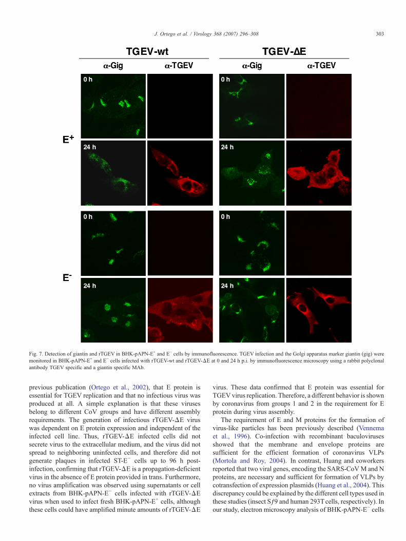
Fig. 7. Detection of giantin and rTGEV in BHK-pAPN-E+ and E− cells by immunofluorescence. TGEV infection and the Golgi apparatus marker giantin (gig) weremonitored in BHK-pAPN-E+ and E− cells infected with rTGEV-wt and rTGEV-ΔE at 0 and 24 h p.i. by immunofluorescence microscopy using a rabbit polyclonalantibody TGEV specific and a giantin specific MAb.
303J. Ortego et al. / Virology 368 (2007) 296–308
previous publication (Ortego et al., 2002), that E protein isessential for TGEV replication and that no infectious virus wasproduced at all. A simple explanation is that these virusesbelong to different CoV groups and have different assemblyrequirements. The generation of infectious rTGEV-ΔE viruswas dependent on E protein expression and independent of theinfected cell line. Thus, rTGEV-ΔE infected cells did notsecrete virus to the extracellular medium, and the virus did notspread to neighboring uninfected cells, and therefore did notgenerate plaques in infected ST-E− cells up to 96 h post-infection, confirming that rTGEV-ΔE is a propagation-deficientvirus in the absence of E protein provided in trans. Furthermore,no virus amplification was observed using supernatants or cellextracts from BHK-pAPN-E− cells infected with rTGEV-ΔEvirus when used to infect fresh BHK-pAPN-E+ cells, althoughthese cells could have amplified minute amounts of rTGEV-ΔE
virus. These data confirmed that E protein was essential forTGEV virus replication. Therefore, a different behavior is shownby coronavirus from groups 1 and 2 in the requirement for Eprotein during virus assembly.
The requirement of E and M proteins for the formation ofvirus-like particles has been previously described (Vennemaet al., 1996). Co-infection with recombinant baculovirusesshowed that the membrane and envelope proteins aresufficient for the efficient formation of coronavirus VLPs(Mortola and Roy, 2004). In contrast, Huang and coworkersreported that two viral genes, encoding the SARS-CoVM and Nproteins, are necessary and sufficient for formation of VLPs bycotransfection of expression plasmids (Huang et al., 2004). Thisdiscrepancy could be explained by the different cell types used inthese studies (insect Sƒ9 and human 293Tcells, respectively). Inour study, electron microscopy analysis of BHK-pAPN-E− cells

Fig. 8. Immunoelectron microscopy of ERGIC in rTGEV-ΔE infected ST-E− cells. Panel A and B show vesicles filled with viruses that react with the anti-ERGIC-53/p58 antibody, specific marker for the intermediate compartment. Panel C shows a detail of the cytoplasm and nucleus where ERGIC-53/p58 specific signal was notobserved. ERGIC-53/p58 specific signal is indicated with arrows. v, viruses; N, nucleus. Scale bars, 100 nm.
304 J. Ortego et al. / Virology 368 (2007) 296–308
infected with rTGEV-ΔE demonstrated that only immatureTGEV virions were assembled in the absence of E protein. Thesevirions contained RNA, and also all the structural TGEVproteins (S,M, and N) except the deleted E protein. These resultsindicate that the E protein is essential for the formation of matureTGEV virions. In the absence of E protein, the assembled virionsdisplayed a morphology different from the full-length virus,were non-infectious, accumulated in the cytoplasm of theinfected cells, and were not released to the extracellular mediumor transported to neighboring cells.
TGEV replication depends on the exocytic pathway tocomplete its morphogenesis (Salanueva et al., 1999). Virionswere assembled as large particles with annular morphology atperinuclear compartment of the cells, and changed theirmorphology into small dense virions during the transportalong the exocytic route (Risco et al., 1998; Tooze et al., 1987).In cells infected with rTGEV-ΔE, an accumulation of the KDELreceptor and the lack of fragmentation and dispersion of Golgimembranes were observed in absence of E protein. Theseeffects and rTGEV-ΔE virus assembly into mature particleswere recovered by providing E protein in trans. In addition,immunoelectron microscopy studies with an intermediatecompartment specific marker confirmed that, in the absenceof the E protein, the maturation of the virus is arrested in the
ERGIC. A similar effect has been described when TGEVinfection is blocked with monensin, a drug that selectivelyaffects the Golgi complex interrupting the secretory pathway.Monensin blocks the viral transport from ERGIC, leading to theaccumulation of numerous large viral particles in dilated pre-Golgi ERGIC elements of vacuolar morphology (Salanueva etal., 1999). The comparable deficiencies observed in viralmorphogenesis in the absence of E protein and as a result oftreatment with monensin suggest an aberrant assembly processdue to the absence of E protein. In principle, E protein could actdirectly on the ERGIC and Golgi compartments, on the virusitself, or in both. It has been postulated that E protein preparesthe membrane or other viral proteins occupying strategicpositions within the budding pathways (Salanueva et al.,1999), as proposed for small acylated glycoproteins of alpha-viruses and orthomyxoviruses that also display ion channelactivity (Gaedigk-Nitschko et al., 1990; Ivanova et al., 1995;Liao et al., 2006; Wilson et al., 2004; Zebedee and Lamb, 1988).The absence of E protein would inhibit the progression throughthe ERGIC-Golgi compartments, resulting in the accumulationof virions in the ERGIC and of the ERGIC itself.
E proteins from MHV and SARS-CoV have viroporinactivity (Liao et al., 2004; Madan et al., 2005). Viroporins act atlate stages of the viral cycle promoting the exit of new virus

305J. Ortego et al. / Virology 368 (2007) 296–308
particles from the cell (Gonzalez and Carrasco, 2003). The exactmechanism by which viroporins alter the permeability of theplasma membrane is unknown, and it is possible that thisactivity is displayed both at the plasma membrane and oncellular organelles (Aldabe et al., 1997; Van kuppeveld et al.,1997). Coronavirus E protein localizes in membranes ofERGIC, where it could modify their permeability, leading tothe disruption of ion gradients at intracellular organelles (Madanet al., 2005), facilitating the transport of the virions along theexocytic pathway and their maturation. The absence of thisviroporin activity mediated by the E protein possibly leads tothe arrest of the transport of virions through the secretorypathway, the accumulation of non-mature virions in ERGIG,and the absence of release of new infectious virus from the cell.The construction of E protein mutants affecting E protein ionchannel activity could assess whether this postulate is correct.
E proteins from the different coronavirus groups showdifferent channel selectivity. The E protein from the group 1coronavirus HCoV-229E is K+ selective, whereas from thegroup 2 MHVand SARS-CoV, and group 3 coronavirus IBV, Eprotein is Na+ selective (Wilson et al., 2006, 2004). The diversechannel selectivity among the coronavirus groups could betranslated in differences in the effect of E protein in thesecretory pathway and may explain the discrepancies among thegene E-defective mutants from the three coronavirus groups,and why E protein is essential for replication of group 1 virusesand non-essential for replication of group 2 viruses. Additionalanalysis of the gene E-defective mutants during assembly isrequired to shed light on the functions of the E protein incoronavirus morphogenesis in the three virus groups of theCoronaviridae family.
In conclusion, the arrest of TGEV maturation and theaccumulation of virions in the secretory pathway due to theabsence of the E protein confirm that this protein is essential forTGEV transportation andmorphogenesis. The ability to generateTGEV-ΔE mutants that are replication-competent but propaga-tion-deficient by complementation in packaging cell lines,supports the potential use of these mutants as vaccine vectors.
Materials and methods
Virus and cells
Recombinant rTGEV-MluI–FseI (rTGEV-wt) and rTGEV-Δ3abΔE (rTGEV-ΔE) (Ortego et al., 2002, 2003) were grownas described (Jiménez et al., 1986). Baby hamster cells (BHK-21) stably transformed with the gene coding for the porcineaminopeptidase N (BHK-pAPN) (Delmas et al., 1992) weregrown in Dulbecco's modified Eagle's medium (DMEM)supplemented with 2% fetal calf serum (FCS) containingGeneticin G418 (1.5 mg/ml) as a selector agent. Porcine kidneycells, LLC-PK1 (European Collection of Cell cultures no.86121112), were grown in medium 199 supplemented with2 mM glutamine and 10% FCS. Cat kidney cells, CRFK (ATCCCCL 94), were grown in DMEM supplemented with 2% FCS.BHK-pAPN-E+ and LLC-PK1-E+ cells were grown asdescribed previously (Ortego et al., 2002). Standard virus
titrations were performed in porcine swine testis (ST) cells.Titrations of virus with the E gene deleted were performed inLLC-PK1 cells expressing the E protein.
Antibodies
The murine MAbs 1D.G3, 3B.B3, and 3B.D10, specific forTGEV S, M, and N proteins, respectively, have been previouslydescribed (Gebauer et al., 1991; Risco et al., 1995; Sánchez etal., 1990). The murine MAb Q3, specific for TGEV E protein,was a kind gift of H. Laude (INRA, Jouy-en Josas, France). Therabbit serum specific for PDI and the murine MAb specific forgiantin was generously provided by A. Nieto (Centro Nacionalde Biotecnología, CSIC, Madrid, Spain) and M. Renz (Instituteof Immunology and Molecular Genetics, Karlsruhe, Germany),respectively. The murine MAb specific for KDEL receptor andthe rabbit polyclonal antibody specific for ERGIC-53/p58 werepurchased from Stressgen Biotechnologies Corp. (Victoria, BC,Canada) and Sigma-Aldrich (St. Louis. MO, USA), respectively.
TGEV virion purification
TGEV virions were sedimented through a 20% sucrosecushion in TEN (Tris–HCl 10 mM [pH 7.4], EDTA 1 mM,NaCl 1M) 0.2% Tween 20 by centrifugation in an SW28Beckman rotor at 25,000 rpm for 2 h at 4 °C. Sucrose wasremoved and the pellet was washed with TEN. Pellet wasrecovered by suspending it in TEN-0.2% Tween 20 andsedimented by centrifugation in a SW 41 Beckman rotor for1 h at 25,000 rpm. Virions were recovered by suspending thepellet in TNE (Tris–HCl 10 mM [pH 7.4], EDTA 1 mM, NaCl100 mM) and analyzed by sodium dodecyl sulfate-polyacryla-mide gel electrophoresis (SDS-PAGE) and silver staining ornegative staining and electron microscopy.
Electron microscopy
Processing of infected cells for embedding in EML-812 forultrastructural studies
Monolayers of BHK-pAPN cells expressing TGEV Eprotein and control cells were infected with rTGEV-wt orrTGEV-ΔE virus. The cells were fixed in situ at 12, 16, and 24 hpost-infection with a mixture of 2% glutaraldehyde and 1%tannic acid in 0.4 M HEPES buffer (pH 7.2) for 2 h at roomtemperature. The fixed monolayers were removed from thedishes in the fixative and transferred to Eppendorf tubes. Aftercentrifugation in a microcentrifuge, the cell pellets were washedwith HEPES buffer and processed for embedding in EML-812(Taab Laboratories, Berkshire, United Kingdom) as described(Risco et al., 1998). The cells were post-fixed with a mixture of1% osmium tetroxide and 0.8% potassium ferricyanide indistilled water for 1 h at 4 °C. After four washes with HEPESbuffer, samples were incubated with 2% uranyl acetate, washedagain, and dehydrated in increasing concentrations of acetone(50, 70, 90, and 100%) for 15 min each at 4 °C. Infiltration inthe resin EML-812 was done at room temperature for 1 day.Polymerization of infiltrated samples was done at 60 °C for

306 J. Ortego et al. / Virology 368 (2007) 296–308
2 days. Ultrathin (50- to 60-nm-thick) sections of the sampleswere stained with saturated uranyl acetate and lead citrate bystandard procedures.
Quick freezing and freeze-substitution of cellsCultures of cells were subjected to a mild fixation with a
solution of 4% paraformaldehyde containing 0.1% glutaralde-hyde in PBS at 4 °C for 30 min. Small pellets of chemicallyfixed cells were cryoprotected with glycerol, applied to smallpieces of filter paper, blotted, and quick frozen in liquidethane. Vitrified specimens were transferred to a Reichert-JungAFS freeze-substitution unit (Leica, Vienna, Austria) andmaintained for 48 h at −90 °C in a mixture of methanol and0.5% (w/v) uranyl acetate. After freeze-substitution, sampleswere infiltrated in Lowicryl K4M (EML Laboratories) at−30 °C and polymerized with UV light. Ultrathin sections ofthe samples were either stained or processed for immunogoldlabeling.
Immunoelectron microscopyImmunogold detection of TGEV proteins on ultrathin-
sections of infected BHK-pAPN cells was performed at roomtemperature with MAbs specific for M, N, S, E, and ERGIC-53proteins by established procedures (Risco et al., 1995). Sectionscollected on Formvar coated gold electron microscopy gridswere incubated for 30 min with Tris buffer-gelatin and thenfloated on drops of diluted primary antibodies for 75 min. Afterjet washing with PBS, samples were incubated for 45 min withsecondary antibodies conjugated with 10-nm-diameter goldparticles and washed again with PBS and distilled water.Samples were then allowed to dry on filter paper before beingstained with saturated uranyl acetate for 25 min, followed by1 min with lead citrate. All samples were analyzed with a JEOL1200 EX II electron microscope.
For ultrastructural detection of RNA, a complex of RNase and10-nm-diameter colloidal gold (EY Laboratories, San Diego,CA) was used as described previously (Risco et al., 1998).Ultrathin sections from Epon-included samples were collectedon gold electron microscopy (EM) grids covered with collodionand incubated for 40 min at room temperature with the goldconjugated RNase diluted 1:20 in PBS. After being washed withPBS and distilledwater, sampleswere stainedwith uranyl acetateand lead citrate. To unmask viral RNA molecules, sections weresubjected to a treatment before incubation with RNase-goldconsisting on 15 min incubation at 37 °C with proteinase K(10 μg/ml in Tris-EDTA), washingwith TE, 10min fixation with4% paraformaldehyde in PBS, washing again with PBS, andincubation for 10 min with 0.2 M NH4Cl. As a cytochemicalcontrol, some sections were pre-incubated for 30 min at 37 °Cwith a solution of non-conjugatedRNase (20μg/ml) before beingtreated with the RNase-gold conjugated.
Negative staining of rTGEV-ΔE virionsVirus from supernatants of BHK-pAPN-E+ and E− cells
infected with rTGEV-ΔE virus, concentrated 200-fold, wereanalyzed by negative staining as described (Bremer et al.,1998). Briefly, samples were adsorbed to UV light-activated
copper grids for 2 min at room temperature. Grids were washedtwo times in H2O (Escors et al., 2001) and stained with 2%uranyl acetate for 1 min. Samples were visualized in a JEOL1200 EXII transmission electron microscope.
Immunofluorescence
Cells were plated on glass coverslips. Infections wereperformed at an m.o.i. =1 pfu/cell at 37 °C in DMEMcontaining 2% FCS. The inoculum was removed at 90 minand the cells were maintained in DMEM 2% FCS. At the timesindicated, cells were washed with PBS and fixed by addition of4% paraformaldehyde for 30 min at room temperature. Fordual-labeling experiments in which, one primary antibody wasderived from mouse and the other one from rabbit, bothantibodies were combined in a PBS-FCS 20% diluent contain-ing 0.2% Saponin (Superfos-Biosector, Vedback, Denmark).Antibodies were allowed to adsorb for 90 min at roomtemperature, and washed three times with PBS-FCS 2%. Cellswere then incubated for 30 min at room temperature with amixture of anti-rabbit and anti-mouse secondary antibodiesconjugated to Alexa 488 or Alexa 594. The coverslips werewashed five times with PBS-FCS 2%, mounted on glass slides,and analyzed with a Laser Scanning Confocal System Radiance2100 (Bio-Rad) on a Zeiss Axiovert 200 microscope. Argon ionand He–Ne lasers at 488 and 543 nm were employed asexcitation sources. Image acquisition was performed usingLaserSharp2000 v.5 software.
Acknowledgments
We thank S. Zuñiga, I. Sola, J.L. Moreno, M.L DeDiego, andE. Alvarez for critically reading the manuscript and helpfuldiscussions. We are also grateful to D. Dorado, and R. Arranzfor their excellent technical assistance. This work was supportedby grants from the Comisión Interministerial de Ciencia yTecnología (CICYT), the Consejería de Educación y Cultura dela Comunidad de Madrid, Fort Dodge Veterinaria, and theEuropean Communities (Projects FMDV Vaccine. QRLT-2001-00825 and DISSECT, SP22-CT-2004-511060).
References
Aldabe, R., Irurzun, A., Carrasco, L., 1997. Poliovirus protein 2BC increasescytosolic free calcium concentrations. J. Virol. 71, 6214–6217.
Almazán, F., González, J.M., Pénzes, Z., Izeta, A., Calvo, E., Plana-Durán, J.,Enjuanes, L., 2000. Engineering the largest RNA virus genome as aninfectious bacterial artificial chromosome. Proc. Natl. Acad. Sci. USA 97,5516–5521.
Arbely, E., Khattari, Z., Brotons, G., Akkawi, M., Salditt, T., Arkin, I.T., 2004.A highly unusual palindromic transmembrane helical hairpin formed bySARS coronavirus E protein. J. Mol. Biol. 341, 769–779.
Baudoux, P., Carrat, C., Besnardeau, L., Charley, B., Laude, H., 1998.Coronavirus pseudoparticles formed with recombinant M and E proteinsinduce alpha interferon synthesis by leukocytes. J. Virol. 72, 8636–8643.
Bremer, A., Häner, M., Aebi, U., 1998. Negative staining, Second ed. In: Celis,E. (Ed.), Cell Biology. A Laboratory Handbook, vol. 3. Academic Press, SanDiego, pp. 277–284.
Brierley, I., Digard, P., Inglis, S.C., 1989. Characterization of an efficient

307J. Ortego et al. / Virology 368 (2007) 296–308
coronavirus ribosomal frameshifting signal: requirement for an RNApseudoknot. Cell 57, 537–547.
Casais, R., Thiel, V., Siddell, S.G., Cavanagh, D., Britton, P., 2001.Reverse genetics system for the avian coronavirus infectious bronchitisvirus. J. Virol. 75, 12359–12369.
Corse, E., Machamer, C.E., 2000. Infectious bronchitis virus E protein istargeted to the Golgi complex and directs release of virus-like particles.J. Virol. 74, 4319–4326.
Curtis, K.M., Yount, B., Baric, R.S., 2002. Heterologous gene expression fromtransmissible gastroenteritis virus replicon particles. J. Virol. 76, 1422–1434.
DeDiego, M.L., Alvarez, A., Almazan, F., Rejas, M.T., Lamirande, E., Roberts,A., Shieh, W.J., Zaki, S., Subbarao, K., Enjuanes, L., 2007. A severe acuterespiratory syndrome coronavirus that lacks the E gene is attenuated in vitroand in vivo. J. Virol. 81, 1701–1713.
Delmas, B., Gelfi, J., L'Haridon, R., Vogel, L.K., Norén, O., Laude, H., 1992.Aminopeptidase N is a major receptor for the enteropathogenic coronavirusTGEV. Nature 357, 417–420.
Enjuanes, L., Brian, D., Cavanagh, D., Holmes, K., Lai, M.M.C., Laude, H.,Masters, P., Rottier, P., Siddell, S.G., Spaan, W.J.M., Taguchi, F., Talbot, P.,2000a. Coronaviridae. In: Wickner, R.B. (Ed.), Virus taxonomy. Classifica-tion and Nomenclature of Viruses. Academic Press, San Diego, California,pp. 835–849.
Enjuanes, L., Spaan, W., Snijder, E., Cavanagh, D., 2000b. Nidovirales. In:Wickner, R.B. (Ed.), Virus Taxonomy. Classification and Nomenclature ofViruses. Academic Press, San Diego, California, pp. 827–834.
Escors, D., Camafeita, E., Ortego, J., Laude, H., Enjuanes, L., 2001.Organization of two transmissible gastroenteritis coronavirus membraneprotein topologies within the virion and core. J. Virol. 75, 12228–12240.
Esper, F., Shapiro, E.D., Weibel, C., Ferguson, D., Landry, M.L., Kahn, J.S.,2005a. Association between a novel human coronavirus and Kawasakidisease. J. Infect. Dis. 191, 499–502.
Esper, F., Weibel, C., Ferguson, D., Landry, M.L., Kahn, J.S., 2005b. Evidenceof a novel human coronavirus that is associated with respiratory tract diseasein infants and young children. J. Infect. Dis. 191, 492–498.
Gaedigk-Nitschko, K., Ding, M.X., Levy, M.A., Schlesinger, M.J., 1990. Site-directed mutations in the Sindbis virus 6K protein reveal sites for fattyacylation and the underacylated protein affects virus release and virionstructure. Virology 175, 282–291.
Gebauer, F., Posthumus, W.A.P., Correa, I., Suñé, C., Sánchez, C.M., Smerdou,C., Lenstra, J.A., Meloen, R., Enjuanes, L., 1991. Residues involved in theformation of the antigenic sites of the S protein of transmissiblegastroenteritis coronavirus. Virology 183, 225–238.
Godet, M., L'Haridon, R., Vautherot, J.F., Laude, H., 1992. TGEV coronavirusORF4 encodes a membrane protein that is incorporated into virions.Virology 188, 666–675.
Gonzalez, M.E., Carrasco, L., 2003. Viroporins. FEBS Lett. 552, 28–34.Hertzig, T., Sacandella, E., Schelle, B., Ziebuhr, J., Siddell, S., Ludewig, B.,
Thiel, V., 2004. Rapid identification of coronavirus replicase inhibitors usinga selectable replicon RNA. J. Gen. Virol. 85, 1717–1725.
Hsieh, P.K., Chang, S.C., Huang, C.C., Lee, T.T., Hsiao, C.W., Kou, Y.H.,Chen, I.Y., Chang, C.K., Huang, T.H., Chang, M.F., 2005. Assembly ofsevere acute respiratory syndrome coronavirus RNA packaging signalinto virus-like particles is nucleocapsid dependent. J. Virol. 79,13848–13855.
Huang, Y., Yang, Z.Y., Kong, W.P., Nabel, G.J., 2004. Generation of syntheticsevere acute respiratory syndrome coronavirus pseudoparticles: implicationsfor assembly and vaccine production. J. Virol. 78, 12557–12565.
Ivanova, L., Lustig, S., Schlesinger, M.J., 1995. A pseudo-revertant of a Sindbisvirus 6K protein mutant, which corrects for aberrant particle formation,contains two new mutations that map to the ectodomain of the E2glycoprotein. Virology 206, 1027–1034.
Jiménez, G., Correa, I., Melgosa, M.P., Bullido, M.J., Enjuanes, L., 1986.Critical epitopes in transmissible gastroenteritis virus neutralization. J. Virol.60, 131–139.
Kapke, P.A., Brian, D.A., 1986. Sequence analysis of the porcine transmissiblegastroenteritis coronavirus nucleocapsid protein gene. Virology 151, 41–49.
Koetzner, C.A., Parker, M.M., Ricard, C.S., Sturman, L.S., Masters, P.S., 1992.Repair and mutagenesis of the genome of a deletion mutant of the
coronavirus mouse hepatitis virus by targeted RNA recombination. J. Virol.66, 1841–1848.
Kuiken, T., Fouchier, R.A.M., Schutten, M., Rimmelzwaan, G.F., vanAmerongen, G., van Riel, D., Laman, J.D., de Jong, T., van Doornum, G.,Lim, W., Ling, A.E., Chan, P.K.S., Tam, J.S., Zambon, M.C., Gopal, R.,Drosten, C., van der Werf, S., Escriou, N., Manuguerra, J.-C., Stohr, K.,Peiris, J.S.M., 2003. Newly discovered coronavirus as the primary cause ofsevere acute respiratory syndrome. Lancet 362, 263–270.
Kuo, L., Masters, P.S., 2003. The small envelope protein E is not essential formurine coronavirus replication. J. Virol. 77, 4597–4608.
Kuo, L., Godeke, G.-J., Raamsman, M.J.B., Masters, P.S., Rottier, P.J.M., 2000.Retargeting of coronavirus by substitution of the spike glycoproteinectodomain: crossing the host cell species barrier. J. Virol. 74, 1393–1406.
Kuo, L., Hurst, R., Masters, P.S., 2002. 21st Annual Meeting, Lexington,Kentucky.
Kuo, L., Hurst, K.R., Masters, P., 2007. Exceptional flexibility in the sequencerequirements for coronavirus small envelope protein function. J. Virol. 81,2249–2262.
Lewicki, D.N., Gallagher, T.M., 2002. Quaternary structure of coronavirusspikes in complex with carcinoembryonic antigen-related cell adhesionmolecule cellular receptors. J. Biol. Chem. 277, 19727–19734.
Liao, Y., Lescar, J., Tam, J.P., Liu, D.X., 2004. Expression of SARS-coronavirusenvelope protein in Escherichia coli cells alters membrane permeability.Biochem. Biophys. Res. Commun. 325, 374–380.
Liao, Y., Yuan, Q., Torres, J., Tam, J.P., Liu, D.X., 2006. Biochemical andfunctional characterization of the membrane association and membranepermeabilizing activity of the severe acute respiratory syndrome coronavirusenvelope protein. Virology 349, 264–275.
Liu, D.X., Inglis, S.C., 1991. Association of the infectious bronchitis virus-3cprotein with the virion envelope. Virology 185, 911–917.
Madan, V., Garcia M de, J., Sanz, M.A., Carrasco, L., 2005. Viroporin activityof murine hepatitis virus E protein. FEBS Lett. 579, 3607–3612.
Maeda, J., Maeda, A., Makino, S., 1999. Release of coronavirus E proteinmembrane vesicles form virus-infected cells and E protein-expressing cells.Virology 263, 265–272.
Masters, P.S., 1999. Reverse genetics of the largest RNA viruses. Adv. VirusRes. 53, 245–264.
Mortola, E., Roy, P., 2004. Efficient assembly and release of SARS coronavirus-like particles by a heterologous expression system. FEBS Lett. 576,174–178.
Narayanan, K., Makino, S., 2001. Cooperation of an RNA packaging signal anda viral envelope protein in coronavirus RNA Packaging. J. Virol. 75,9059–9067.
Ortego, J., Escors, D., Laude, H., Enjuanes, L., 2002. Generation of a replication-competent, propagation-deficient virus vector based on the transmissiblegastroenteritis coronavirus genome. J. Virol. 76, 11518–11529.
Ortego, J., Sola, I., Almazan, F., Ceriani, J.E., Riquelme, C., Balasch, M., Plana-Durán, J., Enjuanes, L., 2003. Transmissible gastroenteritis coronavirusgene 7 is not essential but influences in vivo virus replication and virulence.Virology 308, 13–22.
Penzes, Z., González, J.M., Calvo, E., Izeta, A., Smerdou, C., Mendez, A.,Sánchez, C.M., Sola, I., Almazán, F., Enjuanes, L., 2001. Completegenome sequence of transmissible gastroenteritis coronavirus PUR46-MAD clone and evolution of the Purdue virus cluster. Virus Genes 23,105–118.
Raamsman, M.J.B., Locker, J.K., de Hooge, A., de Vries, A.A.F., Griffiths, G.,Vennema, H., Rottier, P.J.M., 2000. Characterization of the coronavirusmouse hepatitis virus strain A59 small membrane protein E. J. Virol. 74,2333–2342.
Risco, C., Antón, I.M., Suñé, C., Pedregosa, A.M., Martín-Alonso, J.M., Parra,F., Carrascosa, J.L., Enjuanes, L., 1995. Membrane protein molecules oftransmissible gastroenteritis coronavirus also expose the carboxy-terminalregion on the external surface of the virion. J. Virol. 69, 5269–5277.
Risco, C., Muntión, M., Enjuanes, L., Carrascosa, J.L., 1998. Two types ofvirus-related particles are found during transmissible gastroenteritis virusmorphogenesis. J. Virol. 72, 4022–4031.
Rottier, P.J.M., 1995. The coronavirus membrane glycoprotein. In: Siddell, S.G.(Ed.), The Coronavirus. Plenum press, New York, pp. 115–135.

308 J. Ortego et al. / Virology 368 (2007) 296–308
Salanueva, I.J., Carrascosa, J.L., Risco, C., 1999. Structural maturation of thetransmissible gastroenteritis coronavirus. J. Virol. 73, 7952–7964.
Sánchez, C.M., Jiménez, G., Laviada, M.D., Correa, I., Suñé, C., Bullido, M.J.,Gebauer, F., Smerdou, C., Callebaut, P., Escribano, J.M., Enjuanes, L., 1990.Antigenic homology among coronaviruses related to transmissible gastro-enteritis virus. Virology 174, 410–417.
Shen, X., Xue, J.H., Yu, C.Y., Luo, H.B., Qin, L., Yu, X.J., Chen, J., Chen,L.L., Xiong, B., Yue, L.D., Cai, J.H., Shen, J.H., Luo, X.M., Chen, K.X.,Shi, T.L., Li, Y.X., Hu, G.X., Jiang, H.L., 2003. Small envelope protein Eof SARS: cloning, expression, purification, CD determination, andbioinformatics analysis. Acta Pharmacol. Sin. 24, 505–511.
Snijder, E.J., van Tol, H., Pedersen, K.W., Raamsman, M.J.B., de Vries, A.A.F.,1999. Identification of a novel structural protein of anteriviruses. J. Virol. 73,6335–6345.
Sui, J.,Li,W.,Murakami,A.,Tamin,A.,Matthews,L.J.,Wong,S.K.,Moore,M.J.,Tallarico, A.S., Olurinde,M., Choe, H., Anderson, L.J., Bellini,W.J., Farzan,M., Marasco, W.A., 2004. Potent neutralization of severe acute respiratorysyndrome (SARS) coronavirus by a human mAb to S1 protein that blocksreceptor association. Proc. Natl. Acad. Sci. U.S.A. 101, 2536–2541.
Suñé, C., Jiménez, G., Correa, I., Bullido, M.J., Gebauer, F., Smerdou, C.,Enjuanes, L., 1990. Mechanisms of transmissible gastroenteritis coronavirusneutralization. Virology 177, 559–569.
Suñé, C., Smerdou, C., Antón, I.M., Abril, P., Plana, J., Enjuanes, L., 1991. Aconserved coronavirus epitope, critical in virus neutralization, mimicked byinternal-image monoclonal anti-idiotypic antibodies. J. Virol. 65, 6979–6984.
Thiel, V., Herold, J., Schelle, B., Siddell, S., 2001. Infectious RNA transcribedin vitro from a cDNA copy of the human coronavirus genome cloned invaccinia virus. J. Gen. Virol. 82, 1273–1281.
Tooze, J., Tooze, S., Warren, G., 1984. Replication of coronavirus MHV-A59 insac-cells: determination of the first site of budding of progeny virions. Eur. J.Cell Biol. 33, 281–294.
Tooze, J., Tooze, S.A., Fuller, S.D., 1987. Sorting of progeny coronavirus fromcondensed secretory proteins at the exit from the trans-golgi network ofatT20 cells. J. Cell Biol. 105, 1215–1226.
van der Hoek, L., Pyrc, K., Jebbink, M.F., Vermeulen-Oost, W., Berkhout, R.J.,Wolthers, K.C., Wertheim-van Dillen, P.M., Kaandorp, J., Spaargaren, J.,Berkhout, B., 2004. Identification of a new human coronavirus. Nat. Med.10, 368–373.
Van kuppeveld, F.J., Melchers, W.J., Kirkegaard, K., Doedens, J.R., 1997.Structure-function analysis of coxsackie B3 virus protein 2B. Virology 227,111–118.
Vennema, H., Godeke, G.J., Rossen, J.W.A., Voorhout, W.F., Horzinek, M.C.,Opstelten, D.J., Rottier, P.J.M., 1996. Nucleocapsid-independent assemblyof coronavirus-like particles by co-expression of viral envelope proteingenes. EMBO J. 15, 2020–2028.
Wilson, L., McKinlay, C., Gage, P., Ewart, G., 2004. SARS coronavirus Eprotein forms cation-selective ion channels. Virology 330, 322–331.
Wilson, L., Gage, P., Ewart, G., 2006. Hexamethylene amiloride blocks Eprotein ion channels and inhibits coronavirus replication. Virology 353,290–306.
Yount, B., Curtis, K.M., Baric, R.S., 2000. Strategy for systematic assembly oflarge RNA and DNA genomes: the transmissible gastroenteritis virus model.J. Virol. 74, 10600–10611.
Yount, B., Curtis, K.M., Fritz, E.A., Hensley, L.E., Jahrling, P.B., Prentice,E., Denison, M.R., Geisbert, T.W., Baric, R.S., 2003. Reverse geneticswith a full-length infectious cDNA of severe acute respiratory syndromecoronavirus. Proc. Natl. Acad. Sci. U.S.A. 100, 12995–13000.
Yu, X., Bi, W., Weiss, S.R., Leibowitz, J.L., 1994. Mouse hepatitis virus gene5b protein is a new virion envelope protein. Virology 202, 1018–1023.
Zebedee, S.L., Lamb, R.A., 1988. Influenza A virus M2 protein: monoclonalantibody restriction of virus growth and detection of M2 in virions. J. Virol.62, 2762–2772.