Embed Size (px)
Citation preview

Abnormalities of presentation position and lie
Presented by:
Haneen Omar Abuhani

Outlines
• Malpresentation
• Malposition
• Abnormal lies
• Shoulder dystocia

Definitions
• Presentation : the part of the fetus that is lowermost in the pelvis
Malpresentation: any presentation other than vertex
• Position: the relationship of the part of the fetus that presents in the pelvis to the four quadrants of the maternal pelvis
Malpositions: Abnormal positions of vertex relative to maternal pelvis
oAny position other than occcipto-anterior
• Lie: the relationship between the longitudinal axis of the fetus and the longitudinal axis of the mother
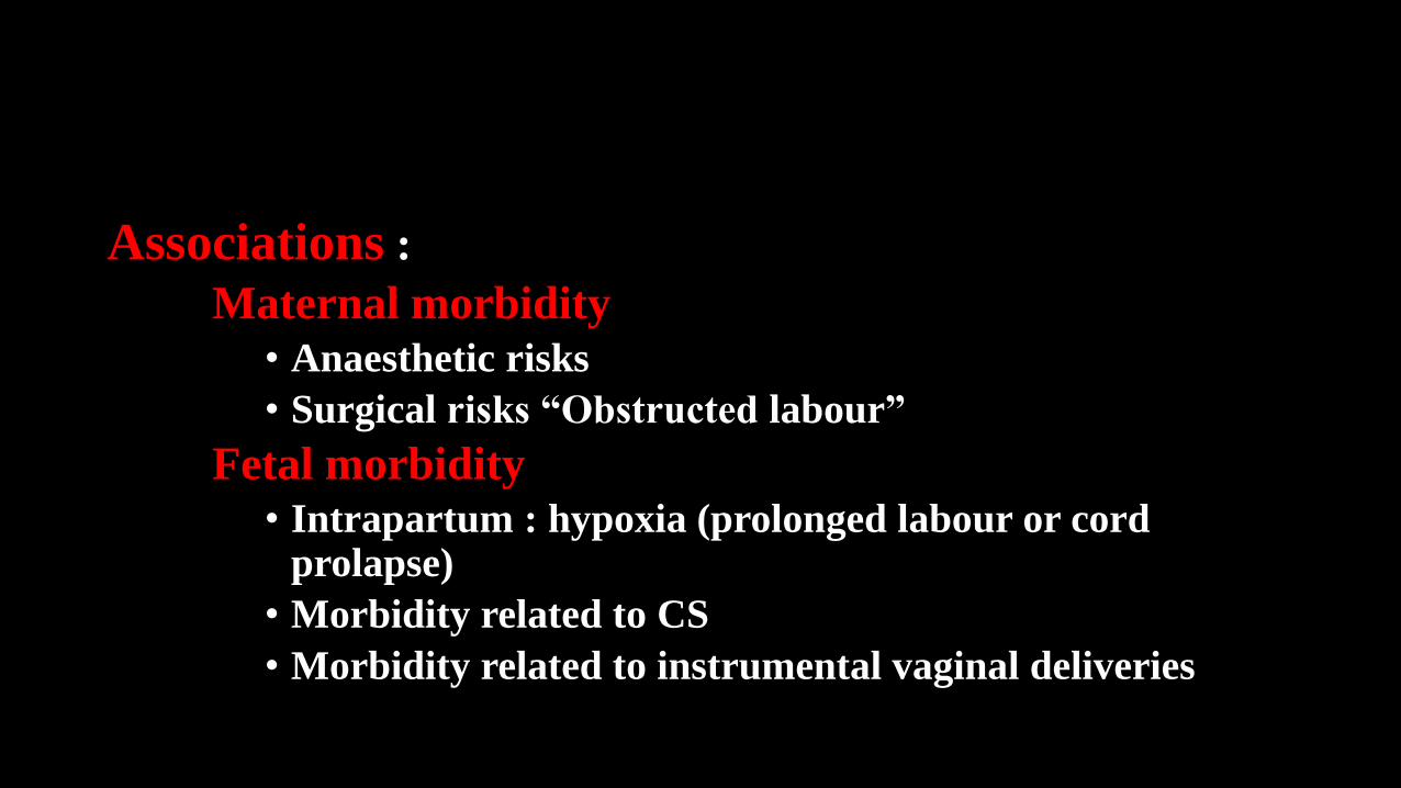
Associations :
Maternal morbidity
• Anaesthetic risks
• Surgical risks “Obstructed labour”
Fetal morbidity
• Intrapartum : hypoxia (prolonged labour or cord prolapse)
• Morbidity related to CS
• Morbidity related to instrumental vaginal deliveries

Presetation
• Portion of the fetus overlying the pelvic inlet.
• The most common presentation is cephalic.
• This is 96% of fetuses at term.
• Malpresentations
• Breech
• Face
• Brow
• Cord presentation and prolapse
• Shoulder / Compound

Breech
• Incidence
28 % at 20 weeks, 15 % at 28 weeks, 3 % at term
• Predisposing factors for breech presentation
oMaternal
Fibroids.
Congenital uterine abnormalities (??).
Uterine surgery. ?
oFetal/placental
Multiple gestation.
Prematurity.
Placenta praevia.
Abnormality (e.g. anencephaly or hydrocephalus).
Fetal neuromuscular condition.
Oligohydramnios or Polyhydramnios.

Types of breech
•Frank Breech (65%)
•Footling Breech (25%)
•Complete Breech (10%)


Antenatal management of breech presentation
•External cephalic version (ECV)
•Vaginal breech delivery
•Elective caesarean section

Antenatal management of breech presentation External cephalic version (ECV)
• Reduces incidence of non-cephalic presentation at
delivery
• Reduces incidence of CS rate
• Recommended to all women with an uncomplicated breech presentation from 36 weeks
• Should not be offered routinely before term, as it has not been shown to improve outcomes if performed before term

ECV • Success rates 40 - 80 %
• Following successful ECV, 97% remain cephalic
Factors that may increase the likelihood of success
oMultiparity
oAdequate liquor volume
oBreech above the pelvic brim
oFetal head easy to feel
oPlacenta not anterior
o? Low BMI

ECV: contraindications
Relative
• Previous lower segment CS
• Maternal disease (hypertension, diabetes)
• IUGR or oligohydramnios
• Maternal high BMI
Absolute
• Multiple pregnancy
• Antepartum haemorrhage (within the last 7 days)
• CS indicated for other reasons
• Ruptured membranes
• Fetal abnormality


Risks of ECV
• Placental abruption.
• Premature rupture of the membranes.
• Cord accident.
• Transplacental haemorrhage (remember anti-D administration to rhesusnegative women).
• Fetal bradycardia.
• Uterine rupture

Breech:
Mode of delivery at term
Planned c section is recommended (Term Breech Trial)
• Reduces: perinatal or neonatal morbidity and death
• Reduces combined complications
Preterm
Controversial
• Decisions should be individualised

Twin breech
First twin breech
• Theoretical risk of interlocked twins (1/817)
• Recommend elective CS
Second twin breech
• does not recommend elective CS
• Second twin‟s position may change following delivery of the first in 20 % of cases

Vaginal delivery of breech
Pre-requisites for vaginal breech delivery:
• The presentation should be either extended (hips flexed, knees extended) or flexed
• There should be no evidence of feto-pelvic disproportion and an estimated fetal weight of <3,500 g (ultrasound or clinical measurement).
• There should be no evidence of hyperextension of the fetal head, and fetal abnormalities

Vaginal delivery of breech
Management of labour
• Fetal wellbeing and progress of labour should be carefully monitored.
• An epidural analgesia is not essential but may be advantageous?
• Fetal blood sampling from the buttocks
• Available operator experienced in delivery.
Technique
• It should be characterized by „masterly inactivity‟ (handsoff).
• Problems are more likely to arise when the obstetrician tries to speed up the process by pulling on the baby, and this should be avoided.

Vaginal delivery of breech
• Delivery of the legs and lower body
If the legs are flexed ?
If extended > Pinard‟s manoeuvre. ?
• Delivery of the buttocks
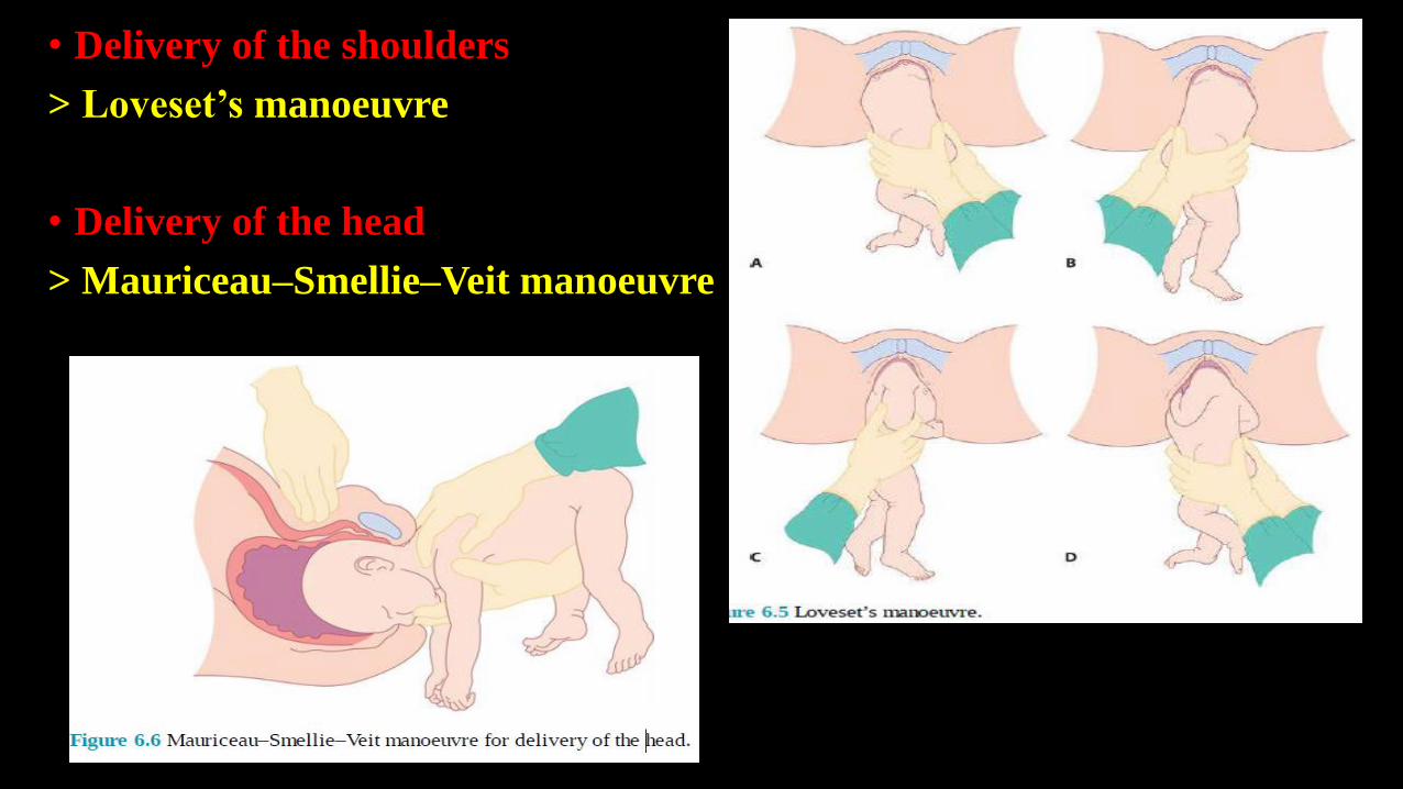
• Delivery of the shoulders
> Loveset‟s manoeuvre
• Delivery of the head
> Mauriceau–Smellie–Veit manoeuvre

Head related abnormal presentations
Face
Brow

Head attitude
Degree of extension-flexion of the fetal head with cephalic presentation.
The most common attitude is vertex.
• Vertex: head is maximally flexed
• Military: head is partially flexed
• Brow: head is partially extended
• Face: head is maximally extended

Flexed
Suboccipito
bregmatic
Deflexed
Occipito-frontal
Brow
Mento-vertical
Face
Submento-bregmatic

Face presentation
• Incidence and aetiology
1/500
• Associated with
• Prematurity
• Fetal goitre
• Uterine anomalies
• Polyhydramnios
• Placenta praevia

Face: clinical finding
• Diagnosis usually made in labour by vaginal examination
• Landmarks: mandible, mouth, nose and orbital ridges
• Avoid damage to the eyes on examination
• Facial oedema: distinction between face and breech ? difficult
• Ultrasound: if there is any doubt
• Delay in the first or second stage of labour may occur

Face: management
• Ultrasound: exclude fetal or pelvic abnormalities
• Vaginal delivery: possible with the mento-anterior position
• In the second stage : o Mento-anterior : head may deliver by flexion
o Mento-posterior: may rotate during the second stage
• Fetal risks: facial soft tissue trauma, causes feeding difficulties
• Maternal risks: perineal injury, sphincter damage, CS
• Augmentation: not advised
• Lack of progress : prompt delivery by CS
• Vacuum delivery: contra-indicated
Why mentoposterior does not deliver vaginally?
• In vertex: delivery of fetal head occurs by extension
•In face : head is already in maximum extension
•In mentoposterior; the head, neck and shoulders enter the pelvis at the same time
•The length of the sacrum is 10 cm
•The length of neck is 5 cm
•The shoulders get impacted
•Labour is obstructed

Brow presentation
Incidence and aetiology
• 1/1000 deliveries
• Due to a deflexed head
Associated with
o Prematurity
o Fetal neck tumours

Brow: Clinical findings
• In labour: failure to progress in first or second stage
• Vaginal examination: forehead is the leading part
• The anteroposterior diameter of the head is „mento-vertical‟: about 13.5 cm at term
• The average anteroposterior and lateral diameters of the female mid-pelvis are 12 × 12 cm

Brow: management
Diagnosis in the early first stage: • Expectant management for a short time (2–3 hours)
• May flex into a vertex or extend to face
Diagnosis often made in late first or second stage: • Caesarean delivery is advised
Augmentation with syntocinon: not advised “uterine Rupture”
Mento-vertical dimensions may be smaller in a preterm fetus, allowing vaginal delivery

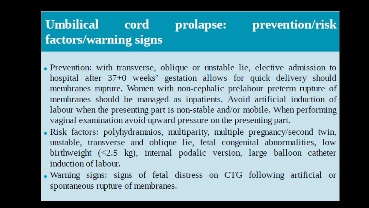
Cord presentation and prolapse
Incidence and aetiology
• Cord presentation/cord prolapse : 0.1–0.6 % of all births
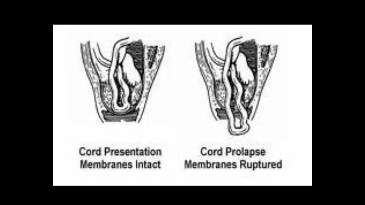
• Cord presentation:
Cord below presenting part, with the membranes intact
• Cord prolapse:
Cord descending through the cervix into the vagina with ruptured membranes
• May follow fetal scalp electrode placement, stabilizing induction of labour, external cephalic version or internal podalic version

Clinical findings
• Presence of a „high‟ presenting part in early labour
• Ultrasound: the presence of a cord presentation
• In advanced labour, the findings are self-explanatory
• The cord may be felt pulsating
• Abnormal cardiotocography, should raise the possibility

Management Cord prolapse
• An emergency delivery
• Fetal hypoxia: o Pressure from the presenting part o Arterial spasm
• The presenting part should be elevated (various methods)
• The cord should be replaced in the vaginal with minimal handling
• In the presence of viable fetus: Immediate delivery
Cord presentation • May be seen by USS in preterm fetuses: No intervention
• Usually diagnosed in labour by VE
• If in labour: CS

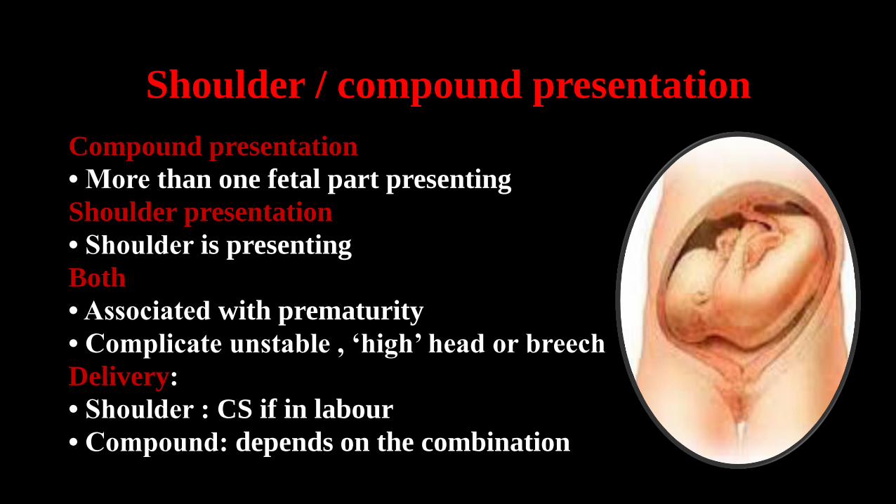
Shoulder / compound presentation
Compound presentation
• More than one fetal part presenting
Shoulder presentation
• Shoulder is presenting
Both
• Associated with prematurity
• Complicate unstable , „high‟ head or breech
Delivery:
• Shoulder : CS if in labour
• Compound: depends on the combination
Shoulder / compound presentation
Compound presentation
• More than one fetal part presenting
Shoulder presentation
• Shoulder is presenting
Both
• Associated with prematurity
• Complicate unstable , „high‟ head or breech
Delivery:
• Shoulder : CS if in labour
• Compound: depends on the combination

Malpositions “Occipito-Posterior” (OP)

OP
Prevalence
• 15 to 32% at the onset of labour
• 10 to 20% early in the second stage
• 5 to 8% at delivery
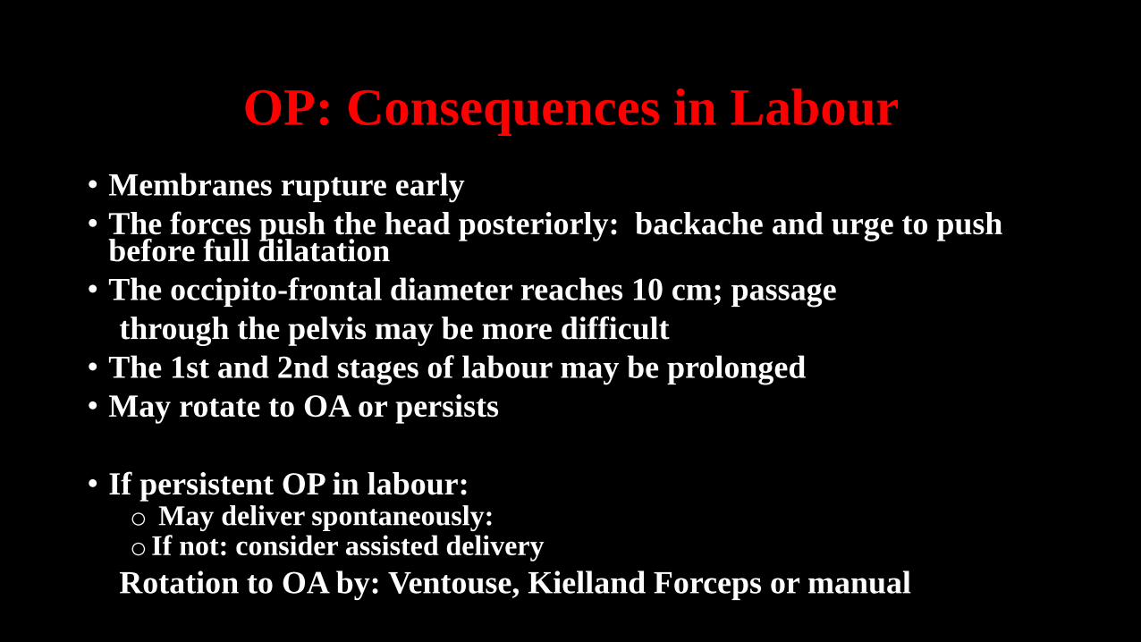
OP: Consequences in Labour
• Membranes rupture early
• The forces push the head posteriorly: backache and urge to push before full dilatation
• The occipito-frontal diameter reaches 10 cm; passage
through the pelvis may be more difficult
• The 1st and 2nd stages of labour may be prolonged
• May rotate to OA or persists
• If persistent OP in labour: o May deliver spontaneously: o If not: consider assisted delivery
Rotation to OA by: Ventouse, Kielland Forceps or manual

Abnormal lie

Unstable/ transverse/ Oblique lies
Incidence and aetiology 1/320
Association Multiparity
Polyhydramnios
Placenta praevia
Pelvic tumour
Uterine anomaly
Contracted maternal pelvis
Hydrocephalus and fetal neck tumours
Fetal neuromuscular dysfunction “reduced FM”
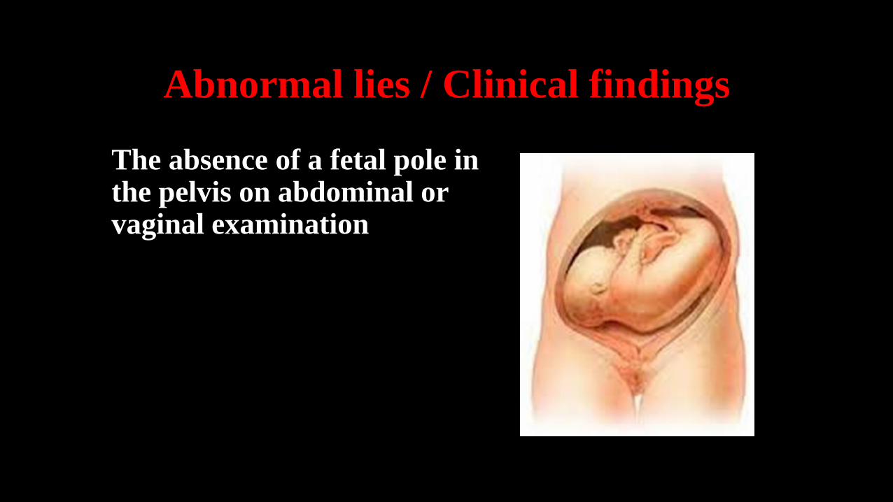
Abnormal lies / Clinical findings
The absence of a fetal pole in the pelvis on abdominal or vaginal examination

Abnormal lie: management
Ultrasound scan
• Confirm findings
• Look for fetal-anomaly
• Measure liquor volume
• Check placental site
• Pelvic tumours or uterine anomalies may be difficult
to identify in late pregnancy

Abnormal lie: management
• In the majority of cases: spontaneous version to longitudinal lie
will occur prior to membrane rupture or labour onset
• Inpatient management : from 37 weeks “ risk of cord prolapse” • Conservative Mx: Lie stabilised longitudinally for 48 H • Active Mx: ECV
• ECV for unstable lie should only be done with immediate induction „stabilizing induction”
• Stabilizing induction requires a favourable cervix
• Should the patient present in early labour, ECV can be attempted

Abnormal lie: management
Caesarean section:
Should be planned at the appropriate gestational age ? 38 weeks
Risk of cord prolapse in the event of contractions or
rupture of membranes

Shoulder dystocia
• It is defined as a vaginal cephalic delivery that requires additional obstetric manoeuvres to deliver the fetus after the head has delivered and gentle traction has been unsuccessful in delivering the shoulders
• It is associated with significant morbidity both for the mother and fetus.

Shoulder dystocia
Maternal complications
• increased perineal trauma (third- and fourthdegree tear)
• postpartum haemorrhage
• psychological trauma.
Fetal complications
• brachial plexus injury (2–7% at birth reducing to 1–3% at 12 months of age)
• fractured clavicle or humerus (1–2%)
• hypoxic brain injury.

Shoulder dystocia
Prevention: • Diagnosis and optimal control of gestational and insulin-dependent diabetics,
reduction of maternal obesity. • Careful plan for mode of delivery in women with previous shoulder dystocia
delivery (recurrence rate 10–15%).
Risk factors: • macrosomia • poorly controlled gestational and insulin-dependent diabetes • maternal obesity • previous shoulder dystocia • instrumental.
Warning signs: • failure of restitution of head following delivery of the head • retraction of the fetal head against the perineum (analogous to a turtle
withdrawing into its shell).


Thank you