Embed Size (px)
Citation preview
A Texture based Tumor detection and automatic Segmentation using Seeded Region Growing Method
Mukesh Kumar, Kamal K.Mehta S.S.C.E.T., Bhilai E-mail [email protected] , [email protected]
Abstract
Detection and segmentation of Brain tumor accurately is a challenging task in MRI. The MRI image is an image that produces a high contrast images indicating regular and irregular tissues that help to distinguish the overlapping in margin of each limb. All automatic seed finding methods may suffer with the problem if there is no growth of tumor and any small white part is there. But when the edges of tumor is not sharped then the segmentation results are not accurate i.e. segmentation may be over or under. This may be happened due to initial stage of the tumors [5]. So , in this paper a method of tumor detection based on texture of the MRI and if it is detected then to segment it automatically is proposed in this paper to separate the irregular from the regular surrounding tissue to get a real identification of involved and noninvolved area that help the surgeon to distinguish the involved area precisely. The method used in this paper is texture analysis and seeded region growing method and it was implemented using MATLAB 7.6.0.324 on 25 Magnetic Resonance Images having brain tumors and also on images without any abnormality to detect the tumor boundaries in 2D MRI for different cases.
Index Terms— Gray level, MRI image, Region growing, tumour, segmentation, Texture analysis etc.
. 1. Introduction
Segmentation is a process of identifying an object or pattern in the given work space. In this project we are considering magnetic resonance image as our work space. Actually the MRI produces a high contrast image representing each part very clearly, but sometimes due to be determined accurately so a problem of segmenting it is always there. In these cases the physiologist always need to have keen observation of the anatomical structure. But this process is too much time consuming and if the initial segmentation result is not correct then other consequent results
like volume calculation also produces incorrect measurement results.
There are a number of methods for brain tumor segmentation like fuzzy logic approach, neuro fuzzy approach, Random walk etc, but these all methods can produces unsatisfactory results due to unsharped edge boundaries and also the time to produce desire result is large[6] . In this paper we are proposing a texture based analysis to detect abnormality in the brain and an automatic region growing method to segment the brain tumours. In this paper we are combining the two parameters to produce more accurate results. Also in this method the users don’t need to select the seed point manually therefore there is no need of human intervention [2]. In this project work our assumption is that the brain tumor have grown in considerable size and their structure may be of any type like snakelike or circular shaped etc[1]. 2. Proposed method
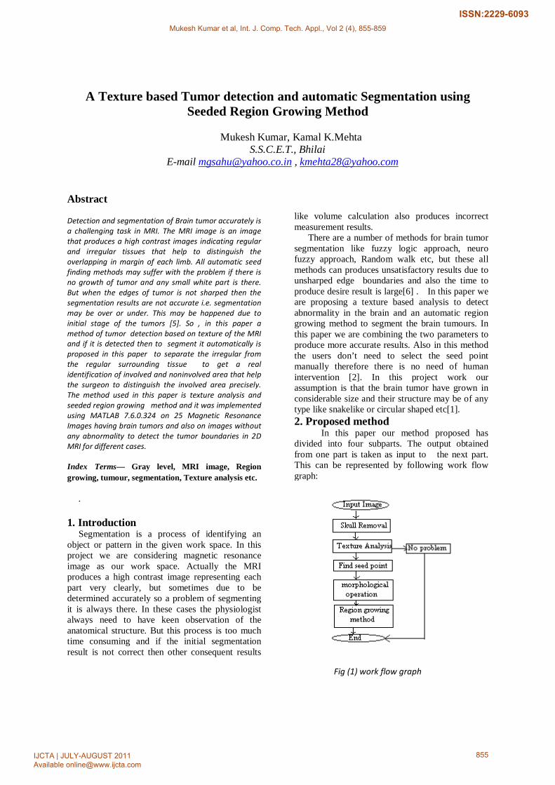
In this paper our method proposed has divided into four subparts. The output obtained from one part is taken as input to the next part. This can be represented by following work flow graph:
Fig (1) work flow graph
Mukesh Kumar et al, Int. J. Comp. Tech. Appl., Vol 2 (4), 855-859
IJCTA | JULY-AUGUST 2011 Available [email protected]
855
ISSN:2229-6093

2.1 Skull Removal This is pre processing step which is required to
produce better results. Skull is outer part of the brain surrounding it i.e. the removal of its non-cerebral tissues. The main problem in skull-stripping is the segmentation of the non-cerebral and the intracranial tissues due to their homogeneity intensities. So it may affect the result of seed point selection [8] .Some observations are required to find the range of gray value of skull portion. Following are involved in skull removal process. (a) First of all find the size of the image and store
the no of rows and columns in separate variables.
(b) Perform iteration for half of the columns and all rows.
(c) Process half of image to convert white pixels into the black pixels by setting their gray value to zero.
(d) Same steps is repeated for the remaining column and row.
2.2 Texture Analysis
MRI contains a lot of microscopic imformation which may not be assessed with the visually and texture analysis technique provides the means for obtaining the information [10, 11]. Texture can be many types such as smooth or rough, regular or irregular, coarse or fine. Some patterns are very complex but they can be clearly represented by texture and can be easily visualize. TA on the images we have performed using the stastical methods of the second order that utilizes gray level run length measures and gray level co-occurence matrix. We have taken Run length parameters in four directions 0, 90, 45 and 135. During the gray level normalization image intensities are distributed from [µ-3α, µ+3α]. Finally, according to the distribution we detect the abnormality in the image.
2.2 Seed Point Selection If texture analysis results indicate the presence
of the abnormality in the MRI then this stage is performed. The image obtained after skull removal is taken as input in this part of the project. For the seed point selection our assumption is that tumour’s region has grown in considerable size .The following steps are performed to find the seed point: (a) Convert the given coloured image into the gray
image. (b) First of all count no pixels whose intensities
are greater than hundred and less than hundred and store them in separate variables.
(c) Find difference between both variables if difference is small then go to step ‘d’ else
convert the image into negative and again set the intensity of external part of the brain to zero and go to step ‘d’
(d) Convert the obtained gray image into the binary image.
(e) Find maximum length and breadth of the brain then
From the center convert the pixels in the area of rectangle shape of size twenty rows and ten columns to black colour.
(f) Find the sum of all rows individually and store it in the array.
(g) Find the sum of all the columns and store the results in another array.
(h) Find the intersection of row and column having maximum sum .This is taken as seed point.
2.3 Morphological Image Enhancement
Morphology mainly deals with the contour and structure of the object. So this is used to perform object extraction, noise removal procedure etc [3]. For the same purpose we are applying these operations to enhance the object boundary and to remove the noise from the image. The most basic morphological operations are dilation and erosion. Dilation adds pixels to the boundaries of objects in an image, while erosion removes pixels on object boundaries. The number of pixels added or removed from the objects in an image depends on the size and shape of the structuring element used to process the image. In the morphological dilation and erosion operations, the state of any given pixel in the output image is determined by applying a rule to the corresponding pixel and its neighbours in the input image. The rule used to process the pixels defines the operation as dilation or erosion.
One important part in morphological operation is to choose the structuring element. A structuring element is a matrix consisting of only 0's and 1's that can have any arbitrary shape and size. The pixels with values of 1 define the neighbourhood .Two-dimensional, or flat, structuring elements are typically much smaller than the image being processed. The centre pixel of the structuring element, called the origin, identifies the pixel of interest -- the pixel being processed. The pixels in the structuring element containing 1's define the neighbourhood of the structuring element. In our project work we are taking DISK shape as structuring element. In the operation of image dilation and erosion we are considering disk structuring element of varying radii so that the obtained image is free from small unwanted parts. In MATLAB working environment there are two built in functions used for dilation and erosion.These morphological functions position the origin of structuring element, its center element over the pixel of interest in the input image. For
Mukesh Kumar et al, Int. J. Comp. Tech. Appl., Vol 2 (4), 855-859
IJCTA | JULY-AUGUST 2011 Available [email protected]
856
ISSN:2229-6093

pixels at the border of the image, parts of the neighbourhood defined by the structuring element can extend past the border of the image. To process border pixels, the morphological functions assign a value to these undefined pixels, as if the functions had padded the image with additional rows and columns. The value of these padding pixels varies for dilation and erosion operations.
If A and B are two sets then dilation is denoted by A B, is obtained by first reflecting B about its origin and then translating the result by x. So that all x such that A and reflected B translated by x that have at least one point in common form the dilated set [7]. Therefore,
AΟB= Ø}.................... (1) Where B’denotes reflection of B. B denotes the
translation of B by x. In our work MRI are gray images therefore,
(2)
Here, f and b denote images f(x,y) and b(x,y). f is being dilated and b is called the structuring element D and Db are the domains of f and b respectively.
Thus, in dilation we choose the maximum value of f+b in a neighborhood defined by b. If all elements of b are positive, the dilated image is brighter than the original and the dark details are either reduced or eliminated. Similarly, Erosion of A by B, denoted by A is the set of all x such that B
translated by x is completely contained in A, i.e., A (3)
For gray scale images,
(4)
Erosion is thus based on choosing the minimum value of (f-b) in a neighborhood defined by the shape of b. If all elements of b are positive, the output image is darker than the original and the effect of bright details in the input image are reduced if they cover a region smaller than b. Therefore, it is concluded that image dilation and erosion operation produces an enhanced and sharpen edge MRI, so that over segmentation and under segmentation can be reduced [7].
2.4 Seeded Region Growing Method The result obtained after morphological operation is taken as input in this stage. This approach to segmentation examines neighboring pixels of initial “seed points” and determines whether the pixel neighbors should be added to the region. The process is iterated on, in the same manner as general data clustering algorithms. The main goal of segmentation is to partition an image into regions. Some segmentation methods such as "Thresholding" achieve this goal by looking for the boundaries between regions based on discontinuities in gray levels or color properties [7]. Region-based segmentation is a technique for determining the region directly. In our project work 8 connected neighbor region growing method has been used. The basic formulation for Region-Based Segmentation is:
0 (a)
Means the segmentation must be complete and every pixel must be in the region. Here Ri is a connected region.
(b)
Equation (b) means the regions must be disjoint. So that a clear separation from each other can be identified.
For i=1, 2,….,n (c)
Means a pixel may belong to the region when it satisfies the condition that gray level of pixel is in the range of Region.
(d)
Means regions Ri and Rj are different in the sense of predicate P
.
Mukesh Kumar et al, Int. J. Comp. Tech. Appl., Vol 2 (4), 855-859
IJCTA | JULY-AUGUST 2011 Available [email protected]
857
ISSN:2229-6093
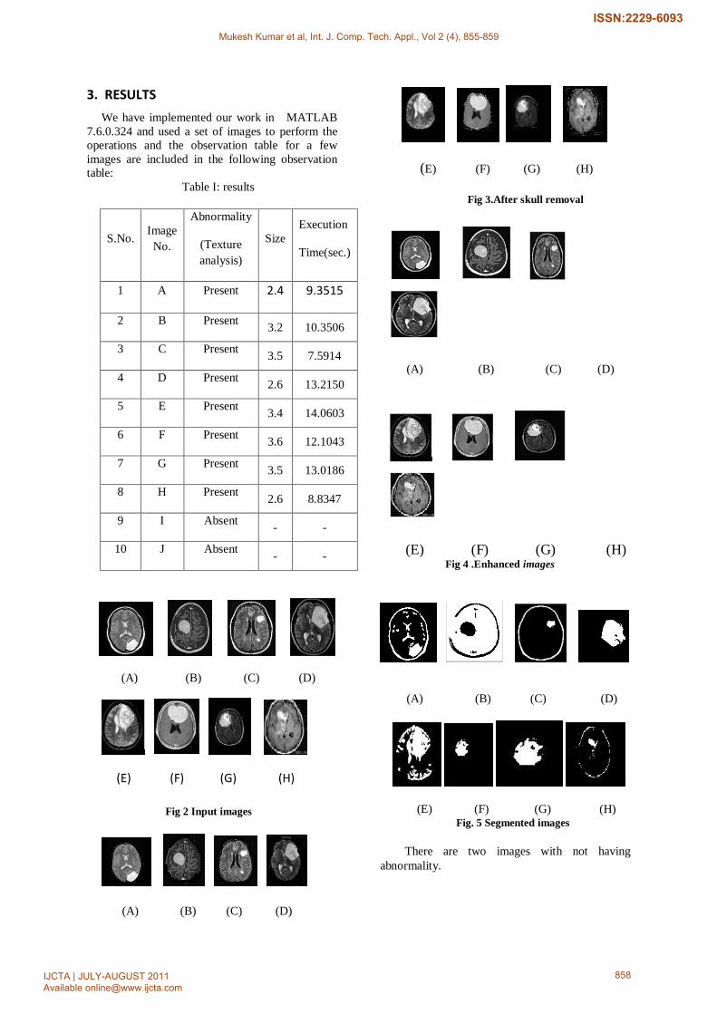
3. RESULTS We have implemented our work in MATLAB
7.6.0.324 and used a set of images to perform the operations and the observation table for a few images are included in the following observation table:
Table I: results
S.No. Image No.
Abnormality
(Texture analysis)
Size Execution
Time(sec.)
1 A Present 2.4 9.3515
2 B Present 3.2 10.3506
3 C Present 3.5 7.5914
4 D Present 2.6 13.2150
5 E Present 3.4 14.0603
6 F Present 3.6 12.1043
7 G Present 3.5 13.0186
8 H Present 2.6 8.8347
9 I Absent - -
10 J Absent - -
(A) (B) (C) (D)
(E) (F) (G) (H)
Fig 2 Input images
(A) (B) (C) (D)
(E) (F) (G) (H)
Fig 3.After skull removal
(A) (B) (C) (D)
(E) (F) (G) (H) Fig 4 .Enhanced images
(A) (B) (C) (D)
(E) (F) (G) (H) Fig. 5 Segmented images There are two images with not having
abnormality.
Mukesh Kumar et al, Int. J. Comp. Tech. Appl., Vol 2 (4), 855-859
IJCTA | JULY-AUGUST 2011 Available [email protected]
858
ISSN:2229-6093
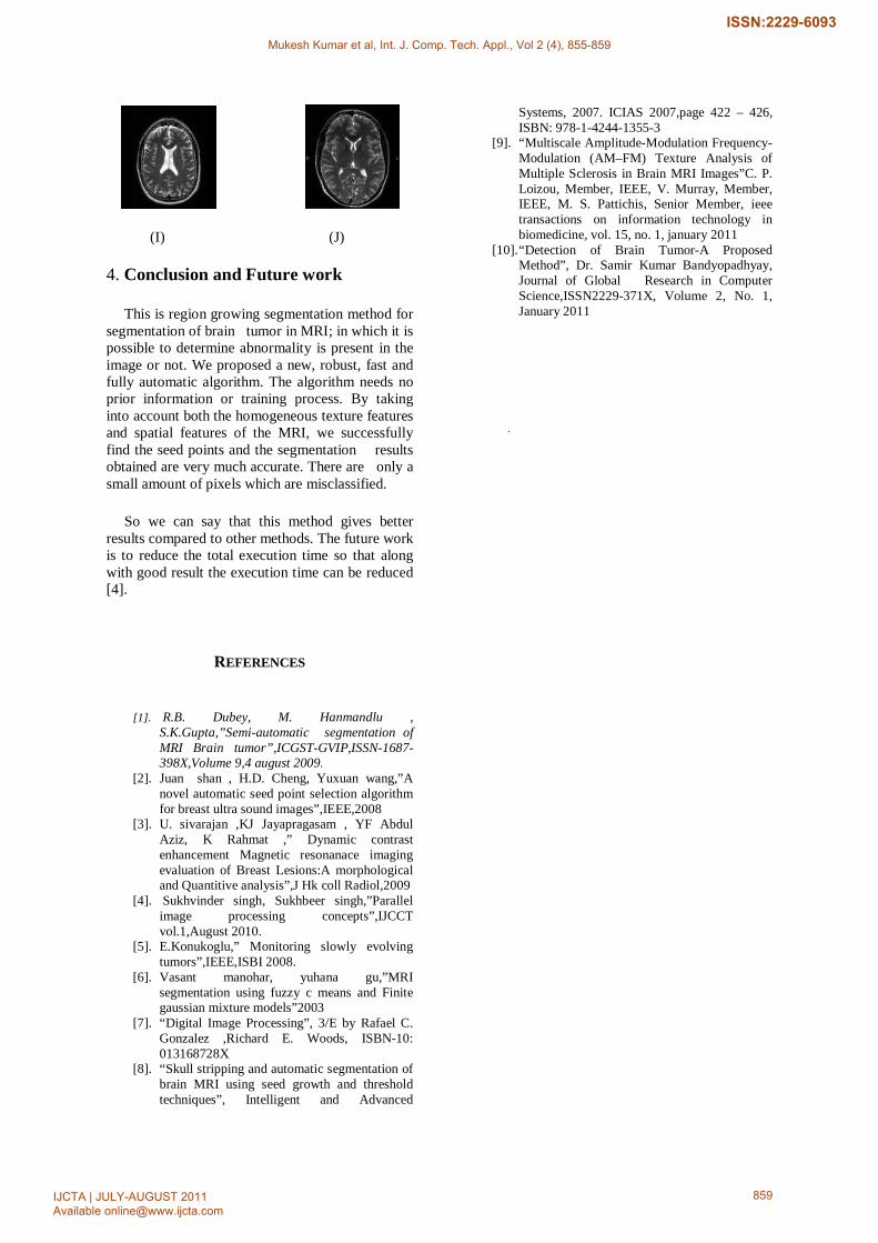
(I) (J)
4. Conclusion and Future work
This is region growing segmentation method for segmentation of brain tumor in MRI; in which it is possible to determine abnormality is present in the image or not. We proposed a new, robust, fast and fully automatic algorithm. The algorithm needs no prior information or training process. By taking into account both the homogeneous texture features and spatial features of the MRI, we successfully find the seed points and the segmentation results obtained are very much accurate. There are only a small amount of pixels which are misclassified.
So we can say that this method gives better results compared to other methods. The future work is to reduce the total execution time so that along with good result the execution time can be reduced [4].
REFERENCES
[1]. R.B. Dubey, M. Hanmandlu ,
S.K.Gupta,”Semi-automatic segmentation of MRI Brain tumor”,ICGST-GVIP,ISSN-1687-398X,Volume 9,4 august 2009.
[2]. Juan shan , H.D. Cheng, Yuxuan wang,”A novel automatic seed point selection algorithm for breast ultra sound images”,IEEE,2008
[3]. U. sivarajan ,KJ Jayapragasam , YF Abdul Aziz, K Rahmat ,” Dynamic contrast enhancement Magnetic resonanace imaging evaluation of Breast Lesions:A morphological and Quantitive analysis”,J Hk coll Radiol,2009
[4]. Sukhvinder singh, Sukhbeer singh,”Parallel image processing concepts”,IJCCT vol.1,August 2010.
[5]. E.Konukoglu,” Monitoring slowly evolving tumors”,IEEE,ISBI 2008.
[6]. Vasant manohar, yuhana gu,”MRI segmentation using fuzzy c means and Finite gaussian mixture models”2003
[7]. “Digital Image Processing”, 3/E by Rafael C. Gonzalez ,Richard E. Woods, ISBN-10: 013168728X
[8]. “Skull stripping and automatic segmentation of brain MRI using seed growth and threshold techniques”, Intelligent and Advanced
Systems, 2007. ICIAS 2007,page 422 – 426, ISBN: 978-1-4244-1355-3
[9]. “Multiscale Amplitude-Modulation Frequency-Modulation (AM–FM) Texture Analysis of Multiple Sclerosis in Brain MRI Images”C. P. Loizou, Member, IEEE, V. Murray, Member, IEEE, M. S. Pattichis, Senior Member, ieee transactions on information technology in biomedicine, vol. 15, no. 1, january 2011
[10]. “Detection of Brain Tumor-A Proposed Method”, Dr. Samir Kumar Bandyopadhyay, Journal of Global Research in Computer Science,ISSN2229-371X, Volume 2, No. 1, January 2011
Mukesh Kumar et al, Int. J. Comp. Tech. Appl., Vol 2 (4), 855-859
IJCTA | JULY-AUGUST 2011 Available [email protected]
859
ISSN:2229-6093


![Image Segmentation based on Deformable Models · Segmentation system Rules • static rules [selection] – lateral ventricles high contrast ⇒good texture map ⇒increase texture](https://img.dokumen.tips/doc/110x75/5f805e616050b07370169abb/image-segmentation-based-on-deformable-models-segmentation-system-rules-a-static.jpg)