Embed Size (px)
Citation preview

8/16/2019 A Systemic Mycobacteriosis in Farmed Meagre (Argyrosomus regius) Caused by Mycobacterium marinum in Turkey
http://slidepdf.com/reader/full/a-systemic-mycobacteriosis-in-farmed-meagre-argyrosomus-regius-caused-by 1/8
The Israeli Journal of Aquaculture - Bamidgeh, IJA_67.2015.1162, 8 pages
The IJA appears exclusively as a peer-reviewed on-line
aderoT.li.rgo.siambwww.://ttphtaalnruo jsseacc-neop
papers free of charge, please register online at.mron f oitastrgire
Sale of IJA papers is strictly forbidden.
A Systemic Mycobacteriosis in Farmed Meagre ( Argyrosomus
regius) Caused by Mycobacterium marinum in Turkey
G. Timur1*, Ç. Ürkü1, Ö. Çanak1, G. Erköse Genç2 and Z. Erturan2
Istanbul, 34470, Ordu Cad. No: 200 Laleli- Istanbul/ Turkey
Istanbul University, Capa, 34093, Istanbul/ Turkey
Keywords: Mycobacterium marinum, meagre, granulomatous lesions
Abstract
This paper describes a systemic mycobacteriosis in farmed meagre
( Argyrosomus regius) caused by Mycobacterium marinum in Turkey. Affected
two year old fish samples generally showed stunted growth, emaciation, slight
ascites and exophtalmia, pale gills and significant mortalities. Only one fish
sample showed haemorrhagic ulcerative skin lesion at the base of caudal fin.
Internally multifocal white colored granulomas in the spleen, kidney and liver
were observed. Ziehl-Neelsen (ZN) and Gram stained fresh squash mounts of
the granulomas revealed Gram and ZN positive rods. Inoculation of sterile
homogenates of the visceral organ granulomas on to Lowenstein-Jensen
slants produced slow-growing (3-4 weeks), yellow to orange colored,
photochromogenic acid fast colonies. ZN positive bacterial isolates wereidentified by using commercially available line probe assays (Genotype
Mycobacterium CM/AS assay) and hsp65 gene sequencing analyses. According
to these molecular analysis results, the isolates were identified as
Mycobacterium marinum. Microscopically, epithelioid cell granulomas were
observed in the visceral organs and gills. ZN stained tissue sections exhibited
heavy acid-fast rods within the granulomas.
Introduction
Mycobacteriosis in fish have been described as a systemic, serious, lethal, chronic,
progressive and worldwide affecting disease in wild and cultured marine, brackish and
853610/0755452.: +90.21leT,mco.ooh@yarumitneslgu:oruthg anidnosperroC*Fax: +90.212 4555861;
1 Department of Fish Disease, Faculty of Aquatic Sciences, University of
2Department of Medical Microbiology, Istanbul Faculty of Medicine,

8/16/2019 A Systemic Mycobacteriosis in Farmed Meagre (Argyrosomus regius) Caused by Mycobacterium marinum in Turkey
http://slidepdf.com/reader/full/a-systemic-mycobacteriosis-in-farmed-meagre-argyrosomus-regius-caused-by 2/8
2 Timur et al.
freshwater fish caused by several species of the genus Mycobaterium. Although
Mycobacterium marinum is considered to be the primary causative agent of fish
mycobacteriosis, a number of Mycobacterium species associated with tubercule
granulomas in cultured, aquarium and wild fish populations such as M. marinum, M.
fortuitium, M. chelonae, M. salmoniphilum, M. smegmatis, M. abscessus, M. neonarum,
M. simiae and M. poriferae (Frerichs, 1993; Chinabut, 1999; Puttinaowarat et al., 2000;
dos Santos et al., 2002; Toranzo et al., 2005; Pourahmad et al., 2009; Jacobs et al.,
2009; Gauthier and Rhodes, 2009; Novotny et al., 2010) have been identified.
Mycobacteriosis was reported in Pacific and Atlantic salmonids (Arakawa and
Frayer 1984), in rabbit fish (Diamant et al., 2000), in sea bass (Colorni, 1992; 1996), in
sea bream (Colorni et al., 1996), in farmed turbot (dos Santos et al., 2002), in cultured
striped bass (Hedrick et al., 1987) and in striped bass (Rhodes et al., 2004). Multiple
granulomas scattered or grouped in the visceral organs of the various fresh water and
salt water fish species (dos Santos et al., 2002; Rhodes et al., 2004; Toranzo et al.,
2005; Gauthier et al., 2009; Jacobs et al., 2009).
Farming meagre in floating marine cages on the coasts of Aegean Sea started in early
2000’s. In September 2013, significant mortalities occurred in 2 years old fish in a
marine cage farm.Clinical, bacteriological, histopathological and molecular examinations indicated
that mycobacteriosis was present in the diseased meagre. This paper describes a
systemic mycobacteriosis in cultured meagre in Turkey.
Materials and Methods
Fish: Six affected fish (350-400g) generally showed loss of appetite, lethargy,
emaciation, floating on the surface of the water were obtained from a floating marine
cage farm located on cost of the Aegean Sea in Turkey.
Bacteriology: Non-fixed tissue samples from liver, spleen and kidney including white
colored granulomas were put into liquid nitrogen at -196˚C to bring to the microbiology
laboratory. These tissue samples were taken from liquid nitrogen and waited until they
reach to room temperature for the preparation of Gram and Ziehl-Neelsen (ZN) stainedsmears. The stained smears were examined by light microscopy.
To obtain pure culture of mycobacteria, decontaminated homogenates were
prepared from the visceral organs granulomas of the affected fish samples. The visceral
granulomas homogenized by using sterile pestles and treated with 4% NaOH (w/v) at
room temperature for 15 minutes and centrifuged at 8000 g for 15 minutes. The
supernatant was removed and the pellet was washed twice with 1ml of sterile phosphate-
buffered solution (PBS) and centrifuged one more time as described above. The pellet
was re-suspended in 150 µl sterile PBS and twenty-five µl of this suspension was
inoculated onto Lowenstein-Jensen medium incubated at 24-25 ˚C (Pourahmad et al.,
2009).
Molecular Study: The isolated ZN positive bacteria were identified using by commercially
available line probe assays are the Genotype CM and AS (HainLife Science, Germany).For preparing DNA, one loopful of cells were suspended in 300 µL of distilled water,
boiled at 95C° for 20 min, sonicated for 15 min, and centrifuged for 5 min. The
GenoType protocol consists of PCR amplification, hybridization of the PCR products to the
probe-containing test strips, and detection of bound products (Richter et al., 2006). Also
sequencing of the 65-kDa heat shock protein gene (hsp65) was performed (Pourahmad
et al., 2009).
Histology: The kidney, liver, spleen, hearth and gill tissues with coloured granulomas
were processed for histopathology by fixing 10% buffered formalin, and processed for
paraffin embedding. Histological sections (4-5µm) were stained haemotoxylin and eosin
(H&E) and tissue ZN staining methods and examined by light microscopy (Bullock, 1978).
Results
Clinical Signs:
Affected two years old six fish weighing between 350 to 400 g, generally
showed nonspecific external clinical signs and included emaciation and stunted growth

8/16/2019 A Systemic Mycobacteriosis in Farmed Meagre (Argyrosomus regius) Caused by Mycobacterium marinum in Turkey
http://slidepdf.com/reader/full/a-systemic-mycobacteriosis-in-farmed-meagre-argyrosomus-regius-caused-by 3/8
Bacteriology: ZN and Gram stained
fresh squash mounts were reviled
ZN and Gram positive rods within
the affected visceral organsnodules. Following 3-4 weeks, steril
homogenate inoculations on
Lowenstein-Jensen slants, slow
growing yellow-orange pigmented
colonies recovered from the
visceral organ granulomas (kidney, spleen and liver) (Fig. 4a). Colonies were examined
for acid-fastness. The ZN stained bacterial smears from these colonies revealed acid fast
cross barring shaped rods (Fig. 4b).
Fig. 4. (a) Yellow-
orange pigmentedcolonies onLowenstein-Jensen
slants,(b) Acid fast rodsfrom Lowenstein-
Jensen slants
Genotype Assay: The ZN positive isolates produced 10th and 15th bants on the Genotip
CM (lane a) (Fig. 5a). According to the interpretation chart, this isolates interpreted as M.
ulcerans or M. marinum. Later this two species were further differentiated with Genotype
AS kit. According to the interpretation chart, this isolates identified as M. ulcerans (Fig. 5
b).
Systemic Mycobacteriosis
3
and ascites (Fig. 1), slight exophthalmia (Fig. 2a), pale gills, and significant mortalities.
Among the six affected fish; only one of them showed hemorrhagic ulcerative skin lesion
at the base of caudal fin (Fig. 2b). Gross internal signs of the affected fish included slight
haemorrhagic ascites and characteristic white multifocal granulomas in the spleen, liver
and kidney measuring 2-7 mm (Fig. 3a, b, c, d).
Fig. 1. Affected fish showed emaciation andstunded growth
Fig. 2. (a) Slight exophthalmia, (b)
Hemorrhagic ulcerative skin lesion at thebase of caudal fin
Fig. 3. Multifocal white granulomas (a)in the liver and spleen, (b) in thespleen and kidney, (c) in the spleen,
(d) in the kidney
8/16/2019 A Systemic Mycobacteriosis in Farmed Meagre (Argyrosomus regius) Caused by Mycobacterium marinum in Turkey
http://slidepdf.com/reader/full/a-systemic-mycobacteriosis-in-farmed-meagre-argyrosomus-regius-caused-by 4/8
2 Timur et al.
Fig. 5. The result of Genotype CM (lane a)and AS (lane b) kit
lane (a): positive bants show to M. ulcerans-
M. marinum, lane (b):
positive bants show toM. ulcerans
hsp65gene Sequencing: A total of 441bp
of the hsp65gene was sequenced
(CEQ8000 Sequence Analysis System,
Beckman-Coulter, USA). The obtained
sequence was compared to those stored
in GenBank using the Basic Local Alignment Search Tool (BLAST; NCBI, Bethesda, MD)
and was shown to be 99% homologous to the Mycobacterium marinum ATCC927
hsp65 gene sequence deposited under accession number AF476470.
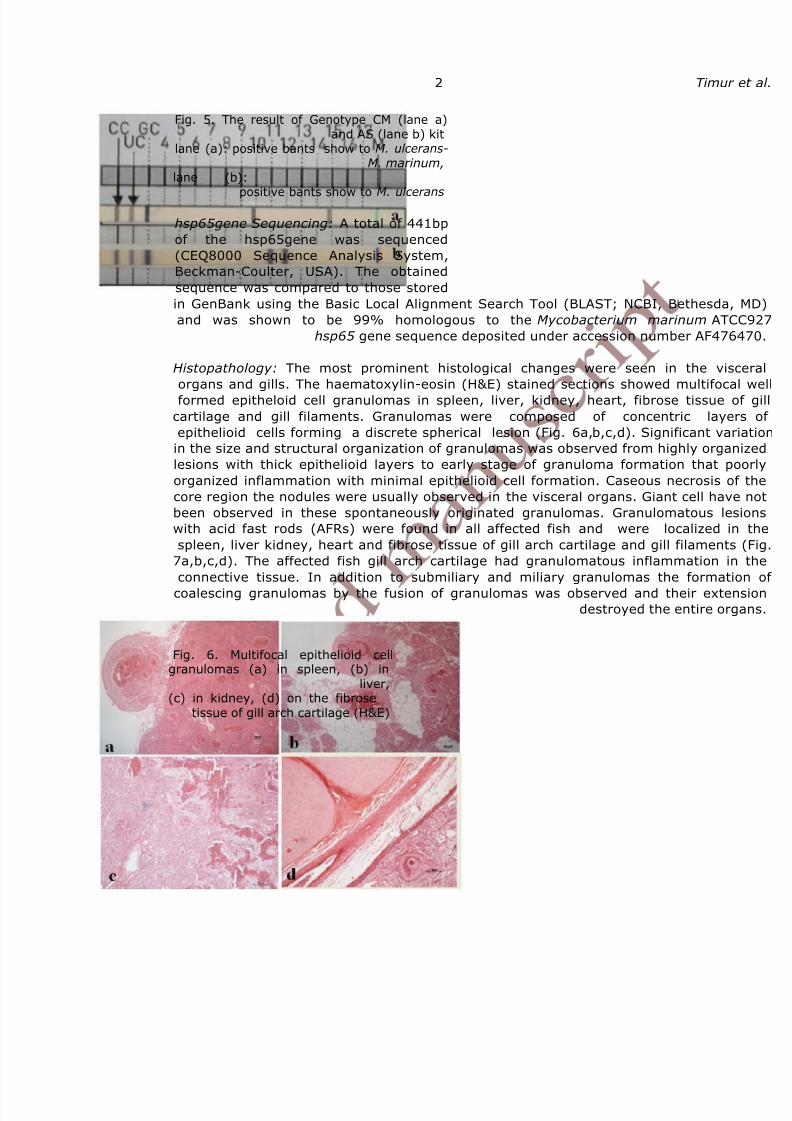
Histopathology: The most prominent histological changes were seen in the visceral
organs and gills. The haematoxylin-eosin (H&E) stained sections showed multifocal well
formed epitheloid cell granulomas in spleen, liver, kidney, heart, fibrose tissue of gill
cartilage and gill filaments. Granulomas were composed of concentric layers of
epithelioid cells forming a discrete spherical lesion (Fig. 6a,b,c,d). Significant variation
in the size and structural organization of granulomas was observed from highly organized
lesions with thick epithelioid layers to early stage of granuloma formation that poorly
organized inflammation with minimal epithelioid cell formation. Caseous necrosis of the
core region the nodules were usually observed in the visceral organs. Giant cell have not
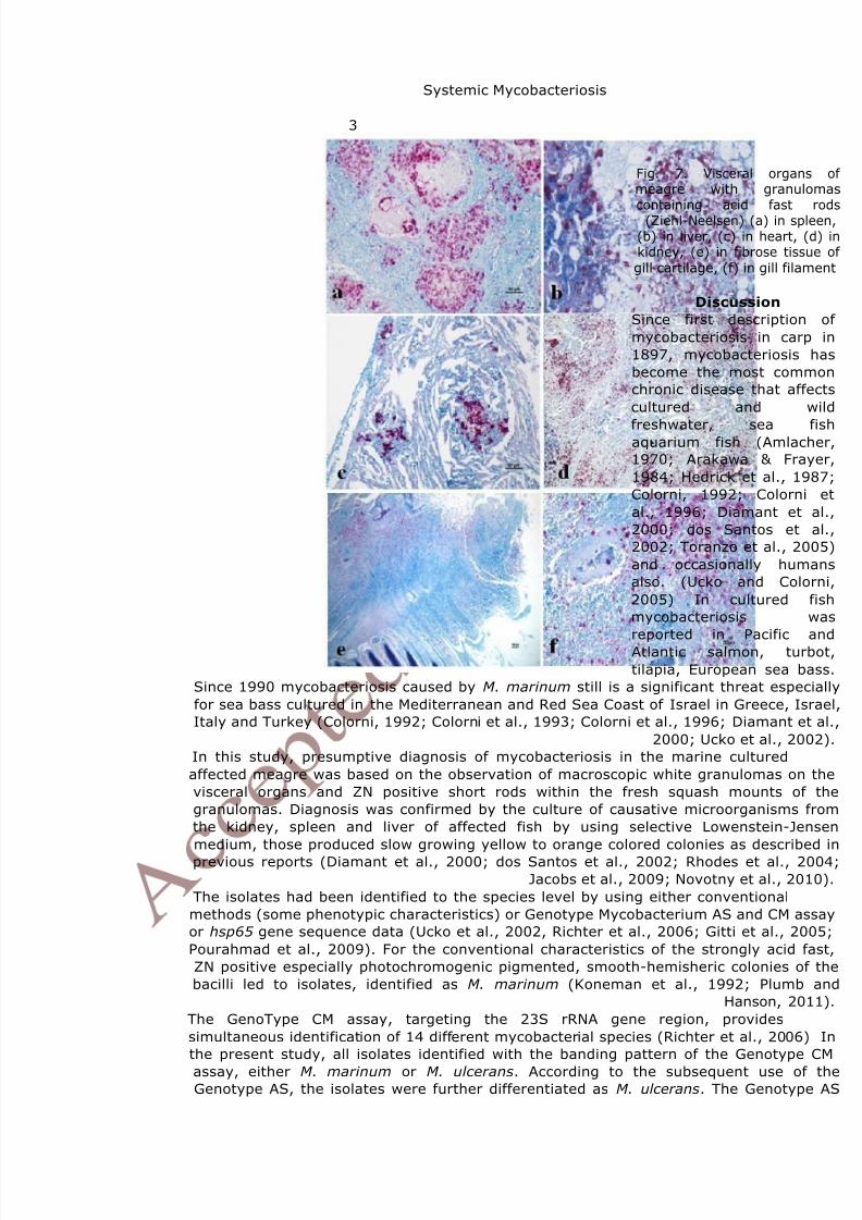
been observed in these spontaneously originated granulomas. Granulomatous lesions
with acid fast rods (AFRs) were found in all affected fish and were localized in the
spleen, liver kidney, heart and fibrose tissue of gill arch cartilage and gill filaments (Fig.
7a,b,c,d). The affected fish gill arch cartilage had granulomatous inflammation in the
connective tissue. In addition to submiliary and miliary granulomas the formation of
coalescing granulomas by the fusion of granulomas was observed and their extensiondestroyed the entire organs.
Fig. 6. Multifocal epithelioid cellgranulomas (a) in spleen, (b) in
liver,(c) in kidney, (d) on the fibrose
tissue of gill arch cartilage (H&E)
8/16/2019 A Systemic Mycobacteriosis in Farmed Meagre (Argyrosomus regius) Caused by Mycobacterium marinum in Turkey
http://slidepdf.com/reader/full/a-systemic-mycobacteriosis-in-farmed-meagre-argyrosomus-regius-caused-by 5/8
Systemic Mycobacteriosis
3
Fig. 7. Visceral organs of meagre with granulomascontaining acid fast rods(Ziehl-Neelsen) (a) in spleen,
(b) in liver, (c) in heart, (d) inkidney, (e) in fibrose tissue of
gill cartilage, (f) in gill filament
Discussion
Since first description of
mycobacteriosis in carp in
1897, mycobacteriosis has
become the most common
chronic disease that affects
cultured and wild
freshwater, sea fishaquarium fish (Amlacher,
1970; Arakawa & Frayer,
1984; Hedrick et al., 1987;
Colorni, 1992; Colorni et
al., 1996; Diamant et al.,
2000; dos Santos et al.,
2002; Toranzo et al., 2005)
and occasionally humans
also. (Ucko and Colorni,
2005) In cultured fish
mycobacteriosis was
reported in Pacific andAtlantic salmon, turbot,
tilapia, European sea bass.
Since 1990 mycobacteriosis caused by M. marinum still is a significant threat especially
for sea bass cultured in the Mediterranean and Red Sea Coast of Israel in Greece, Israel,
Italy and Turkey (Colorni, 1992; Colorni et al., 1993; Colorni et al., 1996; Diamant et al.,
2000; Ucko et al., 2002).
In this study, presumptive diagnosis of mycobacteriosis in the marine cultured
affected meagre was based on the observation of macroscopic white granulomas on the
visceral organs and ZN positive short rods within the fresh squash mounts of the
granulomas. Diagnosis was confirmed by the culture of causative microorganisms from
the kidney, spleen and liver of affected fish by using selective Lowenstein-Jensen
medium, those produced slow growing yellow to orange colored colonies as described in
previous reports (Diamant et al., 2000; dos Santos et al., 2002; Rhodes et al., 2004;
Jacobs et al., 2009; Novotny et al., 2010).
The isolates had been identified to the species level by using either conventional
methods (some phenotypic characteristics) or Genotype Mycobacterium AS and CM assay
or hsp65 gene sequence data (Ucko et al., 2002, Richter et al., 2006; Gitti et al., 2005;
Pourahmad et al., 2009). For the conventional characteristics of the strongly acid fast,
ZN positive especially photochromogenic pigmented, smooth-hemisheric colonies of the
bacilli led to isolates, identified as M. marinum (Koneman et al., 1992; Plumb and
Hanson, 2011).
The GenoType CM assay, targeting the 23S rRNA gene region, provides
simultaneous identification of 14 different mycobacterial species (Richter et al., 2006) In
the present study, all isolates identified with the banding pattern of the Genotype CM
assay, either M. marinum or M. ulcerans. According to the subsequent use of theGenotype AS, the isolates were further differentiated as M. ulcerans. The Genotype AS

8/16/2019 A Systemic Mycobacteriosis in Farmed Meagre (Argyrosomus regius) Caused by Mycobacterium marinum in Turkey
http://slidepdf.com/reader/full/a-systemic-mycobacteriosis-in-farmed-meagre-argyrosomus-regius-caused-by 6/8

8/16/2019 A Systemic Mycobacteriosis in Farmed Meagre (Argyrosomus regius) Caused by Mycobacterium marinum in Turkey
http://slidepdf.com/reader/full/a-systemic-mycobacteriosis-in-farmed-meagre-argyrosomus-regius-caused-by 7/8
Systemic Mycobacteriosis
5
Chinabut, S. 1999. Mycobacteriosis and nocardiosis. In: (Woo TK and Bruno DW, Ed.)
Fish diseases and disorders, Vol. 3: Viral, Bacterial and Fungal Infections, CAB
International, New York.
Colorni, A. 1992. A systemic mycobacteriosis in the European seabass Dicentrarchus
labrax cultured in Eilat (Red Sea). The Israeli Journal of Aquaculture- Bamidgeh 44, 75–81.
Colorni, A., Ankaoua, M., Diamant, A. and W. Knibb, 1993. Detection of
mycobacteriosis in fish using the polymerase chain reaction technique. Bulletin European
Association of Fish Pathologists 13, 195– 198.
Colorni, A., Ucko, M. and W. Knibb, 1996. Epizootiology of Mycobacterium spp. in
seabass, seabream and other commercial fish. Sea bass and sea bream culture:
Problems and prospects. pp. 259– 261. Eur. Aquacult. Soc. Spec. Publ., Verona, Italy.
Colorni, A., Avtalion, R., Knibb, W., Berger, E., Colorni, B. and B. Timan, 1998.
Histopathology of sea bass (Dicentrarchus labrax ) experimentally infected with
Mycobacterium marinum and treated with streptomycin and garlic ( Allium sativum)
extract. Aquaculture, 160: 1–17.
Diamant, A., Banet, A., Ucko, M., Colorni, A., Knibb, W. and H. Kvitt, 2000.Mycobacteriosis in wild rabbitfish Siganus rivulatus associated with cage farming in Gulf
of Eilat, Red Sea. Diseases of Aquatic Organisms, 39: 211–219.
Dos Santos, N. M., do Vale, A., Sousa, M. J. and M. T. Silva, 2002. Mycobacterial
infection in farmed turbot Scophthalmus maximus. Diseases of Aquatic Organisms, 52:
87–91.
Frerichs, G. N. 1993. Mycobacteriosis: nocardiosis. In: (Inglis V, Roberts RJ and
Bromage N.R. Ed.). Bacterial diseases of fish, pp. 219–234. Halsted Press, New York.
Gauthier, D.T. and M.W. Rhodes, 2009. Mycobacteriosis in fishes: A review. Veterinary
Journal 180: 33–47.
Jacobs, J.M., Stine, C.B., Baya, A.M. and M.L. Kent, 2009. A review of
mycobacteriosis in marine fish. Journal of Fish Disease, 32: 119–130.
Gitti, Z., Neonakis, I., Fanti, G., Kontos, F., Maraki, S. and Y. Tselentis, 2006. Use
of the Genotype Mycobacterium CM and AS assays to analyze 76 nontuberculous
mycobacterial isolates from Greece. Journal of Clinical Microbiology, 44: 2244-2246.
Hedrick, R. P., McDowell, T. and J. Groff, 1987. Mycobacteriosis in cultured striped
bass from California. European Journal of Wildlife Research, 23: 391– 395.
Koneman, E. W., Allen, S.D., Janda, W.M., Schreckenberger, P.C. and W.C. Winn,
1992. Color atlas and textbook of diagnostic microbiology. 4th Edition, Lippincott, New
York, USA.
Korun, J., Olgac, V., Akgun, K., Colorni, A. and A. Diamant, 2005. Mycobacteriosis
in European sea bass, Dicentrarchus labrax L., cultured in Turkey. The Israeli Journal of
Aquaculture- Bamidgeh, 4: 215-222.
Mosi, L., Mutoji, N.K., Basile, F. A., Donnell, R., Jackson, K. L., Spangenberg, T.,
Kishi, Y., Ennis, D. G. and P. L. C., 2012. Mycobacterium ulcerans causes minimal
pathogenesis and coloniaztion in Medaka (Oryzias lafmtipes): An experimental fish model
of disease transmission. Microbes and Infection, 9: 719-729.
Novotny, L., Halouzka, R., Matlova, L., Vavra, O., Bartosova, L., Slany, M. and I.
Pavlik, 2010. Morphology and distribution of granulomatous inflammation in freshwater
ornamental fish infected with mycobacteria. Journal of Fish Disease, 33: 947–955.
Plumb, J. A. and L. A. Hanson, 2011. Health maintenance and principal microbial
diseases of cultured fishes. In Striped bass bacterial diseases. Third Edition, Blackwell
Publishing.
Pourahmad, F., Thompson, K. D., Adams, A. and R. H. Richards, 2009. Detection
and identification of aquatic mycobacteria in formalin-fixed, paraffin-embedded fish
tissues. Journal of Fish Disease, 32: 409–419.
Pourahmad, F., Thompson, K. D., Adams, A. and R. H. Richards, 2009.
Comparative evaluation of Polymerase Chain Reaction–Restriction Enzyme Analysis (PRA)

8/16/2019 A Systemic Mycobacteriosis in Farmed Meagre (Argyrosomus regius) Caused by Mycobacterium marinum in Turkey
http://slidepdf.com/reader/full/a-systemic-mycobacteriosis-in-farmed-meagre-argyrosomus-regius-caused-by 8/8
6 Timur et al.
and sequencing of heat shock protein 65 (hsp65) gene for identification of aquatic
mycobacteria. Journal of Microbiological Methods, 76: 128–135.
Puttinaowarat, S., Thompson, K.D., Kolk, A. and A. Adams, 2002. Identification of
Mycobacterium spp. isolated from snakehead, Channa striata (Fowler), and Siamese
fighting fish, Betta splendens (Regan), using polymerase chain reaction-reverse cross
blot hybridization (PCR-RCBH). Journal of Fish Disease, 25: 235–243.
Rhodes, M.W., Kator, H., Kaattari, I., Gauthier, D., Vogelbein, W. and C.A.
Ottinger, 2004. Isolation and characterization of mycobacteria from striped bass Morone
saxatilis from the Chesapeake Bay. Diseases of Aquatic Organisms, 61: 41–51.
Richter, E., Rusch-Gerdes, S. and D. Hillemann, 2006. Evaluation of the GenoType
Mycobacterium assay for identification of mycobacterial species from cultures. Journal of
Clinical Microbiology, 44: 1769–1775.
Timur, G., 1975. A study of giant cells in inflammatory lesions of the plaice
(Pleuronectes platessa L.). Ph.D. thesis, University of Stirling.
Timur, G., Roberts, R.J. and A. McQueen, 1977. The experimental pathogenesis of
focal tuberculosis in the plaice (Pleuronectes platessa L.). Journal of Comparative
Pathology, 87: 83-87.
Toranzo, A.E., Magarinos, B. and J.L. Romalde, 2005. A review of the main bacterialfish diseases in mariculture systems. Aquaculture, 246: 37-61.
Ucko, M., Colorni, A., Kviit, H., Diamant, A., Zlotkin, A. and W.R. Knibb, 2002.
Strain variation in Mycobacterium marinum fish isolates. Applied and Environmental
Microbiology, 11: 5281-5287.
Ucko, M. and A. Colorni, 2005. Mycobacterium marinum Infections in fish and humans
in Israel. Journal of Clinical Microbiology 43(2): 892–895.