Embed Size (px)
Citation preview

Research Journal of Recent Sciences
Vol. 6(6), 13-19, June (2017)
International Science Community Association
A study on effect of ligand on crystallography, morphology and photo
catalytic ability of ZnS nanostructuresBalwinder Kaur
1Department of Chemistry, Punjabi University Patiala2Department of Physics, Punjabi University Patiala
Available online at: Received 30th April
Abstract
A single source precursor zinc complex: Zn(phenyl alanine dithiocarbamate)
nanostructures by solvothermal route. Zn(phenyl alanine dithiocarbamate)
alanine dithiocarbamate (PHEDTC) by simple mixing. Crystalline texture, phase analyses and size
prepared ZnS nanostructures have been carried out by XRD and TEM, respectively. The hexagonal structure ZnS
nanocrystals of various morphologies (nanosheets, nanotriangles
and electron microscope studies, respectively. UV
Photoluminescence (PL) study was carried out to check luminescence of synthes
spectrum. It has been reported that morphology of synthesized nanostructures strongly depends upon the precursor complex
prepared from ligand; L-phenyl alanine dithiocarbamate.
observed in visible light using MB dye as a pollutant in water medium.
Keywords: L-phenyl alanine dithiocarbamate (Spectroscopy, Photoluminescence.
Introduction
A II-VI binary metal-chalcogenide Zinc sulfide semiconductor posses band gap (3.647eV) with various morphologies like nanorods, nanobelts, nanocombs1-6 etc. ZnS nanomaterialspotential applications in the field of semiconductors like optoelectronics, photo-catalysis, bio-imaging, sensorsDithiocarbamates (DTCs) are the class of anionic ligands, which contain two sulfur donor atoms. These ligands stabilize the positive charge on metal ions towards the periphery of complex. The metal bis(phenyl alanine dithiocarbamate) complex have been known to be a precursor for the synthesis of nanomaterial, since they are easy to prepare, nontoxic and stable in natureVariation in derivatives of dithiocarbamate ligands in metal complexes remarkably effects the crystallography and morphology of metal-sulphide nanostructures19
In the present research work, solvothermal route of synthesis has been employed for the synthesis of ZnS nanostructures using single source precursor: Zn(phenyl alanine dithiocarbamate)2. The photo-catalytic activity of ZnS nanostructures have been checked using MB dye as test pollutant in water.
Materials and methods
All the procured reagents were used without further purification. The analytical reagent grade chemicals: L(C9H11O2N) (99.00%), zinc acetate Zn(C
Sciences ___________________________________________
International Science Community Association
A study on effect of ligand on crystallography, morphology and photo
catalytic ability of ZnS nanostructures Balwinder Kaur
1, Karamjit Singh
2 and Ashok Kumar Malik
1*
Chemistry, Punjabi University Patiala-147 002, Punjab, India Department of Physics, Punjabi University Patiala-147 002, Punjab, India
Available online at: www.isca.in, www.isca.me April 2017, revised 18th May 2017, accepted 28th May 2017
A single source precursor zinc complex: Zn(phenyl alanine dithiocarbamate)2 have been chosen to synthesize ZnS
solvothermal route. Zn(phenyl alanine dithiocarbamate)2 has been synthesized from ligand L
by simple mixing. Crystalline texture, phase analyses and size
prepared ZnS nanostructures have been carried out by XRD and TEM, respectively. The hexagonal structure ZnS
nanocrystals of various morphologies (nanosheets, nanotriangles and nano pyramids) have been confirmed by diffraction
and electron microscope studies, respectively. UV-vis. absorption studies have been carried for the detailed optical analyses.
Photoluminescence (PL) study was carried out to check luminescence of synthesized ZnS nanostructures in electromagnetic
spectrum. It has been reported that morphology of synthesized nanostructures strongly depends upon the precursor complex
phenyl alanine dithiocarbamate. Photo-catalytic potential of the ZnS
observed in visible light using MB dye as a pollutant in water medium.
phenyl alanine dithiocarbamate (PHEDTC), Zinc Suphide, Solvothermal, XRD, TEM
chalcogenide Zinc sulfide semiconductor posses band gap (3.647eV) with various morphologies like
etc. ZnS nanomaterials have potential applications in the field of semiconductors like
imaging, sensors7-12 etc. Dithiocarbamates (DTCs) are the class of anionic ligands, which contain two sulfur donor atoms. These ligands stabilize the
itive charge on metal ions towards the periphery of complex. The metal bis(phenyl alanine dithiocarbamate) complex have
for the synthesis of nanomaterial, since they are easy to prepare, nontoxic and stable in nature13-18.
riation in derivatives of dithiocarbamate ligands in metal complexes remarkably effects the crystallography and
19.
In the present research work, solvothermal route of synthesis s of ZnS nanostructures
using single source precursor: Zn(phenyl alanine catalytic activity of ZnS
nanostructures have been checked using MB dye as test
e used without further purification. The analytical reagent grade chemicals: L-phenyl alanine
N) (99.00%), zinc acetate Zn(C2H4O2) (99.50%),
sodium hydroxide (NaOH) (98.00%), and ethanol (C(99.90%) were purchased from Spectrochem, PVT LTD. Mumbai (India), S D Fine Chem Ltd, Mumbai (India), Loba Chemie, Mumbai (India) and Changshu Yangyuan Chemical, China. The laboratory reagent grade chemicals: carbon disulfide (CS2) (99.00%), methylene blue dye (Cand diethylenetriamine (N3C4H12) (98.00%) were procured from Loba Chemie, Mumbai (India) andMumbai (India), respectively. Diethyl ether (Cfor synthesis was procured form Merck Pvt. LTD, Mumbai (India).
Preparation of L-phenyl alanine dith
(PHEDTC): The synthesis route was according to the literature20. CS2 (0.35 mL, 5.70 mmol) was added in 5 mL anhydrous C2H6O and cooled to 0˚C in an ice bath. C(936 mg, 5.70 mmol) and NaOH (456 mg, 11.4 mmol) were added in 10 mL anhydrous C2H5OH and added dropwise to the solution CS2 followed by stirring for 3 h in an ice bath. The white solid was filtered and washed with Etvacuum to yield PHEDTC as a pale yellow solid
Preparation of Zn (PHEDTC)2:
synthesized by dissolving 25 ml water containing 5 mmol zinc acetate to 25 ml of water containg 0.01 mol PHEDTC ligand. Precipitation results immediately and stirring continue for 45 min, filtered and rinsed distilled waterschematic synthesis route of PHEDTC followed by synthesis of Zn(PHEDTC)2 complex.
_____________ISSN 2277-2502
Res. J. Recent Sci.
13
A study on effect of ligand on crystallography, morphology and photo-
have been chosen to synthesize ZnS
has been synthesized from ligand L-phenyl
by simple mixing. Crystalline texture, phase analyses and size-shape analyses of
prepared ZnS nanostructures have been carried out by XRD and TEM, respectively. The hexagonal structure ZnS
) have been confirmed by diffraction
vis. absorption studies have been carried for the detailed optical analyses.
ized ZnS nanostructures in electromagnetic
spectrum. It has been reported that morphology of synthesized nanostructures strongly depends upon the precursor complex
nanostructures has been
TEM, UV-vis. Absorption
sodium hydroxide (NaOH) (98.00%), and ethanol (C2H5OH) (99.90%) were purchased from Spectrochem, PVT LTD.
umbai (India), S D Fine Chem Ltd, Mumbai (India), Loba Chemie, Mumbai (India) and Changshu Yangyuan Chemical, China. The laboratory reagent grade chemicals: carbon disulfide
) (99.00%), methylene blue dye (C16H18N3SCl) (99.00%) ) (98.00%) were procured from
and Spectrochem Pvt. Ltd, Mumbai (India), respectively. Diethyl ether (C2H6O) required for synthesis was procured form Merck Pvt. LTD, Mumbai
phenyl alanine dithiocarbamate
The synthesis route was according to the (0.35 mL, 5.70 mmol) was added in 5 mL
˚C in an ice bath. C9H11O2N (936 mg, 5.70 mmol) and NaOH (456 mg, 11.4 mmol) were
OH and added dropwise to the by stirring for 3 h in an ice bath. The
white solid was filtered and washed with Et2O and dried in vacuum to yield PHEDTC as a pale yellow solid.
: Zn (PHEDTC)2 has been synthesized by dissolving 25 ml water containing 5 mmol zinc acetate to 25 ml of water containg 0.01 mol PHEDTC ligand. Precipitation results immediately and stirring continue for 45 min, filtered and rinsed distilled water21-22. Figure-2 shows the schematic synthesis route of PHEDTC followed by synthesis of
Research Journal of Recent Sciences ______________________________________________________________ ISSN 2277-2502
Vol. 6(6), 13-19, June (2017) Res. J. Recent Sci.
International Science Community Association 14
CS2 NaOH
S Na+S
00-50 C
Stirring
SodiumHydroxide
Carbon Disulphide
S
Zn
S
S
S
Zn(CH3COO)2
O
OH
NH2
O
OH
NH
O
HO
N
HOH
N
H
O
Zn(PHEDTC)2
L-Phenyl alanine
Figure-1: Schematic synthesis route of PHEDTC and Zn (PHEDTC)2.
Preparation of ZnS Nanostructures: ZnS nanostructures were prepared from complexes Zn (PHEDTC)2 via solvothermal route23. ZnS nanostructures were prepared by autoclaving 0.5g of Zn (PHEDTC)2 in 15ml of diethylenetriamine at 170˚C for 10 hours via the formation of intermediate complex as shown in Figure-3. The off white coloured product was centrifuged, filtered and washed with ethanol.
S
Zn
S
S
S
O
HO
N
HOH
N
H
O
Zn(PHEDTC)2 intermediate complex with diethylenetriamine
NH2
NH
H2N
Figure-2: Intermediate Complex.
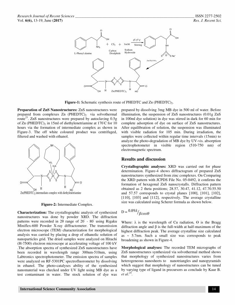
Characterization: The crystallographic analysis of synthesized nanostructures was done by powder XRD. The diffraction patterns were recorded in 2θ range of 20˚ - 80˚ using Rigaku Miniflex-600 Powder X-ray diffractometer. The transmission electron microscope (TEM) characterization for morphological analysis was carried by placing a drop of ethanolic solution of nanoparticles grid. The dried samples were analyzed on Hitachi (H-7500) electron microscope at accelerating voltage of 100 kV .The absorption spectra of synthesized ZnS nanostructures have been recorded in wavelength range 300nm-510nm, using Labtronics spectrophotometer. The emission spectra of samples were analyzed on RF-5301PC spectrofluorometer by dissolving in ethanol. The photo-catalyses ability of the synthesized nanomaterial was checked under UV light using MB dye as a test contaminant in water. The stock solution of dye was
prepared by dissolving 3mg MB dye in 500 ml of water. Before illumination, the suspension of ZnS nanostructures (0.01g ZnS in 100ml dye solution) in dye was stirred in dark for 60 min for complete adsorption of dye on surface of ZnS nanostructures. After equilibration of solution, the suspension was illuminated with visible radiation for 105 min. During irradiation, the samples were collected within regular time intervals (15min) to analyze the photo-degradation of MB dye by UV-vis. absorption spectrophotometer in visible region (510-750 nm) of electromagnetic spectrum.
Results and discussion
Crystallographic analyses: XRD was carried out for phase determination. Figure-4 shows diffractogram of prepared ZnS nanostructures synthesized from zinc complexes. On Comparing the XRD pattern with JCPDS File No. 05-0492, it confirms the formation of hexagonal ZnS nanocrystalls. Diffraction pattern obtained as 2 theta positions; 28.57, 30.47, 41.12, 47.70.55.50 and 57.57 corresponds to crystal planes [100], [101], [102], [110], [103] and [112], respectively. The average crystalline size was calculated using Scherer formula as shown below.
D = 0.89� ����
Where: λ is the wavelength of Cu radiation, ϴ is the Bragg diffraction angle and β is the full-width at half-maximum of the highest diffraction peak. The average crystalline size calculated as ~ 5.7nm. Such a small size was corresponds to peak broadening as shown in Figure-4.
Morphological analyses: The recorded TEM micrographs of ZnS nanostructures synthesized via solvothermal method shows that morphology of synthesized nanostructures varies from heterogeneous nanosheets to nanotriangles and nanopyramids which suggest that morphology of nanostructures can be tuned by varying type of ligand in precursors as conclude by Kaur B. et al.24.

Research Journal of Recent Sciences ______________________________________________________________ ISSN 2277-2502
Vol. 6(6), 13-19, June (2017) Res. J. Recent Sci.
International Science Community Association 15
Figure-3: XRD patteren of ZnS nanostructures prepared from Zn(PHEDTC)2.
Figure-4: TEM micrographs of ZnS nanostructures prepared from Zn(PHEDTC)2; (a) ZnS nanotriangles; (b) ZnS nanosheets.
The dimensions of nanostriangles and nanopyramids i.e length and width, calculated from TEM micrographs recorded for ZnS nanostructures are 95 nm and 24 nm respectively. Optical analyses: The optical properties of nanomaterials can be monitored by UV-vis. absorption spectroscopy. The absorption spectra of ZnS nanostructures prepared from Zn (PHEDTC)2 is shown in Figure-6. The recorded spectra of the synthesized nanostructures have broad absorption profiles in UV-visible region of electromagnetic spectrum, which makes these materials suitable for photo-catalytic applications. Photoluminescence studies: Photoluminescence study provides valuable information about the transitions take place in
nanomaterials. The semiconductor nanostructures have distinctive optical behavior. The room temperature PL spectra of ZnS nanostructures prepared from Zn (PHEDTC)2 were recorded in the wavelength range 420 - 570 nm by exciting at 325 nm wavelength. Figure-7 shows the recorded spectra that PL emission band is highly symmetric and multiple peaks with broadening which demonstrating the participation of diverse emission centers in radiative processes. The ZnS nanostructures prepared from Zn(PHEDTC)2 shows strong and symmetric emission peaks situated at 410 nm and 432 nm, which correspond to interstitial sulphur (IS) lattice defect and interstitial zinc (IZn) lattice defect. A weak shoulder at 469 nm is assigned to dangling sulfur bonds in ZnS grains25-28.
Research Journal of Recent Sciences ____________________________________
Vol. 6(6), 13-19, June (2017)
International Science Community Association
300 330 360 390 420 450
0.4
0.5
0.6
0.7
0.8
0.9
1.0
Ab
so
rba
nce
(a.u
.)
Wavelength(nm)
Figure-5: UV-vis. absorption spectra of ZnS nanostructures.
420 450 480 510
0
5000
10000
15000
20000
25000
30000
Inte
nsity(c
ps)
Wavelength(nm)
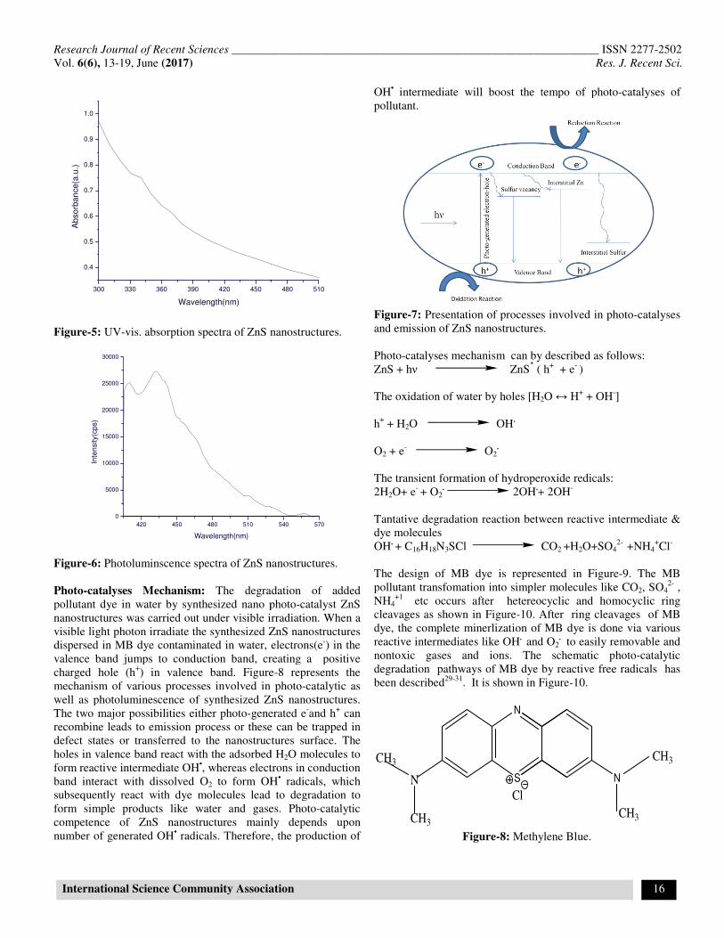
Figure-6: Photoluminscence spectra of ZnS nanostructures. Photo-catalyses Mechanism: The degradation of added pollutant dye in water by synthesized nano photonanostructures was carried out under visible irradiation. When a visible light photon irradiate the synthesized ZnS nanostructures dispersed in MB dye contaminated in water, electrons(valence band jumps to conduction band, creating a positive charged hole (h+) in valence band. Figuremechanism of various processes involved in photowell as photoluminescence of synthesized ZnS nanostructures. The two major possibilities either photo-generated recombine leads to emission process or these can be defect states or transferred to the nanostructures surface. The holes in valence band react with the adsorbed Hform reactive intermediate OH•, whereas electrons in conduction band interact with dissolved O2 to form OHsubsequently react with dye molecules lead to degradation to form simple products like water and gases. Photocompetence of ZnS nanostructures mainly depends upon number of generated OH• radicals. Therefore, the production of
________________________________________________________
International Science Community Association
480 510
vis. absorption spectra of ZnS nanostructures.
540 570
Photoluminscence spectra of ZnS nanostructures.
The degradation of added pollutant dye in water by synthesized nano photo-catalyst ZnS
carried out under visible irradiation. When a visible light photon irradiate the synthesized ZnS nanostructures
in MB dye contaminated in water, electrons(e-) in the valence band jumps to conduction band, creating a positive
ure-8 represents the mechanism of various processes involved in photo-catalytic as well as photoluminescence of synthesized ZnS nanostructures.
generated e-and h+ can recombine leads to emission process or these can be trapped in defect states or transferred to the nanostructures surface. The holes in valence band react with the adsorbed H2O molecules to
, whereas electrons in conduction to form OH• radicals, which
subsequently react with dye molecules lead to degradation to form simple products like water and gases. Photo-catalytic competence of ZnS nanostructures mainly depends upon
radicals. Therefore, the production of
OH• intermediate will boost the tempo of photopollutant.
Figure-7: Presentation of processes involved in photoand emission of ZnS nanostructures. Photo-catalyses mechanism can by described as follows:ZnS + hν ZnS٭ ( h The oxidation of water by holes [H2
h+ + H2O OH.
O2 + e- O2.
The transient formation of hydroperoxide redicals:2H2O+ e- + O2
. 2OH.+ 2OH
Tantative degradation reaction between reactive intermediate & dye molecules OH. + C16H18N3SCl CO
The design of MB dye is represented in Figurepollutant transfomation into simpler molecules like NH4
+1 etc occurs after hetereocyclic cleavages as shown in Figure-10. After ring cleavages of MB dye, the complete minerlization of MB dye is done via various reactive intermediates like OH. andnontoxic gases and ions. The schematic photodegradation pathways of MB dye by reactive free radicals has been described29-31. It is shown in Figure
N
SN
CH3
CH3
Cl
Figure-8: Methylene Blue.
_______________ ISSN 2277-2502
Res. J. Recent Sci.
16
te will boost the tempo of photo-catalyses of
Presentation of processes involved in photo-catalyses
emission of ZnS nanostructures.
catalyses mechanism can by described as follows: ( h+ + e- )
2O ↔ H+ + OH-]
The transient formation of hydroperoxide redicals: + 2OH-
Tantative degradation reaction between reactive intermediate &
CO2 +H2O+SO42- +NH4
+Cl-
The design of MB dye is represented in Figure-9. The MB pollutant transfomation into simpler molecules like CO2, SO4
2- , etc occurs after hetereocyclic and homocyclic ring
10. After ring cleavages of MB ete minerlization of MB dye is done via various
and O2. to easily removable and
nontoxic gases and ions. The schematic photo-catalytic degradation pathways of MB dye by reactive free radicals has
is shown in Figure-10.
N
CH3
CH3
Methylene Blue.

Research Journal of Recent Sciences ______________________________________________________________ ISSN 2277-2502
Vol. 6(6), 13-19, June (2017) Res. J. Recent Sci.
International Science Community Association 17
N
S N
CH3
CH3
N
CH3
CH3
Cl
NH2
N
CH3
CH3S
O
N
CH3
CH3
NH2
N SO3H
COH
CH3
OH
SO3H
OH
NH2
SO3HN
CH3
OH
OHN
CH3
CH3H
Figure-9: Degradation Pathways of MB dye.
The residual dye concentration v/s irradiation time was analyzed by UV-vis. absorption spectrophotometer. The decoloration of dye could be calculated using following formula: C(%)= [(Ao-A)/Ao] x 100,
Where: C is the decoloration degree, A0 and A are the absorption of dye solution before and after photo-catalysis, respectively. Figure-11 shows the spectra of MB dye for 105 min of irradiation on adsorbent ZnS nano photo-catalysts. The photo-catalytic efficiency basically depends upon the generation

Research Journal of Recent Sciences ______________________________________________________________ ISSN 2277-2502
Vol. 6(6), 13-19, June (2017) Res. J. Recent Sci.
International Science Community Association 18
of photo-excited carriers followed by their surface transfer dominance over the radiative recombination. Pristine dye solution was also irradiated for 105 min. in radiation reactor to check the photo-bleaching of MB dye, but no photo-bleaching of MB dye was observed. Hence, the synthesized nanostructures are efficient photo-catalysts to degrade the MB dye.
510 540 570 600 630 660 690 720
0.0
0.2
0.4
0.6
0.8
1.0
1.2 o min
15 min
30 min
45 min
60 min
75 min
90 min
105 min
Ab
so
rba
nce(a
.u.)
Wavelength(nm)
Figure-10: Spectra of absorbed MB dye pollutant for different durations under light.
Conclusion
Highly crystalline ZnS nanostructures have been successfully prepared from single source precursors Zn (PHEDTC)2 by efficient solvothermal route. Mixture of nanosheets, nanotriangles and nanopyramids like morphologies have been formed in case of Zn (PHEDTC)2 precursor. Synthesized ZnS nanostructures show symmetrical and broadened photoluminescence spectra in visible region of electromagnetic spectrum. Synthesized nanostructures seem to be efficient heterogeneous nano photo-catalysts for the degradation of aqueous pollutants.
Acknowledgment
One of the author, Balwinder Kaur is gratified to ministry of minority affairs (MOMA) New Delhi, India for providing Maulana Azad National Fellowships (MANF) for monetary support. The authors are grateful to Sophisticated Instruments Centre, Punjabi University, Patiala for providing essential facilities. A big thank to SAIF, Panjab University, Chandigarh for electron microscopy analyses.
References
1. Onwudiwe D.C. and Strydom C.A. (2015). The Bipyridine Adducts of N-phenyldithiocarbamato Complexes of Zn (II) and Cd (II); Synthesis, Spectral, Thermal Decomposition Studies and Use as Precursors for ZnS and CdS
Nanoparticles. Spectrochim. Acta. Mol. Biomo.l
Spectrosc., 135, 1080-1089.
2. Onwudiwe D.C., Mohammed A.D., Strydom C.A., Young D.A. and Jordaan A. (2014). Colloidal Synthesis of Monodispersed ZnS and CdS Nanocrystals from Novel Zinc and Cadmium Complexes. Superlattices.
Microstruct., 70, 98-108.
3. Zeng X., Pramana S.S., Batabyal S.K., Mhaisalkar S.G., Chen X. and Jinesh K.B. (2013). Low temperature synthesis of wurtzite zinc sulfide (ZnS) thin films by chemical spray pyrolysis. Phys. Chem. Chem.
Phys., 15(18), 6763-6768.
4. Zhu Y.C., Bando Y., Xue D.F. and Golberg D. (2004).
Oriented Assemblies of ZnS One‐Dimensional
Nanostructures. Adv Mater., 16(9‐10), 831-834.
5. Moore D. and Wang Z.L. (2006). Growth of Anisotropic One Dimensional ZnS Nanostructures. J. Mater.
Chem., 16(40), 3898-3905.
6. Yu J.H., Joo J., Park H.M., Baik S.I., Kim Y.W., Kim S.C. and Hyeon T. (2005). Synthesis of Quantum-Sized Cubic ZnS Nanorods by The Oriented Attachment Mechanism. .J.Soc .Chem .Am, 127(15), 5662-5670.
7. Ajibade P.A., Onwudiwe D.C. and Moloto M.J. (2011). Synthesis of Hexadecylamine Capped Nanoparticles Using Group 12 Complexes of N-alkyl-N-phenyl Dithiocarbamate as Single Source precursors. Polyhedron, 30(2), 246-252.
8. Mohamed N.B.H., Haouari M., Zaaboub Z., Hassen F., Maaref H. and Ouada H.B. (2014). Effect of Surface on The Optical Structure and Thermal Properties of Organically Capped CdS Nanoparticles. J. Phys. Chem, Solids., 75(8), 936-944.
9. Zhou X., Zeng X., Yan X., Xia W., Zhou Y. and Shen X. (2014). Shape and Phase Controlled ZnS Nanostructures and Their Optical Properties. Mater. Res. Bull., 59, 25-31.
10. Zhao Q., Xie Y., Zhang Z. and Bai X. (2007). Size-Selective Synthesis of Zinc Sulfide Hierarchical Structures and Their Photo-Catalytic Activity. Cryl. Grow. Des., 7(1), 153-158.
11. Ajibade P.A. and Ejelonu B.C. (2013). Group 12 Dithiocarbamate Complexes: Synthesis, Spectral Studies and Their Use as Precursors for Metal Sulfides Nanoparticles and Nanocomposites. Spectrochim. Acta.
Mol. Biomol. Spectrosc., 113, 408-414.
12. Hu J.S., Ren L.L., Guo Y.G., Liang H.P., Cao A.M., Wan L.J. and Bai C.L. (2005). Mass Production and High Photo-Catalytic Activity of ZnS Nanoporous Nanoparticles. Angew. Chem. Int. Ed., 117(8), 1295-1299.
13. Cesur H., Yazicilar T.K., Bati B. and Yilmaz V.T. (2001). Synthesis, Characterization and Spectral and Thermal Studies of Some Divalent Transition Metal Complexes of

Research Journal of Recent Sciences ______________________________________________________________ ISSN 2277-2502
Vol. 6(6), 13-19, June (2017) Res. J. Recent Sci.
International Science Community Association 19
Benzylpiperazine Dithiocarbamate. Synth. React. Inorg.
Met. Org. Chem., 31(7), 1271-1283.
14. Pickett N.L. and O’Brien P. (2001). Synthesis of Semiconductor Nanoparticles using Single Molecular Precursors. The Chemical Record, 1(6), 467-479.
15. Shahid M., Rüffer T., Lang H., Awan S.A. and Ahmad S. (2009). Synthesis and Crystal Structure of a Dinuclear Zinc(II)-Dithiocarbamate Complex, Bis {[(µ 2-pyrrolidinedithiocarbamato-S, S′)(pyrrolidinedithiocarbamato-S, S′) zinc (II)]}. Coord.
Chem., 62(3), 440-445.
16. Coucouvanis D. (1979). The Chemistry of The Dithioacid and 1, 1-Dithiolate Complexes, 1968-1977. Prog. Inorg.
Chem., 26, 301-469.
17. Efrima S. and Pradhan N. (2003). Xanthates and Related Compounds as Versatile Agents in Colloid Science. CR
Chim, 6(8), 1035-1045.
18. Pike R.D., Cui H., Kershaw R., Dwight K., Wold A., Blanton T.N. and Gysling H.J. (1993). Preparation of Zinc Sulfide Thin Films by Ultrasonic Spray Pyrolsis from Bis (diethyl dithiocarbamato) Zinc (II). Thin Solid
Films, 224(2), 221-226.
19. Kaur B., Singh K. and Malik A.K. (2017). Precursor Dependent Morphological and Photo-Catalytic Behaviour of CdS Nanostructures. Dyes. Pigm., 137, 352-359.
20. Zhu J., Wang Y., Li Z. and Zhang J. (2014). Synthesis and Biological Evaluation of Novel 99mTc-Oxo and 99mTc-Nitrido Complexes with Phenylalanine Dithiocarbamate for Tumor Imaging. J. Radioanal. Nucl. Chem., 302(1), 211-216.
21. Reddy K.H. and Reddy P.S. (2001). Mixed ligand Zinc (II) and Cadmium (II) Complexes with Alkyl Xanthates and 2, 2-Bipyridyl. Ind. J. Chem., 40A, 1118-1120.
22. Onwudiwe D.C. and Ajibade P.A. (2011). Synthesis, Characterization and Thermal Studies of Zn (II), Cd (II) and Hg (II) Complexes of N-Methyl-N-Phenyldithiocarbamate: The Single Crystal Structure of [(C6H5) (CH3) NCS2] 4Hg2. Int. J.Mol.Sci., 12(3), 1964-1978.
23. Prakasam B.A., Lahtinen M., Peuronen A., Muruganandham M., Kolehmainen E., Haapaniemi E. and Sillanpää M. (2015). Phase Selective Synthesis of ZnS Nanoparticles From Structurally New Dithiocarbamate Precursor. Mater. Lett., 144, 19-21.
24. Kaur B., Singh K. and Malik A.K. (2017). Effect of ligands on Crystallography, Morphology and Photo-Catalytic Ability of ZnS Nanostructures. Dyes. Pigm., 142, 153-160.
25. Viswanath R., Naik H.B., Kumar G.Y., Kumar P.P., Kumar G.A. and Praveen R. (2014). EDTA-Assisted Hydrothermal Synthesis, Characterization and Photo luminescent Properties of Mn2+ Doped ZnS. J.lumi., 153, 446-452.
26. Ayodhya D. and Veerabhadram G. (2016). Green Synthesis, Optical, Structural, Photocatalytic, Fluorescence Quenching and Degradation Studies of ZnS Nanoparticles. J. Fluoresc., 26(6), 2165-2175.
27. Kripal R., Gupta A.K., Mishra S.K., Srivastava R.K., Pandey A.C. and Prakash S.G. (2010). Photoluminescence and Photoconductivity of ZnS: Mn2+ Nanoparticles Synthesized via Co-precipitation Method. Spectrochim.
Acta. Mol. Biomol. Spectrosc., 76(5), 523-530.
28. Viswanath R., Naik H.B., Kumar G.Y., Kumar P.P., Harish K.N., Prabhakara M.C. and Praveen R. (2014). Synthesis and Photoluminescence Enhancement of PVA Capped Mn2+ Doped ZnS Nanoparticles and Observation of Tunable Dual Emission: A New Approach. App.Surf.
Sci., 301, 126-133.
29. Chitkara M., Singh K., Sandhu I.S. and Bhatti H.S. (2011). Photo-Catalytic Activity of Zn1-xMnxS Nanocrystals Synthesized By Wet Chemical Technique. Nanoscale res.
Let., 6(1), 438.
30. Houas A., Lachheb H., Ksibi M., Elaloui E., Guillard C. and Herrmann J.M. (2001). Photo-Catalytic Degradation Pathway of Methylene Blue in Water. Appl. Catal.
B., 31(2), 145-157.
31. Sharma M., Jain T., Singh S. and Pandey O.P. (2012). Photocatalytic Degradation of Organic Dyes Under UV–Visible light using Capped ZnS Nanoparticles. Sol.
Energ., 86(1), 626-633.