Embed Size (px)
Citation preview

'TINERALOGIST, VOL. 52, SEPTEMBER-OCTOBER, 1967
A SERPENTINE MINERAL SHOWING DIVERSE STRAIN-RELIEF MECHANISMS
F. AuuBNro, Geological Survey oJ Canad,a, Ottawa, Ontario, Canada
Arsrnlcr
An unstable serpentine polymorph which exhibits the combined properties characteristicof a number of other known polymorphs is described. Macrocrystals of the mineral, fromthe Tilly Foster Mine, New York State, indicate that it is a sixJayered monoclinic serpen-tine, u,'ith each successive layer displaced relative to its neighboursby + a/3 and *bf3 and,or, by a rotation of +60'. Individual layers are further modulated periodically in the ocrystal direction. The resulting crystal has superlattice controlled o and e parameters.
Finer fractions of a porvdered macrocrystal, observed by electron microscopy, show thatit tends to break down into smaller, simpler units: thin corrugated plates are formed, whichthemselves part along weak corrugation joints, forming elongated rods. The formation ofrods deprives the basic serpentine layers of the satisfactory strain relief mechanism attri-buted to corrugation. The rods are seen to compensate for this deprivation by curling paral-lel to their elongation, eventually producing more stable chrysotileJike tubes. The com-plete metamorphosis from plates to tubes has been followed by selected area electron difirac-tion.
INrnonucrroN
During a systematic study of the serpentine minerals, an olive-green,platy mineral, labelled "Serpentine Pseudocrystals", was discoveredamongst the specimens of the National Mineral Collection of Canada.The mineral is part of a hand specimen collected from the Til ly FosterMine, Brewster, Putnam County, New York State. It is closely associ-ated with magnesite, chondrodite, magnetite and chlorite. However,sumcient quantities of the mineral, free from magnetite and otherimpurities, could be selected without difficulty for chemical analysis.
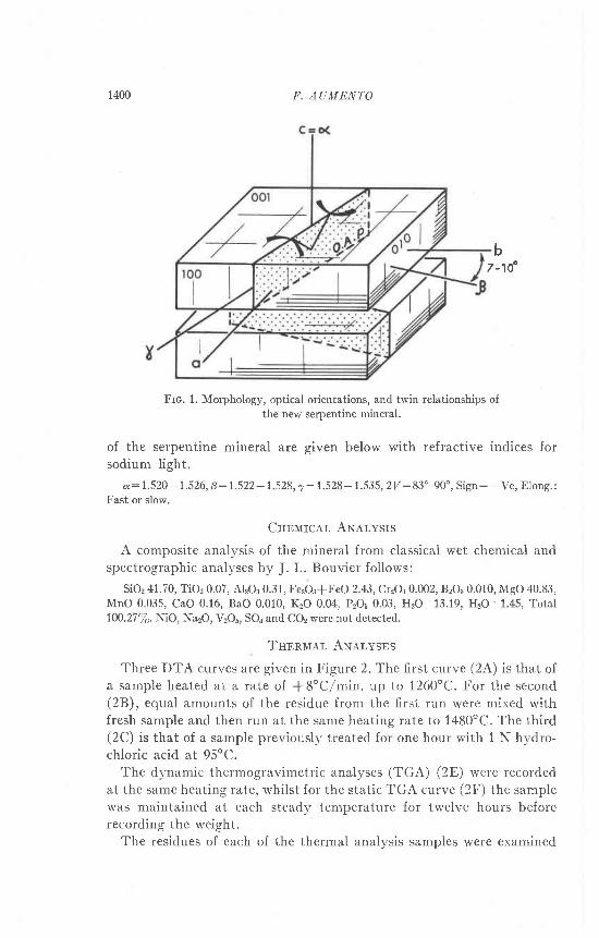
MonpnorocY AND Oprrcar- D.q.ra'fhe mineral exhibits a strong platy habit, with a perfect cleavage
parallel to (001). Flakes, ranging in size up to 5 mm across, can be bentwith ease. Two other good cleavages, parallel to (100) and (010), give thecrystals a euhedral appearance, which, although generally platy, at timescan approximate a cube. The density, determined with a Berman bal-ance, is 2.45+0.01 gmf cc.
Polysynthetic twinning parallel to (001) is apparent under the polar-izing microscope. The twinning, together with the morphology and opti-cal orientations, is shown in Figure (1). The refractive indices deter-mined by the double variation method, were found to vary from plate toplate. Plates parallel to (001) extinguish at 70-10" to the edges, and givea centered biaxial interference figure. The biaxial angle, determined for anumber of plates with a universal stage, is also variable. Optical properties
1399
1400 II. AUMENT'O
b7 -10"
Frc. 1. Morphology, optical orientations, and twin relationships ofthe new serpentine mineral.
of the serpentine mineral are given below with refractive indices forsodium light.
a:1.520-1.526,8:1.522-1.528,7:1.528-1.535, 2V:83"-90", Sign: -Ve, Elong.:Fast or slow.
Cnnurcer, ANervsrs
A composite analysis of the mineral from classical wet chemical andspectrographic analyses by J. L. Bouvier follows:
SiOr 41.70, TiOr 0.07, AlzO:0.31, Fe:O:iFeO 2.43, Cr?OB 0.002, BrO3 0.010, MgO 40.83,MnO 0 035, CaO 0.16, BaO 0.010, KzO 0.O[, PrOi 0.03, H2O+ 13.19, IIzO- 1.45, Total100.27Ta. NiO, Na2O, yrQr, SOa and CO2 were not detected.
TnBnlr.qr, AN,q.lvsns
Three DTA curves are given in Figure 2.The first curve (2A) is that ofa sample heated at a rate of *S"C/min. up to 7260"C. For the second(2B), equal amounts of the residue from the first run were mixed withfresh sample and then run at the same heating rate to 1480'C. The third(2C) is that of a sample previously treated for one hour with 1 N hydro-chloric acid at 95oC.
The dynamic thermogravimetric analyses (TGA) (2E) were recordedat the same heating rate, whilst for the static TGA curve (2F) the samplewas maintained at each steadv temperature for: twelve hours beforerecording the weight.
The residues of each of the thermal analysis samples were examined

STRAIN RNLIF,F IN SERPENTIN]i 1401
by X-ray powder diffraction. Samples heated to 760'C and 900oC gaveidentical powder patterns, showing poorly crystall ized forsterite. Thecomposition of this synthesized forsterite could be calculated from thediffraction patterns (after Jambor and Smith (1964)) to be 97.6 percentFo. Samples heated to between 1150"C and 1405oC showed two wellcrystallized phases: the major one was identified as protoenstatite, theminor one as forsterite. On heating to 1480oC the forsterite pattern in-tensified while the protoenstatite lines grew much fainter. The sampletreated with hydrochloric acid was amorphous to X rays both beforeand after heating to 1040oC.
X-nav Powtnn Drrnnacrrox
Debye-Scherrer, counter diffractometric, and Guinier focusing tech-niques were used in an attempt to resolve all possible reflections and todetermine accurate cell parameters. The counter diffractometer proved
--- - - - - - f
r : m p t n l l u l : , o c .
Frc. 2. Thermal analyses oi the new serpentine mineral. Heating rate (for curves A toB) of *8'C/minute.
A. DTA. of fresh sample.B. DTA. of 50 percent residue from ,4 plus 50 percent o{ fresh sample.C. DTA. of sample previously treated with 1 N HCl ior one hour at 95'C.D. DTG. curve.E. Dynamic TGA curve.F. Static TGA curve.
f
2
o
o
z
o
5
I
3z
s 1 5

I4O2 II, AUMENTO
Teer-o 1. Srpp-scanNno Drrln,q,crocneM oF rHE SrnprNrrxr MrNrner- er 25'C.
d"otnh
nlNo I 1 0 0 da,.
1z.t456789
101 11 2_t.1
741 5161 1
1 819tn
100L J
55
5312I J
I J
t91 1t oo
10866.55
002130200137131132004150060005J J J
006
7 .2654.6 t14 .3704.0203 . 6 3 22 6602 . 6 5 52.5212 . 4 7 52 . 1 1 71 . 8 1 6
1 . 5 3 71 .4531 326| . 2 1 1
7 .3634 5484 . 4 14.093 . 6 3 92.6572.5862.5072 . 4 7 72.0891 8205
.74215372
.4555
.3071 . 2 1 21 1051 .007
962. 9 1 3
001020
Recorded with Ni-filtered Cu radiation and a quartz internal standard. The lines areindexed on the basic cel l d imensions o:5.32, b:9.22, t :7.28 A, B:93.3 ' . Super lat t iceindices based on multipies (n) of o or c are also given lor weak lines at 4.41 and 4.09 A
to be best suited to the problem. The diffractogram (table 1) shows acombination of spacings and intensities similar to those of clino-chryso-ti le, with the addition of a few weak lines. Debye-Scherrer photographshad indicated the possible existence of weak Iines on the higher 20 angleside of the broad (020) peak (as shown by superlattice reflections ofantigorite and ortho-serpentines). The region from 5 A to 2.5 A *u.therefore repeatedly step-scanned with the diffractometer: two veryweak l ines, a l 4 .41 and 4.09 A, *" r . resolved f rom an area of h igh back-ground on the higher 20 angle side of peak (020). No other faint l inescould be resolved, although the high uneven background associated withthese lines mav have been due to a superimposition of a number of weaklines. The pattern, with the exception of the two weak lines mentioned,was indexed using the cell parameters and indexed reflections recorded byprecession photography (next section). More precise cell parameterswere then calculated from the indexed diffractogram, giving a mono-c l in ic ce l l wi th the fo l lowing parameters: -a:5.320* 0.005, 6:9.222+

STRAIN RELIEF IlT SDRPL:NTINE
Frc. 3. Zero-Ievel b* c* precession photograph of new serpentine. Note
continuous streaks parallel to c* when k*O or 6n.
0.005, c: 7 .277 + 0.005 A 0: 93.3'f 0.5o. However, a multiple param-
eter is required if the additionai weak lines at 4.41 and 4.09 A are to be
indexed. Ambiguities as to which parameter is the multiple (the a param-
eter as in antigorite, or the c parameter as in the ortho-serpentines)
can be partially resolved from single crystal X-ray and electron diffrac-
tion photography.A sample treated with 1 N hydrochloric acid at 95"C for one hour
gave an amorphous diffraction pattern, as is attributed to chrysoti les.
SrNcr-n Cnvsrar- X-nev Drnrn.tcrrcN
A combination of precession and Weissenberg photography, using
both Cu and Mo radiation, permitted the recording of up to six levels
about the o*, bx and dx axes.Composites of the zero levels a* c* and 6* cx, taken with the preces-
sion camera, are given in Figures (3) and (4).' The following conditions for systematic reflections, based on the basic
uni t ce l l wi th o:5.32 A and c: 7.28 h, are apparent :
1403

F. AUMENTO
h k l : h * k : 2 n
0k l , : k : 2n
h j l : h : 2 n
h h 0 : h * k : 2 n
h 0 0 : h : 2 n
0 k 0 : k : 2 n
001 : no conditions
Three space groups, namely Cm, C2 or C2f m, are therefore possible forthis serpentine polymorph.
Two very striking features are immediately apparent in the singlecrystal photographs: (1) All reflections recorded show multiple spotsparaliel to the o* axis clustered about eight-fold multiple values of Z.The clusters are interrupted along layer-l ines, and are never made up ofmore than five spots. Taking all the multiple spots separately, the o
Frc. 4 Zero-level a* e* precession photograph of nelv serpentine. Note superlattice con-trolled clustering of spots parallel to o* and elongation of spots parallel to e* when hlo.

STRAIN RELIL,F IN SERPENTINE 1405
parameter has a value of 41 '5 A ( :5.32Xs), the value requi red to index
all the l ines of the powder diffractogram. As in antigorite, the clustering
of reflections arottnd certain rnultiple values of iz could be takcn to repre-
sent a typical superlattice effect. The basic o parameter would be 5.32 A,
but the structure would be modulated periodically in the o direction,
with a "wavelength" of 41.5 A. No change in this superlattice integral
value has been observed between different crystals of the mineral under
X-ray investigation, or by selected area electron diffraction.A series of closely spaced spots parallel to c* are also visible in the
zero level a* c* net (Cu radiation) of Figure 4. Again, they may indicate
a larger superlattice parameter in the c direction, with c:7.277Y.43.66
A. Howener, these multiple spots are not observed in other photographs
involving c*, due either to the poorer resolution of the Mo radiation
used, or to the masking effect of the features described below. Unfortun-
ately it was also impossible to confirm the multiplicity along c* by elec-
tron diffraction due to the preferential alignment of crystallites on sam-
ple grids parallel to the perfect cleavage (001). (2) It is immediately
noticeable that, whilst certain reflections are sharp, others are elongated'
in varying degrees, parallel to c*. On the basis of the extent of the elonga-
tion, the reflections can be subdivided into three types:1. Sharp reflections: these occur only when:
h :0 ior (h0l )
h : 0 or 6n f.or (}hl)
h : 3n and & : 3n lor (hhl)
2. Reflections elongated by c*/2 along c*: these occur when:
h t 0 f . o r ( h O l )
h I 0 or 3n, and k : 3n for (hkl)
3. Continuous streaks parallel to c* of uniform intensity, except for
the falling off of atomic scattering factor with increasing sin O/tr. These
occur when:
h # 0 or 6n f"or (}kl)
h I 3n lor (hkl)
From the evidence of elongation of reflections parallel to c*, it seems
that the serpentine layers making up the crystals are displaced relative
to eachother .Thedisplacementwouldbein theorder of * a/3 and *b/3.
The probability of the stacking mistake, a, calculated from the integral
breadths of the streaks parallel to c* (using the formula b:3a/(4-3q.),
after Rucklidge and Zussman (1965), where b is the integral breadth)

1406 F. AUML,NTO
would be approximately a:0.4 for the o d i rect ion (where b:0.5) . I t isrnore diff icult to give a value to the breadth in the 6 direction, since thestreaks are continuous; these, however, can be interpreted as resultingfrom random stacking of the layers, such that the probabil ity of thestacking mistake, d, must equal or approach unity.
Similar streaking could also be caused by rotation of each successivelayer relative to its neighbours. A + 60o rotation of the layers would pro-duce single crystal photographs comparable to those presented here.However, rotation of basic serpentine layers, should these be individuallycorrugated parallel to o, would cause corrugations to show up in otherdirections as well, and would also inhibit stacking along c with the repeatdistance of 7.28 A. O.r the other hand, it is possible that although thefundamental layers are individually rotated, the corrugations are alwayssuperimposed onto these layers in the same direction with respect to thecrystal as a whole.
ElocrnoN Mrcnoscopv
An ethyl alcohol-based suspension of the finely ground serpentine wasdispersed by vibrating ultrasonically for one minute. Drops of the l iquidwere then pipetted onto sample grids previously covered by 300 A thickcarbon substrates. The specimens were viewed with a Siemens Emiskopr electron microscope. The same specimens were used for selected-areaelectron difiraction.
The first sets of observations were made at 100 kV using a standardsample stage. It was found that the diffraction patterns obtained woulddisappear within one minute of exposure to the electron beam. However,a cold finger stage was subsequently inserted into the microscope, withthe result that the particles could be viewed for five minutes or morewithout any apparent changes taking place.
At high magnifications the mineral exhibits two distinct morphologies,with a complete range of intermediate transitional morphologies betweenthe two end types. These are i l lustrated in Figures 5 and 6. The originalflakes, Iying parallel to (001), are sti l l apparent. A number of these, whenviewed at high magnifications, show the flakes traversed by regularlyspaced parallel l ines, somewhat similar to the electron optical fringesrecorded by Brindley, et al., (1958) and others. The separation of theseparallel l ines is in the ord"er of 160 A, which seems to bear a four-foldrelationship to the superlattice parameter a:41.5 A recorded by X-raydiffraction.
In many cases the plates are clearly seen to be composed of a series oflayers; each layer is made up of parallel-lying elongated laths. Byobserving the orientation of these component laths, one can deduce that
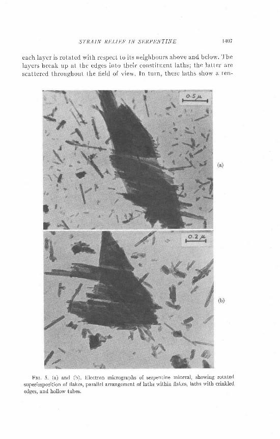
STRAIN RDLIEF IN SI:RPI'NTINII 1407
each la)er is rotated with respect to its neighbours above and below. Thelayers break up at the edges into their constitr.rent laths; the latter arescattered throughout the field of view. In turn, these laths show a ten-
Frc. 5 (a) and (b). Electron micrographs of serpentine
superimposition of flakes, parallel arrangement of laths within
edges, and hollow tubes.
mineral, showing rotated
flakes, laths with crinkled

1408 F. AUMDNTO
Itrc. 6 (a) and (b). Electron micrographs of serpentine mineral, showing flakesu'ith parallel striations, laths, and hollow tubes.
dency to curl up about an axis parallel to their length. Some of themexhibit this tendency in its init ial stage, giving rise to crinkled edges.Other laths have gone a stage further, producing a close approximation

STRAIN RELIEF IN SERPENTINE I1W
to chrysotile tubes. In a few examples, end on views of these "tubes"are visible, showing their hollow axes. The external diameter of these
tubes can vary from 150 A to 250 A.
Er-BcrnoN DrlnnacrroN
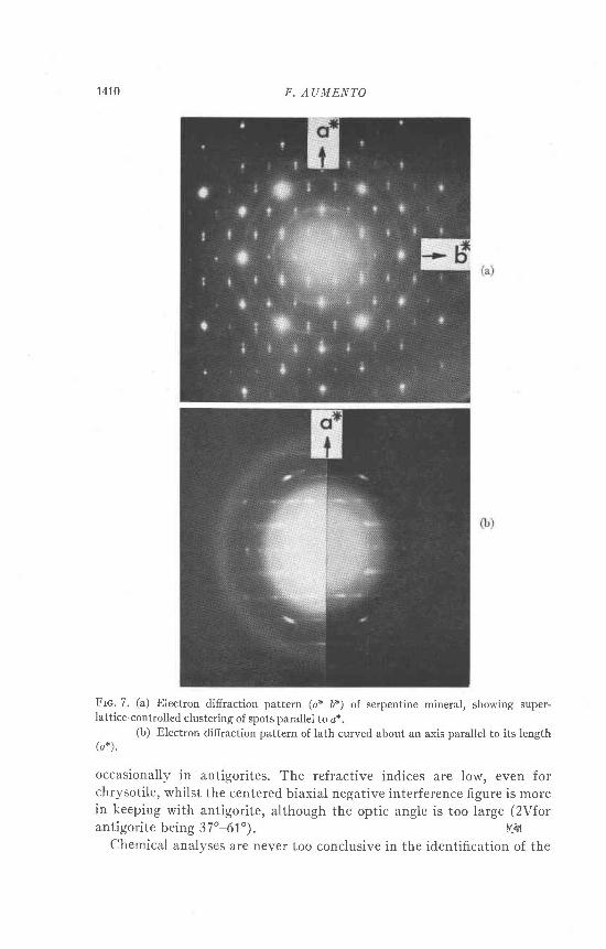
Plates of the serpentine are always found to be lying parallel to the(001) cleavage. These give a well oriented a* b* net (Fig. 7a), showing thesuperlattice-controlled clustering of spots parallel to ox, around certainmultiple values of h. Tn all cases the latter is an integral multiple of 8,suggesting a superlattice parameter of 8X5.32 A:+t.S A.
The process of transformation from plates to tubes can also be followed
by selected area electron diffraction. Whereas the plates give difiractionpatterns as described above, those of the laths and tubes show thatconsiderable curvature has taken place. The resultant patterns aresimilar to the rotation photographs obtained from chrysotile tubes, bothas bundles of fibres in X-ray diffraction, and as single tubes in electron
difiraction. Difficulties were encountered in trying to record diffractionpatterns from single fibres; the microscope was not equipped with abeam stop, so that the main beam generally blacked out the faint pat-
terns given by the fibres. An example of patterns obtained is shown inFigure 7b. Reflections fall along lines perpendicular to the tube axes.The repeat distance between these layers is 5.32 A. As in chrysoti le, thezero layer contains both 001 and 0ft0 spots, while the first layer shows thecharacteristic f lared spots indexed as (hkO). Similarly, the second layercontains 201 spots, and the third layer the flared spots (310) and (330).
The laths have therefore have been curved about the r axis, and nowgive a pattern which approximates an electron diffraction pattern ofchrysotile.
DrscussroN
The mineral described above has many of the properties characteristicof the serpentine group of minerals. Ilowever, the combination of theseproperties cannot be matched to any one of the known serpentine poly-morphs alone, as discussed below. There are two possible explanationsfor the diversity of properties exhibited: this mineral could be an inti-mately compounded mixture of two or more other serpentine poly-morphs, or it cor-rld be a new serpentine polymorph.
The morphology of the mineral is unusual for any serpentine. Singlecrystal plates 5 mm across have not been reported before, except possiblyin the rare case of the l izardite from Kennack Cove (Midgley, 1951).Polysynthetic twinning is also rare in serpentines; it is found only
1410 F. AUMENTO
Irrc. 7. (a) Electron difiraction pattern (ax Dx) of serpentinelattice-controlled clustering of spots parallel to o*.
(b) Electron difiraction pattern of lath curved about an( a * ) .
mineral, showing super-
axis parallel to its length
occasionally in antigorites. The refractive indices are low, even forchrysotile, whilst the centered biaxial negative interference figure is morein keeping with antigorite, although the optic angle is too Iarge (2Vforantigorite being 37'-61'). F:fi
Chemical analyses are never too conclusive in the identifi.cation of the

STRAIN RELIEF IN SERPENTINE I41I
serpentine polymorphs. However, the author has plotted sorne 200
analyses from the literature on triangular diagrams: these show diffusecompositional zones for chrysotile, lizardite and antigorite. The mineralfrom the Tilley Foster Mine falls in the chrysotile zone. The analysisshows a small replacement of silicon by aluminum. Since the sample isknown to be free from magnetite, one can also assume that magnesium is
being replaced to a small extent by ferrous and ferric iron. However,more replacement, especially of Si by AI, would have been expected tohave taken place in order to account for the large platy habit of themineral. The corrugation of the sheets will reduce the necessity of Sireplacement to alleviate mismatching of the layers. The Si:Mg ratio,higher than that required for the ideal serpentine composition, is also inkeeping with corrugated sheets. However, once the crystals are brokendown to dimensions small enough to reduce the effectiveness of thecorrugations, they would be expected, and are indeed observed, to curl.
Thermal analyses, together with the destruction of the crystallinityby 1 N hydrochloric acid and by an electron beam, suggest similaritieswith chrysotile. However, no serpentine polymorph has yet been reportedto produce protoenstatite on heating, either as a major or minor phase.
X-ray diffractograms, were it not for two weak and almost overlookedsuperlattice lines, would identify the mineral conclusively to be clino-chrysotile. However, single crystal photographs exclude this possibility.Without supporting evidence from electron diffraction, it is not possibleto confirm the superlattice along c detected on precession photography.I{owever, a six-layered cell would comply with the other observations ofdisplacement or rotation of individual layers. Hence it is suggested thatthe mineral is a monoclinic six-layer serpentine (c:7.277 X6:43.66 A);it has a B angle approximating that of clino-chrysotile, having individualfundamental layers modulated periodically in the o direction of thecrystal. These photographs show that there are stacking errors betweensuccessive layers, either as displacements of * af 3 and *bf 3, or 6O"rotation of the layers, or both. The electron micrographs suggest thatboth types of stacking errors may exist.
The regularly spaced parallel lines observed at high magnifications onthe plates (Fig. 6b) are not believed to be electron optical fringes; theyare rather corrugations of the plates showing incipient parting. Some ofthe bright lines visible parallel to the axes of the rods could be Fresnelfringes, obtained under slightly out of focus conditions, rather thanhollow tubes. However, in many instances they are definitely tubular,with end sections visible, giving the rotation-type electron diffractionpatterns generally associated with chrysoti les.
The transition from plates to tubes, as followed on the electron micro-

1412 F. AUMENTO
scope, is also unique. The plates have many of the characteristics ofantigorite, whilst the tubes approximate chrysoti le very closely. It ispossible that the plates part along the junctions between adjacentalternating structural waves; these would then be free to curl into tubes.
A polymorphic mixture would offer a second explanation for theelectron microscope observations: rather than being observations ofdifferent stages in the metamorphosis from an unstable platy, to a stabletubular crystal, the photomicrographs and diffraction patterns would beexplained as being from stable material originally present in, and laterreleased from, the macrocrystals. However, with the assumption that thedispersed state contains such a free mixture, one has then to explain whyno such "mixture effect" shows up in the thermal analyses and powderdiffractograms. One mav also expect the complicated single crystalphotographs, if mixture controlled, to vary from crystal to crystal, withdifferent relative proportions of the constituents occurring in differentcrystalsl the photographs are not found to vary, indicating eitheraremarkably constant mixture, which is unlikely, or a different polymorphaltogether.
A number of other properties cannot be controlled or explained by themixing of polymorphs. For example, the refractive indices of the macro-crystals are lower than those of any of the components of a possiblemixture.
CoNcr,usroNs
The mineral from the Til ly Foster Mine is believed to be an unstablepolymorph of the serpentine group of minerals. Its unit cell is made up ofsix superimposed fundamental serpentine layers, with each successivelayer displaced relative to its neighbours by -l a/3 and * bf 3, and, or bya rotation of * 60". Individual layers are further modulated periodicallyin the o crystal direction. The stacking errors and layer modulationsresult in a crystal with superlattice-controlled a and d parameters.
The confi.guration described is stable only in its original macrocrystal-line state, tending to break down into smaller, simpler units if assistedmechanically. Hence the finer fractions of a powdered specimen show thatthe crystals break up in stages, at first forming thin corrugated plates.The latter, in turn, part along weak corrugation joints, giving rise torods which will necessarily be under strain due to the fundamentalmismatching of the constituent sil ica tetrahedral and brucite octahedrallayers. Unable to take up the strain in other ways, the rods resort tocurling parallel to their elongation, as demonstrated in electron micro-graphs, f inally producing chrysoti le-l ike tubes.
STRAIN RELIEF IN SERPENTINE t4l3
Acrwowr,nocourNrs
The author is indebted to Dr. J. Zussman of the Department of Geoiogy and Mineral-
ogy, University of Oxford, for his valuable comments and for critically reading a prelimi-
nary draft of the manuscript.Thanks are also due to Dr. J.A.V. Douglas, Miss C' M. Hunt, Mr. R. N. Delabio, and
Mr. J. L. Bouvier, of the Geological Survey of Canada, Ottawa, and Miss J. Ng Yelim,
Mr. R. Lake, and Mr. J. R. Rowland of the Mines Branch, Ottawa for their techdcal
cooperation.The loan of a Buerger precession camera from the Department of Geology, Dalhousie
University, is also gratefully acknowledged.
RrlnnrNcns
Bnrxotov, G. W., J. J. Counn, R. Uvroe, and J. ZussuaN (1958) Electron-optical obser-
vations with crystals of antigorite. Acla Crystoll'ogr. I l' 99-102
Jaulon, J. L. aNt C. H. Surrn (1964) Olivine composition detremination with small-dia-
meter X-ray powder cameras. Mineral' Mag.33r730-741.
Mrnclrv, H. G. (1951) A serpentine mineral from Kennack Cove, Lizard, Cornwall'
Mineral Mag. 29,52G530
Rucrrmcn, J. C. er"'o J. ZussutN (1965) The crystal structure of the serpentine mineral,
lizardite MSrSirOr(OH)r. Acto Cryslallogr. 19, 381-389
Manuscript ruei't:d., December 17, 1966; accepted' Jor publhation, Iuly 17, 1967.