Embed Size (px)
Citation preview

Copyright © 2005 by the Genetics Society of AmericaDOI: 10.1534/genetics.105.040667
A Screen for Genes Regulating the Wingless Gradient in Drosophila Embryos
Sabrina C. Desbordes,1 Dhianjali Chandraratna and Benedicte Sanson2
Department of Genetics, University of Cambridge, Cambridge CB2 3EH, United Kingdom
Manuscript received January 10, 2005Accepted for publication March 10, 2005
ABSTRACTDuring the development of the Drosophila embryonic epidermis, the secreted Wingless protein initially
spreads symmetrically from its source. At later stages, Wingless becomes asymmetrically distributed in aHedgehog-dependent manner, to control the patterning of the embryonic epidermis. When Wingless ismisexpressed in engrailed cells in hedgehog heterozygous mutant embryos, larvae show a dominant phenotypeconsisting of patches of naked cuticle in denticle belts. This dose-sensitive phenotype is a direct consequenceof a change in Wg protein distribution. We used this phenotype to carry out a screen for identifying genesregulating Wingless distribution or transport in the embryonic epidermis. Using a third chromosomedeficiency collection, we found several genomic regions that showed a dominant interaction. After usinga secondary screen to test for mutants and smaller deficiencies, we identified three interacting genes:dally, notum, and brahma. We confirmed that dally, as well as its homolog dally-like, and notum affect Winglessdistribution in the embryonic epidermis, directly or indirectly. Thus, our assay can be used effectively toscreen for genes regulating Wingless distribution or transport.
SECRETED signaling molecules play an essential role distribution (Bellaiche et al. 1998; The et al. 1999;Takei et al. 2003; Han et al. 2004a,b).in patterning developing metazoans. Work from re-
Gradient formation also depends upon the mecha-cent years shows that the signaling activity of these mole-nism by which signaling ligands move in the plane ofcules is tightly regulated (Freeman 2000). Among vari-epithelia. It is not yet clear what these mechanisms are,ous levels of regulation, controlling the distribution ofbut two main modes of transport have been proposedligands in a field of cells is necessary to establish stable(reviewed by Vincent and Dubois 2002; Gonzalez-gradients that robustly pattern developing tissues (Vin-Gaitan 2003): (1) facilitated diffusion, where ligandscent and Dubois 2002). The distribution of ligandsdiffuse within the extracellular space, and this diffusioncould conceivably be influenced by many factors, includ-is modulated by cell-surface receptors, and (2) trans-ing the concentration of receptors, the composition ofcytosis, where ligands are transported along the planethe extracellular matrix, or the rate of recycling andof the epithelium by repeated cycles of endocytosis anddegradation of the ligand following internalization. Therecycling to the cell surface.best-documented case is the regulation of ligand distri-
In this study, we searched for genes that influencebution by receptors. For example, in Drosophila, Hedge-the distribution or transport of the signaling ligandhog (Hh) range of action is regulated by its receptorWingless (Wg), the homolog of vertebrate Wnt-1 in Dro-Patched: if the Hh-binding domain of Patched is mu-sophila. In the embryonic epidermis, Wg acts at a shorttated, the spread of Hh is extended in the wing imaginalrange to regulate the activity of target genes, whereasdisc (Chen and Struhl 1996). Another case is the mor-it behaves as a long-range morphogen in the wing imagi-phogen Decapentaplegic, whose concentration gradi-nal disc (reviewed by Seto and Bellen 2004). In theent is regulated by the amount of its receptor Thickveinswing, Wg forms a stable gradient with a source localized(Lecuit and Cohen 1998; Tanimoto et al. 2000). Inat the boundary between ventral and dorsal compart-
addition to canonical receptors, low-affinity receptorsments and triggers the expression of target genes at
such as heparan sulfate proteoglycans are likely to affect different distances from its source in a concentration-ligand distribution. For instance, in wing discs, mutants dependent manner (Zecca et al. 1996; Neumann andthat impede the synthesis of heparan sulfate change Hh Cohen 1997).
Both facilitated diffusion and transcytosis have beenproposed to explain how Wg moves from cell to cell. Inthe embryonic and imaginal disc epithelia, Wg protein is1Present address: Laboratory of Stem Cell and Tumor Biology, Neuro-
surgery and Developmental Biology, Memorial Sloan Kettering Can- detected in the basolateral extracellular space as wellcer Center, 1275 York Ave., New York, NY 10021. as in bright intracellular dots localized to the apical side
2Corresponding author: Department of Genetics, University of Cam-of the cells (Pfeiffer et al. 2000; Strigini and Cohenbridge, Downing St., Cambridge CB2 3EH, United Kingdom.
E-mail: [email protected] 2000; Greco et al. 2001; Simmonds et al. 2001). These
Genetics 170: 749–766 ( June 2005)
750 S. C. Desbordes, D. Chandraratna and B. Sanson
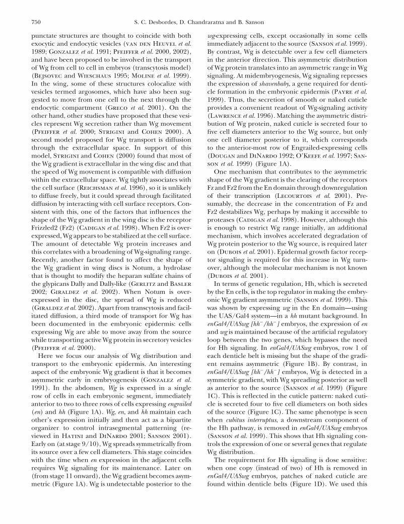
punctate structures are thought to coincide with both wg-expressing cells, except occasionally in some cellsimmediately adjacent to the source (Sanson et al. 1999).exocytic and endocytic vesicles (van den Heuvel et al.
1989; Gonzalez et al. 1991; Pfeiffer et al. 2000, 2002), By contrast, Wg is detectable over a few cell diametersin the anterior direction. This asymmetric distributionand have been proposed to be involved in the transport
of Wg from cell to cell in embryos (transcytosis model) of Wg protein translates into an asymmetric range in Wgsignaling. At midembryogenesis, Wg signaling represses(Bejsovec and Wieschaus 1995; Moline et al. 1999).
In the wing, some of these structures colocalize with the expression of shavenbaby, a gene required for denti-cle formation in the embryonic epidermis (Payre et al.vesicles termed argosomes, which have also been sug-
gested to move from one cell to the next through the 1999). Thus, the secretion of smooth or naked cuticleprovides a convenient readout of Wg-signaling activityendocytic compartment (Greco et al. 2001). On the
other hand, other studies have proposed that these vesi- (Lawrence et al. 1996). Matching the asymmetric distri-bution of Wg protein, naked cuticle is secreted four tocles represent Wg secretion rather than Wg movement
(Pfeiffer et al. 2000; Strigini and Cohen 2000). A five cell diameters anterior to the Wg source, but onlyone cell diameter posterior to it, which correspondssecond model proposed for Wg transport is diffusion
through the extracellular space. In support of this to the anterior-most row of Engrailed-expressing cells(Dougan and DiNardo 1992; O’Keefe et al. 1997; San-model, Strigini and Cohen (2000) found that most of
the Wg gradient is extracellular in the wing disc and that son et al. 1999) (Figure 1A).One mechanism that contributes to the asymmetricthe speed of Wg movement is compatible with diffusion
within the extracellular space. Wg tightly associates with shape of the Wg gradient is the clearing of the receptorsFz and Fz2 from the En domain through downregulationthe cell surface (Reichsman et al. 1996), so it is unlikely
to diffuse freely, but it could spread through facilitated of their transcription (Lecourtois et al. 2001). Pre-sumably, the decrease in the concentration of Fz anddiffusion by interacting with cell surface receptors. Con-
sistent with this, one of the factors that influences the Fz2 destabilizes Wg, perhaps by making it accessible toproteases (Cadigan et al. 1998). However, although thisshape of the Wg gradient in the wing disc is the receptor
Frizzled2 (Fz2) (Cadigan et al. 1998). When Fz2 is over- is enough to restrict Wg range initially, an additionalmechanism, which involves accelerated degradation ofexpressed, Wg appears to be stabilized at the cell surface.
The amount of detectable Wg protein increases and Wg protein posterior to the Wg source, is required lateron (Dubois et al. 2001). Epidermal growth factor recep-this correlates with a broadening of Wg-signaling range.
Recently, another factor found to affect the shape of tor signaling is required for this increase in Wg turn-over, although the molecular mechanism is not knownthe Wg gradient in wing discs is Notum, a hydrolase
that is thought to modify the heparan sulfate chains of (Dubois et al. 2001).In terms of genetic regulation, Hh, which is secretedthe glypicans Dally and Dally-like (Gerlitz and Basler
2002; Giraldez et al. 2002). When Notum is over- by the En cells, is the top regulator in making the embry-onic Wg gradient asymmetric (Sanson et al. 1999). Thisexpressed in the disc, the spread of Wg is reduced
(Giraldez et al. 2002). Apart from transcytosis and facil- was shown by expressing wg in the En domain—usingthe UAS/Gal4 system—in a hh mutant background. Initated diffusion, a third mode of transport for Wg has
been documented in the embryonic epidermis: cells enGal4/UASwg [hh�/hh�] embryos, the expression of enand wg is maintained because of the artificial regulatoryexpressing Wg are able to move away from the source
while transporting active Wg protein in secretory vesicles loop between the two genes, which bypasses the needfor Hh signaling. In enGal4/UASwg embryos, row 1 of(Pfeiffer et al. 2000).
Here we focus our analysis of Wg distribution and each denticle belt is missing but the shape of the gradi-ent remains asymmetric (Figure 1B). By contrast, intransport to the embryonic epidermis. An interesting
aspect of the embryonic Wg gradient is that it becomes enGal4/UASwg [hh�/hh�] embryos, Wg is detected in asymmetric gradient, with Wg spreading posterior as wellasymmetric early in embryogenesis (Gonzalez et al.
1991). In the abdomen, Wg is expressed in a single as anterior to the source (Sanson et al. 1999) (Figure1C). This is reflected in the cuticle pattern: naked cuti-row of cells in each embryonic segment, immediately
anterior to two to three rows of cells expressing engrailed cle is secreted four to five cell diameters on both sidesof the source (Figure 1C). The same phenotype is seen(en) and hh (Figure 1A). Wg, en, and hh maintain each
other’s expression initially and then act as a bipartite when cubitus interruptus, a downstream component ofthe Hh pathway, is removed in enGal4/UASwg embryosorganizer to control intrasegmental patterning (re-
viewed in Hatini and DiNardo 2001; Sanson 2001). (Sanson et al. 1999). This shows that Hh signaling con-trols the expression of one or several genes that regulateEarly on (at stage 9/10), Wg spreads symmetrically from
its source over a few cell diameters. This stage coincides Wg distribution.The requirement for Hh signaling is dose sensitive:with the time when en expression in the adjacent cells
requires Wg signaling for its maintenance. Later on when one copy (instead of two) of Hh is removed inenGal4/UASwg embryos, patches of naked cuticle are(from stage 11 onward), the Wg gradient becomes asym-
metric (Figure 1A). Wg is undetectable posterior to the found within denticle belts (Figure 1D). We used this
751Regulators of Wingless Distribution
it made it impossible to consistently score additional gaps fromdominant phenotype in a F1 screen to find genes givinginteracting deficiencies.a similar phenotype, and thus potentially regulating Wg
Since enGal4/UASwg embryos die at the end of embryogene-distribution. We conducted a deficiency screen on the sis, the collected embryos were aged at least 30 hr so that onlythird chromosome to identify such genes. Two succes- nonviable embryos remain unhatched. Dead embryos were
dechorionated using bleach and their vitelline membrane wassive screens led us to the identification of three genesremoved by hand in Hoyer’s on a slide. Embryos were alignedthat affect Wg distribution in the embryonic epidermis:ventral side up in a mixture of Hoyer’s and lactic acid (1:1).dally, dally-like, and notum. An additional role for dally-Cuticle preparations were baked overnight at 55�.
like in Hh signaling, found while analyzing these results, Gaps were scored by visualizing the cuticles with phase-has been described elsewhere (Desbordes and Sanson contrast microscopy. For abdominal belts A2–A7, we scored
a gap when a patch of smooth cuticle replaced most rows of2003).denticles in the belt (Figure 2, D vs. D�). Because A1 is thinnerthan the other abdominal denticle belts, we scored only com-plete breaches of this belt (Figure 2, E vs. E�). We eliminated
MATERIALS AND METHODS segment A8 from our analysis, because it was too often foldedand difficult to visualize.
Fly stocks: The Bloomington Drosophila Stock Center To assess the significance of our results, we compared the(BDSC) provided the third chromosome deficiency kit (see list number of gaps obtained for each deficiency with the numberof stocks in the appendix) as well as Df(3L)Scf-R11, Df(3R)l26c, of gaps obtained for wild-type chromosomes. We scored fiveDf(3R)ry75, Df(3R)urd, and Df(3R)lc4a. The Umea Stock Center gaps for 354 enGal4/UASwg embryos examined, when chromo-(USC) provided Df(3L)th102 ; Ken Cadigan, Df(3L)Dfz2 ; Daniel somes from the wild-type yw 67 or canton S strains were testedKalderon, Df(3L)A27 ; and Daniel St Johnston, Df(3L)XS-543. in place of a deficiency chromosome in the screening schemeWe obtained the following mutant stocks from the BDSC: (Table 1). We used this value as the expected frequency ofl(3)06464, l(3)01629, arf 72A, brm 2, pip 1, kni 10, Dl 3, and hh AC. gaps and employed a standard � 2 test to compare this expectedDfz2 C1 was provided by Gary Struhl, dally 10 and dally E385 by Steve frequency with the frequency of gaps observed for each defi-Kerridge, Psn B3 and Psn C4 by Mark Fortini, pip 2 and kni 8 by ciency. When the P-value was �0.05, a deficiency was consid-USC, brm T362 and brm T485 by Jessica Treisman, and not 3 and not 5
ered not interacting (appendix).by Steve Cohen. RNA interference: The technique of RNA interference
The transgenic strains used were enGal4 (Andrea Brand, (RNAi) was carried out following the protocol described inUniversity of Cambridge, UK), UASwg (Lawrence et al. 1996), Desbordes and Sanson (2003). Briefly, �300-nucleotide-longand UASGFP (Nick Brown, University of Cambridge, UK). We sequences from the plasmids pBS(KS)-dlp (Baeg et al. 2001)combined these lines to generate the following genotypes: w; and pBS(KS)-dally (Nakato et al. 1995) were amplified usingenGal4/�; UASwg/� (abbreviated as enGal4/UASwg); w; en- primers pairs containing a T7 promoter sequence (5�-TAAGal4/�; UASwg.hh AC/hh AC (abbreviated as enGal4/UASwg[hh�/ TACGACTCACTATAGG-3�) at the 5�-end. The PCR productshh�]); and w; enGal4/�; UASwg.hh AC/� (abbreviated as en- were used as templates for T7 transcription reactions with theGal4/UASwg[hh�/�]). Ribomax large-scale RNA production kit (Roche). Following
To generate germline clones, we used the stocks y w hsflp; phenol/chloroform extraction and ethanol precipitation, theDr/TM3 (BDSC), w; FRT80B ovo D1/TM3Sb (from D. St John- double-stranded RNA (dsRNA) was resuspended in Spradlingston), and w; FRT80B not 3/TM6B (from S. Cohen). y w hsflp; ; injection buffer and injected at a concentration of �8 �g/�lFRT80B ovo D1/ FRT80B not 3 larvae were heat-shocked 1 hr at in blastoderm embryos.37� between 72 and 96 hr of development, and another heat In situ hybridizations of whole-mount embryos: Embryosshock was performed 24 hr later. The females derived from were fixed and hybridized with a digoxygenin single-strandedthese larvae were crossed with either w; FRT80B not 3/TM3actin- RNA probe as described by ( Jowett 1997), except that noGFP males to analyze the cuticle of the embryos or w; enGal4. proteinase K treatment was performed. The wingless cDNAUASGFP; FRT80B not 3/TM3hunchback-lacZ males to analyze was a gift from J. P. Vincent.embryos by immunostainings. The marked balancers TM3ac- Antibody stainings: Immunofluorescence was performed on
fixed embryos according to standard protocols. Primary anti-tin-GFP (TM3GFP) and TM3hunchback-lacZ (TM3LacZ) werebodies used were mouse anti-Wingless 4D4 (1:10) (from thesupplied by the BDSC.Developmental Studies Hybridoma Bank) and rabbit anti-GFPScreening procedure: Each deficiency stock from the third(1:300) (Abcam). Secondary antibodies were Alexa fluores-chromosome kit was crossed to the enGal4 stock to generatecent conjugates from Molecular Probes (Eugene, OR; Alexaengal4/�; deficiency/� F1 males. We used dominant markers488 and Alexa 594). The GFP antibody staining was usedpresent on the balancer chromosome in the deficiency stockto label Engrailed-expressing cells in embryos carrying theto identify unambiguously the F1 males carrying the deficiencyenGal4.UASGFP transgene combination (see Figure 5, E–E��,(in several cases we had to rebalance the stock or use differentand Figure 6, C–D�). To visualize the Wg gradient, five todominant markers to facilitate the identification). For a fewseven confocal sections were collected every 0.3 �m, startingdeficiencies from the kit, the F1 engal4/�; deficiency/� malesfrom the apical surface of the cells, and projected. Sectionsdid not survive or were sterile. About 50 UASwg virgins werewere collected on a Bio-Rad (Richmond, CA) MRC1024 confo-then crossed to 10–30 F1 engal4/�; deficiency/� males (seecal microscope.scheme in Figure 2A), and embryos were collected overnight at
25� on grape juice agar plates. The temperature was controlledcarefully since the UAS/Gal4 system is sensitive to tempera-
RESULTSture. Note that we had to use F1 engal4/�; deficiency/� malesinstead of F1 engal4/�; deficiency/� females in the screen, be- Screen design: In stage 12 wild-type or enGal4/UASwgcause the enGal4; UASwg embryos derived from engal4/� fe-
embryos, the Wg gradient is asymmetric, and as a conse-males were deformed and presented many gaps of nakedquence, naked or smooth cuticle is secreted anterior tocuticle in the denticle belts. The reason for this phenotype is
unclear (enGal4 might be weakly expressed maternally), but the Wg source, whereas a belt of denticles is secreted

752 S. C. Desbordes, D. Chandraratna and B. Sanson
Figure 1.—Shape of the Wingless embryonic gradient in different genetic contexts. The diagrams depict the distribution ofWg protein at stage 12 in different genetic contexts. For clarity, Wingless protein distribution has been drawn above the cells,but in the epithelium the Wingless protein is detected both in the extracellular space and in bright intracellular dots, which aremostly apical (see Introduction). On the same diagrams the type of cuticle secreted by each cell at the end of embryogenesis isrepresented. The corresponding cuticle pattern is shown below. (A) In wild-type embryos, the Wg gradient is asymmetric afterstage 11. Wg is expressed in one row of cells in each segment (green cell in diagram). At stage 12, Wg protein can be detectedup to four to five cells anterior to the Wg source, but only occasionally in the first row of En cells (in red). As a consequenceof this asymmetric gradient, naked cuticle is later on secreted up to four to five cell diameters anterior to the Wg source, butonly in the adjoining En cells in the posterior direction. At the time of cuticle deposition (from stage 15 onward), the En stripeis, on average, two cells wide, with the second cell row secreting the first row of each denticle belt in abdominal segments A2–A7.In these segments, each belt is made of six rows of cells making denticles. (B) When Wg is expressed in the En cells, the gradientremains asymmetric. More En cells are specified, and they express a high level of Wg protein. As a consequence, row 1, whichshould be specified by the posterior-most En cell, disappears completely or partially in all abdominal denticle belts (arrowheads).However, immediately posterior to the En domain, the denticle rows are specified normally, and no Wg protein can be detectedthere. (C) Expressing Wg in the En cells allows us to remove hh without losing En expression. This reveals the role of Hh inmaking the Wg gradient asymmetric: in enGal4/UASwg[hh�/hh�] embryos, Wg protein is detected posterior as well as anteriorto the En domain, and the gradient is symmetric in shape. Consequently, naked cuticle is made over four to five cell diametersposterior as well as anterior to the En domain. Instead of denticle belts, a thin stripe of denticles similar to row 6 denticles issecreted. (D) An intermediate phenotype is seen in enGal4/UASwg[hh�/�] embryos: small regions of naked cuticle are foundwithin denticle belts, often replacing denticle rows 2–5 (arrowheads).
posterior to it in each abdominal segment at the end one copy of enGal4 and one copy of a deficiency chromo-some (or a wild-type or mutant chromosome) wereof embryogenesis (Figure 1, A and B; see also Figure
4, A and B). The only difference between the cuticle crossed with UASwg females to generate enGal4/UASwg[deficiency/�] embryos. The genotype enGal4/UASwg isphenotype of wild-type and enGal4/UASwg embryos is
the loss of the first row of denticles in the latter embryos. embryonic lethal (the embryos do not hatch due to adeformed head) and we therefore collected all the deadThe patches of naked cuticle found within the denticle
belts of enGal4/UASwg [hh�/�] embryos correspond to embryos from this cross for cuticle analysis. Dead en-Gal4/UASwg embryos are easily distinguished from theregions of the epithelium where Wg diffuses symmetri-
cally (Figure 1D) (Sanson et al. 1999). We call these occasional dead wild-type embryos by the fact that row1 is missing from abdominal denticle belt A2–A7 (Figurepatches “gaps” and refer to the dominant phenotype as
a “gap” phenotype. We reasoned that genes controlling 2, C vs. D). In this crossing scheme, half of the enGal4/UASwg embryos are heterozygous for a deficiency chro-the shape of the Wg gradient downstream of Hh signal-
ing should exhibit a similar dominant phenotype. On mosome (or a wild-type or mutant chromosome), whilethe other half is wild type (Figure 2A).the basis of this hypothesis, we screened the Blooming-
ton collection of large deficiencies on the third chromo- We then scored the number of enGal4/UASwg em-bryos that showed patches of naked cuticle in the ab-some for those that give a gap phenotype in the enGal4/
UASwg background. As a control, we also tested in this dominal denticle belts A1–A7 (Figure 2, D� and E�),using strict criteria (see materials and methods). Wesensitized background two wild-type chromosomes (yw67
and cantonS) and three different hh mutations: a hypo- found a very low frequency of gaps when we tested wild-type chromosomes in our screen: yw67 gave 1.9% ofmorph (hh5), a null allele (hhAC), and a deficiency re-
moving the hh locus (Df(3R)hh). embryos with gaps (n � 106) and cantonS, 1.2% (n �248) (Table 1). This gives an average of 1.4% of embryosThe crossing scheme for the screen is given in Figure
2A. First, enGal4 flies were crossed with flies carrying with gaps (n � 354) for the wild-type control. By con-trast, all hh alleles tested gave a high proportion ofthe chromosome to be tested. Then F1 males carrying
753Regulators of Wingless Distribution
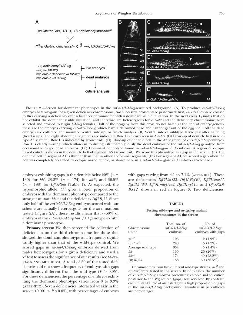
Figure 2.—Screen for dominant phenotypes in the enGal4/UASwg-sensitized background. (A) To produce enGal4/UASwgembryos heterozygous for a given deficiency chromosome, two successive crosses were performed: first, enGal4 flies were crossedto flies carrying a deficiency over a balancer chromosome with a dominant visible mutation. In the next cross, F1 males that donot exhibit the dominant visible mutation, and therefore are heterozygous for enGal4 and the deficiency chromosome, wereselected and crossed to virgin UASwg females. Half of the progeny from this cross do not hatch at the end of embryogenesis:these are the embryos carrying enGal4/UASwg, which have a deformed head and cannot get out of the egg shell. All the deadembryos are collected and mounted ventral side up for cuticle analysis. (B) Ventral side of wild-type larvae just after hatching(head is up). The eight abdominal segments are indicated. Row 1 is clearly seen in A2–A8. (C) Close-up of denticle belt in wild-type A3 segment. Row 1 is indicated by arrowheads. (D) Close-up of denticle belt in the A3 segment of enGal4/UASwg embryos.Row 1 is clearly missing, which allows us to distinguish unambiguously the dead embryos of the enGal4/UASwg genotype fromoccasional wild-type dead embryos. (D�) Dominant phenotype found in enGal4/UASwg[hh�/�] embryos. A region of ectopicnaked cuticle is shown in the denticle belt of segment A3 (arrowhead). We score this phenotype as a gap in the screen. (E) Thedenticle belt in segment A1 is thinner than that in other abdominal segments. (E�) For segment A1, we scored a gap when thebelt was completely breached by ectopic naked cuticle, as shown here in a enGal4/UASwg[hh�/�] embryo (arrowhead).
embryos exhibiting gaps in the denticle belts: 20% (n � with gaps varying from 4.1 to 7.1% (appendix). Theseare deficiencies Df(3L)h-i22, Df(3L)Scf-R6, Df(3L)brm11,130) for hh5, 28.2% (n � 174) for hhAC, and 36.5%
(n � 138) for Df(3R)hh (Table 1). As expected, the Df(3L)VW3, Df(3L)rdgC-co2, Df(3R)ry615, and Df(3R)Dl-hypomorphic allele, hh5, gives a lower proportion of BX12, shown in red in Figure 3. Two deficiencies,embryos with the dominant phenotype compared to thestronger mutant hhAC and the deficiency Df(3R)hh. Sinceonly half of the enGal4/UASwg embryos scored with our TABLE 1scheme are actually heterozygous for the hh mutations Testing wild-type and hedgehog mutanttested (Figure 2A), these results mean that �60% of chromosomes in the screenembryos of the enGal4/UASwg [hh�/�] genotype exhibita dominant phenotype. Total no. of No. of
Chromosome enGal4/UASwg enGal4/UASwgPrimary screen: We then screened the collection oftested embryos embryos with gapsdeficiencies on the third chromosome for those that
showed the dominant phenotype at a frequency signifi- yw 67 106 2 (1.9%)cantly higher than that of the wild-type control. We canton S 248 3 (1.2%)
Average wild type 354 5 (1.4%)scored gaps in enGal4/UASwg embryos derived fromhh 5 130 26 (20%)males heterozygous for a given deficiency and used ahh AC 174 49 (28.2%)� 2 test to assess the significance of our results (see mate-Df(3R)hh 138 50 (36.5%)rials and methods). A total of 59 of the tested defi-
ciencies did not show a frequency of embryos with gaps Chromosomes from two different wild-type strains, yw 67 andcanton S, were tested in the screen. In both cases, the numbersignificantly different from the wild type (P 0.05).of enGal4/UASwg embryos presenting ectopic naked cuticleFor these deficiencies, the percentage of embryos exhib-posterior to the Wg source (gaps) was very low. By contrast,iting the dominant phenotype varies from 0 to 3.3% each mutant allele of hh tested gave a high proportion of gaps
(appendix). Seven deficiencies interacted weakly in the in the enGal4/UASwg background. Numbers in parenthesesare percentages.screen (0.001 � P �0.05), with percentages of embryos
754 S. C. Desbordes, D. Chandraratna and B. Sanson
Fig
ure
3.—
Map
ofth
ede
fici
enci
eson
chro
mos
ome
3te
sted
inth
epr
imar
ysc
reen
.D
efic
ien
cies
from
the
Blo
omin
gton
defi
cien
cyki
tar
epo
siti
oned
ona
draw
ing
ofpo
lyte
ne
chro
mos
ome
3(a
dapt
edfr
omL
ind
sley
and
Zim
m19
92)
acco
rdin
gto
cyto
logi
calp
osit
ion
sgi
ven
inFl
yBas
e(h
ttp:
//fl
ybas
e.bi
o.in
dian
a.ed
u/).
Cyt
olog
ical
posi
tion
sar
ein
dica
ted
unde
rth
edr
awin
g.3L
and
3Rin
dica
tele
ftar
man
dri
ght
arm
ofch
rom
osom
e3,
resp
ecti
vely
.D
efic
ien
cies
that
did
not
inte
ract
are
indi
cate
din
blac
k.T
he
defi
cien
cyth
atun
cove
red
the
hhlo
cus
issh
own
ingr
een
.D
efic
ien
cies
that
inte
ract
edst
ron
gly
(P�
0.00
1)ar
esh
own
inbl
uean
dth
ose
that
inte
ract
edw
eakl
y(0
.001
�P
�0.
05)
are
show
nin
red.

755Regulators of Wingless Distribution
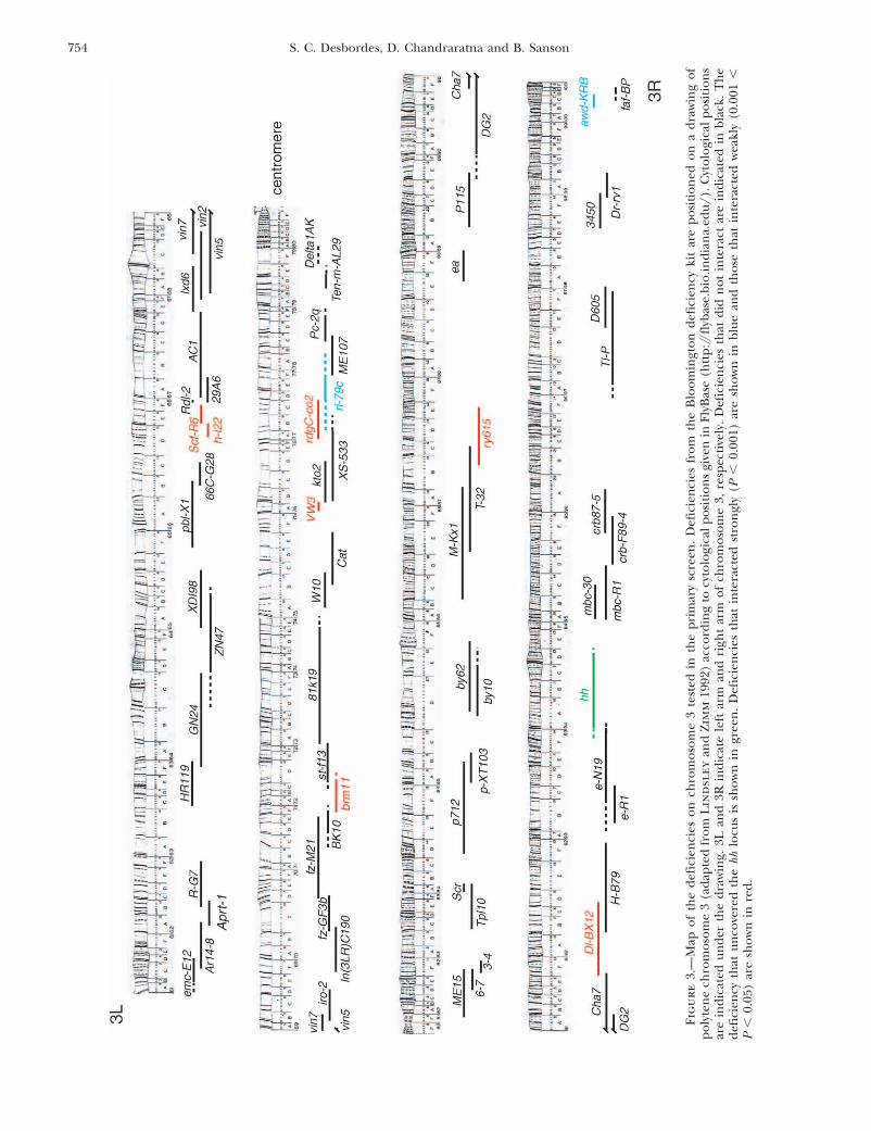
Df(3L)ri-79c and Df(3R)awd-KRB, exhibited a frequency tion of embryos with gaps found for the genotypeenGal4/�; UASwg/� (1.4%) (Table 1). We thus per-of embryos with gaps close to the frequency observed
in enGal4/UASwg embryos derived from males hh�/� formed a secondary screen by testing smaller deficien-cies and point mutations that map to these six regions.(34.2 and 36%, respectively; see appendix). These defi-
ciencies are shown in blue in Figure 3. Table 2 summarizes the findings from the secondaryscreen, and the details about levels of interaction andFor Df(3L)ri-79c and Df(3R)awd-KRB, we tested the
possibility that the dominant phenotype was indepen- cytological positions are given in the appendix.Secondary screen: Region 66: From the primarydent of the enGal4/UASwg background. Of the enGal4/
UASwg embryos, 34.2% (n � 111) derived from Df(3L)ri- screen, two interacting deficiencies overlapped in cyto-logical location 66E: Df(3L)h-i22 (cytological break-79c/� males exhibited gaps in the denticle belts, mainly
in the A4 segment. By crossing Df(3L)ri-79c/� males points 66D10-11 and 66E1-2) and Df(3L)Scf-R6 (66E1-6and 66F1-6). EnGal4/UASwg embryos derived fromwith the yw67 wild-type strain, we found that 25% (n �
60) of the progeny have the same dominant phenotype, Df(3L)h-i22/� or Df(3L)Scf-R6/� males show a domi-nant gap phenotype with a similar frequency of 6.4%thus showing that the dominant phenotype arises inde-
pendently of the enGal4/UASwg-sensitized background. (n � 249) and 7.1% (n � 210), respectively. This sug-gests that the same gene is removed by both deficienciesSince Df(3L)ri-79c removes the gene knirps (kni), we tested
a null mutation in this gene, kni8, and found that 30% of and that the gene should be found in the region ofoverlap between the two deficiencies. Moreover, Df(3L)enGal4/UASwg embryos derived from kni8/� males have
gaps in segment A4. This shows that the dominant phe- Scf-R11, a deficiency that overlaps with the proximal endof Df(3L)Scf-R6, did not interact, suggesting that thenotype detected for Df(3L)ri-79c is due to kni. The excess
naked cuticle found in kni �/� embryos is unlikely to interacting gene is removed by the distal end ofDf(3L)Scf-R6 (Table 2 and appendix). We mapped ge-be a consequence of a change in Wg distribution, since
kni is a gap gene required in the segmentation cascade netically the overlap between Df(3L)h-i22 and Df(3L)Scf-R6 by complementation tests with sequenced P-elementearly in embryogenesis (for a review, see St Johnston
and Nusslein-Volhard 1992). Rather, a perturbation insertions in the region. Df(3L)hi22 failed to comple-ment l(3)10631 (66D14-15), l(3)01629 (66DE1-2), andof the transcription pattern of the segmentation genes
in segment A4 is likely to cause the dominant pheno- l(3)06464 (66E1-2) but complements l(3)10534 (66E2-3). Df(3L)Scf-R6 complemented l(3)10631, but failed totype. In support of this, a compression of the expression
pattern of the pair-rule gene fushi-tarazu has been ob- complement l(3)01629, l(3)06464, and l(3)10534. Thissuggests that the region of overlap between Df(3L)h-i22served in segments A4 and A5 in kni �/� embryos (Car-
roll and Scott 1986). and Df(3L)Scf-R6 falls between l(3)10631 and l(3)10534,which represents an interval of 215 kb on the genomeThe same was found for Df(3R)awd-KRB: 36% (n �
136) of enGal4/UASwg embryos derived from Df(3R)awd- sequence. This interval contains several genes, includ-ing dally, one of the two Drosophila homologs of glypi-KRB /� males exhibit gaps in the denticle belts. Curi-
ously, the gaps are found in alternating segments (not cans (the other one is dally-like ; see below). Dally wasparticularly interesting since it had been implicated inshown) (Desbordes 2004). This dominant phenotype
is also found in Df(3R)awd-KRB /� embryos, showing the regulation of Wg signaling (Lin and Perrimon 1999;Tsuda et al. 1999). A RNA probe corresponding to ex-that it can occur independently of the enGal4/UASwg
background. The excess cuticle found in Df(3R)awd- ons 1–6 of dally cDNA was generated and hybridized topolytene chromosomes from Df(3L)h-i22/� or Df(3L)KRB/� embryos had also been noted by Cox et al.
(2000), and the molecular basis for this phenotype is Scf-R6/� larvae. On wild-type chromosomes, the in situhybridization signal maps at cytological location 66E1-3,unknown. However, because the dominant phenotype
is pair-rule, this again suggests an effect on early segmen- which corresponds to the cytology given in FlyBase (datanot shown) (Desbordes 2004). No signal was observedtation rather than on the regulation of Wg distribution.
We therefore decided to concentrate on the six regions on the deficient chromatid of Df(3L)h-i22/� polytenechromosomes, demonstrating that most dally sequencesdefined by the deficiencies interacting weakly in the
screen. Although the percentage of embryos with gaps are missing in this deficiency (data not shown) (Des-bordes 2004). This shows that Df(3L)h-i22 is an amorphfound for these deficiencies is low (4.1–7.1%), the statisti-
cal test indicates that the interactions detected have a for dally, in contrast to a previous report suggesting thatDf(3L)h-i22 was a hypomorph for this gene (Tsuda etreasonable chance to be significant (0.001 � P � 0.05).
Also, only half of the enGal4/UASwg embryos collected in al. 1999). On Df(3L)Scf-R6/� polytene chromosomes,the signal was clearly reduced on the deficient chroma-a sample are actually heterozygous for the deficiency tested
(the other half are of genotype enGal4/�; UASwg/� ; see tid, suggesting that some of the sequences present be-tween exons 1 and 6 of dally are missing in this deficiencyFigure 2A). This means that the true percentage of
embryos of genotype enGal4/�; UASwg/deficiency show- (data not shown; see Desbordes 2004). Df(3L)Scf-R6 wasgenerated by X-ray irradiation of Scf1, which is an inversioning the dominant phenotype is �8–14% for these defi-
ciencies, which is significantly higher than the propor- (breakpoints 65A12-15; 66F3) (Kopp and Duncan 1997).
756 S. C. Desbordes, D. Chandraratna and B. Sanson
TABLE 2
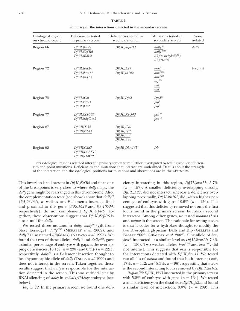
Summary of the interactions detected in the secondary screen
Cytological region Deficiencies tested Deficiencies tested in Mutations tested in Geneon chromosome 3 in primary screen secondary screen secondary screen isolated
Region 66 Df(3L)h-i22 Df(3L)Scf-R11 dally 10 dallyDf(3L)Scf-R6 dally E385
Df(3L)Rdl-2 l(3)06464(dally P2)l(3)01629
Region 72 Df(3L)BK10 Df(3L)A27 brm 2 brm, notDf(3L)brm11 Df(3L)th102 brmT362
Df(3L)st-f13 brmT485
arf 72A
not 3
not 5
Region 75 Df(3L)Cat Df(3L)Dfz2 Dfz2 C1
Df(3L)VW3 pip 1
Df(3L)kto2 pip 2
Region 77 Df(3L)XS-533 Df(3L)XS-543 psn B3
Df(3L)rdgC-co2 psn C4
Region 87 Df(3R)T-32 Df(3R)l26cDf(3R)ry615 Df(3R)ry75
Df(3R)urdDf(3R)lc4a
Region 92 Df(3R)Cha7 Df(3R)Dl-A143 Dl 3
Df(3R)Dl-BX12Df(3R)H-B79
Six cytological regions selected after the primary screen were further investigated by testing smaller deficien-cies and point mutations. Deficiencies and mutations that interact are underlined. Details about the strengthof the interaction and the cytological positions for mutations and aberrations are in the appendix.
This inversion is still present in Df(3L)Scf-R6 and since one ciency interacting in this region, Df(3L)brm11 : 5.7%(n � 157). A smaller deficiency overlapping distally,of the breakpoints is very close to where dally maps, the
dally gene might be rearranged in this chromosome. Also, Df(3L)A27, did not interact, whereas a deficiency over-lapping proximally, Df(3L)th102, did, with a higher per-the complementation tests (see above) show that dally P2
(l(3)06464), as well as two P elements inserted distal centage of embryos with gaps: 18.6% (n � 156). Thissuggested that this deficiency removed not only the firstand proximal to this gene [l(3)01629 and l(3)10534,
respectively], do not complement Df(3L)Scf-R6. To- locus found in the primary screen, but also a secondinteractor. Among other genes, we tested brahma (brm)gether, these observations suggest that Df(3L)Scf-R6 is
also a null for dally. and notum in the screen. The rationale for testing notumis that it codes for a hydrolase thought to modify theWe tested three mutants in dally, dally 10 (gift from
Steve Kerridge), dally E285 (Merabet et al. 2002), and two Drosophila glypicans, Dally and Dlp (Gerlitz andBasler 2002; Giraldez et al. 2002). One allele of brm,dally P2 (also named l(3)06464) (Nakato et al. 1995). We
found that two of these alleles, dally 10 and dally E285, gave brm2, interacted at a similar level as Df(3L)brm11 : 7.3%(n � 150). Two weaker alleles, brmT362 and brmT485, dida similar percentage of embryos with gaps as the overlap-
ping deficiencies, 10.1% (n � 238) and 6.3% (n � 221), not interact. This suggests that brm is responsible forthe interactions detected with Df(3L)brm11. We testedrespectively. dally P2 is a P-element insertion thought to
be a hypomorphic allele of dally (Tsuda et al. 1999) and two alleles of notum and found that both interact (not3,17%, n � 112; not5, 8.3%, n � 96), suggesting that notumdoes not interact in the screen. Taken together, these
results suggest that dally is responsible for the interac- is the second interacting locus removed by Df(3L)th102.Region 75: Df(3L)VW3 interacted in the primary screention detected in the screen. This was verified later by
RNAi silencing of dally in enGal4/UASwg embryos (see with 5.2% of embryos with gaps (n � 134). We testeda small deficiency on the distal side, Df(3L)fz2, and foundbelow).
Region 72: In the primary screen, we found one defi- a similar level of interaction: 8.0% (n � 209). This

757Regulators of Wingless Distribution
deficiency removes fz2, and we therefore tested the only notum, for which at least one mutant allele interacted inour screen. Because brm is a transcriptional regulator,mutant allele available for this gene, fz2C1 (Chen and
Struhl 1999). We did not detect an interaction with we assumed that it is more likely to regulate the expres-sion of segmentation genes than the distribution of Wgthis allele. We also investigated pipe, a gene coding for
a sulphotransferase, which is removed by Df(3L)VW3, (Tamkun et al. 1992; Collins and Treisman 2000). Wethus focused on examining the effect of notum and dallybut two presumed null mutant alleles, pip1 and pip2, did
not interact. on Wg distribution in the embryonic epidermis. Sincedally has a close homolog, dally-like, we also analyzed itsRegion 77: In region 77, we had found two deficiencies
in the primary screen, Df(3L)ri-79c and Df(3L)rdgC-co2. role in this process.Dally: To verify the weak interaction detected withDf(3L)ri-79c interacted strongly (34.2%), but this was
caused by a dominant phenotype due to loss of kni and two alleles of dally, we silenced the gene by RNAi in theenGal4/UASwg background. We found that 44% (n �was independent of the enGal4/UASwg background (see
above). Df(3L)rdgC-co2 interacted weakly: 5.4% (n � 43) of the enGal4/UASwg embryos injected with dallydsRNA exhibit gaps in the denticle belts, which confirms149). We tested one possible candidate gene removed
by this deficiency, Presenilin (Psn), but did not find an its interaction in the screen (compare Table 3 withFigure 4E). By contrast, none of the enGal4/UASwg em-interaction with the two tested alleles, PsnB3 and PsnC4.
We found another deficiency in the region, Df(3L)XS- bryos injected with buffer exhibited any gaps (Table3), and they were indistinguishable from noninjected543, which showed a similar interaction to the primary
screen deficiency: 7.6% (n � 144). However, another enGal4/UASwg embryos (Figure 4B). In the secondaryand primary screen, deficiencies and mutants in dallydeficiency tested in the primary screen, Df(3L)XS533,
which overlaps with a large part of both Df(3L)XS-543 interacted with percentages ranging from 6.3 to 10.1%(appendix). Because only half of the embryos examinedand Df(3L)rdgC-co2, did not interact, thus confusing the
mapping of the interacting locus. Precisely mapped de- in the screen are actually mutant or deficient for dally,12–20% of enGal4/UASwg embryos heterozygous for aficiencies will need to be generated in this region to
pursue the mapping further. dally mutation should exhibit patches of naked cuticle.Knocking down both zygotic and maternal dally mRNAsRegion 87: In region 87, we found one deficiency,
Df(3R)ry615, that interacted very weakly in the primary with RNAi in enGal4/UASwg embryos significantly in-creases this proportion (44%) as expected if dally isscreen (4.1%, n � 169). We then found three smaller
deficiencies in the region that showed slightly higher responsible for the interaction. However, we did notfind any enGal4/UASwg[dally RNAi] embryos exhibitinglevels of interaction: Df(3R)ry75 : 8.3% (n � 120), Df(3R)
urd ; 8.8% (n � 113), and Df(3R)l26C : 9.2% (n � 129). the “full” phenotype found in enGal4/UASwg [hh�/hh�]embryos, where a symmetric expanse of naked cuticleA fourth deficiency in the area, Df(3R)lc4a, did not inter-
act. Although the same level of interaction suggested a is produced anterior as well as posterior to the Engraileddomain (Table 3 and Figure 4C) (Sanson et al. 1999).single locus interacting in region 87, the given cytologi-
cal breakpoints for these four deficiencies did not allow This is expected if dally is not the only factor that regu-lates Wg gradient formation in embryos.us to isolate a small interacting genomic area, suggesting
that either more than one locus interacts or the map- The phenotype of excess naked cuticle seen in en-Gal4/UASwg[dally RNAi] embryos seems to occur inde-ping of the breakpoints for these deficiencies is impre-
cise. pendently of a major role for dally in segmentation.Indeed, dally RNAi in a wild-type background does notRegion 92: In region 92, Df(3R) Dl-BX12 was found to
interact weakly in the primary screen: 6.0% (n � 116). cause segmentation phenotypes at a frequency signifi-cantly different from buffer-injected controls (Des-Since this deficiency removed Delta, we tested one allele
in the screen, Dl3, but did not find an interaction. The bordes and Sanson 2003). This is exemplified by thecontrol in the experiment presented here: half of theoverlapping proximal and distal deficiencies Df(3R)Cha7
and Df(3R)H-b79 did not interact in the screen, sug- embryos injected were of the enGal4/TM3GFP genotype(and thus wild type) and were identified by GFP expres-gesting that the interacting locus is present in the cen-
tral region of Df(3R) Dl-BX12. We tested one smaller sion (the other half of the embryos were enGal4/UASwg).Both buffer-injected and dally dsRNA-injected enGal4/deficiency that removed this central region, Df(3R)Dl-
A143, but did not find an interaction. TM3GFP embryos showed a similar proportion of seg-mentation defects, 23 and 21%, respectively (Table 3).Further investigation of the role of dally, dally-like,
and notum in Wingless distribution: Of six regions se- These segmentation defects are very weak, consisting ofoccasional fusions or deletions of denticle belts, andlected after the primary screen, five (cytology 66, 72,
75, 77, and 87; see Table 2) were found to still interact they are a consequence of the injection process (Des-bordes and Sanson 2003).in the secondary screen. This shows that although weak,
the majority of the interactions detected in the primary In conclusion, these experiments confirm that dallyis the gene responsible for the interaction detected inscreen were meaningful. For two of these regions (cytol-
ogy 66 and 72), we identified three genes, dally, brm, and the screen and suggest that its loss affects the distribu-

758 S. C. Desbordes, D. Chandraratna and B. Sanson
TABLE 3
RNAi silencing of dally and dally-like in enGal4/UASwg embryos
enGal4/UASwg embryos a
Ectopic naked cuticle c enGal4/TM3GFP embryos d
Segmentation Gap Full SegmentationN defects b phenotype phenotype N defects e
Buffer 54 5 (9) 0 0 60 14 (23)dally dsRNA 43 1 (2) 19 (44) 0 42 9 (21)dlp dsRNA 61 23 (38) 9 (15) 24 (39) 71 58 (82)
enGal4 males were crossed to UASwg/TM3GFP females and embryos from the cross were injected with buffer,dally dsRNA, or dlp dsRNA. Embryos were separated in two groups at the end of embryogenesis on the basisof their GFP fluorescence, and their cuticle was examined. Numbers in parentheses are percentages.
a The nonfluorescent embryos are of genotype enGal4/UASwg. Different phenotypes were found.b In all experiments, a proportion of embryos exhibited very weak segmentation defects, which is expected
from damage due to the injection process (see Desbordes and Sanson 2003). However, enGal4/UASwg[dlpRNAi] embryos showed a proportion of segmentation defects significantly higher than the same embryosinjected with buffer or dally dsRNA. Most of these segmentation defects were strong segment polarity pheno-types, suggesting that dlp has another role in segmentation in addition to its requirement for Hh signaling(see text).
c A significant proportion of enGal4/UASwg embryos injected with either dally or dlp dsRNA showed ectopicnaked cuticle. In both experiments, embryos were found with patches of naked cuticle in the ventral denticlebelts (gap phenotype), which was the phenotype looked for in the screen (see Figure 4E). A significantproportion of enGal4/UASwg[RNAi dlp] embryos also showed a phenotype identical to enGal4/UASwg[hh-/hh-]embryos, i.e., naked cuticle on both sides of the Wg source (full phenotype) (see Figure 4D).
d The fluorescent embryos are of genotype enGal4/TM3GFP and thus show a wild-type cuticle pattern.e As shown in Desbordes and Sanson (2003), injection of buffer or dally dsRNA gives a similar proportion
of embryos with very weak segmentation defects, which are due to the injection process. By contrast, mostwild-type embryos injected with dlp dsRNA exhibit strong segment polarity phenotypes, as expected from thefailure in Hh signaling following dlp RNAi (Desbordes and Sanson 2003).
tion of Wg protein in wild-type embryos, without having hh�] embryos (Sanson et al. 1999). The symmetric distri-bution in enGal4/UASwg[dlp RNAi] can be explained ina detectable effect on Wg signaling per se.
Dally-like: At the time we did this work, no mutations two ways: either dlp regulates Wg distribution directly,downstream of Hh signaling, or dlp regulates Hh signal-were available in dlp, so we tested a potential interaction
in our screen by RNAi silencing in the enGal4/UASwg ing, and thus affects Wg distribution indirectly, throughHh. We found that the latter hypothesis is correct. Inbackground. We found that 54% (n � 61) of the enGal4/
UASwg[dlp RNAi] embryos showed excess naked cuticle enGal4/UASwg[hh�/hh�] embryos, endogenous wg ex-pression is lost, because Hh signaling is absent and thein the abdominal segments (whereas enGal4/UASwg
embryos injected with buffer showed no phenotype) maintenance of wg transcription fails (Figure 5, B� vs.C�). If Dlp were regulating Wg distribution directly,(Table 3). Of these 54%, 15% showed gaps in the denti-
cle belts, which was the phenotype looked for in the without affecting Hh signaling, wg endogenous expres-sion would be maintained in enGal4/UASwg[dlp RNAi]screen and is similar to the phenotype seen in enGal4/
UASwg [hh�/�] embryos. Moreover, 39% showed the embryos. We found that this is not the case: wg endoge-nous expression disappears in enGal4/UASwg[dlp RNAi]same phenotype as enGal4/UASwg [hh�/hh�] embryos,
e.g., a symmetric expanse of naked cuticle anterior and embryos as in enGal4/UASwg[hh�/hh�] embryos (Figure5, C and D), suggesting that Dlp is required for eitherposterior to the Engrailed domain (Table 3 and Figure
4, C vs. D). This suggested that the Wg gradient had Hh expression or signaling. We demonstrated elsewhereby additional experiments that Dlp is strictly requiredbecome symmetric in these embryos. To demonstrate that
the cuticle phenotype seen in enGal4/UASwg[dlp RNAi] for Hh signaling in the embryonic epidermis (Des-bordes and Sanson 2003).embryos was due to a change in Wg protein distribution,
we stained stage 12 embryos with a Wg antibody (Fig- We also looked at the effect of dlp RNAi in a wild-type background. In contrast to dally RNAi, dlp RNAiure 5). While in enGal4/UASwg embryos, Wg protein is
never detected posterior to the En domain (Sanson et causes a full segment polarity phenotype (Desbordesand Sanson 2003). This is found as well in the experi-al. 1999); in enGal4/UASwg[dlp RNAi] embryos, Wg is
detected both anterior and posterior to the En domain ment presented here: 82% (n � 71) of the controlembryos, enGal4/TM3GFP, exhibit segmentation defects(Figure 5, E–E���). This symmetric distribution is identi-
cal to the distribution observed in enGal4/UASwg[hh�/ when injected with dlp dsRNA (Table 3). Contrary to

759Regulators of Wingless Distribution
than that in enGal4/UASwg[hh�/hh�] embryos (Figure5, C and C� vs. D and D�). This suggests that en mainte-nance might be impaired in dlp RNAi embryos. More-over, in enGal4/UASwg[RNAi dlp] embryos, 38% exhibitsegmentation defects, whereas the rest exhibit excessnaked cuticle (Table 3). Most of these segmentationdefects are strong segment polarity phenotypes, sug-gesting that normal segmentation fails in these embryos,despite the presence of the artificial autoregulatory loopbetween en and wg. A possibility is that Dlp affects Wgsignaling in addition to being required for Hh signaling(see discussion).
Notum: The two tested alleles of notum, not3 and not5,interact unambiguously in our screen. The interactionwith not3 is stronger than that with not5 (17.0 vs. 8.3%;see appendix), which is consistent with the characteriza-tion of not3 and not5 as amorphic and hypomorphic,respectively (Giraldez et al. 2002). We looked at thephenotype of these mutations in a wild-type back-ground. not5 is homozygous viable so we were able tolook at embryos mutant for the zygotic and maternalcontribution of the gene (i.e., embryos derived fromnot5/not5 mothers). We found that 85% (n � 85) of theembryos have row 1 missing in most denticle belts andoften have a breach of naked cuticle in segment A1.Ten percent of the embryos have stronger phenotypes,with patches of naked cuticle within denticle belts andsometimes complete replacement of denticle belts byFigure 4.—dally-like or dally RNAi in enGal4/UASwg embryos
produces ectopic naked cuticle posterior to the Wingless naked cuticle (Figure 6A). Similar phenotypes, albeitsource. (A) Wild-type larva and (A�) close-up of abdominal weaker, were observed in not3/not3 embryos deriveddenticle belt. Denticle rows 1–6 are visible. (B) enGal4/UASwg from heterozygous mothers (the not3 chromosome islarva: row 1 is completely or partially missing in denticle belts
homozygous embryonic lethal). The presence of a wild-of abdominal segments A2–A7. (B�) Close-up showing a belttype maternal contribution in not3/not3 embryos couldwhere row 1 is missing completely. Rows 2–6 are intact. (C)
enGal4/UASwg[hh�/hh�] embryo: an identical expanse of na- account for the weaker phenotype. To test this, we exam-ked cuticle is found posterior and anterior to the Wg source. ined embryos from mothers bearing germline clones(C�) Close-up showing that rows 2–5 have been replaced by homozygous for the not3 mutant chromosome. Germ-naked cuticle. (D) enGal4/UASwg[dlp RNAi] embryo and close-
line clones bearing females were crossed with not3/up (D�) showing a very similar phenotype to enGal4/UASwg[hh�/TM3GFP males to generate embryos both maternallyhh�] embryos. (E and E�) enGal4/UASwg[dally RNAi] embryo
shows an intermediate phenotype with ectopic naked cuticle and zygotically mutant for notum. The GFP-expressingin some of the denticle belts. embryos, which have a wild-type copy of notum contrib-
uted paternally, hatched and did not show any obviousphenotype. The embryos that do not express GFP, and
what is seen in embryos injected with buffer or dally thus are maternally and zygotically mutants for notum,dsRNA, most of these segmentation defects are actually exhibit a range of severe defects. First, most of the mu-strong segment polarity phenotypes, identical to the phe- tants have an abnormal morphology. For example, outnotypes seen in wg or hh loss of function (not shown; of a collection of 76 mutant embryos, 33 (43%) have asee Desbordes and Sanson 2003). On the basis of our posterior end that is not completely retracted, 9 (12%)study in Desbordes and Sanson (2003), which shows are U-shaped, and 26 (34%) are shaped like a ball (Fig-that Dlp is required for Hh signaling, and the evidence ure 6, B–B��). These phenotypes suggest an early re-from Figure 5, we conclude that the dramatic change quirement of notum in patterning and/or morphogene-of Wg distribution found in enGal4/UASwg[dlp RNAi] sis, which we have not attempted to characterize here.embryos is (at least in part) the indirect consequence We could not analyze reliably the ventral cuticle patternof loss of Hh signaling. of U-shaped or ball-shaped embryos, but we analyzed it
It is worth noting that enGal4/UASwg[RNAi dlp] em- for the 41 remaining embryos: all except one showedbryos do not have a phenotype strictly identical to en- thinner denticle belts in the abdomen, and in addition,Gal4/UASwg[hh�/hh�] embryos. Ectopic wg transcrip- 16 embryos showed either gaps of naked cuticle in the
belts or missing belts (Figure 6B��). Thus the majoritytion in enGal4/UASwg[RNAi dlp] embryos is less robust
760 S. C. Desbordes, D. Chandraratna and B. Sanson
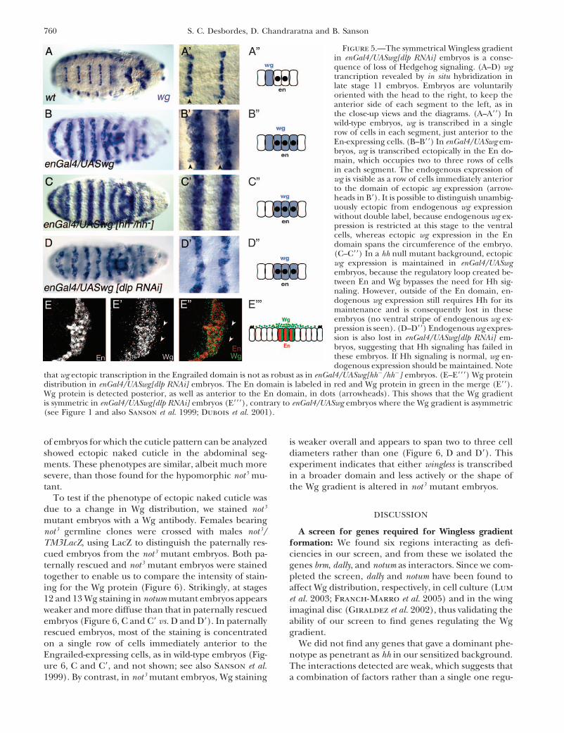
Figure 5.—The symmetrical Wingless gradientin enGal4/UASwg[dlp RNAi] embryos is a conse-quence of loss of Hedgehog signaling. (A–D) wgtrancription revealed by in situ hybridization inlate stage 11 embryos. Embryos are voluntarilyoriented with the head to the right, to keep theanterior side of each segment to the left, as inthe close-up views and the diagrams. (A–A��) Inwild-type embryos, wg is transcribed in a singlerow of cells in each segment, just anterior to theEn-expressing cells. (B–B��) In enGal4/UASwg em-bryos, wg is transcribed ectopically in the En do-main, which occupies two to three rows of cellsin each segment. The endogenous expression ofwg is visible as a row of cells immediately anteriorto the domain of ectopic wg expression (arrow-heads in B�). It is possible to distinguish unambig-uously ectopic from endogenous wg expressionwithout double label, because endogenous wg ex-pression is restricted at this stage to the ventralcells, whereas ectopic wg expression in the Endomain spans the circumference of the embryo.(C–C��) In a hh null mutant background, ectopicwg expression is maintained in enGal4/UASwgembryos, because the regulatory loop created be-tween En and Wg bypasses the need for Hh sig-naling. However, outside of the En domain, en-dogenous wg expression still requires Hh for itsmaintenance and is consequently lost in theseembryos (no ventral stripe of endogenous wg ex-pression is seen). (D–D��) Endogenous wg expres-sion is also lost in enGal4/UASwg[dlp RNAi] em-bryos, suggesting that Hh signaling has failed inthese embryos. If Hh signaling is normal, wg en-dogenous expression should be maintained. Note
that wg ectopic transcription in the Engrailed domain is not as robust as in enGal4/UASwg[hh�/hh�] embryos. (E–E���) Wg proteindistribution in enGal4/UASwg[dlp RNAi] embryos. The En domain is labeled in red and Wg protein in green in the merge (E��).Wg protein is detected posterior, as well as anterior to the En domain, in dots (arrowheads). This shows that the Wg gradientis symmetric in enGal4/UASwg[dlp RNAi] embryos (E���), contrary to enGal4/UASwg embryos where the Wg gradient is asymmetric(see Figure 1 and also Sanson et al. 1999; Dubois et al. 2001).
of embryos for which the cuticle pattern can be analyzed is weaker overall and appears to span two to three celldiameters rather than one (Figure 6, D and D�). Thisshowed ectopic naked cuticle in the abdominal seg-
ments. These phenotypes are similar, albeit much more experiment indicates that either wingless is transcribedin a broader domain and less actively or the shape ofsevere, than those found for the hypomorphic not5 mu-
tant. the Wg gradient is altered in not3 mutant embryos.To test if the phenotype of ectopic naked cuticle was
due to a change in Wg distribution, we stained not3
DISCUSSIONmutant embryos with a Wg antibody. Females bearingnot3 germline clones were crossed with males not3/ A screen for genes required for Wingless gradient
formation: We found six regions interacting as defi-TM3LacZ, using LacZ to distinguish the paternally res-cued embryos from the not3 mutant embryos. Both pa- ciencies in our screen, and from these we isolated the
genes brm, dally, and notum as interactors. Since we com-ternally rescued and not3 mutant embryos were stainedtogether to enable us to compare the intensity of stain- pleted the screen, dally and notum have been found to
affect Wg distribution, respectively, in cell culture (Luming for the Wg protein (Figure 6). Strikingly, at stages12 and 13 Wg staining in notum mutant embryos appears et al. 2003; Franch-Marro et al. 2005) and in the wing
imaginal disc (Giraldez et al. 2002), thus validating theweaker and more diffuse than that in paternally rescuedembryos (Figure 6, C and C� vs. D and D�). In paternally ability of our screen to find genes regulating the Wg
gradient.rescued embryos, most of the staining is concentratedon a single row of cells immediately anterior to the We did not find any genes that gave a dominant phe-
notype as penetrant as hh in our sensitized background.Engrailed-expressing cells, as in wild-type embryos (Fig-ure 6, C and C�, and not shown; see also Sanson et al. The interactions detected are weak, which suggests that
a combination of factors rather than a single one regu-1999). By contrast, in not3 mutant embryos, Wg staining

761Regulators of Wingless Distribution
Figure 6.—Embryos mutant for notum showectopic naked cuticle and a broader Wg gradient.(A) not 5 embryo showing excess naked cuticle insegments A1, A2, A3, A5, and A6. Note that thedenticle belt in segment A5 has been completelyreplaced by naked cuticle (arrowhead). (B–B��)Embryos derived from not 3 germline clones; i.e.,embryos both maternally and zygotically mutantfor notum exhibit a range of cuticle defects. Someembryos are ball shaped (B) or U shaped (B�),while others have an incompletely retracted poste-rior end (B��). In addition, most embryos for whichthe cuticle pattern can be analyzed show a nar-rowing of the ventral denticle belts, with patches ofnaked cuticle in the belts or denticle belts missing(arrowhead in B��). (C and D) Stage 12 embryosderived from females bearing not3 germlines clonesand not3/TM3LacZ males were stained for Wg (Cand D and green signal in C� and D�). These em-bryos also carry enGal4.UASGFP to label the engraileddomain using an antibody against GFP (red signalin C� and D�). LacZ-positive embryos are paternallyrescued and show a Wg gradient indistinguishablefrom wild-type embryos (C and C�). LacZ-negativeembryos are maternally and zygotically mutant fornotum and show a Wg gradient that is broader andweaker (D and D�).
late the shape of the Wg gradient in embryos, down- the interactors. Indeed, the current deficiency kit doesnot cover the whole genome and many deficienciesstream of Hh signaling. It also seems likely that most
factors involved are maternally provided, as is the case either are imprecisely mapped or represent complexaberrations [such as Df(3L)ScfR6], which renders analy-for dally and notum. Our screen did not remove any
maternal contribution since we tested heterozygous sis difficult. Also, the current deficiency kit containssome very large deficiencies, which might not interactmales for a given deficiency chromosome, due to the
constraints in the reagents available at the time (see because they remove modifiers. Another problem withlarge deficiencies is that some are haplo-insufficient andmaterials and methods). Hence, the wild-type mater-
nal contribution would weaken the interactions de- could not be tested in the screen because heterozygousmales did not survive or were sterile.tected for genes contributed maternally and zygotically,
such as dally and notum. This can also explain why not Another use of the enGal4/UASwg-sensitized back-ground is to test the role of candidate genes in Wgall the mutant alleles tested for dally interact: weak mod-
ifiers present elsewhere on the third chromosome of gradient formation. For example, deep orange (dor) andChlathrin (Chc), two genes required for lysosomal degra-mutant alleles might be enough to mask an interaction
already diminished by the maternal contribution. dation, have already been found to produce ectopicnaked cuticle when their zygotic contribution is re-However, although weak, most interactions detected
in our primary screen were confirmed in the secondary moved in the enGal4/UASwg background (Dubois et al.2001). This contributed to the demonstration that Wgscreen and we believe that it can be applied to the
whole genome to find additional factors regulating Wg is degraded at a faster rate posterior to the En domain.It is worth noting that dor and Chc zygotic mutants dogradient formation. In light of our results, two changes
could be made to improve screen efficiency. First, fe- not exhibit any cuticle phenotype and thus do not revealtheir role in Wg distribution without the sensitized back-males heterozygous for a given deficiency, instead of
males, could be used to remove half of the maternal ground. Moreover, the maternal contribution of dorand Chc cannot be removed genetically in the maternalcontribution in addition to halving the zygotic contribu-
tion (note that F1 UASwg/�; Deficiency/� females should germline, since these genes are required for oogenesis.Thus the use of the enGal4/UASwg background providesbe used rather than enGal4/�; Deficiency/� females,
since the progeny of the latter display an interfering an assay for genes that could not be tested otherwise inembryonic gradient formation.phenotype; see materials and methods). This should
uncover maternal factors that might have been over- Role of Dally-like in Wingless gradient formation:RNAi silencing of dally or dlp in enGal4/UASwg embryoslooked in our screen. Second, the use of precisely
mapped deficiencies from the new Drosdel collection generated ectopic naked cuticle in both cases, but dlpsilencing had a much stronger effect (Figure 4 and(Ryder et al. 2004), instead of the current deficiency
collection, should improve dramatically the mapping of Table 3). This strong phenotype is explained at least in

762 S. C. Desbordes, D. Chandraratna and B. Sanson
part by the strict requirement for Dlp in Hh signaling and Desbordes and Sanson 2003) or zygotically andmaternally mutant for dally (Franch-Marro et al. 2005)(Desbordes and Sanson 2003) (Figure 5). In enGal4/
UASwg [dlp RNAi] embryos, the failure of Hh signaling do not exhibit any segment polarity phenotype. How-ever, Dally might cooperate with Dlp to modulate Wggenerates a symmetrical gradient of Wg protein and
results in a phenotype identical to enGal4/UASwg [hh�/ signaling because embryos mutant for both dally anddlp have a stronger segment polarity phenotype than hhhh�] embryos (Figure 5) (Sanson et al. 1999).
Dlp could be regulating Wg distribution as well, but mutants (Dlp is strictly required for Hh signaling) andphenocopy the phenotype of wg hh double mutantsits requirement for Hedgehog signaling makes this dif-
ficult to assess. We noted, however, that a significant (Franch-Marro et al. 2005).Role of Notum in Wingless gradient formation: Halv-proportion of enGal4/UASwg [dlp RNAi] embryos shows
a segment polarity phenotype (Table 3) and that the ing the dose of notum in enGal4/UASwg embryos gener-ates ectopic naked cuticle (appendix). Moreover, notumstripe of wg transcription in the en domain of these
embryos is weak compared to the control enGal4/UASwg mutants exhibit variable amounts of excess naked cuti-cle (Figure 6) (see also Giraldez et al. 2002). Consistent[hh�/hh�] embryos (Figure 5, C and C� vs. D and D�).
The simplest way to explain these phenotypes is if en with this effect, overexpression of Notum in embryosinhibits the formation of naked cuticle (Giraldez etexpression is somewhat compromised in enGal4/UASwg
[dlp RNAi] embryos. Surprisingly, this effect would be al. 2002). We found that in embryos maternally andzygotically mutant for notum, the Wg gradient appearsspecific to en, since naked cuticle can still form in the
absence of dlp (Figure 4D) (Desbordes and Sanson broader than in wild-type embryos (Figure 6, D and D�).This broadening is seen clearly for the anterior Wg2003; Franch-Marro et al. 2005). It is possible that in
contrast to naked cuticle specification, En maintenance gradient, whereas it is less easy to observe posterior tothe Wg source. This is expected if the fast degradationrequires high levels of Wg. In support of this idea, only
the cells immediately adjacent to the Wg-expressing cells of Wg occurring posterior to the source (Dubois etal. 2001) is operating normally in notum mutants, thusare able to maintain En expression at stage 10 (Vincent
and Lawrence 1994). Thus, it is possible that Dlp is masking changes in the distribution of Wg protein. Wesuggest that in notum mutants more Wg is available forrequired to maintain a high concentration of Wg at the
surface of the Wg-expressing cells and that this contrib- signaling before it is targeted for degradation, resultingin the formation of ectopic naked cuticle posterior toutes to the maintenance of En expression in adjoining
cells. In the absence of Dlp, Wg would be released from the Wg source. A broadening of the Wg gradient hasalso been found for notum mutant clones in the wingthe cells, thus compromising En maintenance without
affecting naked cuticle specification (this might even disc (Giraldez et al. 2002). In contrast to what has beenobserved in the wing disc, however, the broader Wgpromote naked cuticle specification over a longer dis-
tance). Consistent with this hypothesis, very recent re- gradient in notum mutant embryos also appears weaker(Figure 6, D and D�). This does not seem to have ansults show that in the wing disc Dlp has opposite effects
on short-range and long-range Wg signaling (Kirkpat- impact on signaling, however, since no segment polarityphenotypes were observed in notum mutant embryos,rick et al. 2004; Kreuger et al. 2004; Franch-Marro
et al. 2005). suggesting that engrailed maintenance is normal.Notum has homologies to pectin acetylesterases andRole of Dally in Wingless gradient formation: The
excess naked cuticle observed in enGal4/UASwg [dally appears to modify the activity of glypicans, in particularDlp (Gerlitz and Basler 2002; Giraldez et al. 2002).RNAi] embryos could be due to a partial requirement
in Hh signaling or, alternatively, to a direct effect on Recent work indicates that Notum directly or indirectlyinduces cleavage of the glycosylphosphatidylinositol an-Wg distribution. Both hypotheses are plausible since
Dally is required redundantly with Dlp for Hh signaling chor of Dlp (but not Dally), leading to shedding of thisglypican from the cell surface (Kreuger et al. 2004). Itin wing discs (Han et al. 2004a) and recent data indicate
that Dally can affect Wg distribution (Lum et al. 2003; has been hypothesized that Wg bound to Dlp could bemade unavailable to signaling because of shedding fromFranch-Marro et al. 2005). In Drosophila cultured
cells, Dally transfection causes accumulation of exoge- the cell surface, thus explaining the increase in Wgactivity associated with loss of Notum. What is the sub-nous Wg at the cell surface (Franch-Marro et al. 2005),
whereas dally RNAi results in loss of Wg staining (see strate of Notum in the embryo? In the epidermis, bothnotum and dlp are upregulated in the cells that respondnote 20 in Lum et al. 2003). Taken together, these obser-
vations suggest that Dally is able to retain Wg protein to Wg (Khare and Baumgartner 2000; Giraldez etal. 2002). By contrast, dally is downregulated in the sameat the cell surface. It may be that, in the embryo, loss
of dally releases Wg and allows it to signal at a longer cells, suggesting that Wg signaling might repress dallyexpression (Tsuda et al. 1999). This could reflect arange to make naked cuticle.
This effect of Dally seems to be independent of a different or even opposite role of the two glypicans inWg gradient formation. Since in the wing disc Notumrequirement for Wg signaling per se, since embryos ei-
ther depleted of dally mRNA following RNAi (this study appears to act on Dlp and not Dally, the effects that we

763Regulators of Wingless Distribution
Giraldez, A. J., R. R. Copley and S. M. Cohen, 2002 HSPG modifi-observe could be attributed to the regulation of Dlp bycation by the secreted enzyme Notum shapes the Wingless mor-
Notum. If this is true, this would reveal a role for Dlp phogen gradient. Dev. Cell 2: 667–676.Gonzalez, F., L. Swales, A. Bejsovec, H. Skaer and A. Martinezin Wg gradient formation in the embryo, in addition
Arias, 1991 Secretion and movement of wingless protein into its role in Hh signaling. Interestingly, in addition tothe epidermis of the Drosophila embryo. Mech. Dev. 35: 43–54.
changes in the cuticle pattern, loss of notum causes se- Gonzalez-Gaitan, M., 2003 Endocytic trafficking during Drosoph-ila development. Mech. Dev. 120: 1265–1282.vere defects in the patterning and/or morphogenesis
Greco, V., M. Hannus and S. Eaton, 2001 Argosomes: a potentialof the early embryo (Figure 6), suggesting that Notumvehicle for the spread of morphogens through epithelia. Cell
may have substrates other than the glypicans. 106: 633–645.Han, C., T. Y. Belenkaya, B. Wang and X. Lin, 2004a DrosophilaWe thank Clive McKimmie for his early contribution to the screen,
glypicans control the cell-to-cell movement of Hedgehog by aMargit Pal for the in situ hybridizations on polytene chromosomes,dynamin-independent process. Development 131: 601–611.
Daniel St. Johnston and Jean-Paul Vincent for their comments on the Han, C., T. Y. Belenkaya, M. Khodoun, M. Tauchi and X. Lin,manuscript, and Francois Balloux for advice on the statistics. We also 2004b Distinct and collaborative roles of Drosophila EXT familythank K. Cadigan, S. Cohen, M. Fortini, D. Kalderon, S. Kerridge, proteins in morphogen signalling and gradient formation. Devel-D. St Johnston, G. Struhl, J. Treisman, the Hybridoma Bank, the opment 131: 1563–1575.
Hatini, V., and S. DiNardo, 2001 Divide and conquer: patternBloomington and Umea stock centers, and FlyBase for fly strains,formation in Drosophila embryonic epidermis. Trends Genet 17:reagents, and valuable information. This work was supported by a574–579.Career Development Award from the Wellcome Trust (054525/Z/
Jowett, T., 1997 Tissue in Situ Hybridization: Methods in Animal Devel-98) to B.S. B.S. was also supported by the Cambridge Newton Trustopment. John Wiley & Sons, New York.and S.D. by the Cambridge European Trust.
Khare, N., and S. Baumgartner, 2000 Dally-like protein, a newDrosophila glypican with expression overlapping with wingless.Mech. Dev. 99: 199–202.
Kirkpatrick, C. A., B. D. Dimitroff, J. M. Rawson and S. B. Selleck,LITERATURE CITED2004 Spatial regulation of Wingless morphogen distributionand signaling by Dally-like protein. Dev. Cell 7: 513–523.Baeg, G. H., X. Lin, N. Khare, S. Baumgartner and N. Perrimon,
2001 Heparan sulfate proteoglycans are critical for the organiza- Kopp, A., and I. Duncan, 1997 Control of cell fate and polarity inthe adult abdominal segments of Drosophila by optomotor-blind.tion of the extracellular distribution of Wingless. Development
128: 87–94. Development 124: 3715–3726.Kreuger, J., L. Perez, A. J. Giraldez and S. M. Cohen, 2004 Oppos-Bejsovec, A., and E. Wieschaus, 1995 Signaling activities of the
Drosophila wingless gene are separately mutable and appear to ing activities of Dally-like glypican at high and low levels of Wing-less morphogen activity. Dev. Cell 7: 503–512.be transduced at the cell surface. Genetics 139: 309–320.
Bellaiche, Y., I. The and N. Perrimon, 1998 Tout-velu is a Drosoph- Lawrence, P. A., B. Sanson and J. P. Vincent, 1996 Compartments,wingless and engrailed: patterning the ventral epidermis of Dro-ila homologue of the putative tumour suppressor EXT-1 and is
needed for Hh diffusion. Nature 394: 85–88. sophila embryos. Development 122: 4095–4103.Lecourtois, M., C. Alexandre, L. Dubois and J. P. Vincent, 2001Cadigan, K. M., M. P. Fish, E. J. Rulifson and R. Nusse, 1998 Wing-
less repression of Drosophila frizzled 2 expression shapes the Wingless capture by Frizzled and Frizzled2 in Drosophila em-bryos. Dev. Biol. 235: 467–475.Wingless morphogen gradient in the wing. Cell 93: 767–777.
Carroll, S. B., and M. P. Scott, 1986 Zygotically active genes that Lecuit, T., and S. M. Cohen, 1998 Dpp receptor levels contributeto shaping the Dpp morphogen gradient in the Drosophila wingaffect the spatial expression of the fushi tarazu segmentation
gene during early Drosophila embryogenesis. Cell 45: 113–126. imaginal disc. Development 125: 4901–4907.Lin, X., and N. Perrimon, 1999 Dally cooperates with DrosophilaChen, C. M., and G. Struhl, 1999 Wingless transduction by the
Frizzled and Frizzled2 proteins of Drosophila. Development 126: Frizzled 2 to transduce Wingless signalling. Nature 400: 281–284.Lindsley, D. L., and G. Zimm, 1992 The Genome of Drosophila melano-5441–5452.
Chen, Y., and G. Struhl, 1996 Dual roles for patched in sequester- gaster. Academic Press, New York.Lum, L., S. Yao, B. Mozer, A. Rovescalli, D. Von Kessler et al.,ing and transducing Hedgehog. Cell 87: 553–563.
Collins, R. T., and J. E. Treisman, 2000 Osa-containing Brahma 2003 Identification of Hedgehog pathway components by RNAiin Drosophila cultured cells. Science 299: 2039–2045.chromatin remodeling complexes are required for the repression
of wingless target genes. Genes Dev. 14: 3140–3152. Merabet, S., F. Catala, J. Pradel and Y. Graba, 2002 A greenfluorescent protein reporter genetic screen that identifies mod-Cox, R. T., D. G. McEwen, D. L. Myster, R. J. Duronio, J. Loureiro
et al., 2000 A screen for mutations that suppress the phenotype ifiers of Hox gene function in the Drosophila embryo. Genetics162: 189–202.of Drosophila armadillo, the -catenin homolog. Genetics 155:
1725–1740. Moline, M. M., C. Southern and A. Bejsovec, 1999 Directionalityof wingless protein transport influences epidermal patterning inDesbordes, S., 2004 The role of the Drosophila glypicans in Wing-
less and Hedgehog signalling during embryogenesis. Ph.D. The- the Drosophila embryo. Development 126: 4375–4384.Nakato, H., T. A. Futch and S. B. Selleck, 1995 The divisionsis, University of Cambridge, Cambridge, UK.
Desbordes, S. C., and B. Sanson, 2003 The glypican Dally-like is abnormally delayed (dally) gene: a putative integral membraneproteoglycan required for cell division patterning during postem-required for Hedgehog signalling in the embryonic epidermis
of Drosophila. Development 130: 6245–6255. bryonic development of the nervous system in Drosophila. Devel-opment 121: 3687–3702.Dougan, S., and S. DiNardo, 1992 Drosophila wingless generates
cell type diversity among engrailed expressing cells. Nature 360: Neumann, C. J., and S. M. Cohen, 1997 Long-range action of Wing-less organizes the dorsal-ventral axis of the Drosophila wing.347–350.
Dubois, L., M. Lecourtois, C. Alexandre, E. Hirst and J. P. Vin- Development 124: 871–880.O’Keefe, L., S. T. Dougan, L. Gabay, E. Raz, B. Z. Shilo et al., 1997cent, 2001 Regulated endocytic routing modulates wingless sig-
naling in Drosophila embryos. Cell 105: 613–624. Spitz and Wingless, emanating from distinct borders, cooperate toestablish cell fate across the Engrailed domain in the DrosophilaFranch-Marro, X., O. Marchand, E. Piddini, S. Ricardo, C. Alex-
andre et al., 2005 Glypicans shunt the Wingless signal between epidermis. Development 124: 4837–4845.Payre, F., A. Vincent and S. Carreno, 1999 ovo/svb integrateslocal signalling and further transport. Development 132: 659–
666. Wingless and DER pathways to control epidermis differentiation.Nature 400: 271–275.Freeman, M., 2000 Feedback control of intercellular signalling in
development. Nature 408: 313–319. Pfeiffer, S., C. Alexandre, M. Calleja and J. P. Vincent, 2000 Theprogeny of wingless-expressing cells deliver the signal at a distanceGerlitz, O., and K. Basler, 2002 Wingful, an extracellular feedback
inhibitor of Wingless. Genes Dev. 16: 1055–1059. in Drosophila embryos. Curr. Biol. 10: 321–324.

764 S. C. Desbordes, D. Chandraratna and B. Sanson
Pfeiffer, S., S. Ricardo, J. B. Manneville, C. Alexandre and J. P. through synthesis of heparan sulfate proteoglycans. Development131: 73–82.Vincent, 2002 Producing cells retain and recycle Wingless in
Tamkun, J. W., R. Deuring, M. P. Scott, M. Kissinger, A. M. Patta-Drosophila embryos. Curr. Biol. 12: 957–962.tucci et al., 1992 brahma: a regulator of Drosophila homeoticReichsman, F., L. Smith and S. Cumberledge, 1996 Glycosamino-genes structurally related to the yeast transcriptional activatorglycans can modulate extracellular localization of the winglessSNF2/SWI2. Cell 68: 561–572.protein and promote signal transduction. J. Cell Biol. 135: 819–
Tanimoto, H., S. Itoh, P. ten Dijke and T. Tabata, 2000 Hedge-827.hog creates a gradient of DPP activity in Drosophila wing imaginalRyder, E., F. Blows, M. Ashburner, R. Bautista-Llacer, D. Coul-discs. Mol. Cell 5: 59–71.son et al., 2004 The DrosDel collection: a set of P-element inser-
The, I., Y. Bellaiche and N. Perrimon, 1999 Hedgehog movementtions for generating custom chromosomal aberrations in Drosoph-is regulated through tout velu-dependent synthesis of a heparanila melanogaster. Genetics 167: 797–813.sulfate proteoglycan. Mol. Cell 4: 633–639.Sanson, B., 2001 Generating patterns from fields of cells. Examples
Tsuda, M., K. Kamimura, H. Nakato, M. Archer, W. Staatz etfrom Drosophila segmentation. EMBO Rep. 2: 1083–1088.al., 1999 The cell-surface proteoglycan Dally regulates WinglessSanson, B., C. Alexandre, N. Fascetti and J. P. Vincent, 1999signalling in Drosophila. Nature 400: 276–280.Engrailed and hedgehog make the range of Wingless asymmetric
van den Heuvel, M., R. Nusse, P. Johnston and P. A. Lawrence,in Drosophila embryos. Cell 98: 207–216.1989 Distribution of the wingless gene product in DrosophilaSeto, E. S., and H. J. Bellen, 2004 The ins and outs of Winglessembryos: a protein involved in cell-cell communication. Cell 59:signaling. Trends Cell Biol. 14: 45–53. 739–749.Simmonds, A. J., G. dosSantos, I. Livne-Bar and H. M. Krause, Vincent, J. P., and L. Dubois, 2002 Morphogen transport along
2001 Apical localization of wingless transcripts is required for epithelia, an integrated trafficking problem. Dev. Cell 3: 615–623.wingless signaling. Cell 105: 197–207. Vincent, J. P., and P. A. Lawrence, 1994 Drosophila wingless sus-
St Johnston, D., and C. Nusslein-Volhard, 1992 The origin of tains engrailed expression only in adjoining cells: evidence frompattern and polarity in the Drosophila embryo. Cell 68: 201–219. mosaic embryos. Cell 77: 909–915.
Strigini, M., and S. M. Cohen, 2000 Wingless gradient formation Zecca, M., K. Basler and G. Struhl, 1996 Direct and long-rangein the Drosophila wing. Curr. Biol. 10: 293–300. action of a wingless morphogen gradient. Cell 87: 833–844.
Takei, Y., Y. Ozawa, M. Sato, A. Watanabe and T. Tabata, 2003Three Drosophila EXT genes shape morphogen gradients Communicating editor: K. V. Anderson
APPENDIX
Deficiencies and mutations tested in the primary and secondary screens
% enGal4/UASwgDeficiency Cytology embryos with gaps Interaction
Primary screenDf(3L)emc-E12 61A; 61D3 2.7 (n � 111) NoDf(3L)Ar14-8 61C5-8; 62A8 1.6 (n � 126) NoDf(3L)Aprt-1 62A10-B1; 62D2-5 1.7 (n � 275) NoDf(3L)R-G7 62B9; 62E7 1.5 (n � 136) NoDf(3L)HR119 63C2;63F7 3.3 (n � 121) NoDf(3L)GN24 63F4-7;64C13-15 1.2 (n � 86) NoDf(3L)ZN47 64C;65C 1.0 (n � 98) NoDf(3L)XDI98 65A2;65E1 0.9 (n � 109) NoDf(3L)pbl-X1 65F3;66B10 3.0 (n � 101) NoDf(3L)66C-G28 66B8-9;66C9-10 0 (n � 123) NoDf(3L)h-i22 66D1011;66E1-2 6.4 (n � 249) Yes (P � 0.01)Df(3L)Scf-R6 66E1-6;66F1-6 7.1 (n � 210) Yes (P � 0.01)Df(3L)Rdl-2 66F5;66F5 3.3 (n � 153) NoDf(3L)29A6 66F5;67B1 2.2 (n � 89) NoDf(3L)AC1 67A2;67D11-13 0 (n � 74) NoDf(3L)lxd6 67E1-2;68C1-2 0 (n � 109) NoDf(3L)vin2 67F2-3;68D6 2.7 (n � 113) NoDf(3L)vin5 68A2-3;69A1-3 0 (n � 92) NoDf(3L)vin7 68C8-11;69B4-5 0 (n � 92) NoDf(3L)iro-2 69B1-5;69D1-6 0.8 (n � 124) NoIn(3LR)C190 69F3-4;70C3-4 � 89;89 0 (n � 83) NoDf(3L)fz-GF3b 70C2;70D5 1.2 (n � 85) NoDf(3L)fz-M21 70D2-3;71E4-5 2.1 (n � 94) NoDf(3L)BK10 71C;71F 1.2 (n � 81) NoDf(3L)brm11 71F1-4;72D1-10 5.7 (n � 157) Yes (P � 0.02)Df(3L)st-f13 72C1-D1;73A3-4 1.6 (n � 127) NoDf(3L)81k19 73A3;74F 0 (n � 88) NoDf(3L)W10 75A6-7;75C1-2 1.7 (n � 119) NoDf(3L)Cat 75B8;75F1 0 (n � 127) NoDf(3L)VW3 76A3;76B2 5.2 (n � 134) Yes (P � 0.02)
(continued)

765Regulators of Wingless Distribution
APPENDIX
(Continued)
% enGal4/UASwgDeficiency Cytology embryos with gaps Interaction
Df(3L)kto2 76B1-2;76D5 2.4 (n � 83) NoDf(3L)XS-533 76B4;77B 2.3 (n � 130) NoDf(3L)rdgC-co2 77A1;77D1 5.4 (n � 149) Yes (P � 0.02)Df(3L)ri-79c 77B-C;77F-78A 34.2 (n � 111) Yes (P � 0.001)Df(3L)ME107 77F3;78C8-9 0.9 (n � 107) NoDf(3L)Pc-2q 78C5-6;78E3-79A1 2.0 (n � 153) NoDf(3L)Ten-m-AL29 79C1-3;79E3-8 2.9 (n � 104) NoDf(3L)Delta1AK 79F;80A 2.1 (n � 96) NoDf(3R)ME15 81F3-6;82F5-7 1.9 (n � 104) NoDf(3R)6-7 82D3-8;82F3-6 1.2 (n � 81) NoDf(3R)3-4 82F3-4;82F10-11 1.3 (n � 76) NoDf(3R)Tpl10, 83C1-2;84B1-2 0 (n � 77) NoDf(3R)Scr 84A1-2;84B1-2 0 (n � 78) NoDf(3R)p712 84D4-6;85B6 0 (n � 145) NoDf(3R)p-XT103 85A2;85C1-2 0 (n � 102) NoDf(3R)by10 85D8-12;85E7-F1 0.8 (n � 122) NoDf(3R)by62 85D11-14;85F6 0 (n � 91) NoDf(3R)M-Kx1 86C1;87B1-5 0 (n � 79) NoDf(3R)T-32 86E2-4; 87C6-7 1.2 (n � 82) NoDf(3R)ry615 87B11-13;87E8-11 4.1 (n � 169) Yes (P � 0.05)Df(3R)ea 88E7-13;89A1 2.1 (n � 141) NoDf(3R)P115 89B7-8;89E7-8;20 2.8 (n � 145) NoDf(3R)DG2 89E1-F4;91B1-2 1.9 (n � 104) NoDf(3R)Cha7 90F1-4;91F5 2.0 (n � 98) NoDf(3R)Dl-BX12 91F1-2;92D3-6 6.0 (n � 116) Yes (P � 0.01)Df(3R)H-B79 92B3;92F13 2.7 (n � 113) NoDf(3R)e-R1 93B6-7;93D2 2.9 (n � 103) NoDf(3R)e-N19 93B2-13; 94A3-8 1.6 (n � 127) NoDf(3R)mbc-30 95A5-7;95C10-11 2.0 (n � 101) NoDf(3R)mbc-R1 95A5-7;95D6-11 2.5 (n � 79) NoDf(3R)crb-F89-4 95D7-11;95F15 0 (n � 105) NoDf(3R)crb87-5 95F7;96A17-18 1.1 (n � 88) NoDf(3R)Tl-P 97A;98A1-2 0 (n � 95) NoDf(3R)D605 97E3;98A5 0.9 (n � 115) NoDf(3R)3450 98E3;99A6-8 0 (n � 109) NoDf(3R)Dr-rv1 99A1-2;99B6-11 1.4 (n � 72) NoDf(3R)awd-KRB 100D1;100D3-4 36.0 (n � 136) Yes (P � 0.001)Df(3R)faf-BP 100D;100F5 0.9 (n � 107) No
Secondary screenRegion 66
Df(3L)Scf-R11 66E3-4; 66F1-2 1.8 (n � 172) Nodally 10 66E1-3 10.1 (P � 0238) Yes (P � 0.001)dally E385 66E1-3 6.3 (P � 0221) Yes (P � 0.05)l(3)06464(dally P2) 66E1-2 2.5 (n � 80) Nol(3)01629 66E1-2 1.1 (n � 347) No
Region 72Df(3L)A27 70E; 72A3-4 1.0 (n � 99) NoDf(3L)th102 72B1-72D12 18.6 (n � 156) Yes (P � 0.001)brm2 72C1 7.3 (n � 150) Yes (P � 0.001)brmT362 72C1 1.9 (n � 103) NobrmT485 72C1 0 (n � 139) Noarf 72A 72C1 2.0 (n � 108) NoNot 3 72D1 17.0 (n � 112) Yes (P � 0.001)Not 5 72D1 8.3 (n � 96) Yes (P � 0.001)
Region 75Df(3L)Dfz2 75F10-11;76A1-5 8.0 (n � 209) Yes (P � 0.001)
(continued)

766 S. C. Desbordes, D. Chandraratna and B. Sanson
APPENDIX
(Continued)
% enGal4/UASwgDeficiency Cytology embryos with gaps Interaction
Dfz2 C1 75F9-10 0 (n � 92) Nopip1 76A3 0 (n � 99) Nopip 2 76A3 3.2 (n � 124) No
Region 77Df(3L)XS-543 76B; 77A 7.6 (n � 144) Yes (P � 0.001)psn B3 77B9-C1 1.5 (n � 135) NopsnC4 77B9-C1 1.0 (n � 111) No
Region 87Df(3R)l26c 87E1; 87F11 9.2 (n � 129) Yes (P � 0.001)Df(3R)ry75 87D2; 87D14 8.3 (n � 120) Yes (P � 0.001)Df(3R)urd 87F1-15 8.8 (n � 113) Yes (P � 0.001)Df(3R)lc4a 87E5-4; E11 1.7 (n � 113) No
Region 92Df(3R)Dl-A143 91F13; 92A2-3 3.3 (n � 151) NoDl 3 92A1-2 3.3 (n � 123) No
The cytology of each deficiency and gene is given according to FlyBase (http://flybase.bio.indiana.edu/).The percentage of enGal4/UASwg embryos presenting ectopic naked cuticle (gaps) is given for each chromosometested. A � 2 was performed for each result, using the number of embryos with gaps found with wild-typechromosomes as the expected frequency (see materials and methods). A mutant chromosome was consideredinteracting when the P-value was �0.05. Interacting deficiencies or mutants are underlined. n, the total numberof embryos of genotype enGal4/UASwg examined.