Embed Size (px)
Citation preview

www.wjpps.com │ Vol 10, Issue 2, 2021. │ ISO 9001:2015 Certified Journal │
625
More et al. World Journal of Pharmacy and Pharmaceutical Sciences
A REVIEW ON-PULMONARY DRUG DELIVERY SYSTEM
Swapnil M. More*1 and Sagar S. Kale
1
1Asst Prof, Sahyadri College of Pharmacy Methwade Sangola.
ABSTRACT
Pulmonary drug delivery has attracted tremendous scientific and
biomedical interest in recent years and has progressed considerably
within the context of local treatment for lung diseases, by virtue of
enhanced local targeting and reduced systemic side effects with the
administration of minute drug dosages. Furthermore, with the high
surface area and permeability of the lung, the 21st century has seen a
paradigm shift to inhaled therapy for systemic use. Because of
limitations associated with the conventional treatment of various
chronic diseases a growing attention has been given to the
development of targeted drug delivery systems. Pulmonary route of drug delivery gaining
much importance in the present day research field as it enables to target the drug delivery
directly to lung both for local and systemic treatment. Pulmonary tract tends to be considered
as very promising and attractive route for the administration of active substances intended to
treat local pulmonary e.g., asthma, chronic obstructive pulmonary disease (COPD), microbial
infections). Among the different respiratory cells, the ciliated epithelial cells of the larger and
smaller airways and the type I and type II pneumocytes are the key players in pulmonary drug
transport.With their diverse cellular characteristics, each of these cell types displays a unique
uptake possibility.Hence, the better understanding of complexes and challenges facing the
development of pulmonary drug delivery system offer an opportunity to the pharmaceutical
scientist in minimizing the clinical and technical gaps.
KEYWORDS: Dry powder inhaler, lung deposition, pulmonary route, targeted drug
delivery, Meter dose inhaler, nebulizer, Fine particle fraction.
INTRODUCTION
Pulmonary route have been used to treat various respiratory diseases for centuries. Ancient
inhalation therapies included the use of leaves from plants, vapors from aromatic plants,
WORLD JOURNAL OF PHARMACY AND PHARMACEUTICAL SCIENCES
SJIF Impact Factor 7.632
Volume 10, Issue 2, 625-641 Review Article ISSN 2278 – 4357
*Corresponding Author
Swapnil M. More
Asst Prof, Sahyadri College
of Pharmacy Methwade
Sangola.
Article Received on
01 Dec. 2020,
Revised on 22 Dec. 2020,
Accepted on 11 Jan. 2021
DOI: https://doi.org/10.17605/OSF.IO/29J6A

www.wjpps.com │ Vol 10, Issue 2, 2021. │ ISO 9001:2015 Certified Journal │
626
More et al. World Journal of Pharmacy and Pharmaceutical Sciences
balsams, However, around the turn of the 19th century, with the invention of liquid
nebulizers, these early treatments developed into legitimate pharmaceutical therapies. In the
1920 s adrenaline was introduced as a nebulizer solution, in 1925 nebulizer porcine insulin
was used in experimental studies in diabetes, and in 1945 pulmonary delivery of the recently
discovered penicillin was investigated. steroids had been introduced in the mid 1950s for the
treatment of asthma and nebulizers were enjoying widespread use.
In 1956 the pressured metered dose inhaler (pMDI) was introduced,
over the past 5 decades, helped by the advances in molecule design and drug discovery the
pMDI has risen to become the main stay of asthma treatment.[1]
The respiratory tract is an attractive route for the administration of therapeutics and
genomics. The past decade has been marked by intensive research efforts on pulmonary drug
delivery (PDD) for local and systemic drug and gene delivery including diagnostic agents
because it has several advantages over other routes of administration.[2,3]
Various
pharmaceuticals, biopharmaceuticals, anesthetics, smoke or steam have been successfully
inhaled for medical purposes for centuries.[4]
Over the years inhalation therapy has
established itself as a valuable tool in the local therapy of pulmonary diseases such as asthma,
chronic obstructive pulmonary disease (COPD)[4]
, cystic fibrosis (CF), and pulmonary
hypertension.[5]
The local application of therapeutic agents to the respiratory system has
several advantages over other routes of administration, i.e., a large surface area available for
absorption, high to blood flow, highly vascularized tissue, rapid absorption, avoidance of first
pass effect, avoidance of the effects of gastric stasis and pH, smaller doses required than by
the oral route to achieve equivalent therapeutic effects useful for local treatment and systemic
distribution.[6,7]
Site-specific delivery facilitates a reduction of the therapeutic dose to be
administered, and thus, decreases the associated adverse effects.[7]
In addition, inhalation
represents a non-invasive alternative for systemic delivery of biopharmaceuticals, which are
labile to gastric acid, and thus, improves patient compliance.[8]
The efficiency of inhalation
therapy depends on the delivery system, the devices used and the fate of the delivered
medication in the respiratory tract. Once the therapeutic agent has been deposited in the lung,
elimination is instantly initiated, decreasing the initial high local concentrations of the
therapeutic agent in lung tissue.[8,9]
Because the concentration of the drug can decrease
quickly, it often requires multiple daily inhalations which can cause difficulty for the
patient‟s compliance.[10]

www.wjpps.com │ Vol 10, Issue 2, 2021. │ ISO 9001:2015 Certified Journal │
627
More et al. World Journal of Pharmacy and Pharmaceutical Sciences
Advantages
Provides local action within the respiratory tract
Provides rapid drug action
Provides reduced dose
Allows for a reduction in systemic side-effects It can be
Employed as an alternative route to drug interaction when two or more medications are
used concurrently Reduces extracellular enzyme levels compared to GI tract
Due to the large alveolar surface area Reduces evasion of first pass hepatic metabolism by
Absorbed drug Offers the potential for pulmonary administration of
Systemically active materials
Disadvantages
The duration of activity is often short-lived due to the rapid removal of drug from the lungs
or due to drug metabolism. Necessitates frequent dosing[11]
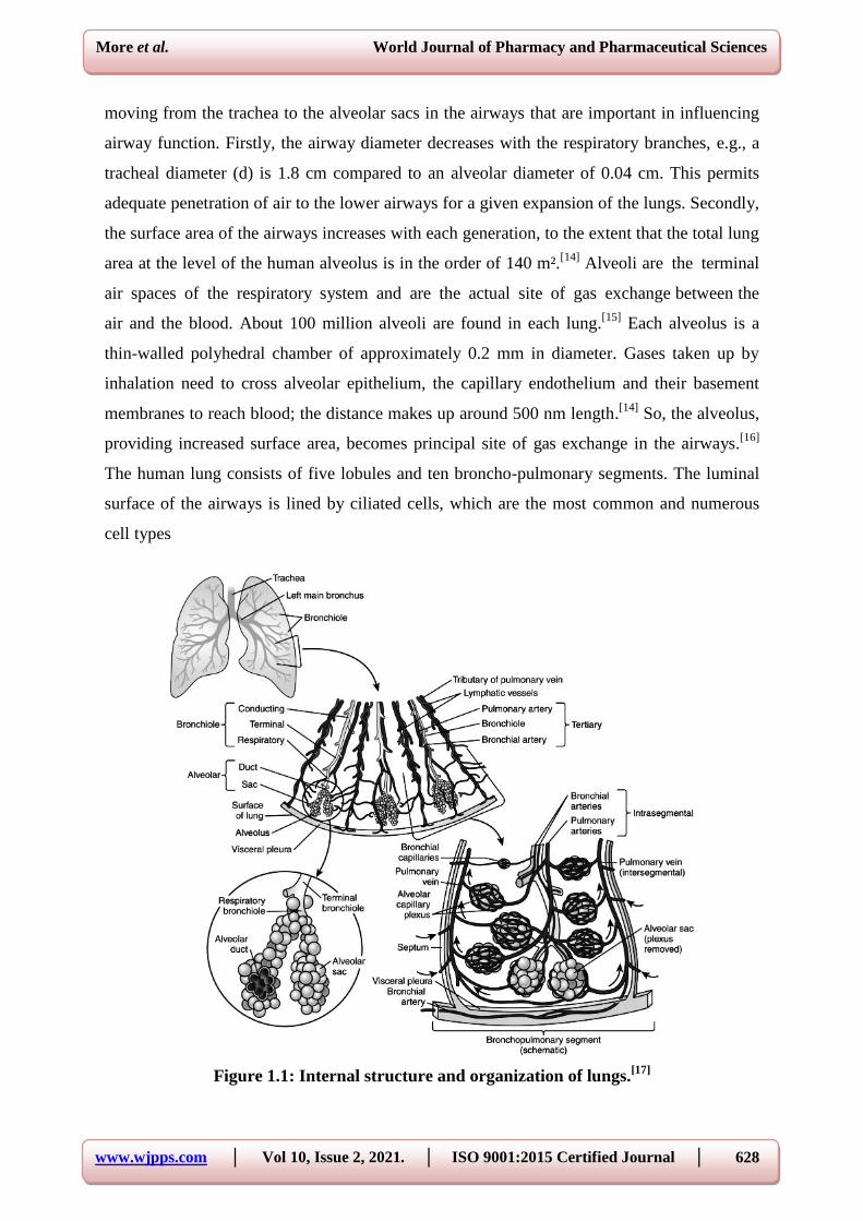
Anatomy and Physiology of Human Respiratory Tract
For the development of new PDD systems, one should have detailed knowledge of lung
anatomy and physiology. The respiratory tract is mainly divided into two regions i.e., the
upper airway and the lower airway with the line of division being the junction of the larynx
and trachea.[12]
The upper airway acts as an air transport system and consists of the nose,
mouth, pharynx and larynx. The lower respiratory tract consists of the tracheobronchial, gas-
conducting airways and the gas exchanging acini. The lower airway is divided into three
zones: conducting, transitional, and respiratory zones. The conducting zone is responsible for
the bulk movement of air and blood. In the central airways, air flow is rapid and turbulent and
no gas exchange occurs. The transitional zone has a limited role in gas exchange.[12,13]
The
respiratory zone mainly comprises of respiratory bronchioles and alveoli, where the actual
gas exchange takes place.[13]
Figure 1.1 shows a schematic representation of the lung. The
bronchial tree trunk begins with the trachea, which bifurcates to form the main bronchi: the
left and right primary bronchi. Each primary bronchus divides into still smaller secondary
bronchi. The secondary bronchi branch into many tertiary bronchi that further branch several
times, ultimately giving rise to tiny bronchioles that sub-divide many times, finally forming
terminal bronchioles and respiratory bronchioles. Each respiratory bronchiole sub-divides
into several alveolar ducts that end in clusters of small thin-walled air sacs called alveoli,
which open into a chamber called the alveolar sac.[14]
Two physical changes occur while
www.wjpps.com │ Vol 10, Issue 2, 2021. │ ISO 9001:2015 Certified Journal │
628
More et al. World Journal of Pharmacy and Pharmaceutical Sciences
moving from the trachea to the alveolar sacs in the airways that are important in influencing
airway function. Firstly, the airway diameter decreases with the respiratory branches, e.g., a
tracheal diameter (d) is 1.8 cm compared to an alveolar diameter of 0.04 cm. This permits
adequate penetration of air to the lower airways for a given expansion of the lungs. Secondly,
the surface area of the airways increases with each generation, to the extent that the total lung
area at the level of the human alveolus is in the order of 140 m².[14]
Alveoli are the terminal
air spaces of the respiratory system and are the actual site of gas exchange between the
air and the blood. About 100 million alveoli are found in each lung.[15]
Each alveolus is a
thin-walled polyhedral chamber of approximately 0.2 mm in diameter. Gases taken up by
inhalation need to cross alveolar epithelium, the capillary endothelium and their basement
membranes to reach blood; the distance makes up around 500 nm length.[14]
So, the alveolus,
providing increased surface area, becomes principal site of gas exchange in the airways.[16]
The human lung consists of five lobules and ten broncho-pulmonary segments. The luminal
surface of the airways is lined by ciliated cells, which are the most common and numerous
cell types
Figure 1.1: Internal structure and organization of lungs.[17]

www.wjpps.com │ Vol 10, Issue 2, 2021. │ ISO 9001:2015 Certified Journal │
629
More et al. World Journal of Pharmacy and Pharmaceutical Sciences
Mucus cells are intermingled among the ciliated cells. The walls of the conducting airways
are coated by an adhesive, viscoelastic mucus layer (thickness: 5–55 μm) which is secreted
by the mucus cells. The major components of mucus are glycoproteins and water.[18]
This
mucus fulfils important functions, i.e., the protection of the respiratory epithelium from
dehydration, the water in the mucus promotes saturation of inhaled air, mucus contains
antibacterial proteins and peptides, such as defensins and lysozyme that inhibit microbial
colonization of the airways, and mucus is also involved in airway protection from inhaled
xenobiotics or chemicals. Clearance of mucus from the lung is driven by the motion of the
ciliated cells „mucociliary escalator‟, which generates a mucus flow rate of ~5 mm/min. Thus,
the mucus blanket is replaced every 20 minutes in healthy subjects.[18]
The mucociliary
escalator serves as an important protective mechanism for removing small inhaled particles
from the lungs. The composition, thickness, viscosity and clearance of the mucus is often
altered in patients suffering from airway diseases such as asthma, COPD and CF. The
alveolar epithelium is composed of Type I and Type II alveolar cells and occasional brush
cells. The Type I pneumocytes are thin cells, cover most of the surface of the alveoli (95% of
the surface area) and the Type II pneumocytes are cuboidal secretory cells are interspersed
among the Type I cells.[19]
The alveolar space is coated by a complex surfactant lining that
reduces surface tension to minimize the work of breathing and prevents collapse of the
alveoli during expiration.[17]
The majority of insoluble particles deposited in the upper
airways are eliminated by mucociliary clearance.[20]
The most prominent defense mechanism
of the respiratory region is macrophage clearance. The particles deposited in the deeper lung
will be taken up by alveolar macrophages, which slowly migrate out of the lung, either
following the broncho-tracheal escalator or the lymphatic system .The blood supply to the
lung is provided by a pulmonary circulation and a systemic circulation. A drug delivered to
the lower airways can enter the systemic circulation by absorption into the alveolar capillaries
of the pulmonary vascular bed.[17,19]
Cellular Aspectsof Pulmonary Drug Transport
The transepithelial transport of compounds along the respiratory epithelium from the upper
airways with nasopharynx, trachea and large bronchi to the lower respiratory tract with small
bronchioles and alveoli is characterized bylarge quantitative di¡erences. In this respect, the
transport in the upper airways is limited by a smaller surface area and lower regional blood
£ow. Also, the upper airways possess a high ¢ltering capacity and remove 70 ^90% of
pressurized particles. In contrast, the smaller airways and alveolar space account for more

www.wjpps.com │ Vol 10, Issue 2, 2021. │ ISO 9001:2015 Certified Journal │
630
More et al. World Journal of Pharmacy and Pharmaceutical Sciences
than 95% of the lung‟s total surface area.[21]
Also, this compartment is directly connected to
the systemic circulation via the pulmonary circulation.
There are two major cell types found in the alveolar epithelium: type Iand type
IIpneumocytes. Whereas type Icells have a very thin cell body with long membranous
extensions, occupying an area of about 95% of the alveolar surface[22]
, the type II
pneumocytes are characterized by a more cuboidal morphology and cover about 5% of the
total alveolar surface.[23,24]
Studies on the subcellular morphology of type Icells revealed the
presence of endocytotic vesicles which may function as carriers in the absorption processes
of larger proteins such as insulin (5.7 kDa).[25,26]
Although type II pneumocytes express a
variety of transport proteins , it is generally accepted that their main functions are the
production of surfactant proteins and the di¡erentiation into type Icells after epithelial barrier
injuries.The pulmonary blood ^ gas barrier consists of a thick and a thin side, which are
composed of the alveolar epithelium, the capillary endothelium, and the intervening
extracellular matrix (basement membranes of the two cell layers).[27]
Out of the two cell types involved in the blood^ gas barrier, the type Icells display most likely
the rate-limiting step concerning the uptake of compounds into the pulmonary circulation as
previous studies reported a 103 times lower permeability for substances such as sucrose in
comparison to endothelial cells.[28]
This is based on the di¡erence in pore size between
alveolar cells (0.6 ^ 1nm) and endothelial cells (4 ^5.8 nm)[29]
and the tight junctions depth
which is 0.26170.023 mm (signi¢cantly) higher than the tight junctions depth of the capillary
endothelial cells (0.16670.011 mm).[30]
In contrast to these conditions at the blood^ gas barrier, three other di¡erent types of tight
junctions have been identi¢ed for extra- and intrapulmonary airways.[31]
Most importantly,
they di¡er in the degree of luminal ¢bril interconnections which are sparsely interconnected in
type I, more densely interconnected in type II and most densely interconnected in type III
tight junctions. While the type Iis almost exclusively found between extrapulmonary airway
ciliated cells, the type II is primarily present in smaller airways and between Clara cells and
the type III between mucous cells[31]
with a secretory cycle-associated change in permeability
This dependence upon the secretory cycle, which is leakier when the mucous cells are in a
state of active secretion has also been reported for other cell types such as mammary gland
epithelial cells.[32]
It is most likely that the regional di¡erencesin tightjunctionmorphology are

www.wjpps.com │ Vol 10, Issue 2, 2021. │ ISO 9001:2015 Certified Journal │
631
More et al. World Journal of Pharmacy and Pharmaceutical Sciences
directly linked to the transepithelial transport capacities of water and ions in contrast to
actively transported larger molecules.
RECENT ADVANCES IN PULMONARY DRUG DELIVERY DEVICES
Following types of inhalation devices are present
Inhalation drug delivery system by‐ metered dose inhalers
Inhalation drug delivery system by - dry powder inhalers
Inhalation drug delivery system by ‐nebulizer
Pressurized Metered-Dose Inhalers
Traditional asthma therapy has primarily used the pMDI. Since the 1950s, they have been the
backbone of inhalation therapy (33). In a pMDI, a propellant pressurized to a liquid state is
holding the drug either in a suspended or dissolved form. Metered release of the fluid through
a valve causes expansion and evaporation of propellant leaving the drug in the form of a high
velocity aerosol. Rapid release of the propellant into the valve stem along with the actuator
seating forms an expansion chamber where propellant starts to boil (Figure 1.). The liquefied
propellant serves both as a source of energy for expelling the formulation from the valve in
the form of rapidly evaporating droplets and as a dispersion medium for the drug and other
excipients.
How to use the MDI,
Shake the inhaler well before use (3 to 4 shakes)
Remove the cap
Breathe out, away from your inhaler
Bring the inhaler to your mouth. Place it in your mouth between your teeth and close your
Mouth around it. Start to breathe in slowly. Press the top of your inhaler once and keep
breathing in slowly
Until you have taken a full breath. Remove the inhaler from your mouth, and hold your
breath for about 10 seconds, then
Breath out.

www.wjpps.com │ Vol 10, Issue 2, 2021. │ ISO 9001:2015 Certified Journal │
632
More et al. World Journal of Pharmacy and Pharmaceutical Sciences
Figure 1: Schematic representation of a typical pressurized metered-dose inhaler.[33]
The essential components of an MDI are the container, the metering valve, and the actuator.
Usual valve volumes range from 25-100 µl, which deliver a drug dose of about 50 µg to 5
mg.
Nebulizers
Nebulizers have been used in inhalation therapy since the early 19th
century. Marketed
respiratory solutions are generally composed of drug dissolved in aqueous, isotonic solvent
systems that may contain preservatives to reduce microbial growth. There are two traditional
devices: air-jet and ultrasonic nebulizers. For a typical jet nebulizer (Figure 2), compressed
air passes through a narrow hole and entrains the drug solution from one or more capillaries
mainly by momentum transfer. Large droplets impact on baffles and gets refined to the size
required, while droplets with smaller size run in a streamline flow of air bypassing the impact
on baffles.[36]
Approximately 50-60% of the particles produced are in the respirable range.
Figure 2: Schematic presentation of a jet nebulizer.[34]
www.wjpps.com │ Vol 10, Issue 2, 2021. │ ISO 9001:2015 Certified Journal │
633
More et al. World Journal of Pharmacy and Pharmaceutical Sciences
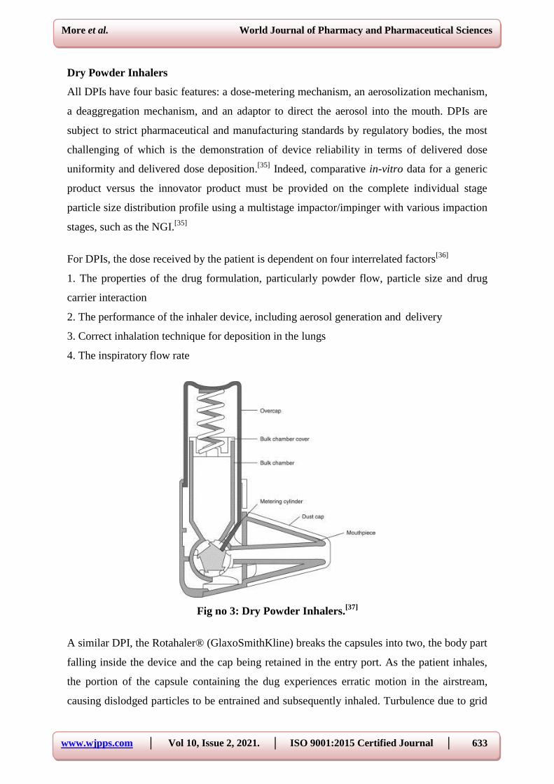
Dry Powder Inhalers
All DPIs have four basic features: a dose-metering mechanism, an aerosolization mechanism,
a deaggregation mechanism, and an adaptor to direct the aerosol into the mouth. DPIs are
subject to strict pharmaceutical and manufacturing standards by regulatory bodies, the most
challenging of which is the demonstration of device reliability in terms of delivered dose
uniformity and delivered dose deposition.[35]
Indeed, comparative in-vitro data for a generic
product versus the innovator product must be provided on the complete individual stage
particle size distribution profile using a multistage impactor/impinger with various impaction
stages, such as the NGI.[35]
For DPIs, the dose received by the patient is dependent on four interrelated factors[36]
1. The properties of the drug formulation, particularly powder flow, particle size and drug
carrier interaction
2. The performance of the inhaler device, including aerosol generation and delivery
3. Correct inhalation technique for deposition in the lungs
4. The inspiratory flow rate
Fig no 3: Dry Powder Inhalers.[37]
A similar DPI, the Rotahaler® (GlaxoSmithKline) breaks the capsules into two, the body part
falling inside the device and the cap being retained in the entry port. As the patient inhales,
the portion of the capsule containing the dug experiences erratic motion in the airstream,
causing dislodged particles to be entrained and subsequently inhaled. Turbulence due to grid

www.wjpps.com │ Vol 10, Issue 2, 2021. │ ISO 9001:2015 Certified Journal │
634
More et al. World Journal of Pharmacy and Pharmaceutical Sciences
upstream of the mouthpiece deaggregates particles. A FPF of 26% has been reported for this
low resistance device.[37]
Devices for generating particles.[38,39]
Device Particle size (mm) Particle size uniformity
Metered-doseinhaler 1^35 Heterogeneous
Jet nebulizer 1.2^6.9 Heterogeneous
Ultrasonic nebulizer 3.7^10.5 Heterogeneous
Spinning disc 1.3^30 Monodisperse
Dry powder Flow-related Heterogeneous
Vibrating ori¢ce 0.5^50 Monodisperse
Mechanisms and Ways Of Pulmonary Drug Administration
Over the last decade, the systemic absorption of a broad range of therapeutic agents after
pulmonary application has been demonstrated in animals as well as humans. Through
pulmonary route, the drug can be administered by two primary modes: first, intranasal
administration, which has anatomical limitation, such as narrower airway lumen, second, oral
inhalative administration. By oral inhalative administration far better results can be expected
as it allows to administer very small particles with a concentration loss of only 20% in
comparison with 85% by nasal route. Oral inhalative administration can again be classified as
intratracheal instillation and intratracheal inhalation. The most common method used in
laboratory is the intratracheal instillation. In the intratracheal instillation, a small amount of
drug solution or dispersion is delivered into the lungs by a special syringe. This provides a
fast and quantifiable method of drug delivery to the lungs. The localized drug deposition is
achieved with a comparatively small absorptive area. So, the instillation process is much
simple, non-expensive, and has non-uniform drug distribution.
In preclinical animal studies, intratracheal instillation has frequently been used to assess the
pulmonary absorption and systemic bioavailability, especially with regard to the precise
dosing and effectiveness associated with this method.[40]
However, intratracheal instillation is
not a physiological route for application, and results obtained from these studies may not be
transferable to aerosol applications in humans. On the contrary, inhalation method uses
aerosol technique by which we can get more uniform distribution with great penetration.
However, this method is more costly and difficult to measure the exact dose in lungs. The
deposition of drug by aerosol administration in the pulmonary airway mainly takes place by
three mechanisms:-gravitational sedimentation, inertial impaction, and diffusion. If the drug
particle size is comparatively bigger, then, deposition takes place by first two mechanisms

www.wjpps.com │ Vol 10, Issue 2, 2021. │ ISO 9001:2015 Certified Journal │
635
More et al. World Journal of Pharmacy and Pharmaceutical Sciences
where, either sedimentation occurs due to gravitational force or inertial impaction occurs due
to hyperventilation. When the particle size is smaller they deposit mainly by diffusion
mechanism, which in turn is based on the Brownian motion. Apart from the pulmonary
morphological aspects and ventilatory parameters size of the particles or droplets and the
geometry is quite important. The size of particle or droplet in terms of diameter along with
the surface electrical charges, shape of the particulate matter if it is a fiber and hygroscopy
also having profound influence on drug deposition through pulmonary route.[41]
The term
mass median aerodynamic diameter is used and it depends on size, shape, and density of the
particulate system.
Techniques of Making Particulate Matter For Lung Delivery
Many conventional techniques have been reported to produce DPI formulations. However,
these methods have number of limitations, such as particle size, size distribution, shape and
poor control over powder crystallinity. These problems can be rectified by specialized milling
techniques. Jet-milling of drug under nitrogen gas with new nanojet milling instrument is the
most suitable method for creating nanoparticles meant for pulmonary drug delivery. Here,
some of the important techniques are discussed in brief.[41]
Spray Drying Technique
Spray drying is an advanced pharmaceutical manufacturing process used to efficiently
produce respirable colloidal particles in the solid state.[42,43]
Spray drying was explored in the
1980s as an alternative means of producing fine particles for pulmonary delivery. In this
process, the feed solution is supplied at room temperature and pumped to the nozzle where it
is atomized by the nozzle gas. The atomized solution is then dried by preheated drying gas in
a special chamber to remove water moisture from the system, thus forming dry particles. This
method is more promising in producing the particles of above 2-μm size. This method is
reported to have better control on particle formation and hence can be easily translated to
large scale production. This process is also suitable for thermolabile materials, such as
proteins and peptides, because mechanical high- energy input is avoided in this process. More
importantly, spray-drying can result in uniform particle morphology.[44,45]
Spray Freeze Drying Method
This method was explored for pharmaceutical application in early 1990s. It is an advanced
particle engineering method, which combines spray-drying and freeze-drying processing
steps. It involves spraying the drug solution into liquid nitrogen as a freezing medium

www.wjpps.com │ Vol 10, Issue 2, 2021. │ ISO 9001:2015 Certified Journal │
636
More et al. World Journal of Pharmacy and Pharmaceutical Sciences
followed by lyophilization.[46]
This method produces light and porous particles and high fine
particle fraction with improved aerosol performance and almost 100% yield at subambient
temperatures[47]
Thermolabile protein and peptide substances, such as insulin[48]
and plasmid
DNA[49]
can also be formulated into dry powder inhalation products. However, this is an
expensive process restricted for only expensive drug.
Supercritical Fluid Technology
The basic feature of this process is the controlled crystallization of drugs from dispersion in
supercritical fluids, carbon dioxide. This method has been used in the pharmaceutical field
for production of microparticles, nanoparticles, liposomes, and inclusion complexes. This
method is used for the production of particulate pulmonary drug delivery systems containing
proteins and peptides, and also used to improve the formulation properties of certain drug
candidates.[50,51]
Solvent Precipitation Method
This method involves sono-crystallization and micro-precipitation by opposing liquid jets.
Crystalline drug particles with narrow size distribution could be prepared by direct controlled
crystallization[52]
Inhalable particles can be produced by rapid precipitation from aqueous
solutions using antisolvents. Recently, ultrasonic radiation has been applied to control the
precipitation. Various antiasthmatic drugs were prepared using the sono-crystallization
technique.
Double Emulsion/Solvent Evaporation Technique
This method involves preparation of oil/water emulsion with subsequent removal of the oil
phase through evaporation. The organic solvent diffuses out of the polymer phase and into
the aqueous phase, and is then evaporated, forming drug- loaded polymeric nanoparticles. By
this method, biodegradable polymers have been intensively investigated as carriers for
respiratory solid drug nanoparticles.
Particle Replication In Nonwetting Templates
Particle replication in non wetting templates (PRINT) is top-down particle fabrication
technique developed by Dr. Joseph DeSimone and his group. This technique is able to
produce uniform-sized organic micro- and nanoparticles with complete control of size, shape,
and surface functionality, and helps in loading of small organic therapeutics, proteins,
peptides, oligonucleotides, RNA contrast agents, radiotracers, and flurophores.[53–55]

www.wjpps.com │ Vol 10, Issue 2, 2021. │ ISO 9001:2015 Certified Journal │
637
More et al. World Journal of Pharmacy and Pharmaceutical Sciences
CONCLUSIONS
It can be concluded from the whole literature that pulmonary drug targeting offers several
advantages which can improve drug‟s efficacy and reduce unwanted systemic side effects. It
provides a large surface area for absorption, thin alveolar epithelium permitting rapid
absorption, absence of first-pass metabolism, rapid onset of action and high bioavailability.
Since pulmonary disorders are engulfing a major portion of population every day, it has been
a major issue of discussions and debates. Various new technologies for pulmonary drug
delivery as well as novel targeting methodologies have directed researchers to focus upon this
route as a more preferred one for targeting various pulmonary disorders.
REFERENCES
1. Michael T. Newhouse, “Encyclopedia of Pharmaceutical Technology”, second edition,
Dekker, New York Informa Healthcare USA, 2000; 19: 1279‐1285.
2. Groneberg DA, Eynott PR, Doring F, Dinh QT, Oates T, Barnes PJ, et al. Distribution
and function of the peptide transporter PEPT2 in normal and cystic fibrosis human lung.
Thorax, 2002; 57(1): 55-60.
3. Groneberg DA, Nickolaus M, Springer J, Doring F, Daniel H, Fischer A. Localization of
the peptide transporter PEPT2 in the lung: implications for pulmonary oligopeptide
uptake. The American journal of pathology, 2001; 158(2): 707-14.
4. Hiller FC. In: Hickey AJ, editor. Pharmaceutical Inhalation Aerosol Technology. New
York: Marcel Dekker Inc., 1992; 385.
5. Olschewski H, Simonneau G, Galie N, Higenbottam T, Naeije R, Rubin LJ, et al. Inhaled
iloprost for severe pulmonary hypertension. The New England journal of medicine, 2002;
347(5): 322-9.
6. Courrier HM, Butz N, Vandamme TF. Pulmonary drug delivery systems: recent
developments and prospects. Critical reviews in therapeutic drug carrier systems, 2002;
19(4-5): 425-98.
7. Groneberg DA, Witt C, Wagner U, Chung KF, Fischer A. Fundamentals of pulmonary
drug delivery. Respiratory medicine, 2003; 97(4): 382-7.
8. Patton JS, Byron PR. Inhaling medicines: delivering drugs to the body through the lungs.
Nature reviews Drug discovery, 2007; 6(1): 67-74.
9. Geiser M, Kreyling WG. Deposition and biokinetics of inhaled nanoparticles. Particle and
fibre toxicology, 2010; 7: 2.

www.wjpps.com │ Vol 10, Issue 2, 2021. │ ISO 9001:2015 Certified Journal │
638
More et al. World Journal of Pharmacy and Pharmaceutical Sciences
10. Gessler T, Seeger W, Schmehl T. Inhaled prostanoids in the therapy of pulmonary
hypertension. Journal of aerosol medicine and pulmonary drug delivery, 2008; 21(1): 1-
12.
11. Jaspart S, Bertholet P, Piel G, Dogne J, Delattre L, Evrard B, Solid lipid microparticles as
a sustained release system for pulmonary drug delivery, Eur. J. Pharm. Biopharm, 2007;
65: 47-56.
12. JB. West PW. Ventilation-perfusion relationships.in The Lung: Scientific Foundations.
In: R.G. Crystal JBW, P.J. Barnes and E.R. Weibal, Raven Press, editor, 1991; 1289.
13. Yu J, Chien YW. Pulmonary drug delivery: physiologic and mechanistic aspects. Critical
reviews in therapeutic drug carrier systems, 1997; 14(4): 395-453.
14. Thompson AJHaDC. In: A.J. Hickey MD, editor. Pharmaceutical Inhalation Aerosol
Technology. 2nd Edition ed. New York, NY, USA, 2004; 1.
15. Stone KC, Mercer RR, Gehr P, Stockstill B, Crapo JD. Allometric relationships of cell
numbers and size in the mammalian lung. American journal of respiratory cell and
molecular biology, 1992; 6(2): 235-43.
16. Crapo JD, Barry BE, Gehr P, Bachofen M, Weibel ER. Cell number and cell
characteristics of the normal human lung. The American review of respiratory disease,
1982; 126(2): 332-7.
17. C.A. D‟Angelis JJCaRMR. In: Zimmerman BPFaJJ, editor. Pediatric Critical Care. 4th
Edition ed. Philadelphia, PA, USA2011.
18. Lai SK, Wang YY, Wirtz D, Hanes J. Micro- and macrorheology of mucus. Advanced
drug delivery reviews, 2009; 61(2): 86-100.
19. Thompson RJAaDC. Inhalation Aerosols: Physical and Biological Basis for Therapy.
New York, NY, USA, 1996; 83.
20. Van der Schans CP. Conventional chest physical therapy for obstructive lung disease.
Respiratory care., 2007; 52(9): 1198-206; discussion 206-9.
21. Weibel ER. Morphometry of the human lung: the state of the art after two decades. Bull
Physiopathol Respir (Nancy), 1979; 15: 999–1013.
22. Hirai K, Ogawa K. Cytochemical quantitation of cytochrome oxidase activity in rat
pulmonary alveolar epithelial cells and possible defect in type I cells. J Electron Microsc
(Tokyo), 1986; 35: 19–28.
23. Mason RJ, Crystal RG. Pulmonary cell biology. Am J Respir Crit Care Med., 1998; 157(4
Part 2): S72–S81.

www.wjpps.com │ Vol 10, Issue 2, 2021. │ ISO 9001:2015 Certified Journal │
639
More et al. World Journal of Pharmacy and Pharmaceutical Sciences
24. Sorokin SP. Properties of alveolar cells and tissues that strengthen alveolar defenses.
Arch Intern Med 1970; 126: 450–463. 14. Gil J, Silage DA, McNiff JM. Distribution of
vesicles in cells of air– blood barrier in the rabbit. J Appl Physiol, 1981; 50: 334–340.
25. Gil J. Number and distribution of plasmalemmal vesicles in the lung. Fed Proc, 1983; 42:
2414–2418.
26. Low F. Electron microscopy of the rat lung. Anat Rec., 1952; 113: 437–443.
27. Wangensteen OD, Wittmers LE Jr, Johnson JA. Permeability of the mammalian blood–
gas barrier and its components. Am J Physiol, 1969; 216: 719–727.
28. Taylor AE, Gaar KA Jr. Estimation of equivalent pore radii of pulmonary capillary and
alveolar membranes. Am J Physiol, 1970; 218: 1133–1140.
29. Inoue S, Michel RP, Hogg JC. Zonulae occludentes in alveolar epithelium and capillary
endothelium of dog lungs studies with the freeze-fracture technique. J Ultrastruct Res.,
1976; 56: 215–225.
30. Inoue S, Hogg JC. Freeze-etch study of the tracheal epithelium of normal guinea pigs
with particular reference to intercellular junctions. J Ultrastruct Res., 1977; 61: 89–99.
31. Nguyen DA, Neville MC. Tight junction regulation in the mammary gland. J Mammary
Gland Biol Neoplasia, 1998; 3: 233–246.
32. Nguyen DA, Parlow AF, Neville MC. Hormonal regulation of tight junction closure in
the mouse mammary epithelium during the transition from pregnancy to lactation. J
Endocrinol, 2001; 170: 347–356.
33. Newman SP. Principles of metered-dose inhaler design. Respiratory care., 2005; 50(9):
1177- 90.
34. Le Brun PP, de Boer AH, Heijerman HG, Frijlink HW. A review of the technical aspects
of drug nebulization. Pharmacy world & science : PWS, 2000; 22(3): 75-81.
35. Newman SP, Busse WW. Evolution of dry powder inhaler design, formulation, and
performance. Respiratory medicine, 2002; 96(5): 293-304.
36. Atkins PJ. Dry powder inhalers: an overview. Respiratory care., 2005; 50(10): 1304-12;
discussion 12.
37. Dunbar CA, Hickey AJ, Holzner P. Dispersion and Characterization of Pharmaceutical
DryPowder Aerosols. Kona Powder and Particle Journal, 1998; 16: 7-45.
38. Newman SP. Aerosol generators and delivery systems. Respir Care, 1991; 36: 939–951.
39. Newman SP, Wilding IR, Hirst PH. Human lung deposition data: the bridge between
invitro and clinical evaluations for inhaled drug products? Int J Pharm., 2000; 208: 49–60.

www.wjpps.com │ Vol 10, Issue 2, 2021. │ ISO 9001:2015 Certified Journal │
640
More et al. World Journal of Pharmacy and Pharmaceutical Sciences
40. Lizio R, Klenner T, Borchard G, Romeis P, Sarlikiotis AW, Reissmann T, et al. Delivery
of the GnRH antagonist centrolix by intratracheal instillation in Anesthetized rats. Eur J
Pharm Sci., 2000; 9: 253–8. [PubMed] [Google Scholar]
41. Chono S, Tanino T, Seki T, Morimoto K. Influence of particle size on drug delivery to rat
alveolar macrophages following pulmonary administration of ciprofloxacin incorporated
into liposomes. J Drug Target., 2006; 14: 557–66. [PubMed] [Google Scholar]
42. Mosen K, Backstrom K, Thalberg K. Particle formation and capture during spray drying
of inhale particles. Pharm Dev Technol., 2004; 9: 409–18. [PubMed] [Google Scholar]
43. Duddu SP, Sisk SA, Walter YH. Improved lung delivery from a passive dry powder
inhaler using an engineered pulmosphere powder. Pharm Res., 2002; 19: 689–
95. [PubMed] [Google Scholar]
44. White S, Bennett DB, Cheu S. Pharmaceutical development of a novel product for
pulmonary delivery of insulin. Diabetes Technol Ther., 2005; 7: 896–
906. [PubMed] [Google Scholar]
45. Gilani K, Najafabad AR, Berge M, Rafiee-Tehrani M. The effect of water to ethanol feed
ratio on physical properties and aerosolization behavior of spray dried cromolyn sodium
particles. J Pharm Sci., 2005; 94: 1048–59. [PubMed] [Google Scholar]
46. Rogers T, Johnston K, Williams R. Solution-based particle formation of pharmaceutical
powders by supercritical or compressed fluid CO2 and cryogenic spray-freezing
technologies. Drug Dev Ind Pharm., 2001; 27: 1003–16. [PubMed] [Google Scholar]
47. Maa YF, Prestrelski SJ. Biopharmaceutical powders: Particle formation and formulation
considerations. Curr Pharm Biotechnol, 2000; 1: 283–302. [PubMed] [Google Scholar]
48. Yu Z, Garcia AS, Johnston KP, Williams RO. Spray freezing into liquid nitrogen for
highly stable protein nanostructured microparticles. Eur J Pharm Biopharm, 2004; 58:
529–37. [PubMed] [Google Scholar]
49. Kuo JH, Hwang R. Preparation of DNA dry powder for non-viral gene delivery by spray-
freeze drying: Effects of protective agents on the stability of DNA. J Pharm Pharmacol,
2004; 27–33. [PubMed] [Google Scholar]
50. Chattopadhyay P, Shekunov BY, Yim D, Cipolla D, Boyd B, Farr S. Production of solid
nanoparticle suspensions using supercritical fluid extraction of emulsions for pulmonary
delivery using the AERx system. Adv Drug Deliv Rev., 2007; 59: 4 44–
53. [PubMed] [Google Scholar]
www.wjpps.com │ Vol 10, Issue 2, 2021. │ ISO 9001:2015 Certified Journal │
641
More et al. World Journal of Pharmacy and Pharmaceutical Sciences
51. Rehman M, Shekunov BY, York P. Optimization of powders for pulmonary delivery
using supercritical fluid technology. Eur J Pharm Sci., 2004; 22: 1–17. [PubMed] [Google
Scholar]
52. Rasenack N, Steckel H, Mullar BW. Micronization of anti-inflammatory drugs
forpulmonary delivery by a controlled crystallization process. J Pharm Sci., 2003; 92: 35–
44. [PubMed] [Google Scholar]
53. Gratton SE, Pohlaus PD, Lee J, Cho MJ, DeSimon JM. Nanofabricated particles for
engineered drug therapies: A preliminary biodistribution study of
PRINT™
nanoparticles. J Control Rel., 2007; 121: 10–8. [PMC free
article] [PubMed] [Google Scholar]
54. Gratton SE, Napier ME, Ropp PA, Tian S, DeSimon JM. Microfabricated particles for
engineered drug therapies: Elucidation into the mechanisms of cellular internalization of
PRINT particles. Pharm Res., 2008; 25: 2845–52. [PMC free article] [PubMed] [Google
Scholar]
55. Heidi MM, Yun-Seok R, Xiao W. Nanomedicines in pulmonary delivery. Int J Nano
med., 2009; 4: 299–319. [Google Scholar]


![Pulmonary drug delivery system [PDDS]](https://img.dokumen.tips/doc/110x75/587c0a411a28ab03768b542f/pulmonary-drug-delivery-system-pdds.jpg)