Embed Size (px)
Citation preview

A REVIEW OF THE IRONCLAD BEETLES OF THE WORLD (COLEOPTERA
ZOPHERIDAE: PHELLOPSINI AND ZOPHERINI)
by
Ian Andrew Foley
A thesis submitted in partial fulfillment of the requirements for the degree
of
Master of Science
in
Entomology
Montana State University Bozeman, Montana
May 2006

COPYRIGHT
by
© Ian Andrew Foley
2006
All Rights Reserved

ii
APPROVAL
of a thesis submitted by
Ian Andrew Foley
This thesis has been read by each member of the thesis committee and has been found to be satisfactory regarding content, English usage, format, citations, bibliographic style, and consistency, and is ready for submission to the Division of Graduate Education.
Michael A. Ivie, Ph. D.
Chair
Approved for the Department of Plant Sciences and Plant Pathology
John E. Sherwood, Ph. D.
Approved for the Division of Graduate Education
Joseph J. Fedock, Ph. D.

iii
STATEMENT OF PERMISSION TO USE
In presenting this thesis in partial fulfillment of the requirements for a master’s
degree at Montana State University, I agree that the Library shall make it available to
borrowers under rules of the Library.
If I have indicated my intention to copyright this thesis by including a copyright
notice page, copying is allowable only for scholarly purposes, consistent with “fair use”
as prescribed in the U.S. Copyright Law. Requests for permission for extended quotation
from or reproduction of this thesis in whole or in parts may be granted only by the
copyright holder.
Ian Andrew Foley May 2006
iv
ACKNOWLEDGEMENTS
This thesis would not have been possible without the guidance, understanding,
and assistance of Michael Ivie, as well as funding for this project through the Montana
State Agricultural Experiment Station. Richard Hurley provided much needed relief and
support. My committee, Matt Lavin, Richard Miller, and Kevin O’Neill, offered useful
suggestions and discussion on drafts of this document. I am thankful to the graduate
students, staff, and faculty of the Entomology Graduate program, as well as the
Department of Plant Sciences and Plant Pathology at Montana State University,
especially past members of the Ivie Lab Katharine Marske, Sardis-Medrano Cabral, J.
Joseph Giersch, Alistair Ramsdale and Jessica Fultz, Sarah Wallace and Rebecca Baril.
Michael Caterino afforded me the opportunity to learn molecular techniques and use of
the SEM at the Santa Barbara Natural History Museum, Chuck Triplehorn, Alfred
Newton, and Warren Steiner kindly offered unpublished data on specimens, literature and
thoughts on the group. This research would also not have been possible without the
timely response to loan request from all of the curators and collection managers who
facilitated specimen loans, particularly Maxwell Barclay. Translations of Russian type
labels were kindly provided by Alex Konstantinov. Finally, I am grateful for the constant
encouragement and support from my parents, John and Mary Ellen, and siblings Aaron,
Jenna, and Patrick as well as my friends.

v
TABLE OF CONTENTS 1. THE FAMILY ZOPHERIDAE .......................................................................................1 Introduction......................................................................................................................1 Diagnostic Morphology ...................................................................................................2 Taxonomic History of the Family Zopheridae.................................................................2 2. REVISION OF THE GENUS PHELLOPSIS..................................................................9 Introduction......................................................................................................................9 Taxonomic History ..........................................................................................................9 Materials ........................................................................................................................12 Methods -- Morphology.................................................................................................16 Methods-- Phylogenetic Analysis ..................................................................................18 Taxonomy of the Genus Phellopsis LeConte ................................................................23 Biology of the Genus .....................................................................................................27 Key to the Species of Phellopsis....................................................................................28 Species Descriptions ......................................................................................................30 Phellopsis obcordata (Kirby) ........................................................................................30 Phellopsis porcata (LeConte) ........................................................................................35 Phellopsis suberea Lewis ..............................................................................................41 Phellopsis amurensis (Heyden) ....................................................................................45 Phellopsis chinensis Semenow ......................................................................................48 Phellopsis yulongensis Foley and Ivie NEW SPECIES ................................................52 Synoptic Catalog of Phellopsis Species ........................................................................54 Results of Phylogenetic Analysis...................................................................................55 Biogeography.................................................................................................................57 Habitat Conservation .....................................................................................................60 Future Research .............................................................................................................62 3. REVIEW OF THE ZOPHERINI ...................................................................................64 Introduction....................................................................................................................64 Zopherini Solier .............................................................................................................65 Taxonomic History of the Zopherini .............................................................................65 Materials ........................................................................................................................68 Methods -- Morphology.................................................................................................68 Methods-- Phylogenetic analysis ...................................................................................70 Results -- Phylogenetic Analysis ...................................................................................76 Key to World Genera of Zopherini................................................................................79 Zopherus Gray ...............................................................................................................82 Synopsis of Zopherus Gray Species ..............................................................................83

vi
TABLE OF CONTENTS-CONTINUED Zopherosis White ...........................................................................................................85 Synopsis of Zopherosis White Species .........................................................................86 Zopher Ślipiński and Lawrence .....................................................................................86 Synopsis of Zopher Ślipiński and Lawrence Species ...................................................86 Noserinus Casey.............................................................................................................87 Key to Species of Noserinus Casey ..............................................................................88 Synopsis of Noserinus Casey Species............................................................................88 Scoriaderma Fairmaire ..................................................................................................89 Synopsis of Scoriaderma Fairmaire Species .................................................................91 Meralius Casey ..............................................................................................................91 Key to species of Meralius Casey..................................................................................93 Synopsis of Meralius Casey Species .............................................................................93 Meralius clavaplius Foley and Ivie NEW SPECIES.....................................................93 Sesaspis Casey NEW SENSE........................................................................................96 Key to Species of Sesaspis Casey................................................................................100 Synopsis of Sesaspis Casey Species ............................................................................102 Sesaspis denticulata (Solier) NEW COMBINATION ................................................102 Sesaspis emarginatus (Horn) NEW COMBINATION................................................104 Sesaspis doyeni (García-París et al.) NEW COMBINATION ....................................104 Sesaspis lutosus (Champion) NEW COMBINATION................................................108 Sesaspis adami Foley and Ivie NEW SPECIES ..........................................................106 Sesaspis ashei Foley and Ivie NEW SPECIES............................................................109 Sesaspis triplehorni Foley and Ivie NEW SPECIES...................................................110 Phloeodes LeConte ......................................................................................................113 Synopsis of Phloeodes LeConte Species .....................................................................112 Nosoderma Solier NEW SENSE .................................................................................113 Synopsis of Nosoderma Solier ....................................................................................119 Key to Species of Nosoderma Solier ...........................................................................121 Future Research ...........................................................................................................123 4. REVISION OF THE GENUS PHLOEODES..............................................................126 Introduction..................................................................................................................126 Taxonomic History ......................................................................................................126 Materials ......................................................................................................................132 Methods -- Morphology...............................................................................................136 Taxonomy of the Genus Phloeodes LeConte ..............................................................138 Synoptic Catalog of Phloeodes Species.......................................................................142 Biology of the Genus ...................................................................................................143 Key to the Species of Phloeodes..................................................................................145 Species Descriptions ....................................................................................................146 Phloeodes diabolicus (LeConte)..................................................................................146

vii
TABLE OF CONTENTS-CONTINUED Phloeodes plicatus (LeConte)......................................................................................153 Phloeodes venustus (Champion) NEW COMBINATION ..........................................157 Biogeography...............................................................................................................160 Future research.............................................................................................................163 REFERENCES CITED....................................................................................................164 FIGURES.........................................................................................................................176 APPENDICES APPENDIX A: Specimens examined of Phellopsis obcordata ..................................219 APPENDIX B: Specimens examined of Phellopsis porcata.......................................228 APEENDIX C: Specimens examined of the Asian spp. of Phellopsis........................249 APPENDIX D: Specimens examined of Phloeodes spp. ............................................250 APPENDIX E: Specimens examined of Sesaspis spp.................................................282

viii
LIST OF TABLES Table Page 1. Changes in the classification of Zopheridae from Doyen and Lawrence (1979) to Ślipiński and Lawrence (1999) ............................7 2. Character matrix used in the phylogenetic analysis of Phellopsis................................22 3. Locality data for species used in the phylogenetic analysis of the Zopherini ..............71 4. Character matrix used in the phylogenetic analysis of the Zopherini...........................78

ix
LIST OF FIGURES
Figure Page 1. Distribution of the North American species of Phellopsis .......................................................36 2. Distribution of the East Asian species of Phellopsis ................................................................45 3. Distribution of the Chinese species of Phellopsis.....................................................................51 4. Distribution of the Central American genus Sesaspis.............................................................103 5. Broad view of the distribution of the Central American genus Nosoderma...........................120 6. Narrow view of the distribution of the genus Nosoderma near Mexico City.........................120 7. Distribution of Phloeodes diabolicus......................................................................................152 8. Distribution of Phloeodes plicatus..........................................................................................157 9. Distribution of Phloeodes venustus ........................................................................................159 10. Phellopsis obcordata (Kirby) habitus...................................................................................176 11. Phellopsis porcata (LeConte) habitus ..................................................................................176 12. P. obcordata hypomeron ......................................................................................................176 13. P. porcata hypomeron ..........................................................................................................176 14. P. amurensis (Heyden) habitus. ............................................................................................177 15. P. suberea Lewis habitus. .....................................................................................................177 16. P. chinensis (Semenow) habitus. ..........................................................................................177 17. P. yulongensis Foley and Ivie NEW SPECIES habitus........................................................177 18. P. suberea dorsal view of head. ............................................................................................178 19. P. yulongensis dorsal view of head.......................................................................................178 20. P. amurensis dorsal view of head. ........................................................................................179 21. P. chinensis dorsal view of head...........................................................................................179 22. P. obcordata dorsal view of head. ........................................................................................180 23. P. porcata dorsal view of head. ............................................................................................180 24-29. 24-28 lateral views of male parameres. 24. P. obcordata, 25. P. porcata, 26. P.
yulongensis, 27. P. chinensis, 28. P. suberea. 29. Ventral view of male parameres P. yulongensis.......................................................................................................................181
30. P. chinensis ventrites 2-5. .....................................................................................................182 31. P. yulongensis ventrites 2-5. .................................................................................................182 32. P. obcordata ventral view of larval A8. ...............................................................................182 33. P. porcata ventral view of larval A8. ...................................................................................182 34. P. yulongensis ventral view of head. ...................................................................................183 35. P. suberea ventral view of head. ..........................................................................................183 36. P. chinensis ventral view of head. ........................................................................................184 37. Usechus lacerta Motschulsky ventral view of head. ............................................................184 38. P. chinensis metatarsus. ........................................................................................................185 39. P. chinensis pronotal scale-like setae....................................................................................186 40. P. yulongensis pronotal hair-like setae. ................................................................................186 41. P. amurensis mesepimeron. ..................................................................................................186 42. P. yulongensis mesepimeron.................................................................................................186

x
Figure Page 43. P. yulongensis mandible. .....................................................................................................187 44. P. suberea mandible..............................................................................................................187 45. P. yulongensis maxilla. .........................................................................................................187 46. P. suberea maxilla. ...............................................................................................................187 47. P. chinensis metasternum behind mesocoxa. .......................................................................188 48. P. obcordata metasternum behind mesocoxa. ......................................................................188 49. P. yulongensis antenna..........................................................................................................188 50. P. porcata female genitalia. ..................................................................................................188 51. P. obcordata prosternal process. ..........................................................................................189 52. P. yulongensis prosternal process. ........................................................................................189 53-55. Elytra showing ridges and nodules. 53. P. chinensis, 54. P. yulongensis, 55. P. suberea ..................................................................................................................190 56-17. Elytra showing ridges and nodules. 56. P. amurensis, 57. P. obcordata. ........................191 58. P. obcordata lateral pronotal margin. ..................................................................................192 59. P. porcata lateral pronotal margin. .......................................................................................192 60. Usechus lacerta Motschulsky habitus. ................................................................................192 61. Hyporagus sp. habitus. .........................................................................................................192 62. Single most parsimonious tree found in NONA/WINCLADA (Length 23, CI 0.86, RI 0.87)...........................................................................................193 63. Phloeodes diabolicus habitus, California, San Diego Co. ....................................................194 64. P. diabolicus habitus, California, Napa Co...........................................................................194 65. P. plicatus habitus.................................................................................................................194 66. P. venustus habitus................................................................................................................194 67. P. diabolicus prothorax.........................................................................................................195 68. P. plicatus prothorax.............................................................................................................195 69. P. diabolicus antennal cavity. ...............................................................................................195 70. P. venustus prothoracic hypomeron......................................................................................196 71. P. venustus ventral view of tarsus.........................................................................................196 72. P. diabolicus male genitalia..................................................................................................197 73. P. plicatus male genitalia......................................................................................................197 74. P. venustus male genitalia.....................................................................................................197 75. Sesaspis triplehorni Foley and Ivie NEW SPECIES male genitalia. ...................................197 76. P. diabolicus labium. ............................................................................................................198 77. P. plicatus labium. ................................................................................................................198 78. P. plicatus tarsal claw SEM. .................................................................................................198 79. P. diabolicus male femoral nodule SEM. .............................................................................199 80. P. plicatus male femoral nodule SEM. .................................................................................199 81. P. diabolicus male femoral nodule, apical edge SEM. .........................................................200 82. P. plicatus male femoral nodule, apical edge SEM. .............................................................200 83. Nosoderma inaequalis habitus..............................................................................................201 84. Nosoderma exsculptum habitus.............................................................................................201 85. Nosoderma asperatum habitus..............................................................................................201

xi
Figure Page 86. Nosoderma scabrosum habitus. ............................................................................................201 87. Nosoderma insigne habitus. ..................................................................................................202 88. Nosoderma sparsus habitus. .................................................................................................202 89. Nosoderma zunilensis habitus...............................................................................................202 90. Nosoderma guatemalensis habitus........................................................................................202 91. Nosoderma aequalis habitus. ................................................................................................203 92. Sesaspis denticulata habitus. ................................................................................................203 93. Sesaspis ashei Foley and Ivie NEW SPECIES habitus. .......................................................203 94. Sesaspis doyeni habitus.........................................................................................................203 95. Sesaspis triplehorni Foley and Ivie NEW SPECIES habitus. ..............................................204 96. Sesaspis emarginatus habitus. ..............................................................................................204 97. Sesaspis lutosus habitus. .......................................................................................................204 98. Sesaspis adami Foley and Ivie NEW SPECIES habitus.......................................................205 99. Phloeodes diabolicus mandible. .........................................................................................205 100. Sesaspis adami mandible. ...................................................................................................205 101. Nosoderma aequalis mandible............................................................................................205 102. Sesaspis adami prothoracic hypomeron..............................................................................206 103. Nosoderma exsculptum prothoracic hypomeron.................................................................206 104. Noserinus dormeanus habitus. ............................................................................................207 105. Noserinus furcatus NEW COMBINATION habitus. .........................................................207 106. Zopher iviei habitus.............................................................................................................207 107. Meralius NEW SPECIES habitus.......................................................................................207 108. Meralius echinatus habitus. ................................................................................................208 109. Scoriaderma sp. habitus......................................................................................................208 110. Zopherus championi habitus. ..............................................................................................208 111. Zopherus jansoni habitus. ...................................................................................................208 112. Zopherosis georgei habitus. ................................................................................................209 113. Zopherosis georgei antennal club. ......................................................................................209 114. Zopherus championi antennal club. ....................................................................................209 115. Sesaspis adami antennal club..............................................................................................209 116. Meralius NEW SPECIES mesotibia...................................................................................210 117. Scoriaderma sp. dorsal view of pronotum..........................................................................210 118. Scoriaderma sp. prothoracic hypomeron............................................................................211 119. Scoriaderma sp. ventral view of head.................................................................................211 120. Sesaspis adami ventral view of head. .................................................................................212 121. Nosoderma aequalis ventral view of head..........................................................................212 122. Nosoderma inaequalis ventral view of head.......................................................................213 123. Meralius echinatus ventral view of prothorax. ...................................................................213 124. Mesepimeron tubercles. ......................................................................................................214 125. Mesepimeron setose punctures. ..........................................................................................214 126. Meralius apex of last antennomere. ....................................................................................215 127. Scoriaderma apex of last antennomere...............................................................................215

xii
Figure Page 128. Noserinus furcatus epipleuron. ...........................................................................................215 129. Noserinus furcatus tarsal claw. ...........................................................................................216 130. Zopherus championi tarsal claw. ........................................................................................216 131. Strict consensus of 4 most parsimonious trees (MPT) with Phellopsis as the outgroup found in NONA/WINCLADA (Length 83, CI 0.55, RI 0.78). ..............217 132. Strict consensus of 20 MPT with Zopherus as the outgroup (Length 78, CI 0.58, RI 0.81).................................................................218

xiii
ABSTRACT
Phellopsis LeConte is revised. Phellopsis porcata (LeConte) is returned to valid status and P. yulongensis NEW SPECIES is described. Phellopsis montana Casey NEW SYNONYMY (NS) and P. robustula Casey NS = P. porcata (LeConte). Phellopsis imurai Masumoto = P. amurensis (Heyden) NS. Species redescriptions, a key to species and notes on the history, biology and biogeography of the group are provided. Phylogenetic analyses support several changes to sustain monophyletic genera of Zopherini because the genera Meralius Casey, Nosoderma Solier, and Phloeodes LeConte were found to be polyphyletic.
The genus Sesaspis Casey NEW STATUS is re-recognized, and redescribed to include the following species: Sesaspis denticulata (Solier), S. emarginatus (Horn) NEW COMBIATION (NC), S. lutosus (Champion) NC, S. doyeni (García-París et al.) NC, S. adami NEW SPECIES, S. ashei NEW SPECIES, and S. triplehorni NEW SPECIES.
Phloeodes LeConte is clarified with 10 new synonymies [P. diabolicus (LeConte) = P. pustulosus (LeConte) NS, P. latipennis Casey NS, P. ovipennis Casey NS, P. elongatus Casey NS, P. scaber Casey NS, P. angustus Casey NS, and P. remotus Casey NS). Phloeodes plicatus (LeConte) = Noserus torvus Casey NS, Noserus collaris Casey NS, Noserus corrosus Casey NS, Noserus convexulus Casey NS]. Phloeodes venustus (Champion) NC is supported as a member of this clade extending the known range of the genus into Central America.
Noserinus furcatus (Kirsch) NC is moved from Meralius Casey. Meralius clavapilus NEW SPECIES is described from Venezuela. Eleven new synonyms are proposed in Nosoderma Solier (N. championi Casey NS, N. prominens Casey NS, N. senex Casey NS, N. brevicolle Casey NS, and N. subglabrum Casey NS = N. inaequalis (Say); N. interruptum Champion NS = N. insigne Champion; N. carinatum Champion NS, N. anceps Champion NS, N. impressum Champion NS, and N. longipennis Casey NS = N. exsculptum Champion; and N. squalidus Casey NS = N. guatemalensis Champion). Scoriaderma congolense Fairmaire NS is a junior synonym of Nosoderma scabrosum Solier.
Keys to the genera of Zopherini and species of Meralius Casey, Noserinus Casey, Nosoderma Solier, Phloeodes LeConte and Sesaspis Casey are provided, with illustrations of all included species of the genera mentioned.
Disclaimer: This thesis is not intended to meet the provision of the ICZN (1999) regarding publication of new nomenclatural acts [Art. 8.2]. No name or nomenclatural act proposed herein should be considered available as defined by the ICZN

1
CHAPTER 1
THE FAMILY ZOPHERIDAE
Introduction
The family Zopheridae (sensu Ślipiński & Lawrence 1999) is a diverse-looking
combination of species that were recently placed in three different families (Zopheridae,
Monommatidae, and Colydiidae) (e.g. Lawrence & Newton 1995). The sense of the
Zopheridae includes two subfamilies, Zopherinae, and Colydiinae. The Colydiinae were
not well represented in the analysis of Ślipiński & Lawrence (1999) and the
demonstration of the monophyly of that group has been questioned (Ivie 2002a, c). On
the other hand, although highly variable in general morphological appearance, the
definition of the subfamily Zopherinae (including the Monommatidae) is well supported
by morphological phylogenetic analysis (Ślipiński & Lawrence 1999, Ivie 2002a, c).
The large, often rough-bodied members of this subfamily commonly referred to as
ironclad beetles are now placed in two zopherid tribes -- the Zopherini and Phellopsini.
My research focused on these two tribes and the taxonomic problems that exist therein.
Prior to this study these tribes contained a total of 72 valid species names, many of which
were poorly defined and of uncertain taxonomic status. The problem within this group is
that taxa have been defined by the intensity of the dorsal sculpture and vestiture. These
types of characters have been found to be highly variable and almost always only useful
in a presence/absence sense, i.e. ridge in 3rd elytral interval present or not,
intertuberculate setae present or not. This study has better defined individual species, and

2
significantly improved the confused species-level taxonomy of the ironclad beetles.
Diagnostic Morphology
The Zopheridae belong to the superfamily Tenebrionoidea (Lawrence and
Newton 1995), and share many morphological characters with typical members of the
Tenebrionidae, including antennal insertions concealed under a lateral expansion of the
frons (suprantennal ridge) and a maximum of 5-5-4 tarsal formula. The Zopheridae can
be distinguished from the Tenebrionidae based on the four connate ventrites (three in
Tenebrionidae) and procoxal cavities closed (or nearly so, rarely broadly open as in
Usechimorpha) by a laterad expansion of the prosternal process rather than a mesad
expansion of the hypomeron. In larvae, the presence of thoracic and/or abdominal
asperities and the dorsal surface of the head with lyriform or v-shaped frontal arms are
also useful as diagnostic characters, but known larvae are quite rare and remain
undescribed for many genera.
Taxonomic History of the Zopheridae
Historical references to zopherid species have mostly been based on the large,
hard-bodied and rough appearance of flightless species with heteromerus (5-5-4) tarsi
commonly referred to as ironclad beetles. Originally placed within the Tenebrionidae,
the group has recently received broad support as a family taxon (Doyen and Lawrence
1979, Ślipiński & Lawrence 1999, Ivie 2002a, b, c, d)
Most of the species of Zopherinae (sensu Ślipiński & Lawrence 1999) were
described before the broad recognition of the group as a family taxon. Based on this

3
history, the current higher-level components of the subfamily are discussed in a
chronological manner.
The family-group name was first proposed as the tribe Zophérites by Solier
(1834), for the New World genus Zopherus Gray. Solier (1841) expanded the definition
to include his New World (Mesoamerica and Cuba) genus Nosoderma Solier. These two
genera contain a majority of the species in, and are the historical core of the current
Zopherini.
Horn (1870) proposed the tribe Usechini in the Tentyriinae for the atypical North
American and Japanese genus Usechus Motschulsky 1845.
In Casey’s (1907a, 1907b) revision of the zopherid complex, he proposed a
classification of four tribes and 14 genera: Usechini (Usechus), Zopherini (Megazopherus
Casey, Zopherodes Casey, Zopherinus Casey, Zopherus), Nosodermini (Meralius Casey,
Phloeodes, Noserus, Noserinus Casey, Noserodes Casey, Nosoderma, Phellopsis,
Sesaspis Casey, and Verodes Casey) and the Australian Zopherosini (Zopherosis georgei
White).
Zopheridae were first elevated to family rank by Böving and Craighead (1931).
Their action was based on the larval characters of divided cardines, the lack of a
frontoclypeal suture (fused frontoclypeal region), lyriform frontal arms and distinct
hypostomal rods.
Gebien (1936), in his catalog of tenebrionids, did not recognize the zopherids as a
distinct family, and returned to Horn’s (1870) tribal classification, recognizing only two
zopherid tribes, the Zopherini and Usechini, but moved them to the tenebrionid subfamily

4
Asidinae. While Gebien (1936) ignored Casey’s tribes Nosodermini and Zopherosini, he
did recognize all of the genera Casey described (1907a, 1907b), and added the South
American genus Exeniotis Pascoe 1871 to the Zopherini.
Crowson (1955) returned the Zopheridae to the family-level definition of Böving
and Craighead (1931), i.e. with the tribes Zopherini and Nosodermini, and modified and
expanded it with supporting adult characters. Crowson (1955) noticed marked structural
similarities between specimens of Phellopsis and Monomma (Monommatidae) in the
antennae, general head structure, the incompletely closed pro-coaxal cavities,
metendosternite, largely connate abdominal ventrites, and oblique impressions of the fifth
ventrite. He also noticed metendosternite similarities between Phellopsis, the Colydiidae,
and the Mycetophagidae. Kamiya (1963) tentatively moved the Usechini to the
Zopheridae, based on similar adult structural characters. Boddy (1965) used open
procoxal cavities (Phellopsis, Usechus, and Usechimorpha), and clavate antennae with
last 3 segments suddenly wider to define the group, primarily based on Phellopsis. Watt
(1967, 1974) added further support for the recognition of the family. Other authors
(Arnett 1963, 1971, 1973; Triplehorn 1972; Arnett and Jacques 1981; Downie and Arnett
1996) continued to argue that the group belonged as a tribe or tribes within the
tenebrionid subfamily Asidinae, all without providing phylogenetic evidence.
Watt (1974) performed the first phylogenetic analysis of the zopherid group using
adult and larval synapomorphies to construct overall similarity matrices for the
Tenebrionidae and related taxa. He showed that Zopheridae and Tenebrionidae are
closely related but have significant dividing characters, such as the zopherid-type of

5
procoxal cavity closure. To his definition of the Zopheridae, Watt (1974) also added 5
Tenebrionidae genera (Arthopus Sharp, Brouniphylax Strand, Exohadrus Broun,
Parahelops Waterhouse, Syrphetodes Pascoe), the Ulodini (Dipsaconia Pascoe, Ganyme
Pascoe, Trichulodes Carter and Ulodes Latreille), as well as the Australian genera
Cotulades Pascoe and Docalis Pascoe from the Stenosini and Latometus Erichson from
the Bolitophagini. Watt (1974) stated that the Ulodinae all resemble Zopheridae in the
transversely flat prosternal process expanded behind the coxae to partly close the
procoxal cavity; the simple, lightly scelerotized tegmen; and the aedeagus with setose
parameres that lack the inflected alae (winglike structure) characteristic of true
tenebrionids.
The family Merycidae was established (Crowson 1955) for the genus Meryx
Latrielle from Australia. This genus was suggested to belong to the Zopheridae (Watt
1974), and was formally so placed in the Ulodinae by Doyen and Lawrence (1979)
Doyen and Lawrence (1979) explored the relationship and higher classification of
the Zopheridae and suggested a relationship with the Colydiidae and Monommatidae
based on the procoxal structure, preapical groove of ventrite 5, and structure of the
aedeagus. They removed Exeniotis from the Zopherini (Gebien 1936) and returned it to
the Tenebrionidae (sensu stricto) and divided the Zopheridae into three subfamilies:
Zopherinae, Usechinae, and Ulodinae, adding 3 Tenebrionidae genera to the Ulodinae
(Notocerastes Carter, Phaennis Champion, and Trachyderas Philippi). They also
returned Trichulodes to the Tenebrionidae and noted that Cotulades, Docalis, and
Latometus were closely related to the Usechinae, but placed them incertae sedis. Doyen

6
et al. (1989) added the Australian genus Melytra to the Zopheridae without specific
infrafamilial placement.
Lawrence and Britton (1991) recognized the Zopherinae but divided the Ulodinae
(sensu Doyen and Lawrence 1979) into three subfamilies: Ulodinae, Parahelopinae, and
Merycinae. In Lawrence’s (1994) phylogenetic analysis of the Perimylopidae,
Zopheridae, and Chalcodryidae, he showed that the Zopherinae, Usechinae, and
Cotulades form a monophyletic group with the Colydiidae and Monommatidae which are
more closely related to each other than any of the components are to the Ulodinae (sensu
Doyen and Lawrence 1979). He excluded the Ulodinae from the Zopheridae and
recognized that group as the family Ulodidae. Based on his analysis Lawrence (1994)
modified the components of the Ulodidae (from Ulodinae sensu Doyen and Lawrence
1979) by adding Pteroderes Germain and Trachyderastes Kaszab and moving
Parahelops Waterhouse and Melytra Pascoe to the Perimylopidae. Lawrence and
Newton’s (1995) classification followed Lawrence (1994), limiting the family to two
subfamilies: Usechinae and Zopherinae, with 3 (Cotulades, Docalis, and Latometus)
incertae sedis genera. In a typographical error, the Zopheridae are stated to include the
Merycidae (p. 891, line 8), even though the only genus, Meryx Latreille, is correctly
placed within the Ulodidae.
Ślipiński and Lawrence (1999) examined the phylogenetic relationship of the
families Colydiidae, Monommatidae, and Zopheridae. Their results suggest that the three
families form a monophyletic group, and proposed a new definition of the family
Zopheridae, dividing it into two subfamilies: Colydiinae and Zopherinae. This definition

7
was followed by Lawrence et al. (1999 b) but not Lawrence et al. (1999 a). In the new
sense, the Colydiinae contain the old colydiids (sensu Ivie and Ślipiński 1990) minus the
Pycnomerinae which are placed in the Zopherinae with the core zopherids. The
Zopherinae also include the old Monommatidae. They recognized six tribes within their
Zopherinae: Latometini (3 genera), Usechini (2 genera), Monommatini (16 genera),
Phellopsini (1 genus), Pycnomerini (4 genera), and Zopherini (8 genera).
Table. 1. Changes in the Zopheridae from Doyen and Lawrence (1979) to Ślipiński and Lawrence (1999). Historical Taxa 1979 Placement 1999 Placement ulodids (12 genera) sub-family Ulodinae family Ulodidae Usechus Usechinae Usechini Usechimorpha Usechinae Usechini Phellopsis Zopherinae Phellopsini Meralius Zopherinae Zopherini Noserinus Zopherinae Zopherini Noserus Zopherinae syn. of Phloeodes Nosoderma Zopherinae Zopherini Phloeodes Zopherinae Zopherini Scoriaderma Zopherinae Zopherini Zopherosis Zopherinae Zopherini Zopherus Zopherinae Zopherini Zopher gen. nov. in 1999 Zopherini Cotulades incertae sedis Pycnomerini Docalis incertae sedis Pycnomerini Latometus incertae sedis Latometini Orthocerodes gen. nov. in 1999 Latometini Notorthocerus gen. nov. in 1999 Latometini Pycnomerus Colydiidae: Pycnomerini Zopherinae: Pycnomerini Pycnomerodes Colydiidae: Pycnomerini Zopherinae: Pycnomerini Monommatidae Family-taxon Monommatini Colydiidae Family-taxon Colydiinae of Zopheridae
While the higher level relationships have been critically reviewed, many problems
still exist in the definition and placement of individual species. Ślipiński & Lawrence
(1999) have provided an excellent framework (Table 1) to examine the genera of

8
Zopherinae at the species level, and most species of Zopherini (excluding Zopherus) are
reviewed in the second chapter of this thesis. The need for species-level revisions in this
group has been noted repeatedly (Campbell 1991, Ivie 2002c, Garcia-Paris 2001).

9
CHAPTER 2
A REVISION OF THE GENUS PHELLOPSIS
Introduction
The genus Phellopsis LeConte is the only component of the tribe Phellopsini
(Ślipiński and Lawrence 1999), and can be separated from other large Zopherinae by
having 11-segmented antennae and slightly-open procoxal cavities. It forms a distinct
cluster based on a unique combination of characters (Ślipiński and Lawrence 1999) and
hypothesized synapomorphies including the laterally lobed ventrites and tuberculate body
surfaces. The members of Phellopsis are widely distributed in the Holarctic, but exhibit
very little interspecific morphological diversity. This similarity between species has led
to considerable taxonomic confusion. This revision is the first for the entire genus, and I
provide a key, full species descriptions, illustrations, taxonomic history and distribution
maps for all included species.
Taxonomic History
LeConte (1862) described Phellopsis for Bolitophagus obcordatus Kirby 1837,
from Canada and New England, and Nosoderma porcatum LeConte 1853, from
California and Oregon. In his 1853 description LeConte noted the strong resemblance
between P. porcata and P. obcordata, an observation that started the confused history of
the taxonomy of the North American species.
Several authors have dealt with the problem of distinguishing these allopatric
species in North America. Horn (1870) stated that the species were difficult to diagnose,

10
but treated them as distinct species based on color and sculpture. Henshaw (1881) first
synonymized the two species in his catalog of species described by LeConte. LeConte’s
speculation about the species’ validity continued even after his death in 1881. In a
posthumous (for LeConte) publication, LeConte and Horn (1883) stated that the two
named populations were likely parts of a single species, but then retained both names as
valid. Casey (1907a), adding to the quandary, stated that they were in fact quite distinct
and also supported both as full species. No further changes were made for over 80 years
and the purported species were distinguished by geographic origin due to the lack of a
good diagnostic character (C.A. Triplehorn pers. com. to M.A. Ivie). Then, without
comment, Campbell (1991) returned to Henshaw’s (1881) placement of P. porcata as a
junior synonym of P. obcordata. This placement was recognized and commented on by
Ivie (2002b).
In the meantime, Casey (1907a) described two additional western species, P.
robustula Casey from Idaho and P. montana Casey from California. These names have
been mostly ignored, and were mentioned only by Boddy (1965) and Ivie (2002b), and in
some catalogs and checklists (e.g. Gebien 1936). A decisions on their official status has
languished for almost 100 years.
The Asian species have a very different taxonomic history. In 1885, Heyden
described Pseudonosoderma Heyden 1885 (Type species Pseudonosoderma amurensis
Heyden 1885) from the Russian Far East. Unfortunately, the genus was mistakenly
placed in the Byrrhidae section of the Zoological Record of 1885 (Sharp 1886, Champion
1894, Lewis 1895) and was not recognized as a zopherid when Phellopsis suberea Lewis

11
1887 was described from Japan and Russia. Fellow Russian, Semenow (1893), did note
the correct relationship of Heyden’s genus, and added Pseudonosoderma chinense from
the Gansu Province of China.
The following year Champion (1894) synonymized Pseudonosoderma and
Phellopsis. The fact that P. suberea was based on a series of specimens from both Japan
and Russia (Lewis 1887) led Champion (1894) to place P. suberea as a synonym of the
Russian P. amurensis. Lewis contradicted Champion’s synonymy in a note (Lewis 1895)
asserting that the two were in fact distinct species. He acknowledged that the single
specimen he originally cited (Lewis 1887) from Russia as P. suberea was in fact P.
amurensis, and restricted the former name to the Japanese populations. However, since
no holotype was established, the syntype series was mixed, and no lectotype has been
designated, the correct interpretation of Lewis’s name remained uncertain. Establishment
in this paper of a lectotype from the Japanese syntypes finally solidifies Lewis’ and
subsequent authors view as the correct one.
The most recent species addition to Phellopsis is P. imurai Masumoto 1990 from
South Korea. Masumoto (1990) also provided the first key to the Asian Phellopsis.
However, he based that key at least in part on misidentified specimens, further adding to
the confusion in this difficult genus.
At the beginning of this study, we were left with one recognized Trans-North-
American species, and two North American names of uncertain status, as well as four
east-temperate Asian species of confused definition. My work has shown that there are
two North American and four Asian species, these being significantly different in their

12
species limits from the previous assessment.
Materials
The current study was based on the examination of over 3,400 adult specimens of
the genus Phellopsis, as well as an equal number of related genera. The number of North
American specimens available for the current work is considerably larger than that of the
Asian species and is a better representation of the expected variability of the species. The
numbers of adults examined in each species are: P. porcata (n = 2628), P. obcordata (n =
752), P. amurensis (n = 22), P. suberea (n = 61), P. chinensis (n = 12), P. yulongensis
NEW SPECIES (n = 10). The availability of adult specimens was sufficient to complete
a thorough examination of morphology and delimit each species. Larval representatives
were examined for only the North American species, with a single late instar of each
species examined. The larva of P. amurensis is described from Russia, but specimens
were not available for examination, and the larval character used in the phylogenetic
analysis for this species is based on the published description (Keleinikova and Mamaev
1971).
The material for this investigation was obtained on loan from most of the North
American entomological collections, as well as many collections in Europe, and a few
Asian collections. It proved extremely difficult to obtain loans from Chinese institutions,
and I was ultimately unsuccessful. All of the material examined from China is housed in
collections outside of that country. Specimens were obtained from or are deposited in the

13
following institutions and collections (the curator responsible for the loan is listed in
parenthesis):
AAPC - Albert Allen Personal Collection, Boise, Idaho (Albert Allen).
ASUT - Arizona State University, Tempe, Arizona (David Pearson).
BMNH - The Natural History Museum, London, United Kingdom (Maxwell V. L.
Barclay).
BPBM - Bernice P. Bishop Museum, Honolulu, Hawaii (Alistair S. Ramsdale).
BYUC - Brigham Young University, Provo, Utah (Shawn M. Clark)
CASC- California Academy of Sciences, San Francisco, California (Norm Penny and
David H. Kavanaugh).
CHICO - Chico State University, Chico, California (Donald Miller).
CMNC - Canadian Museum of Nature, Ottawa, Ontario (François Génier).
CNCI - Canadian National Collections of Insects, Ottawa, Ontario (Patrice Bouchard).
CSCA - California State Collection of Arthropods, Sacramento, California (Chuck
Bellamy).
CSUC - Colorado State University, Fort Collins, Colorado (Boris C. Kondratieff).
DBTC - Donald B. Thomas Personal Collection, Weslaco, Texas (Donald Thomas).
DEI - Deutsches Entomologisches Institut, Leibniz-Zentrums für
Agrarlandschaftsforschung, Müncheberg, Germany.
DKYC - Daniel K. Young Personal Collection, Madison, Wisconsin (Daniel K. Young).
EIHU - Hokkaido University, Sapporo, Japan (Mashiro Ohara).
EMEC - University of California, Berkeley, California (Cheryl Barr).

14
ENMU - Eastern New Mexico University, Portales, New Mexico (Darren A. Pollock).
FMNH - Field Museum, Chicago, Illinois (James H. Boone).
FSCA - Florida State Collection of Arthropods, Gainesville, Florida (Paul E. Skelley).
HNHM - Hungarian Natural History Museum, Budapest, Hungary (Otto Merkl).
HUMB - Humboldt State University, Arcata, California (Michael Camann).
INHS - Illinois Natural History Survey, Champaign (Colin Favret).
IRCW - University of Wisconsin, Madison, Wisconsin (Steven Krauth).
JEWC - James E. Wappes Personal Collection, Bulverde, Texas (James E. Wappes).
LACM - Natural History Museum of Los Angeles County, Los Angeles, California
(Weiping Xie).
LSAM - Louisiana State Arthropod Museum , Baton Rouge, Louisiana (Victoria Bayless).
LUND - Lund University, Lund, Sweden (Roy Danielsson).
MCPM - Milwaukee Public Museum, Milwaukee, Wisconsin (Susan Borkin).
MAIC - Michael A. Ivie Private Collection, Bozeman, Montana (Michael. A. Ivie).
MCZ - Museum of Comparative Zoology, Harvard University, Cambridge,
Massachusetts (Philip D. Perkins).
MIZ - Museum and Institute of Zoology, Polish Academy of Sciences, Warszawa, Poland
(Wioleta Tomaszewska).
MSUC - Michigan State University, East Lansing, Michigan (Gary L. Parsons).
MTEC - Montana Entomology Collection, Montana State University Bozeman (Michael
A. Ivie).

15
NCSU - North Carolina State University Insect Collection, Raleigh, North Carolina
(Robert Blinn).
NHMB - Natural History Museum Basel, Switzerland (Michael Brancucci and Eva
Sprecher).
NHMW - Naturhistorisches Museum Wien, Vienna, Austria (Heinrich Schönmann).
NMNH - National Museum of Natural History, Washington D.C. (Warren E. Steiner).
NMPC - National Museum of Natural History, Prague, Czech Republic (Svatopluk Bílý).
OSAC - Oregon State Arthropod Collection, Oregon State University, Corvallis (Andrew
Brower).
RLAC - Rolf L. Aalbu Personal Collection, Sacramento, California (Rolf L. Aalbu).
ROME - Royal Ontario Museum, Toronto, Ontario (Brad Hubely).
SBMN - Santa Barbara Museum of Natural History, California (Michael S. Caterino).
SEMC - Snow Entomological Collections, University of Kansas, Lawrence (Zachary H.
Falin).
SMDV - Spencer Entomological Museum, University of British Columbia, Vancouver
(Karen M. Needham).
TAMU - Texas A&M University, College Station (Edward G. Riley).
UTSC -Utah State University, Logan (Colin Brammer).
UASM - Strickland Museum, University of Alberta, Edmonton (Danny Shpeley).
UCDC - Bohart Museum of Entomology, University of California-Davis (Steve L.
Heydon).
UCMC - University of Colorado Museum, Boulder (Virginia Scott).

16
UCRC - University of California-Riverside (Douglas Yanega).
UGCA - Georgia Museum of Natural History, Athens (Cecil L. Smith).
UMMZ - University of Michigan, Ann Arbor (Mark F. O'Brien).
WFBM - William F. Barr Entomological Museum, Univ. of Idaho, Moscow (Frank W.
Merickel).
WSUC - Washington State University, Pullman (Richard Zack).
WVDA - West Virginia Dept. of Agriculture Charleston (Laura T. Miller).
ZIN - Russian Academy of Sciences, St. Petersburg (Mark G. Volkovitsh).
Methods -- Morphology
This revision was based on morphological characters primarily of adult specimens
following the operational species concept of Whitehead (1972). This concept assumes
that if one group of organisms shares a set morphological characters which are absent in
another group, then the difference is due to genetic differences between the groups. If
these supposedly genetically-based gaps between the groups are significant, then the
groups are defined as different species.
Specimens were initially separated based on geographic location, and then
compared to other populations looking for unique morphological characters that would
support the recognition of different species. Because all members of the genus Phellopsis
are relatively homogeneous morphologically, a set of informative species-level characters
did not exist. I was able to identify a working set of morphological characters that varied

17
within the genus, and allowed for discrimination of the species, as well as phylogenetic
comparison between the species.
Frequently, specimens of the genus are encrusted with a greasy exudate as well as
accumulated debris, such as sand and soil, making structures difficult to examine. In
order to examine both external and internal morphological structures, specimens were
relaxed and cleaned (Ivie 2002d) by first placing them in hot water (90-100° C) for 5-10
minutes. Once relaxed, specimens were placed in an ammonium hydroxide solution
(Parsons’® household ammonia) in an ultrasonic cleaner for 10-15 minutes, followed by
a distilled water rinse. Any remaining encrustations were then scraped away using the
point of an insect pin. The cleaning process significantly enhances the visibility of the
surface sculpture, without compromising the specimen. Specimens thus prepared were
then ready for dissection and disarticulation, which was used to study structures such as
mouthparts and genitalia.
Nomenclature of morphological structures follows Doyen (1966), Doyen and
Lawrence (1979), Lawrence and Britton (1991) and Ślipiński and Lawrence (1999).
Definitions specific to this study are few. A tubercle is defined as rounded protuberance
of the cuticle that has a single inserted seta. This differs from a nodule, which is used to
refer to the large rounded or tear-drop shaped elevation of an entire cuticular area that
may have multiple setae and/or tubercles on its surface. The term “setiferous fossae” was
recently used in the Zopherinae (García-París et al. 2001) to refer to pits in the cuticular
surface with a single inserted seta. Here they are referred to as setose punctures
following Harris (1979). Other sculpture definitions follow Harris (1979) and Nichols

18
(1989).
Specimens were studied on a Leica® Wild M3C stereoscope equipped with a
150w fiber optic illuminator. Habitus images of larger specimens were made using an
Olympus DP11 digital camera system, mounted to a NIKON® micro-NIKKOR 105mm
lens. Images of smaller morphological characters and structures were made using a JVC
(DC Ky-F75U) digital camera mounted on a Leica® MS5 stereoscope, attached to an
IBM IntelliStation M Pro® with a 1GHz Pentium4® processor. Enhancements to digital
images were made using the Syncroscopy AutoMontagePro® version 5.03.0020 Beta
5005 software and edited in Adobe PhotoShop® 5.5. Line drawings were made by
tracing digital images with a drawing tablet in Adobe PhotoShop® 5.5.
Type specimens were examined for all named species except P. amurensis (DEI)
which was not available for loan. Specimens of this species were examined from the type
locality and confidently represent the true identity of the species.
Transcription of label data from type specimens follows Ivie (1985): the end of
each line on a label is indicated by a “;” (semicolon); the individual labels are separated
by a “/” (backslash). The summarized distribution data follows the format COUNTRY:
PROVINCE or STATE: County, Borough, Census Area, or Municipality.
Methods -- Phylogenetic Analysis
A cladistic analysis was conducted based on morphological characters and was
used to produce a hypothesis of the relationship between the six known species of
Phellopsis. Outgroups were chosen using Ślipiński and Lawrence’s (1999) phylogeny,

19
with a choice of one exemplar from the sister-group and one from the next-lower clade.
Species with available larvae were chosen as exemplars. Following the conventions of
WINCLADA, Usechus lacerta Motschulsky was coded as the designated outgroup, and
the Monommatini Hyporaghus sp. was included within the ingroup, representing the
reported sister-linage to Phellopsis (Ślipiński and Lawrence 1999).
Fourteen characters represented by 34 states were ultimately selected for the
analysis and construction of cladograms. The initial matrix contained over 40 characters,
but most were discarded because they were plesiomorphies shared by all taxa in the
analysis, autapomorphies for individual species, or phylogenetically uninformative.
Characters, states and definitions are as follows (adult characters unless noted) [Character
state numbering starts with “0”, because this is the data entry format used by
WINCLADA.]:
0. Lateral margin of elytra appearing: (0) smooth, Figs. 10, 11, 57, 60; (1) serrate, Figs.
14-17, 53-56. In the dorsal view the lateral margin of the elytra appears serrate
due to protruding tubercles and nodules in the 7th elytral interval.
1. Subgenal sculpture: (0) with setose punctures, Fig. 37; (1) with distinct raised
tubercles, Fig. 36; (2) with indistinct, flat tubercles, Fig. 35. In the Chinese
species of Phellopsis, the ventral surface of the head is relatively smooth, because
the tubercles are flattened and merged together. In the other species, the
tuberculation in this area is similar to other surfaces of the body. The sculpture of
both outgroup taxa consists of setose punctures. Subgenal tubercles occur as a

20
homoplasy in some members of the Zopherini (Phloeodes), but are hypothesized
as independently derived synapomorphy in Phellopsis.
2. Lateral margins of ventrites 3 and 4: (0) even; (1) posterolaterally expanded, Figs. 30-
31. Ventrites 3 and 4 of all Phellopsis species are lobed along the lateral margin.
This character is not seen in any of the other Zopherinae, and represents a
hypothesized synapomorphy for Phellopsis. It is found in some other flightless
species of beetles including members of the Ulodidae and Tenebrionidae, but this
is considered as a homoplasy in distantly related derived taxa.
3. Elytral punctures: (0) rounded, Figs. 10-11, 14; (1) elongate, Figs. 14-17. The
puncture on the dorsal surface of the elytra in several species are fused together or
indistinct (1), or rounded and in regular rows.
4. Subgenal ridge: (0) longer, extending below eye, eye set below surface of gena; (1)
shorter, produced forward, eye even with gena; (2) absent. The subgenal ridge is
well defined in the genus, but varies in the relative position. Species with a
longer ridge (1) also have a median depression on the margin.
5. Ventrites: (0) with setose punctures; (1) tuberculate, Figs. 30-31. All species of
Phellopsis have tubercles, Usechus and Hyporaghus have setose punctures.
Tubercles on the ventral surface of the body have independently evolved in other
lineages of Zopherini (Phloeodes and Nosoderma).
6. Lateral margin of epistoma anterior to suprantennal ridge: (0) fused; (1) short, Figs.
22- 23; (2) long, Fig. 19-21; (3) short due to median depression on dorsal surface
of head, Fig. 18. The distance from the apicolateral angle of the epistoma to the

21
insertion of the suprantennal ridge is either shorter or longer than the 3rd
antennomere, or entirely fused with the epistoma. Character state 3 occurs as an
autapomorphy in P. suberea where
7. Profemur grooved ventrally: (0) complete; (1) incomplete. The ventral surface of the
profemur is grooved for the reception of the tibia. It either extends to the base of
the femur (0) or ends short of the base (1).
8. Tubercles on head between frons: (0) absent, with punctures; (1) present and weak,
Fig. 19-23; (2) present and strong, Fig. 18. Distinct large tubercles are present on
the apex of the head in P. suberea. These tubercles decrease in intensity in the
other species, and are weakly present in many Zopherinae. The presence of
clearly visible large tubercles on the dorsal surface of the head is a hypothesized
synapomorphy for Phellopsis.
9. Hypomeron: (0) without intertuberculate setae, Fig. 12; (1) with dense intertuberculate
setae, Fig. 13.
10. Scutellum: (0) visible; (1) not visible.
11. Larva: ventral asperites of A8: (0) absent; (1) present, Figs. 32-33. This character
shows up as a synapomorphy for Phellopsis, but because of missing data in 3 of 6
species (and many other members of the subfamily), this is poorly supported.
Ventral asperities are also present in some Zopherini (Zopherus).
12. Male parameres in lateral view: (0) without basal stop, Fig. 27; (1) with clear basal
stop, Figs. 24-26, 28.

22
13. Tarsomere with: (0) simple setae only; (1) with ventral setae thickened, modified as
tiny spurs, distinctly heavier than dorsal surface, Fig. 38; (2) with dense setose
pads.
Table 2. Character matrix used for phylogenetic analysis.
Taxon\Character 0 1 2 3 4 5 6 7 8 9 10 11 12 13 Usechus lacerta 0 0 0 0 0 0 0 0 0 0 1 0 0 0 Hyporaghus sp. 0 0 0 0 2 0 0 0 0 0 0 0 1 2 P. porcata 0 1 1 0 0 1 1 1 1 1 0 1 1 0 P. obcordata 0 1 1 0 0 1 1 1 1 0 0 1 1 0 P. chinensis 1 2 1 1 1 1 2 0 1 1 0 ? 0 1 P. yulongensis (n. sp) 1 2 1 1 1 1 2 0 1 0 0 ? 1 1 P. suberea 1 1 1 1 1 1 3 1 2 0 0 ? 1 1 P. amurensis 1 1 1 0 1 1 2 1 1 0 0 1 1 1
Parsimony analysis was conducted using the software programs WINCLADA
version 1.00.08 (Nixon 2002) and NONA 2.0 (Goloboff 1999). All characters, except 1
and 6, were treated as unordered using non-additive Fitch parsimony (Wiley 1981, Wiley
et al. 1991, Lipscomb 1998, Maddison and Maddison 2000). Characters 1 and 6 were
treated as additive using Farris optimization, because intermediate states are hypothesized
in these characters. All characters were equally weighted. Missing characters were coded
as question marks (“?”). All searches relied on heuristic parsimony approaches of NONA
2.0 (Goloboff 1999) run with WINCLADA version 1.00.08 (Nixon 2002) as a shell
program to find the most parsimonious trees. The commands ‘hold 1000’, ‘500 mult*N’,
‘1 hold/’ were used for the unconstrained multiple TBR + TBR ‘mult*ma*’ search
strategy (TBR is the tree bisection-reconnection method of branch-swapping).
Bootstrap support was estimated for nodes with the ‘bootstrap’ option in NONA based on
1000 replicates and ‘max TBR’. Characters were traced on the most parsimonious tree

23
using WINCLADA. The relationships between taxa are hypothesized on the assumption
of synapomorphies and parsimony. The cladistic principles delineated by Wiley (1981),
Wiley et al. (1991), Lipscomb (1998), and Maddison and Maddison (2000), were used to
infer relationships based on the results of the analysis.
Taxonomy of the Genus Phellopsis LeConte
(Figs. 10-36, 38-59, 62)
Phellopsis LeConte 1862: 216. (Type species Bolitophagus obcordatus Kirby 1837,
designated by Casey 1907b: 470). Horn 1870: 271, 273. Henshaw 1881: 203, 255.
LeConte and Horn 1883: 365. Champion 1884: 44. Fairmaire 1894: C1. Lewis
1887: 218-220. Lewis 1894: 379, pl. xiii, fig. 1. Champion 1894: 114. Lewis
1895: 447. Casey 1907a: 44-46. Casey 1907b: 470, 480-481. Reitter 1916: 130-
131. Leng 1920: 223. Bradley 1930: 183, 322. Böving and Craighead 1931: 41,
pl. 52. Gebien 1936: 668. Crowson 1955: 127. Arnett 1962: 650, 668. Boddy
1965: 77-78, pl. x. Arnett 1968: 650, 668. Arnett 1971: 650, 668. Keleinikova and
Mamaev 1971: 125-128. Arnett 1973: 650, 668. Doyen 1976: 267, 270-271.
Doyen and Lawrence 1979: 341-345. Papp 1984: 162-163. Arnett 1985: 350.
Masumoto 1990: 87-91. Campbell 1991: 252. Lawrence 1991: 518-519. Egorov
1992: 504-505. Steiner 1992: 25-30. Lawrence 1994: 341-344. Ślipiński and
Lawrence 1999: 21, 23. Steiner 1999: 125, 138-139. García-París et. al. 2001:
145. Ivie 2002: 460.
Pseudonosoderma Heyden 1885: 305. (Type species Pseudonosoderma amurensis

24
Heyden 1885 by monotypy). Semenow 1893: 499. Synonymy by Champion
1894: 114.
Diagnosis: The members of this genus are easily distinguished from all other large
(> 9mm) Zopherinae by the presence of 11-segmented antennae (Fig. 39) associated with
slightly open procoxal cavities (Figs. 41 and 42).
Description (male): Length 11-22mm. Body elongate, parallel sided; elytra 1.9-
2.4X longer than pronotum; reddish brown to black; dorsal surface granulose, covered in
small dark tubercles, each with single setae; secondary vestiture setose; elytral and
pronotal sculpture forming similar pattern in all species (Figs. 53-57), pronotum with
lyriform ridge and lateral nodule, elytra with parallel ridges, two subapical nodules, and
single nodule near apex.
Head with suprantennal ridges distinctly raised above widely separated antennal
insertions (Figs. 18-23); not constricted behind eyes; dorsal surface of head with varying
intensity of tubercles between suprantennal ridges and frons; margin of suprantennal
ridge emarginate, flat, or convex. Antennae 11-segmented (Fig. 49), not reaching beyond
middle of prothorax; capitate with moderate 3 segmented club antennomere 2 transverse,
shorter than 1 or 3; antennomere 3 slightly elongate but less than twice as long as wide,
shorter than 4 and 5 combined; antennomeres 9 and 10 with lateral patch of sensilla, 11
with apex covered in sensilla. Subgenal ridges present. Eye emarginate, coarsely faceted,
frontal margin with golden setae; area directly behind with small elevated glabrous piece.
Labrum visible, transverse, punctate, apical margin with dense fringe of setae. Mandible
acutely bidentate, apex curved mesally (Figs. 43-44); median tooth, setose-fringed

25
membranous prostheca, and mola present. Maxillary surfaces with setae inserted in small
punctures, variably sculptured; maxillary palpifer and basistipes with long bristle-like
setae; apical maxillary palpomere rounded; galea and lacinia densely setose, lacinia
hooked laterally, with one or two small teeth (Figs. 45-46). Labial palps broadly
separated, inserted laterally; ligula shallowly emarginate to smooth, setose. Submentum
with setose pit (Fig. 36). Gula strongly narrowed or not; posterior tentorial pits present
along suture, often indistinct.
Pronotum with lyriform ridge divided by midline; large apicolateral nodule;
lateral margin slightly explanate or thickened; pronotum widest anterior to midline,
anterior angles produced and broadly rounded; posterior angles obtuse; base narrower
than elytral base; lateral margin of pronotum variably arcuate, with dense intertuberculate
setae at margin, presence of setae varying in species on hypomeron. Hypomeron lacking
any hint of antennal cavities. Prosternum in front of procoaxe longer than midlength
between procoxal cavities; prosternum anterior to procoaxe at midline longer than
prosternal process; prosternal process gradually expanded then narrowed, apical margin
concave or biconcave; strongly elevated and curved dorsally behind coxae; procoxal
cavities circular, widely separated, and narrowly open.
Scutellum abruptly elevated, notched anteriorly to rounded; elytron with
scutellary striole; with 7 rows distinct to obscure, rounded, irregular or elongate
punctures; epipleuron complete; distinct paired tubercles on elytron at top of apical
declivity, a single tubercle near apex, apex rounded and slightly emarginate; elytra not
fused.

26
Mesepisternum widely separated, with round fovea or vermiculate, occasionally
with small tubercles (Figs. 41-42); mesocoxal cavities closed laterally, moderately
separated; mesoventral process extending to middle of mesocoxal cavity.
Exposed portion of metepisternum long and narrow; metaventral median line
long; metacoxal extending laterally to reach elytron; cavities moderately separated.
Brachypterous, flight wings reduced to small elongate or rounded membranous pads.
Tarsal formula 5-5-4; tarsi and claws simple; tarsal setae on ventral surface of
tarsomeres variably shorter and thicker than dorsal surface (Fig. 38); all tibia with paired
apical spurs; apical margin ringed with small spines; femora and tibia with length of
pro<meso<meta; length of meta- tibia 0.06-0.12X longer than the femur; ventral surfaces
of all femora with elongate glabrous area.
Intercoxal process of Ventrite 1 (V1) broadly truncate; abdomen with 5 ventries;
first 4 connate, V1 weakly to strongly depressed behind the coxae, V5 with preapical
groove divided into two sinuous pits (Figs. 21 and 22); V3 and V4 with laterally
expanded lobes, V2 occasionally with hint of expansion; laterotergite 3 variably
expanded and coupled with internal surface of elytron. Adeagus as in Figs. 30-31.
Female: The female lacks the setose pit on the submentum, but is similar in all
other external morphological characters. The female genitalia (Fig. 50) distally
terminates in a single-segmented, elongate gonostylus that is setose on the apex. Tergite
eight and the proctiger are densely clothed in appressed setae. The pleated membrane
lacks setation, and the coxite is setose laterally and at the apex.
Larva: (see description of the larva of P. obcordata below).

27
Notes: The generic identity of this group has been relatively stable since
LeConte’s description. The only issue was Heyden’s description of Pseudonosoderma,
which contained P. amurensis and P. chinensis for a short time. Pseudonosoderma is
clearly synonymous with Phellopsis (Champion 1894).
Biology
Adults and larvae both feed on fungi associated with decaying trees in old growth
boreal forests (Steiner 1992, Ivie 2002). Adults are surface feeders, while larvae burrow
into the substrate.
Adults of the genus have been collected on a variety of xylophilous fungi growing
on both coniferous and deciduous trees in various states of decay (label data, pers. obs.,
Steiner 1992, Ivie 2002). Several reports in the literature have associated adults with
specific habitats. Phellopsis obcordata adults have been reported feeding on Piptoporus
betulinus (Bull.:Fr.) P. Karst. on a paper birch log (Betula papyrifera Marsh), and Fomes
annosus (Fr.) Cooke on dead balsam fir (Abies balsamea Mill.) in Maine, and from P.
betulinus on sweet birch (Betula lenta L.) in Maryland (Steiner 1992). Phellopsis
porcata adults have been associated with fungi on western hemlock (Tsuga heterophylla
(Raf.) Sarg.) on Vancouver Island (Guppy 1951) and on Lentinus fungus in Montana
(Russell 1968). In Japan, P. suberea was described from Boletus fungus on large oak
trees (Quercus sp.) (Lewis 1894). Phellopsis amurensis was reported on Poriaceae fungi
from a mixed forest of spruce, fir, birch, and other deciduous trees in the Sikhote-Alin
Mountains of southern Primorski Krai, Russia (Lafer 2002).

28
Adults use thanatosis (death feigning) as a predator avoidance mechanism, a
behavior that has been documented in several groups of beetles (Chemsak and Linsley
1970, Allen 1990, Oliver 1996, Miyatake 2001, and Miyatake et al. 2004) and
specifically in the Zopherini (Evans and Hogue 2004). When disturbed, adults drop to
the ground with appendages retracted, and remain motionless for a significant time period
(pers. obs., Steiner 1992). Their rough bodies blend perfectly with bark chips and
detritus at the base of trees or litter on the ground, making a good search image critical to
collecting species of this group.
The larvae of Phellopsis obcordata have been found living in shelf fungi in dense
woodland (Peterson 1951) and conks of the fungi Piptoporus betulinus (Polyporales:
Fomitopsidaceae) (Steiner 1999). In Western North America, the larvae of P. porcata
bore through soft wood, where they feed on white sheet fungi between the laminae of
large rotting spruce (Picea sp.) stumps (Ivie 2002). Wood boring has also been reported
for the larva of P. amurensis (Keleinikova and Mamaev 1971), but this is probably
another case of fungal association.
Key to the Species of Phellopsis
1. Lateral margin of elytra appearing smooth in dorsal view (Figs. 10-11); North
America……………………………………………...……………………..……..2
1’. Lateral margin of elytra appearing serrate in dorsal view (Figs. 14-17); Asia
……………..………………………………………………………………………3

29
2. Hypomeron lacking intertuberculate setae (Fig. 12); lateral margin of pronotum
strongly bisinuate in lateral view (Fig. 58); elytral punctures large, discal
tubercles less than ¼ diameter of puncture; posterior margin of prointercoxal
process straight to weakly concave (Fig. 51). Eastern North America.
………………………………………………….…………….P. obcordata (Kirby)
2’. Hypomeron with dense setae between tubercles (Fig. 13); lateral margin of pronotum
arcuate to weakly bisinuate in lateral view (Fig. 59); elytral punctures small,
discal tubercles about ½ diameter of punctures; posterior margin of prointercoxal
process bi-sinuate. Western North America...........................P. porcata (LeConte)
3. Outer margin of suprantennal ridge emarginate; head between frons with very large
tubercles (Fig. 18); gula at narrowest point approximately 0.2X the width the apex
of the submentum (Fig. 35). Japan………………………………P. suberea Lewis
3’. Outer margin of suprantennal ridge straight to convex (Fig. 19-23); head between
frons with at most small indistinct tubercles (Fig. 19-23); gula approximately
0.65X width of the apex of the submentum (Fig. 34, 36). Mainland Asia.
.……………..….......................................................................................................4
4. Elytral humerus with nearly 90° notch (Fig. 14); striae with linear series of rounded
punctures; subgena with raised tubercles. Russian Far East and Korean Pennisula.
……………….………………………………..……………P. amurensis (Heyden)
4’. Elytral humerus rounded or flattened but not distinctly notched (Fig. 15-17); striae
with irregular punctures; subgena with indistinct, flat tubercles (Fig. 34, 36).
…………………………………………………………………………...…….…..5

30
5. Setae of pronotal disc broad, scale-like, golden (fig. 39); elytral disc irregularly
squarrose; ventrites with very large flat tubercles (Fig. 30); pronotal disc between
and anterior to lyriform ridges lacking distinct tubercles; disc of hypomeron with
intertuberculate setae. Central China…………………..…P. chinensis (Semenow)
5’. Setae of pronotal disc narrow, hair-like, red (Fig. 40); elytral disc plain and striate;
ventrites with smaller tubercles (Fig. 31); pronotal disc between and anterior to
lyriform ridges with distinct tubercles; disc of hypomeron lacking intertuberculate
setae. Western China…..………………..………..P. yulongensis NEW SPECIES
Species Descriptions
Phellopsis obcordata (Kirby)
(Fig. 1, 10, 12, 22, 24, 32, 48, 51, 57, 58)
Bolitophagus obcordatus Kirby 1837: 236.
Boletophagus obcordatus LeConte 1853: 235. LeConte 1862: 216. LeConte and Horn
1883: 365. Casey 1907b: 470. Ślipiński and Lawrence 1999: 23. (lapsus calami)
Nosoderma obcordatum: Heyden 1885: 307. LeConte 1853: 235.
Phellopsis obcordata: Horn 1870: 273. Hubbard and Schwarz 1878: 640. Henshaw 1881:
255 [in part]. Lewis 1887: 219. Champion 1894: 114. Hamilton 1895: 341. Leng
1920: 223. Leonard 1928: 401. Böving and Craighead 1931: pl. 52. Chagnon
1935: 278. Geiben 1936: 668. Brimley 1938: 190. Triplehorn 1952: 1-3. Peterson
1960: 180, fig. C48. Dillon and Dillon 1961: 464, pl. xlvi. Boddy 1965: 78. Pielou
1966: 1235. Pielou and Verma 1968: 1184. Keleinikova and Mamaev 1971: 125.

31
Arnett 1983: 17. Papp 1984: 162. Lawrence 1991: 518-519. Campbell 1991:
252[in part]. Steiner 1992: 25-30. Lawrence 1994: 341. Downie and Arnett 1996:
1080. Ślipiński and Lawrence 1999: 21. Steiner 2000: 138-139. Ivie 2002: 458-
460[in part]. Triplehorn and Johnson 2005: 435.
Type Material Examined: LECTOTYPE, here designated: Specimen of
undetermined sex in the BMNH. Circle label with “N. Scotia” on backside; “5969, B” on
front/ Round red-ringed “Type” label/ Bolitoph obcordatus; N. Scotia 5969; Rev. W.
Kirby/ white card Lectotype underlined in red, Bolitophagus obcordatus; Kirby 1873;
designated by M.A. Ivie 2005.
Diagnosis: Distinguished from the other North American species, P. porcata, by
the lack of intertuberculate setae on the hypomeron (Fig. 12). Other useful characters
include, the presence of large distinct round punctures on the elytra, 10-13 between the
apical edge of the scutellary striole and subapical nodule, the ridge in the 3rd elytral
interval very weak or absent medially and more strongly arcuate around the scutellum,
strongly bisinuate lateral margin of the pronotum, the basal connecting ridge between the
1st and 3rd elytral interval smooth and lacking a depression, and the lateral subapical
nodule of the declivity strongly directed away from the plane of the body. This species
can also be confused with P. amurensis, but has the tuberculation on the apical half of the
pronotum stronger, and the lateral elytral margin smooth rather than serrate.
Description (male): Length 11-16mm. Reddish brown to dark brown; dorsal
setose vestiture sporadic; most elytral punctures large and clearly visible; vestiture
consisting of short, slightly thickened setae, lacking significant intertubercle setation on

32
prothoracic and elytral surfaces. Head on dorsal surface with weak indistinct tubercles
between frons; outer margin of suprantennal ridges concave; lateral margin of epistoma
anterior to suprantennal ridge short (0.12-0.17mm, Fig. 22); ventral surface of head with
distinct and regular tubercles; gula wide; subgenal ridge rounded, with slight depression
medially; subgenal ridge longer, extending below eye; eye set below genal surface. Post-
occipital suture deep and narrowly divided. Last antennomere with micro-setose patch of
sensilla oval; preapical patches oblong. Ligula shallowly emarginate.
Pronotum evenly tuberculate; paired elevations on apical margin of pronotum
broadly and weakly divided at midline; lateral margin of pronotum strongly bisinuate
(Fig. 58); hypomeron without intertuberculate setae, cuticular surface between tubercles
with smooth microsculpture.
Dorsal elytral surfaces with small, often obscure tubercles; humerus slightly
flattened; scutellum oval, shallowly set below elytral ridges; scutellary striole distinct;
10-13 large, rounded elytral punctures along midline between scutellary striole and large
nodule at start of apical declivity; median subapical elytral nodule usually noticeably
larger than lateral nodule; lateral nodule of apical declivity usually strongly projected
away from body plane at approximately 45° angle; paired nodules considerably larger
than single nodule near apex; ridges in 3rd and 5th elytral intervals, overlapping at most
only at base and apex but not medially; nodule in 3rd interval never connected to ridge;
area around elytral suture weakly elevated and flattened, never with large tubercles.
Metasternum with small, relatively uniform tubercles (Fig. 48); ventrite tuberculation
reduced medially, uniformly spaced laterally. Tarsal setation denser on ventral surface,

33
setae relatively uniform in thickness.
Aedeagus (Fig. 24), relatively short and broad; apex of parameres almost straight; apical
margin of basal stop with moderate concave depression medially, and lightly setose.
Female: Same as male except lacking setose pit on submentum.
Variation: The dorsal elytral sculpture is less variable than in P. porcata.
Southern Appalachian specimens (North Carolina, Tennessee, West Virginia, Georgia)
frequently have the humeri more flattened and pointed apically.
Larva: (modified from Peterson 1951 and Lawrence 1991). Body elongate, 15-
25mm when extended, subcylindrical, white or creamy in color; head, pronotum,
asperites, and legs lightly sclerotized; tarsal claws, urogomphi, and mouthparts heavily
sclerotized.
Head subquadrate, lateral margins rounded, dorsoventrally flattened, prognathus,
with sparse patches of short setae, 5 stemmata on each side, frontal arms lyriform.
Antennae 3-segmented, anterolaterally inserted. Mandibles large, prognathus, apical tip
bidentate. Maxilla thick and fleshy, with apical setae; cardo bifid.
Prothorax enlarged, partly enclosing head when retracted. Meso-and metathorax
with arcuate rows of asperites, followed by sparse patches of asperites; spiracles annular-
biforous.
Abdominal segments subequal in size, segments 1-6 with dorsal rows and patches
of asperites, 9 with sclerotized granulate basal spots and well-developed recurved
urogomphi; ventral surface of 2-8 with progressively strong patches of asperites, 8 with
dense patch (Fig. 32). Legs short, 5-segmented; coxae large and transverse, short setae

34
concentrated on apical surfaces, claw elongate hook.
Distribution (Fig. 1): Restricted to boreal forests of eastern portions of North
America, widespread but uncommon from Newfoundland south to Northern Georgia and
west to northern Wisconsin and the Upper Peninsula of Michigan. In more southern
locales the species appears to be confined to higher elevations of the Appalachian
Mountains.
Recorded Distribution: The full data for individual specimens are recorded in
Appendix A. A summary of the distribution is presented here as COUNTRY:
PROVINCE or STATE: county (when available).
CANADA: NEW BRUNSWICK, NEWFOUNDLAND, NOVA SCOTIA, ONTARIO,
QUEBEC. UNITED STATES: CONNECTICUT: Litchfield. GEORGIA: Rabun.
MASSACHUSETTS: Worcester. MARYLAND: Garrett. MAINE: Cumberland,
Hancock, Kennebec, Knox, Lincoln, Penobscot, Piscataquis, Washington. MICHIGAN:
Cheboygan, Emmet, Marquette, Schoolcraft. NORTH CAROLINA: Avery, Buncombe,
Burke, Haywood, Macon, Watauga. NEW HAMPSHIRE: Coos, Grafton, Rock, Carr.
NEW JERSEY: Hudson (Steiner 1992) NEW YORK: Erie, Essex, Franklin, Herkimer,
St. Lawrence, Tompkins, Ulster, Wayne. PENNSYLVANIA: Dauphin, Forest,
Huntington, Monroe, Westmoreland. TENNESSE: Blount, Carter, Sevier, Tusculum.
VIRGINIA: Albemarle, Giles, Highland, Lee, Madison, Page, Washington. VERMONT:
Addison, Bennington, Lamoille. WISCONSIN: Florence, Forest. WEST VIRGINIA:
Greenbrier, Pendleton, Pocahontas, Preston.
Notes: The name P. obcordata has recently been applied to an assumed Trans-

35
North American species (Campbell 1991, Ivie 2002, label data), but is here restricted to
the species occurring in Eastern North America. The historical discussion of the North
American species names has been tumultuous and has included very little diagnostic
support. A summary of this history is provided in the Introduction. The difficulty in
separating the two species was frequently acknowledged by ambiguous characterizations
of the relationship (LeConte 1853, Horn 1870, LeConte and Horn 1883), or stated as very
clearly defined (Casey 1907a). I have found that 100% of specimens can be assigned to
one of the 2 North American species recognized here using the characters in the key and
diagnosis. These characters are 100% correlated with a geographic division across the
North American Great Plains. This division is well supported based on the glaciation and
subsequent drying out of the North American Great Plains (Kavanaugh 1988, Marek and
Kavanaugh 2005). Hopefully the taxonomic confusion has now been resolved.
Roughly 40% of specimens examined were collected pre-1940. This may be an
indication of a negative correlation between human disturbance in Eastern North America
and habitat availability. The post-World War II distribution of this species appears to be
restricted to highly fragmented old growth remnants of a historically widespread forested
habitat.
Phellopsis porcata (LeConte)
(Fig. 1, 11, 13, 23, 25, 33, 50, 59)
Nosoderma porcatum LeConte 1853: 235. LeConte 1862: 216. LeConte and Horn 1883:
365.

36
Fig. 1. The distribution of North American species of Phellopsis. P. obcordata red circles and P. porcata green circles.
Phellopsis porcata: Horn 1870: 273. Henshaw 1881: 255. LeConte and Horn 1883: 365.
Champion 1894: 114. Casey 1907a: 44-45. Leng 1920: 223. Geiben 1936: 668.
Guppy 1951: 28. Peterson 1960: 180, fig. C48. Boddy 1965: 78, pl. x. Papp 1984:
163. Campbell 1991: 252. Downie & Arnett 1996: 1080. Arnett 1983: 17. Boddy
1965: 78. Ivie 2002: 458-460.
Noserus plicatus (not LeConte) Milne and Milne 2000: 585, pl. 224.
Phellopsis robustula Casey 1907a: 45. Leng 1920: 223. Geiben 1936: 668. Boddy1965:
78. Arnett 1983: 17. NEW SYNONYMY
Phellopsis montana Casey 1907a: 46. Leng 1920: 223. Geiben 1936: 668. Arnett 1983:

37
17. NEW SYNONYMY
Phellopsis obcordata, (Not Kirby). Henshaw 1881: 255[in part]. Campbell 1991: 252[in
part]. Ivie 2002: 460 [in part].
Type Material Examined: Nosoderma porcatum LeConte. HOLOTYPE in MCZ
♂ missing right meta-tarsus. Gold circle/ Red square type 4508 label/ Phellopsis porcata
Lec./ Jan.-Jul. 2005 MCZ Image Database.
Phellopsis robustula Casey. HOLOTYPE in NMNH ♀ missing left pro-leg and antenna.
“Id.[aho]./ Casey bequest 1925/ Orange rectangle Type USNM 46373/ hand written
robustula Csy.”.
Phellopsis montana Casey. HOLOTYPE in NMNH ♀ intact. “Cal.[ifornia]/ Casey
bequest 1925/ orange rectangle Type USNM 46372/ hand written Montana Csy.”.
PARATYPE in NMNH ♀ missing right mesotarsus. USNM orange label stating “This
ex.; clearly placed with porcata in Csy box; is doubtless the Placer Co. paratype of
montana.”/ Placer Co. Cal.
Diagnosis: Distinguished from the other North American species, P. obcordata,
by having dense intertubercle setation on the hypomeron (Fig. 13). Other useful
characters include, the 14-18 smaller elytral punctures between the apical edge of the
scutellary striole and subapical nodule, arcuate lateral margin of the pronotum, more
complete ridge in the 3rd elytral interval, generally denser setose vestiture, the basal
connecting ridge between the 1st and 3rd elytral interval distinctly depressed, and the
nodules of the apical declivity directed almost parallel with the body plane.

38
Description (male): Length 12-18mm. Reddish brown to black; dorsal vestiture
very dense slightly obscuring elytral punctures; vestiture consisting of dense long,
relatively thin setae; considerable amount of intertuberculate setation on pronotal and
elytral surfaces.
Head on dorsal surface with weak indistinct tubercles between frons (Fig. 23);
outer margin of suprantennal frontal ridges concave; lateral margin of epistoma anterior
to suprantennal ridge short (0.04-0.10mm), almost in same plane; ventral surface of head
with irregular tubercles; gula wide; sub-genal ridge rounded, with slight depression
medially; sub-genal ridge longer, extending below eye; eye set below genal surface. Post
occipital suture deep and narrowly divided. Last antennomere with micro-setose sensilla
patch oblong. Ligula shallowly emarginate; maxillary palpifer with thickened setae;
basistipes with paired thickened setae inserted laterally and strongly recurved; lacinia
narrowed with median tuft of setae.
Pronotum evenly tuberculate across median portion; paired elevations on apical
margin of pronotum broad and weakly divide by midline, nearly fused; lateral margin of
pronotum arcuate to weakly bisinuate; hypomeron with dense intertuberculate setae.
Dorsal surface of elytra usually with obvious tubercles; humerus rounded;
scutellum rounded set below elytral ridges, scutellary striole distinct; 14-18 elytral
punctures along midline between scutellary striole and large median nodule at start of
apical declivity; median and lateral nodule of declivity usually subequal in size; lateral
nodule almost parallel with plane of body; paired nodules slightly smaller to subequal in
size to single nodule near apex; ridges in 3rd and 5th elytral intervals usually overlapping

39
weakly for most of length; nodule in 3rd elytral interval almost always connected to
ridge.; area around elytral suture strongly elevated usually with row of distinct tubercles.
Metasternum with uniform moderately sized tubercles; ventrite tuberculation reduced
medially, uniformly spaced laterally. Tarsus with ventral setae slightly thickened and
spur like, heavier than dorsal setae.
Aedeagus (Fig. 25) elongate, apical margin of basal stop with weak concave
depression medially; parameres laterally with short setae along margin of basal stop; apex
of parameres curved dorsally.
Female: Similar to male except lacking setose pit on the submentum. Genitalia
(Fig. 50).
Larva: Differs from P. obcordata having more pronounced asperites on all
surfaces with the patch on the ventral surface of A8 much denser (Fig. 33); urogomphus
more strongly recurved; lyriform frontal arms more bulbous.
Variation: Probably the most variable of all Phellopsis species, this could be
attributed to the fact that this species represents the greatest number of available
specimens, and/or because it has the largest range. This variation includes the strength of
the ridge in the 3rd elytral interval, size and shape of elytral punctures, and density of
dorsal vestiture. Specimens from the inland forests east of the Columbia Plateau in
Idaho, Montana, eastern Oregon and Washington, and eastern British Columbia and
Alberta (including the Casey type of P. robustula) have the elytral humerus slightly
flattened and sharpened apically, the depression between the 1st and 3rd connecting ridge
weaker, the ridge in the 3rd sharpened apically, the pronotal margin more arcuate but not

40
notched, and a generally denser setose vestiture. Specimens from Alaska and western
British Columbia occasionally have the pronotal margins weakly bisinuate, a stronger
connecting ridge between the 1st and 3rd elytral interval, more distinct elytral punctures,
and reduced dorsal tuberculation. The 2 eastern Nevada specimens are typical of
specimens found in California populations.
Notes: The confusion regarding the identity of this species is covered under P.
obcordata. The type specimens and others identified by Casey as P. robustula or P.
montana show typical variation of the species throughout the known range.
Distribution: Restricted to western North America, can be locally common in suitable
habitat. This species tracks the forests of the western cordilleras from Alaska as far south
as the Transverse Ranges of California and in the east from the western edge of Alberta
south through western Montana and Idaho north of the Snake River. An outlying
population in Nevada’s Ruby Mountains needs more study.
Recorded Distribution: The full data for individual specimens is recorded in
Appendix B. A summary of the distribution is presented here as COUNTRY: STATE or
PROVINCE: County, Borough, or Census Area (when available).
CANADA: BRITISH COLUMBIA: ALBERTA. UNITED STATES: ALASKA: Haines,
Ketchikan, Matanuska, Skagway-Hoonah-Angoon, Wrangell-Petersburg.
CALIFORNIA: Alpine, Amador, Butte, Calaveras, Del Norte, Douglas, El Dorado,
Glenn, Humboldt, Inyo, Lassen, Mendocino, Modoc, Nevada, Placer, Plumas, San Diego,
San Bernadino, Santa Cruz, Shasta, Sierra, Siskiyou, Sonoma, Tehama, Trinity,
Tuolumne, Yuba Co. IDAHO: Ada, Boise, Bonner, Boundary, Clearwater, Idaho.

41
Kootenai, Latah, Shoshone, Valley. MONTANA: Flathead, Glacier, Lake, Lincoln,
Missoula, Ravalli. NEVADA: Elko, Washoe. OREGON: Benton, Clackamas, Coos,
Deschutes, Douglas, Hood River, Jackson, Jefferson, Josephine, Kane, Klamath, Lane,
Lincoln, Linn, Union, Wallowa, Wasco, Yamhill. WASHINGTON: Asotin, Chelan,
Clallam, Clark, Cowlitz, Jefferson, King, Kitsap, Mason, Pacific, Pierce, San Juan,
Skagit, Snohomish, Thurston, Walla Walla, Whatcom, Yakima.
Phellopsis suberea Lewis
(Fig. 2, 15, 18, 28, 35, 44, 46, 55)
Phellopsis suberea Lewis 1887: 219. Champion 1894: 114. Lewis 1894: 379, pl. xiii, fig.
1. Lewis 1895: 447. Reitter 1916: 131. Geiben 1936: 668. Nakane 1963: 235, pl.
118. Chujo 1985: 344, pl. 58. Masumoto 1990: 87-91. Kim et. al. 1994: 176.
Phellopsis suberosus Fairmaire 1894: C1. (lapsus calami)
Phellopsis subaenea Weon et. al. 2000: 119. (lapsus calami)
Type Material Examined: Phellopsis suberea Lewis. LECTOTYPE here
designated, specimen of undetermined sex in BMNH mounted on card. Yuyama written
in pencil on backside of card /round red-ringed type label / Phellopsis suberea Lewis
Type/ Japan underlined in yellow; G. Lewis 1910-320; Yuyama 10.V.-14.V.81/ white
card Lectotype underlined in red; Phellopsis suberea; Lewis 1887; designated by M.A.
Ivie 2005. PARALECTOTYPES here designated: 8 specimens: 6 in BMNH with same
data as lectotype, 1 in BMNH labeled in pencil Sapporo/Japan underlined in yellow; G.
Lewis. 1910-320/Sapporo 5.VIII.-16.VIII.1880, 1 in the HNHM, Yuyama; 10.V.-

42
14.V.81/ Japan underlined in yellow; G. Lewis; 1910-320/ British Museum/ Suberea/
Sammiuna Adr. Schuster/ Rectangle red banded Paratypus label 1887; Phellopsis
suberea; Lewis / white card Paralectotype underlined in red; Phellopsis suberea; Lewis
1887; designated by M.A. Ivie 2005.
The specimen that probably correlated to that mentioned by Lewis in the original
description form “Chiuzenji” was excluded from the type series because the label date
did not match the data that was published. Other specimens in the BMNH bearing the
Lewis “Yuyama” label were excluded from the type series because they are clearly
smaller than the described length, and were pinned unlike the card mounted types or were
collected by Lewis from localities not mentioned in the description of the species.
The incorrectly identified syntype mentioned by Lewis (1887) in the original
description from Siberia that was later corrected to P. amurensis (Lewis 1895), was not
present with the rest of the Lewis material in the BMNH. If found, it would qualify for
paralectotype status.
Diagnosis: The species is probably the most distinct member of the genus and is
easily separated from all other Phellopsis species by the large distinct tubercles on the
apex of the head limited by a strong transverse depression (Fig. 18), the presence of a
narrowed gula (Fig. 35), and emarginate outer margin of the suprantennal ridges (Fig.
18).
Description (male): Length 14.5-22 mm. Elongate species, light to dark brown;
dorsal vestiture consisting of golden scale like setae. Dorsal surface of head (Fig. 18)
with strong tuberculation between frons; suprantennal frontal ridges with outer margin

43
distinctly emarginate; lateral margin of epistoma anterior to suprantennal ridge short
(0.12-0.15mm); ventral surface of head with very large tubercles, lateral profile arced;
gula strongly narrowed, narrowest point apical of gular pits; sub-genal ridge produced
apically, rounded margin; sub-genal ridge shorter, not extending below eye, eye at same
level as gena.
Ligula transverse with smooth apical edge; mandible (Fig. 44) with inner tooth
rounded, small tooth apical of prostheca blunt, prostheca setose only on apex not
wrapping around mola, molar surface smooth; maxilla (Fig. 46) with galea, palpifer, and
basistipes ruggedly sculptured; galea with secondary row of setae; palpifer with setae of
several widths, twice as wide as setae on galea and lacinia; two thick setae on basistipes
set in deep fossae; cardo with depressed apical margin; lateral profile of galea arced;
densely setose.
Pronotum evenly tuberculate, widest apical to midline and strongly angled to
narrowed base; paired elevations on apical margin of pronotum narrowly pointed and
well divide along midline; lateral margin of pronotum bisinuate; hypomeron with large
tubercles, intertuberculate area glabrous. Mesepisternum with irregularly shaped
elongate punctures and elevations.
Elytra narrow and elongate, only slightly wider than widest portion of pronotum;
scutellum rounded, scutellary striole distinct; elytral humerus sharply produced
posteriorly, not rounded or flattened laterally; lateral margins of elytra appearing strongly
serrate; elytral punctures variable, typically elongate, but also rounded; 3rd and 4th rows of
punctures indistinct from each other at midpoint, staggered and overlapping, not forming

44
distinct rows; ridge in 1st elytral interval slightly elevated around scutellum; ridge in 3rd
interval strongly elevated and almost straight in basal 1/3 becoming weak in middle
terminating in large tear drop shaped tubercle; ridge in 5th interval indistinct in apical
portion originating where ridge in 3rd weakens becoming strong and arcuate outwards,
terminating in weak tubercle followed by a gap and then a large tubercle joining 5th and
7th lateral serrate ridge; paired nodules of apical declivity considerably larger than single
nodule near apex. Metasternum bituberculate with small and large sized tubercles. Base
of pro-tibia strongly narrowed and curved. Ventrites 1-3 obtusely ridged along midline.
Tarsus with ventral setae modified as small spurs, distinctly heavier than dorsal setae.
Aedeagus (Fig. 28); apical margin of basal stop with v-shaped depression
medially; lateral portion of parameres with elongate depression near base; tip of median
lobe laterally flattened.
Female: Similar to male except lacking setose pit on the submentum.
Distribution: JAPAN: SHIKOKU: Ehime. HONSHU: Gumma, Mie, Shizuoka,
Tochigi. KYŪSHŪ: Fukuoka, Ōita. HOKKAIDŌ: Sapporo.
Notes: This species has been mistakenly recorded from South Korea (Weon et al.
2000), but these records almost certainly refer to P. amurensis. The rather convoluted
history of this species is covered in the introduction.
Variation: This species has been recorded from all of the large islands of Japan,
and individual populations are presumed to be isolated on these islands, but no
observable morphological variation between populations has been noted.

45
Fig. 2. The distribution of P. suberea, green circles, and P. amurensis, red circles.
Phellopsis amurensis (Heyden)
(Figs. 2, 14, 20, 41, 56)
Pseudonosoderma amurense Heyden 1885: 306. Semenow 1893: 499. Champion 1894:
114. Fairmaire 1894: C1. Lewis 1895: 447.
Phellopsis amurensis: Reitter 1916: 130. Leng 1920: 223. Geiben 1936: 668. Keleinikova
and Mamaev 1971: 124-128. Gaedike 1986: 360. Masumoto 1990: 91. Egorov
1992: 505. Slipinski and Lawrence 1999: 23.
Phellopsis imurai Masumoto 1990: 87. NEW SYNONYMY.

46
Type Material Examined: Pseudonosoderma amurense Heyden. Holotype in DEI
not examined.
Phellopsis imurai Masumoto. PARATYPE ♂ in HNHM with median lobe on
card, missing both mesotarsi and left meta-leg. Chonwangbong, 950-;1500m, Mt. Chiri-
san; Kyongsangnam-do; 7.VI.1989 [KOREA]; Imura & Mizunuma lg/ pink rectangle
label Paratype; handwritten Phellopsis; imurai Masumoto. The holotype is deposited in
the National Science Museum, Tokyo and was not examined.
Diagnosis: This species is similar in general elytral structure to P. obcordata but
has the lateral elytral margins serrate and suprantennal frontal ridges strongly elevated
like the other Asian species. It is distinguished from these species by the more stout
form, rounded elytral punctures and presence of strongly notched humeri (Fig. 14).
Description (male): Length 14.5-19mm. Dark brown to black; dorsal vestiture
consisting of golden hair-like setae. Head with dorsal surface (Fig. 20) with
tuberculation between the frons weak; outer margin of suprantennal frontal ridges
smooth; post occipital suture narrow and deep; lateral margin of epistoma anterior to
suprantennal ridge longer (0.24-0.29mm); ventral surface of head distinct raised
tubercles; gula wide, narrowest point at gular pits; sub-genal ridge produced apically,
rounded margin; sub-genal ridge shorter, not extending below eye; eye at same level as
gena. Ligula subquadrate with slightly emarginate apical edge; mandibular prostheca
with brush of setae at apex.
Pronotum evenly tuberculate, apical margins acutely rounded; paired elevations
on apical margin of pronotum distinctly separated along midline; lateral margin weakly

47
bisinuate; hypomeron lacking intertuberculate setae.
Lateral profile of elytra flattened medially in dorsal plane; elytral humerus
flattened with a distinct notch; lateral margins of elytra serrate; scutellum oval set well
below elytral rides; scutellary striole distinct and deep; 10-12 rounded elytral punctures
between striole and start of nodule at apical declivity; 3rd and 4th rows of punctures
forming distinct rows at midpoint; ridge in 5th elytral interval starting at mid-point of
meta-sternum and extending to the apical margin of ventrite 3, distinct gap between ridge
and nodule of declivity; nodule in 3rd elytral interval connected to ridge; median
subapical nodule larger than lateral one; nodule at apex of elytron smaller than median
subapical nodule; paired subapical nodules of declivity overlapping for a significant
width in the same lateral plane, almost entire width. Mesepisternum with regular small
punctures (Fig. 41); metasternum bituberculate. Ventrites 1-3 flat, with numerous
uniformly distributed small tubercles. Tarsus with ventral setae modified as spurs,
distinctly heavier than dorsal setae.
Aedeagus with basal stop of parameres only moderately enlarged, shorter in
length; apical margin of stop smooth.
Distribution: RUSSIA: Coastal mountains from Amur River valley to Khrebet
Sikhote Alin Mountains north of Vladivostok, SOUTH KOREA: Chonwangbong.
Variation: Little morphological variation is seen in specimens throughout the
species range.
Notes: While correctly identifying the generic synonymy of Pseudonosoderma,
Champion (1894) incorrectly synonymized P. suberea with P. amurensis. Lewis (1895)

48
corrected a previous misidentification of P. amurensis as a syntype of P. suberea and
recognized both species as distinct returning all of the available Asian names to valid
status within Phellopsis.
Masumoto (1990) commented that P. imurai was a relative of P. amurensis, but
compared it to P. suberea, from which it clearly differs. A paratype of P. imurai is the
only Korean specimen examined, and is lighter in color than many typical specimens of
P. amurensis, but morphologically does not differ from Russian specimens.
While only reported from the Sikhote-Alin Mountain Range of the Primorski
Krai, Amur River valley and mountainous regions of South Korea, I expect that P.
amurensis occurs in suitable habitat areas all along the western slope of the Sea of Japan.
This includes North Korea, and possibly far-eastern areas of the Jilin and/or Heilongjiang
provinces of China in the Changbai Shan Mountains where deforestation may be less
than total. Specimens from Korea (identified as P. suberea almost certainly incorrectly)
have been reported from Cheongrangli, Hongcheon, Kangwon, Kwangheung, Kyeongki,
and Seoul (Weon et al. 2000).
Phellopsis chinensis Semenow
(Fig. 3, 16, 21, 27, 30, 36, 38, 39, 47, 53)
Pseudonosoderma chinense Semenow 1893: 499.
Phellopsis chinensis: Champion 1894: 114. Reitter 1916: 131. Geiben 1936: 668.
Masumoto 1990: 87-91.
Type Material Examined: Pseudonosoderma chinense Semenow HOLOTYPE ♂

49
in ZIN missing left protarsus. Hand written on card [in Russian] pass near village
Morpin to south; 4.VII85/ folded piece of paper 2-4.811.85.; [in Russian] top of a pass
from village of Morpin to the south/ handwritten on graph paper Pseudonosoderma
chinense; m; typum, unknown character–II-92/ gold hexagon dot/ red rectangle label
“Holotypus”/ yellow rectangle Zoological Institute; Russian Academy; of Sciences,; St.
Petersburg, RUSSIA.
Material Examined: CHINA: GANSU: Min Shan Mts.,h-2700; 70km W. from
Wudu; 15.06.2005.; Leg. A. Gorodinski (4 MAIC). Min Shan Range; 70km N.W. Wudu;
1 vi 1997; 2100m; Leg: A. Gorodinski (2 AAPC). Hin Shan Range; 2100m 70km N.W.;
Hodo I-VI-97; Leg.A.Gorodinski (1 AAPC). Min Shan Mts.; 76km N.W. Wudu; 25 vii
2000; Leg: A. Gorodinski (1 AAPC). SICHUAN: Near Pingwu; 1.07.2005.; Leg. A.
Gorodinski (3 MAIC). SHAANXI: 100km.S.W. of Xian; Taibai Shan Mt.; Near
Houzhezi village; 30.07.2005.; Leg. A. Gorodinski (1 MAIC).
Diagnosis: This species can be distinguished from all other species of Phellopsis
by the absence of the basal stop on the parameres of the male (Fig. 27). It can be
differentiated externally from P. amurensis by the rounded to flat elytral humeri (Fig.
16), elongate and irregular elytral punctures, rugged nature of the elytral ridges (Fig. 53),
and smooth ventral surface of the head (Fig. 36), and from P. yulongensis by the very
large tubercles on the ventral surface (Fig. 30) and overall golden scale like vestiture
(Fig. 39).
Description (male): Length 12.5-21mm. Robust species; dorsal setose vestiture
golden consisting of dense flattened broad scale like setae (Fig. 39); dorsal elytral

50
sculpture very rugged and squarrose.
Head with dorsal surface (Fig. 21) with tuberculation almost completely reduced;
lateral margin of epistoma anterior to suprantennal ridge long (0.32-0.35mm); tubercles
on ventral surface of head large and flattened creating a relatively smooth surface; gular
sutures weakly defined; gula wide, narrowest point near gular pits; sub-genal ridge
produced apically, rounded margin; sub-genal ridge shorter, not extending below eye; eye
at same level as gena. Ligula subquadrate with slightly emarginate apical edge.
Pronotal disc between and anterior to lyriform ridges lacking distinct tubercles;
paired elevations on apical margin of pronotum distinctly separated along midline; lateral
margin of pronotum bisinuate, thickened; hypomeron with very large tubercles, dense
intertuberculate setae present; prosternal process slightly bi-impressed. Metasternum bi-
tuberculate with very large and very small tubercles (Fig. 47)
Lateral profile of elytra rounded in dorsal plane; elytral humerus broadly rounded;
scutellum rounded; scutellary striole distinct and deep; lateral margin strongly serrate;
ridge in 5th elytral interval starting at or just before meta-coxae and extending to mid
point of ventrite 3; nodule at apex of elytron slightly smaller to subequal in size to lateral
subapical nodule; medial subapical nodule larger than lateral and apical; paired subapical
nodules overlapping for a modest width in the same lateral plane, slightly offset.
Tubercles on ventrites large and flattened (Fig. 30). Tarsus with ventral setae modified as
small spurs, distinctly heavier than dorsal setae (Fig. 38).
Aedeagus (Fig. 27); ventral surface of parameres broadly arcuate in lateral view, lacking
basal stop.

51
Female: Similar to male but lacking setose pit on submentum
Distribution: CHINA: GANSU, SICHUAN, and SHAANXI.
Notes: The specimen identified as P. chinensis (HNHM), and assumedly
examined by Masumoto (1990), does not accurately represent the holotype of P.
chinensis Semenow (ZIN). That specimen is here designated a paratype of P. yulongensis
NEW SPECIES. The misidentification P. chinensis, addition of P. yulongensis, and
inclusion of P. imurai (here synonymized with P. amurensis) renders the key to Asian
species (Masumoto 1990) outdated.
Variation: Two specimens from Gansu (1 ♂, 1 ♀) have the dorsal elytral sculpture
reduced in intensity, and the lateral aspect of the elytra slightly flattened
Fig. 3. The distribution of the species of Phellopsis found in China, P. chinensis green circles and P. yulongensis red circles.

52
Phellopsis yulongensis Foley and Ivie NEW SPECIES
(Fig. 3, 17, 19, 26, 29, 31, 34, 40, 42, 43, 45, 49, 52, 54)
Type Material: HOLOTYPE: CHINA: YUNNAN, 50km N. Lijiang, Yulongshan
Nat. Res., 24-29-VI-1993, E. Jendek & O. Sausa leg. (MAIC). PARATYPES; CHINA:
YUNNAN, 50km N. Lijiang, Yulongshan Nat. Res., 24-29-VI-1993, E. Jendek & O.
Sausa leg. (4 MAIC). CHINA: YUNNAN, Heishui, 35km N Lijiang, 27°13’N 100°19’E,
1-19-VII-1992, E. Jendek leg. (2 MAIC --1 disarticulated). CHINA: YUNNAN, Heishui,
35km N Lijiang, 27°13’N 100°19’E, 1-19-VII-1992, S. Becvar lgt. (1 HNHM).
Etymology: This species is named for the type locality, the Yulongshan Nature
Reserve, in acknowledgement of the role that the conservation of old growth habitat areas
will play in the preservation of so many rare animals in all regions of the world,
especially developing nations.
Diagnosis: This species can be distinguished from P. amurensis by having
reduced tuberculation on the ventral surface of the head (Fig. 34), the humeri flattened
not notched, and the elytral punctures elongate rather than rounded. It is distinguished
from P. chinensis by having a more uniformly tuberculate pronotal disc, reduced
sculptural intensity (Fig. 17), smaller and more frequent tubercles on the ventrites (Fig.
31), the vestiture reddish and composed of thin setae (Fig. 40), and the presence of a
distinct stop at the base of the male parameres (Fig. 26, 29).
Description (male): 14-19mm long. Reddish to black; dorsal vestiture consisting
of thin reddish thread like setae, red especially on vertex of head and pronotum (Fig. 40);

53
elytral sculpture consisting of sinuate ridges and distinct nodules.
Head with dorsal surface with tuberculation almost completely reduced (Fig. 19);
lateral margin of epistoma anterior to suprantennal ridge long (0.34-0.37mm);
antennomeres 9 and 10 with elongate sensilla patch extending partially around base of
previous segment; antennomere 11 conical at apex (Fig. 49); tubercles on ventral surface
of head large and flattened creating a relatively smooth surface; gular sutures weakly
defined; gula wide, narrowest point at gular pits; sub-genal ridge produced apically,
rounded margin; sub-genal ridge shorter, not extending below eye; eye at same level as
gena.
Mandible (Fig. 43) with molar surface smooth without distinct ridges,
membranous prostheca with short dense fringe of setae; maxilla (Fig. 45); lacinia with
two small hooks laterally in the same plane, surrounded by a whorl of setae restricted to
the margin creating hollow space in lateral width; palpifer with four thick setae, twice as
wide as setae on galea and lacinia; basistipes with two thinner setae set in small fossae.
Post occipital suture shallow and broad.
Pronotum with tuberculation uniformly distributed and similar in size; pronotal
disc between and anterior to lyriform ridges with distinct tubercles; paired elevations on
apical margin of pronotum broad and weakly divide by midline, nearly fused; lateral
margin of pronotum arcuate to weakly bisinuate, weakly explanate; hypomeron with
moderate sized tubercles, lacking intertuberculate setae; prosternal process bisinuate.
Lateral profile of elytra flattened medially in dorsal plane; lateral margin weakly serrate;
humerus flattened; scutellum oval, scutellary striole shallow; ridge in 5th elytral interval

54
starting approximately at mid point of meta-sternum and extending to the mid-point of
ventrite 3; paired nodules of declivity not overlapping for a considerable width in the
same lateral plane, strongly offset; medial subapical nodule larger than lateral or apical;
nodule at apex sub-equal in size to lateral subapical nodule. Metasternum bi-tuberculate,
with moderate and small sized tubercles.
Tubercles on ventrites average sized and rounded (Fig. 31); laterotergite 3 not
greatly expanded, in dorsal view with elytron removed; intercoxal process of V1 broad,
flattened at the apex, strongly elevated. Tarsus with ventral setae slightly thickened, not
distinctly spur like, heavier than dorsal setae.
Aedeagus (Figs. 26, 29); basal stop of parameres with well developed fringe of setae;
apical margin concave medially.
Female: Similar to male but lacking setose pit on submentum.
Distribution: CHINA: YUNNAN.
Variation: The type series is morphologically very consistent.
Notes: The two species found to occur in China are narrowly divided by the
boundary between the Southwest China and Central China zoogeographic regions (Jach
and Ji 1995). The area of Southwest China where P. yulongensis is found has been
considered one of the most diverse temperate regions in the world (UNESCO 2005).
Synoptic Catolog of the Phellopsis Species
Phellopsis LeConte 1862
Pseudonosoderma Heyden 1885

55
P. obcordata (Kirby) 1837
P. porcata (LeConte) 1853, RETURN TO VALID STATUS
P. robustula Casey 1907, NEW SYNONYMY
P. montana Casey 1907, NEW SYNONYMY
P. amurensis (Heyden) 1885
P. imurai Masumoto 1990, NEW SYNONYMY
P. suberea Lewis 1887
P. chinensis (Semenow) 1893
P. yulongensis Foley and Ivie, NEW SPECIES
Results -- Phylogenetic Analysis
The present analysis was conducted primarily for insight into the species-area and
biogeographic relationships shown by the disjunct distribution of this genus. A single
most parsimonious tree (MPT, Fig. 62) was found using heuristic methods in NONA 2.0.
The most parsimonious tree required 23 steps, had a Consistency Index (CI) of .86 and
Retention Index (RI) of .87. Bootstrap support values are shown for all well (>50)
supported nodes.
This cladogram provides strong support for a monophyletic Phellopsini based on
the synapomorphies of the tuberculate subgenal sculpture 1; (1, 2), ventrites 3 and 4
laterally expanded 2; (1), and tuberculate ventrites 5; (1).
The only poorly supported internal node is that between the two North American
species. A sister-species relationship is weakly supported in the analysis because there

56
are no known apomorphies that hold these species together. The uniting characters (0 (0)
smooth lateral margin of the elytra, 4 (0) long sub-genal ridge, 13 (0) tarsomere with
simple setation, and 6 (1) short lateral margin of epistoma) are plesiomorphies shared
with one or more of the outgroups. However, both species posses all of the defining
synapomorphies of the genus and a unique combination of these characters with the
above mentioned plesiomorphies weakly support the sister-species relationship of the
North American species.
Strong support is given for an Asian group derived from the North American
lineage. The Asian species are a well supported monophyletic group based on the
synapomorphies of the serrate elytra margin 0; (1), short subgenal ridge 4; (1), extended
lateral epistomal margin 6; (2), (secondarily modified as an autapomorphy in P. suberea
6; (3)) and thickened or spur like tarsal setation 13; (1). The mainland Asian group (P.
amurensis + the Chinese species) is not supported as a monophyletic group. Likewise,
the data also does not support a sister group relationship between the two species across
the Sea of Japan, even though these two species are geographically close. These data
support P. amurensis as basal to the remaining lineage P. suberea + Chinese species,
with this group defined by the irregular and elongate elytral punctures 3; (1). The species
from Japan is the most divergent of all the Phellopsis species and is defined by several
autapomorphies including the large tubercles on the dorsal surface of the head 8; (2).
The Chinese species are shown to be sister-species based on the subgenal
sculpture of large flattened tubercles 1; (2). Reversals occur in the Chinese species for

57
character 7 (complete groove on the ventral surface of the prefemora) and in P. chinensis
for character 12 (male parameres lacking basal stop).
A homoplasy occurs in character 9 (hypomeron with dense intertuberculate setae,
state 1), with the setae occurring only in P. porcata and P. chinensis.
Biogeography
The genus Phellopsis shows a disjunct distribution pattern across the northern
Pacific Ocean. This Nearctic-Eastern Palearctic distribution pattern has been found in a
variety of organisms (Sanmartin et. al. 2001, Xiang et. al. 1998, Enghoff 1995, Enghoff
1993, Campbell 1993, etc.) and was identified as a possible result of the vicariance of a
pan-Holartcic ancestor (Enghoff 1993) with subsequent extinctions in adjacent areas
(Sanmartin et. al. 2001) such as Eurasia and the North American Great Plains (Marek and
Kavanaugh 2005). During the last glacial maximum members of the genus Phellopsis
were probably restricted to refugial areas of suitable mesic forest habitat, and individuals
have subsequently weakly dispersed into the current limited species ranges. Or, they
dispersed north and uphill with the glaciers’ retreat, remaining in refugial mountains in
the south, but becoming more continuous in northern boreal forests as those forests
expanded. There is evidence for glacial refugia in or adjacent to all known species ranges,
such as the Southern Appalachians in North America (Campbell 1980), and areas of the
Pacific Coast of North America (Howden 1970, Lavoie 2001, Kavanaugh 1988, Marek
and Kavanaugh 2005), the southern Russian Primorye region (Serizawa 2002), Western

58
Yunnan (Ren E.M. et al 1957) and coastal areas of Japan (Tsukada 1985, Ono 1991,
Shoda et al. 2003).
Fauna and flora diversity is variable between the different Holarctic infraregions,
with the Eastern Palearctic usually the richest, and the Western Palearctic the most
depauperate (Sanmartin et. al. 2001). Phellopsis fits this pattern well with four species
present in the Eastern Palearctic and none documented from the Western Palearctic. This
difference in diversity has been explained by two hypotheses. The refugium argument
suggests the climactic effects of the Pleistocene glaciations were less in Asia, and the
extent of land recently covered by ice was considerably smaller, leaving areas of suitable
mesic forest habitat (Sanmartin et al. 2001). The other argument is the diversification
hypothesis that contends the Eastern Palearctic was already more diverse in the Tertiary,
and more of this diversity survived the Quaternary climactic hardships (Sanmartin et al.
2001). The absence of the genus in the Western Palearctic can possibly be explained by
extinction during the Pleistocene glaciations due to climatic changes (Sanmartin et. al.
2001). It has also been suggested that increased diversification in Asia is more important
than extinction in Europe, and that this explains the difference in diversity between the
regions (Latham & Ricklefs 1993). Eastern Asia has been suggested as the center of
origin of boreotropical floral and faunal elements because of its high present-day
diversity (Sanmartin et. al. 2001). This diversification is hypothesized to be driven by
topological diversity which provided a rich variety of habitats and climates (Sanmartin et.
al. 2001) and presumably accelerated speciation rates compared to the other Holarctic
infraregions. This is a possible explanation of the species diversity of the genus

59
Phellopsis and other groups of insects in Asia (Qian & Ricklefs 2000, Nordlander et. al.
1996, Enghoff 1993).
Phellopsis is its present on islands off both the Atlantic and Pacific coasts of
North America and the Pacific coast of Asia. The presence of large flightless beetles on
islands has previously been explained by the theory of dispersal over large emergent
coastal areas during times of lowered sea level (Howden 1970). However, these
emergent areas which could have been part of the mainland during the Pleistocene could
have served as glacial refugia (Kavanaugh 1988). In places such as Alaska and Northern
British Colombia where the genus reaches its northern limits but is only reported from
islands and coastal areas, these populations may represent vicariant remnants from the
Pliestocene. Another possibility that has been proposed in other flightless tenebrionoid
beetles (Peck 1994, Finston and Peck 1995, Finston, Peck and Perry 1997) is dispersal by
oceanic drift on pleuston or floating debris, which could explain the presence of
Phellopsis on coastal islands. The large size of these beetles make it an unlikely
candidate to travel any significant distance through aerial pleustonic methods, but
individuals could float on woody debris. Ulke (1902) supports the possibility of the
genus traveling considerable distances floating on woody debris with this note: “The
Potomac River every spring carries down quantities of flood debris from the mountain
districts, containing insects which properly belong to higher elevations, for example,
Phellopsis obcordata”.
All speciation events in the genus appear to be the result of broad allopatric
isolation, either on large islands (Japan), or by broad areas that were once or still are

60
unsuitable habitat. In North America it seems that the boreal forest areas of Northern
Canada would be suitable habitat for the genus, but specimens have not been reported
east of Alberta, or west of Ontario. The absence of the genus in the north-central boreal
forests could be the result of the previous isolation of the group to glacial refugia (Marek
and Kavanaugh 2005). This isolation would have been followed by the inability of the
species to recolonize suitable habitat based on its limited dispersal abilities. There may
be a temperature limit on Phellopsis or its hosts that has restricted rebound into this area
from the more temperate coastal areas, or there may simply not yet have been enough
time for propigules to extend the range that far.
Habitat Conservation
The fact that this genus appears to require suitable old growth habitat could be a
reason why the North American species were collected more commonly in the past (Post-
1940). Recent collections frequently occur in protected “natural areas” of preserved old
growth habitat in places such as Glacier and Great Smoky Mountain National Parks, and
other protected U.S. Forest Service lands. The fragmentation of old growth boreal forests
and limited dispersal abilities of the genus suggest that current populations are highly
isolated and gene flow between them is perhaps non-existent. I would not expect
flightless beetles to be capable of moving between fragmented areas like other more
mobile animals.
Species of Phellopsis have drawn some interest as biological indicators of old
growth forest habitat (Steiner 1992, USGS 2003). In Western North America, P. porcata

61
was considered for indicator status of old growth habitat in Oregon but was ultimately
rejected as a useful species because it could not be adequately collected (USGS 2003).
On the Asian continent species of Phellopsis have been collected in or near the
protected habitat of large mammalian flagship conservation species including the Giant
Panda (Ailuropoda melanoleuca) and the Amur or Siberian Tiger (Panthera tigrus
altaica). The shared habitat of beetles requiring old growth habitat and large vertebrates
that receive considerable international conservation support probably means that the
habitats of this genus of beetles will be preserved as long as broader conservation efforts
continue. The Amur Tiger range is mostly in the Russian Primorski Krai, and there are
probably at most a handful of Amur Tigers inhabiting adjacent regions of northeast China
(Lu Binxin 1993). These areas of possible tiger habitat directly coincide with a large area
of the expected distribution of P. amurensis.
In South-Central China the two species of Phellopsis that occur along the edge of
the Tibetan Plateau occur in areas protected in various nature reserves designated to
protect Giant Panda habitat (Mittermeier et. al. 1999). Logging and encroachment of
small-scale agriculture in a country where almost all available land is influenced by a
dense human population are the major threats to suitable habitat for this sensitive species
(Schaller 2005). The Yunnan province of China where Phellopsis yulongensis occurs had
an estimated 23% forest covered area in 1996 (MacKinnon et. al. 1996) compared to 5.1-
12% in the surrounding provinces. Several nature preserves have been established in
Yunnan in recent years (MacKinnon et. al. 1996). This includes the United Nations
Educational, Scientific and Cultural Organization (UNESCO) natural heritage site the
62
Three Parallel Rivers of Yunnan site (2003) which covers approximately 1.7 million
hectares (UNESCO World Heritage Centre 2006).
The wide range persistence of the genus Phellopsis will require broad
conservation efforts directed at the limited remaining suitable old growth habitat. There
will be no reintroductions or captive breeding programs to increase population levels of
an animal that few people ever see or notice alive. The persistence of relatively natural
boreal and temperate forest ecosystems will be vital to the continued survival of this
genus.
Future Research
Phellopsis is a group that is widely distributed geographically and theoretically
old, but exhibits very little interspecific morphological diversity. This genus as a whole
is an excellent candidate for future molecular studies if sufficient material from Asia can
be obtained for data collection. If molecular data were available for all known species, it
would significantly increase the available number of informative characters for the
phylogenetic analysis and increase support for a hypothesis about the biogeographic and
evolutionary history of the genus. At present only North American material is available
in sufficient quantity and condition to be useful for molecular analysis.
In the western North American molecular data could be used to examine the
population structure of P. porcata and recognize potential cryptic species in isolated
populations. The Ruby and inland Rocky Mountain populations warrant future genetic
study, to determine whether they are distinct at the species level. Comparison to other

63
populations could also provide evidence about the biogeographic history, i.e. whether
they are remnants of a widespread ancestor or recent introductions. The potential exists
for other outlying populations of P. porcata to occur in the Black Hills of South Dakota
as well as other Great Basin ranges. Population comparisons could also determine
whether the genetic structure throughout the range follows a south to north diversity
decline (as has been seen in other animals and plants, Kuchta and Tan 2005, Soltis et al.
1997) and hypothesize the age of Alaskan populations. The results of this analysis could
also offer insight into the origins of P. porcata. If a south to north diversity decline is
seen that would suggest a northward recolonization by a widespread ancestor. On the
other hand, if Alaskan populations were found to be older that would suggest a
colonization event from Asia across a Beringian land bridge and persistence in
Pleistocene refugia along the coast.
The discovery of unknown populations in Asia in places such as the Kuril Islands
and Kamchatka would require extensive specifically directed collecting effort because the
genus is rarely collected in traps, with the vast majority of specimens being collected by
hand. The possibility for the discovery of new species exists in the diverse temperate
regions of Asia, but this also would require extensive collecting, and often an effort
directed specifically at the under bark/shelf fungus micro-habitat occupied by these
beetles.

64
CHAPTER 3
REVIEW OF THE ZOPHERINI
Introduction
In the process of revising the genus Phloeodes LeConte, and examining most
members of the allied genera in the Zopherini, it became obvious that several member
species were incorrectly placed. The generic definitions provided by Ślipiński and
Lawrence (1999) are accurate based on the type species of each genus, and those
definitions are not significantly modified here. The problems within the tribe arise in the
placement of individual species. The historical lack of useful characters (LeConte 1862,
Casey 1907b, Gebien 1936) to identify the genera of zopherines with 10-segmented
antennae was a significant obstacle to placing individual Central American species in one
of the two genera recognized by Ślipiński and Lawrence (1999), Phloeodes and
Nosoderma Solier. This difficulty required a review of all known species of Zopherini in
order to revise the genus Phloeodes. Therefore, all recognizable species of the Zopherini
(excluding the revised and well-defined genus Zopherus Gray) were examined and a
phylogenetic analysis was performed based on morphological characters of these species.
This resulted in a reclassification of the genera, including the re-recognition of Sesaspis
Casey. A key to the genera of Zopherini, keys to the species of Nosoderma Solier,
Sesaspis Casey, Meralius Casey, Noserinus Casey and Phloeodes LeConte (in the
following chapter) are provided, along with illustrations of most species, and the
description of four new species.

65
Zopherini Solier
Included in the Zopherini Solier (sensu Ślipiński and Lawrence 1999) are all of
the genera of larger zopherids, of the typical "ironclad" appearance that have always
formed the core of the zopherid complex except Phellopsis LeConte. With the exclusion
of the genus Phellopsis, the remaining members of the Zopherini form a stable, probably
monophyletic group (Ślipiński and Lawrence 1999). Members of the tribe Zopherini
exhibit an immense amount of intraspecific variation (Triplehorn 1972), which has made
species level definitions complex and difficult to establish. While the higher level
relationships of the subfamily Zopherinae appear well resolved (Ślipiński and Lawrence
1999), many problems exists in the definition and placement of individual species.
Previous to this study the Zopherini contained 8 genera: Meralius, Noserinus,
Nosoderma, Phloeodes LeConte, Scoriaderma Fairmaire, Zopher Ślipiński and
Lawrence, Zopherosis White, and Zopherus.
Taxonomic History of the Zopherini
The tribe Zopherini was originally proposed by Solier (1834) as Zophérites, for
the New World genus Zopherus. Solier (1841) expanded this definition to include his
New World (Mesoamerica and Cuba) genus Nosoderma and several new species. These
two genera contain the majority of the species and represent the core of the current
Zopherini.
White (1859) next described the monotypic genus Zopherosis from Australia.
While it has remained unchanged for almost 150 years, the other genera have not been

66
nearly as stable. LeConte (1862) moved several species described in Nosoderma and
Bolitophagus Illiger into 3 new genera, recognizing 5 genera of Zopherini in his
tenebrionid subfamily Tentyriinae: Noserus LeConte, Nosoderma, Phellopsis, Phloeodes,
and Zopherus.
Further additions to the Zopherini included the African genus Scoriaderma
Fairmaire 1894 for Nosoderma cordicolle Waterhouse 1880 and Scoriaderma congolense
Fairmaire 1894. Scoriaderma was described without explicit tribal placement but with
noted affinities to Nosoderma echinatus Guérin-Méneville 1838, later the type-species of
Meralius Casey. The close relationship between Scoriaderma and Meralius has been
confirmed (Ślipiński and Lawrence 1999, and here).
The vast majority of names (many questionable) applied to species of zopherids
(sensu Solier) were described in the late 19th and early 20th centuries (e.g. Champion
1884, Casey 1907a, 1907b). Champion (1884) described 10 new species of Zopherus (of
which only 2 names are currently considered valid vide Triplehorn (1972), and 13 new
species of Nosoderma (of which 7 are valid based on the current analysis). Casey
(1907b) divided what is now the Zopherini into three tribes, the Zopherini, Nosodermini
and Zopherosini: the first containing Zopherus (and Casey’s now-synonymized genera
Megazopherus Casey, Zopherodes Casey, Zopherinus Casey), the next the rest of the
large ironclads with 10-segmented antennae (Meralius, Noserus, Noserinus, Noserodes
Casey, Nosoderma, Phloeodes, Sesaspis Casey, and Verodes Casey) plus Phellopsis, and
the last for the Australian Zopherosis. This tribal level separation was not recognized by

67
Gebien (1936), or subsequent authors (Triplehorn 1972, Doyen and Lawrence 1979,
Ślipiński and Lawrence 1999, etc.) and is not recognized here.
Casey’s papers (1907a, 1907b) created a plethora of generic and specific
synonyms. Megazopherus, Zopherinus, and Zopherodes were synonymized with
Zopherus by Triplehorn (1972) and Noserodes, Sesaspis, and Verodes were synonymized
with Nosoderma by Doyen and Lawrence (1979). Only two of the genera described by
Casey, Meralius and Noserinus, are currently considered valid (Ślipiński & Lawrence
1999), although the current research supports the recognition of the genus Sesaspis
Casey. Casey created 21 specific synonyms and 1 subspecies in Zopherus (Triplehorn
1972), 10 in Phloeodes (discussed in the following chapter), 7 in Nosoderma (listed
below), 1 in Noserinus (Doyen and Lawrence 1979), and 2 in Phellopsis (discussed in the
previous chapter). All in all, Casey described 42 species of zopherids and only one,
Zopherus uteanus (Casey 1907a), appears to be a truly valid species (Triplehorn 1972).
These are perfect examples of Casey’s well-known approach to taxonomy where he
described individual variation rather than true species (Triplehorn 1972).
Gebien (1936) established several specific synonyms, moved Nosoderma furcatus
Kirsch 1866 to Meralius, and added the South American genus Exeniotis Pascoe 1871 to
the Zopherini which was later returned to the Tenebrionidae (Doyen and Lawrence
1979). Ślipiński and Lawrence (1999) made additional changes to the Zopherini by
synonymizing Noserus LeConte with Phloeodes LeConte (discussed in following
chapter) and describing the genus Zopher from Southeast Asia.

68
Materials
The current study was based on the examination of almost all definable species
belonging to the tribe Zopherini (excluding a few species of Zopherus). The material for
this investigation was obtained on loan from most of the North American entomological
collections, as well as many collections in Europe, and a few Central American
collections. The availability of adult specimens was sufficient to complete a thorough
examination of morphology and delimit each species and genus, as well as generate a
character matrix for phylogenetic analysis. At least representative type material was
examined in all genera except Zopherus and Zopherosis, and these genera were
adequately represented by specimens from the type localities or by species that are easily
identifiable. Specimens were obtained from the institutions and collections listed in the
following chapter (pgs. 135-138).
Methods -- Morphology
This review was based on morphological characters of adult specimens following
the operational species concept of Whitehead (1972). This concept hypothesizes that
unique morphological characters have a genetic basis. These unique characters can be
inherited, and therefore used to delimit species and genera.
Frequently specimens of the tribe are encrusted with a greasy exudate as well as
accumulated environment debris. This makes morphological structures very difficult to
examine. In order to examine external morphological structures, specimens were
cleaned. To clean the specimens they were first relaxed in hot water (90-100° C) for 5-10

69
minutes. Once relaxed, specimens were placed in an ammonium hydroxide solution
(Parsons’® household ammonia) in an ultrasonic cleaner for 10-15 minutes, followed by
a distilled water rinse. Any remaining obstructions were then scraped away using the
point of an insect pin. The cleaning process significantly enhances the visibility of the
surface sculpture, without compromising the specimen. Dissection and disarticulation
were used to study certain morphological characters (i.e. mouthparts, genitalia) of relaxed
specimens.
Specimens were studied on a Leica® Wild M3C stereoscope equipped with a
150w fiber optic illuminator. Habitus images of larger specimens were made using an
Olympus DP11 digital camera system, mounted to a NIKON® micro-NIKKOR 105mm
lens. Images of smaller morphological characters and structures were made using a JVC
(DC Ky-F75U) digital camera mounted on a Leica® MS5 stereoscope, attached to an
IBM IntelliStation M Pro® with a 1GHz Pentium4® processor. Enhancements to digital
images were made using the Syncroscopy AutoMontagePro® version 5.03.0020 Beta
2005 software and edited in Adobe PhotoShop® 5.5. Line drawings were made by
tracing digital images with a drawing tablet in Adobe PhotoShop® 5.5.
Nomenclature of morphological structures follows Doyen (1966), Doyen and
Lawrence (1979), Lawrence and Britton (1991) and Ślipiński and Lawrence (1999). A
tubercle is defined as rounded protuberance of the cuticle that has a single inserted seta.
This differs from a nodule, which is used to refer to the large rounded or tear-drop shaped
elevation of an entire cuticular area that may have setae and/or tubercles on the surface.
Specifically, the “male nodule” refers to an elevated circular to elliptical area near the

70
base of the femora of the male in many species of Zopherini. The term “setiferous
fossae” was recently used in Noserus (García-París et al. 2001) to refer to pits of the
cuticular surface with a single inserted setae, I use setose punctures to refer to these
features following Harris (1979). Other sculpture definitions follow Harris (1979) and
Nichols (1989).
The label transcription for type specimens follows Ivie (1985). Data on each line
within a label is separated by “;” (semicolon); each individual label is separated by a “/”
(backslash).
Methods -- Phylogenetic Analysis
A cladistic analysis was conducted based on morphological characters and was
used to determine individual species placement and hypothesize evolutionary
relationships between the genera. This analysis was based on 27 species of Zopherini
representing all known genera, and all definable species possessing 10-segmented
antennae. This extensive within genus sampling included all known species of Meralius,
Noserinus, Nosoderma, Phloeodes, Scoriaderma, Zopher, and Zopherosis.
Parsimony analysis was conducted using the software programs WINCLADA
version 1.00.08 (Nixon 2002) and NONA 2.0 (Goloboff 1999). All characters were
treated as unordered throughout using non-additive Fitch parsimony (Wiley 1981, Wiley
et al. 1991, Lipscomb 1998, Maddison and Maddison 2000) and were equally weighted.
Missing characters were coded as question marks (?). All searches relied on heuristic
parsimony approaches of NONA 2.0 (Goloboff 1999) run with WINCLADA version

71
1.00.08 (Nixon 2002) as a shell program to find the most parsimonious trees.
Table. 3. Locality data for species used in phylogenetic analysis. Species Locality Collection Zopherus n. haldemani USA: Texas, Burleson Co. UCRC Phloeodes diabolicus USA: California, Orange Co. CAS Phloeodes plicatus USA: California, Los Angeles Co. LACM Zopherosis georgei AUSTRALIA: Victoria MAIC Noserus emarginatus USA: Texas, Bexar Co. TXAM Nosoderma denticulata MEXICO: Nuevo Leon MAIC Sesaspis n. sp. 1 MEXICO: Tampaulipas TXAM Nosooderma venusta COSTA RICA: Guanac. Prov. INBC Noserus doyeni MEXICO: Nuevo Leon MAIC Noserus n. sp. nr. doyeni MEXICO: Hidalgo SEMC Nosoderma inaquale MEXICO: Puebla UCMC Nosoderma insigne MEXICO: Oaxaca UCMC Nosoderma scabrosum MEXICO MNHN Nosoderma exsculptum MEXICO: Oaxaca HNHM Sesapis n. sp. 2 BELIZE: Toledo dist. USNM Nosoderma lutosus MEXICO: Oaxaca MAIC Nosoderma zunilensis HONDURAS: Olancho CMN Nosoderma guatemalensis MEXICO: Chiapas CMN Nosoderma sparsus GUATEMALA: Quezalten CMN Nosoderma aequalis MEXICO: Oaxaca MAIC Noserinus dormeanus ARGENTINA: Iguazu AAPC Meralius echinatus CUBA MAIC Meralius n. sp. 1 VENEZUELA: N. Grenada NHMB Meralius furcatus VENEZUELA: Merida MAIC Scoriaderma sp. 1 KENYA: Kwale Dist. MAIC Scoriaderma sp. 2 COMOROS NHMB Zopher ivie MALAYSIA: Cameron Highlands MAIC Phellopsis porcata USA: California, Nevada Co. UCDC
The commands ‘hold 1000’, ‘500 mult*N’, ‘10 hold/’ were used for the
unconstrained multiple TBR + TBR ‘mult*ma*’ search strategy (TBR is the tree
bisection-reconnection method of branch-swapping). Bootstrap support was estimated
for nodes with the ‘bootstrap’ option in NONA based on 1000 replicates. Characters
were traced on the most parsimonious tree using WINCLADA. The relationships
between taxa are hypothesized on the assumption of synapomorphies and parsimony.

72
The cladistic principles delineated by Wiley (1981), Wiley et al. (1991), Lipscomb
(1998), and Maddison and Maddison (2000), were used to infer relationships based on
the results of the analysis.
Two separate analyses were conducted, differing in the designated outgroup. The
species Phellopsis porcata (LeConte) was used as the designated outgroup in the first
analysis because it has been historically placed with the larger zopherines. In the results
of the phylogenetic analysis of Ślipiński and Lawrence (1999), Phellopsis is part of the
linage that is sister to the Zopherini + Pycnomerini. In the second analysis, Zopherus
nodulosus haldemani Horn was used as the designated outgroup. This genus, along with
Zopherosis, is the reported sister-clade to the rest of the Zopherini (Ślipiński and
Lawrence 1999).
The 32 characters used in the analysis include a mixture of those used by Ślipiński
and Lawrence (1999) and newly employed ones.
Characters, states and definitions are as follows: (Character numbering started with “0”,
because this is the data entry format used by WINCLADA).
0. Number of antennomeres: (0) 11; (1) 10; (2) 9.
1. Antennal cavity: (0) absent; (1) present. The size, shape, presence or absence of
antennal cavities on the prothorax is highly variable. The antennal cavity is
present if a small distinct cavity (Fig. 102) or broad depression that clearly can
receive the antennae (Fig. 67-69, 118) is present.
2. Nodule on male femora: (0) absent on all femora; (1) present on at least two pairs of
femora. This secondary sexual character (Figs. 79, 80) in the male is found only

73
within the Zopherini, and is absent from all other tribes in the Zopherinae. The
character was not used in the analysis by Ślipiński and Lawrence (1999), and is
variable within the tribe. The nodule is present on all femora in Phloeodes,
Zopher, Noserinus, most Nosoderma, and some Sesaspis but is absent on some or
all femora in Meralius, some Sesaspis, and rarely Nosoderma. This character is
totally absent in members of Scoriaderma, Zopherus, and Zopherosis.
3. Transverse groove on prosternum: (0) absent; (1) present (Fig. 123). A transverse
groove anterior of the procoxal cavities is present in some species.
4. Flight wings: (0) reduced to membranous flap or absent; (1) present and fully
developed. Most members of the Zopherinae have lost flight capabilities.
5. Area between tarsal claw: (0) simple exposed insertions (Fig. 78); (1) overlapping
cuticle (Fig. 130); (2) with empodium (Fig. 129). In Zopherus the cuticle
overlaps between the tarsal claws enclosing the base of the claw (1), often there is
a tuft of setae on the dorsal surface. In those species with a large scutellum, the
area between the insertions of the tarsal claws has a membranous (Fig. 129)
and/or setose empodium that may have a few or a tuft of setae (2). The
plesiomorphic condition (0) of simple exposed insertions between the claws is
present in most members of the Zopherini (Fig. 78)
6. Epipleuron: (0) tuberculate/smooth; (1) with distinct ridge (Fig. 128).
7. Labial palp insertions: (0) dorsal/lateral (Fig. 76, 77, 119-121); (1) ventral (Fig. 122).
8. Labial palp insertions: (0) exposed; (1) concealed by mentum (Fig. 119).
9. Scutellum: (0) present; (1) absent.

74
10. Eye: (0) wide, greater than 10 facets at narrowest point; (1) narrow, less than 10
facets at narrowest point.
11. Antennal-club number of apparent segments enlarged: (0) 3 segments (Fig 113); (1) 2
segments (Fig. 115); (2) 1 segment (Fig. 114).
12. Penultimate antennomere with sensillae: (0) on both sides; (1) protruding on lateral
sided (Fig. 115); (2) absent. All of the species of Zopherini with 10-segmented
antennae have a protruding area that is variably covered in sensilla.
13. Apical antennomere with micro-setose field: (0) covering most of apex (Fig. 126); (1)
depressed and emarginate (Fig. 127).
14. Mandibular membraneous prostheca: (0) present (Fig. 101); (1) absent (Figs. 99, 100)
15. Lateral margin of pronotum: (0) smooth; (1) with distinct teeth or lobes.
16. Maxillary palp with last segment: (0) pointed/rounded at apex; (1) flattened at apex
(Fig. 121). The apex is considered flattened if the apical sensilla area is longer
than wide, and the apex is wider than the base.
17. Maxillary palpomere 2: (0) not or slightly expanded at base; (1) strongly and abruptly
expanded at base.
18. Subgenal ridge: (0) weak (Fig. 103); (1) strong (Fig. 102).
19. Secondary setose vestiture on pronotum: (0) present; (1) absent; (2) entire pronotum
glabrous. The secondary setose vestiture is that which is present or absent
between the setae that are singly inserted on tubercles.
20. Apical margin of at least first basal antennomere: (0) similar setose density to other
surfaces; (1) with very short golden setae; (2) with dense patch of setae.

75
21. Hypomeron with: (0) no antennal cavity; (1) broad depression (Fig. 118); (2)
complete cavity (Figs. 67, 69); (3) incomplete (Fig. 68); (4) arcuate depression
(Fig. 102); (5) with thumb like projection where prosternal suture meets margin
(Figs. 103).
22. Ventral surface with: (0) tubercles with single setae (Fig. 124); (1) setose punctures
(Fig. 125); (2) only setae.
23. Ligula: (0) with margin evenly setose; (1) with tuft of setae inserted in fossae (Fig.
77).
24. Antennomere 3: (0) elongate longer than 4; (1) transverse, subequal in length to 4.
25. Preapical groove of last ventrite: (0) divided; (1) sinuate groove; (2) single narrow
groove along nearly entire margin.
26. Tarsal strip: (0) absent; (1) weak, usually tufts of golden setae; (2) well developed,
(Fig. 71).
27. Procoxal-cavities: (0) externally open; (1) externally closed.
28. Dorsal sculpture: (0) combination of setae and tubercles; (1) only setae and punctures;
(2) mostly glabrous.
29. Vestiture on legs: (0) uniform setose; (1) bi-setose (Fig. 116); (2) glabrous with strip.
A bi-setose vestiture consists of setae of two distinctly different lengths.
30. Submentum: (0) with setose pit; (1) without setose pit. The plesiomorphic condition
(0) is found in all other tribes of Zopherinae, but its loss in the Zopherini is
considered a synapomorphy.

76
31. Apical and pre-apical antennomeres: (0) distinct and free (Fig. 113); (1) partly or
entirely fused together (Figs. 114, 115).
Results -- Phylogenetic Analysis
In the analysis with Phellopsis porcata as the outgroup, 4 most parsimonious trees
(Length=81, Consistency Index=.56, Retention Index=.80) were found using heuristic
search methods in NONA 2.0. Based on these cladograms a strict consensus tree was
found using WINCLADA (Fig. 131, L=83, CI=.55, RI=.78). The strict consensus option
retains only those monophyletic groups occurring in all input trees (Schuh 2000).
With Zopherus as the designated outgroup, 20 most parsimonious tress (L=70,
CI=.61, RI=.83) were found using the same heuristic search methods in NONA 2.0.
Based on these cladograms a strict consensus tree was found using WINCLADA (Fig.
132, L=78, CI=.58, RI=.81).
Both consensus cladograms support the recognition of 9 genera of Zopherini
(Table 4, Fig. 131, 132). The analysis with Phellopsis as the outgroup (Fig. 131) is
preferred over the analysis with Zopherus as the outgroup (Fig. 132) to look at
relationships within the Zopherini because the polarization of certain characters (number
of antennomeres, presence of complete antennal cavity, etc.) is unclear within Zopherus.
The characters states represented by Phellopsis are also shared by most members of the
other basal lineages of the subfamily (Ślipiński and Lawrence 1999), and are assumed to
be plesiomorphic.

77
The clade Zopherus + Zopherosis is supported as basal to the rest of the Zopherini
by the synapomorphy of having maxillary palpomere 2 strongly and abruptly expanded at
base 17 (1) as it was in the analysis of Ślipiński and Lawrence (1999). In that analysis
the Zopher + Noserinus linage arose within the rest of the Zopherini, but as a distinct
group. The present analysis supports Zopher as an independent linage from the rest of
the Zopherini based on the presence of functional flight wings 4 (1), which also occurs as
a homoplasy in Noserinus dormeanus. Zopher and the independent Noserinus clade are
shown as basal to the rest of the Zopherini, but are weakly supported. In the analysis
with Zopherus as the outgroup, this group forms a linage that terminates in a trichotomy,
but the group is well supported by several synapomorphies: 5 (2) presence of a setose
empodium between the tarsal claws, 6 (1) distinct epipleural ridge, 9 (0) large scutellum,
and 10 (0) wider eye. The relationship between Zopher and Noserinus warrants further
investigation. In those genera possessing 10-segmented antennae, the data of Ślipiński
and Lawrence (1999) shows the genus Nosoderma in varying phylogenetic positions. In
two of the consensus cladograms presented, it occurs basally as the sister-group to the
rest of the Zopherini, which are defined by the synapomorphy of loss of the membranous
mandibular prostheca (14 (1) in this analysis). In the other two cladograms Nosoderma is
placed within the other Zopherini where the presence of the mandibular prostheca occurs
as a homoplasic reversal. The current analysis supports Nosoderma as arising within the
10-segmented antennal group, with the reversal of the mandibular prostheca as
synapomorphy at the generic level and a homoplasy with basal members of the

78
Zopherinae. Nosoderma is also defined by the synapomorphies of the weak sub-genal
ridge 18 (0), and flattened apex of the last maxillary palpomere 16 (1).
Table 4. Character matrix used for phylogenetic analysis.
Taxon\Character 0 1 2 3 4 5 6 7 8 9 10 11 12 13 14 15 16 17 18 19 20 21 22 23 24 25 26 27 28 29 30 31
Zopherus n. haldemani 2 1 0 0 0 1 0 1 1 1 1 2 0 1 1 0 0 1 1 2 0 2 2 0 0 1 2 1 2 2 1 1
Phloeodes diabolicus 1 1 1 0 0 0 0 0 0 1 1 1 1 0 1 0 0 0 1 0 1 2 0 1 0 2 2 1 0 1 1 1
Phloeodes plicatus 1 1 1 0 0 0 0 0 0 1 1 1 1 0 1 0 0 0 1 0 1 3 0 1 0 2 2 1 0 1 1 1
Zopherosis georgei 0 1 0 1 0 2 0 0 0 0 1 0 0 1 1 0 0 1 0 0 0 2 1 0 0 0 2 1 2 1 1 0
Noserus emarginatus 1 1 0 0 0 0 0 0 0 1 1 1 1 0 1 0 0 0 1 0 2 4 1 0 1 2 1 1 1 1 1 1
Nosoderma denticulata 1 1 1 0 0 0 0 0 0 1 1 1 1 0 1 0 0 0 1 0 2 4 1 0 0 2 1 1 1 1 1 1
Sesaspis n. sp. Tampas. 1 1 1 0 0 0 0 0 0 1 1 1 1 0 1 0 0 0 1 0 2 4 1 0 0 2 1 1 1 1 1 1
Nosooderma venustus 1 0 1 0 0 0 0 0 0 1 1 1 1 0 1 0 0 0 1 0 1 5 0 1 0 2 2 1 0 1 1 1
Noserus doyeni 1 1 1 0 0 0 0 0 0 1 1 1 1 0 1 0 0 0 1 0 1 4 1 0 0 2 1 1 1 1 1 1
Noserus n. sp. Hidalgo 1 1 1 0 0 0 0 0 0 1 1 1 1 0 1 0 0 0 1 0 1 4 1 0 0 2 1 1 1 1 1 1
Nosoderma inaquale 1 0 1 0 0 0 0 1 0 1 1 1 1 0 0 0 1 0 0 0 0 5 0 1 0 2 0 1 0 0 1 1
Nosoderma insigne 1 0 1 0 0 0 0 1 0 1 1 1 1 0 0 0 1 0 0 0 0 5 0 1 0 2 0 1 0 0 1 1
Nosoderma scabrosum 1 0 1 0 0 0 0 1 0 1 1 1 1 0 0 0 1 0 0 0 0 5 0 1 0 2 0 1 0 0 1 1
Nosoderma exsculptum 1 0 1 0 0 0 0 1 0 1 1 1 1 0 0 0 1 0 0 0 0 5 0 1 0 2 0 1 0 0 1 1
Nosoderma denticulata 1 1 1 0 0 0 0 0 0 1 1 1 1 0 1 0 0 0 1 0 0 4 1 0 0 2 1 1 1 1 1 1
Nosoderma lutosus 1 1 1 0 0 0 0 0 0 1 1 1 1 0 1 0 0 0 1 0 2 4 1 0 0 2 1 1 1 1 1 1
Nosoderma zunilensis 1 0 1 0 0 0 0 0 0 1 1 1 1 0 0 0 1 0 0 1 0 5 0 1 0 2 1 1 0 0 1 1
Nosoderma guatemalensis 1 0 1 0 0 0 0 0 0 1 1 1 1 0 0 0 1 0 0 1 0 5 0 1 0 2 1 1 0 0 1 1
Nosoderma sparsus 1 0 1 0 0 0 0 0 0 1 1 1 1 0 0 0 1 0 0 1 0 5 0 1 0 2 1 1 0 0 1 1
Nosoderma aequalis 1 0 0 0 0 0 0 0 0 1 1 1 1 0 0 0 1 0 0 1 0 5 0 1 0 2 1 1 0 0 1 1
Noserinus dormeanus 1 0 1 0 1 2 1 1 0 0 0 1 1 0 1 1 1 0 1 0 0 5 2 1 0 2 0 1 1 0 1 1
Meralius echinatus 1 1 1 1 0 0 0 1 0 1 1 1 1 0 1 1 0 0 1 0 0 4 2 1 0 2 0 1 1 1 1 1
Meralius n. sp. VENZ 1 1 1 1 0 0 0 1 0 1 1 1 1 0 1 1 0 0 1 0 0 4 2 1 0 2 0 1 1 1 1 1
Meralius furcatus 1 0 1 0 0 2 1 1 0 0 0 1 1 0 1 1 0 0 1 0 0 5 2 1 0 2 0 1 1 0 1 1
Scoriaderma sp. 1 1 1 0 1 0 0 0 0 1 1 1 1 1 1 1 1 0 0 1 0 0 1 2 0 1 2 0 1 1 1 1 1
Scoriaderma sp. 2 1 1 0 1 0 0 0 0 1 1 1 1 1 1 1 1 0 0 1 0 0 1 2 0 1 2 0 1 1 1 1 1
Zopher ivie 0 0 1 0 1 2 1 1 0 0 0 0 0 0 1 0 0 0 1 0 0 5 2 1 0 2 0 1 1 0 1 0
Phellopsis porcata 0 0 0 0 0 0 0 0 0 0 0 0 0 0 0 0 0 0 0 0 0 0 0 0 0 0 0 0 0 0 0 0
This clade is supported as the sister-group of Phloeodes which together are defined by
synapomorphies of tuberculate ventral 22 (0) and dorsal sculpture 28 (0). In the
remaining clade defined by the synapomorphy of short arcuate groove on the hypomeron
21 (4), (this character is reversed in Scoriaderma) Meralius + Scoriaderma are supported
as the sister group of the Sesaspis linage. The sister-group relationship between Meralius
and Scoriaderma was previously supported (Ślipiński and Lawrence) and is supported by

79
the analysis with Phellopsis as the outgroup. The hypothesized relationship between is
Sesaspis and Meralius + Scoriaderma is not unreasonable based on a geographic
proximity and generally similar morphological appearance between Meralius clavapilus,
from Venezuela, and species of Sesaspis. The relationship of Meralius, Noserinus +
Zopher, Sesaspis, and Phloeodes + Nosoderma is an unresolved polytomy in the
cladograms with Zopherus as the outgroup. In that cladogram Scoriaderma is basal to
the other genera with 10-segmented antennae plus Zopher.
The independent status of the genus Sesaspis Casey from the Nosoderma +
Phloeodes lineage, where all of its previously described members were placed, is well
supported by the arcuate depression on the hypomeron 21(4), patch of setae on the apical
margin of the basal antennomeres 20 (2), ventral body surfaces with setose punctures 22
(1), and the weak tarsal strip 26 (1).
The results of the analysis support changing the generic placement of several
species that are discussed under the generic headings below. The species within the
genera Sesaspis and Nosoderma are mostly presented as unresolved polytomies (with two
separate clades within Nosoderma), these are also discussed below.
Key to World Genera -- Zopherini
(modified from Slipinski & Lawrence 1999, and García-París, et. al. 2001)
1. Antennae appearing 9-segemented, club composed of 3 fused segments (Fig. 114);
femora and tibia with paired rows of golden setae on inner surface; dorsal
80
vestiture glabrous (Southwestern U.S., Central America). Figs. 110, 111.
……………………………………………….……………………..Zopherus Gray
1’. Antennae 10 or 11-segemented; club variable, weakly 1 segmented to distinctly 3
segmented (Fig. 113, 115); dorsal vestiture densely setose or tomentose..…...…...2
2. (1’). Antennae 11-segmented with weak to strong 3-segmented club…….…...………3
2’. Antennae 10-segmented with 1 or 2-segmented club………………..……..……….…4
3. (2). Scutellum partially visible; flight wings absent; hypomeron with complete cavity
to contain antennae (Fig. 69); weak 3-segmented antennal club (Fig. 113); dorsal
surface glabrous, sculpture strongly nodulate (Australia). Fig. 112.
………………………………………………...........................…Zopherosis White
3’. Scutellum large and visible; functional flight wings; hypomeron lacking antennal
cavity; strong 3-segmented antennal club; dorsal surface with dense patchy
pubescence (Penn. Malaysia). Fig. 106. …….……Zopher Ślipiński and Lawrence
4. (2’). Scutellum large and visible; epipleural ridge distinct in basal 2/3rd of elytra (Fig.
128); setose empodium present between tarsal claws (Fig. 129) (South America).
Figs. 104, 105. ........................................................Noserinus Casey NEW SENSE
4’. Scutellum not visible or very small; epipleural ridge totally absent; between tarsal
claws simple………………………………………………………......…………...5
5. (4’). Lateral margin of pronotum with distinct teeth or lobes (Fig. 107, 108, 109);
prosternum densely setose with transverse groove (Fig. 123).
…………………………………………………………….………...…….….…...6
5’. Lateral margin of pronotum without distinct lobes, at most irregularly denticulate;

81
prosternum densely setose to tuberculate, rarely with broad transverse
depression….….......................................................................................................7
6. (5). Sensilla area at apex of last antennomere medially constricted, reduced and
recessed, not visible laterally (Fig. 127); hypomeron with shallow antennal
cavities; labial palps inserted laterally concealed by mentum (Fig. 119); apical
portion of pronotum with sinuate channel (Fig. 117); thin ridge connecting
suprantennal and sub-genal ridge, enclosing apical margin on antennal insertion
(Fig. 118) (Africa, Comoro Islands). Fig. 109
…………………..…..............……………………………..Scoriaderma Fairmaire
6’. Micro-setose field at apex of last antennomere large, covering
approximately half of antennomere, visible laterally (Fig. 126); labial palps
inserted ventrally, visible; pronotal hypomera without antennal cavities;
apicolateral portion of pronotum with indistinct arcuate depression; thin ridge
between suprantennal and sub-genal ridges absent (Cuba, South America). Figs.
107, 108. …………………………………….………………...…Meralius Casey
7. (5’). Ventral surfaces with setose punctures (Fig. 125), setation often very dense
obscuring punctures; apical margin of hypomeron with short arcuate depression
(Fig. 102); apical margin of at least 1st antennomere, sometimes antennomeres 1-
6, with dense tuft of golden setae not present on basal margin (Central America,
Texas). Figs. 92-98. .……………………………..Sesaspis Casey NEW SENSE
7’. Ventral surfaces with tubercles (Fig. 124); hypomeron variable from small break in
margin to complete antennal cavity; apical margin of 1st antennomere usually with

82
only bristle like setae, rarely with patch of short flat golden setae. …….………..8
8. (7’). Mandible with membraneous prostheca (Fig. 101); hypomeron without large
antennal cavity, occassionaly with weak straight depression (Fig. 103); ventral
surface of head with dense flattened tubercles lateral of gula; gula with punctures;
sub-genal ridge weak, slightly arcuate with median depression; last segment of
maxillary palp with flattened apical margin, flat sensillary surface of apex wider
than base (Fig. 121) (Central America). Figs. 83-92.
………………………...………………….……..Nosoderma Solier NEW SENSE
8’. Mandible without membraneous prostheca (Figs. 99, 100); hypomeron with complete
antennal cavity to weak depression (Figs. 69, 70); ventral surface of head
variable; gula densely setose; sub-genal ridge strong with distinct curve toward
base of head; last segment of maxillary palp acutely narrowed at apex, sensillary
surface of apex narrower than base. (California Floristic Province and Central
America). …………………………………..…Phloeodes LeConte NEW SENSE
Zopherus Gray
(Figs. 110, 111, 114, 130)
The genus was described by Gray (1832) (Type species Zopherus mexicanus Gray
1832, subsequent designation by Casey 1907a). This is the largest genus in the tribe with
19 valid species, and is the only one that has been critically examined (Triplehorn 1972).
It is well supported based on several synapomorphies including: 0 (2) the apparent 9-
segmented antennae, 11 (2) with a fused club, 5 (1) the overlapping cuticle between the

83
tarsal claws, 19 (2) glabrous pronotum and 25 (1) ventrite 5 with a sinuate preapical
groove.
Diagnosis: The genus can be distinguished from all Zopheridae by having 9-
segmented antennae with the club composed of 3 fused segments, and complete antennal
cavities on the hypomeron. Key to Species (see Triplehorn 1972).
Synopsis of Zopherus Gray Species
Z. chilensis Gray 1832 Central America Z. insignis Blanchard 1861 Z. bremei Guerin-Meneville 1844 Z. moreletii Lucas, 1852 Z. nervosus Solier 1841 Mexico Z. pectoralis LeConte 1851 Z. reticulatus (var.) Champion 1884 Z. compactus Champion 1884 Z. marmoratus Casey 1907a Z. nodulosus nodulosus Solier 1841 Mexico Z. variolosus Sturm 1843 Z. sallaei (var.) Champion 1884 Z. verrucosus (var.) Champion 1884 Z. nodulosus haldemani Horn 1870 Texas Z. jourdani Salle 1849 Central America Z. mexicanus (auct., nec. Gray) Z. costaricensis Champion 1884 Z. jansoni Champion 1884 Costa Rica, Nicaragua Z. mexicanus Gray 1832 Mexico Z. maculatus Champion 1884 Z. angulicollis Champion 1884 Mexico Z. laevicollis Solier 1841 Mexico Z. venosus (var.) Champion 1884 Z. tuberculatus Champion 1884

84
Z. limbatus (Casey) 1907a Z. xestus Triplehorn 1972 Texas Z. solieri Triplehorn 1972 Mexico Z. elegans Champion (nec. Horn) 1892 Z. championi Triplehorn 1972 Mexico elegans Champion (nec. Horn) 1892 Z. tristis LeConte 1851 Arizona, California, Mexico Z. aequalis (Casey) 1907a Z. variabilis (Casey) 1907a Z. incrustans (Casey) 1907a Z. concolor LeConte 1851 New Mexico, Texas, Z. guttulatus Horn 1867 Z. morosus (Casey) 1907a Z. gracilis Horn 1867 Arizona, New Mexico, Mexico Z. pudens (Casey) 1907a Z. caudalis (Casey) 1907a Z. lugubris (Casey) 1907a Z. pruddeni (Casey) 1907a Z. luctuosus (Casey) 1907a Z. elongatus (Casey) 1907a Z. germinatus (Casey) 1907a Z. uteanus (Casey) 1907a SW United States Z. mormon (Casey) 1907a Z. granicollis granicollis Horn 1885 Arizona, Nevada, Baja Mexico Z. induratus (Casey) 1907a Z. californicus (Casey) 1907a Z. prominens (Casey) 1907a Z. granicollis ventriosus (Casey) 1907a California Z. parvicollis (Casey) 1907a Z. opacus Horn 1867 SW United States Z. elegans Horn 1870 SW United States Z. otiosus (Casey) 1907a Z. verrucipennis (Casey) 1907a Z. circumductus (Casey) 1907a

85
Z. woodgatei (Casey) 1907a Z. sanctaehelenae (Blaisdell) 1931 California (Napa Co.)
Zopherosis White
(Figs. 112, 113)
This monotypic genus was described by White (1859) (Type species Zopherosis
georgei White 1859). Casey (1907b) described the separate tribe, Zopherosini, for this
species but this division never was subsequently recognized (Gebien 1936, Doyen and
Lawrence 1979, Ślipiński and Lawrence 1999).
This genus is clearly distinct from Zopherus, and is defined by a unique
combination of plesiomorphies (11-segmented antennae 0 (0), scutellum present 9 (0)) as
well as apomorphies (groove on prosternum 3 (1) and setose vestiture 22 (1)) that occur
as homoplasies higher in the cladograms.
Adults of Zopherosi georgei have been observed feeding on fruiting bodies of the
fungi Ganoderma applanatum (Fries) Karsten on a standing dead coachwood tree
Ceratopetalum apetalum D. Don. (Cunoniaceae) in coastal rainforests (Hawkeswood
2003).
Diagnosis: Distinguished from other flightless species by having 11-segmented
antennae and a small scutellum. The presence of complete antennal cavities on the
hypomeron will distinguish this species from all Zopherini except Zopherus (9-
segmented antennae) and Phloeodes diabolicus (10-segmented antennae).

86
Synopsis of Zopherosis White Species
Z. georgei White 1859 Australia, N.S.W
Zopher Ślipiński and Lawrence
(Fig. 106)
Type Material: PARATYPE ♂, intact in MAIC. MALAYA: Cameron; Highlands,
Mt.; Brichang, 2-7.I.59/ L. W. Quate; Collector/ blue rectangle Paratype; Zopher; iviei;
Slip. & Lawe.
This monotypic genus was described by Ślipiński and Lawrence (1999) (Type
species Zopher iviei Ślipiński and Lawrence 1999). The relationship with the genus
Noserinus is unresolved by the cladograms, but in the analysis with Phellopsis as the
outgroup, Zopher lacks the synapomorphy of Noserinus (lateral margins of pronotum
with distinct teeth or lobes 15 (1)).
Diagnosis: The only Zopherini genus with 11-segmented antennae and functional
flight wings. The distinct 3-segmented club and smooth lateral pronotal margins lacking
lobes or teeth will distinguish this species from the closely related Noserinus.
Synopsis of Zopher Ślipiński and Lawrence Species
Z. iviei Ślipiński and Lawrence 1999 Peninsular Malaysia

87
Noserinus Casey
(Figs. 104, 105, 128, 129)
Type Material Examined: COTYPE, Noserinus dormeanus ♀, in NHMB-Frey
Collection. Hand written on paper what appears to be Brazil; Dep “unknown word”/ pink
rectangle label hand written Noserinus; printed Cotype; hand written dormenaus.
The genus Noserinus was described by Casey for his new Brazilian species
Noserinus annulatipes Casey 1907b (Type species Noserinus annulatipes Casey 1907, by
original designation). Casey mentioned that his species was related to Nosoderma
dormeanum Fairmaire 1889, but this species was not officially moved to Noserinus until
Gebien (1936) synonymized the Casey species with Noserinus dormeanus. The larva of
Noserinus dormeanus was described by Costa et al. (1988). While only reported in the
literature from Brazil, specimens have also been seen from Argentina and Paraguay
(NEW COUNTRY RECORDS).
The species Noserinus dormeanus, Zopher iviei Ślipiński and Lawrence, and
Meralius furcatus form a distinct cluster in the phylogenetic analysis. This group shares
the presence of a large setose scutellum 9 (0), distinct epipleural ridge 6 (1), empodium
between tarsal claw 5 (2) (occurs as a homoplasy with Zopherosis), and the wider eye 10
(0). The present phylogenetic data, with Phellopsis as the outgroup, supports Meralius
furcatus as the sister-species to Noserinus dormenaus. Therefore, based on the
phylogenetic analysis Meralius furcatus is moved to Noserinus, rather than recognizing a
third monotypic genus; NEW COMBINATION. The species N. furcatus, differs from

88
Noserinus dormeanus in lacking functional flight wings and possessing small pits along
the basal margin of the pronotum.
Diagnosis: The presence of a large setose scutellum and 10-segmented antennae
is unique among the Zopherini.
With the inclusion of Noserinus furcatus, the generic description of Noserinus
should be slightly modified as follows from Ślipiński and Lawrence (1999).
Description (male): Length 13-24 mm. Flight wings present or absent. Anterior clypeal
margin, deeply to weakly emarginate. Pronotal sides weakly to strongly lobed. Male
femora with nodules on meso and metafemora only.
Key to Species of Noserinus Casey
1. Functional flight wings present; apical margin of elytral smooth. South America. Fig.
104. ……………………………………………….…… N. dormeanus (Fairmaire)
1’. Functional flight wings absent; apical margin of elytra with strongly projecting
conical nodules. Venezuela. Fig. 105. …………..……………N. furcatus (Kirsch)
Synopsis of Noserinus Casey Species
N. dormeanus (Fairmaire) 1889 South America
N. annulatipes Casey 1907
N. furcatus (Kirsch) 1866, NEW COMBINATION South America

89
Scoriaderma Fairmaire
(Figs. 109, 117, 118, 119, 127)
Type Material Examined: Scoriaderma congolense: HOLOTYPE ♂, missing right
proleg and left protarsus in MNHN. Handwritten on rectangle Congo/ red rectangle
TYPE/ Scoriaderma; congolense; Fairmaire 1894; Congo./ light blue rectangle Muséum
Paris; 1906; Coll. L. Fairmaire.
Scoriaderma comoriense: LECTOTYPE here designated, of undetermined sex, intact in
MNHN. Handwritten Comores/ red rectangle TYPE/ Scoriaderma; comorianum; n-g.; J-
Comores/ light blue rectangle Muséum Paris; 1906; Coll. L. Fairmaire/ red square
LECTOTYPE; Scoriaderma; comoriense; Fairmaire 1894; designated; I.A.Foley 2006.
PARALECTOTYPE here designated, of undetermined sex, missing right protarsus, in
NHMB-Frey Collection. Handwritten (in same hand as Lectotype) J. Comores/ on pink
card hand written; typed Cotype; handwritten comoriense/ red square
PARALECTOTYPE; Scoriaderma; comoriense; Fairmaire 1894; designated; I.A.Foley
2006.
This genus was described by Fairmaire (1894a) for the African species S.
comoriense Fairmaire 1894 (Type species S. comoriense by monotypy). The type species
is incorrectly identified by Gebien (1936) as Nosoderma cordicolle Waterhouse 1880.
Gebien (1936) also moved Nosoderma cordicolle into Scoriaderma after it was
mentioned by Fairmaire (1894b) along with the description of S. congolense.
Based on the examination of the type of Scoriaderma congolense, it is clear that
the locality information was either misinterpreted or incorrect. The information on the

90
specimen is simply handwritten “Congo”, but the specimen is actually an identical match,
of the opposite sex, to the type of Nosoderma scabrosum Solier and clearly not
congeneric with S. comoriense. One possible cause of the error could be that there is a
town of El Congo (25° 58’ N, 105° 12’ W) in the Mexican state of Durango, and could
explain why no additional specimens from the Congo region have been discovered. This
scenario also makes considerably more sense biogeographically, rather than a genus that
occurs in both tropical forests of Western Africa as well as drier upland forests of Eastern
Africa, the genus is restricted to the later and the Comoro Islands (this confirms
suspicions held by M. A. Ivie on the distribution of the genus). Whatever the reason for
the misidentification, Scoriaderma congolense Fairmaire 1894b is proposed as a junior
synonym of Nosoderma scabrosum Solier 1841, NEW SYNONYMY.
In the analysis with Phellopsis as the outgroup, Scoriaderma is defined by several
synapomorphies that are homoplastic with one or both of the basal Zopherus +
Zopherosis clade. These characters include; 2 (0) absence of femoral nodules on all
femora in the male (also homoplasy in Sesaspis emarginatus and Nosoderma aequalis, 8
(1) labial palp insertions concealed by the mentum, 13 (1) sensilla area at apex of last
antennomere in recessed and emarginate area and 24 (1) antennomere 3 transverse (also
occurs as a homoplasy in Sesaspis emarginatus. The hypomeron with a broad distinct
depression (21 (1)) is a synapomorphy that does not occur elsewhere. The lateral bridge
between the suprantennal ridge and subgenal ridge that encloses the antennal insertion
apically (Fig. 107) is probably also a synapomorphy, but was not included in the
phylogenetic analysis. The characters that are shared with the Zopherus + Zopherosis

91
clade, result in Scoriaderma occurring as basal to the rest of the Zopherini in the analysis
with Zopherus as the outgroup.
Diagnosis: Similar in general appearance to Meralius echinatus, but distinguished
from that genus by having a sinuate channel near the apical margin of the pronotum (Fig.
117), lateral bridge between the suprantennal ridge and subgenal ridge that encloses the
antennal insertion apically (Fig. 118), the labial palp insertions concealed by the mentum
(Fig. 119), the micros-setose field of the last antennomere depressed and emarginate at
the apex (Fig. 127), well developed but incomplete antennal cavity on the hypomeron,
and lack of femoral nodules in the male.
Synopsis of Scoriaderma Fairmaire Species
S. cordicolle (Waterhouse) 1880 Africa
S. comoriense Fairmaire 1894a Comoro Islands
Meralius Casey
(Figs. 107, 108, 116, 123, 126)
The genus Meralius was described by Casey (Type species Nosoderma
duponchelii Solier 1841, by original designation) without examining any actual
specimens of the genus. It was distinguished based on the crenulate lateral margins of the
prothorax, and being the only member of the tribe from the West Indies (Cuba). The type
species was later synonomized with Nosoderma echinatus Guérin-Méneville 1838
(Gebien 1936).

92
The species Nosoderma furcatus Kirsch (1866) from Venezuela has been stated to
resemble Scoriaderma cordicolle (Fairmaire 1894b), but was placed in Meralius by
Gebien (1936) without comment. Meralius furcatus (Kirsch) does not fit the definition of
the genus (sensu Ślipiński and Lawrence) and is incorrectly placed. It differs from true
Meralius species most obviously in having a large scutellum and lacking a groove on the
prosternum which is only broadly depressed. Coincidentally, there is an undescribed
species of Meralius from Venezuela that was represented in Gebien’s collection (label
data- “Sammlung H. Gebien”) and probably assumed to be Meralius furcatus. Gebien
could have based his placement of Nosoderma furcatus in Meralius based on this
specimen. There was nothing done with the genus for nearly 140 years until 2004, when
two new species were described from Cuba. Garrido (2004) added two questionable
species to the genus, Meralius turquinensis and Meralius montanus. These species were
differentiated based on size, sculpture and variation in the femoral nodules of the male,
all of these are variable within species of Zopherini. The male nodule is a particularly
variable character across species, within species, and even within individual specimens.
Rare specimens of the genera Sesaspis and Nosoderma, have the nodules occurring
asymmetrically, present on one leg but not the other in a single individual.
This genus is defined by the synapomorphy of 7 (1) ventrally inserted labial palps
(homoplasy with Nosoderma). The presence of very narrow channel at least along the
apical third of the lateral margin of the pronotum occurs nowhere else in the Zopherini
and is probably a synapomorphy, but was not included in the analysis because similar

93
pronotal channels occur in different position in Scoriaderma, Noserinus and Zopher and
the charcter states could not be clearly defined except in Scoriaderma.
Diagnosis: The combination of the transverse groove on the prosternum (Fig.
123), distinct pit on the metasternum, and lobed lateral margins of the pronotum will
separate this genus from all Zopherini with 10-segmented antennae except Scoriaderma.
It can be distinguished from Scoriaderma by the ventrally inserted labial palps not
concealed by mentum, sensilla at apex of last antennomere covering the apical half of the
segment (Fig. 126), presence of nodules on the femora of the male, and a very narrow
channel at least along the apical third of the lateral margin of the pronotum.
Key to Species of Meralius
1. Apical half of elytra, excluding lateral margin, with 5 or 6 small nodules all similar in
size; secondary setation on legs, uniformly thickened and apressed; apex of elytra
along suture smooth to weakly emarginate. Cuba. Fig. 108.
…………………………………………………M. echinatus (Guérin-Méneville)
1’. Apical half of elytra, excluding lateral margin, with single large median nodule, and 2
or 3 weak elevations; secondary setation on legs, very long, projecting, and club
shaped (Fig. 116); apex of elytra along suture strongly emarginate. Venezuela.
Fig. 107. ………………………..…………Meralius clavapilus NEW SPECIES
Synopsis of Meralius Casey Species
M. echinatus (Guérin-Méneville) 1838 Cuba
M. duponchelii (Solier) 1841

94
M. turquinensis Garrido 2004, NEW SYNONYMY Cuba
M. montanus Garrido 2004, NEW SYNONYMY Cuba
Meralius clavapilus Foley and Ivie, NEW SPECIES Venezuela
Meralius clavapilus Foley and Ivie NEW SPECIES
Description (of male): 17.5 mm. Fig. 107. Brachypterous. Dorsal vestiture
multicolored tomentose, consisting of short, appressed scales; secondary setation of long
club shaped golden setae. Anterior clypeal margin, weakly concave; bridge connecting
suprantennal ridge and subgenal ridge absent; subgenal ridge strongly developed;
antennomere 3 approximately 1.75 X longer than 4; labial palps inserted ventrally; labial
margin with tufts of setae inserted in fossae; mentum acute at apex.
Scutellum not visible; lateral margin of pronotum with distinct teeth; apical angle with
large posteriorly directed fin-shaped tooth; hypomeron with short arcuate groove near
apical margin; well developed transverse groove medially on prosternum. Elytra
elongate, approx. 1.75 X as long as wide, 1.78 X longer than pronotum; lateral margin of
declivity with two strongly projecting, acute nodules; apex with similar large nodules one
either side of suture, creating deeply emarginate tip; start of declivity with large nodule
medially. Metasternum with distinct small pit. Profemora only, with small nodule; tibial
spurs present but short, barely extending beyond setose fringe; tarsal strip absent.
Ventrites 1-4 rounded; margin between ventrites 1-3 with median groove; ventrite 5 with
narrow preapical groove flattened at apex.
Female: Unknown

95
Type Material: HOLOTYPE 1 ♂ - Handwritten on green rectangle N. Grenada;
underside same label 246/ Meralius; furcatus; unknown character/ Sammlung; H. Gebien
(NHMB-Frey Collection).
Distribution: The only known specimen is from the locality N. Grenada.
Undoubtedly more specimens occur in collections, probably in the AMNH (Garrido
2004).
Biology: No biological data is present on the label of the holotyope.
Etymology: The name refers to the setae that are strongly club-shaped and
present on the body surfaces. It is a combination of the Latin clava=club and pilus=hair.
Diagnosis: This species is distinguished from all mainland American species of
Zopherini without a visible scutellum by the distinct transverse groove on the
prosternum. It is distinguished from Meralius echinatus by the elongate form, large
elytral nodules, and long club shaped secondary setose vestiture (Fig. 116). In general
form it resembles some species of Sesaspis but it is easily distinguished from that genus
by lacking setose punctures on the ventral surface, and presence of the distinct prosternal
groove.
Notes: The holotype bears a label identifying the specimen as Meralius furcatus,
as well as a label that places it in the collection of Gebien. This is strong evidence to
suggest that when Gebien (1936) moved Nosoderma furcatus Kirsch to Meralius, he was
in fact examining the new species described here which is true member of Meralius. The
species name moved by Gebien (1936) is supported as more closely related to Noserinus
dormeanus, and is now placed in that genus.

96
Sesaspis Casey NEW SENSE
(Figs. 75, 92-98, 100, 102, 115, 120, 125)
This genus was described by Casey without examining actual specimens and was
based on the published description of Nosoderma denticulata Solier (Type species
Nosoderma denticulata Solier 1841 by original designation). The genus was later
synonymized with Nosoderma Solier by Doyen and Lawrence (1979). The current
phylogenetic analysis supports the recognition of Sesaspis as a distinct genus that occurs
from Mesoamerica to Texas. The four described species placed in this genus were most
recently placed in the genera Nosoderma (S. denticulata and S. lutosus) and Phloeodes
(=Noserus) (S. emarginatus and S. doyeni). The independent phyletic lineages of
Noserus (sensu Doyen and Lawrence 1979) were recognized by García-París et al.
(2001), who suggested the genus was possibly paraphyletic, with a Mexican Gulf and
Pacific lineage, but took no action to correct the generic definition or component species.
García-París et al. (2001) also returned Noserus to valid status after it had been
synonymized with Phloeodes (Ślipiński and Lawrence 1999). From this sense of
Noserus, Noserus plicatus is confirmed as a member of Phloeodes (Ślipiński and
Lawrence 1999, Ivie 2002) and the other two described species are here placed in
Sesaspis.
The name Nosoderma denticulata has historically been applied to a species that is
relatively common in Central America. Based on the examination of the Solier type of
this species these identifications are incorrect. Solier (1841) described the species
Nosoderma denticulata from Mexico, but only a single specimen of the presumed species

97
has been examined from Mexico, and it was in Southern Chiapas. The true identity of
Nosoderma denticulata lies with a species that has only been recorded from a handful of
localities in Eastern and Central Mexico and is closely related (possibly the sister-
species) to Noserus emarginatus Horn from Texas. Both of the species that the name
Nosoderma denticulata has been applied to are supported as true members of the genus
Sesaspis Casey. Therefore the type species of the new sense of the genus remains
Nosoderma denticulata Solier 1841.
While there has been some confusion about species identity, amazingly there are
no synonyms described in Sesaspis. This could be due to the fact that the genus is rarer
and more isolated in habitat than the genera Phloeodes and Nosoderma. For example,
Noserus emarginatus was described in 1878 from Texas. This species has been reported
from a relatively wide geographic area in Southern Texas, some of which are highly
populated, but relatively few specimens (very few when compared to Phloeodes) have
been collected. The rarity of specimens (Casey never saw any 1907a, 1907b) during a
time of questionable taxonomy, saved the genus from the accumulation of synonyms that
have plagued the related genera. Almost all of the type material from which the three
new species are described has been collected since 1980, and is the result of relatively
recent collecting efforts in Central Mexico and Central America.
The species of the genus Sesaspis appear as a totally unresolved polytomy in the
strict consensus cladograms. However, general patterns can be identified based on
morphology. The species Sesaspis doyeni and Sesaspis ashei, Sesaspis emarginatus and
Sesaspis denticulata, and Sesaspis lutosus and Sesaspis adami are all probably sibling

98
species. This leaves the species Sesaspis triplehorni relatively isolated in Central
America and sharing general sculptural patterns with four of the other species (S.
emarginatus, S. denticulata, S. lutosus and S. adami) which almost certainly represent a
separate clade within Sesapis defined by the loss of the male nodule on at least one
femora, the dense setose vestiture, and distinct sculpture of elevated ridges and nodules.
On the other hand, Sesaspis doyeni and Sesaspis ashei are similar in the reduced elytral
sculpture, and presence of nodules on all femora of the male.
Diagnosis: This genus is defined by having the ventral body surfaces covered
with punctures with single inserted setae (tubercles in Phloeodes and Nosoderma) and the
presence of a short arcuate cavity on the hypomeron (also present in Meralius, but that
genus has a distinct transverse groove on the prosternum).
Description (of male): Length 11-19 mm. Brachypterous. Dorsal vestiture
tomentose to densely setose; vestiture consisting of dense bristle like setae, or
combination of very short matted woolly setae and golden setae inserted in punctures,
never with obvious tubercles; ventral surface similar with setae inserted in punctures.
Head not constricted behind eyes; suprantennal ridges distinctly raised above antennal
insertions. Anterior clypeal margin relatively straight to concave. Subgenal ridges
present and strong. Eye elongate oval, posterior margin slightly emarginate. Antenna 10
segmented; antennomere 2 shorter than 1 or 3; antennomere 3 short and transverse equal
in size to 4, or slightly to considerably longer than 4; at least antennomere 1 and up to
basal 6 antennomeres with dense patch of golden setae on apical margin; antennal club 2-
segmented, weak. Mandible (Fig. 100) bidentate, without membranous prostheca; last

99
segment of maxillary palp acutely acuminate; labial palps inserted laterally, insertions
exposed; apical margin of labium with setose fringe or setae inserted in fossae.
Lateral margin of pronotum weakly arcuate to weakly bisinuate; disc with strong
lyriform to weakly arcuate ridge laterally, simple along midline to paired parallel ridges
running entire length, more strongly elevated at apex and base; anterior angles rounded,
produced forward. Hypomeron with distinct short arcuate cavity near apical margin,
shallow depression behind cavity; prosternal process 2.0-2.5X as wide as coxal cavity,
apex concave; procoxal cavities externally closed.
Scutellum not visible. Elytra with or without distinct ridges and nodules; with 8
rows of fairly regular punctures; never with obvious tubercles.
Mesocoxae separated by distance subequal in width to coaxal cavity; laterally
closed. Tarsal formula 5-5-4, tarsi with all segments with dense patches of golden setae
creating weak median strip; apex between claws simple without setose empodium.
Nodules on femora absent, present on all legs, or present only on pro and mesofemora.
Abdomen with ventrites 1-4 medially flattened or rounded; with setose punctures, never
with tubercles; ventrite 5 with narrow arcuate preapical groove. Aedeagus without basal
stop.
Female: Similar to male except lacking nodules on femora. When the nodules are
absent in male (S. emarginatus), there is no distinguishable difference.
Larva: Unknown
Distribution: The genus occurs in Central America north up the Sierra Madre
Oriental into Texas. This is a relatively small geographic area to support 7 species, but

100
the mountainous Pine Forest of this area are highly fragmented and have led to the
formation of several allopatric species.
Key to Species of Sesaspis Casey
1. Elytral sculpture reduced, at most a faint straight elevation of the 3rd or 5th elytral
interval, and weak elevation at start of declivity; apex of elytra almost smooth, if
weakly emarginate not swollen along suture…………………...…………………2
1’. Elytral sculpture well developed with any combination of distinct parallel or arcuate
ridges and large nodules; apex of elytra weakly to strongly emarginate, if weakly
emarginate then swollen along suture……………………………………………3
2. (1). Elytra with faint elevation in 5th interval; lateral margin of apical declivity smooth.
Mexico, Nuevo Leon. Fig. 94.…………………..…S. doyeni (García-París et al.)
2’. Elytra with faint elevation in 3rd interval; lateral margin of apical declivity weakly
serrate. Mexico, Hidalgo. Fig. 93.…………..……….…..S. ashei NEW SPECIES
3. (1’). Elytra with two distinct almost straight parallel ridges from base to declivity,
ridge in 3rd terminating in small nodule before apical margin; apex of elytra
strongly emarginate; antennomere 3 short and transverse subequal in size to 4;
male nodules absent on all femora. Texas. Fig. 96 ……….S. emarginatus (Horn)
3’. Ridge in 3rd elytral interval weak, indistinct, or series of disconnected nodules for
most of length, ridge in 5th if present strongly arcuate; apex of elytra weakly
emarginate; antennomere 3 slightly to obviously elongate, longer than 4; male
nodules present on at least pro and mesofemora…………………...…………….4

101
4. (3’). Elytra ridge in 3rd interval reduced medially, distinct apically extending almost to
apical margin; apex of elytra only slightly emarginate; nodule at start of apical
declivity weak, connected to ridge; vestiture of dense bristle like setae. Mexico.
Fig. 92. …………………….…………………………....S. denticulata (Solier)
4’. Elytral ridge in 3rd interval reduced to almost absent, or consisting of series of
rounded nodules; 3rd interval terminating in large round or arcuate nodule at start
of apical declivity; vestiture tomentose……...……………………………………5
5. (4’) Ridge in 3rd elytral interval formed by disconnected round nodules; elytral suture
at apex not swollen; nodules at start of declivity strongly rounded or conical;
subgenal ridge strongly thickened with enclosed median depression or pit.
Mexico, Chiapas and Central America. Fig. 95.
……………………………………………………S. triplehorni NEW SPECIES
5’. Ridge in 3rd elytral interval indistinct, elytra flattened dorsally; elytral suture strongly
to weakly swollen; nodules at start of declivity arcuate; subgenal ridge well
developed but with distinct posterior bend…………………………….………….6
6. (5’) Apical margin of elytra bifid with two distinct nodules, suture weakly swollen;
posterior angles of pronotum obtuse and rounded; dorsal surface of head with
apical margin of frons flat, margin of suprantennal ridge slightly emarginate,
channels at base of suprantennal ridge straight. Mexico, Oaxaca. Fig. 97.
………………………………………………………………S. lutosus (Champion)
6’. Apex of elytra with single nodule, suture strongly swollen with short ridge; posterior
angles of pronotum expanded sharpened to approximately 90° angle; dorsal

102
surface of head with apical margin of frons concave, margin of suprantennal ridge
rounded, channels at base of suprantennal ridge arcuate. Mexico. Fig. 98.
…………………………………………………………...S. adami NEW SPECIES
Biology: At least two species have been collected from Oak (Quercus sp.), and
Sesapis doyeni was described from under loose bark of dead pine stumps (García-París et
al. 2001).
Synopsis of Sesaspis Casey Species
S. denticulata (Solier) 1841, NEW COMBINATION Mexico
S. emarginatus (Horn) 1878, NEW COMBINATION Texas
S. triplehorni Foley and Ivie, NEW SPECIES Central America
S. lutosus (Champion) 1884, NEW COMBINATION Mexico
S. adami Foley and Ivie, NEW SPECIES Mexico
S. doyeni (García-París et al. 2001), NEW COMBINATION Mexico
S. ashei Foley and Ivie, NEW SPECIES Mexico
Sesaspis denticulata (Solier) NEW COMBINATION
(Fig. 92)
Type Material Examined: Nosoderma denticulata: HOLOTYPE ♂ intact in
MNHN. Small green square Denti-;culatum/ green circle nosoderm.; denticulata.; May
Sol.; Det. 61/red rectangle TYPE/ Solier-9641-34; Mus. Paris/ denticulatum Sol 33, 1
Mex/ hand written nosoderma; denticulatum; Sol.; mexique; on backside of same label

103
microglossa; denticulata Sol; mexique (m. gory.).
Fig. 4. The known distribution of the genus Sesaspis Casey. S. emarginatus Texas, green squares. S. denticulata Mexico, Nuevo Leon, white circles. S. doyeni Mexico, Nuevo Leon, orange square. S. adami Mexico, Tampaulipas, Hidalgo, and Queretaro, blue circles. S. ashei Mexico, Hidalgo, yellow square. S. lutosus Mexico, Hidalgo red square. S. triplehorni Mexico, Chiapas, and Belize, Guatemala, Honduras, and Nicaragua, orange circles.
Diagnosis: Vestiture similar to S. emarginatus, consisting of dense bristle like
setae, but with the elytral ridges arcuate rather than straight and strongly parallel and
femoral nodule present in the male. The elytral apex only slightly emarginate, the nodule
at start of apical declivity weak, and the nodule connected to the ridge will distinguish
this species from other members of Sesaspis with obvious elytral sculpture.

104
Sesaspis emarginatus (Horn) NEW COMBINATION
(Fig. 96)
Type Material: HOLOTYPE of undetermined sex, mounted on card, with left
meso-tarsus glued to card in MCZ. Small square label Tex(as)/red rectangle Holotype
label with #3924 crossed out/red square label MCZ; Holotype 33955/ Noserus
emarginatus Horn/Jan-Jul. 2005; MCZ Image Database.
Diagnosis: Distinguished by very dense bristle like setose vestiture, antennomere
3 transverse subequal in length to 4, distinct parallel ridges running almost entire length
of elytra, elytra strongly emarginate at apex and male lacking nodules on all femora (also
occurs in Nosoderma aequalis Champion).
Sesaspis doyeni (García-París, Coca-Abia, & Parra-Olea)
NEW COMBINATION
(Fig. 94)
Type Material: Noserus doyeni: PARATYPES #5-33 in EMEC. MEX: Nuevo
Leon, Cerro Potosi; 10,300’ IX-25-75/J. Powell, J. Chemsak & T. Friedlander/red
Rectangle Noserus doyeni García-París, Coca-Abia, & Parra-Olea 2000, Paratipo. #5
with Nosoderma n. sp., Det. J. Doyen, 1989. #32 with Noserus sp., Det. J. Doyen, 1987.
PARATYPES #34-36 in CASC. Cerro Potosi, 11000’, Galena, Nuevo Leon, Mex.,
AUG.5,1938, Harry Hoogstraal/ under pine bark/ Gift to the California Academy of
Sciences from Nevada Dept. of Agriculture via Robert C. Bechtel received 26 October

105
1990/ Red Rectangle Noserus doyeni García-París, Coca-Abia, & Parra-Olea 2000,
Paratipo.
Diagnosis: Distinguished from all members of Sesaspis except S. ashei, by
smooth, non-prominent dorsal and lateral tuberculation, completely covered by dense but
very short tomentose setae. Distinct from S. ashei based on the thinner body, and very
weakly elevated ridge present in the 5th rather than 3rd elytrl interval.
Sesaspis lutosus (Champion) NEW COMBINATION
(Fig. 97)
Type Material: Nosoderma lutosum: LECTOTYPE ♂, intact in BMNH. Orange
ringed circle Type/Orizaba-Mexico.; Salle Coll./Nosoderma lutosum,; Champ. MS/1674
light blue square/Phellopsis sp. Salle/Sp. Figured/ B.C.A.Col.IV.1.; Nosoderma
lutosum/red square Lectotypus; Nosoderma; lutosum; Champion; García-París des 2000.
PARALECTOTYPE ♀, intact in BMNH. Same data as LECTOTYPE except no type or
1674 label.
Diagnosis: Similar to S. adami, but distinguished by the non-prominent posterior
angles of the pronotum, and bifid tubercle at the elytra apex. See discussion of S. adami
below.
Notes: Only a single specimen besides the types has been examined. This
specimen from Mexico, Oaxaca (MAIC), differs slightly from the types in the intensity of
the sculpture but shows the same pattern.

106
Sesaspis adami Foley and Ivie NEW SPECIES
(Figs. 98, 100, 102, 115, 120)
Description (male): Length 16-20 mm. Black to dark brown appearing furry
when in natural state, cleaned specimens have a mottled dark red to black pattern. Dorsal
vestiture of very short bristle like setae, not scales, secondary vestiture of longer golden
setae. Anterior clypeal margin distinctly concave. Antennomere 3 approximatley 1.20 X
longer than 4; antennomeres 1 and 2 with distinct tuft of golden setae. Labium with tufts
of setae inserted in fossae. Pronotum with weak arcuate lateral ridges; lateral margin
slightly serrate; hind angles strongly produced into acute angles. Elytral sculpture with
weak elevation at base of 3rd interval; strong sinuate ridge from base to start on ventrite 2
in 5th interval terminating in nodule; arcuate nodule at start of declivity tapering into 3rd
interval; area around suture swollen with nodule at apex. Femoral nodules always large
and distinct on pro and mesofemora, almost always absent on metafemora, rarely smaller
nodule. Ventrites 1-3 flattened medially. Parameres dorsally not fused; apex strongly
emarginate and pointed, knob like.
Female: Same as male except lacking femoral nodules.
Type Material: HOLOTYPE 1 ♂ intact in IEXA. MEXICO: Tamaulipas,; El
Cielo. 1080m. Tronco-; Quercus . 28-IV-82.; M. A. Morón, col.
PARATYPES: 1 ♂, 1♀ same data as Holotype (IEXA). 1 ♂ - MEXICO: Rancho del;
Cielo, Gomez Farias, Edo.; Tampas. 8-II-1980.; Bosq. mexifilo de montaña; P. Reyes-C.
col. (IEXA). 1 ♂ - MEXICO: Rancho del; Cielo, Gomez Farias, Edo.; Tampas.
Mayo.1981; P. Reyes-C. col. (IEXA). 1 ♂ - MEXICO: Rancho del Cielo,; Mpio.

107
Ģ.Farias, Tampas.; 7-V-81; Alt. 1188m; Bosque Mesofilo.; M.L. Castillo (IEXA). 1 ♂ -
MEXICO: Tamaulipas,; El Cielo.1080m.col.divrna; 26-V-82; R. Terrón, col.; M. Hist. N.
Cd. Méx. (IEXA). 1 ♂, 1 ♀ - MEXICO: Tamaulipas, Gómez; Farías, Ranch El Cielo.;
11-X-2002, Alt. 1200 m.; L. Cervantes y L. Delgado cols. (IEXA). 1 ♂ - MEXICO:
Hidalgo; 48.5 km S Tamazunchale; Hwy.85, 10 July 1990; 1030 m, J.S. Ashe, K.J.; Ahn,
R.Leschen #141; ex: Polyporales (SEMC). 1 ♀ - MEXICO: Queretaro; Hy. 120- El
Madroño; D. Brzoska 21-VII-1991 (SEMC). 1 ♀ - MEXICO: Queretaro; 8.3 km W San
Luis; Potosi/Queratero border; Hwy.120, 8 July 1990/ 1560m, J.S.Ashe, K.J.; Ahn,
R.Leschen #87; ex: under bark (SEMC). 2 ♂ - MEXICO: Tampas., El; Cielo, Ejido San
Jose, 7.4 km W Gomez; Farias, 1400 m/ VII-21-1994; Wm. Godwin (TXAM). 1 ♀ -
MEXICO: Tampaulipas, Mpio.; Gomez Farias nr. Rancho; deCielo, 1200m.; III-19-1992.
R. Jones; T. Carlow, & W. Godwin (TXAM). 1 ♀ - MEXICO: Tampaulipas, Ejido San
José, 7.5 km.; W. Gomez Farias, 1400 m; VII-20-1994, Wm.; Godwin, rotten log pile
(TXAM). 20 ♂, 6 ♀ - MEXICO: Tamps., El Canindo; nr. Ejido San José,; 7.5 km.W.
Gomez Farias,; 1400 m., VII-28-30-;1993, J.C. Schaffner (TXAM).
Distribution: Known from the Mexican provinces of Tamaulipas, Queretaro, and
Hidalgo, where the species apparently occurs at elevations between 1000-1400 m in the
Sierra Madre Oriental.
Biology: Three specimens were associated with Oak (Quercus sp.) and several
others were collected in “mesófilos”, mountain forests or cloud forests.
Etymology: This species is named in honor of Stanisław Adam Ślipiński, who
along with John F. Lawrence, recently reviewed the genera of Zopherinae (Ślipiński and

108
Lawrence 1999) and provided an excellent framework to address the Zopheridae at the
species level.
Diagnosis: The species is closely related to S. lutosus, but can be distinguished
from that species by the acutely pointed hind angles of the pronotum, concave anterior
clypeal margin, weakly serrate lateral pronotal margin and single dominant nodule
expanded along the apex of the elytral suture, compared to the bi-fid large nodule at the
apex in S. lutosus and strongly serrate lateral margin of the pronotum.
Notes: Sesaspis lutosus apparently replaces S. adami in more southern Mexican
locales such as Oaxaca.
Sesaspis ashei Foley and Ivie NEW SPECIES
(Fig. 93)
Description (male): Length 17.5-22.5 mm. Black to dark brown; in natural state
covered in greasy exudates and environmental debris; cleaned specimens have the cuticle
uniformly red to black. Dorsal vestiture of very short bristle like setae slightly expanded,
secondary vestiture of short golden setae. Anterior clypeal margin weakly concave.
Antennomere 3 approximatley 1.25 X longer than 4; antennomere 1-6 with short dense
golden setae on apical margin. Labium with thin fringe of setae. Pronotum with weak
arcuate lateral ridges; lateral margin smooth; hind angles obtusely rounded. Elytral
sculpture with weak elevation running nearly entire length in interval 3, slightly more
elevated at start of declivity; lateral margin of declivity weakly serrate. Femoral nodules
large and distinct on all femora. Ventrites 1-3 flattened medially. Parameres emarginate

109
apex, sides not strongly projecting forward.
Type Material: HOLOTYPE: 1 ♂, MEXICO: Hidalgo; 4.4 km N Tlanchinol;
Hwy. 105, 8 July 1992; 1420 m, J. S. Ashe #31; ex: misc. collecting (SEMC).
PARATYPES: 1 ♂, 1 ♀, MEXICO, Hgo.,; La Mojonera,; 28.X.1992,; leg. J. Pál
(HNHM).
Distribution: Known only from the Mexican state of Hidalgo.
Biology: No biological information is present on the label data. Sesaspis doyeni,
the most closely related species, has been reported under the loose bark of dead pine trees
(Pinus sp.) (García-París et al. 2001), and this species probably inhabits a similar Pine-
Oak forest ecosystem.
Etymology: This species is named in honor of the late Dr. J. Steve Ashe, who
was the collector of the holotype and a significant number of other Central American
Zopherini.
Diagnosis: This species is closely related to S. doyeni, but can be distinguished
from that species by the weakly serrate lateral elytral margin of the declivity, the weak
elytral ridge in the 3rd rather than 5th interval, shallow dorsal elytral depressions medio-
laterally, and a noticeably thicker body -- visually the epipleuron is wider than the
metepisternum for nearly the entire length.
Notes: This species is probably the sister species to S. doyeni which is restricted
to higher elevations of Nuevo León. The two species are restricted to sky islands in the
Sierra Madre Oriental, and are divided by a considerable distance of unsuitable habitat.

110
Sesaspis triplehorni Foley and Ivie NEW SPECIES
(Fig. 75, 95)
Description (male): Length 11-22 mm. Vestiture consisting of dense setae short
and bristle like to tomentose, color mottled darks and light. Anterior clypeal margin
concave. Antennomere 2 short and transverse; 3 only slightly longer than 4; antennomere
1-6 with short dense golden setae on apical margin. Labium with two lateral tufts and
median few setae inserted in broad depression; subgenal ridge thickened with median
depression or pit. Pronotum with arcuate lateral ridges; lateral margin sinuate with weak
notch; hind angles obtusely rounded. Elytral sculpture with arcuate rows of nodules in 3rd
and 5th elytral intervals; large teardrop shaped nodule in 3rd at start of declivity. Femoral
nodules present only on pro and meso femora. Ventrites 1-3 flattened medially.
Parameres strongly narrowed at the tip almost fused, weakly emarginate; near apex with
distinct color band, light yellow at the tip (Fig. 75).
Type Material: HOLOTYPE: 1 ♂ - BELIZE: Toledo Dist.; Blue Creek Village;
16 June 1981; W. E. Steiner/ EARTHWATCH Belize; Expedition 1981; D. H.
Messersmith,; W. E. Steiner, et al. (USNM). PARATYPES: 7 ♂, 5 ♀ - same data as
Holotype (USNM). 3 ♂, 3 ♀ - same data as Holotype, except 1 July 1981 (USNM). 1 ♂
- BELIZE: Orange Walk District; Rio Bravo Conservation Area; Mahogony Trail;
17°50’N, 89°01’W 4May1996; C.E. Carlton Lot#093/ LSAM0024746 (LSAM). 2 ♂ -
BELIZE, 28.v.1997; Chiquibul For. Res.; Las Cuevas Field Station; 88°59’W, 16°44’N/
Pitfall with dung; coll. D. Inward; BMNH{E}; 2005-78 (BMNH). 1 ♀ - BELIZE: Orange
Walk Dist.; Rio Bravo Conserv. Area; Mahogany Trail, 2-3.IX.1995, P. W. Kovarik,
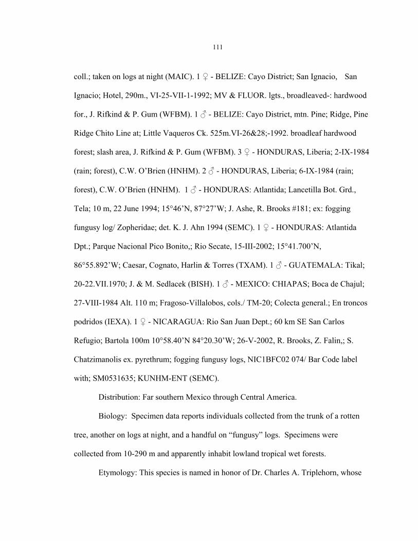
111
coll.; taken on logs at night (MAIC). 1 ♀ - BELIZE: Cayo District; San Ignacio, San
Ignacio; Hotel, 290m., VI-25-VII-1-1992; MV & FLUOR. lgts., broadleaved-: hardwood
for., J. Rifkind & P. Gum (WFBM). 1 ♂ - BELIZE: Cayo District, mtn. Pine; Ridge, Pine
Ridge Chito Line at; Little Vaqueros Ck. 525m.VI-26&28;-1992. broadleaf hardwood
forest; slash area, J. Rifkind & P. Gum (WFBM). 3 ♀ - HONDURAS, Liberia; 2-IX-1984
(rain; forest), C.W. O’Brien (HNHM). 2 ♂ - HONDURAS, Liberia; 6-IX-1984 (rain;
forest), C.W. O’Brien (HNHM). 1 ♂ - HONDURAS: Atlantida; Lancetilla Bot. Grd.,
Tela; 10 m, 22 June 1994; 15°46’N, 87°27’W; J. Ashe, R. Brooks #181; ex: fogging
fungusy log/ Zopheridae; det. K. J. Ahn 1994 (SEMC). 1 ♀ - HONDURAS: Atlantida
Dpt.; Parque Nacional Pico Bonito,; Rio Secate, 15-III-2002; 15°41.700’N,
86°55.892’W; Caesar, Cognato, Harlin & Torres (TXAM). 1 ♂ - GUATEMALA: Tikal;
20-22.VII.1970; J. & M. Sedlacek (BISH). 1 ♂ - MEXICO: CHIAPAS; Boca de Chajul;
27-VIII-1984 Alt. 110 m; Fragoso-Villalobos, cols./ TM-20; Colecta general.; En troncos
podridos (IEXA). 1 ♀ - NICARAGUA: Rio San Juan Dept.; 60 km SE San Carlos
Refugio; Bartola 100m 10°58.40’N 84°20.30’W; 26-V-2002, R. Brooks, Z. Falin,; S.
Chatzimanolis ex. pyrethrum; fogging fungusy logs, NIC1BFC02 074/ Bar Code label
with; SM0531635; KUNHM-ENT (SEMC).
Distribution: Far southern Mexico through Central America.
Biology: Specimen data reports individuals collected from the trunk of a rotten
tree, another on logs at night, and a handful on “fungusy” logs. Specimens were
collected from 10-290 m and apparently inhabit lowland tropical wet forests.
Etymology: This species is named in honor of Dr. Charles A. Triplehorn, whose

112
excellent species level revision of Zopherus was motivation to understand the other
genera of the tribe, and for considerable additional unpublished observations on the group
which were kindly shared with M. A. Ivie.
Diagnosis: Resembles the species S. emarginatus (Texas) and S. denticulata
(Mexico), in the dense setose vestiture, but is quite distinct in sculpture. The pronotum
has sub-parallel ridges beside the midline that have weakly divided elevations at the apex
and base (not quite as strong as S. emarginatus), the elytral sculpture consists of arcuate
rows of small round nodules, with one distinctly larger and teardrop shaped nodule at the
start of the apical declivity. The dense mottled tomentose vestiture, and banded apex of
the parameres are also quite distinct.
Phloeodes LeConte
(Figs. 63-74, 76-82, 99, 124)
This genus is revised in the following chapter, and only a diagnosis and species
synopsis of the genus is provided here.
Diagnosis: This genus is separated from all others of the tribe with 10-segmented
antennae, by having tubercles on the ventral surface and lacking a membranous
mandibular prostheca.
Synopsis of Phloeodes LeConte Species
P. diabolicus LeConte 1851 California, BAJA MEXICO, possibly Arizona
P. pustulosus LeConte 1859, NEW SYNONYMY

113
P. latipennis Casey 1907a P. ovipennis Casey 1907a, NEW SYNONYMY P. elongatus Casey 1907a, NEW SYNONYMY P. scaber Casey 1907a, NEW SYNONYMY P. angustus Casey 1907a, NEW SYNONYMY P. remotus Casey 1907b, NEW SYNONYMY
P. plicatus (LeConte) 1859 California, BAJA MEXICO, possibly Arizona
Noserus torvus (Casey) 1907a, NEW SYNONYMY N. collaris (Casey) 1907a, NEW SYNONYMY N. corrosus (Casey) 1907b, NEW SYNONYMY
N. convexulus (Casey) 1907b, NEW SYNONYMY P. venustus (Champion) 1884 NEW COMBINATION Central America
Nosoderma Solier NEW SENSE
(Figs. 83-91, 101, 103, 121-122)
The genus Nosoderma is the second largest in the tribe behind Zopherus, and at
the beginning of the study contained 23 valid named species. Nosoderma was described
by Solier into which he placed 5 species (Nosoderma denticulatum, Nosoderma
duponchelii, Nosoderma scabrosum, Nosoderma morbillosum, and Nosoderma vicinium)
without designating a type species. Casey (1907b) incorrectly designated the type
species as Eurychora inaequlais Say, which was not one of the species included in the
genus at the time of its description by Solier, this designation was followed by Doyen and
Lawrence (1979). The type species was subsequently correctly identified as Nosoderma
morbillosum Solier by Gebien (1936) who synonymized it and Nosoderma vicinium with

114
Nosoderma inaequale (Say), this designation was followed by Ślipiński and Lawrence
(1999).
Champion (1884) in his Biologia Centrali-Americana described 13 new species of
Nosoderma and Casey (1907b) added an additional 7. After Casey’s revision of the
zopherids, the 23 named Nosoderma species were placed in the genera Sesaspis,
Noserodes Casey, and Verodes Casey. This placement was followed by Gebien (1936),
but the three genera were all later synonymized with Nosoderma (Doyen and Lawrence
1979).
Phylogenetic analysis of this group suggests that as currently constituted it is not
monophyletic. The species group centered on Nosoderma denticulata forms a distinct
group and is here recognized as the reconstituted Casey genus Sesaspis NEW SENSE.
When Sesaspis is recognized as a genus, Nosoderma, Phloeodes, and Sesaspis all form
monophyletic genera with the only remaining problem being the placement of
Nosoderma venustus. This species does not fit the definition of either Nosoderma or
Sesaspis. It is excluded from Nosoderma because it lacks a membranous mandibular
prostheca, and from Sesaspis because the ventral surface is covered with tubercles and it
lacks an arcuate groove on the hypomeron. The phylogenetic analysis of the tribe
supports Nosoderma venustus at the base of a monophyletic lineage containing the two
California species Phloeodes diabolicus and Phloeodes plicatus. Based on this evidence,
Nosoderma venustus is moved to Phloeodes, and becomes the only member of that genus
to occur outside the California Floristic Province.

115
The genus Nosoderma as here defined contains nine species in two separate
clades, that both appear as polytomies in the consensus cladogram. A group of five
species occurs generally from Oaxaca, Mexico, north possibly into the United States
(there are unconfirmed localities of Nosoderma exsculptum in Texas with simply state
data), and another group containing four species that generally occur from Oaxaca,
Mexico, south into Central America.
The species-group occurring northwards from Oaxaca is generally restricted to
high-elevation cloud forests and contains the two most widespread species of Nosoderma,
Nosoderma inaequalis and Nosoderma exsculptum, and three that are quite rare.
Nosoderma scabrosum and Nosoderma asperatum appear to be sister-species that are
isolated to high elevation cloud forests, only Nosoderma scabrosum was included in the
phylogenetic analysis because the two species are coded exactly the same in the character
matrix. These species were presumed to be synonymous until the types of Nosoderma
scabrosum was examined. The type specimens are the only representatives of this species
that have been examined. Nosoderma insigne is the other rare species that is known only
from the state of Oaxaca. The elytral sculpture of this species, consisting of ridges that
are hooked at the apex is unique in Nosoderma.
The linage occurring in more southern locales in Central America contains the
species that Casey placed into the genera Noserodes (Nosoderma zunilensis, Nosoderma
guatemalensis, and Nosoderma sparsus) and Verodes (Nosoderma aequalis). The three
species previously placed in Noserodes are closely allied, and Nosoderma zunilensis and
Nosoderma guatemalensis are probably sister-species. The species previously placed in

116
Verodes is distinct among species of Nosoderma in having the elytral sculpture
completely reduced, even more so than Sesaspis doyeni, and lacking nodules on all
femora of the male, but these characters do not warrant generic distinction based on the
current analysis.
Diagnosis: The true core Nosoderma group is supported by the presence of a
distinct membranous prostheca on the mandible (Fig. 101), ventrally inserted labial palps
(Fig. 122), lack of any hint of an antennal cavity on the hypomeron, and the presence of
tubercles on the ventral surface (also in Phloeodes).
Type Material Examined: Nosoderma scabrosus: HOLOTYPE ♀ missing right
meta tarsus in MNHN. Red rectangle TYPE/ Solier-9641-34; Mus. Paris; hand written
on green square Nosoderma; scabrosum. Solier; denticla? (Chev. Dej.); Mexico/
scabrosus Sol 36, 3 Mex Bves/ Museum Paris; Coll. De. Marseul; 2842-90.
Nosoderma asperatum: LECTOTYPE %, both antennae broken off in BMNH.
Orange ringed circle Type/Mexico; Salle Coll./Nosoderma; asperatum Champ MS/ Sp.
Figured.;B.C.A.Col.IV.1./ red square Lectotypus; Nosoderma; asperatum Champion;
García-París des 2000. PARALECTOTYPE %, right antennae broken off in BMNH.
1671 on light blue square/Mexico; Salle Coll./chewed up label no visible
lettering/Nosoderma; asperatum Champ MS/B.C.A.Col.IV.1.;Nosoderma asperatum/Foto
on white square/red square Paralectotipo; Nosoderma; asperatum Champion; M. García-
París et al det. 2002.
Nosoderma exsculptum: LECTOTYPE ♀, right pro-leg missing in BMNH.
Orange ringed circle Type/Jalapa, Mexico; Hoege./& on square/Nosoderma; exsculptum

117
Champ- B.C.A.Col.IV.1.;Nosoderma exsculptum/red square Nosoderma; exsculptum
Champion; Lectotype García-París 2000. PARALECTOTYPES %, intact and % left
meta leg missing, left meso tarsus missing in BMNH. Jalapa, Mexico; Hoege./153 on
square/G.C. Champion Coll.; B.M. 1927/409/red square Paralectotipo; Nosoderma;
exsculptum Champion; M. García-París et al 2002.
Nosoderma anceps: LECTOTYPE ♀, intact in BMNH. Orange ringed circle
Type/Jalapa, Mexico; Hoege./Nosoderma anceps; Champ. ;? var.
exsculptum/B.C.A.Col.IV.1.; Nosoderma anceps/red square Lectotypus; Nosoderma
anceps; Champion; García-París 2000. PARALECTOTYPES two ♂, intact in BMNH.
Same data as LECTOTYPE
Nosoderma impressum:LECTOTYPE ♀, intact in BMNH. Orange ringed circle
Type/1668 on light blue square/Jacale, Mexico; Salle Coll./Nosoderma impressum,
Champ. MS./B.C.A.Col.IV.1.;Nosoderma impressum/red square Lectotypus; Nosoderma;
impressum; Champion; García-París des 2000. PARALECTOTYPE ♂ missing right
meta leg. Same data as LECTOTYPE without circle type label.
Nosoderma interruptum: LECTOTYPE ♀, missing left antennae, tarsus on card in
BMNH. Orange ringed circle Type/Las Vigas, Mexico; Hoege./Nosoderma interruptum
Champ./B.C.A.Col.IV.1.;Nosoderma interruptum/red square Lectotypus; Nosoderma;
interruptum Champion; García-París des 2000. PARALECTOTYPE ♂, intact in BMNH.
Same data as LECTOTYPE without circle type label.
Nosoderma insigne: LECTOTYPE ♀, intact in BMNH. Orange ringed circle
Type/Capulalpam/1657 on light blue square/Mexico; Salle Coll./Nosoderma insigne

118
Champ. MS/B.C.A.Col.IV.1.;Nosoderma insigne/red square Lectotypus; Nosoderma;
insigne Champion; Garcia-Paris des 2000. PARALECTOTYPE ♀, last 4 antennomeres
glued on, palps broken in BMNH. Same data as LECTOTYPE without type
label/1656/pink F on white square/M. García-París el al det. 2002.
Nosoderma carinatum: LECTOTYPE ♂, intact in BMNH. Orange ringed circle
Type/1659 on light blue square/Jacale, Mexico.; Salle Coll./Nosoderma carinatum,
Champ./B.C.A.Col.IV.1.;Nosoderma carinatum/red square Lectotypus; Nosoderma;
carinatum; Champion; García-París des 2000. PARALECTOTYPES ♀, broken right
meso and meta tarsus in BMNH. Las Vigas, Mexico.; Hoege./G.C. Champion Coll.; B.M.
1927-409/red square Paralectotypus; Nosoderma; carinatum; Champion; García-París des
2000. Intact ♀, same data as above/Paralectotipo M. Garcia-Paris et al det. 2002.
Nosoderma guatemalense: LECTOTYPE ♂, intact in BMNH. Orange ringed
circle Type/Calderas, Guatemala; Champion./Nosoderma guatemalense, Champ.; MS-Sp.
Figured./B.C.A.Col.IV.1.;Nosoderma guatemalense./red square Lectotypus Nosoderma;
guatemalense; Champion; García-París des 2000. PARALECTOTYPE ♀, intact in
BMNH. Same data as LECTOTYPE.
Nosoderma zunilense: LECTOTYPE ♀ missing left meta leg in BMNH. Orange
ringed circle Type/Cerro Zunil, 4000ft.; Champion./Nosoderma zunilense Champ; MS-
Sp. Figured./B.C.A.Col.IV.1.;Nosoderma zunilense/ red square Lectotypus Nosoderma;
zunilense; Champion; García-París des 2000. PARALECTOTYPE ♀, intact in BMNH.
El Zumbador, 2500ft.; Champion./Nosoderma; zunilense Champ MS; B.C.A.Col.IV.1.
Nosoderma zunilense/pink F on white square/ red square Paralectotipo Nosoderma;

119
zunilense; Champion; García-París et al det. 2002.
Nosoderma sparsum: LECTOTYPE ♀, missing left meso tarsus in BMNH.
Orange ringed circle Type/Totonicapama,; 85-10,500 ft. Champion./Nosoderma sparsum
Champ./Sp. Figured/B.C.A.Col.IV.1.; Nosoderma sparsum/red square Lectotypus
Nosoderma; sparsum; Champion; García-París des 2000. PARALECTOTYPE ♂, intact
in BMNH. Same data as LECTOTYPE.
Nosoderma aequale: HOLOTYPE ♀, left antennae broken, left pro and meso
tarsus broken, right meso and meta tarsus broken in BMNH. Orange ringed circle
Type/Parada/1654 light blue square/Mexico.; Salle Coll./Nosoderma aequale; Champ.
MS./Sp. Figured/B.C.A.Col.IV.1.;Nosoderma aequale/red square Holotypus Nosoderma
aequale García-París des 2000.
Synopsis of Nosoderma Solier Species
N. inaequale (Say) 1835 Mexico
N. morbillosum Solier 1841 N. vicinum Solier 1841 N. championi Casey 1907b, NEW SYNONYMY N. prominens Casey 1907b, NEW SYNONYMY N. senex Casey 1907b, NEW SYNONYMY N. brevicolle Casey 1907b, NEW SYNONYMY N. subglabrum Casey 1907b, NEW SYNONYMY
N. insigne Champion 1884 Mexico
N. interruptum Champion 1884, NEW SYNONYMY
N. scabrosum Solier, 1841 Mexico
Scoriaderma congolense Fairmaire 1894b, NEW SYNONYMY

120
5
6 Figs. 5 and 6. 5. Wide angle distribution of specific localities of examined specimens of Nosoderma. 6. Closer view of distribution of specimens of Nosoderma centered about Mexico City, MEXICO. N. inaequalis, blue squares. N. insigne, yellow circles. N. asperatum, green square. N. exsculptum, red squares. N. scabrosum, white circles. N. aequalis, blue squares. N. guatemalensis, white squares. N. sparsus, yellow square. N. zunilensis, red circles. N. asperatum Champion 1884 Mexico
N. exsculptum Champion 1884 Mexico, possibly Texas
N. carinatum Champion 1884, NEW SYNONYMY N. anceps Champion 1884, NEW SYNONYMY

121
N. impressum Champion, 1884, NEW SYNONYMY N. longipennis Casey 1907b, NEW SYNONYMY N. zunilensis Champion 1884 Central America
N. guatemalensis Champion 1884 Central America
N. squalidus Casey 1907b, NEW SYNONYMY
N. sparsus Champion 1884 Guatemala
N. aequalis Champion 1884 Mexico
Key to the Species of Nosoderma Solier
1. Dorsal surface of pronotum with all setae singly inserted at the base of tubercles;
tubercles on pronotum uniformly distributed………………………….…………2
1’. Dorsal surface of pronotum with dense setae between tubercles inserted directly on
surface; tubercles on pronotum more dense on ridges, and absent in intervening
areas………………….……………………………………………………………5
2. (1). Pronotal and elytral surface smooth with dense flattened tubercles lacking any
distinct ridges or nodules; male femoral nodules absent. Mexico, Oaxaca. Fig. 91.
……………….....................…………………………..…… N. aequalis Champion
2’. Pronotal and elytral surfaces with distinct ridges and nodules; male femoral nodules
present on at least meso and metafemora………….…….……………………..…3
3. (2’). Metepisternum irregularly foveolate; elytra elongate, 1.95-2.05X longer than
widest point; elytral smooth at apex. Guatemala. Fig. 88.
……………………………………………………...………..N. sparsus Champion

122
3’. Metepisternum tuberculate; elytra more stout, 1.60-1.75 X longer than widest point;
elytra very shallowly to deeply emargiate at apex. Mexico and Central
America………… ……………..…………………….............................................4
4. (3’). Elytra shallowly emarginate at apex, rounded; 5th elytral interval with weak
ridge. Mexico and Central America. Fig. 90.
………………………………………………………. N. guatemalensis Champion
4’. Elytra deeply emarginate at apex, sharply pointed; 5th elytral interval containing three
large disconnected nodules. Mexico and Central America. Fig. 89.
………...........................................................................…..N. zunilensis Champion
5. (1’). Base of elytra with two broad shallow transverse depression across suture;
posterior angles of pronotum weakly to moderately expanded; prosternum
anterior of procoxae flat; 5th elytral interval with straight ridge starting at base;
apical third of elytron with 4 small nodules. Mexico. Fig. 84.
......................................................................................… N. exsculptum Champion
5’. Base of elytra without transverse depressions; posterior angles of pronotum strongly
expanded; prosternum anterior of procoxae with broad depression; 5th elytral
interval with arcuate ridge starting at base, beyond level of mesocoxal cavity, or
only nodules; apical third of elytron with less than 4 nodules…..………………...6
6. (5’). Third elytral interval with subapical ridge distinctly hook at apex; apex of elytra
with swollen ridge around suture; 5th elytral interval with arcuate ridge starting at
base. Mexico. Fig. 87. …………........................................… N. insigne Champion

123
6’. Third elytral interval with short ridge at base, only nodules, or combination of both;
apex of elytra flat around suture; 5th elytral interval simple at base........................7
7. (6). Third elytral interval with short ridge at base; 5th elytral interval with arcuate ridge
from level of just behind mesocoxal cavities to the suture between ventrites 2 and
3; apical third of elytron with three moderate sized nodules subequal in size, one
near apex slightly smaller than subapical two. Mexico. Fig. 83.
………………………………………….......…………….……. N. inaequalis Say
7’. (6’). Third elytral interval with nodule at base; 5th elytral interval with disconnected
nodules arranged in a row approximately from the level of the midpoint of
metasternum to suture between ventrites 1 and 2; apical third of elytron with three
large projecting conical nodules, subapical very large, much larger than one near
apex. Mexico ………………...……………………………………………………8
8. (7’). Apical margin of elytra weakly serrate; male nodule present on all femora.
Mexico. Fig. 86. ……………………………………………..N. scabrosum Solier
8’. (7’). Apical margin of elytra with strongly projecting conical nodules, appearing very
strongly serrate; male nodule present only on meso and metafemora. Mexico, Hidalgo.
Fig. 85. ……………………………………….N. asperatum Champion
Future Research
More data are needed on the Zopherini in all areas where they occur. Many
species are highly restricted in their habits and require specific collecting efforts to
discover specimens. The known distributions for most Central American species are

124
probably incomplete and represent only a portion of the actual populations that exist.
The collection of additional specimens preserved for molecular study would bring an
entirely knew data set into the phylogenetic analysis of the tribe. Even representative
samples of the genera are not currently available for molecular analysis. Obtaining
specimens of the genera Meralius, Sesaspis, and Nosoderma for molecular analysis
would add characters that could go a long way to support or reject the current hypothesis
on the classification of the described species.
The larvae of several genera remain undescribed, and those that are described are
usually only based on a single species. A more complete representation of larvae in the
Zopherini would add significant characters to future phylogenetic analyses.
It is also possible that new species are yet to be discovered in isolated high
elevation forests and/or lowland tropical forests throughout Central America. Only the
genus Zopherus has been recorded from El Salvador, but other genera of the tribe almost
certainly occur in that country.
The relationship between Zopher and Noserinus needs further investigation as to
whether the group forms a single monophyletic genus, or is in fact two distinct genera.
New species could also be discovered in Southeastern Asia, and South America.
The very large body-sized monotypic genus Zopherosis would be interesting to
examine at the population level. This genus is predicted to be relatively old and highly
fragmented. It would be interesting to examine the intraspecific population structure to
see if it indicates low level of gene flow between distinct identifiable populations.

125
The species level taxonomy of the genus Scoriaderma has yet to be resolved, due
to a lack of specimens. It is possible that only one relatively widespread species exists in
Eastern Africa and on the Comoro Islands. Molecular data from this genus would also
offer insight into its relationship within the Zopherini, as to whether it is more closely
related to Zopherus + Zopherosis, or to Meralius and the American genera, or if it is
possibly intermediate between the two clades.
Many members of the Zopherini are quite rare in collections, especially
considering their large size, and future collecting efforts will be important to continuing
to advance our knowledge of this diverse assemblage of remarkable beetles.

126
CHAPTER 4
REVISION OF THE GENUS PHLOEODES
Introduction
Phloeodes is closely related to the other large flightless “ironclad” beetles with
10-segmented antennae and closed procoxal cavities. It is one of eight recognized genera
in the apparently monophyletic tribe Zopherini (sensu Ślipiński & Lawrence 1999).
While the genera have recently been redescribed (Ślipiński & Lawrence 1999), the
species are poorly defined and their placement is unresolved. I examined the species of
Phloeodes LeConte 1862 (=Noserus LeConte 1862, sensu Ślipiński & Lawrence 1999,
Ivie 2002c) to evaluate the many names available for the group (Doyen and Lawrence
1979, Ivie 2002c). There are currently 14 names available but they have not been
evaluated since Casey (1907a and 1907b). The surplus of available names in this genus
makes it difficult to achieve accurate determinations for individual specimens. The
desperate need for a species-level revision of this group was pointed out by Ivie (2002c),
and was undertaken with the explicit purpose of clearing up the nomenclature of the
genus. I have revised the species, redefined the genus based on a phylogenetic analysis
of the tribe, provided descriptions, illustrations, a key, as well as documented the
distribution of the species.
Taxonomic History
The genera Phloeodes LeConte and Noserus LeConte were originally described

127
for species described in Nosoderma Solier 1841. The taxonomic histories of the species
have followed separate paths since 1862. These paths recently converged when Noserus
was synonymized with Phloeodes (Ślipiński and Lawrence 1999). This synonymy was
not recognized by García-París et al. (2000, 2001) who described a Mexican species,
Noserus doyeni García-París et al. 2001, in the old sense of Noserus which returned the
genus to independent status. Ivie (2002c) restored it to synonymy following Ślipiński
and Lawrence (1999), but little actual analysis of this question has been conducted.
The genus Phloeodes originally included two California species, Nosoderma
diabolicum LeConte 1851, and Nosoderma pustulosum LeConte 1859 (LeConte 1862).
This genus was differentiated from Zopherus Gray and Phellopsis LeConte by having 10-
segmented antennae and from Noserus LeConte by the degree of closure of the
prothoracic antennal cavity. The genus Nosoderma Solier, occurring in Central America,
was not included in LeContes’ generic key (1862), but he did mention that it differed
from Phellopsis in the number of antennal segments (10 vs. 11). He did not comment on
any differences between the three genera with 10-segmented antennae (Noserus,
Nosoderma, and Phloeodes).
The only attempted revision of the components of Phloeodes was conducted by
Casey (1907a, 1907b). In these two papers Casey described 6 new species of California
Phloeodes: P. latipennis 1907a, P. ovipennis 1907a, P. elongatus 1907a, P. scaber 1907a,
P. angustus Casey 1907a, and P. remotus Casey 1907b. Casey’s (1907a) contention was
that the two LeConte Phloeodes species were divided into a northern species (Phloeodes
diabolicus) and a southern species (Phloeodes pustulosus) separated by the presence of a

128
dense pale whitish vestiture on the apical declivity of the elytra in Phloeodes. To this
interpretation, Casey (1907a, 1907b) then described his 6 new Phloeodes species from
few or single specimens. These specimens were distinguished in typical Casey key
format based on variations in sculpture, body proportions, velvety spots on the elytra, and
vestiture of the apical declivity. All of these characters have been observed to be highly
variable within the genus, frequently because the external appearance of individuals is
concealed by the accumulation of exudate and collected environmental debris. Casey had
some inkling of this condition when he commented that some of his species may prove to
be subspecies (1907a) and that some of his previously described Phloeodes species
seemed doubtful (1907b). Despite these reservations, he concluded after examining
additional specimens, that all of the species were valid and proceeded to described
Phloeodes remotus from a single specimen (Casey 1907b). LeConte did not recognize a
type species when he erected the genus, therfore Casey (1907b) designated Phloeodes
diabolicus as the type species. To date, only one of the Casey names, Phloeodes
latipennis, has been synonymized. It was listed as junior synonym of Phloeodes
pustulosus by Gebien (1936).
LeConte (1862) described the genus Noserus, in the same paper that he described
Phloeodes, with Nosoderma plicatum LeConte 1859 from California as the sole member
of the genus. Noserus emarginatus Horn 1878 was added from Texas, considerably
expanding the geographic range of the genus.
In a situation similar to his treatment of Phloeodes, Casey (1907a, 1907b) also
revised Noserus. In addition, he recognized the close relationship between Phloeodes

129
and Noserus and noted that in both groups the species forms were difficult to define
(Casey 1907b). However, Casey decribed several new California Noserus species based
on single or a few specimens as he had done in Phloeodes: Noserus torvus Casey 1907a,
Noserus collaris Casey 1907a, Noserus corrosus Casey 1907b, and Noserus convexulus
Casey 1907b. In describing these taxa, Casey primarily used differences in the strength
of elytral ridges and tubercles as well as degree of body convexity; these characters are
highly variable in most members of the tribe and are not very useful in defining species
limits.
The most recent species addition to the genus was Noserus doyeni García-París et
al. (2001) from Nuevo Leon, Mexico. The confusion surrounding the valid names in
California Noserus species has been noted by García-París et al. (2001), who published a
key to species of adult Noserus without including the Casey species. They stated that
these species are a “group of poorly defined forms allied to Noserus plicatus, which do
not deserve a specific taxonomic status because most of their diagnostic characters are
variable features widespread along the geographic range” (García-París et al 2001).
However, they never formally synonymized the names, leaving them in limbo.
This group has proven to be a perfect example of Casey’s approach to species
description, where he describes individual specimens rather than true species. Triplehorn
(1972) gave an excellent critical review of the genus Zopherus, in which he retained only
one of the 23 species described by Casey, recognizing a maximum of seven synonyms for
a single previously-described species (Zopherus gracilis Horn).
At the generic level, I have found that two of four valid species (Noserus

130
emarginatus and Noserus doyeni) do not belong to either available generic name, but
rather belong to a genus mistakenly synonymized elsewhere. Thus, at the beginning of
this study the genus Phloeodes (=Noserus) contained 14 validated species names with
only one published synonym. Virtually all of these names are unused in collections and
the literature, and no available resources existed that provided any characteristics by
which species-level decisions could be made. In their genera of Zopherinae (sensu lato),
Doyen and Lawrence (1979) stated there were only eight valid names in this group, two
in Phloeodes and six in the old Noserus, but they published no synonymies and did not
indicate which species names were in fact valid.
When Noserus LeConte was synonymized with Phloeodes LeConte by Ślipiński
and Lawrence (1999) in their review of the genera of Zopherinae, the synonymy was
based on the fact that there are no distinguishing characters between the taxa besides the
extent of the antennal cavity. All available evidence supports that Phloeodes diabolicus
and Phloeodes plicatus, the respective type-species of the genera Phloeodes and Noserus,
belong to the same genus, and that synonymy is recognized here.
The problems with the classification of Phloeodes in this sense comes from the
two described species that occur outside of California, Noserus emarginatus from Texas
[possibly Northern Mexico (García-París et al. 2000)] and Noserus doyeni, from Nuevo
Leon, Mexico. Casey, who divided the Zopherini (sensu Ślipiński and Lawrence 1999)
into several genera that were later synonymized (Triplehorn 1972, Doyen and Lawrence
1979) was the first to acknowledge that true Phloeodes, would be confined to California
(1907b). Even though Casey did not examine specimens of Noserus emarginatus, he
131
stated that it would prove to be generically different and possibly belong to the genus
Nosoderma Solier of Central America. The placement of the eastern species in Noserus
has recently been questioned (García-París et al. 2000), and some similarities with
species of Nosoderma have been acknowledged but without action. Horn (1878)
mentioned a general similarity in sculpture between Noserus emarginatus and Meralius
echinatus (Guérin-Méneville) from Cuba but did not suggest any actual relationship
between these species, but probably recognized differences from the California species.
These two species of “Noserus” differ from the current diagnosis of Phloeodes in
having setiferous punctures on the ventral body surfaces, a different type of antennal
cavity, a poorly defined tarsal strip, variation in the male nodules, and the generally dense
setose vestiture. They also differ from species of Nosoderma in lacking a membranous
mandibular prostheca, in the presence of ventral setose punctures and laterally inserted
labial palps. Therefore, these two species are removed from the genus Phloeodes, and
added to the genus Sesaspis Casey NEW SENSE as Sesaspis emarginatus (Horn) NEW
COMBINATION and Sesaspis doyeni (García-París et al.) NEW COMBINATION (see
previous chapter for further discussion). In the process of resolving this quandary, the
species currently placed in Nosoderma were examined, and unexpectedly an additional
species belonging to Phloeodes was discovered. Nosoderma venustus was described
from Guatemala by Champion (1884), and is here recognized as a member of Phloeodes.
The results of a phylogenetic analysis of the Zopherini (Chapter 2, above) have
led to significant changes in the membership of Phloeodes proposed here. The two
Phloeodes species from the Gulf of Mexico region (emarginatus and doyeni) were found

132
to belong to a group with members in Nosoderma, but that cluster together as a separate
linage. Those species are now to be placed in a reconstituted Sesaspis Casey.
Phloeodes (sensu strictu) is here restricted to the two California species
(Phloeodes diabolicus and Phloeodes plicatus) and the Central American Phloeodes
venustus (Champion) NEW COMBINATION, which was discovered to be a member of
Phloeodes during the course of this study. The species here considered to be members of
Phloeodes LeConte represent quite distinct forms. While the habitat and geographic
range of the California species seem to overlap entirely, the two species are easily
distinguishable. Phloeodes venustus, moved into Phloeodes from Nosoderma, was called
the “finest and most distinct” of all the Central American species (Champion 1884), so it
is also readily identifiable. Limiting the valid species names in California to two, while
synonymizing nine of Casey’s names and one of LeConte’s, will hopefully clear up some
of the confusion surrounding these large and very interesting beetles.
The reader is referred to Chapter 2 for methods and detailed results of the
phylogenetic analysis and rationale for these actions.
Materials
The current study was based on the examination of over 5,000 adult specimens
including all types of the genus Phloeodes, as well as an equal number of related genera.
Adults of the two California species recognized here are relatively common and are very
well represented in museum collections. In contrast, the Texan, Mexican, and Central
American species are far less collected. The following numbers of adults of each species

133
were examined: Phloeodes diabolicus (n=3,875), Phloeodes plicatus (n=1035),
Phloeodes venustus (n=21), Sesaspis doyeni (n=113), Sesaspis emarginatus (n=70). The
availability of adult specimens was sufficient to complete a thorough examination of
morphology and delimit each species.
The material for this investigation was obtained on loan from most of the North
American entomological collections, as well as many collections in Europe, and a few
Central American collections. Specimens were obtained from the following institutions
and collections (the curator responsible for the loan is listed in parenthesis):
AAPC - Albert Allen Personal Collection, Boise, Idaho (Albert Allen).
ASUT - Arizona State University, Tempe, Arizona (David Pearson).
BMNH - The Natural History Museum, London, United Kingdom (Maxwell V. L.
Barclay).
BPBM - Bernice P. Bishop Museum, Honolulu, Hawaii (Alistair S. Ramsdale).
BYUC - Brigham Young University, Provo, Utah (Shawn M. Clark)
CASC- California Academy of Sciences, San Francisco, California (Norm Penny and
David H. Kavanaugh).
CHICO - Chico State University, Chico, California (Donald Miller).
CMNC - Canadian Museum of Nature, Ottawa, Ontario (François Génier).
CNCI - Canadian National Collections of Insects, Ottawa, Ontario (Patrice Bouchard).
CSCA - California State Collection of Arthropods, Sacramento, California (Chuck
Bellamy).
CSUC - Colorado State University, Fort Collins, Colorado (Boris C. Kondratieff).

134
DBTC - Donald B. Thomas Personal Collection, Weslaco, Texas (Donald Thomas).
DEI-ZALF - Deutsches Entomologisches Institut (DEI) in Leibniz-Zentrums für
Agrarlandschaftsforschung (ZALF), Müncheberg, Germany.
DKYC - Daniel K. Young Personal Collection, Madison, Wisconsin (Daniel K. Young).
EMEC - University of California, Berkeley, California (Cheryl Barr).
ENMU - Eastern New Mexico University, Portales, New Mexico (Darren A. Pollock).
FMNH - Field Museum, Chicago, Illinois (James H. Boone)
FSCA - Florida State Collection of Arthropods, Gainesville, Florida (Paul E. Skelley).
HNHM - Hungarian Natural History Museum, Budapest, Hungary (Otto Merkl).
HUMB - Humboldt State University, Arcata, California (Michael Camann).
IEXA- Instituto De Ecología, A.C., Xalapa, Veracruz, Mexico (Miguel Angel Morón
Ríos).
INBC - Instituto Nacional de Biodiversidad (INBio), Santo Domingo de Heredia, Costa
Rica (Angel Solís).
INHS - Illinois Natural History Survey, Champaign (Colin Favret).
IRCW - University of Wisconsin, Madison, Wisconsin (Steven Krauth).
JEWC - James E. Wappes Personal Collection, Bulverde, Texas (James E. Wappes).
LACM - Natural History Museum of Los Angeles County, Los Angeles, California
(Weiping Xie).
LSAM - Louisiana State Arthropod Museum , Baton Rouge, Louisiana (Victoria Bayless).
LUND - Lund University, Lund, Sweden (Roy Danielsson).
MAIC - Michael A. Ivie Private Collection, Bozeman, Montana (Michael. A. Ivie).

135
MCPM - Milwaukee Public Museum, Milwaukee, Wisconsin (Susan Borkin).
MCZ - Museum of Comparative Zoology, Harvard University, Cambridge,
Massachusetts (Philip D. Perkins).
MNHN - Museum National d'Histoire Naturelle, Paris, France.
MIZ - Museum and Institute of Zoology, Polish Academy of Sciences, Warszawa, Poland
(Wioleta Tomaszewska).
MSUC - Michigan State University, East Lansing, Michigan (Gary L. Parsons).
MTEC - Montana Entomology Collection, Montana State University Bozeman (Michael
A. Ivie).
NHMB - Natural History Museum Basel, Switzerland (Michael Brancucci and Eva
Sprecher).
NHMW - Naturhistorisches Museum Wien, Vienna, Austria (Heinrich Schönmann).
NMPC - National Museum of Natural History, Prague, Czech Republic (Svatopluk Bílý).
OSAC - Oregon State Arthropod Collection, Oregon State University, Corvallis (Andrew
Brower).
RLAC - Rolf L. Aalbu Personal Collection, Sacramento, California (Rolf L. Aalbu).
ROME - Royal Ontario Museum, Toronto, Ontario (Brad Hubely).
SBMN - Santa Barbara Museum of Natural History, California (Michael S. Caterino).
SEMC - Snow Entomological Collections, University of Kansas, Lawrence (Zachary H.
Falin).
SMDV - Spencer Entomological Museum, University of British Columbia, Vancouver
(Karen M. Needham).

136
TAMU - Texas A&M University, College Station (Edward G. Riley).
UTSC -Utah State University, Logan (Colin Brammer).
UCDC - Bohart Museum of Entomology, University of California-Davis (Steve L.
Heydon).
UCMC - University of Colorado Museum, Boulder (Virginia Scott).
UCRC - University of California-Riverside (Douglas Yanega).
UGCA - Georgia Museum of Natural History, Athens (Cecil L. Smith).
NMNH - National Museum of Natural History, Washington D.C. (Warren E. Steiner)
WFBM - William F. Barr Entomological Museum, Univ. of Idaho, Moscow (Frank W.
Merickel).
WSUC - Washington State University, Pullman (Richard Zack).
ZIN - Russian Academy of Sciences, St. Petersburg (Mark G. Volkovitsh).
Methods -- Morphology
This revision was based on morphological characters primarily of adult specimens
following the operational species concept of Whitehead (1972). This concept
hypothesizes that unique morphological characters have a genetic basis. These unique
characters can be inherited, and therefore used to delimit species. The California species
of Phloeodes are better defined and morphologically quite distinct when compared to
other members of the subfamily, so initial sorting of the “species” was done based on
general gestalt, and then specific characters were examined to delimit the species and
generate a character set for phylogenetic analysis.

137
Frequently specimens of the tribe are encrusted with a greasy exudate as well as
accumulated environment debris, such as sand and soil. This makes morphological
structures very difficult to examine. In order to examine external morphological
structures, specimens were cleaned. To clean the specimens they were first relaxed in hot
water (90-100° C) for 5-10 minutes. Once relaxed, specimens were placed in an
ammonium hydroxide solution (Parsons’® household ammonia) in an ultrasonic cleaner
for 10-15 minutes, followed by a distilled water rinse. Any remaining obstructions were
then scraped away using the point of an insect pin. Dissection and disarticulation were
used to study certain morphological characters (i.e. mouthparts, genitalia) of relaxed
specimens.
Specimens were studied on a Leica® Wild M3C stereoscope equipped with a
150w fiber optic illuminator. Habitus images of larger specimens were made using an
Olympus DP11 digital camera system, mounted to a NIKON® micro-NIKKOR 105mm
lens. Images of smaller morphological characters and structures were made using a JVC
(DC Ky-F75U) digital camera mounted on a Leica® MS5 stereoscope, attached to an
IBM IntelliStation M Pro® with a 1GHz Pentium4® processor. Enhancements to digital
images were made using the Syncroscopy AutoMontage® software and edited in Adobe
PhotoShop® 5.5. Line drawings were made by tracing digital images with a drawing
tablet in Adobe PhotoShop® 5.5.
Nomenclature of morphological structures follows Doyen (1966), Doyen and
Lawrence (1979), Lawrence and Britton (1991) and Ślipiński and Lawrence (1999). A
tubercle is defined as a rounded protuberance of the cuticle that has a single inserted seta.

138
This differs from a nodule, which is used to refer to the large rounded or tear-drop shaped
elevation of an entire cuticular area that may have setae and/or tubercles on the surface.
Specifically, the “male nodule” refers to an elevated circular to elliptical area near the
base of the femora of the male in many species of Zopherini. The term “setiferous
fossae” was recently used in Noserus (García-París et al. 2001) to refer to pits of the
cuticular surface with a single inserted setae, I use setose punctures to refer to these
features following Harris (1979). Other sculpture terms follow Harris (1979) and Nichols
(1989).
The label transcription for type specimens follows Ivie (1985). Data on each line
within a label is separated by “;” (semicolon); each individual label is separated by a “/”
(backslash).
Taxonomy of the Genus Phloeodes LeConte
(Figs. 63-74, 76-82, 99, 124)
Phloeodes LeConte 1862: 216. (Type species: Nosoderma diabolicum LeConte, 1851,
designated by Casey 1907b). LeConte 1853: 235. Horn 1870: 272-273. LeConte
and Horn 1883: 364. Champion 1884: 44. Casey 1907a: 42-43. Casey 1907b:
471-473. Leng 1920: 223. Essig 1926: 429. Bradley 1930: 183, 322. Böving and
Craighead 1931: 41. Essig 1934: 110. Gebien 1936: 667-668. Arnett 1973: 651.
Doyen 1976: 267-272; fig. 1-11. Doyen and Lawrence 1979: 341-345. Powell and
Hogue 1979: 295, 297. Doyen 1980: 3. White 1983: 250, fig. 107. Arnett 1985:
350. Costa et al. 1988: 213. Hogue 1993: 296, 298. Ślipiński and Lawrence 1999:

139
19; fig. 69-82. Ivie 2002c: 458-461. Caterino 2004: 7.
Noserus LeConte 1862: 216. (Type species: Nosoderma plicatum LeConte, 1859, by
monotypy). Synonymy by Ślipiński and Lawrence (1999). Horn 1870: 273.
LeConte and Horn 1883: 365. Champion 1884: 44. Casey 1907a: 43-44. Casey
1907b: 473-474. Leng 1920: 223. Bradley 1930: 183, 322. Böving and Craighead
1931: 41. Gebien 1936: 668. Arnett 1960: 651, 668. Arnett 1968: 651, 668. Arnett
1973: 668. Arnett 1983: 16. Doyen and Lawrence 1979: 345. Doyen 1980: 3.
Arnett 1981: 145. Arnett 1985: 350. Ślipiński & Lawrence 1999: 19. García-París
et al. 2000: 473. García-París et al. 2001: 151-153. Ivie 2002: 461.
Ageonoma Pascoe, 1866: 478. (Type species: Nosoderma diabolicum LeConte, 1851, by
monotypy). Synonymy by Horn (1870). Horn 1870: 272.
Phoeodes Arnett 1962: 668 (lapsus calami).
Noserosus Doyen and Lawrence 1979: 345 (lapsus calami).
Noserodes [in part] Gebien 1936: 670.
Diagnosis: The current phylogenetic analysis supports this group based on the
synapomorphy of a well-developed glabrous tarsal strip (Fig. 71), this character also
occurs as a homoplasy in the distantly related Zopherus, and weakly in some Nosoderma,
but Zopherus has 9-segmented antennae (10 in Phloeodes) and Nosoderma has no hint of
an antennal cavity (well developed in Phloeodes, Figs. 67-70). This genus is very closely
related to the genus Nosoderma Solier, and can be further distinguished from that genus
(Ślipiński and Lawrence 1999) by the lack of a mandibular prostheca; the broad mentum;
and the laterally inserted labial palps. The presence of a tomentose whitish vestiture on

140
the humeri and apical declivity in many specimens of Phloeodes diabolicus, and covering
a large portion of the body in Phloeodes venustus will separate those two species from all
other Zopherini with 10-segmented antennae that lack a scutellum. The third member of
the genus, Phloeodes plicatus is uniformly colored, but has a unique antennal cavity
structure that is distinct but not clearly limited in the posterior margin (Fig. 68).
Description (male): 9.5-29 mm. Body elongate and subcylindrical, dark brown,
gray to black, often with whitish vestiture on portions or majority of elytra. Dorsal
surface densely granulose, covered with small black tubercles bearing single laterally
inserted golden setae; secondary vestiture consisting of short appressed setae dark to
white; arcuate velvety spots often present; elevated ridges and tubercles distinct to absent.
Head inserted into prothorax to subgenal ridge; not constricted behind eyes; eyes
transverse, slightly emarginate; interfacetal setae absent; occiput with median
longitudinal ridge; frontal ridges clearly raised above antennal insertions; subgenal ridge
strong. Antenna 10-segmented; relatively short, not extending beyond middle of
pronotum; antennomere 2 transverse, shorter than 1 or 3; antennomere 3 quadrate, longer
than wide; antennomere 9 with anterior slightly protruding smooth area; weakly
developed 1- or 2- segmented club; antennomere 10 with micro-setose field covering
entire apex, visible laterally; antennomeres with regularly spaced bristles. Gular suture
arcuate; tentorial pits near base along suture, small rounded to slightly elongate; gular
incisions u-shaped; mentum narrowed anteriorly, anterior edge sharply emarginate;
ventrally setose; submentum without setose pit. Labrum transverse, margin rounded,
fringed with golden setae; labial palps inserted laterally (Fig. 76-77); maxillae with

141
lacinia and galea densely covered with golden setae; mandible bidentate, without
membranous prostheca, more than half of basal portion strongly punctured laterally, apex
smooth.
Pronotum with anterior edges produced forwards; lateral margins arcuate; disc
with shallow depressions, and weak arcuate ridges; prothoracic hypomera with distinct
antennal cavities or shallow depression, complete or not (Fig. 67-70); prosternum with
deep pits to shallow depression along inner margin on prosternal suture; prosternal
process concave; procoxal cavities externally closed (Fig. 67-68).
Flight wings absent; suture distinct but elytra fused; scutellum not externally
visible; dorsum of elytra with irregular punctures and small black tubercles, often with
paired velvety patches or elevated ridges; tip of elytra at most slightly emarginate.
Femora with small black tubercles covering surface; tibia with moderate to weak
setose ridges, with slightly too strongly expanded distal nodule containing tuft of golden
setae extending between paired apical spurs; setose nodules on inner surface of all femora
(Fig. 79-82); tarsal formula 5-5-4; tarsi with all segments bearing rows of golden setae
with median area with glabrous strip (Fig. 71); last segment with simple but strong claws
(Fig. 78).
Metasternum sub-equal in length to abdominal V1; intercoxal process of
abdominal V1 broad; abdomen with ventrites 1 to 4 connate, 5 free; all with irregular
small black tubercles on surface; V5 with irregular semicircular preapical groove. Male
genitalia (Figs. 72-74) with basal piece scoop shaped; parameres fused basally, strongly
to weakly emarginate at tip, with tufts of setae. Median lobe narrow, acute.

142
Female: Differs from male in lacking nodules near base on the ventral surface of the
femora.
Larva: (see description of the larva of Phloeodes diabolicus below).
Notes: The inclusion of Phloeodes venustus in this genus is based on the absence
of a membranous mandibular prostheca (which excludes it from Nosoderma), presence of
tubercles on all body surfaces (which excludes if from Sesaspis), a well defined tarsal
strip, broad apical margin of the mentum, and laterally inserted labial palps. The whitish
vestiture that is seen in many specimens of Phloeodes diabolicus, and was the historical
distinction between that species and Phloeodes pustulosus, is exactly the same vestiture
that covers nearly the entire surface of the body in Phloeodes venustus. This whitish
vestiture is seen in no other species of Zopherini with tuberculate sculpture.
Synoptic Catalog of Phloeodes Species
Phloeodes LeConte 1862
Ageonoma Pascoe 1866
Noserus LeConte 1862
P. diabolicus (LeConte) 1851
P. pustulosus (LeConte) 1859, NEW SYNONYMY
P. latipennis Casey 1907a, synonomy by Gebien 1936
P. ovipennis Casey 1907a, NEW SYNONYMY
P. elongatus Casey 1907a, NEW SYNONYMY

143
P. scaber Casey 1907a, NEW SYNONYMY
P. angustus Casey 1907a, NEW SYNONYMY
P. remotus Casey 1907b, NEW SYNONYMY
P. plicatus (LeConte) 1859
Noserus torvus Casey 1907a, NEW SYNONYMY
Noserus collaris Casey 1907a, NEW SYNONYMY
Noserus corrosus Casey 1907b, NEW SYNONYMY
Noserus convexulus Casey 1907b, NEW SYNONYMY
P. venustus (Champion) 1884, NEW COMBINATION
Biology
A general evolutionary trend towards wood boring larvae has been suggested in
the Zopherini (Triplehorn 1972, Doyen and Lawrence 1979, Doyen 1976, Ślipiński and
Lawrence 1999, Ivie 2002c). The larval morphology of Phloeodes diabolicus is
obviously modified for a wood-boring habitat possessing an enlarged thoracic region and
reduced legs (Doyen 1976). The larva of Phloeodes diabolicus was described from
cottonwood logs (Populus sp.), oak logs (Quercus sp.), and mulberry roots (Morus) and
was suggested as a nonspecific decomposer that probably utilizes a variety of tree species
(Doyen 1976). While for many larval Zopherinae the associated substrate is rotting
wood, the nutritive source may be associated fungi (Doyen 1976, Ivie 2002) possibly
white rot fungi (Lawrence 1991).

144
Adults of Phloeodes diabolicus and Phloeodes plicatus are most commonly
collected under bark of decaying oak (Quercus sp.) and cottonwood (Populus sp.) trees,
but have also been found associated with woody material of willow (Salix sp.), alder
(Alnus sp.), sycamore (Platanus sp.), walnut (Juglans sp.), eucalyptus, cedar (Cedrus
sp.), pine (Pinus sp.), madrone (Arbutus sp.), and laurel (Umbellularia sp.), as well as
under several shrubs (including Salicornia sp. and Baccharis sp.), on fungi, in leaf litter,
and under rocks. Multiple individuals of both species were found in the same
microhabitat, with both species being relatively abundant under the same standing dead
oak trees in Monterey Co., California in 2005 (pers. obs. with M. A. Ivie).
Typically species of Zopherini are covered in a waxy cuticular secretion that
accumulates environmental debris. Such secretions have been hypothesized to be
mechanisms to protect against water loss, for sexual attraction, and defense (Lawrence
and Hlavac 1979). All three functions seem reasonable within the Zopherini. The
secretions in Phloeodes probably serve at least some sexual attractant function, this is
attributed to the apparent secretory pores on the secondary sexual character of the male
(Figs. 81-82). These waxy secretions would also provide a useful barrier against water
loss in the dry summer months that occur in the Southwestern United States and Central
America, where many members of the tribe are found.
Many Zopherinae have the ability to feigndeath (thanatosis), a behavior
documented in several groups of beetles (Chemsak and Linsley 1970, Allen 1990, Oliver
1996, Miyatake 2001, and Miyatake et al. 2004) and specifically in the Zopherini (Evans
and Hogue 2004). Both species of the genus Phloeodes in California when disturbed will
145
drop from an attached substrate and become immobile for an extended period of time,
probably as a defense mechanism (pers. obs).
Individuals have also been reported (label data) to have the ability to survive long
periods of time without food or water. This potential has also been documented in
members of Zopherus (Sallé 1849, Triplehorn 1972).
Key to the Species of Phloeodes
1. Prothoracic hypomera with deep antennal cavities that are clearly limited posteriorly,
and completely contains the antennae while in repose (Figs. 67, 69); elytra usually
with at least partially visible velvety arcuate patches; elytra with elevated
sculpture reduced, at most to single weak median nodule at start of apical
declivity. California Floristic Province. Figs. 63, 64.
……………………………………………………...……..P. diabolicus (LeConte)
1’. Prothoracic hypomera with deep antennal cavities that are incomplete and not limited
posteriorly (Fig. 68), or broad shallow depression (Fig. 70); elytral vestiture
variable; elytra with three distinct nodules at start of declivity, two offset
medially, and one laterally. ……….……………………………………………..2
2. (1’). Prothoracic hypomera with deep antennal cavity that is not limited posteriorly
(Fig. 68); elytral vestiture uniform in coloration from dark red to black; with only
three nodules in area of declivity; length rarely over 20mm. California Floristic
Province. Fig. 65. ……………………………………...…....P. plicatus (LeConte)

146
2’. (1’). Prothoracic hypomera with shallow depression, lacking distinct cavity (Fig. 70);
elytral vestiture mottled whitish to black, with velvety spots along suture and on
ridges; with three nodules in declivity area and fourth nodule near apex of elytra;
length over 20mm. Central America. Fig. 66.……...……P. venustus (Champion)
Species Descriptions
Phloeodes diabolicus (LeConte)
(Figs. 7, 63-64, 67, 69, 72, 76, 79, 81, 99)
Nosoderma diabolicum LeConte 1851: 130. LeConte 1853: 235. LeConte 1857: 49; pl. 2.
LeConte 1859: 77. Pascoe 1866: 487. Heyden 1885: 307.
Phloeodes diabolicus: Horn 1870: 273. Casey 1907a: 42. Leng 1920: 223. Essig 1926:
429. Gebien 1936: 667. Doyen 1976: 267-272; fig. 1-11. Powell and Hogue 1979:
295, 297. Arnett 1983: 16. White 1983: 250, fig. 107. Costa et al. 1988: 213.
Ślipiński and Lawrence 1999: 19, figs. 69-81. Ivie 2002: 458, 461.
Nosoderma diabolica: Casey 1907b.
Ageonoma diabolica Pascoe 1866: 487.
Type Material: LECTOTYPE here designated: ♂ intact. Hand written Phloeodes
diabolicus (LeC.)/ Red square Type-4505/ gold circle/ Jan.-Jul MCZ image database/ red
rectangle Lectotype; Nosoderma ♂; diabolicum LeConte 1851; designated by I. A. Foley
2006.
Nosoderma pustulosum LeConte 1859: 77.

147
Phloeodes pustulosus: Horn 1870: 273. Casey 1907a: 43. Leng 1920: 223. Essig1926:
428. Gebien 1936: 668. Doyen 1980: 3. Arnett 1983: 16. White 1983: 250. Hogue
1993: 296, 298. Ivie 2002c: 461. NEW SYNONYMY.
Type Material: HOLOTYPE ♀, missing left meso, and right meta tarsomeres in
MCZ. . Red square Type 4506/ hand written P. pustulosus: (LeC.): Tejon/ Jan.-Jul. 2005
MCZ Image Database.
Phloeodes latipennis Casey 1907a: 43. Casey 1907b: 472. Leng 1920: 223. Gebien 1936:
668. Arnett 1983: 16. (Synonymy by Gebien 1936: 668)
Type Material: LECTOTYPE, here designated: ♂, intact in USNM. Cal.(ifornia)/
Casey; bequest; 1925;/ orange rectangle Type USNM; 46376/ hand written latipennis;
Csy./ Casey determ.; pustulosus-10/ red square LECTOTYPE; Phloeodes; ♂ latipennis;
Casey 1907; designated I. A. Foley 2006.
Phloeodes ovipennis Casey 1907a: 42. Casey 1907b: 472. Leng 1920: 223. Gebien 1936:
667. Arnett 1983: 16. NEW SYNONYMY.
Type Material: LECTOTYPE, here designated: ♀, right pro leg missing last two
tarsomeres in USNM. Cal.(ifornia)/ Casey; bequest; 1925;/ orange rectangle Type
USNM; 46375/ hand written ovipennis Csy./ Casey determ.; elongatus- 4/ red square
LECTOTYPE; Phloeodes; ♀ ovipennis; Casey 1907; designated I. A. Foley 2006.
PARALECTOTYPE, here designated: ♂, intact in USNM. Cal.(ifornia)/ Casey;
bequest; 1925/ orange rectangle ovipennis-2; Paratype USNM; 46375/ Casey
determ.;elongatus-5/ red square PARALECTOTYPE; Phloeodes; ♀ ovipennis; Casey
1907; designated I. A. Foley 2006.

148
Phloeodes elongatus Casey 1907a: 42. Casey 1907b: 472. Leng 1920: 223. Gebien 1936:
668. NEW SYNONYMY.
Type Material: LECTOTYPE, here designated: ♂, missing left meso tarsus in
USNM. Kern; Co. Cal.,(ifornia)/ Casey; bequest; 1925/ orange rectangle Type USNM
46374/ hand written elongatus; Csy./ red square LECTOTYPE; Phloeodes; ♂ elongatus;
Casey 1907; designated I. A. Foley 2006.
Phloeodes scaber Casey 1907a: 43. Casey 1907b: 472. Leng 1920: 223. Gebien 1936:
668. Arnett 1983: 16. NEW SYNONYMY.
Type Material: LECTOTYPE, here designated: ♂, intact in USNM. Handwritten
S. Diego/ Casey; bequest; 1925/ Casey determ.; scaber – 4/ orange rectangle Probably;
original; TYPE; of scaber/ red square LECTOTYPE; Phloeodes; ♀ scaber; Casey 1907;
designated I. A. Foley 2006. Of the 7 specimens identified as Phloeodes scaber by Casey,
only 2 are from San Diego, and the one designated the Lectotype is closer to the length of
the specimen in the original description.
Phloeodes angustus Casey 1907a: 43. Casey 1907b: 472. Leng 1920: 223. Gebien 1936:
668. Arnett 1983: 16. NEW SYNONYMY.
Type Material: LECTOTYPE, here designated: ♀, left antenna missing last two
antennomeres in USNM. S. Cal.(ifornia)/ Morrison/ Casey; bequest; 1925/ red rectangle
Neo-; Type USNM; 46378, hand written angustus; Csy./ red square LECTOTYPE;
Phloeodes; ♀ angustus; Casey 1907; designated I. A. Foley 2006.

149
Phloeodes remotus Casey 1907b: 472. Leng 1920: 223. Gebien 1936: 668. Arnett 1983:
16. NEW SYNONYMY.
Type material: LECTOTYPE, here designated: ♂, right meta-tarsus missing in
USNM. No locality label/ Casey; bequest; 1925/ Casey determ.; remotus – 4/ red square
LECTOTYPE; Phloeodes; ♂ remotus; Casey 1907; designated I. A. Foley 2006. This
species was differentiated by Casey based on the femoral swelling in the male, this
specimen is the only male of the 4 Phloeodes remotus identified by Casey. It is also from
an unrecorded locality as the original description states.
Discussion of Casey Types: The variation in the Casey types is typical of the
species throughout its geographic range. The specimens uniquely identified by Casey
show the following variation: Phloeodes remotus is densely clothed through out with a
gray-brown vestiture, the velvety spots of the elytra not black, but a more reddish-brown,
in Phloeodes angustus the body is more elongate and narrower in form, the velvety spots
indistinct, the dorsal surface is very unevenly and more sparsely tuberculate, with large
areas along the middle and lateral third devoid of tubercles, Phloeodes scaber the
prothorax is narrower than the elytra, the pronotum is strongly elevated along median
third, the slopes of the median elevation on the prothorax slightly concave and devoid of
tubercles, Phloeodes elongatus the prothorax is elongate, the finer tubercles are
aggregated slightly stronger arcuate ridges, the elytral velvety spots are large and distinct,
Phloeodes ovipennis with paler grayish vestiture, the central velvety spot slightly arcuate
and oblique, the basal short, the dorsal surface of the head with smaller tubercles.
Diagnosis: The presence of large antennal cavities that are clearly limited

150
posteriorly and completely contain the antennae while in repose (Fig. 69), a few arcuate
velvety spots on the elytra, and the reduced pattern of elevated ridges to at most one
small nodule at the start of the declivity will distinguish individuals of the species from
all other Zopherini with 10-segmented antennae.
Description (male): Length 12.5-24 mm. Dark gray to black; usually black,
occasionally with whitish vestiture on elytral humeri, coxae, apical ventrites and lateral
portions of apical declivity. Pronotum .80-.95 X as long as broad, sides arcuate, widest
slightly anterior of midline, anterior angles produced forward and rounded, margin
narrowed towards base, hind angles obtuse; prothoracic hypomeron with large distinct
antennal cavities that completely contain the antennae. Elytra 1.50-1.85 X as long as
broad, and 1.7-2.0 X as long as pronotum; dorsal profile of elytra rounded; base of elytra
frequently with depressed area containing black velvety vestiture, and variable series of
black velvety patches along length of elytra; dorsal surface densely granulose; usually
without distinct elevations or ridges; rarely with single raised tubercle at start of apical
declivity. Ridges of dark setae usually visible on tibia; tibial setae inserted in tubercles
dark in color. Male nodules present on all femora (Fig. 79, 81). Parameres strongly
emarginate at tip, sides produced forward and slightly sharpened, tuft of setae long (Fig.
72).
Larva: (modified from Doyen 1976). Body elongate up to 51mm when extended,
white or creamy in color; head, pronotum, asperities, and legs lightly sclerotized; tarsal
claws, urogomphi, and mouthparts heavily sclerotized.
Head subquadrate, dorsoventrally flattened, prognathus, with sparse patches of

151
short setae, stemmata absent, frontal arms V-shaped. Antennae 3-segmented
anterolaterally inserted. Mandibles large, prognathus, apical tip bidentate, incisor lobe
multidentate, mola distinct with series of ridges on lateral surface. Maxilla thick and
fleshy, with apical setae; cardo bifid.
Prothorax enlarged, partly containing head when retracted. Meso and metathorax
with arcuate rows of asperites; spiracles annular-biforous.
Abdominal segments rounded; first 2 segments with rows of asperities; 9 with
short recurved urogomphi. Legs short, 5 segmented; coxae large and transverse, claw
basally thickened.
Notes: Gebien (1936) listed a single Casey species, Phloeodes latipennis, as a
synonym of Phloeodes pustulosus (LeConte), but did not address any of the other species
in this genus. The remaining Casey species are all individuals that are well within the
normal character range of Phloeodes diabolicus LeConte. For example the type series of
Phloeodes scaber Casey contains seven specimens all of which are a fairly uniform
chalky gray color, with the velvety spots on the dorsum of the elytra difficult to see. This
is a common situation in specimens where environmental debris from the activity of the
insect accumulates on the surface and obstructs some of the external structures. While
this series appears slightly different than the other specimens in the Casey collection, it is
clearly not a unique and distinct species across the spectrum of conditions that can be
found in unprepared specimens of Phloeodes diabolicus.
The only non-Casey synonymy proposed is that of Phloeodes pustulosus. When
LeConte described this species (1859) he stated that it was of the “size and form” of

152
Phloeodes diabolicus, but based his discrimination of the two types based on smaller and
less regular tubercles, the lack of velvety spots on the elytra, and sculpture near the
scutellum. These characters are all clearly variable, and do not justify the division of the
continuous population into more than one true species. Clinal variation is seen in the
presence (Fig. 64) or absence (Fig. 63) of the whitish vestiture on the elytral humeri,
apical declivity, and on the apical ventrites. Most northern California populations show
the whitish vestiture while central and southern populations are uniformly black or dark
grey. A similar pattern is seen in the distribution of Zopherus nodulosus nodulosus Solier
and Zopherus nodulosus haldemani Horn. Populations north of the Rio Grande River in
Texas are predominately white, and those south in Mexico have a greater proportion of
the dorsal surface black (Triplehorn 1972).
Fig. 7. The distribution of Phloeodes diabolicus.

153
Distribution: California Floristic Province as far north as Shasta Co, east possibly
all the way to the edge of Arizona (6 specimens: 4 AZ label only, 1-Yuma, 1-“Grand
Canon”/VII-1924; Van Dyke), and south into Baja Mexico, Norte. Specimens have also
been recorded from Alaska, Oklahoma, Oregon, Pennsylvania, Texas and Wisconsin
though none are from specific localities and can almost certainly be discounted as
labeling errors, or hitchhiking individuals.
Phloeodes plicatus (LeConte)
(Figs. 8, 65, 68, 73, 77, 78, 80, 82)
Nosoderma plicatum LeConte 1859: 77. LeConte 1862: 216. LeConte and Horn 1883:
365.
Noserus plicatus: Horn 1870: 273. Casey 1907a: 44. Leng 1920: 223. Gebien 1936: 668.
Doyen 1980: 3. Arnett 1981: 145. Arnett 1983: 16. Arnett 1985: 350. Ślipiński
and Lawrence 1999: 19. García-París et al. 2000: 473. Milne 2000: 585, pl. 224
(excepting plate of Phellopsis porcata LeConte). García-París et al. 2001: 151-
153. Ivie 2002: 461.
Phloeodes plicatus: Ivie 2002c: 461.
Type Material: LECTOTYPE, here designated: ♀, intact in MCZ. Cal.(ifornia)/
red square Type 4507/Noserus; plicatus (LeC.); Tejon/ Jan.-Jul. 2005 MCZ Image
Database/ red rectangle Lectotype; Nosoderma; ♀ plicatum; LeConte 1859; designated
by I. A. Foley 2006.

154
Noserus torvus Casey 1907a: 44. Leng 1920: 223. Gebien 1936: 668. Arnett 1983: 16.
NEW SYNONYMY.
Type Material: LECTOTYPE, here designated: ♀, intact in USNM. Cal. (ifornia)/
Casey; bequest 1925/ Casey determ.; corrosus – 8/ orange rectangle probably; one of the;
original; set of; torvus/ red square LECTOTYPE Noserus; ♀ torvus; Casey 1907;
designated; I.A. Foley 2006. All of the specimens in the Casey collection labeled torvus
are ♂♂ rather then ♀ as stated in the description, therefore based on the note attached to
the specimen this female is designated the Lectotype.
Noserus collaris Casey 1907a: 44. Leng 1920: 223. Gebien 1936: 668. Arnett 1983: 17.
NEW SYNONYMY.
Type Material: LECTOTYPE, here designated: ♂, missing right antenna in
USNM. Cal.(ifornia)/ Casey; bequest; 1925/ orange rectangle Type USNM; 46380, hand
written collaris; Csy./ Casey determ.; plicatus-5/ red square LECTOTYPE Noserus; ♂
collaris; Casey 1907; designated; I.A. Foley 2006.
Noserus corrosus Casey 1907b: 474. Leng 1920: 223. Gebien 1936: 668. Arnett 1983:
17. NEW SYNONYMY.
Type Material: LECTOTYPE, here designated: ♀, missing right metatibia and left
metatarsus in USNM. Hand written Los Angeles; Co.; Calif.(ornia)/ Casey; bequest;
1925/ orange rectangle Type USNM; 46381/ hand written corrosus; Csy./ red square
LECTOTYPE Noserus; ♀ corrosus; Casey 1907; designated; I.A. Foley 2006.
PARALECTOTYPE, here designated: ♀, several tarsi missing in USNM. Same data as
Lectotype/ orange rectangle corrosus-2, Paratype USNM, 46381/ red square

155
PARALECTOTYPE Noserus; ♀ corrosus; Casey 1907; designated; I.A. Foley 2006.
Noserus convexulus Casey 1907b: 474. Leng 1920: 223. Gebien 1936: 668. Arnett 1983:
17. NEW SYNONYMY.
Type Material: HOLOTYPE ♀, missing several tarsi in USNMM. Cal(ifornia)/
Casey; bequest; 1925/ orange rectangle Type USNM; 46383/ hand written convexulus
Csy./ red square HOLOOTYPE Noserus; ♀ convexulus; Casey 1907; designated; I.A.
Foley 2006.
Phloeodes sp.: Ślipiński and Lawrence 1999: 20, fig. 82.
Diagnosis: The presence of distinct antennal cavities that are not clearly limited
posteriorly and a dorsal elytral sculpture consisting of a series of distinct arcuate ridges
and tubercles will easily separate this species from Phloeodes diabolicus. The uniform
coloration distinguishes it from Phloeodes venustus.
Description (male): 9.5-19mm. Uniformly dark reddish brown, gray to black;
usually grayish, never with any whitish vestiture, rarely light area around scutellum.
Pronotum .90-.95 X as long as broad; lateral margins of pronotum with dense tubercles.
Elytra 1.45-1.65 X as long as broad, 1.7-1.9 X as long as pronotum; sculpture distinct,
consisting of arcuate ridges and tubercles, especially strong in apical third and laterally;
short ridge in 3rd elytral interval; longer arcuate ridge in 5th, pointing at v-shaped nodule;
three distinct nodules at apical declivity. Dorsal profile of elytral apical declivity, square
shaped. Antennal cavities present but not complete, large depression along prosternal
suture. Ridges of dark setae on tibia usually indistinct; tibial setae inserted on tubercles

156
golden in color. Mentum, labium (Fig. 77). Male femoral nodules (Fig. 80, 82).
Parameres weakly and shallowly emarginate at tip, sides not produced forward, tuft of
setae moderately short (Fig. 73).
Female: Similar to male except lacking nodule on femora.
Larva: Unknown.
Notes: The Casey types show typical variation of the species, and slightly vary in
the following characterizations of their description: Noserus torvus is smaller and
narrower than typical Phloeodes plicatus, with more obvious ridges, and larger lateral
nodules, Noserus collaris has weaker basal ridges than Phloeodes plicatus and Noserus
torvus, and a strongly elevated apical nodule, in Phloeodes corrosus, the elytral ridges
and nodules are weak, and the exudate is thick, obscuring many of the tubercles, Noserus
convexulus has a slightly rounded lateral elytral profile, stronger lateral tuberculation,
slightly broader and shorter elytra, and smaller scattered ventral tubercles. Clearly all of
these sculptural differences are well within the normal variation of the single recognized
species Phloeodes plicatus.
Distribution: California Floristic Province as far north as Mendocino Co., east
possibly all the way to Arizona (1 specimen: AZ; Yuma; 3-VII-1936; D.L. Tiemann),
south into Baja Mexico, Norte, and west onto the Channel Islands. Specimens have also
been reported from Washington and Oaxaca, Mexico, but are probably errors.

157
Fig. 8. The distribution of Phloeodes plicatus.
Phloeodes venustus (Champion) NEW COMBINATION
(Figs. 66, 70, 71, 74)
Nosoderma venustus Champion 1884: 45.
Noserodes venusta: Casey 1907b: 481. Gebien 1936: 670. Blackwelder 1945: 515.
Arnett 1983: 16.
Type material examined: Nosoderma venustum: LECTOTYPE ♂, intact in
BMNH. Orange ringed circle Type/V. de Atitlan, 25-3500 ft.; Champion./Nosoderma
venustum; Champ MS./Sp. Figured/B.C.A.Col.IV.1.; Nosoderma venustum/red square

158
Lectotypus; Nosoderma; venustum Champion; García-París des 2000.
PARALECTOTYPE ♀, intact in BMNH. Zapote; Guatemala; G.C.
Champion./Nosoderma venustum; Champ MS./Sp. Figured/B.C.A.Col.IV.1.;Nosoderma
venustum/red square ;Paralectotypus; Nosoderma; venustum Champion García-París des
2000.
Diagnosis: Large elongate species is distinguished from all Zopherini with 10-
segmented antennae by lacking a scutellum, having tubercles on the ventral surface and
the tomentose vestiture which is mottled in color, primarily whitish, golden, and dark
brown to black. The species can appear largely black if the vestiture has worn off, or is
obstructed by greasy exudate.
Description (male): Length 22-29 mm. Dorsal vestiture mottled tomentose,
whitish to black, with granulate tubercles with single inserted setae. Pronotum 1.00-1.10
X as long as broad, sides arcuate, widest slightly anterior of midline, anterior angles
produced forward and rounded, margin weakly narrowed towards base, hind angles
obtuse. Prothoracic hypomeron with large shallow depressions (Fig. 70); tuberculation
absent or obviously reduced in depression. Elytra elongate, 1.89-2.15 X as long as broad,
and 1.75-2.0 X as long as pronotum; dorsal profile of elytra slightly flattened; area along
suture and/or elevated ridges with irregular dark brown to black velvety patches. Tibia
with weak ridges with golden setae; setae of legs inserted in very small tubercles;
vestiture on legs consisting of mottled dark and light patches. Male nodules present on
all femora, with only a few inserted setae; tibia with strongly expanded distal nodule with
median depression containing tuft of golden setae extending between paired apical spurs.

159
Parameres strongly emarginate at tip, sides strongly produced forwards and rounded,
setose tuft long.
Female: Similar to male except lacking nodule on femora.
Larva: Unknown
Notes: This species was described in the genus Nosoderma but was placed in the
genus Noserodes Casey by Gebien (1936) based on Casey’s suggestion (1907b) that it
was probably a member of that genus. The genus Noserodes was then synonymized with
Nosoderma (Doyen and Lawrence 1979) moving the species back into the later genus. In
comparison with the Californian species of this genus, very little information has been
published on this large and very distinct species.
Fig. 9. The distribution of Phloeodes venustus.

160
Distribution: Central America from the countries of Costa Rica, Guatemala,
Nicaragua, and Panama, at elevations between 600 – 1500 m.
Biogeography
The genus Phloeodes as currently defined occupies two disjunct geographic areas;
the California Floristic Province (CFP), where it occurs from northern California (as far
north as Shasta County) south into northwestern Mexico (Baja California, Norte) and
Central America. In the CFP, specimens have been recorded from the Coastal,
Transverse, and Peninsular ranges, as well as the Central Valley, Sierra Nevada and
Channel Islands (Phloeodes plicatus). In California species are most commonly
associated with oak woodlands.
The uniqueness and biodiversity value of CFP is without question (Mittermeier et
al. 2004). It encompasses a biologically rich and threatened eco-region defined by its
Mediterranean-type vegetation and climate (Dallman 1998, Mittermeier et al. 2004). The
climate type consists of mild, wet winters and hot, dry summers (Dallman 1998). The
distinctive climate and topography of the area support a region that is home to over 30%
of the known insect species north of Mexico (Calsbeek et al. 2003).
Due to the habitat complexity of the CFP, a variety of phylogeographic studies
have been conducted on the genetic structure of animal populations to determine if broad
biogeographic patterns can be applied to the region (Calsbeek et al. 2003). These studies
indicate that there have been three significant topographic barriers that have influenced
population structure and variation: the Plio-Pliestocene inundation of the Monterey delta,

161
the uplift of the Transverse ridge, and elevation of the Sierra Nevada (Maldonado et al.
2001, Calsbeek et al. 2003). While these barriers have not usually been significant
enough to induce speciation events, they have influenced the genetic and morphological
structure of some populations of animals. The Monterrey delta was probably a vast
enough embayment to prevent the dispersal of large flightless beetles such as Phloeodes,
as it has been suggested to have done in small vertebrates (Peabody & Savage 1958).
Several vertebrate species show specific phylogeographic clades associated with the
different northern and southern sub-regions (Wake 1997, Maldonado et al. 2001,
Calsbeek et al. 2003).
Since species of Phloeodes exhibit a high degree of morphological diversity,
patterns are difficult to decipher. The only significant variation that appears reasonably
consistent is the presence of a dense whitish vestiture on portions of the elytra in
Northern California populations of Phloeodes diabolicus. This coloration is certainly
influenced by the activity of the animal, and can be found weakly in central California
populations, and certainly does not merit species-level distinction. But the distribution of
the variation could be an indication of a historical vicariance between populations divided
by the Monterey delta. This pattern of distinction between northern and southern
populations by the delta has been documented in the genetic structure of several
vertebrates: the ornate shrew, Sorex ornatus (Maldonado et al. 2001), California Newt,
Taricha torosa (Tan & Wake 1995), the plethodontid salamander, Batrachoseps (Yanev
1980), the California mouse, Peromyscus californicus (Smith 1978), the pocket gopher,
Thomomys bottae (Patton & Smith 1994), and the California mountain kingsnake,

162
Lampropeltis zonata, (Rodríguez-Robles et al. 1999). It is possible that a similar
population structure could be seen in Phloeodes diabolicus, and research on the
phylogeography of this species has recently been undertaken (M. S. Caterino pers.
comm.).
In morphological phylogenetic analyses (Fig. 119, 120) Phloeodes diabolicus and
Phloeodes plicatus come out supported as sister-species and it is interesting that they
occur sympatrically throughout their range, despite being very distinct morphologically.
The only area where the species are not known to co-occur is the slightly extended far
northern edge of the range in Phloeodes diabolicus, and on the California, Channel
Islands (Santa Cruz and Santa Catalina) where only Phloeodes plicatus is known. The
presence of Phloeodes plicatus on the Channel Islands could be the result of recent
colonization/introduction or historic isolation.
The inclusion of Phloeodes venustus in this genus significantly increases the
known range of the genus. In the phylogenetic analysis of the genus, this species comes
out as basal to the California clade. It is somewhat intermediate between the genus
Nosoderma, and Phloeodes, but shares more characters with the later. The only observed
character difference between the Central American Phloeodes venustus and the California
species is the strength of the antennal cavity of which Phloeodes plicatus is intermediate
between Phloeodes venustus (broad depression) and Phloeodes diabolicus (complete
cavity). The taxonomy of this genus hopefully has been resolved, and the species here
retained within the genus well defined.

163
Future Research
The California species of Phloeodes are one part of a group of beetles being
investigated for general phylogeographic patterns in the California Floristic Province (M.
S. Caterino pers. comm.). This project is based on molecular characters, and Phloeodes
diabolicus and Phloeodes plicatus have both been sequenced for the mitochondrial gene
cytochrome oxidase 1 (COI). Obtaining specimens of Phloeodes venustus for molecular
analysis would be a huge step towards supporting, or rejection the morphologically
supported hypothesis of its placement within Phloeodes.

164
REFERENCES CITED
Allen, A. A. 1990. Death-feigning in Exochomus quadripustulatus L. (Col.: Coccinellidae). Entomological Record 102: 23. Arnett, R. H. 1962. The Beetles of the United States (a Manual for Identification). Part 5
pgs. 645-850. Catholic University of America Press, Washington D.C., 1112 pp. Arnett, R. H. 1968. Beetles of the United States (A Manual for Identification). American Entomological Institute. Ann Arbor, MI, 1112 pp. Arnett, R. H. 1971. Beetles of the United States (A Manual for Identification). American Entomological Institute. Ann Arbor, MI, 1112 pp. Arnett, R. H. 1973. Beetles of the United States (A Manual for Identification). American Entomological Institute. Ann Arbor, MI, 1112 pp. Arnett, R. H. 1983. Family 103. Tenebrionidae, pg. 1-91. In: R. H. Arnett, (ed.),
Checklist of the Beetles of North and Central America and the West Indies, Vol. 6 The Darkling beetles, Strepsiptera, and related groups. E.J. Brill Publishing Co., New York.
Arnett, R. H. 1985. American Insects, A handbook of the Insects of America North of Mexico. Van Nostrand Reinhold Co. Inc., New York. 850 pp. Arnett, R. H. and R. L. Jacques. 1981. Simon and Schuster’s Guide to Insects. Simon and Schuster, New York. 511 pp. Blackwelder, R. E. 1945. Checklist of the Coleopterous insects of Mexico, Central
America, the West Indies, and South America. Part 3. Smithsonian Institution, United States National Museum Bulletin 185: 343-550.
Blasdell, F. E. 1929. A revision of the beetles of the tenebrionid tribe Usechini, with descriptions of a new genus and new species. Proceedings of the United States National Museum 75: 1-14. Blaisdell, F. E. 1934. Note regarding the secondary sexual characters in Phloeodes
(Coleoptera: Tenebrionidae). Pan-Pacific Entomologist 10: 110. Blanchard, É. 1845. Histoire des Insectes, traitent de leurs moers et de leurs
métamorphoses en général, et comprenant une nouvelle classification fondée sur leurs rapports naturels. Hyménoptères et Coléoptères. Dídot, Paris, 1, v + 396 p.

165
Boddy, D. W. 1965. Zopheridae. Pp. 77-79. In: M. H. Hatch, (ed.) The beetles of the Pacific Northwest. Part IV: Macrodactyles, Palpicornes and Heteromera. University of Washington Publications in Biology 16(4): 1-268. Böving, A. G. and F. C. Craighead. 1931. An illustrated synopsis of the principal larval forms of the order of Coleoptera. Entomologica Americana 11: 1-351. Bradley, J. C. 1930. A Manual of the Genera of Beetles of America North of Mexico. Daw, Illston and Co. Ithaca, NY. 366 pp. Brimley, C. S. 1938. The Insects of North Carolina. North Carolina Deptartment of Agriculture, Raleigh. 560 pp. Calsbeek, R., J. N. Thompson and J. Richardson 2003. Patterns of molecular evolution and diversification in a biodiversity hotspot: the California Floristic Province. Molecular Ecology 12: 1021-1029 Campbell, J. M. 1991. Zopheridae. P. 252. In: Y. Bousquet, (ed.) Checklist of the beetles of Canada and Alaska. Agriculture Canada Publication 1861/E. Canada Communications Group-Publishing. Ottawa. Casey, T. L. 1907a. Notes on Chalcolepidius and the Zopherini. Canadian Entomologist 39: 35-46. Casey, T. L. 1907b. A revision of the American components of the tenebrionid subfamily Tentyriinae. Proceedings of the Washington Academy of Sciences 9: 275-522. Caterino, M. S. 2004. The Beetles (Coleoptera) of the UC-Landels-Hill Big Creek Reserve. http://www.sbnature.org/collections/invert/entom/bcbeetles.pdf. pp. 1-9. Chagnon, G. 1935. Contribution à l’étude des coléoptères de la Province de Québec. Université de Montréal, Montréal. Cachet. 385 pp. Chagnon, G. and A. Robert. 1962. Principaux Coléoptères de la Province de Québec. Les Presses de l’ Université de Montréal, Montreal, Quebec. 440 pp. Champion, G. C. 1884. Fam. Tenebrionidae (part), Pp. 1-88. In: F. D. Godman and O. Salvin, (eds.) Biologia Centralia-Americana. Insecta. Coleoptera. Vol. IV, Part 1. Porter, London. Champion, G. C. 1894. [Note] The genus Pseudonosoderma, v. Heyd. Entomologist’s Monthly Magazine, 30: 114. Chemsak, J. A. & Linsley, E. G. 1970 Death-feigning among North American

166
Cerambycidae (Coleoptera). Pan-Pacific Entomologist 46: 305–307. Chûjô, M. T. 1985. Zopheridae, pg. 344, pl. 58. In: Y. Kurosawa et al., (ed.) Colored Illustrations of the Coleoptera of Japan Vol. III, Hoikusha, Osaka, Japan. Costa, C., S. A. Vanin and S. A. Casari-Chen. 1988. Larvas de Coleoptera do Brasil. Museu de Zoologia, Universidade de São Paulo. São Paulo, Brazil. 282 pp, 165 pls. Crowson, R. A. 1955. The Natural Classification of the Families of Coleoptera. Nathaniel Lloyd, London. 187 pp. Dallman, P. R. 1998. Plant Life in the World’s Mediterranean Climates. University of California Press, Berkeley and Los Angeles. 258 pp. Dillon, E. S., and L. S. Dillon. 1961. A Manual of Common Beetles of Eastern North America. Row, Peterson and Co., Evanston, Illinois. 884 pp. Downie, N. M, and R. H. Arnett. 1996. The Beetles of Northeastern North America. Vol. II: Polyphaga: Series Bostrichiformia through Curculionoidea. Sandhill Crane Press. Gainsville, FL. 1721 pp. Doyen, J. T. 1966. The skeletal anatomy of Tenebrio molitor (Coleoptera: Tenebrionidae). Miscellaneous Publications of The Entomological Society of America 5: 103-150. Doyen, J. T. 1976. Description of the larva of Phloeodes diabolicus Leconte (Coleoptera: Zopheridae). Coleopterists Bulletin 30: 267-272. Doyen, J. T., and J. F. Lawrence. 1979. Relationship and higher classification of some Tenebrionidae and Zopheridae (Coleoptera). Systematic Entomology 4: 333- 377. Doyen, J. T., Matthews, E. G., and J. F. Lawrence. 1990. Classification and annotated
checklist of the Australian genera of Tenebrionidae (Coleoptera). Invertebrate Taxonomy 3 (1989): 229-260.
Doyen, J. T., and S. E. Miller. 1980. Review of Pleistocene darkling ground beetles of the California asphalt deposits (Coleoptera: Tenebrionidae, Zopheridae). Pan- Pacific Entomologist 56: 1-10. Egorov, A. B. 1992. 94. Cem. Zopheridae, Pp. 504-505. In: P.A. Ler, (ed.) Key to the Insects of the Russian Far East. Vol. III. Part 2. Dal’nauka, Vladivostok [in Russian].

167
Enghoff, H. 1993. Phylogenetic biogeography of a Holarctic group: the julidan millipedes. Cladistic subordinates as an indicator of dispersal. Journal of Biogeography 20: 525-536. Enghoff, H. 1995. Historical Biogeography of the Holarctic: Area relationships, ancestral areas, and dispersal of non-marine animals. Cladistics 11: 223-263. Essig, E. O. 1926. Insects of Western North America. MacMillian Co., New York. 1035 pp. Evans, A. V. and J. N. Hogue. 2004. Introduction to California Beetles. California Natural History Guides No. 78. University of California Press, 299 pp. Fairmaire, L. 1894a. [Note]. Bulletin de la Société Entomologique de France 1894: C-CI. Fairmaire, L. 1894b. Coléoptéres de l’Afrique intertropicale et australe. Annales de la Société Entomologique de Belgique 38: 651-679. Gaedike, H. 1986. Katalog der in den Sammlungen der Abteilung Taxonomie der Insekten des Institutes für Pflanzenschutzforschung, Bereich Eberswalde (ehemals Deutsches Entomologisches Institut) aufbewahrten Typen-XXIV. Beiträge zur Entomologie 36: 321-429. García-París, M., G. Parra-Olea and M. Coca-Abia. 2000. First records of the genus Noserus LeConte (Coleoptera: Zopheridae) in Mexico. Proceedings of the Entomological Society of Washington 102: 473-474. García-París, M., G. Parra-Olea and M. Coca-Abia. 2001. A new species of Noserus (Coleoptera: Zopheridae) from Mexico. Pan-Pacific Entomologist 77: 144-155. Gebien, H. 1936. Katalog der Tenebrioniden (Col.: Heteromera). Teil I. (abgeschlossen August 1936) Pubblicasioni del Museo Entomologico “Pietro Rossi” 2: 505-883. Goloboff, P. 1999. NONA (NO NAME) ver. 2 Published by the author, Tucumán,
Argentina. Guppy, R. 1951. Habitat of Phellopsis porcata LeC. Coleopterists Bulletin 5: 28. Guérin Méneville, F. É. 1838. Revue et magasin de zoologie pure et appliquée. Paris. Pp.
280.

168
Hamilton, J. 1895. Catalogue of the Coleoptera of southwestern Pennsylvania, with notes and descriptions. Transactions of the American Entomological Society 22: 317- 381. Harris, R. A. 1979. A glossary of surface sculpturing. Occasional papers in Entomology
No. 28. Bureau of Entomology of the California Department of Agriculture, 31 pp.
Hawkeswood, T. J. 2003. New host records for adults of some fungus-feeding beetles (Coleoptera) from New South Wales and Queensland, Australia. Calodema 1: 7- 11. Henshaw, S. 1881. Index to the Coleoptera described by J. L. LeConte, M.D. Transactions of the American Entomological Society 9: 197-272. Heyden, L. 1885. Weitere Beiträge zur Coleoperen-Fauna des Amur-Gebietes. Deutsche Entomologische Zeitschrift, Berlin 29: 299-307, pl. 4. Hogue, C. L. 1993. Insects of the Los Angeles Basin. Natural History Museum of Los Angeles County Foundation. 446 pp. Horn, G. H. 1870, Revision of the Tenebrionidae of America, North of Mexico. Transactions of the American Philosophical Society 2 : 271-273 Hubbard, H. G., and E. A. Schwarz. 1878. The Coleoptera of Michigan [with] descriptions of new species by John L. LeConte, M.D. Proceedings of the American Philosophical Society 17: 593-669, pl. #1. ICZN. 1999. International Code of Zoological Nomenclature, 4th Edition International Trust for Zoological Nomenclature. 126 pp. Ivie, M. A. 1985. Nomenclatorial notes on West Indian Elaphidiini (Coleoptera:
Cerambycidae). Pan-Pacific Entomologist 64: 303-314. Ivie, M. A. 2002a. 103. Colydiidae, pp. 445-453. In: Arnett, R. H., M. C. Thomas, P. E. Skelley, and J. H. Howard (eds.), Vol. 2 American Beetles. Polyphaga: Scarabaeoidea through Curculionoidea. CRC Press, Boca Raton Florida. Ivie, M. A. 2002b. 104. Monommatidae, pp. 454-456. In: Arnett, R. H., M. C. Thomas, P. E. Skelley, and J. H. Howard (eds.), Vol. 2 American Beetles. Polyphaga: Scarabaeoidea through Curculionoidea. CRC Press, Boca Raton Florida.

169
Ivie, M. A. 2002c. 105. Zopheridae Solier 1834, pp. 457-462. In: Arnett, R. H., M. C. Thomas, P. E. Skelley, and J. H. Howard (eds.), Vol. 2 American Beetles. Polyphaga: Scarabaeoidea through Curculionoidea. CRC Press, Boca Raton Florida. Ivie, M. A. 2002d. Key to families of beetles in American north of Mexico, pp. 816-835.
In: Arnett, R. H., M. C. Thomas, P. E. Skelley, and J. H. Howard (eds.), Vol. 2 American Beetles. Polyphaga: Scarabaeoidea through Curculionoidea. CRC Press, Boca Raton, Florida
Ivie, M. A. and S. A. Ślipiński. 1990. Catalog of the genera of world Colydiidae
(Coleoptera). Annales Zoologici (Warszawa) 43 (Suppl. 1): 32 pp. Jäch, M. A. and L. Ji. 1995. Introduction, pp. 5-32. In: Jäch, M. A. and L. Ji. (eds.), Water Beetles of China (Vol. 1). Zoologisch-Botanische Gesellschaft and Wiener Coleopterologenverein, Vienna, Austria. Kamiya, H. 1963. On the systematic position of the genus Usechus Motschulsky, with a
description of a new species from Japan (Coleoptera). Mushi 37 : 19-26. Kavanaugh, D. H. 1988. The insect fauna of the Pacific Northwest Coast of North America: present patterns and affinities and their origins. Memoirs of the Entomological Society of Canada 144: 125–149. Keleinikova, S. I., and B. M. Mamaev. 1971. Phellopsis amurensis Heyd. (Coleoptera, Zopheridae) a specific wood destroyer in the South Primorski Krai. Builletin, Moskovskoe Obshchestvo Ispytatelei Prirody, Novaya Seriya. Otdel Biologicheskii 76(4): 124-128. [in Russian] Kim, J. I., Y. J. Kwon, J. C. Paik, S. M. Lee, S. L. Ahn, H. C. Park and H. Y. Chu. 1994. Order 23. Coleoptera, pp. 117-214. In: The Entomological Society of Korea and Korean Society of Applied Entomology (ed.). Check List of Insects from Korea. Kon-Kuk Univ. Press, Seoul. Kirby, W. 1837. Part 4. The Insects. In: J. Richardson, Fauna Boreali-Americana, or the
zoology of the northern parts of British America. John Murray, London, Norwich, 325 pp., 8 pls.
Kirsch, T. 1866. Beiträge zur Käferfauna von Bogotà. Berliner Entomologische
Zeitschrift 10: 189-190. [in German] Kuchta, S. R. and A.-M. Tan. 2005. Isolation by distance and post-glacial range expansion in the rough-skinned newt, Taricha granulosa. Molecular Ecology 14:

170
225-244. Lawrence, J. F. 1991a. Zopheridae (Tenebrionoidea) (including Merycidae), pp. 518-519. In: F.W. Stehr, ed. Immature Insects. Vol. II. Kendall/Hunt. Dubuque, Iowa. Lawrence, J. F. 1994. The larva of Sirrhas variegates, sp. nov., with notes on the Perimylopidae, Ulodidae (stat. nov.), Zophridae and Chalcodryidae (Coleoptera: Tenebrionoidea). Invertebrate Taxonomy 8: 329-349. Lawrence, J. F. and E. B. Britton. 1991. Coleoptera (Beetles), pp. 543-683. In: CSIRO, (eds.). The Insects of Australia. 2nd Edition. Vol. 2. Melbourne University Press, Melbourne. Lawrence, J. F., A. M. Hastings, M. J. Dallwitz, and T. A. Paine and E. J. Zurcher.
1999a. Beetle larvae of the world: Descriptions, Illustrations, Identification, and Information Retrieval for Families and Subfamilies. CD-ROM Version 1.1 for MS-Windows. CSIRO Publishing: Melbourne.
Lawrence, J. F., A. M. Hastings, M. J. Dallwitz, T. A. Paine and E. J. Zurcher. 1999b. Beetles of the World: A key and Information System for Families and Subfamilies. Version 1.0 for MS-Windows. CSIRO Publishing: Melbourne. Lawrence, J. F., and A. F. Newton. 1995. Families and subfamilies of Coleoptera (with selected genera, notes, references and data on family-group names). In: J. Pakaluk and S. A. Ślipiński (Eds.), Biology, Phylogeny, and Classification of Coleoptera: Papers Celebrating the 80th Birthday of Roy A. Crowson, pp. 779-1006. Muzeum I Instytut Zoologii PAN, Warszaw, Poland. LeConte, J. L. 1851. Description of a new species of Coleoptera, from California. Annals of the Lyceum of Natural History, New York, 5: 125-216. LeConte, J. L. 1853. Description of twenty n. sp. of Coleoptera inhabiting the U.S., Proceedings of the Academy of National Science of Philadelphia 6: 226-235. LeConte, J. L. 1857. Report upon the insects collected on the survey. In: Reports of explorations and surveys for a railroad route from the Mississippi River to the Pacific Ocean. 12: 1-79, pl 2. LeConte, J. L. 1859. Catalog of the Coleoptera of Fort Tejon, California. Proceedings of the Academy of National Science of Philadelphia 11: 69-90. LeConte, J. L. 1861. Classification of the Coleoptera of North America. Prepared for the Smithsonian Institution. Smithsonian Miscellaneous Collections 13: 1-208.

171
LeConte, J. L. 1862. Classification of the Coleoptera of North America. Prepared for the Smithsonian Institution. Smithsonian Miscellaneous Collections 136: 209- 286. LeConte, J. L., and G. H. Horn. 1883. Classification of the Coleoptera of North America. Smithsonian Miscellaneous Collections 507: i-xxxviii + 1-567. Leng, C. W. 1920. Catalogue of the Coleoptera of America, North of Mexico. John D. Sherman, Jr., Mount Vernon, New York. x + 470 pp. Leonard, M. D. 1928. A List of the Insects of New York. Cornell University Press, Ithaca, New York. 1121 pp. Lewis, G. 1887. On a new species of Phellopsis found in Japan and Siberia. The Entomologist 20: 218-220. Lewis, G. 1894. On the Tenebrionidae of Japan. Annals and Magazine of Natural History (London) 6: 377-400, pl. xiii. Lewis, G. 1895. On the Cistelidae and other heteromerous species of Japan. Annals and Magazine of Natural History (London) 6: 422-448, pl. viii. Maldonado, J. E., C. Vilà and R. K. Wayne. 2001. Tripartite genetic subdivisions in the ornate shrew (Sorex ornatus). Molecular Ecology 10: 127. Marek, P. E., and D. H. Kavanaugh. 2005. The evolutionary relationships of North American Diplous Motschulsky (Coleoptera: Carabidae: Patrobini) inferred from morphological and molecular evidence. Invertebrate Systematics 19: 145-168. Masumoto, K. 1990. A new Phellopsis species from South Korea (Zopheridae, Coleoptera). Entomological Review of Japan 45: 87-91. Milne, L., M. Milne, and S. Rayfield. 2000. National Audubon Society Field Guide to
Insects & Spiders, North America. Random House, Ltd. New York, pp. 1-989. Mittermeier, R. A., P. R. Gil, M. Hoffman, J. Pilgrim, T. Brooks, C. G. Mittermeier, J. Lamoreux, and G. A. B. Da Fonseca. 2004. HOTSPOTS Revisited. Earths Biologically Richest and Most Endangered Terrestrial Ecoregions. Cemex, Conservation International. Toppan Printing Co. Japan, pp. 1-431. Miyatake, T. 2001. Diurnal periodicity of death-feigning in Cylas formicarius (Coleoptera: Brentidae). Journal of Insect Behavior 14: 421–432. Miyatake, T., K. Katayamay, Y. Takeda, A. Nakashima, A. Sugita and M. Mizumoto. 2004. Is death-feigning adaptive? Heritable variation in fitness difference of

172
death-feigning behaviour. Proceedings of the Royal Society Biological Sciences Series B 271 : 2293-2296. Motschulsky, V. de. 1845. Remarques sur la collections de Coléoptères russes. ler
Article. Bulletin de la Société Impériale des Naturalistes de Moscou 18: 3-127, pl. 1.
Nakane, T. 1963. Zopheridae, pg. 235, pl. 118. In: T. Nakane, K. Ohbaryashi, S. Nomura,
Y. Kurosawa. Iconographia insectorum Japonicorum colore naturali edita, Vol. II (Coleoptera). Hokuryukan, Tokyo. [in Japanese]
Nichols, S. W. 1989. The Torre-Bueno glossary of entomology (ed. R. T. Schuh). The
New York Entomological Society. 840 pp. Nixon, K. C. 2002. WinClada ver. 1.00.08. Published by the author, Ithaca, NY, USA. Oliver, M. K. 1996. Death-feigning observed in Hippopsis lemniscata (Fabricius) (Coleoptera: Cerambycidae). Coleoperist Bulletin 50: 160–161. Ono, Y. 1991. Glacial and periglacial paleoenvironments in the Japanese Islands. Journal
of Quaternary Research 30: 203-211. Papp, C. S. 1984. Introduction to North American Beetles. Entomography Publications, Sacramento, CA. 335 pp. Pascoe, F. P. 1866. Notices of new or little-known genera and species of Coleoptera. Journal of Entomology 2: 487. Peabody, F. E. and J. M. Savage. 1958. Evolution of the Coast Range corridor in
California and its effect on the origin and dispersal of living amphibians and reptiles, pp. 159–186. In: Zoogeography (ed. Hubbs, C. L.). American Association for the Advancement of Science, Washington, D.C.
Peterson, A. 1951. Larvae of Insects. Part II. Coleoptera, Diptera, Neuroptera, Siphonaptera, Mecoptera, Trichoptera. Edwards Brothers, Inc., Ann Arbor, Michigan 416 pp. Pielou, D. P. 1966. The fauna of Polyporus betulinus (Bulliard) Fries (Basidiomycetes: Polyporaceae) in Gatineau Park, Quebec. Canadian Entomologist 98 : 1233- 1237. Pielou D. P., and A. N. Verma. 1968. The arthropod fauna associated with the birch bracket fungus, Polyporus betulinus, in eastern Canada. Canadian Entomologist

173
100: 1179-1199. Powell, J. A., and C. L. Hogue. 1979. California Insects. University of California Press,
Berkeley and Los Angeles, 388 pp. Reitter, V. E. 1916. Bestimmungstabelle der Tenebrioniden, enthaltend die Zopherini, Elenophorini, Leptodini, Stenosini und Lachnogyini. Wiener Entomologische Zeitung 35: 129-171. Russell, K. L. 1968. The Faunal Relationships of the Coleoptera of Montana, West of the Continental Divide, with a List of Species Known to Occur There. Masters Thesis, University of Washington. 208 pp. Sanmartin, I., H. Enghoff, and F. Ronquist. 2001. Patterns of animal dispersal, vicariance, and diversification in the Holarctic. Biological Journal of the Linnean Society 73: 345-390. Schaller, G. B. 2005. Giant Panda, a symbol of conservation under immediate threat.
World Wildlife Fund (WWF) http://panda.org/about_wwf/what_we_do/species/our_solutions/endangered_species/giant_panda/index.cfm.
Schuh, R. T. 2000. Biological Systematics. Comstock Publishing Assoc., Cornell
University Press, Ithaca, New York. 236 pp. Semenow, A. (also cited as Semenov). 1893. Coleoptera asiatica nova. Horae Societatis Entomologicae Rossicae, variis sermonibus in Rossia usitatis editae 27: 494-507. Serizawa, K. 2002. A spatial aspect on mitochondrial DNA genealogy in Apodemus
peninsulae from East Asia. Biochemical Genetics 40: 149-161. Sharp, D. 1886. Insecta, 264 pgs. In: F. Jefferey Bell (ed.), Zoological Record 1885.
Record of Zoological Literature, Vol. 22, Johnson Reprint Corp. New York. Sharp, D. 1894. Insects, 384 pgs. In: D. Sharp, M. A., F. R. S., F. Z. S., & c. (eds.),
Zoological Record 1893, Record of the Zoological Literature, Vol. 30, Part 13, Johnson Reprint Corp. New York.
Shoda, E., K. Kubota, and H. Makihara. 2003. Geographical structure of morphological characters in Semanotus japonicus (Coleoptera: Cerambycidae) in Japan. Applied Entomology and Zoology 38: 369-377. Ślipiński, S. A. and J. F Lawrence. 1999. Phylogeny and classification of Zopheridae
sensu novo (Coleoptera: Tenebrionoidea) with a review of the genera of

174
Zopheridae (excluding Monommatini). Annales Zoologici (Warszawa) 49: 1-53. Solier, A. J. J. 1834. Essai d’une division des Coleoptéres Heteromeres, et d’une monographie de la famille des Collapterides. Annales de la Société Entomologique de France 3: 479-636, pls. 12-15. Solier, A. J. J. 1841. Essai sur les Collaptérides (suite). Annales de la Société Entomologique de France 10: 29-51, pl. 2. Soltis, D. E., M. A. Gitzendanner, D. D. Strenge, and P. S. Soltis. 1997. Chloroplast
DNA intraspecific phylogeography of plants from the Pacific Northwest of North America. Plant Systematics and Evolution 206: 353–373.
Steiner, W. E., Jr. 1992. “Ironclad Beetles,” family Zopheridae, in Maryland: notes on the natural history and distribution of Phellopsis obcordata (Kirby). Maryland Naturalist [1991] 35: 25-30. Steiner, W. E., Jr. 1999. Flightless Beetles in Appalachian “deserts:” Studies on the distribution and localized habitats of some species of Tenebrionidae (Coleoptera). Proceedings of the Appalachian Biogeography Symposium, VMNH Special Publication 7: 125-144. Suzuki, H., S. Yasushib, N. Ohba, J.-S. Bae, B.-R. Jin, H.-D. Sohn, and S.-E. Kim. 2004. Phylogeographic Analysis of the Firefly, Luciola lateralis, in Japan and Korea Based on Mitochondrial Cytochrome Oxidase II Gene Sequences (Coleoptera: Lampyridae). Biochemical Genetics 42: 287-300. Triplehorn, C. A. 1952. The Darkling Beetles of Ohio (Coleoptera: Tenebrionidae). Masters Thesis, The Ohio State University. 159 pp. Triplehorn, C. A. 1972. A review of the genus Zopherus of the World (Coleoptera: Tenebrionidae). Smithsonian Contributions to Zoology 108: 1-24. Triplehorn, C. A. and N. F. Johnson. 2005. Borror and DeLong’s Introduction to the
Study of Insects 7th edition. Thomson, Brooks/Cole, Belmont, California, 864 pp. Tsukada, M. 1985. Map of the vegetation during the last glacial maximum in Japan.
Journal of Quaternary Research 23: 369-381. United Nations Educational, Scientific and Cultural Organization World Heritage Centre.
2006. http://whc.unesco.org/en/list/1083. United States Geological Survey (USGS). 2003. Coarse Wood Chewers and
Comminuters, Fort Collins Science Center Online (ed. L. Everette).

175
http://www.mesc.usgs.gov/resources/education/arthropods/wood_chewers.asp Fort Collins, Colorado.
Watt, J. C. 1967. The families Perimylopidae and Dacoderidae (Coleoptera, Heteromera).
Proceedings of the Royal Entomological Society of London, Series B. Taxonomy, 36: 109-118.
Watt, J. C. 1974. A revised subfamily classification of Tenebrionidae (Coleoptera). New Zealand Journal of Zoology, 1: 381-452. Weon, G.-J., W.-Y. Jang, K.-S. Oh, S.-C. Shin, and J.-D. Park. 2000. List of Species in
Insect Collection of the Korean Forest Research Institute, 7. Coleoptera (except Superfamily Chrysomeloidea). Korean Forest Research Institute, Journal of Forestry Science 63: 108-123.
White, A. 1859. Descriptions of unrecorded species of Australian Coleoptera of the families, Carabidae, Buprestidae, Lamellicornia, etc. Proceedings of the Zoological Society of London 27: 117-123. White, R. E. 1983. A field guide to the beetles of North America. Houghton Mifflin Co., Boston, 368 pp. Whitehead, D. R. 1972. Classification, phylogeny, and zoogeography of Schizogenius
Putzeys (Coleoptera: Carabidae: Scaritini) Quaestiones Entomologicae, 8: 131-348.
Xiang, Q.-Y., D. E. Soltis, and P. S. Soltis. 1998. The eastern Asian and eastern and western North American floristic disjunction: congruent phylogenetic patterns in seven diverse genera. Molecular Phylogenetics and Evolution 10: 178-190.

5 mm 5 mm
1 mm
176
10. P. obcordata (Kirby) habitus, Canada, Quebec. 11. P. porcata (LeConte) habitus, Claifornia, Shasta Co. 12. P. obcordata hypomeron. 13. P. porcata hypomeron.
10 11
12 13

5 mm
177
14. P. amurensis (Heyden) habitus, Russia. 15. P. suberea Lewis habitus, Japan, Honshu, Tochigi Pref. 16. P. chinensis (Semenow) habitus, China, Gansu Prov. 17. P. yulongensis Foley and Ivie habitus, China, Yunnan Prov.
14 15
16 17

18. P. suberea dorsal view of head. 19. P. yulongensis dorsal view of head.
1 mm
178
18
19

20. P. amurensis dorsal view of head. 21. P. chinensis dorsal view of head.
1 mm
179
20
21

22. P. obcordata dorsal view of head. 23. P. porcata dorsal view of head.
1 mm
180
22
23

24-29. 24-28, lateral views of male parameres. 24. P. obcordata, 25. P. porcata, 26. P. yulongensis, 27. P. chinensis, 28. P. suberea. 29. Ventral view of male parameres P. yulongensis.
24 25
26 27
28 29
1 mm
181

30. P. chinensis ventrites 2-5. 31. P. yulongensis ventrites 2-5. 32. P. obcordata ventral view of larval A8. 33. P. porcata ventral view of larval A8.
1 mm
1 mm
182
30 31
32 33

34. P. yulongensis ventral view of head. 35. P. suberea ventral view of head.
1 mm
183
34
35

36. P. chinensis ventral view of head. 37. Usechuslacerta Motschulsky ventral view of head.
1 mm
184
36
37

1 mm
185
38. P. chinensis metatarsus.
38

39. P. chinensis pronotal scale-like setae. 40. P. yulongensis pronotalhair-like setae. 41. P. amurensis mesepimeron. 42. P. yulongensismesepimeron.
1 mm
1 mm
18639 40
41 42

43. P. yulongensis mandible. 44. P. suberea mandible. 45. P. yulongensis maxilla. 46. P. suberea maxilla.
1 mm
187
43 44
45 46

47. P. chinensis metasternum behind mesocoxa. 48. P. obcordatametasternum behind mesocoxa. 49. P. yulongensis antennae. 50. P. porcata female genitalia.
18847 48
49
50

51. P. obcordata prosternal process. 52. P. yulongensisprosternal process.
18951
52

53-55, elytra showing ridges and nodules. 53. P. chinensis. 54. P. yulongensis. 55. P. suberea.
5 mm
190
53 54 55

56-57, elytra showing ridges and nodules. 56. P. amurensis. 57. P. obcordata.
5 mm
191
56 57

58. P. obcordata lateral pronotal margin. 59. P. porcata lateral pronotal margin. 60. Usechus lacerta habitus California, Monterey Co. 61. Hyporagus sp. habitus B.W.I, Montserrat.
192
58 59
60 61

Fig.
62.
Sin
gle
mos
t par
sim
onio
us tr
ee fo
und
in N
ON
A/W
INC
LAD
A (L
engt
h 23
, CI
.86,
RI .
87),
boot
stra
p su
ppor
t val
ues a
re sh
own
at a
ll w
ell (
>50)
supp
orte
d no
des.
100
99
92
60
78
193

Fig. 63. Phloeodes diabolicus habitus, California, San Diego Co. 64. P. diabolicus habitus, California, Napa Co. 65. P. plicatus habitus, California, Los Angeles Co. 66. P. venustus habitus, Costa Rica/
5 mm5 mm
5 mm 5 mm
194
63 64
65
66

67. P. diabolicus prothorax. 68. P. plicatus prothorax. 69. P. diabolicus antennal cavity.
195
67 68
69

70. P. venustus prothoracic hypomeron. 71. P. venustus tarsal strip.
196
70
71

Fig. 72. P. diabolicus male genitalia. 73. P. plicatus male genitalia. 74. P. venustus male genitalia. 75. Sesaspis triplehorni male genitalia.
197
72 73
74
75

76. P. diabolicus labium. 77. P. plicatus labium. 78. P. plicatustarsus SEM.
1mm
198
76 77
78

79. P. diabolicus male femoral nodule SEM. 80. P. plicatus male femoral nodule SEM.
1mm
199
79
80

81. P. diabolicus male femoral nodule, apical edge SEM. 82. P. plicatus male femoral nodule, apical edge SEM.
100 μm
100 μm
200
81
82
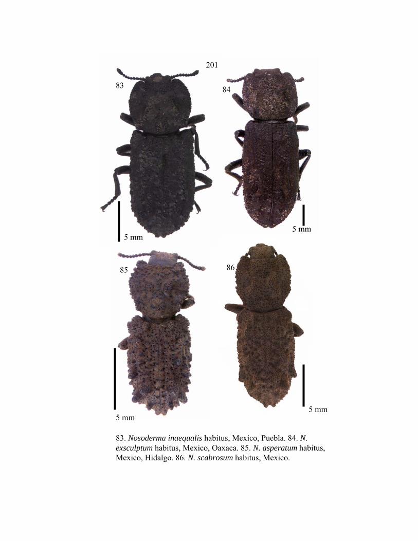
83. Nosoderma inaequalis habitus, Mexico, Puebla. 84. N. exsculptum habitus, Mexico, Oaxaca. 85. N. asperatum habitus, Mexico, Hidalgo. 86. N. scabrosum habitus, Mexico.
5 mm5 mm
5 mm5 mm
201
83 84
85 86

Fig. 87. N. insigne habitus, Mexico, Oaxaca. 88. N. sparsus habitus , Gutaemala. 89. N. zunilensis habitus, Mexico, Chiapas. 90. N. guatemalensis habitus, Mexico, Chiapas.
5 mm 5 mm
5 mm5 mm
202
87 88
89 90

91. N. aequalis habitus, Mexico, Oaxaca. 92. S. denticulata habitus, Mexico, Nuevo Leon. 93. S. ashei NEW SPECIES habitus, Mexico, Hidalgo. 94. S. doyeni habitus , Mexico, Nuevo Leon.
5 mm
5 mm
5 mm
5 mm
203
91 92
93 94

95. S. triplehorni NEW SPECIES habitus, Belize. 96. S. emarginatus habitus, Texas, Comal Co. 97. S. lutosus habitus, Mexico, Oaxaca. 98. S. adami NEW SPECIES habitus, Mexico, Tamaulipas.
5 mm
5 mm
5 mm5 mm
204
95 96
97 98

99. Phloeodes diabolicus mandible. 100. Sesaspis adami mandible. 101. Nosoderma aequalis mandible.
205
99 100
101

102. Sesaspis adami prothorax. 103. Nosoderma exsculptumprothorax.
206
102
103

104. Noserinus dormeanus habitus, Brazil. 105. Noserinus furcatus NEW COMBINATION habitus, Venezuela. 106. Zopher iviei habitus, Penn. Mayalysia. 107. Meralius clavapilusNEW SPECIES habitus, Venezuela.
5 mm 5 mm
5 mm
5 mm
207
104105
106 107

108. M. echinatus habitus, Cuba. 109. Scoriaderma sp. Habitus, Tanzania. 110. Zopherus championi habitus, Mexico, Nuevo Leon. 111. Z. jansoni habitus, Costa Rica.
5 mm 5 mm
5 mm 5 mm
208
108 109
110 111

112. Zopherosis georgei habitus, Australia, N.S.W. 113. Zopherosis antennal club. 114. Zopherus antennal club. 115. Sesaspis antennal club.
5 mm
209
112 113
114 115

116. Meralius clavapilus NEW SPECIES, tibia. 117. Scoriaderma sp. pronotum, sinuate groove.
210
116
117

118. Scoriaderma sp. prothoracic hypomeron. 119. Scoriderma sp. ventral view of head.
211
118
119

120. Sesaspis adami ventral view of head. 121. Nosoderma aequalis ventral view of head.
212120
121

122. Nosoderma inaequalis ventral view of head. 123. Meralius echinatus ventral view of prothorax.
213
122
123

124. Mesepimeron tubercles. 125. Mesepimeron setose punctures.
214
124
125

126. Meralius apex of last antennomere. 127. Scoriaderma apex of last antennomere. 128. Noserinus furcatus epipleuron.
0.25 mm
215
126 127
128

129. Noserinus furcatus tarsal claw. 130. Zopherus championi tarsal claw.
216
129
130

Fig. 131. Strict consensus of 4 MPT with Phellopsis as the outgroup found in NONA/WINCLADA (Length 83, CI .55, RI .78). Bootstrap support values are shown at all well (>50) supported nodes.
100
86
100
100
60
5564
64
76
217

Fig. 132. Strict consensus of 20 MPT with Zopherus as the outgroup(Length 78, CI .58, RI .81).
7680
82
80
218
82
100


![Ironclad C++ - Department of Computer Sciencesantosh.nagarakatte/papers/ironclad... · ing [8] on pointer operations. Ironclad C++ requires that all bare C++ pointer types be replaced](https://img.dokumen.tips/doc/110x75/5cc34b0588c993ab2e8c8460/ironclad-c-department-of-computer-science-santoshnagarakattepapersironclad.jpg)


