Embed Size (px)
Citation preview

J Med Syst (2016) 40:104 DOI 10.1007/s10916-016-0459-8
EDUCATION & TRAINING
A Review of Simulators with Haptic Devices for MedicalTraining
David Escobar-Castillejos1 · Julieta Noguez1 ·Luis Neri2 ·Alejandra Magana3 ·Bedrich Benes4
Received: 3 November 2015 / Accepted: 1 February 2016© Springer Science+Business Media New York 2016
Abstract Medical procedures often involve the use of thetactile sense to manipulate organs or tissues by using spe-cial tools. Doctors require extensive preparation in orderto perform them successfully; for example, research showsthat a minimum of 750 operations are needed to acquiresufficient experience to perform medical procedures cor-rectly. Haptic devices have become an important trainingalternative and they have been considered to improve med-ical training because they let users interact with virtualenvironments by adding the sense of touch to the simula-tion. Previous articles in the field state that haptic devicesenhance the learning of surgeons compared to current train-ing environments used in medical schools (corpses, animals,or synthetic skin and organs). Consequently, virtual envi-ronments use haptic devices to improve realism. The goalof this paper is to provide a state of the art review ofrecent medical simulators that use haptic devices. In par-ticular we focus on stitching, palpation, dental procedures,
This article is part of the Topical Collection on Education &Training
� David [email protected]
1 Escuela de Ingenieria y Ciencias, Instituto Tecnologico y deEstudios Superiores de Monterrey Campus Ciudad de Mexico,Distrito Federal, Mexico
2 Escuela de Educacion, Humanidades y Ciencias Sociales,Instituto Tecnologico y de Estudios Superiores de MonterreyCampus Ciudad de Mexico, Distrito Federal, Mexico
3 Associate Professor of Computer and Information Technology,Purdue University, West Lafayette, Indiana, USA
4 Professor of Computer Graphics Technology, Purdue University,West Lafayette, Indiana, USA
endoscopy, laparoscopy, and orthopaedics. These simulatorsare reviewed and compared from the viewpoint of used tech-nology, the number of degrees of freedom, degrees of forcefeedback, perceived realism, immersion, and feedback pro-vided to the user. In the conclusion, several observations perarea and suggestions for future work are provided.
Keywords E-learning · Medical training · Haptic devices ·3D simulators · Training
Introduction
Teaching process is a key element during the training of pro-fessionals. This has been characterized as a professor-basedapproach, which is considered as the most effective meanto acquire new knowledge [1]. Since the origin of comput-ers, researchers have been looking for new ways to improveand reduce the costs in the teaching process. Therefore, dif-ferent types of simulations, control systems and learningenvironments via the Internet have emerged in the field ofTechnology Enhanced Learning (TEL) and e-learning [2].
One of the main areas where TEL environments havebeen developed is the medical field. Surgical procedureshave a high degree of difficulty and complexity. It is nec-essary to provide students or practitioners a proper andextensive learning process before they can perform surg-eries correctly. The learning curve in the medical process isa concept focused on two aspects: optimization of operatingtime [3] and reduction of patients bleeding [4]. Vickers et al.[5] stated that about 750 operations are needed to improvesurgical procedures. They also found that patients who havebeen treated by doctors with surgical experience, that haveperformed between 750 and 10,250 procedures, tend to havefewer health problems than patients who were treated bydoctors with less experience.

104 Page 2 of 22 J Med Syst (2016) 40:104
Considering advances in technology and new laws in themedical field, various solutions have been pursued to allowmedical students to acquire the necessary skills involvedin medical procedures. Some solutions involve the use ofanimals in operating rooms, which must have similar condi-tions as those related to humans. Devices that use syntheticmaterials to resemble human skin have also been imple-mented. Nevertheless, they fail to emulate the real char-acteristics of human skin. Other solutions suggest the useof virtual environments, which can simulate situations withdifferent levels of risk. Former virtual environments onlyexploited the senses of sight and hearing; consequently, theyexcluded other interaction possibilities.
Several companies and authors had created e-learningenvironments for medical education. These advances havebeen reviewed to assess the impact in medical education [6,7]. However, Secin et al. [8] stated that current surgical envi-ronments did not provide the realism needed to train futuredoctors during operations, they did not provide users thesense of force strength during the task, or they were not ableto adequately develop the necessary skills.
As a solution, haptic simulation has great potential toimprove medical training. A haptic device is a mechanicalinput/output device that enables users interact with virtualenvironments by adding the sense of touch, which enhancesthe learning quality [9]. The incorporation of haptic tech-nologies in medical software and simulations has grown [10,11] and various companies developed medical stations usinghaptic devices [12–17].
The use of haptic simulators provides new alternativesolutions that also allow the development of new teachingmethods. The advantage of haptic technologies in virtualenvironments is the ease they have to recreate difficult sit-uations originated during real practices. They provide newmeans of exploration and representation. They enable thecreation of systems that are capable of implementing newmethods or procedures, and in some cases, these systemscan generate uncommon anatomies by modifying specificmodels with patients information, as it has been reviewed[9]. Therefore, they provide students the ability to practicesurgery as often as necessary.
This paper reviews recent advances in medical trainingsimulators with the focus on haptic technologies. In particu-lar, virtual environments that use commercial haptic devices
were chosen because these have already been benchmarkedand they accomplish security and performance standards.This review is focused on stitching, palpation, dental,endoscopy, laparoscopy, orthopaedics, and miscellaneousprocedures. Existing works and simulators are reviewed andcompared from the point of view of used technology, thenumber of degrees of freedom and degrees of force feed-back, immersion, and learning feedback provided to theuser. At the end, several observations per area are providedand suggestions for future work are proposed.
Haptics
Haptic devices
Haptic devices are electro-mechanical devices with handlers(Fig. 1) that allow motion with several Degrees of Freedom(DoF). When coupled with virtual simulators, they providethe user the sense of touch in addition to the sight. In hapticenvironments, touch sensation can be performed by humans,machines or a combination of both, while objects and/orenvironments may be real, virtual, or a combination of both.Current physical haptic devices always present a residualinner friction that can be perceived as noise, which can evenfatigue the user in some cases. Additionally, the device itselfhas a certain degree of inertia, which present a problem ifthe user moves the haptic device quickly. Haptic devices bal-ance to compensate for external forces of the systems, suchas gravity. They also have mechanisms that provide suffi-cient reaction to stimulation and effort sense to detect hardsurfaces [19]. Their resolution, i.e., the amount of feedbackper unit of distance, needs to be high in order to providea greater detail of textures in virtual environments, and themovement area must be large enough in order to simulatethe actual workspace. Moreover, haptic devices have differ-ent DoF depending on the amount of directions they canmove. The most common are haptic devices with 3-DoF thatcan follow the XYZ axis.
Current haptic devices use two basic variations to controlinteraction: impedance control and admittance control. Inimpedance control, the user moves the device, and it sendsthe data back to the computer; therefore, the application is
(a) (b) (c) (d)
Fig. 1 Commonly used haptic devices in virtual simulators. a) Phantom Omni �, b) Phantom Desktop �. c) Phantom Premium �, and d)Novint Falcon �

J Med Syst (2016) 40:104 Page 3 of 22 104
responsible for controlling the feedback. Prime examplesof this type of device are Phantom �, built by Geomagic� (previously known as SensAble Technologies �), andFalcon � from Novint Technologies Inc � (Fig. 1).
In contrast, by using the admittance control devices usersexert a force on the device, which reacts by displacing itin a proportional distance. This action is translated in thedevice as displacement of the input and force feedback asthe output of the system. This type of control providesthe users with the freedom in the mechanical design ofthe devices, and these devices are able to produce move-ment with greater force and stiffness. However, due to theircomplexity, they are usually very large and they must bedesigned carefully to interact safely with humans. Conse-quently, they are not commonly used in the training field.An example of these devices is HapticMaster �, manufac-tured by Mog Corporation � (previously known as FCSControl Systems �) [20].
Haptic rendering
The goal of haptic rendering is to enable the user to feel,touch, and manipulate virtual objects through a haptic inter-face. The type of interaction defines the procedure for hapticrendering and how the forces are rendered. These methodscan be identified by the way they model the interaction ofthe haptic device in the virtual environment. There are threekinds of haptic rendering: a) point-based, b) ray-based, andc) based on a 3D object made by a group of points, lines andpolygons [18] as shown in Fig. 2.
Rendering of deformable objects is often required inmedical procedures. Visual rendering has been studiedextensively in the area of computer graphics [21]. Basdo-gan et al, have divided rendering techniques of deformableobjects into two types: geometry-based and physics-based[18].
Geometry-based techniques deform the object based ongeometric manipulations. For instance, the user manipulatesthe vertices or control points around the 3D object to mod-ify the shape of the object. These techniques are usuallyfast and relatively easy to implement; however, they focus
mainly on the visual representation, which does not neces-sarily simulates the underlying mechanical deformation.
Physics-based algorithms add physics simulation to themodification of geometry by modeling the physical lawsinvolved in the movement of the object and the dynamicsof the interaction within it. Physical approaches are neces-sary to simulate a realistic behavior of deformable objects.Nevertheless, physical rendering is computationally moreexpensive than pure geometry-based modeling.
Haptics in medical training
We describe and analyze recent virtual simulators that usehaptic devices to practice medical procedures. These areorganized by types of medical practices they simulate.
Stitching techniques
Simulation of stitching procedures is one of the areas wherehaptic technology has been implemented to create learn-ing simulators. Skin and organs have flexible features, sostitching simulators consider mainly rendering techniquesof deformable objects. Additionally, haptic simulators com-bined with active learning environments can provide userswith features such as deformation of the suture thread, knottying, and interaction between tools, the needle and theenvironment, as can be seen in [22]. Jia and Pan devel-oped a stitching simulator which can simulate interactionand deformation of the suture thread and objects [23]. Theyapplied the Follow the Leader (FTL) algorithm [24] to sim-ulate the thread, where each link can rotate freely at theirconnecting vertex. On the side of the deformable object,a tensor mass-spring deformation model in a tetrahedralmesh was implemented to simulate the skin. For collisiondetection in the environment their simulator uses an Axis-Aligned Bounding Box (AABB) tree, where the shape ofthe bounding boxes are updated during the simulation. Thesimulator uses a single Phantom Desktop � haptic device;nevertheless, the proper process of stitching simulators istwo handed. The novelty of the work presented by Jian
Force
ForceTorque
Force
Torque
Fig. 2 Haptic rendering techniques: based on points (left), line segments (middle), and a 3D object (right) [18]

104 Page 4 of 22 J Med Syst (2016) 40:104
Fig. 3 Suture simulatordeveloped by Payandeh and Shi[23]. The skin model (a) isconstructed as a mass-springsystem (b) and the system usestwo haptic device to operate thesuture thread
and Pan is the description of four possible states for theposition of the needle according to its interaction with thevirtual skin:
1. the needle is completely outside of the deformableobject,
2. the end of the needle is in contact with the skin (endtouching),
3. the tip of the needle is in contact with the skin, but it hasnot pierced the skin yet (tip touching), and
4. the needle is piercing the skin.
Their environment provides a functional approach tosuture tasks, and it uses shadows for the 3D location of thehaptic probe; however, the simulator lacks the skin texture,the knot tying action of the suture, the needle is modeled as aline segment, and, as mentioned before, it only implementsa single haptic device.
Similar research was used by Payandeh and Shi [25]who proposed a serious gaming platform to teach suturingand knotting for simple skin or soft tissue wound closure.The haptic devices used in the experimental study were twoPhantom Omni �. They also used a mass-spring model tocreate the deformable skin and the suturing material andthey used Bounding-Volume Hierarchy (BVH) to detect col-lisions and self-collisions in the suture thread (Fig. 3). Theyused Virtual Reality Modeling Language (VRML) files pro-duced in Autodesk � 3ds Max � to create 3D models ofpre-wounded skin. Latter, the simulator imports these filesto build the virtual objects.
Payandeh and Shi presented a suture simulator based onphysics models, and it allows users to tie a knot. One featurethat has to be remarked in this work is that it implements
tissue tearing. This is made by finding intersection pointsbetween each polygon and the tearing path. However, ifthe user repeatedly inserts the needle at the same location,the mesh can be subdivided, which can generate unwantedsmall triangles.
Another interesting research is the one made by Choiet al. [27]. They used the physical rendering engineNVIDIA � PhysX � in collaboration with OpenGL tocreate a system for developing manual skills in the fieldsof medicine and nursing. The system uses two PhantomOmni � haptic devices. The prototype uses a model ofsprings interconnected to simulate the deformable skin. Themodels of the needle and thread are created as a chainof spheres interconnected as a segment, which facilitatesthe flexibility of the elements and allows a simple way toreprodcure thread trimming. The FTL algorithm was usedto simulate the suture thread; however, FTL was modified touse springs and springs with mass as the joints of the thread.This approach provide a realistic sensation. Furthermore,bending can obtained by adding torsion springs in the model.
A recent work in the suture field is the one made byRicardez et al. [26] that was focused on generating an exter-nal suture environment named SutureHap. This simulatorsimulates sensations that are perceived in medical rooms oroffices, as can be seen in Fig. 4. This feature enables a newalternative in the learning process of doctors. The system isbased on the architecture proposed by [28]. SutureHap usedNVIDIA � PhysX � physical engine, OpenGL � to ren-der the graphical environment, and Phantom Omni � hap-tic devices. The system is based on the elaboration of asuturing knot using a real technique, which was consultedamong medical staff.
Fig. 4 SutureHap environmentcreated by Ricardez et al. [26]The left image shows a userperforming the stitching processin the simulator. The use of twoPhantom Omni � hapticdevices is implemented torecreate similar conditions asthe ones experienced in realstitching process. On the rightside a close-up of the virtualenvironment can be seen

J Med Syst (2016) 40:104 Page 5 of 22 104
For haptic rendering, three blocks were developed. Col-lision detection and calculation of force feedback responsebetween the cloth and the haptic cursor were performed byusing the simplified model, where a novel algorithm wasused. This algorithm is based on the addition of multiplerays that are emanating from the center of the haptic avatarin different directions. Finally, response force between solidobjects was developed according to the ”penalty method”standard technique.
In the environment of Ricardez et al. further changessuch as improvement of interactions between objects in thescene, and the implementation of collision handling andhaptic feedback between surgical tools should be consideredto improve the performance of the simulator.
Both Jia and Pan and Payandeh and Shi do not implementor use proper medical tools. The incorporation of tweezerswith realistic appearance to the simulator will show studentsthe correct tools to use in operations and surgical tasks, asRicardez et al. did. Moreover, by simulating physical prop-erties of tools, such as shape, students will learn how tointeract with them and take them into consideration in theworkspace. Finally, another aspect that Ricardez et al. andPayandeh and Shi should consider to provide is appropriatespace location. Sometimes, students could get lost if they donot perceive a visual cue in navigation. Therefore, the addi-tion of shadows of objects could improve the perception ofthe 3D environment.
Palpation
Palpation is the process where surgeons analyze, via theirfingers, tissues or organs to detect anomalies on the surface.Stiffness is essential in this medical procedure. Areas thatare stiffer than other can be considered as potential tumors.Therefore, correct calculation of force feedback is neces-sary during the creation of virtual simulators with hapticdevices.
Li et al. [29] developed a tumor location simulatorbased on soft tissue probing data. They used a Microsoft� Kinect � device to create a tissue model using depth datafrom a silicone phantom tissue and Finite Element Model
(FEM) techniques. This enables the skin to be a geometricaldeformable soft tissue that can be modified in real time. Thenovelty of this study is the use of a rolling indentation sen-sor to obtain friction and stiffness values, which are storedin a look-up table. Therefore, the force feedback to the useris based on the values from the table that is accessed duringthe simulation.
A case test study was made with twenty students, whoused the phantom tissue and the simulator. They were askedto identify two zones that are stiffer than others. For the sim-ulator, they used a Phantom Omni � to navigate the system.Results showed that the simulator could be seen as a feasi-ble and efficient alternative solution of manual palpation totrain students in this medical field.
Another palpation simulator is described in the workof Ullrich and Kuhlen [30]. The system implements nee-dle insertion features, which is a task that is usuallydone after palpation. In their approach, the authors mod-eled a deformable skin using FEM methods and tetrahe-dral meshes. For collision detection, they used the Bullet� Physics Library [31]. The simulation implements the useof hand models and illumination to provide space locationcues; therefore, the authors proposed a new algorithm fortissue dragging to provide proper interactions. The simu-lator uses two Phantom Omni � haptic devices; however,Ullrich and Kuhlen modified the end effector of one to pro-vide a lightweight palpation pad (Fig. 5). This enables thehaptic device to be used by two fingers, which is the mini-mum number of fingers used during palpation. The systemwas evaluated by 23 beginner anesthesiology students and17 experts, where results established average acceptance ofthe simulator. The simulator should be modified and betterimprovements of the hardware should be included; however,the results also showed that the modification of the hap-tic device provided better sensations that the ones that areexperienced with the original stylus.
Finally, modifications to typical work stations have beenmade in this area, as the incorporation of Augmented Reality(AR). PalpSim is a simulator made by Coles et al. [32]. Thissimulator can be seen in Fig. 6. PalpSim was designed toreplicate real scenarios in virtual environments by using AR.
Fig. 5 Palpation environmentshave been developed bymodifying current hapticdevices. Ullrich and Kuhlendeveloped one where they use alightweight palpation pad. [30].Left figure shows currentrendering of the virtualenvironment. Right figuredisplays a user using theenvironment. It can be seen themodification to the haptic deviceon the left hand

104 Page 6 of 22 J Med Syst (2016) 40:104
Fig. 6 Another simulator thatmodified haptic devices is theone developed by Coles et al.[32]. This simulator uses twoFalcon � haptic devicescoupled with a palpation pad(right side). Additionally, theauthors used Augmented Realitytechniques to provide a realisticapproach (left side)
In their workstation, the authors have guaranteed visuohap-tic alignment by fixing a LCD display and using a cameraunder it to capture users’ hands movements. By doingthe latter, the authors could use chroma-key techniques toattain AR. Therefore, users can see in the screen the virtualenvironment with their hands’ real time movements.
Coles et al. created a work station where the user han-dles three haptic devices under the LCD screen: two Falcon� haptic devices are coupled to emulate palpation, and onePhantom Omni � haptic device to manipulable the sutureinsertion task. The end effector of both devices were mod-ified to provide real sensation for the tasks. Additionally,the authors did in vivo force measurement for palpation andneedle insertion tasks. Data obtained were used to calculateand recreate force feedback in the simulator. The simula-tor was validated by a user test, where seven experts in thefield used the workstation. At the end of the simulation, theywere asked to answer a 29 question survey to provide feed-back, and it was strongly agreed that the simulator providesrealistic and correct stimuli.
Dental procedures
Dental training has been done using haptics feedback withPhantom Head � [33] or artificial teeth. However, thesemethods present similar limitations as other surgical areas,such as spatial location difficulties. Ethical constraints pro-hibit the use of real human teeth, and different teeth materi-als lack the appropriate standards or they can be used onlyonce per procedure on the same tooth.
A simulator for typical dental procedures called hapTELwas created by Tse et al. [34] and it allows dental drilling,caries removal, and cavity preparation for tooth restora-tion. In the first stages of hapTEL, Falcon � and ForceDimension � Omega 3 � haptic devices were used. Theresults stated that the quality of haptic rendering neededto be improved and modifications to the physical environ-ment were necessary that were implemented in a secondprototype. Falcon � devices were modified by adding an
additional 6-DoF passive measurement device, and theywere mounted in an upward position. Additionally, a mag-netic ball bearing socket system was developed to provide180◦ of rotation. This socket system avoided the presenceof singularities.
The simulation uses polygonal models that were con-verted into tetrahedra by Tetgen � [35]. The haptic algo-rithm to render forces displays four different types of mate-rials and physical properties: enamel, dentine, pulp, andcavity. hapTEL novelty includes the use of different forcefeedback depending on the material, but mainly the additionof a device to the Falcon �. This setup improved the real-ism of the virtual mouth model and let students feel as ifthey were in a typical dental operation.
Another common field of dental procedures is oralimplant therapy. It has been accepted worldwide as anintegral part of dental practice and it has become evenmore important over the past few years. The placementof implants takes into consideration anatomically complexoperation sites in the cranio-maxillofacial region, whichincrease the risk of damage in operations. Computer-aidedsurgeries have helped students improve their skills; how-ever, their application in dental implant surgery is rarelyreported.
A virtual training system for oral implantation was devel-oped by Chen et al. [36]. CAPPOIS (Computer AssistedPreoperative Planning for Oral Implant Surgery) helpsusers establish a medical environment, and lets users trans-fer the data to the training station. Chen et al. stated that thispilot study proved that CAPPOIS helps inexperienced doc-tors grasp the feel of the osteotomy procedure during dentalimplant surgery. However, more clinical cases need to bedone to show the feasibility and reliability of CAPPOIS.CAPPOIS used various algorithms in the field of computergraphics, and several medical image processing methodsare involved, including: DICOM � file parsing, imagesegmentation and 3D-visualization, surface decimation, cut-ting, spatial search and 3D distance computing, volumemeasurement, spline curve generation, multi(curved)-planar

J Med Syst (2016) 40:104 Page 7 of 22 104
reconstruction, registration, among others. CAPPOIS wasimplemented by using Chai3D � and Qt �, and the sys-tem uses Omega 6 � haptic device from Force Dimension�. Additionally, CAPPOIS allows the use of 3D glassesto view the work place in 3D. This environment was madeto guide users in preoperative planning. Moreover, CAP-POIS allows users perceive the position and orientation oforal implants, do bone density analysis and bone volumemeasurements for maxillary defects, among others.
Soft tissue simulation has been studied extensively incomputer graphics area [18]. A recent research made byHui and Dang-xiao in the area has expanded this topic byincluding bimanual interaction to attain practical applica-tions in the medical field [38]. They developed a dentalsurgery focused on the interactions between the dental toolsand tissues, in this case cheeks and tongue. They based theirdeformation model on a mass-spring model. The novel partof their work is the addition of a special kind of springs,”returning spring”. This spring enable their meshes to beincompressible and resistant to bending by connecting eachmass of the mesh to its original position.
Force feedback provided to the user is calculated fromthe penetration depth made by the tip of the tool. Hui andDang-xiao simplified the interaction of the tools and thedeformable skin as a case of rigid bodies. Therefore, onlythe tip of the tool is interacting with the mesh and theforce that is acting on the mesh is calculated by distributingthe force applied to the contact point to all of its neighborvertexes.
They applied their approach to a virtual simulator wherethey modeled a face, in which cheeks are made up of 143masses and 951 springs and the tongue is made up of 213
masses and 1747 springs. Hui and Dang-xiao used twoPhantom Omni haptic devices in the simulator, and theymodeled their cursors as a Mouth Mirror and a DentalExplorer for exploration and interaction purposes.
One type of teaching-learning techniques used in edu-cation are collaborative environments where students caninteract among them or with a professor to solve prob-lems and learn as a team. As stated before, TEL optionsallow students to enhance their learning by allowing them toacquire knowledge and skills via virtual environments [39].The implementation of these environments in the medicalarea can help teachers guide students in medical operations.Kosuki and Okada developed a collaborative training sys-tem [37]. The simulator was created using a tool calledIntelligentBox, which is a 3D visual software developmentsystem based on the Model-View-Controller architecturalpattern [40]. The authors have implemented components formedical operations in IntelligentBox, and they have devel-oped specific modules for touch and drilling actions todesign a dental training environment (Fig. 7).
IntelligentBox defines 3D objects as polygonal models,which are represented as a set of several polygons. Forobject deformations, the system uses a very simple methodbased on the movements of the haptic device pointer. Thismethod enables the user to use push and pull interaction onthe environment. On the side of collision detection, Intel-ligentBox solves an equation based on the vertex locationsand the interaction point, where only if the interaction pointis the opposite side of the vertex, the system detects a colli-sion. Finally, the dental training station uses Phantom Omni� haptic device and IntelligentBox provides a distributedmodel sharing mechanism called RoomBoxes. RoomBoxes
Fig. 7 Current prototype fortypical dental procedures madeby Kosuki and Okada [37]. Thesimulator was made usingIntelligentBox, and it providesstudents a collaborativeenvironment where they canwork in turns to solve a task
104 Page 8 of 22 J Med Syst (2016) 40:104
provides students a virtual environment in which they canshare user-operation events.
Evaluations of the learning curve and acquisition of skillsusing haptic technologies have been reviewed in previouswork [41]. Assessment and evaluation of students trainingcan be found in the literature [42]. Wang et al. [42] designeda simulator called iDental to provide a preliminary user eval-uation. Teeth models were obtained via point-cloud datafrom Computed Tomography (CT) images and scans andwere refined using 3ds Max �. The result is a VRML filethat is the input for the virtual environment. The meshes oftissues for rigid and elastic objects are handled as triangles,and GHOST SKD � handled collision [43].
The simulator uses one Phantom Desktop � hapticdevice and runs on a workstation with one display. Perfor-mance and user tests with 10 dentists and 19 postgraduatestudents with one to two years of experience in dental proce-dures were made. Results obtained showed that the currentsimulator provides relative realism similar to that experi-enced with real patients. The results suggest that virtualreality simulators give undergraduate students more flexibletraining experiences. However, the current simulator needsto improve its haptic and graphic feedback, as well as thephysical hardware, which is not consistent with the currenttechnique.
Endoscopy
Endoscopy procedures examine the interior of a holloworgan or cavity of the body using an endoscope. Neuro-surgery is one of the surgical specialties within endoscopywith the highest risks [44]. It requires a safe manipulation ofinstruments around fragile tissues; consequently, the mostfrequently reported errors in neurosurgery are technical ones[45].
Delorme et al. [46] developed NeuroTouch �, a virtualsimulator for cranial microsurgery training (Fig. 8). Theneurosurgical simulator was developed using a physic-basedengine and is composed of a 3D graphics rendering sys-tem (stereoscope), haptic devices, other controls, and oneor two computers. Two different types of haptic systemscan be used: the Phantom Desktop � or a Freedom 6S� from MPB Technologies �. Delorme et al. developeda software for the simulations to design and mimic a neu-rosurgical microscope. The simulation uses multiple rates:graphics at 60 Hz, haptics at 1k Hz, and tissue mechanicsat 100 Hz. NeuroTouch was developed to enable residentsto practice their skills in tumor-debulking and tumor cauter-ization tasks. This system computes the deformation of thetissues and topology changes according to tissue rupture,cut, or removal. The mechanical behavior of tissues is mod-eled as viscoelastic solids using a quasilinear viscoelasticconstitutive model for the viscous part of the environment.
Fig. 8 Neurosurgery resident testing a NeuroTouch prototype [46].The workstation implements a stereoscope as an immersion deviceto help the user locate himself in the virtual world. The system usestwo Freedom 6S � haptic devices. Picture comes from the NationalResearch Council Canada
NeuroTouch is a virtual simulator with haptic feedbackthat was designed for the acquisition and assessment oftechnical skills involved in craniotomy-based procedures.Delorme et al. stated that usability feedback from doctorsprovided NeuroTouch � ways to improve its functionality.They have praised the visual portion of the simulator, andsuggested modifications in the ergonomic design. Finally,by adding a modification of parameters of the model, thissystem can be extended to cover patient-specific operations,which would increase the adaptability of the environmentand cover a wider range of scenarios.
Another simulator was developed by Jiang et al. [47]and it allows a simulation of endoscopic third ventricu-lostomy. This procedure is used to treat hydrocephalus bymaking a perforation in the floor of the third ventricle of thebrain under endoscopic guidance. Jiang et al. adapted thestereoscope design by Delorme et al. The proposed systemadapted the NeuroTouch � microneurosurgery to achievethe practices needed in the simulation. They used four mir-rors and a screen instead of two mirrors and two screens. Toprovide haptic feedback, a neuro-endoscope handle with ablunt probe instrument inserted in its working channel wasmounted on a Phantom Omni � haptic device. The sim-ulator was created using their software simulation engineBlade. Jiang et al. designed the environment through closecollaboration with the end-users. It enables two tasks: oneis the burr-hole position and entry orientation selection,and the second is the navigation inside the ventricular sys-tem, reaching the third ventricle and perforating the thirdventricle floor.
Other studies have focused on developing prototypes ofother kinds of endoscopic surgeries. Previous work in thearea of endonasal endoscopy made by Neubauer [48], useda joystick to navigate and access tumor areas in the envi-ronment. This approach reduces the realism of the simulatordue to its lack of haptic interactions with tools. Additionally,

J Med Syst (2016) 40:104 Page 9 of 22 104
they did not implement deformations modules of tissues dueto its navigation nature. Perez-Gutierrez et al. [49]. divideda research study in three steps to develop an endoscopicendonasal haptic surgery simulator and they simulated arigid endoscope device with 4-DoF and modeled the nasaltissue.
Perez-Gutierrez et al. used a force feedback joystickto provide the haptic interaction. The simulator providesthe user graphical feedback using the camera movementsin the virtual environment, and through the joystick, theuser can sense the interaction forces to reach the tumor,as can be seen in Fig. 9. The system was created usingNukak3D � [50]. In this research, further work shouldbe considered to improve aspects such as immersion andinteraction.
Advances in endoscopy simulators can also be foundin the area of surgical suturing. Punak et al. developed asurgical suturing simulator for wound closure using the sim-ulation of the Covidien � (previously known as Autosuture�) Endo Stich � suturing tool in [51]. The simulator usestwo Phantom Omni � haptic devices: one for a grasper andthe other for the simulated Endo Stich � suturing device.
The model of the wound is based on FEM techniques,which in this case used the linear hexahedral model. Addi-tionally, the wound is simulated as a triangular surface meshembedded in the linear hexahedral. This allowed Punaket al. to modify the surface of the wound or change the gridresolution of the mesh without changing the global environ-ment . On the other hand, the suture model was based ona simplified Cosserat theory of elastic rods, which uses theCoRDE model [52], enabling the model to be discretizedinto a coupling of a chain of mass points and a chain oforientations. The simulator uses bounding volumes in thetools and BVH for the collision detection of between theopen wound and the thread. Finally, they used a finite statemachine to animate the knot tying sequence.
The suture was simulated with 100 points, and the trian-gular surface mesh of the wound was composed of 2,178
vertices and 4,352 triangles. The simulation ran at 20 fpswhen there were null or minor intersections, and it ran atapproximately 10 fps with complex collisions and inter-actions. Punak et al. aimed to create low-cost and simplesimulators that help trainees learn surgical skills. User test-ing of the system is necessary to observe if the environmentprovides realistic behavior and allows the user to be trainedin the correct way. Additionally, diversity of wounds andmethods of suture are being planned. Most of the surgicalprocedures are done with at least 6 DoF. However, Punaket al. proposed to test if simulators with three degrees offorce feedback can be a feasible option instead of usinga higher cost simulator offering more degrees of forcefeedback, so further field research should be done.
Laparoscopy
One of the most common surgeries is laparoscopic chole-cystectomy, which is the removal of a diseased gallblad-der. This surgery is often used as the training case forlaparoscopy due to its high frequency and perceived lowrisk. Park et al. [53] developed a low cost virtual real-ity surgical simulator. It consists of three elements: virtualreality, motion sensor, and haptic feedback. The authorsused the Microsoft � Kinect � 3D camera to capture andstore information associated with the movements of thebody of a user. To provide haptic feedback, this work usesNintendo � Wiimote � controllers.
To create the virtual environment, Park et al. constructan expert knowledge database derived from video footagetaken from a surgical endoscope during real cholecystec-tomy operations (Fig 10). A set of 15 key stages weredefined and a database links the corresponding imageframes of the video footage to the relevant key stagesthat were used to construct an interactive video tutorial.Finally, the system uses Hidden Markov Models (HMMs) tocompare the actions of users with actions from the expertknowledge database.
Fig. 9 Current visualization foran endoscope approaching to themiddle turbinate in an endonasalhaptic surgery simulator madeby Perez-Gutierrez et al. [49]

104 Page 10 of 22 J Med Syst (2016) 40:104
Fig. 10 Virtual realitysimulator made by Park et al. fora laparoscopic operation. Theimage depicts the video tutorialthat users can watch (right), andthe surgical instruments that theuser manipulates usingWiimotes � (left) [53]
By using Wiimotes � and the Kinect � sensor, it ispossible to capture the actions of the user. However, theenvironment loses realism due to the fact that it lacks sim-ulated physical surgical devices. Laparoscopic workstationsusually involve the handling of physical parts while the sur-geon proceeds with the operation. However, the workstationproposed by Park et al. was aimed as an in-home trainingsystem for students, and it can help them practice anywhere.
Another low cost training simulator is eLaparo4D, whichwas created by Gaudina et al. [54]. They designed train-ing exercises for medicine students in realistic scenariosof videolaparoscopic surgeries. The system is based on anode.js application server, which enables all the visualiza-tion, communications and administration. The user interfaceuses HTML5 �, which runs a Unity3D � engine plugin[55]. The meshes in the simulation were developed inBlender � 3D [56].
Haptic feedback is provided by using three PhantomOmni �, where the first two are used as tool handlers(grasper, hook or scissors) and the third one is used tomove the camera within the virtual abdomen. By usingthree haptic devices, students would experience a realis-tic approach of the laparoscopic equipment. Gaudina et al.used an Arduino � board connected to a vibrating motorthat also has a vibration feedback. Additionally, the way thesimulator was made allows haptic-based remote guidanceprovided by a supervisor. This guidance can be provided viaweb to show students the proper way to execute a criticaltask. Finally, the system enables users to have their own pro-file, which is tracked over time and users can monitor theirprogress. The system was designed in a way in which eachexercise has its allowed and not allowed actions, which addsor subtracts points from the user’s score.
Gaudina et al. stated that eLaparo4D has many aspectsthat need to be improved. One is the physical modeling tosimulate the interactions of elements properly, such as bodyparts that could interfere with the surgical procedure. More-over, the definition of exercises in the simulator are being
developed to reach a certain level of mastery. Finally, sur-gical procedures need to provide performance feedback, soeLaparo4D is evaluating the implementation of a trackingmodule of the students’ learning curve.
Serious games are employed to teach skills on surgery-based training applications. Serious games provide highfidelity simulations of environments and situations. De Pao-lis presented a serious game for training suture skills inlaparoscopic surgery [57]. The system is focused on phys-ical modeling, and it established a set of parameters todetermine the development of trainees. The simulator usesa pair of haptic devices, which can be two Phantom Omni� or two Falcon � using the multidevice HAPI � library.The system uses PhysX � physics engine and Ogre3D� graphics engine [58].
De Paolis used the mass-spring method to design the tis-sue in PhysX �, and he used Ogre3D � to render the tissue.On the other hand, the thread model was built using follow-the-leader techniques in PhysX �. The thread is composedof cylinders that are connected through a spherical joint.This enables the rotation of the elements relative to eachother. The architecture of the system was developed usingthe architectural pattern of Model-View-Controller, whichmanages the behavior and responses of the environment andobjects.
In the assessment of suturing procedure, De Paolis con-sidered the following parameters to evaluate the medicalprocess: duration time, accuracy, force peak, tissue damage,angle of entry, and needle distance. The software architec-ture was developed using the architectural pattern Model-View-Controller, which manages the behavior of objects inthe environment, responds to requests for information abouttheir states and to instructions to change state. De Paolis[57] concluded that the serious game of laparoscopic sutur-ing could be improved by modifying some aspects of theenvironment. One is improving the interaction between thetissue and suture thread, which is one of the areas in suturemodeling that is currently researched.

J Med Syst (2016) 40:104 Page 11 of 22 104
Other improvements in the area of laparoscopic simu-lators include the addition of visual effects. For instance,smoke and bleeding models are not developed due to itslesser significance compared to other major modules, suchas collision detection and physics simulation. Neverthe-less, Halic and De developed a GPU based method toimplement realistic smoke and bleeding effects in virtualreality simulators [59] (Fig. 11). These effects were appliedin the Laparoscopic Adjustable Gastric Banding (LAGB)simulator [60].
In order to obtain the desired effects, Halic and De ana-lyzed videos obtained from LAGB and they identified twodifferent kinds of smoke effects. One is the environmentalsmoke effect, which often obstructs the camera view. Theother is the smoke effect originated at the tip of the cauterytool that spreads around the tissue slowly. Both types areblended to generate the final environment. For bleedingeffects, an attribute variable was assigned to all vertexes ofthe mesh structure. The processing is made when the inter-polated value of this variable is compared with the noisetexture. If it exceeds a certain value, bleeding is rendered inthat particular fragment.
The simulator and the effects were written with GLSL� shader. The smoke video fetching and bleeding render-ing were performed within the same shader. Halic and Deused the GPU for bleeding and smoke simulation; therefore,it decreased the computational load of the CPU with a slightreduction in the rendering performance. However, the per-formance is better compared to a full-fledged simulation ofboth effects. Halic and De specified that they are planningto optimize the texture processing in CPU and CPU-GPUdata transfer to avoid the loss of performance obtained.
Finally, assistant modules are currently being applied inthe area of virtual simulators, where one of the most com-mon is the technique of Virtual Fixtures (VF). VF is a
technique that is used to guide students while they are work-ing on a specific task. VF applies forces in the haptic deviceto maintain the students outside of prohibited work spaceareas. Research made by Hernansanz et al. is focused on VF[61]. The paper discusses the advantages of VR to improvebasic learning skills in laparoscopic surgery. They used theSkill-Rule-Knowledge taxonomy [62] to assess assistingtechnologies applied in abilities acquisition.
Hernansanz et al. implemented as VF: visual guidance,audio guidance, motion scaling, magnification, and forcefeedback. These provide the student an assistance environ-ment. The simulator that Hernansanz et al. used was theone developed by Zerbato [63]. The system uses a singlePhantom Omni � haptic device.
Orthopaedics
Arthroscopy [9] is another medical surgery that has beenimplemented in virtual environments. Arthroscopic proce-dures are performed to evaluate or treat orthopaedic condi-tions. Heng et al. developed a virtual reality system for kneearthroscopic surgery using OpenGL � and C++ [73]. Theycreated soft tissue deformable model using Finite-ElementAnalysis (FEA) with realistic feedback. They developedtheir own 4-DoF haptic device with 3-Degrees of ForceFeedback (DoFF) to satisfy their requirements, and theyused slides from the Visible Human Project image datasetcombined with CT real images to obtain better visual real-ism. In this simulator, two types of meshes are generated:one to model non-deformable organs (i.e., bones), andthe other using tetrahedral meshes to represent deformableorgans. Using an in-house architecture, these two meshesare rendered in a preprocessing phase of the system. Toimprove the performance, they propose regions with dif-ferent properties to balance the computation time and the
Fig. 11 In order to promote realism in simulators, Halic and De developed (a) smoke effects and (b) bleeding effects [59]. These effects wereapplied to LAGB simulator [60]

104 Page 12 of 22 J Med Syst (2016) 40:104
Fig. 12 Chen and He developeda bone drilling simulator, whereusers can feel, by forcefeedback, the differentcomponents of the bone [64]. Infigure (a) a user is performingthe task using a PhantomPremium �, and on figure (b) aclose-up of the environment canbe seen
level of simulation realism. Finally, the simulator devel-oped procedures to cut tetrahedral meshes and implementedoptimized collision detection methods. An endoscope and acutter were modeled to provide real tool models in this sim-ulation. The simulator includes the functionality to recordthe surgical process. This feature enables medical studentsto playback the recorded procedure. This system was eval-uated by medical professionals and the results reveal asatisfactory tactile feedback.
A typical task in orthopaedics is bone drilling. This pro-cess is an essential step before the insertion of screws orpins. Chen and He created a simulator for bone drilling ofa femur bone in a hip fracture surgery [64]. This simulatorcan be seen in Fig. 12. The key feature of this study is theuse of a Phantom Premium �, which provide 6 DoFF. In thesimulator, the bone is modeled as a voxel, which is obtainedfrom CT or Magnetic Resonance Imaging (MRI) scans andthe drilling probe is modeled as a cylindrical volume.
In this simulator two important aspects for drilling weretaken into consideration: bone properties and drill param-eters. The bone properties was modeled according to its
heterogeneity (hyaline cartilage, spongy bone, marrow cav-ity, and compact bone). For drill parameters, drill speed,type of drills and feed rates were considered. Finally, calcu-lation of force feedback (drilling force and rolling torque)is based on the material removal rate, which depends ondrill diameter and feed-rate. Additionally, the travel distanceof the drill is calculated using feed-rate and time intervalparameters.
In orthopaedics, one specific surgery process for frac-tures on the human femur is the Less Invasive StabilizationSystem (LISS). LISS allows surgeons to insert a percuta-neous plate and fix it in the distal femur with screws to helpbones recover from fractures. For this operation, Cecil et al.developed a virtual collaborative simulator for LISS training[65]. The simulator uses FEM approach to model deforma-tions when surgical tools interact with the surfaces of thebone (Fig. 13).
The importance of this research was the implementationof the collaborative mode. Each user has their own virtualenvironment on their work station; the control of the simula-tion remains with a specific user until he grants permissions
Fig. 13 Another environment that uses the technique of collaborativelearning is the one made by Cecil et al. [65]. Picture on the right showsthe surgery process for fractures on the human femur was designed. Byapplying the methodology of collaborative environments, Cecil et al.
enables users to practice the surgery process, and on another work-station an expert surgeon can check the process. On the right side, aclose-up of the virtual simulator can be seen

J Med Syst (2016) 40:104 Page 13 of 22 104
to another user, in a token-based approach. In addition tothe collaborative mode, Cecil et al. designed this simulatoras two different platforms to be used as an evaluation andplanning tool for surgeons. The first platform is a simulator,which was created using C++ and Chai3D �. This simula-tor incorporates a Phantom Omni � haptic device to interactwith the environment and obtain force feedback from it. Thesecond platform was created to visualize the surgery processobtained in the process. This system was considered as atool to help surgeons teach the process and evaluate medicalresidents.
Miscellaneous procedures
Additional to the previous medical areas, there have beensimulators in other specific areas or tasks. One of them isin the training of biopsy, which is the sampling of cells ortissues for examination. This procedure is most commonlyperformed to evaluate whether there are inflammatory orcancerous conditions in organs. Training of this task canonly be performed on live patients, where consequent risksmay occur in patients. Therefore, the importance of creatingsystems that provide visual realism and controlled environ-ments is emphasized. In this area, Ni et al. developed avirtual reality simulator for Ultrasound-Guided (UG) liverbiopsy training [66].
The authors combined images obtained from CT withultrasound images to provide higher realism. In addition, thegenerated deformable model is able to simulate the breath-ing of a patient by changing controlled parameters. For hap-tic feedback, they used two haptic devices that are managedin independent routines, a Phantom Omni � to simulatean ultrasonic scanning probe and a Phantom Premium tohandle the virtual needle.
Ni et al. show in the simulator the liver model and theultrasonic sensor image that can be seen in real opera-tions, as can be seen in Fig. 14. User tests were made toevaluate different aspects of realism, such as images andtactile effects. The evaluation was conducted with expertsand trainees, where satisfactory results were obtained. Nev-ertheless, it was highlighted that the performance of expertsis not as high as expected when breathing is enabled in thesimulation.
A Transrectal Ultrasound (TRUS) guided prostatebiopsy virtual environment using haptic devices has beendeveloped in recent studies [67]. Selmi et al. designeda complete learning environment where various exerciseswere implemented to provide didactic learning. To achievethis goal, the dedicated clinical case database from Koelis� UroStation � system was connected to the systemto provide diverse patient cases. This database covers alarge variety of important situations that surgeons typicallyencounter during real surgeries.
Fig. 14 Ni et al. developed a virtual biopsy simulator [66]. In this, theuser can perfom interactive anatomical navigations, where accordingto the movements of the probe, a CT view that corresponds to it isshown
The simulator let practitioners use a Phantom Omni� haptic device to navigate the environment on one hand,and on the other hand they use CamiTK � GUI [68], whichprovides data and feedback to the user during the simula-tion. Moreover, the simulator allows practitioners to accesstheir performance record, such as average score and proce-dure average time of the procedure. Additionally, to improverealism, students have to complete a checklist, which istypically done prior to the operation.
The simulator of Selmi et al. is used in two phases. In thefirst one, students are asked to perform seven specific exer-cises, which help them to understand the procedures duringthe biopsy and let them practice hand-eye interaction in thesimulator. The second phase offers them the option to per-form a virtual biopsy process, where the student can selectthe desired position to do the biopsy (decubitus or lateral)and the location to start it (left central base, left lateral base,right central base or right lateral base).
Training environments have also been created for liversurgery. Yi et al. proposed a method to adapt and calibrateshape and size of organs by establishing feature points ontheir surface [69]. The organs are formed by triangle meshesthat follow the spring-mass damper model to achieve defor-mations. Yi et al. used a simplified Lin-Canny algorithm tocalculate the nearest point from the surgical tool in orderto deploy the corresponding interaction forces and collisiondetection. This simulator enables medical students practicesurgeries in virtual environments by applying images ofpatient’s organs in the simulator.
Virtual training environments have also been designed inoptometry. Optometry is usually practiced in close super-vision of experts due to the fact that these procedures aretime-consuming and unrepeatable. Tradicional techniquesin the area focus on the use of artificial or bull eyes. Med-ical students in optometry need training environments to
104 Page 14 of 22 J Med Syst (2016) 40:104
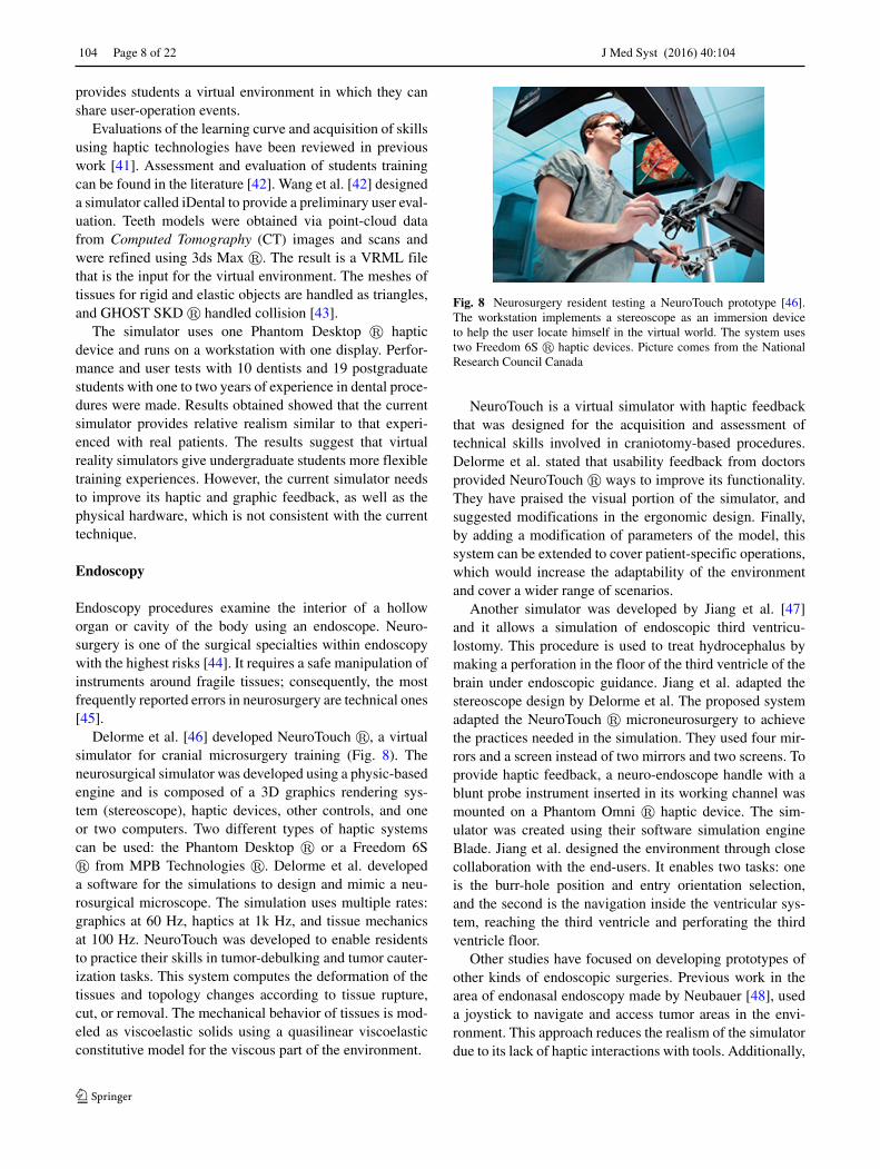
develop their skills with superior realism. Wei et al. [70]created an immersive extendable and configurable work sta-tion that implements the use of a Phantom Omni � hapticdevice and a Head Mounted Display (HMD) to provide ARtechniques. This work station can be seen in Fig. 15. Thesimulation was developed using C++ and Chai3D �. Theeye model was implemented as a polygonal mesh with sep-arate component and textures. By doingthis segmentation,interaction between the surgical tools and the eye providedifferent physical properties, according to the region that theuser is navigating. Moreover, the model was built implicitlyfrom the interactions with the deformable object; therefore,data-driven models were used in this simulator.
In the virtual environment, illumination and needlesharpness modification were adapted to provide the bestsuitable and similar conditions as the ones experienced dur-ing real surgeries . Additionally,, the simulator consideredthe use of the HMD to adjust the visual rendering effect,and AR techniques to modify the slit lamp orientation dur-ing the simulation. Finally, the simulator was tested by fiveoptometry students who already knew the procedures. Theyhad to perform five different tests and their performance wasrecorded by the system. By using the simulator, they wereevaluated by assessing 1) distance estimation between thetool and the eye, 2) foreign body location, 3) foreign bodyremoval task, 4) the angle and position of the needle, and 5)needle insertion.
The authors stated that in the first trials made by thestudents, they had bad performance because they were stillunfamiliar with the workstation. The principal problem wasin the area of space location (Z-axis or depth). At the end ofthe evaluation, students were asked to answer a survey aboutthe system. In this questionnaire, immersion, interactivityand effectiveness of the system were asked. Most studentsagreed that the simulator could enable users to finish thetasks faster and safer due to the force feedback provided ontheir hand.
Besides advances in training students for specific medi-cal tasks, physio-therapeutic systems have been made usinghaptics to relieve the work load of physiotherapists. Surg-eries are usually followed by a rehabilitation program. Typi-cal feedback provided to patients is given using audiovisualcues [71]. However, in some cases, using a mechanical orhaptic feedback can be implemented as an alternative orcomplement treatment. A recent research made by Rajannaet al. proposed a system called KinoHaptics, which is anautonomous, wearable, haptic device to help during therecovery process of patients [72].
KinoHaptics hardware is based on an in-house devel-oped armband (with vibration motors), and a Kinect �2.0 to track the motion of users. By adding the armbandwith vibro-haptic feedback, Rajanna et al. simulates a phys-iotherapist that accompanies patients during the trainingsession. Kinect � 2.0 is used to capture the movementof the patient to recreate it in a virtual scheme. Thevirtual scheme and the user interface were made usingUnity3D � framework.
The system was tested by 14 students. Test subjectswere chosen according to the following categories: hassuffered an injury, has limited motion in their limbs, andhas no prior injury or limited motion of their limbs.Finally, results shows that users liked KinoHaptics andfelt it convenient to be used for self-care after having asurgery.
Discussion
In this section we provide a comparison and summary oftechnical aspects used in haptic virtual simulations. Thenwe discuss the visual realism of the simulations and then wefocus on the training aspects of virtual environments. Thepapers discussed in this work are summarized in Tables 1and 2.
Fig. 15 A workstation foroptometry tasks was developedby Wei et al. [70]. Theworkstation provides a realisticapproach to real operations.Figure (a) shows that the systemis made from a head display andthe equipment that is used in theoperation. Additionally, adisplay and a Phantom Omni aremounted. On figure (b)screenshot of the body removaltask is shown

J Med Syst (2016) 40:104 Page 15 of 22 104
Table1
Com
pari
son
ofha
ptic
sim
ulat
ions
Aut
hor
Med
ical
HaD
Mod
ific
atio
nN
umbe
rD
oFD
oFF
Type
Task
perf
orm
edTo
ols
sim
ulat
edG
ravi
tySp
ace
LF
to
area
toH
aDto
HaD
ofFF
loca
tion
user
Jia
and
Pan
[23]
Stitc
hing
Des
ktop
No
16
3Fo
rce
Clo
sea
wou
ndN
eedl
eN
oSh
adow
sN
o
Paya
ndeh
and
Shi[
25]
Stitc
hing
Om
niN
o2
63
Forc
eC
lose
aw
ound
Nee
dle
and
No
Non
eN
o
Nee
dle
Dri
ver
Ric
arde
zet
al.[
26]
Stitc
hing
Om
niN
o2
63
Forc
eC
lose
aw
ound
Nee
dle,
Nee
dle
No
Non
eN
o
Hol
der
and
Twee
zers
Cho
ieta
l.[2
7]St
itchi
ngO
mni
No
26
3Fo
rce
Clo
sea
wou
nd−
No
Non
eN
o
Lie
tal.
[29]
Palp
atio
nO
mni
No
16
3Fo
rce
Tum
or−
No
Non
eN
o
Loc
aliz
atio
n
Ullr
ich
and
Kuh
len
[30]
Palp
atio
nO
mni
Yes
26
3Fo
rce
Palp
atio
nan
dH
ands
and
Nee
dle
No
Vir
tual
Rea
lity
No
Nee
dle
Inse
rtio
n
Col
eset
al.[
32]
Palp
atio
nFa
lcon
and
Om
niY
es2/
13/
63
Forc
ePa
lpat
ion
and
Han
dsan
dN
eedl
eN
oN
one
No
Nee
dle
Inse
rtio
n
Tse
etal
.[34
]D
enta
lFa
lcon
Yes
13
6Fo
rce
Dri
lling
,Car
ies
Den
talD
rill
No
3DL
CD
No
rem
oval
and
Cav
itypr
epar
atio
n
Che
net
al.[
36]
Den
tal
Om
ega
6N
o1
43
Forc
eO
rali
mpl
ants
Den
talD
rill
No
3DL
CD
Yes
Hui
and
Dan
g-xi
ao[3
8]D
enta
lD
eskt
opan
dO
mni
No
26
3Fo
rce
Man
ipul
atio
nof
Mou
thM
irro
rN
oN
one
No
Che
ekan
dan
dD
enta
l
Tong
ueE
xplo
rer
Kos
ukia
ndO
kada
[37]
Den
tal
Om
niN
o1
63
Forc
eTo
uch
and
Dri
lling
−N
oN
one
Yes
Wan
get
al.[
42]
Den
tal
Des
ktop
No
16
3Fo
rce
Rem
ove
calc
ulus
Den
talD
rill
No
Non
eN
o
Del
orm
eet
al.[
46]
End
osco
pyFr
eedo
m6s
No
26
3Fo
rce
Tum
or-d
ebul
king
Ultr
ason
icN
oSt
ereo
scop
eN
o
and
Tum
orA
spir
ator
,Bip
olar
caut
eriz
atio
nE
lect
rocu
ster
y
task
san
dM
icro
scis
sors
Jian
get
al.[
47].
End
osco
pyO
mni
Yes
26
3Fo
rce
End
osco
pic
thir
dPr
obe
No
Non
eY
es
vent
ricu
lost
omy
Pere
z-G
utie
rrez
etal
.[49
]E
ndos
copy
Joys
tick
No
11
No
Vib
ratio
nE
ndos
copi
cR
igid
No
Non
eN
o
endo
nasa
lsur
gery
End
osco
pem
odel
Puna
ket
al.[
51]
End
osco
pyO
mni
No
26
3Fo
rce
Clo
sea
wou
ndTw
eeze
ran
dN
oSh
adow
sN
o
Nee
dle
Han
dler
HaD
:Hap
ticD
evic
e,FF
:For
ceFe
edba
ck,L
F:L
earn
ing
Feed
back
,UG
:Ultr
asou
nd-G
uide
d,T
RU
S:T
rans
rect
alU
ltras
ound

104 Page 16 of 22 J Med Syst (2016) 40:104
Table2
Com
pari
son
ofha
ptic
sim
ulat
ions
(con
t)
Aut
hor
Med
ical
HaD
Mod
ific
atio
nN
umbe
rD
oFD
oFF
Type
Task
perf
orm
edTo
ols
sim
ulat
edG
ravi
tySp
ace
LF
to
area
toH
aDof
HaD
ofFF
user
loca
tion
Park
etal
.[53
]L
apar
osco
pyW
iimot
eN
o2
3N
oV
ibra
tion
Cho
lecy
stec
tom
y2
No
Kin
ect
Yes
Gau
dina
etal
.[54
]L
apar
osco
pyO
mni
No
36
3Fo
rce
Vid
eola
paro
scop
ic1
No
Non
eY
es
surg
ery
De
Paol
is[5
7]L
apar
osco
pyFa
lcon
orO
mni
No
23/
63
Forc
eL
apar
osco
pic
−Y
esN
one
No
sutu
re
Hal
ican
dD
e[5
9]L
apar
osco
pyO
mni
No
26
3Fo
rce
Lap
aros
copi
cTw
eeze
rN
oN
one
No
Adj
usta
ble
Gas
tric
Ban
ding
task
Her
nans
anz
etal
.[61
]L
apar
osco
pyD
eskt
opN
o1
63
Forc
eV
ideo
Smok
ean
dN
oN
one
No
lapa
rosc
opic
blee
ding
mod
ules
surg
ery
Hen
get
al.[
73]
Ort
hopa
edic
sO
wn
devi
ceN
o2
43
Forc
eK
nee
Art
hros
copy
End
osco
pean
dN
oN
one
No
Cut
ter
Che
nan
dH
e[6
4]O
rtho
paed
ics
Prem
umN
o1
66
Forc
eB
one
drill
ing
Dri
ller
No
Non
eN
o
inhi
p
frac
ture
Cec
ilet
al.[
65]
Ort
hopa
edic
sPr
emiu
mN
o1
66
Forc
eL
ISS
surg
ery
Dri
ller
No
Non
eY
es
Nie
tal.
[66]
Bio
psy
Om
nian
dPr
emiu
mN
o1/
16
3/6
Forc
eL
iver
UG
biop
syT
rans
duce
ran
dN
oN
one
No
Bio
psy
Nee
dle
Selm
ieta
l.[6
7]B
iops
yO
mni
No
16
3Fo
rce
TR
US
Gui
ded
Ultr
ason
icN
oN
one
Yes
Pros
tate
Scan
ning
Prob
e
biop
syan
dN
eedl
e
Yie
tal.
[69]
Liv
erSu
rger
yO
meg
a3
No
13
3Fo
rce
Liv
ersu
rger
yC
uttin
gPr
obe
No
Non
eN
o
Wei
etal
.[70
]O
ptom
etry
Om
niN
o1
63
Forc
eO
ptom
etry
task
sFo
reig
nbo
dyN
oH
MD
No
rem
oval
and
Inje
ctio
npr
oced
ure
Raj
anna
etal
.[72
]R
ehab
ilita
tion
Arm
band
No
No
No
No
Vib
ratio
nPh
ysio
-the
rape
utic
Non
eY
esK
inec
t2.0
No
task
s
HaD
:Hap
ticD
evic
e,FF
:For
ceFe
edba
ck,L
F:L
earn
ing
Feed
back
,UG
:Ultr
asou
nd-G
uide
d,T
RU
S:T
rans
rect
alU
ltras
ound

J Med Syst (2016) 40:104 Page 17 of 22 104
Technical comparison
Haptic devices in virtual simulators
Current simulators mainly use haptic devices with threeor six DoF. The most common ones are Falcon � andPhantom Omni � due to their affordable price (Falcon� costs around $150 - $200 US Dollar as of July 2015).Falcon � devices only provide 3-DoF, Phantom Omni� provides 6-DoF, but it has only 3-DoFF. Addition-ally, its cost is around $2,200 US Dollar, which makesit less accessible. Several authors have attempted to com-bine low cost haptic devices with external modules toprovide additional degrees of freedom [32, 34], or theyprovide better realism by adapting surgical tools to them[30, 47].
The prevailing used haptic devices in stitching proce-dures are from the Phantom family. This kind of hapticsuses 6-DoF, but they only have 3-DoFF, except for the Phan-tom Premium, which has all its DoF coupled with forcefeedback. A typical task used in the development of stitch-ing simulators is open-wound operations, where they mainlyfocus the task on the dermis layer. The nature of this pro-cedure implies that torsion is needed when the needle ispiercing the skin; consequently, simulators need to provide6-DoF. Particularly in this area, none of the authors adaptedhaptic devices or created their own to provide forces in casessuch as skin perforation by the needle.
Palpation virtual environments target to achieve sensa-tions felt in real procedures. Palpation is a procedure that iscommonly made in parallel with needle insertion. Authorsusually adapt haptic devices with pads to provide realsensations. Other authors focus their attention to provideproper sensations of tumors by scanning current tumor pads.They translated the texture and force sensations to pro-vide better haptic feedback and rendering. Palpation maybenefit from using new haptic devices that use piezoelec-tric sensor in gloves to enhance the sensation and depthperception.
Dental simulators focus on the most common actions indental procedures, as caries and calculus removal. Applica-tions use diverse haptic devices; nevertheless, the main aimis the reproduction of realistic workstations. Tse et al. [34]adapted the workstation to provide these conditions, andalso added an extension to a Falcon � device in order toprovide 6-DoFF. Additionally, undergraduate students useda common drill as the handler of the haptic device, whichenhanced the learning procedure. Further work in this areacould consider the addition of two haptic devices becausemedical tasks are done by using two hands.
Endoscopy and laparoscopy environments belong to theclass of Minimally Invasive Surgeries (MIS). Becausethis procedures need to be performed with two hands,simulations are usually manipulated with two PhantomOmni � haptic devices. Virtual environments in thisarea focus principally in endoscopy navigation [47, 49].Laparoscopy simulations are commonly developed to pro-vide practice in videolaparoscopic surgery [54, 61]. Newalternatives, such as [53], provide low-cost simulations byusing haptic devices coupled with video games by usingWiimotes �.
Orthopaedics environments usually focus in arthroscopictasks, principally drilling. In this type of simulations, FEMshould be considered. Researchers should focus on how tomodel bone properties and structure. By doing this, theycan model proper interaction in drilling and force feedback.Finally, miscellaneous procedures are being developed.Important cases in the area are biopsies and optometry. Bothkind of simulators use Phantom Omni �. However, manip-ulation of tools requires 6-DoFF, as in the case of stitching.Finally, post-surgery rehabilitation has started to add hapticfeedback as a complementary resources of audiovisual cues[72].
In short, simulators in these areas are developed with 3-DoFF due to their nature, where only 3-DoF are needed.Commonly used tools in medical procedures involve manip-ulations using triggers, so their addition to haptic devicesdo not require the incorporation of more DoF such asin [54].
Degrees of freedom and modifications
State of the art articles on medical applications focus ondeveloping simulations, which leaves the addition and mod-ifications of haptic devices as another field of study. Themodification of haptic devices can be found in robotics androbot-assisted surgeries [74]. Modification to haptic devicesusually improves haptic sensations in order to make themcloser to real surgeries or procedures [30, 32, 34, 47].
In contrast, several authors decreased the physical real-ism in virtual simulators coupled with different hapticdevices [53] to make the simulations more affordable.Whereas it decreases the cost of a virtual simulator forundergraduate students by using a Kinect � and Wiimotes�, the type of force feedback and the form of Wiimotes� reduced realism in the practice of skills.
Additionally, due to the technical properties of the forcefeedback, only one force aiming in one direction can beusually applied. One exception is the work of [53] thatprovided vibrations as force feedback. Laparoscopic appli-cations need to provide at least 3-DoFF. Consequently, theWiimote � is not a feasible option because it only provides

104 Page 18 of 22 J Med Syst (2016) 40:104
vibration as feedback. Also, twisting forces require 6-DoFFand they are found and used rarely [34].
Visual realism
Another important aspect that should be considered in thedevelopment of simulators is to reproduce a high level ofvisual realism. Recent progress in the GPU processing andcomputer graphics in general allows an extremely high levelof realism. Very few works [46, 54, 60, 61] focus on pho-torealistic rendering and high visual quality. Most of theworks use only schematic rendering with shadow to showthe location of the probe and simple Phong shading todepict the surfaces. Finally, only a limited number of workssimulate gravity [57].
The most commonly used visualization is displayingthe position of the probe as a sphere, sometimes withshadow, and in some cases 3D immersion is added. Thereis usually some learning required for the users to becomefamiliar with the system as they first need to allocate them-selves properly in the simulator. Current simulators let usersexperiment and move freely in the 3D scene before start-ing the medical procedures, as exemplified in the workof [26].
Users very often report the difficulty in detecting theposition of the probe in the 3D space. It is extremely impor-tant to locate the coordinate system of the virtual 3D spacecorrespondingly to the real one so when the probe movesto the right the virtual one should move in the same direc-tion. Moreover, scaling of the virtual environment is veryimportant as well. In many cases the forces as well as thesizes of the objects do not correspond to the real world.The minimal requirement for the correct perception of the3D location is the inclusion of shadows to locate the hapticprobe. Some works add 3D immersion to increase realismand to add the 3D perception. The immersion is providedeither by using active 3D head mounted displays [34, 36] orby using 2D screens with displaying in stereo [46]. The 3Dhead mounted displays allow free movement, and their posi-tion and orientation is coupled with the virtual camera, butthey are usually bound to a single user. Passive 2D screensallow multiple users to view the scene, but the movementof the user is not transferred to the movement of the vir-tual camera. The most common solution is using active 3Ddisplays, where the position of the person performing theprocedure is critical, as for example, in the treatment ofocclusions.
One of the main challenges is to improve the way stu-dents perceive the location of objects in space, especiallythe z-axis (depth) as described in [70]. An alternative solu-tion was made by Coles et al. [32], where the authors usedAR to ensure visual-optical alignment.
Physics
Computer graphics has provided an unprecedent progressin simulation of physics. Recent GPU-supported physicssimulation engines such as PhysX � [75], Havok � [76],Newton � [77], or Bullet � [31] allow simulation of fluiddynamics, kinematics, or FEM and soft tissues. These simu-lations are physically precise, they can use very large scenesand meshes, and are usually provided in real time. This hasnot been exploited to the full extent in the area of hap-tic simulations for two reasons; first, users rely on existinghaptic libraries and APIs that usually do not provide accessto the most advanced and recent algorithms. Second, theselibraries are usually implemented on the GPU and theyrequire usage of advanced visualization techniques to befully used. Moreover, combining haptic feedback with thoselibraries is non-trivial. Nevertheless, it would be extremelybeneficial for haptic simulations to fully exploit and usemodern GPU-oriented physics-based approaches as it couldbe the next quantum leap in the quality and realism of thesimulators.
Feedback in training
An important aspect is to provide learning feedback andevaluation during training sessions. While most of the exist-ing simulators provide visuo-haptic feedback, some of themalso provide a detailed feedback of the training session byrecording the number of clicks, motion of the haptic cur-sor, applied forces in the environment, actions made by theuser, etc. The assessment of the tasks and user performanceis made via the registration of the interaction parametersof the environment. For instance, some systems focus onquantifying the number of operations carried out in a giventime interval, such as the system described in [42], wherethe authors measured the number of calculus removed overa period of ten minutes. Other systems analyzed parame-ters and evaluation checklists from the users by checkingthe reliability of the simulation [66, 70]. Nonetheless, theseresults and checklists were measured internally, so no eval-uation nor feedback was given to the user. Researcheshave also considered increasing the adaptability of virtualenvironments. Students can use different medical trainingscenarios by modifying the parameters of the models in thework of Delorme et al. [46].
Contrary to the closed systems that do not provide feed-back to the user, simulators introduced in [36, 47, 53]provided real-time feedback to the user. They send immedi-ate messages on how the user is performing and they alsoprovide tips during the task. Additionally, Jiang et al. [47]presented a system that includes an evaluation of the wholetask by displaying an appropriate assessment metrics.

J Med Syst (2016) 40:104 Page 19 of 22 104
Gaudina et al. [54] used an assessment method to eval-uate the learning by measuring user performance. Theirsimulator predicted the score obtained during the trainingsession and provided students with feedback that helpedthem to know the aspects that needed to be improved.VF has been used in the area as guidance during trainingsimulations; however, they are not properly considered aslearning feedback. VF are used to keep students out of pro-hibited areas within the workspace. This type of assistancewas used by Hernansanz et al. [61]. Nonetheless, VF hasbeen shown to be generally ineffective in improving resultsin training [78, 79]. Authors should consider using otherkinds of virtual assistance methods to attain better resultsduring training and avoid disturbances during user’s move-ments, such as Temporally Separated Assistance or SpatiallySeparated Assistance [80]. Another methodology that hasbeen used in virtual environments with haptic devices is col-laborative learning. These environments are based on theinteraction between users or user-expert. These are prin-cipally based in a token-based approach, where one usercannot do anything until she has the token [37, 65]. How-ever, these environments require large time from experts,and they are not easily accessible for training simulators.
Several studies performed usability tests with studentsand experts in the field of medicine [26, 29, 30, 32, 37,42, 66, 70]. Most of the researchers ask the medical staffto answer surveys and functionality questionnaires to assessthe simulation fidelity, and these evaluations helped virtualenvironments with haptic devices to gain acceptance in themedical community.
Even though the above-presented simulators allowassessment of user performance in the virtual environment,they do not usually provide assistance, guidance or hintsafter simulation. It is important to decide whether it is betterto provide immediate ongoing feedback while interactingwith the simulation, or to provide users a post-feedback afterhaving interacted with the system. The use of expert humantutors could help to train users appropriately. Indeed, thesesystems have contributed in various areas in the training ofprofessionals [1].
Considerations
By analyzing the state of art of haptic simulators in medicaltraining, we identified several aspects that are important toconsider.
The target audience It is extremely important to actuallyknow the audience, the level of knowledge they hold, andto make an ex ante decision about the targeted group beforethe simulator is actually developed. The audience affectsthe choice of the technology (DoF, DoFF, immersion, visual
realism) as well as appropriate learning feedback to beprovided.
The required level of visual realism varies significantlyfor each area. While the high visual quality is important inareas where the user can actually see the performed opera-tion in daylight (dental, stitching) it may not be so importantin endo- and laparoscopy. However, a general tendency toincrease the realism and the quality of rendering is given bythe increasing quality of GPUs and modern APIs for com-puter graphics, such as OpenGL and shading languages suchas GLSL.
Another challenge is the incorporation of medical toolsmodels in the virtual environment that promotes realism inthe simulations. Students tend to learn better if they havetraining stations that provide simulator resources as the onesthey use on real life. On the other hand, modifications ofcurrent haptic devices may be important to increase thequality of haptic feedback, and they represent a lower costchoice for situations when a large number of users need tobe trained. According to users and medical staff, differencesbetween the virtual procedure and the real one representa change in traditional operation habits, which generates adifficulty in the procedures of surgical tasks. This aspectinvolves the creation of simulators that add, adapt or modifycurrent haptic devices.
The feedback provided by current simulators is often themost neglected part of the system. Future work should befocused on providing in-depth feedback for the users, eval-uating the learning of an individual user over time, andincluding intelligent assistants or tutors that could allow stu-dents to enhance dexterity and improve performance. Thesemodules should measure and guide them to perform the taskproperly, providing an enhanced learning process.
In addition to the previous remarks, it is important tomention that the development of adaptive simulators, capa-ble of modifying the degree of difficulty of surgical pro-cesses depending on the user performance, will be highlysuitable for training purposes. Indeed, these systems couldprovide appropriate challenges aimed to improve suturingskills acquisition. Also, this kind of simulators should pro-vide modules in order to monitor the curve of expertisereached by users and display new scenarios according totheir progress.
Moreover, there are also challenges that must be resolvedwhen researchers want to incorporate haptic devices in vir-tual training environments. One of them is the calibrationof force feedback that is going to be sent to students whenthey interact with the simulated tissues and organs. Propermodeling of forces should be considered by applying propermathematical theorems and constrictions.

104 Page 20 of 22 J Med Syst (2016) 40:104
Finally, it is worth to mention that simulators shouldbenefit from the latest advances in GPU design and novelalgorithms for physically based modeling.
Conclusion
In this work, we have presented a survey of medical train-ing simulators that use haptic feedback. Existing worksand simulators have been reviewed and compared from theviewpoint of surgical techniques modeled, the number ofdegrees of freedom and degrees of haptic feedback, per-ceived realism, immersion, and learning feedback providedto the user.
Visual and auditory channels have been privileged in theevolution of the interaction between humans and computers.Tactile sensations have been added gradually by develop-ing devices intended for hands, such as mouse, keyboardsand touch screens, which are primarily used to input data.Recent advances in the development of haptic devices allowthe perception of the environment through an active exami-nation by the sense of touch, feeling, and palpating its shapeand texture. Training virtual environments that incorporatehaptic devices pose an important alternative to train and gainhand-operated skills.
While haptic simulations are an interesting and low costalternative to training by using real tissues, they are stillhindered by the low realism of the visual environment orthe high price for high quality devices. Visual realism hasbeen driven by the entertainment industry, whereas hapticfeedback is usually developed in research laboratories.
One important aspect of the medical simulators isthe quality of the feedback provided to the user. Manyapproaches limit themselves to only mimicking the realworld. However, simulators should have the capability tolog the entire session and provide detailed feedback thatcould be coupled with the use of an expert evaluationeither in the form of human evaluation or an intelligenttutor. This would certainly have the potential to increasethe usage of medical haptic training devices in medicalpractices.
References
1. Vanlehn, K., The Behavior of Tutoring Systems. Int. J. Artif. Intell.Educ. 16(3):227–265.
2. Gazibara, T., Marusic, V., Maric, G., Zaric, M., Vujcic, I.,Kisic-Tepavcevic, D., Maksimovic, J., Maksimovic, N., Denic,L.M., Grujicic, S.S., Pekmezovic, T., and Grgurevic, A., Intro-ducing E-learning in Epidemiology Course for Undergradu-ate Medical Students at the Faculty of Medicine, Univer-sity of Belgrade: A Pilot Study. J. Med. Syst. 40(3):1–12,2015.
3. Ito, M., Sugito, M., Kobayashi, A., Nishizawa, Y., Tsunoda,Y., and Saito, N., Influence of learning curve on short-termresults after laparoscopic resection for rectal cancer. Surg. Endosc.23(2):403–408, 2009.
4. Tseng, J.F., Pisters, P.W., Lee, J.E., Wang, H., Gomez, H.F., Sun,C.C., and Evans, D.B., The learning curve in pancreatic surgery.Surgery 141(4):456–463, 2007.
5. Vickers, A.J., Savage, C.J., Hruza, M., Tuerk, I., Koenig, P.,Martınez-Pineiro, L., Janetschek, G., and Guillonneau, B., Thesurgical learning curve for laparascopic compared to open rad-ical prostatectomy: a retrospective cohort study. Lancet Oncol.10(5):475–480, 2009.
6. Lau, F., and Bates, J., A review of e-learning practices forundergraduate medical education. J. Med. Syst. 28(1):71–87,2004.
7. Juanes, J.A., and Ruisoto, P., Computer applications in healthscience education. J. Med. Syst. 39(9):1–5, 2015.
8. Secin, F.P., Savage, C., Abbou, C., de La Taille, A., Salomon, L.,Rassweiler, J., Hruza, M., Rozet, F., Cathelineau, X., Janetschek,G., Nassar, F., Turk, I., Vanni, A.J., Gill, I.S., Koenig, P.,Kaouk, J.H., Martinez Pineiro, L., Pansadoro, V., Emiliozzi, P.,Bjartell, A., Jiborn, T., Eden, C., Richards, A.J., Van Velthoven,R., Stolzenburg, J.-U., Rabenalt, R., Su, L.-M., Pavlovich, C.P.,Levinson, A.W., Touijer, K.A., Vickers, A., and Guillonneau,B., The learning curve for laparoscopic radical prostatectomy:an international multicenter study. J. Urol. 184(6):2291–2296,2010.
9. Coles, T.R., Meglan, D., and John, N.W., The Role of Haptics inMedical Training Simulators : A Survey of the State of the Art.IEEE Trans. Haptic 4(1):51–66, 2011.
10. Cotin, S., Delingette, H., and Ayache, N., Real-Time ElasticDeformations of Soft Tissues for Surgery Simulation. IEEE Trans.Vis. Comput. Graph. 5(1):62–73, 1999.
11. Brown, J., Sorkina, S., Latombea, J.-C., Montgomery, K., andStephanides, M., Algorithmic Tools for Real-Time MicrosurgerySimulation. Med. Image Anal. 6(3):289–300, 2002.
12. Immersion Medical, Medical Solutions, [accessed Oct-15-2014].http://www.immersion.com/markets/medical/solutions/index.html.
13. Mentice, About us, [accessed Oct-15-2014]. http://www.mentice.com/about-us/.
14. ReachIn Technologies, About ReachIn Technologies, [accessedOct-15-2014]. http://www.reachin.se/companyinfo/.
15. Science, Surgical, About us, [accessed Oct-15-2014]. http://www.surgical-science.com/surgical-science/about-us/.
16. Simbionix, GI Mentor, [accessed Jan-18-2016]. http://simbionix.com/simulators/gi-mentor/.
17. CAE Healthcare, CAE Healthcare, [accessed Feb-15-2015]. http://www.caehealthcare.com/eng/.
18. Basdogan, C., and Srinivasan, M.A., Haptic Rendering in VirtualEnvironments. In: Handbook of Virtual Environments, 2002, pp.117–134.
19. Massie, T.H., Design of a Three Degree of Freedom Force-Reflecting Haptic Interface, Ph.D. thesis, 1993.
20. Van der Linde, R.Q., Lammertse, P., Frederiksen, E., and Ruiter,B., The hapticmaster, a new high-performance haptic interface. In:Proc. Euro-haptics (2002), pp. 1–5.
21. Basdogan, C., De, S., Kim, J., Muniyandi, M., Kim, H., and Srini-vasan, M.A., Haptics in minimally invasive surgical simulation.IEEE Comput. Graph. Appl. 24(2):56–64, 2004.
22. Marshall, P., Payandeh, S., and Dill, J., A study on haptic render-ing in a simulated surgical training environment. In: 14th sympo-sium on haptic interfaces for virtual environment and teleoperatorsystems, 2006, pp. 241–247.

J Med Syst (2016) 40:104 Page 21 of 22 104
23. Jia, S., and Pan, Z., A preliminary study of suture simulation invirtual surgery. In: International conference on audio language andimage processing (ICALIP), 2010, pp. 1340–1345.
24. Brown, J., Latombe, J.-C., and Montgomery, K., Real-time knot-tying simulation. Vis. Comput. 20(2-3):165–179, 2004.
25. Payandeh, S., and Shi, F., Interactive multi-modal suturing. VirtualReality 14(4):241–253, 2010.
26. Ricardez, E., Noguez, J., Neri, L., Munoz-Gomez, L., andEscobar-Castillejos, D., SutureHap : A suture simulator withhaptic feedback. In: Workshop on virtual reality interaction andphysical simulation VRIPHYS, 2014, pp. 79–86.
27. Choi, K.-S., Chan, S.-H., and Pang, W.-M., Virtual suturing sim-ulation based on commodity physics engine for medical learning.J. Med. Syst. 36(3):1781–1793, 2012.
28. Salisbury, K., Conti, F., and Barbagli, F., Haptic rendering: intro-ductory concepts. IEEE Comput. Graph. Appl. 24(2):24–32, 2004.
29. Min, L., Faragasso, A., Konstantinova, J., Aminzadeh, V., Senevi-ratne, L., Dasgupta, P., and Althoefer, K., A novel tumor localiza-tion method using haptic palpation based on soft tissue probingdata. In: IEEE international conference on robotics and automa-tion (ICRA), 2014, pp. 4188–4193.
30. Ullrich, S., and Kuhlen, T., Haptic palpation for medical simu-lation in virtual environments. IEEE Trans. Vis. Comput. Graph.18(4):617–625, 2012.
31. Coumans, E.: Bullet physics library, [accessed April-15-2015].http://bulletphysics.org/wordpress/.
32. Coles, T., John, N., Gould, D., and Caldwell, D., Integrating hap-tics with augmented reality in a femoral palpation and needleinsertion training simulation. IEEE Trans. Haptic 4(3):199–209,2011.
33. Phantom Head Dental Ltd, Phantom Head, [accessed Oct-15-2014]. http://www.phantomhead.com/.
34. Tse, B., Harwin, W., Barrow, A., Quinn, B., San Diego, J., andCox, M., Design and development of a haptic dental trainingsystem - hapTEL. In: Vol. 6192 of Lecture Notes in Computer Sci-ence - Haptics: Generating and Perceiving Tangible Sensations,Springer Berlin Heidelberg, pp. 101–108, 2010.
35. Si, H., Tetgen, [accessed Oct-15-2014]. http://wias-berlin.de/software/tetgen/.
36. Chen, X., Lin, Y., Wang, C., Shen, G., and Wang, X., A vir-tual training system using a force feedback haptic device fororal implantology. In: Transactions on edutainment VIII, SpringerBerlin Heidelberg, 2012, pp. 232–240.
37. Kosuki, Y., Okada, Y., 3D visual component based developmentsystem for medical training systems supporting haptic devicesand their collaborative environments. In: Sixth International Con-ference on Complex, Intelligent and Software Intensive Systems(CISIS), 2012, pp. 687–692.
38. Hui, Z., and Dang-xiao, W., Soft tissue simulation with biman-ual force feedback. In: International conference on audio languageand image processing (ICALIP), 2010, pp. 903–907.
39. Yanng, B., Intelligent learning system based on HMM model,2011 Intl. Symposium on Knowledge Acquisition and Modeling(KAM), 560–564, 2011.
40. Okada, Y., and Tanaka, Y., Intelligentbox: A constructive visualsoftware development system for interactive 3D graphic applica-tions. In: Proceedings computer animation, 1995, pp. 114–125,213.
41. Basdogan, C., Sedef, M., Harders, M., and Wesarg, S., VR-based simulators for training in minimally invasive surgery. IEEEComput. Graph. Appl. 27(2):54–66, 2007.
42. Wang, D., Zhang, Y., Hou, J., Wang, Y., Lv, P., Chen, Y., and Zhao,H., iDental: A haptic-based dental simulator and its preliminaryuser evaluation. IEEE Trans. Haptic 5(4):332–343, 2012.
43. SensAble, Ghost SDK, [accessed April-10-2015]. http://www.dentsable.com/support-ghost-sdk.htm.
44. Rodwin, M.A., Chang, H.J., Ozaeta, M.M., and Omar, R., Mal-practice premiums in massachusetts, a high-risk state: 1975 to2005. Health Aff. 27(3):835–844, 2008.
45. Stone, S., and Bernstein, M., Prospective error recording insurgery: an analysis of 1108 elective neurosurgical cases. Neuro-surgery 60(6):1075–1080, 2007.
46. Delorme, S., Laroche, D., DiRaddo, R., and Del Maestro,R.F., NeuroTouch: a physics-based virtual simulator for cranialmicroneurosurgery training. Neurosurgery 71:32–42, 2012.
47. Jiang, D., Hovdebo, J., Cabral, A., Mora, V., and Delorme, S.,Endoscopic third ventriculostomy on a microneurosurgery simula-tor. SIMULATION: Transactions of The Society for Modeling andSimulation International 89(12):1442–1449, 2013.
48. Neubauer, A., Wolfsberger, S., Forster, M.-T., Mroz, L.,Wegenkittl, R., and Buhler, K., Advanced virtual endoscopic pitu-itary surgery. IEEE Trans. Vis. Comput. Graph. 11(5):497–507,2005.
49. Perez-Gutierrez, B., Martinez, D. M., Rojas, O.E., Endoscopicendonasal haptic surgery simulator prototype: A rigid endoscopemodel, 2010 IEEE Virtual Reality Conference (VR), 2010.
50. Bioingenium Research Group, Nukak3D, [accessed Oct-15-2014]. http://nukak3d.sourceforge.net/index.php.
51. Punak, S., Kurenov, S., and Cance, W., Virtual interrupted sutur-ing exercise with the Endo stitch suturing device. In: Advances invisual computing, Springer Berlin Heidelberg, 2011, pp. 55–63.
52. Spillmann, J., and Teschner, M., CoRdE: Cosserat Rod elementsfor the dynamic simulation of one-dimensional elastic objects. In:Eurographics/ACM SIGGRAPH symposium on computer anima-tion, 2007, pp. 1–10.
53. Park, C.H., Wilson, K.L., and Howard, A.M., Examining thelearning effects of a low-cost haptic-based virtual reality sim-ulator on laparoscopic cholecystectomy. In: Proceedings of the26th IEEE international symposium on computer-based medicalsystems, 2013, pp. 233–238.
54. Gaudina, M., Zappi, V., Bellanti, E., and Vercelli, G., eLaparo4D:A step towards a physical training space for virtual video laparo-scopic surgery. In: IEEE seventh international conference oncomplex, intelligent, and software intensive systems, 2013, pp.611–616.
55. Unity Technologies, Unity - Game engine, tools, and multiplat-form, [accessed Jan-18-2015]. https://unity3d.com/es/unity.
56. Blender Foundation, Blender, [accessed Oct-15-2014]. http://www.blender.org/.
57. De Paolis, L.T., Serious game for laparoscopic suturing training.In: IEEE sixth international conference on complex, intelligent,and software intensive systems (CISIS), 2012, pp. 481–485.
58. Torus Knot Software Ltd, Ogre3D, [accessed Oct-15–2014].http://www.ogre3d.org/.
59. Halic, T., and De, S., Lightweight bleeding and smoke effectfor surgical simulators. In: IEEE virtual reality conference (VR),2010, pp. 271–272.
60. De, S., Ahn, W., Lee, D.Y., and Jones, D.B., Novel virtual lap-band simulator could promote patient safety. In: Medicine meetsvirtual reality 16, 2008, pp. 98–100.
61. Hernansanz, A., Zerbato, D., Gasperotti, L., Scandola, M., Fiorini,P., and Casals, A., Improving the development of surgical skillswith virtual fixtures in simulation. In: Information processingin computer-assisted interventions, Springer Berlin Heidelberg,2012, pp. 157–166.
62. Rasmussen, J., and Member, S., Skills, Rules, and Knowledge;Signals, Signs, and Symbols, and Other Distinctions in HumanPerformance Models. IEEE Trans. Syst. Man Cybern. 13(3):257–266, 1983.

104 Page 22 of 22 J Med Syst (2016) 40:104
63. Zerbato, D., Baschirotto, D., Baschirotto, D., Botturi, D., andFiorini, P., GPU-based physical cut in interactive haptic simula-tions. Int. J. Comput. Assist. Radiol. Surg. 6(2):265–72, 2011.
64. Chen, Y., and He, X., Haptic simulation of bone drilling based onhybrid 3d part representation. In: 2013 IEEE international confer-ence on computational intelligence and virtual environments formeasurement systems and applications (CIVEMSA), 2013, pp.78-81.
65. Cecil, J., Ramanathan, P., Rahneshin, V., Prakash, A., and Pirela-Cruz, M., Collaborative virtual environments for orthopedicsurgery. In: IEEE international conference on automation scienceand engineering (CASE), 2013, pp. 133-137.
66. Ni, D., Chan, W.-Y., Qin, J., Chui, Y.-P., Qu, I., Ho, S., and Heng,P.-A., A virtual reality simulator for ultrasound-guided biopsytraining. IEEE Comput. Graph. Appl. 31(2):36–48, 2011.
67. Selmi, S.-Y., Fiard, G., Promayon, E., Vadcard, L., and Troccaz,J., A virtual reality simulator combining a learning environmentand clinical case database for image-guided prostate biopsy. In:IEEE 26th international symposium on computer-based medicalsystems (CBMS), 2013, pp. 179-184.
68. TIMC-IMAG laboratory, Computer Assisted Medical InterventionTool Kit, [accessed Jan-01-2016]. http://camitk.imag.fr/.
69. Yi, N., Xiao-jun, G., Xiao-ru, L., Xiang-feng, X., and Wanjun,M., The implementation of haptic interaction in virtual surgery.In: International conference on electrical and control engineering(ICECE), 2010, pp. 2351-2354.
70. Wei, L., Najdovski, Z., Abdelrahman, W., Nahavandi, S., andWeisinger, H., Augmented optometry training simulator withmulti-point haptics. In: IEEE international conference on systems,man, and cybernetics (SMC), 2012, pp. 2991-2997.
71. Gamecho, B., Silva, H., Guerreiro, J., Gardeazabal, L., andAbascal, J., A context-aware application to increase elderlyusers compliance with physical rehabilitation exercises athome via animatronic biofeedback. J. Med. Syst. 39(11):1–11,2015.
72. Rajanna, V., Vo, P., Barth, J., Mjelde, M., Grey, T., Oduola, C.,and Hammond, T., Kinohaptics: An automated, wearable, hapticassisted, physio-therapeutic system for post-surgery rehabilitationand self-care. J. Med. Syst. 40(3):1–12, 2015.
73. Heng, P.-A., Cheng, C.-Y., Wong, T.-T., Xu, Y., Chui, Y.-P., Chan,K.-M., and Tso, S.-K., A virtual-reality training system for kneearthroscopic surgery. IEEE Trans. Inf. Technol. Biomed. 8(2):217–227, 2004.
74. Hirche, S., and Buss, M., Human-oriented control for hapticteleoperation. Proc. IEEE 100(3):623–647, 2012.
75. NVIDIA, PhysX FAQ - NVIDIA, [accessed April-15-2015].http://www.nvidia.com/object/physxfaq.html.
76. Havok, About Havok, [accessed April-15-2015]. http://www.havok.com/about-havok/.
77. Newton dynamics, Newton Dynamics - About Newton, [accessedApril-15-2015]. http://newtondynamics.com/forum/newton.php.
78. Reinkensmeyer, D.J., How to retrain movement after neuro- logicinjury: a computational rationale for incorporating robot (or ther-apist) assistance. IEEE Engineering in Medicine and BiologySociety Meeting 2:1479–1482, 2003.
79. Crespo, L. M., Reinkensmeyer, D.J., Effect of robotic guidance onmotor learning of a timing task, 2008.
80. Powell, D., and O’Malley, M.K., Efficacy of shared-control guid-ance paradigms for robot-mediated training. In: IEEE worldhaptics conference, pp. 427–432, 2011.