Embed Size (px)
Citation preview

Carbohydrate Research, 255 (1994) 27-39 Elsevier Science B.V., Amsterdam
27
A reversed phase HPLC method for the simultaneous determination of all monosaccharides contained in galactosaminoglycan isomers from human aorta proteoglycans
Marilena Format0 ‘,*, Alessandro Senes a, Francesco Soccolini b, Rita Coinu a and Gian Mario Cherchi ’
‘Institute of General Physiology and Biological Chemktry, via Muroni, 25, b Department of Chemistry, via viinna, 2, ’ Institute of Applied Biology, via Muroni, 25, University of Sassari, I-07100 Sassari (Italy)
(Received March 3rd, 1993; accepted September 24th, 1993)
ABSTRACT
Monosaccharides obtained by reduction and hydrolysis of galactosaminoglycan isomers, are entirely determined as their perbenzoyl derivatives by reversed phase HF’LC, without removal of hexosamines prior to benzoylation. The method is suitable for the analysis of arterial proteoglycan constituent galactosaminoglycans, providing specific, precise and reproducible results. Moreover, synthesis and characterization of tri-O-benzoyl-1,6+anhydroidose and N-benzoyl-tetra-O-benzoy1-w and -B-D-
galactosamine have been accomplished.
INTRODUCTION
Changes in the distribution of proteoglycans (PGs) seem to play an important role in the pathogenesis of atherosclerosis1-3.
In a previous study, comparing PGs isolated from normal and atherosclerotic human aorta, we observed an increase of dermatan sulphate-rich PGs CDS-rich PGs) in atherosclerotic areas4. Since at least two types of DS-rich PGs differing in iduronic acid content have been found in bovine aorta tissue’,6, it seems of considerable interest to establish whether the increase of DS-rich PGs associated with atherosclerosis is related to a proportional increase in the degree of DS iduronation. Such data could provide insights into the mechanisms involved in post-translational processes in PG biosynthesis. In fact, the level of iduronation
* Corresponding author.
0008-6215/94/$07.00 8 1994 - Elsevier Science B.V. All rights reserved SSDI 0008-6215(93)E0377-D

28 M. Forrnato et al. / Carbohydr. Res. 255 (1994) 27-39
depends on the glucuronic acid-iduronic acid epimerase, the activity of which could be different in normal and atherosclerotic areas.
The structural study of arterial PGs requires the identification of the predomi- nating glycosaminoglycan (GAG) on a single core protein. GAGS can display a considerable heterogeneity with respect to their molecular size, disaccharide
composition, and sulphate content. Moreover, hexuronate containing GAGS show many differences with respect to the ratio of iduronic to glucuronic acid units7*8.
Enzymic reactions, electrophoresis, and ion-exchange chromatography have
been widely used for the analysis of GAGS 9~10 The highly heterogeneous structure . of copolymeric chains makes it difficult to separate isomers with differing iduronate
content by means of conventional electrophoretic or chromatographic procedures. Recently, high performance liquid chromatography (HPLC) has been successfully used for the structural studies of GAGS, both as intact chains” and products of
chemical and/or enzymic depolymerization’2-23. Karamanos et al.= used a re-
versed phase HPLC isocratic system for the determination of glucuronic and iduronic acids in standard GAGS after stoichiometric reduction, hydrolysis, hex-
osamine removal, and per-O-benzoylation of neutral sugars derived from hex- uronates.
The method described in this paper, which is a modification of the HPLC
technique described by Karamanos et al.“, consists of: (i) the reduction of uranic
acids in GAGS; (ii) the hydrolysis of the reduction products; (iii) the direct per-N, O-benzoylation of the resulting mixture of monosaccharides; (iu) the analysis of the perbenzoyl derivatives by reversed phase HPLC with gradient elution.
The aim of the present work is to verify the applicability of our method for the simultaneous determination of iduronic acid, glucuronic acid, and galactosamine in galactosaminoglycans from purified human arterial PGs.
RESULTS AND DISCUSSION
Reduction of galactosaminoglycans was performed in the system 1-(3-dimethyl- aminopropylj-3-ethylcarbodiimide-NaBH, in aqueous solution, according to Kara-
manos et al.“. The crude product so obtained was hydrolyzed with 2 M trifluoro-
acetic acid to D-glucose, 1,6-anhydro-L-idose, and D-galactosamine. The mixture of the three monosaccharides was treated with benzoic anhydride in pyridine at 37”C, in order to obtain the corresponding perbenzoyl derivatives to be submitted to HPLC analysis.
A set consisting of penta-O-benzoyl-D-glucose, anhydro-tri-O-benzoyl-1,6+idose and penta-Z’V,@benzoyl-D-galactosamine, to be used as a standard, was preliminar- ily prepared by submitting commercial sugars to the previously described proce- dure, with the exception only of the reduction step *. The structures of the
* Treatment of the commercial monosaccharides with CF,CO,H allowed us to obtain the same anomeric mixtures as those resulting from the hydrolysis of the biological material (GAGS), as well as to generate the anhydro form of L-idose.
M Form&u et al. /Carbohydr. Res, 255 @94j 27-39 29
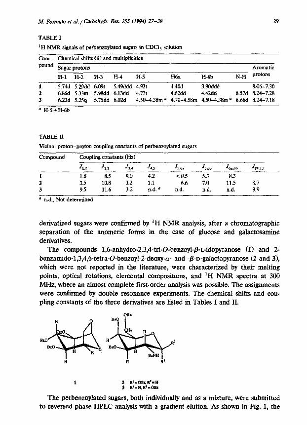
TABLE I
‘H NMR signals of ~r~~oy~ted sugars in CDCi, ~Iution
Corn- Chemical shifts (8) and multiplicities pound Sugar protons
H-l H-2 H-3 H-4 H-5 H6a H-6b
Aromatic N_H protons
1 5.74d 5.29dd 6.091: 5.49ddd 4.93t 4.4Od 3.9Oddd 8.06-7.30 2 6.86d 5.33m 5.98dd 6.13dd 4.73t 4.62dd 4.42dd 6.57d 8.24-7.28 3 6.23d 5.2.5q 5.75dd 6.02d 4.50-4.38m a 4.70-4.58m 4.50-4.38m o 6.66d 8.24-7.18
= H-S+H-6b
TABLE II
Vicinal proton-proton coupling constants of perbenzoylated sugars
Compound
f.
2 3
Coupling constants (Hz)
JLZ J 29 J 3*4
1.8 8.5 9.0 3.5 10.8 3.2 9.5 11.6 3.2
J4P JS,& JS,6b J 6a,6b JHH,Z
4.2 < 0.5 5.3 8.3 1.1 6.6 7.0 11.5 8.7 n.d. a n.d. n.d. n.d. 9.9
0 n.d., Not dete~ned
derivatized sugars were confirmed by ‘H NMR analysis, after a chromatographic separation of the anomeric foxms in the case of glucose and g~acto~~ne derivatives.
The compounds 1,6-anhydro-2,3,4-tri-O-benzoyl-P_L-idopyranose (1) and 2- be~~do-l,3,4~6-tetra-~-be~oyl-2-deo~~- and -~-D-gala~topyr~ose (2 and 31, which were not reported in the literature, were characterized by their melting points, optical rotations, elemental compositions, and ‘H NMR spectra at 300 MHz, where an almost complete first-order analysis was possible. The assignments were confirmed by double resonance experiments. The chemical shifts and cou- pling constants of the three derivatives are listed in Tables I and II.
1 2 R’=OB&R’=H 3 R’rH,R’nORz
The perbenzoylated sugars, both individually and as a mixture, were submitted to reversed phase HPLC analysis with a gradient elution. As shown in Fig. 1, the

30
0.4-
R
a? 0.3-
0.2-
O.l-
0.0-A A h I I
0 20 40
4 OS- 3
0.4-
0.0 v ' 0 20 40
2 ‘tl
;o .c;’
%
!5 u :
.;
yc 60
100
75 8
3 I
IN. Fonnato et al. / Carbohydr. Res. 255 (1994) 27-39
100
3 k
75 v
0.5- c, Glc ,100
0.4-
o'3~_._---/
____~..
/ _.___.. -..... 75 g -
Y .d Ll
.50 .*
0.2 g
0.1 .*,j
0.0 ..' \
d I I -0
20 40 min 60 Fig. 1. Reversed phase chromatography oE a, tribenzoyl anhydroidose, b, pentabenzoyl galactosamine, and c, pentabenzoyl glucose. The elution was performed with gradients of acetonitrile in water, as described in the text, flow rate 1 mL/min.
retention times were quite different and in the case of galactosamine derivatives, a satisfactory anomer resolution was also achieved, the major peak corresponding to the (Y anomer (panel b). The minor peaks at t, 47.6 and 48.1 which appear in the chromatogram of tribenzoyl anhydroidose (panel a), are probably due to the pentabenzoyl derivatives originating from some unmodified L-idose. As for pentabenzoyl glucose (panel c), the major peak corresponds to the /3 anomer.
Table III shows the retention times of the perbenzoylated sugars. Our chro- matographic conditions allowed us to obtain a good separation of all derivatives, whereas the isocratic elution employed by Karamanos et al.M does not separate
M. Fomzato et ai. /Carbohydr. Res. 255 (1994) 27-39
TABLE III
31
Retention times of perbenzoylated monosaccharides on reversed phase HPLC
Sugar Retention times (min)
1,6Anhydroidose Galactosamine Glucose
(I anomer
42.2 41.8
/3 anomer
37.8 41.2 48.2
1,6-anhydroidose from galactosamine, thus making the removal of hexosamines essential. The previous separation of hexosamine by means of ion-exchange chromatography, after GAG hydrolysis, can be omitted in our set-up, thus elimi- nating an additional step which is time consuming and might cause incomplete recoveries.
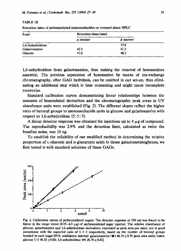
Standard calibration curves demonstrating linear relationships between the amounts of benzoylated derivatives and the chromatographic peak areas in UV absorbance units were established (Fig. 2). The different slopes reflect the higher ratio of benzoyl groups to monosaccharide units in glucose and galactosamine with respect to 1,6-anhydroidose (5 : 5 : 3).
A linear detector response was obtained for injections up to 4 pg of compound. The reproducibility was 2.9% and the detection limit, calculated as twice the baseline noise, was 10 ng.
To establish the reliability of our modified method in determining the relative proportion of L-iduronic and D-glucuronic acids in tissue galactosaminoglycans, we first tested it with standard solutions of these GAGS.
0 2 4 6 8 10
nmol
Fig. 2. Calibration curves of perbenzoylated sugars. The detector response at 230 nm was found to be linear in the range tested (0.05-4.0 PB) of perbenzoylated sugar injected. The relative absorbance of glucose, galactosamine and 1,6-anhydroidose derivatives, expressed as peak area per nmol, are in good accordance with the expected ratio of 5: 5 :3 respectively, based on the number of benzoyl groups bonded to each sugar [95% confidence interval: galactosamine (~146.76 f 0.70 peak area units/nmol; glucose (0) 46.211tO.60; 1,danhydroidose (0) 26.76+0.42].

32 M. Form&o et a!. /Carbohydr. Res. 255 (1994) 27-39
0.15
P 0.10
0.05
0.00
Fig. 3. Reversed phase ch~~atagraphy of ~r~~lated ~on~a~ha~des derived by the reduction and hydrolysis of: a, standard C6S and b, DS.
Fig. 3 shows the separation obtained utilizing dermatan sulphate (DS) and chondroitin 6-sulphate (CXS). The hexosamine-hexuronate molar ratio and the iduronate percentage relative to total hexuronate in the standard gaiactosamino- g&cans are listed in Table IV. The relative percentage of iduronic acid with respect to total hexuronates in standard preparations of commercial 665, chon- droitin 4-sulphate (CXS), and DS were 5,5, and 85%, respectively. These percent- ages are only slightIy different from those obtained by Karamanos et al.24 on standard CS and DS. These differences could be due to variability in the commer- cial preparations used.
TABLE IV
Monosaccharide oomposition of standard galactos~in~lyca~s
Standard GAG Hexuronate : hexosamine lduranate, per cent molar ratio B of total hemonate
c4s 0.98 5 C6S 1.02 5 DS 1.03 85
a Determined by reversed phase cbromato~aphy. The sum of tribenzoyl ~hydr~id~ and pentaben- zoyl glucose was compared to pentabenzoyl galactosamine.

hf. Formato et al. / Carbohydr. Rex 255 (1994) 27-39 33
9
8 25-
.L!
E _
is x o-
0 50 100 150 200
Elutlon vol (mL)
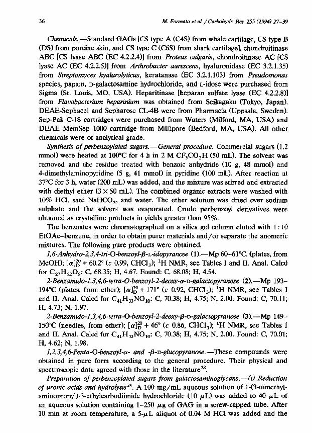
Fig. 4. DEAE-Sephacel chromatography of PGs extracted from human aortas. Elution was performed with a linear salt gradient (0.25-2.0 M NaCl). Two fractions were collected: fraction A contained mostly HS-PGs; fraction B contained a mixture of CS- and DS-PGs.
Control experiments showed that the recovery of the full assay protocol for each
standard GAG was ca. 80-90%. However, hydrolysis, perbenzoylation, and C-18 cartridge purification steps gave almost quantitative recoveries, suggesting that
ethanol precipitation represents the most critical step.
Moreover, as shown in Table IV, the perbenzoylated products obtained from
standard GAG solutions after the whole procedure always gave a molar ratio of galactosamine to hexuronic acid derivatives close to 1, thus indicating similar recoveries for all components.
Experiments performed on galactosaminoglycan mixtures of different relative composition showed that this method gives good results even with samples contain- ing very low concentrations of DS and an excess of CS isomers.
The effective applicability of the modified procedure to the analysis of the composition of PG preparations from human aorta tissue was tested on chon-
droitin sulphate-rich PGs and dermatan sulphate-rich PGs, purified by means of ion-exchange chromatography on DBAE-Sephacel (Fig. 4) and gel-filtration chro-
matography on Sepharose CL-4B (Fig. 5), as described in the experimental section. The identity of constituent GAGS was assessed, after papain digestion, by elec-
trophoretic analysis25,26 associated with the use of specific lyases, as previously described4.
Ion-exchange chromatography showed two hexuronate containing peaks: the first consisted of heparan sulphate (HS)-PGs, and the second of CS-PGs, DS-PGs, and only traces of HS-PGs and hyaluronic acid (HA). The latter, when submitted to gel-filtration chromatography, produced two peaks: one was eluted with the void volume of the column and consisted almost exclusively of CS-PGs, and the other, roughly separated, eluted in the included volume, was composed mainly of DS-PG. Both fractions were analyzed for their sugar composition.
The galactosaminoglycan analysis of aorta PGs requires a preliminary digestion with papain, in order to obtain polysaccharide chains, followed by the sequential

M. Fomaato et al. / Carbohydr. Res. 255 (I994) 27-39
Elution vol (mL) Fig. 5. Sepharose CL4B gel chromatography of fraction B obtained from the DEAE-Sephacel Two fractions were collected, as shown.
use of hyaluronidase, heparitinase, and keratanase to ensure the removal of glycosaminoglycans, which could be derived from hyaluronic acid, heparan sul- phate-PGs, and keratan sulphate-PGa, occasionally present as contaminants in purified galactosaminoglycan containing PG preparations. It should be noted that direct analysis of heparan sulphate chains under the experimental conditions used
is not possible owing to its resistance to hydrolysis27.
,100
_ a, CS-rich PG R N O.lO-
d
0.15 1 b, DS-rich PG /
Fig. 6. Reversed phase chromatography of perbenzoyl monosaccharides derived by the reduction and hydrolysis of GAGS from human aorta: a, CS-rich PG, b, DS-rich PG.
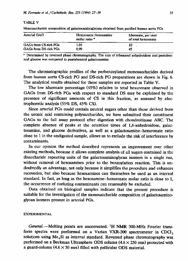
M. Fonmm et a~~Ca~~. Res. 255 (199a) 27-39 35
TABLE V
Monosaccharide composition of galactosaminogiycans obtained from purified human aorta PGs
Arterial GAG Hexuronate : hexosamine Idurouate, per cent molar ratio * of total hexuronate
GAGS from C&rich PGs 1.05 10 GAGS from DS-rich PGs 0.99 45
@ Determined by reversed phase chromatography. The sum of tribenzoyl anhydroidose and pentaben- zoyl glucose was compared to pentabenzoyl galactosamine.
The chromatographi~ profiles of the perbenzoylated monosa~harides derived from human aorta CS-rich PG and D&rich PC preparations are shown in Fig. 6. The analytical results obtained for these samples are reported in Table V.
The low iduronate percentage (45%) relative to total hexuronate observed in GAGS from DS-rich PGs with respect to standard DS may be explained by the presence of significant amounts of CS in this fraction, as assessed by elec- trophoretic analysis (55% DS, 45% CS).
Since arterial PGs could contain neutral sugars other than those derived from the uranic acid containing polysaccharides, we have submitted their constituent GAGS to the full assay protocol after digestion with chondroitinase ABC. The complete absence of peaks at the retention times of 1,6-~hydroidose, galac- tosamine, and glucose derivatives, as well as a ~alactos~ine-he~ronate ratio close to 1 in the undigested sample, allows us to exclude the risk of interference by contaminants.
In our opinion the method described represents an improvement over other existing methods, because it allows complete analysis of all sugars contained in the disaccharide repeating units of the galactosam~o~y~ isomers in a single run, without removal of hexosamines prior to the benzoylation reaction. This is un- doubtedly an advantage, not only because it simplifies the procedure and enhances recoveries, but also because hexosamines can themselves be used as an internal standard. In fact, as long as the hexosamine-hexuronate molar ratio is close to 1, the occurrence of coeluting ~ntaminants can reasonably be excluded.
Data obtained on biological samples indicate that the present procedure is suitable for the investigation of the monosaccharide composition of galactosamino- glycan isomers present in arterial PGs.
EXPERIMENT
Gemral.---Melting points are uncorrected. ‘H NMR 300-MHz Fourier trans- form spectra were performed on a Varian VXR-300 spectrometer in CDCl, solutions using Me,Si as internal standard. Reversed phase ~hromato~aphy was performed on a Beckman Ultrasphere ODS cohmm (4.6 x 250 mm) protected with a guard-column (4.6 X 30 mm) filled with pellicular ODS material.
36 M. Fomato et al. / Carbohydr. Res. 255 (1994) 27-39
Chemicals. -Standard GAGS [CS type A (C4S) from whale cartilage, CS type I3
(DS) from porcine skin, and CS type C (C6S) from shark cartilage], chondroitinase ABC [CS lyase ABC (EC 4.2.2.4)J from Proteus vulguti, chondroitinase AC [CS
lyase AC (EC 4.2.2.511 from Arthrobucter aurescens, hyaluronidase (EC 3.2.1.35) from Streptomyces hyalurotyticus, keratanase (EC 3.2.1.103) from Pseudomonas species, papain, p-galactosamine hydrochloride, and L-idose were purchased from Sigma (St. Louis, MO, USA). Heparitinase [heparan sulfate lyase (EC 4.2.2.8)] from Flavobacterium heparinium was obtained from Seikagaku (Tokyo, Japan).
DEAE-Sephacel and Sepharose CL-4B were from Pharmacia (Uppsala, Sweden). Sep-Pak C-18 cartridges were purchased from Waters (Milford, MA, USA) and
DEAE MemSep loo0 cartridge from Millipore (Bedford, MA, USA), All other chemicals were of analytical grade.
Synthesk of perbenzoylated sugars. -General procedwe. Commercial sugars (1.2 nnnol) were heated at 100°C for 4 h in 2 M CF,CO,H (50 mL). The solvent was removed and the residue treated with benzoic anhydride (10 g, 48 mmol) and 4-dimethylaminopyridine (5 g, 41 mmo1) in pyridine (100 mL>. After reaction at
37°C for 3 h, water (200 mL) was added, and the mixture was stirred and extracted with diethyl ether (3 x 50 mL). The combined organic extracts were washed with 10% HCl, satd NaHC@, and water. The ether sohrtion was dried over sodium
sulphate and the solvent was evaporated. Crude perbenzoyl derivatives were obtained as crystalline products in yields greater than 95%.
The benzoates were chromatographed on a silica gel column eluted with 1: 10 EtOAc-benzene, in order to obtain purer materials and/or separate the anomeric mixtures. The following pure products were obtained.
1,6-Anhydro-2,3,4-tti-O-benzoyQ3-r;idopyranose (l).-Mp 60-61°C. (plates, from
MeOH); [a]g + 60.2* (c 0.99, CHCl,); ‘H NMR, see Tables I and II. Anal. Calcd for C,H,,O,: C, 68.35; H, 4.67. Found: C, 68.08; H, 4.54.
2-Benzumido-1,3,4,6-tetra-Q-benzoyl-2-deoxy-a-o-galactopyranose (2).-Mp 193- 194°C (plates, from ether); [cY]~ + 171” (c 0.92, CHCI,); ‘H NMR, see Tables I and II. Anal. Calcd for C,,H33N0,,: C, 70.38; H, 4.75; N, 2.00. Found: C, 70.11; H, 4.73; N, 1.97.
2-Benzamido-1,3,4,6-tetra-Q-benzoyl-2-deoxy-13-D-galacto~ra~se (3).-Mp 149-
150°C (needles, from ether); [1x]g + 46” (c 0.86, CHCl,); ‘H NMR, see Tables I and II. Anal. Calcd for C,,H,,NO,,,: C, 70.38; H, 4.75; N, 2.00. Found: C, 70.01; H, 4.62; N, 1.98.
1,2,3, #, 6-Pentrr -O-benzoyZ-cr- and -o-D-glucopyrunose. -These compounds were obtained in pure form according to the general procedure. Their physical and
spectroscopic data agreed with those in the literature? Preparuhun of perbenzoylated sugars from galactosuminoglycans.-(i) Reduction
of uranic acids and hydrolysis”. A 100 mg/mL aqueous solution of l-(3-dimethyl- aminopropyl)-3-ethylcarbodiimide hydrochloride (10 PL) was added to 40 PL of an aqueous solution containing l-250 pg of GAG in a screw-capped tube. After 10 min at room temperature, a 5-pL aliquot of 0.04 M HCl was added and the

M. Formuto ef al. /Ca~~y~. i&x. 255 (1994) 27-39 37
mixture was regularly stirred in an ultrasonic bath. After 50 min another 5-PL aliquot of 0.04 M HCl was added and the mixture was stirred for a further hour.
To the above solution 2 M aq NaBH, (100 PL) was added and the mixture was heated at 50°C for 1 h. A second aliquot of 2 M NaBI& WO PL) was then added and the heating was continued for another 30 min. After cooling to room temperature, 25 FL of glacial acetic acid was cautiously added, and then 1.3 mL of abs EtOH. The sample was briefly vortexed and kept at 4°C overnight. After centrifugation in a high speed microcentrifuge (9OOOg), the pellet obtained was dried under a stream of N,.
The sample obtained from the above procedure was dissolved in 2 M CF&O,H (0.5 mL) and heated at 100°C for 9 h.
(ii) ~~r~e~~oy~r~~~ of the ~o~sacc~~de maples. ‘l%e hydrolyzed mixture was transferred into a 10 mL glass syringe and the solvent removed by Iyophilization. The dried product was treated with 0.5 mL of pyridine containing 10% <w/v) benzoic anhydride and 5% (w/v) 4-dimethylaminopyridine at 37°C for 90 min. The reaction was quenched with 4.5 mL of water and the mixture was passed three times through a C-18 cartridge. After washing with 10 mL aq 10% pyridine and 10 mL of water, the benzoyl derivatives were eluted with 15 mL of MeCN, reversing the flow through the cartridge. The solvent was evaporated.
The sample was dissolved in MeCN to a final concentration of 4-40 pg/mL of the original GAG uranic acid content, and then the solution was centrifuged at. 9OOOg to precipitate particulate materials and filtered on a Millipore filter (pore size 0.22 pm) prior to injection into the HPLC system.
Reversed phase HPLC.---The elution was performed with a discontinuous linear gradient of MeCN in water. The chromatographic conditions were the following: O-10 min 25% MeCN, lo-25 min 25-60% MeCN, 25-35 min 60% MeCN, 35-40 min 60-75% MeCN, and 40-60 min 75% MeCN. The flow rate was 1.0 mL/min at room temperature, and detection was at 230 nm. A 20-PL loop was used for injection. Identity of the peaks was confirmed by coelution with authentic samples. Calibration curves were obtained by injecting known boots of standard perben- zoylated monosaccharides, ranging from 0.05 to 4 pg.
Preparatim uf tissue glycosamirwglycam .-PGs were obtained from samples of normal human thoracic aorta intima and media taken at autopsy within 24 h of death. Aortas from two subjects aged 39 and 46 years were used after obtaining appropriate consent from the relatives. After removing the adventitia and outer media, only normal areas were selected. Aorta minces were suspended in 15 times their weight of 6 M urea in 1 M NaCI, ~ntaining protease ~hibitors (10 mM ethylenediaminetetraacetic acid disodium salt, 5 mM benzamidine hydrochloride, 10 mM phenylmethanesulphonyl fluoride, 10 mM e-aminocaproic acid, 10 mM ~-ethy~ale~ide~, and shaken at 4°C for 24 h. The extract, c~nta~ing ca. 60% of the total uranic acids, was centrifuged at I~~g for 1 h at 4°C and the pellet discarded. The supernatant was dialysed against 6 M urea, containing protease inhibitors, until a concentration of 0.25 M NaCl was reached.
38 M. Format0 et al. /Carbohydr. Res. 255 (1994) 27-39
Samples were submitted to ion-exchange chromatography on a DEAE-Sephacel column (1.5 X 25 cm), equilibrated with a 50 mM sodium acetate buffer, pH 6.2
containing 6 M urea, 0.25 M NaCI, 10 mM CaCl,, and protease inhibitors at a flow rate of 10 mL/h. The column was washed with 3 bed volumes of the same buffer to elute contaminating proteins and the PG elution was carried out with a linear
gradient of NaCl in the same buffer, ranging from 0.25 to 2.0 M. The uranic acid
profile showed two hexuronate containing peaks. Hexuronate containing material of the second peak was applied onto a Sepharose CL4B column (1.0 X 50 cm) and
eluted with 6 M urea, 1 M NaCI, containing protease inhibitors, at a flow rate of 10
mL/h, producing two PG fractions. Both of these were exhaustively dialyzed and digested sequentially with papain, hyaluronidase, heparitinase, and keratanase before HPLC analysis.
Other anafytkal methods.-Hexuronic acid content was determined by the method of Bitter and Muir29, using D-glucuronolactone as a standard. Neutral sugar content in reduced GAGS was measured by the anthrone reaction3’. Protein concentration was determined by the method of Lowry31.
Protein digestion was carried out with papain (150 pg/mg protein) in the
presence of 5.0 mM cysteine hydrochloride and 5.0 mM ethylenediaminete-
traacetic acid, in 0.1 M sodium acetate buffer, pH 5.8 at 60°C for 48 h3*. The digest was clarified by centrifugation at 8OOOg and the residue washed. Washings were
combined with the original digest and loaded onto an anion-exchange cartridge to
remove oligosaccharides. GAGS were eluted with 1 M NaCl and precipitated by adding 4 vol of EtOH. The mixture was left overnight at 4°C and the precipitate
separated by centrifugation, washed twice with EtOH and diethyl ether, and dried. Electrophoretic separation of GAGS was carried out according to Cappelletti et
al z,26 . .
When necessary, aliquots of tissue GAGS (100 pg hexuronic acid/ml) were digested with the following lyases at 37°C for 24 h: hyaluronidase, 10 U/mL in 0.5
M sodium acetate, pH 5.6; keratanase, 0.02 U/mL in 0.05 M tris(hydroxymethyl)- aminomethane hydrochloride, pH 7.4; heparitinase, 0.02 U/mL in 0.05 M sodium acetate, pH 7.0; chondroitinase ABC or AC, 0.1 U/mL in 0.1 M sodium acetate,
pH 8.0. Every digestion step was followed by precipitation with EtOH.
ACKNOWLEDGMENTS
This work was supported by funds from M.U.R.S.T, Italy. We wish to express our gratitude to Dr. Antonio Zucca for kindly recording the NMR spectra.
REFERENCES
1 W.D. Wagner, B.G.J. Salisbury, and H.A. Rowe, Arteriosclerosis, 6 (1986) 407-417.
2 R. Dalferes, B. Radhakrishnamurthy, H.A. Ruiz, and G.S. Berenson, Exg. Mol. Pathol., 47 (1987) 363-376.

M. Format0 et al/Carbohydr. Res. 255 (1994) 27-39 39
3 S. Berenson, B. Radhakrishnamurthy, S.R. Srinivasan, P. Vijayagopal, and E.R. Dalferes, Arch. Pathol. Lab. Med, 112 (1988) lOO2-1010.
4 G.M. Cherchi, R. Coinu, P. Demure, M. Formato, G. Sanna, M. Tidore, M.E. Tira, and G. De Luca, Mati, 10 (1990) 362-372.
5 R. Kapoor, C.F. Phelps, L. Caster, and L. Fransson, Biochem. J., 197 (1981) 259-268. 6 R. Kapoor, C.F. Phelps, and T.N. Wight, Biochem. J., 240 (1986) 575-583. 7 A.R. Poole, Biochem. J., 236 (1986) l-14. 8 T.N. Wight, Arteriosclerosk, 9 (1989) l-20. 9 N.B. Beaty and R.J. Mello, J. Chromatogr., 418 (1987) 187-222.
10 C. Kodama, T. Kodama, and Z. Yosizawa, 1. Chromurogr., 429 (1988) 293-313. 11 J. Lee, B. Radhakrishnamurthy, E.R. Dalferes, Jr., and G.S. Berenson, J. Chromatogr., 419 (1987)
275-279. 12 J. Lee and H. Tieckehnann, Anal. Biochem., 94 (1979) 231-236. 13 N. Ototani, N. Sato, and Z. Yosizawa, J. Biochem. (Tokyo), 85 (1979) 1383-1385. 14 L. Huharty, J.A. Glick, N.M. Matusewicz, and H. Kihara, Biochem. Med., 27 (1982) 352-360. 15 A. Hjerpe, C.A. Antonopoulos, B. Classon, B. Engfeldt, and M. Nurminen, J. Chroma~ogr., 235
(1982) 221-227. 16 K. Murata and Y. Yokoyama, Anal. Biochem., 146 (1985) 327-335. 17 C. Seldin, N. Seno, K.F. Austin, and R.L. Stevens, Anal. B&hem., 141(1984) 291-300. 18 I. Takazono and Y. Tanaka, J. Chromatogr, 288 (1984) 167-176. 19 E. Zebrower, F.J. Kieras, and W.T. Brown, Anal. Biochem., 157 (1986) 93-99. 20 J. Macek, J. Krajckova, and M. Adam, J. Chromarogr., 414 (1987) 156-160. 21 K. Murata and Y. Yokoyama, J. Chromatogr., 423 (1987) 51-61. 22 T. Gherezghiher, M.C. Koss, R.E. Nordquist, and C.P. Wilkinson, J. Chromarogr., 413 (1987) 9-15. 23 J. Linhardt, K.N. Gu, D. Loganathan, and S.R. Carter, Anal. Bbchem., 181 (1989) 288-296. 24 N.K. Karamanos, A. Hjerpe, T. Tsegenidis, B. Engfeldt, and C.A. Antonopoulos, Anal. Biochem.,
172 (1988) 410-419. 25 R. Cappelletti, M. Del Rosso, and V.P. Chiarugi, Anal. Biochem., 99 (1979) 311-315. 26 R. Cappelletti, M. Del Rosso, and V.P. Chiarugi, Anal. Biochem., 105 (1980) 430-435. 27 J.E. Shively and E. Conrad, Biochemistry, 15 (1976) 3932-3942. 28 N.B. D’Accorso, I.M.E. Thiel, and M. Shuller, Carbohydr.Res., 124 (1983) 177-184. 29 T. Bitter and H.M. Muir, Anal. Biochem., 4 (1962) 330-334. 30 J.H. Roe, J. Biol. Chem., 212 (1955) 335-343. 31 O.H. Lowry, N.J. Rosenbrough, A.L. Farr, and R.J. Randall, J. Biol. Chem., 193 (1951) 265-275. 32 G.C. Gillard, D. Caterson, and D.A. Loowther, Biochem, J., 145 (1975) 209-213.


![Increasing Sensitivity in HPLC...[W.R.Melander, C.Horvath, Reversed-Phase Chromatography, in HPLC Advances and Perspectives, V2, Academic Press, 1980] Challenge of Making “2 µm”](https://img.dokumen.tips/doc/110x75/5e668ee1c92e374c9200dfb5/increasing-sensitivity-in-hplc-wrmelander-chorvath-reversed-phase-chromatography.jpg)