Embed Size (px)
Citation preview

756
Biochimica et Biophysica Acta~ 600 (1980) 756--768 © Elsevier/North-Holland Biomedical Press
BBA 78862
A RE-EVALUATION OF THE SURFACE COMPLEXITY OF THE INTACT ERYTHROCYTE
STEPHEN THOMPSON, CAROLYN M. RENNIE and ALUN H. MADDY *
Department of Zoology, University of Edinburgh, Edinburgh, EH9 3JT (U.K.)
(Received February 21st, 1980)
Key words: Giycoprotein; Lactoperoxidase; Surface protein; (Erythrocyte)
Summary
Surface proteins and glycoproteins of intact human red blood cells were labelled with '2sI by the lactoperoxidase method. The radioactive proteins were then separated in each of the Fairbanks and Laemmli one-dimensional poly- acrylamide gel electrophoresis systems. The radioactive polypeptides had dif- ferent mobilities in the two systems, largely due to the anomalous migration of glycoproteins in polyacrylamide gels. A two-dimensional system was therefore developed using the Fairbanks and Laemmli buffer systems to exploit these anomalies. This procedure clearly resolved radioactive glycoproteins and proteins and enabled the identification of many more surface components than had previously proved possible.
Introduction
Many techniques have been developed to label surface components of ery- throcyte membranes [ 1--3 ]. Techniques utilising radioactive iodine have proved to be the most popular, presumably due to the ease of detection of radioactive iodine in labelled proteins after their separation in polyacrylamide gels. The best known, and most used, of these techniques is the lactoperoxidase- catalysed iodination procedure using H202 directly [4] or H202 generated by the glucose-glucose oxidase system [5]. Other techniques utilising radioactive iodine involve the direct covalent binding of radioactively labelled chemicals, often diazonium salts, to surface proteins [6,7]. More recently, another iodinating reagent, chloroglycoluril, has been used to label erythrocyte surface
* T o w h o m correspondence shou ld be addressed. Abbreviat ion: Hepes , N-2-hydroxyethylpiperazine-NP-ethanesulphonic acid.

757
proteins [8]. Surprisingly, relatively few membrane proteins have been shown to have a surface orientation in intact red blood cells by these techniques and such a simplicity is inconsistent with the many functions that may be attributed to the cell's surface [9]. Other techniques [10,11], especially tritiation of the surface carbohydrates [12,13], have revealed many more components. The apparent discrepancies could result from several factors, variation in the susceptibility of different surface groups to different labelling techniques, variation in fractionation of labelled components, and aggregation of labelled components.
The commonest fractionation procedure used to study surface labelled groups is sodium sulphate (SDS) gel electrophoresis in various guises, and although it is well known that glycoproteins behave anomalously in these systems [14] frequently insufficient attention is paid to these anomalies. Here we compare the fractionation of erythrocyte membrane proteins by two well-established gel buffer systems, those of Fairbanks et al. [15] and Laemmli [16], and combine the two methods into a two-dimensional technique which exploits the effects of gel matrix and buffer on the mobilities of glycoproteins. This combination results in a much more complex pattern of labelled proteins after lactoperoxidase-catalysed iodination than has previously been described as it reveals many components which are obscured when either buffer system is used alone. Some of the complexity can be interpreted in terms of glycoprotein aggregations which have been reported elsewhere [ 17 ].
Materials and Methods
Materials 12sI as Na~2SI in dilute NaOH was purchased from the Radiochemical Centre
(Amersham, Bucks, U.K.). Biochemicals and enzymes were purchased from Sigma Chemical Co. (St. Louis, MO, U.S.A.). Chemicals were of analytical grade and were obtained from BDH Chemicals Limited (Poole, Dorset, U.K.).
Methods Preparation of erythrocyte suspensions for 12sI labelling. Blood was obtained
by venipuncture from healthy male and female adults using heparin as an anti- coagulant. White cells were removed by using the method of Beutler et al. [18]. The blood was passed through an a-ceUulose/microcrystalline cellulose (2 : 1, w/w) column in Hepes-buffered isotonic saline (133 mM NaCl, 4.5 mM KC1, 10 mM Hepes, pH 7.4) [19]. The filtered blood was washed three times in the same Hepes-buffered isotonic saline and then labelled with 12sI. Blood was iodinated within 3 h of collection.
Surface labelling with 12sI. Lactoperoxidase-catalysed iodination was carried out using H202 generated by the glucose oxidase system of Hubbard and Cohn [5]. 1 ml of inct~bation mixture contained 5 - l 0 s cells in Hepes-buffered isotonic saline, 5 mM glucose, 0.1/zg glucose oxidase (Sigma, Type V), 10/~g lactoperoxidase (Sigma, powder), 0.5/~M K127I and 50/~Ci of Na12SI: (in our experience, 0.5 #M K12TI gave the maximal level of iodination). Incubations were carried out for 20 min at 37°C then the reaction was terminated by the addition of 10-ml of Hepes-buffered isotonic saline (4°C)conta in ing 10 pM

758
Na2S2Os: (all subsequent operations were performed at 4~C). The cells were washed three times with 12-ml of this buffer and twice with 12-ml of this buffer containing 5 #M KI to remove any free Na12SI. Ghosts were prepared by lysing the cells in 12-ml of 5 mM phosphate buffer (pH 7.2) and centri- fuging the resulting suspension at 38 000 × g for 25 min. The pink ghosts were then washed in 12-ml of 5 mM phosphate buffer (pH 7.5) and recentrifuged. The resultant off-white ghost pellet was prepared for electrophoresis by the addition of 1 vol. of 0.125 M Tris-HC1, pH 6.8, containing 10% (w/v) SDS, 20% glycerol, 0.002% bromophenol blue, 1 mM EDTA and 80 mM dithio- threitol to 1 vol. of ghosts. This mixture was immediately heated for 5 min at 100°C to inactivate proteases and maximise protein solubilisation.
One-dimensional polyacrylamide gel electrophoresis. Proteins were separated by SDS-polyacrylamide gel electrophoresis in the continuous buffer system described by Fairbanks et al. [ 15] or the discontinuous buffer system described by Laemmli [16]. The slab gel apparatus used was that first described by Reid and Bieleski [20] and later modified by Studier [21]. The slab gels were 15 cm wide, 14 cm high and 1.5 mm thick. A 14-sample comb was used, each sample well 7 mm wide, 15 mm deep and 3 mm apart. In the system of Fairbanks, the acrylamide stock solution contained 40% (w/v) acrylamide and 1.5% (w/v) bis-acrylamide. This was used to make linear gradient acrylamide slabs from 4--6% acrylamide. Electrophoresis took approx. 4 h at a constant current of 40 mA. In the discontinuous buffer system of Laemmli, the acrylamide stock solution was 30% acrylamide and 0.8% bis-acrylamide. This was used to prepare running gels of 11.5% acrylamide and stacking gels of 5% acrylamide. Electro- phoresis took approx. 4 h at a constant current of 30 mA. Pre-set Tris-HC1 crystals (pH 8.7 at 25°C, Sigma) were used to make up the running-gel buffer; this was essential for reproducible high-resolution gels. The total polypeptide content of the cells was determined by staining the gels with Coomassie brilliant blue [ 15]. Radioactive polypeptides were detected by autoradiography of the dried gels.
Two-dimensional gel electrophoresis. To produce a two-dimensional gel of erythrocyte ghost proteins the protein samples were first run in a one-dimen- sional Falrbanks slab gel (1.5 mm thick) with empty sample wells between samples. After electrophoresis, strips (7--8 mm wide)containing the samples were cut from the acrylamide slab. These strips were equilibrated for 30 min in Laemmli sample buffer containing 0.0625 M Tris-HC1, pH 6.8, 5% (w/v) SDS, 10% (w/v) glycerol, 0.5 mM EDTA and 5% (v/v) mercaptoethanol. After equilibration the strips Could be run in the second (Laemmli) dimension immediately or stored frozen at --20°C for up to 2 or 3 weeks. Longer periods of storage led to dehydration and shrinkage. Second-dimension Laemmli gels (1.75 mm thick) were prepared as for one-dimensional gels except that the stacking gel was 1.5 cm high with a flat surface which was itself 1 cm below the buffer surface. A strip from the Fairbanks gel was then pushed into the space above the stacking gel, fixed into position with 1.0% (w/v) agarose in electrode buffer containing 0.001% bromophenol blue as a marker dye and subjected to electrophoresis.

759
Results and Discussion
Iodination of the red cells Under the conditions described in Methods with 0.5 pM K127I, the radio-
active iodine incorporation into membrane surface proteins was approx. 3 • 107 dpm/mg of protein. At 1.5 /~M or 0/zM K127I, the radioactive iodine incorporation dropped by 65 and 25%, respectively. Non-specific labelling (control without any lactoperoxidase) was less than 0.5%.
Polyaerylamide gel electrophoresis in the Fairbanks system The continuous buffer system of Fairbanks et al. [15] has been the most
widely used system for the examination of erythrocyte proteins in recent years. We therefore separated our labelled membrane proteins in this system for direct comparison with other studies. Fig. 1 compares the total polypeptide content of the erythrocytes with those labelled with radioactive iodin'b. This
A B C D
i
P
Hb
PASl
l ib p s2 m m m
3
4 . 5 a , ,
6 /
i s , m S 3
H b ÷ D F - -
m
l~Sl
i
~ i P A S 2
~ Slm~SS
Fig. 1 . (A) T h e t o t a l p o l y p e p t i d e c o n t e n t b y C o o m a s s / e s t a i n i n g o f e r y t h r o c y t e m e m b r a n e s s e p a r a t e d b y the F a i r b a n k s s y s t e m , a n d (B) a u t o r a d i o i ~ a p h o f t h e r a d i o a e t / v e i o d i n e - l a b e l l e d s u r f a c e p o l y p e p t i d e s o f A. (C) T h e t o t a l p o l y p e p t i d e c o n t e n t o f e r y t h r o c y t e m e m b r a n e s s e p a r a t e d b y t h e L a e m m l i s y s t e m , a n d (D) a u t o r a d i o i ~ a p h o f t h e r a d i o a c t i v e i o d i n e - l a b e l l e d s u r f a c e p o l y p e p t i d e s o f C. P o l y p e p t i d e s a re n u m b e r e d as in t h e Fa l rb .n l rA s y s t e m . H b = h a e m o g l o b i n ; D F = d y e f r o n t .

760
labelling agrees very well with that obtained in other studies [7,8,22,23] through which we can identify one of the labelled peptides in band 3 as PAS 1 and the labelled peptides in the 4.5 and 7 positions as PAS 2 and PAS 2, respectively. These assignments are confirmed and amplified by our two- dimensional system (see below), as is the apparently anomalous labelling coincident with actin (band 5).
Polyacrylamide gel electrophoresis in the Laemmli system The total polypeptide content of erythrocytes and the radioactively labelled
surface proteins obtained when labelled membranes were separated in the system described by Laemmli are also shown in Fig. 1. The Laemmli system gives a much better resolution than the Fairbanks system and many differences can be seen between the polypeptide patterns produced by the two systems.
In Laemmli gels there is no 2.1 band, band 3 stains as a broad rather diffuse band containing sharp discrete bands within it, band 4.1 is always a doublet [24], up to 15 polypeptides can be clearly detected between bands 4.2 and 5, making the '4.5 region' very complex, and an extra band above band 7 (labelled as 6.9) is always present. To allow comparison with other studies that have used the Laemmli polyacrylamide gel system [8,13,24,25] two relatively strong bands in the 4.5 region are labelled as 4.5a and 4.5b. Band 4.5a migrates fractionally slower than catalase and is the strongest band in the 4.5 region in incompletely washed (pink) normal ghosts and in ghosts obtained from patients with hereditary spherocytosis, it is, in fact, a major constituent of the cyto- plasm. Band 4.5b migrates fractionally faster than catalase and is the strongest band in the 4.5 region in completely washed (white) normal ghosts (Thompson, S. and Maddy, A.H., unpublished results). Band 6.9 is extracted along with spectrin (bands 1 and 2) and actin (band 5) when erythrocyte ghosts are dialysed overnight against 0.5 mM EDTA and is, presumably, the component designated as band 7 in the spectrin extract of Dunbar and Ralston [26]. Up to 20 surface-labelled polypeptide bands can be seen in autoradiograph of the one-dimensional gel.
When the four most radioactive polypeptide bands (PAS 1, PAS 2, PAS 3 and band 3) are compared between each gel system, as would be predicted from the literature [14,27], they have different relative mobilities. In Falrbanks gels at the concentration used here, PAS 1, 2 and 3 have apparent molecular weights of 95 000, 55 000 and 29 000, respectively; in Laemmli gels they migrate with apparent molecular weights of 80 000, 38 000 and 24 000. We therefore decided to exploit this anomalous migration of the glycoproteins and to subject the labelled proteins to a combination of the two buffers as a two-dimensional system. Such a technique would be expected to separate labelled glycoproteins from each other and from any labelled proteins which co-migrate with them in either of the two one-dimensional systems.
Two-dimensional polyacrylamide gel electrophoresis Erythrocyte protein samples were separated in two dimensions as described
in Methods. A Coomassie blue-stained two-dimensionai gel does not give very much extra detail compared with a one dimensional Laemmli gel (Fig. 2). It does, however, show that the Fairbanks 2.1 component runs as a band 1 corn-

761
ii i
m - J ~
Q 6
" 7
Fig. 2. The two-dimensional Coomassle-stalning pat tern of e ry throcyte membrane polypept ides . Inset: 1, 2 and 2.1 region at a lower protein loading.
ponent in Laemmli gels, hence explaining the lack of a 2.1 band in Laemmli gels. Band 3 is also seen to migrate as a large diffuse area away from the main polypept ide arc, presumably because of its carbohydrate content [28,29] . Four or five small spots are seen on the polypept ide arc in positions where the broad band 3 obscures them in one-dimensional systems. This heterogenei ty might account for some of the apparent multiplicity of function of the band 3 region.
The autoradiographed gel (Fig. 3) is much more informative, revealing more than 50 surface-labelled components . The strongest radioactivity is associated with components that do no t stain well with Coomassie blue, only four of the radioactive spots (bands 3, 4.5a, 4 .5b and haemoglobin, Fig. 4) are stained. The
Fig. 3. Autoradiographs of surface-labeiled components of e ry throcyte membranes. (a) Short exposure, (b) long exposure.

I!
LA
EM
ML
I ...
...
..
LA
E
. !
e e
®
i ¸
~ ~ ii
i~
i~ iii!!
i ~
me
m
~P
AS
3
I IP
~Sl
mP
As
2
m E
l
FAIRBANKS
°
tU I . ~ v , , ,
0
0
0 %o °
763
O
H b Q
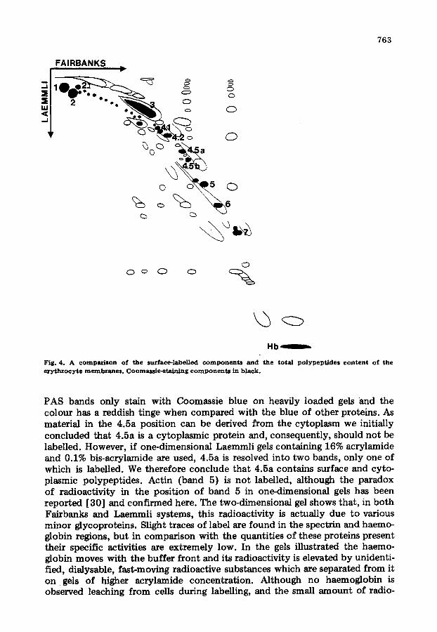
Fig. 4. A comparison of the surface-labelled componen ts and the to ta l polypeptides content of the ery throcytc membranes. Coomassie-staining componen ts in black.
PAS bands only stain with Coomassie blue on heavily loaded gels and the colour has a reddish tinge when compared with the blue of other proteins. As material in the 4.5a position can be derived from the cytoplasm we initially concluded that 4.5a is a cytoplasmic protein and, consequently, should not be labelled. However, if one-dimensional Laemmli gels containing 16% acrylarnide and 0.1% bis-acrylamide are used, 4.5a is resolved into two bands, only one of which is labelled. We therefore conclude that 4.5a contains surface and cyto- plasmic polypeptides. Actin (band 5) is not labelled, although the paradox of radioactivity in the position of band 5 in one-dimensional gels has been reported [30] and confirmed here. The two-dimensional gel shows that, in both Fairbanks and Laemmli systems, this radioactivity is actually due to various minor glycoproteins. Slight traces of label are found in the spectrin and haemo- globin regions, but in comparison with the quantities of these proteins present their specific activities are extremely low. In the gels illustrated the haemo- globin moves with the buffer front and its radioactivity is elevated by unidenti- fied, dialysable, fast-moving radioactive substances which are separated from it on gels of higher acrylamide concentration. Although no haemoglobin is observed leaching from cells during labelling, and the small amount of radio-

764
activity would probably pass unnoticed in other gel systems, especially when gels have been sliced for counting, this radioactivity could represent a degree of leakage of the erythrocytes.
The PAS bands 1, 2, 3 and 4 of the Fairbanks dimension each resolve into several components in the Laemmli second dimension (Fig. 5).
The major components of PAS 1 and 2 form a rectangle which we interpret as the two interconvertible forms of PAS 1 and 2 which have been reported [14,31]. It is probable that these PAS 1 and 2 components represent the dimeric and monomeric forms of the same major erythrocyte glycoprotein (glycophorin A) both of which might exist in the membrane [ 32]. Glycophorin A (Fig. 6a) can also contribute to PAS 4 [33]. Although the two glycoprotein A components comprise the vast majority of the PAS 1 band, the PAS 2 band contains another family of glycoproteins. This family of glycoproteins probably represents part of glycophorin B (Fig. 6b) reported by Furthmayr et al. [17] (the PAS 2' components of Mueller and Morrison [22]) and can explain other reports demonstrating that the PAS 2 glycoproteins are different
FAIRBANKS
C~
f
PAS 1 4 2 3
%
Fig. 5. T h e c o m p o n e n t s f o r m e d in th e L a e m m l i s e c o n d d i m e n s i o n from PAS bands 1, 2, 3 and 4 o f the Fafrbanks first dimension.

FAIRBANKS FAIRBANKS
~ (2) C?
a b
765
FAIRBANKS FAIRBANKS
. &
Q o O Q ~ O ~ ~ O ~
c d
Fig . 6. ( a - -c ) D i a g r a m s i l l u s t r a t i n g t h e p r o b a b l e r e l a t i o n s h i p b e t w e e n t h e P A S b a n d s 1 - - 4 a n d t h e g / y c o - p h o r i n s A , B a n d C, r e s p e c t i v e l y . T h e r e l a t i o n s h i p s p o s t u l a t e d b e t w e e n t h e P A S b a n d s a n d t h e t h r e e g / y c o p h o r i n s d e p e n d u p o n o b s e r v e d c o - m i g z a t i o n o f c o m p o n e n t s a n d t h e r e p o r t b y F u r t h m a y r e t al . [ 1 7 ] t h a t g l y e o p h o r i n A z~uas p r e d o m i n a n t l y i n t h e p o s / t i o n o f P A S 1 w i t h a lesser c o m p o n e n t as P A S 2 , t h a t g l y c o p h o r l n B r u n s p r e d o m i n a n t l y as P A S 2 w i t h m i n o r c o m p o n e n t s in p o s i t i o n s P A S I a n d P A S 4, a n d t h a t $ l y c o p h o r i n C r u n s c h i e f l y as P A S 2 a n d P A S 3 w l t h a t r a c e o f P A S 4. (d ) R a d i o a c t i v e s u r f a c e - l a b e l l e d c o m p o n e n t s w h i c h c a n n o t b e a s s i g n e d t o t h e g l y c o p h o r i n s .
to PAS 1 glycoproteins [34,35]. The PAS 3 band appears to be a fairly discrete entity comprising one family of glycoproteins which are probably the major constituents of glycophorin C (Fig. 6c). PAS 4 is made up of traces of glyco- proteins which run predominantly in the PAS 1, 2 and 3 positions. This agrees with earlier evidence presented by Tanner [27]. Our allocations of labelled components to specific glycoproteins is confirmed by the method of Hama- guchi and Cleve [36] for extracting glycoproteins from ghosts. The radioactive

766
surface-labelled glycoproteins extracted are shown in Fig. 7. The major labelled glycoprotein extracted is glycophorin A (PAS 1 and 2), although another PAS 4 component is also extracted. This agrees with the result of Luthra et al. [30] who also examined extracted glycoproteins from surface-labelled mem- branes.
Our conclusion that the many high molecular weight components resolved from PAS 2 and 3 in the second dimension are aggregates of the relatively low molecular weight glycophorins B and C, depends upon the reports that these glycophorins can form SDS-resistant aggregates [17,37] . However, it is diffi- cult to explain how these seemingly large complexes in the Laemmli gels move as small entities in the Fairbanks gel. Careful examination shows that the molecular weights (or at least electrophoretic mobilities) of the aggregates of glycophorins B and C are not identical, which suggests that the two glyco- phorins are distinct, albeit similar, molecules. The mobilities of the aggregates of C are logarithmically related, but the inter-relationships within the B zone are more complex as additional components are present.
Fig. 6d illustrates the many labelled components that cannot be assigned to the glycophorins or band 3. Most appear as diffuse areas of radioactivity, bu t the pattern is highly reproducible between different blood samples.
Earlier reports on the surface of the ery throcyte have tended to conclude
i ..J X
IAI
Y
Fig. 7. Autozadlo~raph of a t w o - d i m e n s i o n a l gel of the su~.ace-labelled glycoproteins extracted by the method o f H a m a g u c h i and Cleve [ 8 6 ] . V i r t u a l l y al l of the a c t i v i t y is confined to glycophorin A (F ig . 6a) .

767
that the diversity of results obtained by different labelling techniques is a con- sequence of the variation in the activity of the exposed groups to the different labels. The diverse results are succinctly described by Sears etal. [7]. Generally, lactoperoxidase has appeared to label rather few components, while tritiation after galactose oxidase treatment has labelled many components. This discrepancy may result from the fact that the galactose oxidase tritiates exposed glycosyl residues and lactoperoxidase the exposed polypeptide chains, but the disparity may also arise partially from the differing resolving powers of the gels used by the various authors to fractionate the labelled membranes. It is apparent from our results that a complex labelling pattern is obtainable after lactoperoxidase labelling. The complexity does not appear to be a con- sequence of proteolysis. The membranes are boiled in the presence of SDS as soon as they have been prepared and addition of phenylmethylsulphonyl fluoride plus EDTA during lysis and washing procedures has no effect on the pattern. The pattern is highly reproducible between the eight individuals so far examined with the exception that the relative amounts of the high molecular weight aggregates in the Laemmli dimension can vary slightly between samples of the same individual prepared on different days.
The two-dimensional system described in this report greatly facilitates the identification of surface-labelled components and should be of particular value in the study of membrane glycoproteins. The two-dimensional gels clearly show that conflicting results can be obtained from the same labelled membane preparation by different one-dimensional systems. Combination of the two SDS gel systems also clarifies some ambiguities of nomenclature which have been caused by the application of a band nomenclature developed for the Fairbanks gel to the Laemmli system.
Acknowledgements
We are grateful for the cooperation of many members of the Haematology Department, University of Edinburgh, for blood samples. The work was supported by the Wellcome Trust. C.M.R. holds an S.R.C. Studentship.
References
1 Carzaway, K.L. (1975) Biochim. Biophys. Acta 415, 379--410 2 Hubbard, A.L. and Cohn, Z.A. (1976) in Biochemical Analysis of Membranes (Maddy, A.H., ed.),
pp. 427--501, Chapman and Hall, London 3 Hynes, R.O. (1976) in New Techniques in Biophysics and Cell Biology. (Pain, R.H. and Smith, B~I.,
eds.), Vol. 3, pp. 147--212, J. Wiley, New York 4 phillil~, D.R. and Mo~'ison, M. (1971) Biochemistry 10, 1766--1771 5 Hubbard, A.L. and Cohn, Z.A. (1972) J. Cell Biol. 55, 390--405 6 Cabantchik, Z.I. and Rothstein, A. (1972) J. Membrane Biol. 10, 311~330 7 Seats, D.A., Friedman, J.M. and George, J.N. (1977) J. Biol. Chem. 252, 712--720 8 Markwell, M.A.K. and Fox, C.F. (1978) Biochemistry 17, 4807---4817 9 Steck, T.L. (1974) J. Cell Biol. 62, 1--19
10 Staros, J.V. and Richazds, F.M. (1974) Biochemistry 13, 2720--2726 11 Itaya, K., Gamberg, C.G. and Hakomori, S. (1975) Biochem. Biophys. Res. Commun. 64, 1028--1035 12 Steck, T.L. and Dawson, G. (1974) J. Biol. Chem. 249, 2135--2142 13 Gambexg, C.G. (19"/6) J. Biol. Chem. 251,510---515 14 Matron, L.S.G. and Garvln, J.E. (1973) Biochem. Biophys. Res. Commun. 52, 1457--1462 15 Faizbanks, G., Steck, T.L. and Wallach, D.F.H. (1971) Biochemistry 10, 2606--2617
7 6 8
16 Laemmli , U.K. (1970) Nature 227, 680--685 17 FurthmaYr, H.T., Tomita , M. and Marchesi, V.T. (1975) Biochem. Biophys. Res. C o m m u n . 65, 113--
121 18 Beutler, E., West, C. and Blume, K.G. (1976) J. Lab. Clin. Med. 6 6 , 3 2 8 - - 3 3 3 19 Rermie, C.M., T h o m p s o n , S., Parker, A.C. and Maddy, A.H. (1979) Clin. Chim. Acta 96,119---126 20 Reid, M.S. and Bieleski, R.L. (1966) Anal. Biochem. 2 2 , 3 7 4 - - 3 6 1 21 Studier, F.W. (1973) J, Mol. Biol. 7 9 , 2 3 7 - - 2 4 6 22 Mueller, T.J. and Morrison, M. (1974) J. Biol. Chem. 249, 7566--7573 23 Cabantchik, I.Z., Baishin, M., Breuer, W. and Roths te ln , A. (1975) J. Biol. Chem. 250, 5130--5136 24 Mueller, T.J. and Morrison, M. (1977) J. Biol. Chem. 252, 6573--6576 25 Allen, D.W. and Cadman, S. (1979) Biochim. Binphys. Acta 551, 1---9 26 Dunbar , J.C. and Rals ton, G.B. (1976) Biochim. Biophys. Acta 510, 283--291 27 Tanner , M.J.A. (1976) In Current Topics in Membranes and Transpor t (Juliano, R.L. and Roths te in ,
A., eds.), Vol. 11, pp. 279--325, Academic Press, London 28 Tanner , M~I.A. and Boxer, D.H. (1972) Biochem. J. 129, 333--347 29 Juliano, R.L. and Roths te in , A. (1971) Biochim. Biophys. Acta 2 4 8 , 2 2 7 - - 2 3 5 30 Luthra , M.G., Fr iedman, J.M. and Sears, D.A. (1978) J. Biol. Chem. 253, 6647- -5653 31 Fu r thmayr , H. and Marchesi, V.T~ (1976) Biochemistry 16, 1137- -1144 32 Slutzky, G.M. and Ji, T.H. (1974) Biochim. Biophys. Acta 373, 337--346 33 Potempa, L.A. and Garvin, J.E. (1976) Biochem. Biophys. Res. Commun . 72, 1049--1055 34 FuJita, S. and Cleve, H. (1975) Biochim. Biophys. Acta 382. 172--180 35 Drzdniek, Z. and Lisowska, E. (1979) Arch. I m m u n . Ther. Exp. 2 7 , 2 6 3 ~ 2 7 0 36 Hamaguchi , H. and Cleve, H. (1972) Blochim. Biophys. Acta 276, 271--280 37 Fu r thmayr , H. (1979) J. Supramol. Struct. 9, 79--95