Embed Size (px)
Citation preview

A rapid diagnostic test for clubroot
R Faggian and S Parsons NRE Knoxfield
Project Number: VG99008
VG99008
This report is published by Horticulture Australia Ltd to pass on information concerning horticultural research and development undertaken for the vegetable industry.
The research contained in this report was funded by Horticulture Australia Ltd with the financial support of the vegetable industry.
All expressions of opinion are not to be regarded as expressing the opinion of Horticulture Australia Ltd or any authority of the Australian Government.
The Company and the Australian Government accept no responsibility for any of the opinions or the accuracy of the information contained in this report and readers should rely upon their own enquiries in making decisions concerning their own interests.
ISBN 0 7341 0530 4
Published and distributed by: Horticultural Australia Ltd Level 1 50 Carrington Street Sydney NSW 2000 Telephone: (02) 8295 2300 Fax: (02) 8295 2399 E-Mail: [email protected]
© Copyright 2002
Horticulture Australia
A rapid diagnostic test for clubroot
Final report for project VG99008 (June 2002)
Robert Faggian and Sarah Parsons
NRE Knoxfield
A rapid diagnostic test for clubroot
Final report for project VG99008
Project Leader Caroline Donald Agriculture Victoria (Knoxfield) Private Bag 15 Ferntree Gully Business Centre, Vic. 3156 Ph: 03 9210 9222 Fax: 03 9800 3521
Principal investigators Rob Faggian and Sarah Parsons Agriculture Victoria (Knoxfield) Private Bag 15 Ferntree Gully Business Centre, Vic. 3156 Ph: 03 9210 9222 Fax: 03 9800 3521
Scope of the report This report presents a summary of work conducted at NRE Knoxfield (the Institute for Horticultural Development) between August 1999 and June 2002. Whilst every attempt has been made to present as complete a summary as possible, some sections (i.e. experimental failures deemed irrelevant to the final recommendations) have been omitted to ensure the report is succinct and comprehensible. Further details can be obtained from the author on request.
Research team: Ian porter (team leader), Caroline Donald (project leader), Robert Faggian and Sarah Parsons (scientists) - NRE Knoxfield.
Ann Lawrie (scientist) - RMIT University.
Funded by: Horticulture Australia Ltd, NRE and the Australian Vegetable Brassica Growers through the AUSVEG levy.
Any recommendations contained in this publication do not necessarily represent current HAL policy. No person should act on the basis of the contents of this publication, whether as to matters of fact or opinion or other content, without first obtaining specific, independent professional advice
in respect of the matters set out in this publication.
Department of Natural Resources and Environment, June 2002.
Contents
Media summary 3
Technical summary 4
Introduction 5
Chapter 1. Validation of the diagnostic test ...6
Materials and methods 6
False negatives - agricultural chemicals 6
Fasle positives - soil organsims 7
Movement of P. brassicae through plant - seed testing 8
PCR reaction conditions 9
Improved DNA extraction from soil 9
Results 9
Agricultural chemicals 9
Soil organisms 9
Movement of P. brassicae through plant 9
Discussion 12
Agricultural chemicals 12
Soil organisms 12
Movement of P. brassicae through plant 12
Summary 12
Chapter 2. Effect of non-viable resting spores on PCR 14
Materials and methods 14
Preparation of resting spore
suspensions 14
Zoospore
germination 14
Persistence of P. brassicae DNA in non-sterile soil 15
Results 15
1

Zoosp
germination 15
Persistence of DNA in soil 15
Discussion 18
Relevance to industry 18
Chapter 3. Development of quantitative assay 19
Materials and methods 19
Assay design and production of standard curves 19
Predicting disease severity at trial sites nationally 20
Results 20
TaqMan amplification plots ; 20
TaqMan standard curve 20
Disease prediction 21
Discussion 24
Commercialisation path 25
Recommendations 26
Technology transfer 27
References 29
Media Summary
A DNA-based forensic type test developed in Australia will provide vegetable growers with a valuable tool to help manage clubroot, one of the most costly diseases of brassica vegetables. Clubroot is estimated to wipe out about 10% of brassica vegetable production worldwide. Designed for soil, water and plant material, this technology can be used to test fields to get a measure of potential yield loss due to clubroot, and to test water sources and nursery stock for possible contamination with the clubroot organism. The Victorian Department of Natural Resources and Environment is already providing growers with a commercial service based on the diagnostic tests developed in this research project.
The organism that causes clubroot is a soil-borne protozoan which can persist for twenty years and can be inadvertently transported in contaminated irrigation water and soil. The difficulties of controlling clubroot, together with the lack of hygiene practices to avoid its spread, make this disease one of the most significant impediments to efficient production of brassica crops. There are several control measures growers can use to manage clubroot but they are costly and dependent on the soil concentration of the clubroot spores. However, growers can use the DNA test to quickly determine the quantity of clubroot spores in their fields which gives a measure of potential disease and yield loss. This then enables them to make informed decisions about which control measures are the most cost-effective, depending on the disease risk, potentially saving time and money where control was unnecessary or saving the crop from serious damage by clubroot. Water and nursery stocks can also be tested to prevent contamination of uninfected areas.
The diagnostic test, which has proved to be quick, reliable, accurate and able to detect minute quantities of the Plasmodiophora in soil, water and plant samples, has attracted world-wide attention. Negotiations are currently under way with Horticulture Research International (Wellesbourne, England) and the UK Department of Environment, Food and Rural Affairs to develop an on-farm test kit for clubroot. This kit will enable growers to do preliminary on-farm tests which will indicate whether they need more accurate laboratory tests to determine the clubroot status of their farms. The clubroot diagnostic assay developed in Australia is seen as the 'gold standard' for clubroot detection worldwide and Australian expertise is considered vital to the successful development of an on-farm kit.
In brief, the outcomes of this project are:
1. A robust, rapid, accurate diagnostic test for clubroot which is already available commercially through Crop Health Services
2. New quantitative assays to predict crop disease before planting 3. New tools that will accelerate soil-borne pathogen research 4. Collaborative research between NRE and HRI to develop inexpensive on-farm test kits for
clubroot and other soil-borne plant pathogens
3
Technical Summary
An existing diagnostic PCR test for clubroot (Faggian et ah, 1999) has been used successfully in the past to trace the source of clubroot outbreaks in Australia. Although the test represented a breakthrough in P. brassicae research, it's robustness and accuracy could not be relied upon for several reasons. Firstly, the test returned positive results where no evidence of clubroot could be found and conversely, on occasion, returned negative results where clear evidence of clubroot existed. Therefore, there was a risk of obtaining false positives and false negatives. Secondly, the test provided very limited information - the presence or absence of clubroot DNA - rather than more desirable information such as inoculum density and inoculum infectivity.
To test the assay's specificity, it was screened against the DNA from a number of common soil organisms. No false positives were encountered, bringing the total number of organisms the assay has been screened against to nearly 150. Also, the assay was spiked with a number of common agricultural chemicals likely to be encountered in field soil samples. No false negatives were encountered. Therefore, under normal circumstances, the chemicals tested in this study are unlikely to inhibit the diagnostic assay. However, unknown inhibitory compounds in soil resulted in poor amplification efficiency from some samples. To combat this, a new extraction procedure was successfully developed which utilised a bead-beating mill and a PVPP spin column. The new procedure generated DNA that did not contain PCR-inhibitory substances. Also, by testing different parts of clubroot-infested plants, it was confirmed that P. brassicae cannot move systemically to aerial parts of the plant, and therefore positive seed tests are the result of surface contamination, not internal contamination. The test is therefore considered robust with a negligible risk of containing false negative or false positive results.
The test was used to confirm that P. brassicae contamination of seed is external only and not internal by systemic upward movement of P. brassicae within the plant. The results showed that the test detects DNA in both live and dead spores. Residual DNA from dead spores remains 'intact' for up to 9-12 weeks, and this interval needs to be accounted for when determining the most appropriate time to sample paddocks.
Finally, a quantitative real-time PCR probe was designed and used to generate standard curves and disease risk ratings using known inoculum level : dose-response curves. The preliminary data generated using soil samples from several trial sites around the country indicate that the quantitative assay could form the basis of a predictive model. This would allow growers to test their paddocks before planting to get an estimate of the inoculum level of P. brassicae, and make informed disease management/control decisions.
In summary, the outcomes of this project are:
1. A robust, rapid, accurate diagnostic test for clubroot which is already available commercially through Crop Health Services
2. New quantitative assays to predict crop disease before planting 3. New Research tools that will accelerate soil-borne pathogen research 4. Collaborative research between NRE and HRI to develop inexpensive on-farm test kits for
clubroot and other soil-borne plant pathogens
4
Introduction
Clubroot is one of the most serious soil-borne diseases in the world, wiping out 10% of all brassica crops annually. Control is difficult, expensive and often hard to target because there is no reliable and rapid detection method for Plasmodiophora brassicae, the organism that causes clubroot.
The Polymerase Chain Reaction (PCR) is an important tool for the detection of a wide range of plant pathogens. It is being used successfully in many diagnostic laboratories all over the world where commercially available PCR tests provide a valuable means of detecting pathogens with great sensitivity.
An existing diagnostic PCR test for clubroot (Faggian et ah, 1999) has been used successfully in the past to trace the source of clubroot outbreaks in Australia. The test has been shown to be specific and sensitive, with the capacity to detect fewer than 1000 P. brassicae resting spores in water, soil, plant tissue and dust. In brief, the test amplifies a specific segment of the P. brassicae ribosomal RNA gene, and relies on a crude DNA extraction method directly from suspect soil samples. Although the test represented a breakthrough in P. brassicae research, it's robustness and accuracy could not be relied upon for several reasons. Firstly, the test returned positive results where no evidence of clubroot could be found, and conversely, on occasion, returned negative results where clear evidence of clubroot existed. Therefore, there was a risk of obtaining false positives and false negatives. Secondly, the test provided very limited information - the presence or absence of clubroot DNA - rather than more desirable information such as inoculum density and inoculum infectivity.
Therefore, the goals of this project are: 1) To validate the results obtained with the existing diagnostic test for clubroot and develop
the test further as necessary. Further development may comprise optimisation of the PCR reaction conditions, design of new primers, development of new DNA extraction procedures, screening against DNA from common soil organisms
2) To investigate whether common agricultural chemicals are inhibitory to PCR and therefore how they can affect the results obtained with the diagnostic test.
3) To investigate ways of detecting only viable clubroot inoculum with the diagnostic test. Methods to be looked at include inducing resting spore germination and determining the persistence of clubroot DNA in soil.
4) To develop a quantitative molecular assay, based on real-time PCR, for estimating soil inoculum levels and predicting subsequent crop disease levels.
6
Chapter 1: Validation of the diagnostic test.
INTRODUCTION
False negative PCR results, due to complete reaction failure, are one of the consequences of PCR inhibition (De Lomas et al., 1992; St. Pierre et al, 1994), or, more commonly, a reduced PCR sensitivity is observed. Inhibition is of particular concern when trying to perform PCR from soil and other environmental samples, as soil contains humic acids, phenolic compounds, heavy metals, fulvic acids, clay and excessive non-target DNA (Wilson, 1997), all of which can interfere with DNA and inhibit the polymerase chain reaction.
On the other hand, false positives are the result of non-specific amplification, which can be due to a number of factors including poor PCR primer design (i.e. binding of PCR primers to non-target species) or poor PCR optimisation (i.e. non-specific amplification of non-target genes). For instance, in a single gram of soil (dry weight), microbial numbers can exceed 108 for bacteria, 106
for actinomycetes and 105 for fungi (Turco and Sadowsky, 1995). The majority of these, 90-99%, are impossible to culture (Trevors, 1996) and it is unlikely that sequence data exists for these unknown species - therefore specific PCR primer design becomes difficult.
This section of the report describes experiments that were carried out to make the clubroot diagnostic test more robust by reducing the risk of obtaining false positives and false negatives. Three hypotheses were investigated: 1) do agricultural chemicals (i.e. pesticides) inhibit PCR resulting in false negatives?; 2) do unknown soil-inhabiting organisms result in false positives? and 3) does P. brassicae migrate from the roots to aerial parts of the infected plant to confuse the interpretation of PCR seed testing for clubroot?
MATERIALS AND METHODS
1. False Negatives - Agricultural Chemicals
Agricultural chemicals used during the production of brassica crops (Table 1), and those used during the production of common rotation crops onion and lettuce, were tested for their inhibitory properties in the clubroot diagnostic PCR (the effects of some pesticides on PCR were not assessed due to the hazards associated with handling them, for example Metham Sodium). The chemicals, which included fungicides, insecticides, herbicides and fertilisers, were prepared according to their label rates and added directly to PCR reaction tubes in quantities that would be encountered in a 0.5 g field soil sample (i.e. typical size of sample taken for PCR testing). The chemicals were also aliquoted into Eppendorf tubes, with and without soil, and subjected to the DNA extraction procedure outlined by Faggian et al. (1999). One microlitre of the resultant crude 'DNA' extract was combined with 1 uL of pure P. brassicae DNA, and the mixture was PCR amplified using the clubroot diagnostic PCR protocol.
7
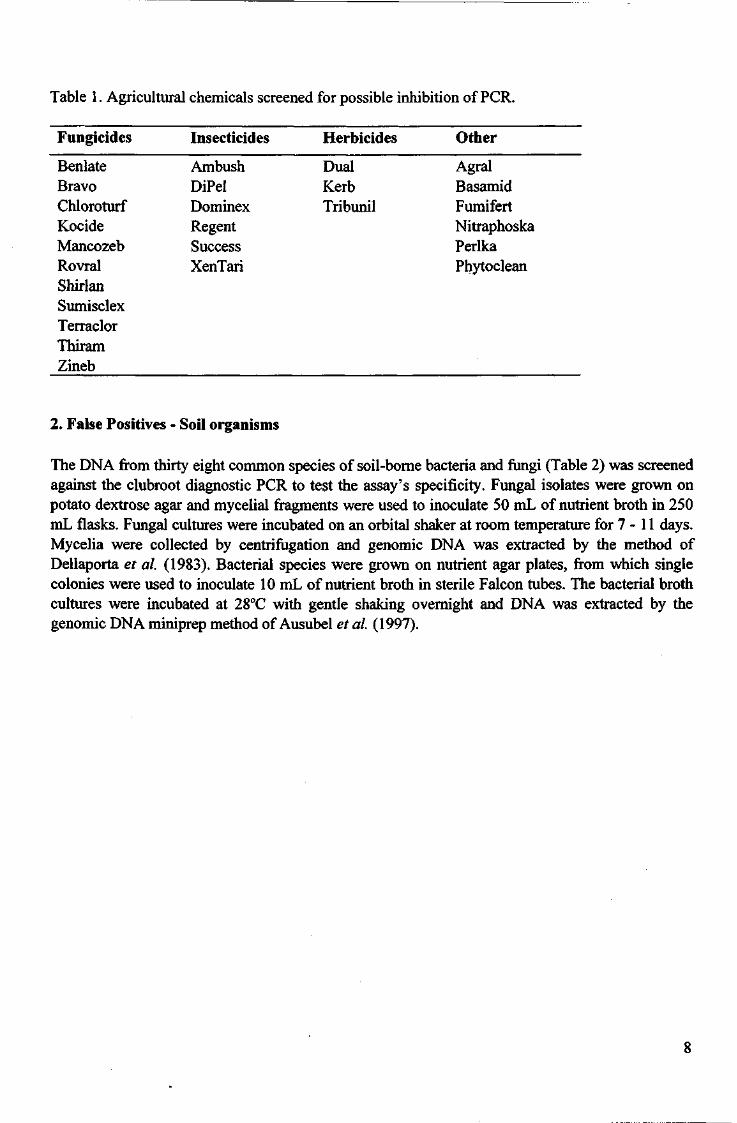
Table 1. Agricultural chemicals screened for possible inhibition of PCR.
Fungicides Insecticides Herbicides Other
Benlate Ambush Dual Agral Bravo DiPel Kerb Basamid Chloroturf Dominex Tribunil Fumifert Kocide Regent Nitraphoska Mancozeb Success Perlka Rovral XenTari Phytoclean Shirlan Sumisclex Terraclor Thiram Zineb
2. False Positives - Soil organisms
The DNA from thirty eight common species of soil-borne bacteria and fungi (Table 2) was screened against the clubroot diagnostic PCR to test the assay's specificity. Fungal isolates were grown on potato dextrose agar and mycelial fragments were used to inoculate 50 mL of nutrient broth in 250 mL flasks. Fungal cultures were incubated on an orbital shaker at room temperature for 7 -11 days. Mycelia were collected by centrifugation and genomic DNA was extracted by the method of Dellaporta et al. (1983). Bacterial species were grown on nutrient agar plates, from which single colonies were used to inoculate 10 mL of nutrient broth in sterile Falcon tubes. The bacterial broth cultures were incubated at 28°C with gentle shaking overnight and DNA was extracted by the genomic DNA miniprep method of Ausubel et al. (1997).
8
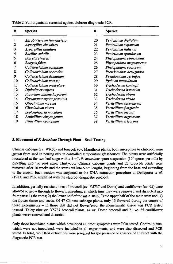
Table 2. Soil organisms screened against clubroot diagnostic PCR.
# Species # Species
1 Agrobacterium tumefaciens 20 Penicillium digitatum 2 Aspergillus chevalieri 21 Penicillium expansum 3 Aspergillus nidulans 22 Penicillium italicum 4 Bacillus subtilis 23 Penicillium spinulosum 5 Botrytis cinerea 24 Phytophthora cinnamomi 6 Botrytis fabae 25 Phytophthora megasperma 7 Colletotrichum acutatum; 26 Phytophthora cactorum 8 Colletotrichum coccodes 27 Pseudomonas aeruginosa 9 Colletotrichum dematium; 28 Pseudomonas syringae 10 Colletotrichum musae; 29 Pythium mamillatum 11 Colletotrichum orbiculare 30 Trichoderma koningii 12 Diplodia erumpens 31 Trichoderma hamatum 13 Fusarium chlamydosporum 32 Trichoderma virens 14 Gaeumannomyces graminis 33 Trichoderma viride 15 Gliocladium roseum 34 Verticillium albo-atrum 16 Gliocladium virens 35 Verticillium fungicola 17 Leptosphaeria maculans 36 Verticillium lecanii 18 Penicillium chrysogenum 37 Verticillium nigrescens 19 Penicillium cyclopium 38 Verticillium tricorpus
3. Movement of P. brassicae Through Plant - Seed Testing
Chinese cabbage (cv. WR60) and broccoli (cv. Marathon) plants, both susceptible to clubroot, were grown from seed in potting mix in controlled temperature glasshouses. The plants were artificially inoculated at the two leaf stage with a 1 mL P. brassicae spore suspension (107 spores per mL) by pipetting into the root zone. Thirty-four Chinese cabbage plants and 23 broccoli plants were removed after 10 weeks and the stems cut into 5 cm lengths, beginning from the base and extending to the crown. Each section was subjected to the DNA extraction procedure of Dellaporta et ah (1983) and PCR amplified with the clubroot diagnostic protocol.
In addition, partially resistant lines of broccoli (cv. Y5737 and Dome) and cauliflower (cv. 65) were allowed to grow through to flowering/seeding, at which time they were removed and dissected into four parts: 1) the roots; 2) the lower half of the main stem; 3) the upper half of the main stem and; 4) the flower tissue and seeds. Of 47 Chinese cabbage plants, only 13 flowered during the course of these experiments - in those that did not flower/seed, the meristematic tissue was PCR tested instead. Thirty nine cv. Y5737 broccoli plants, 44 cv. Dome broccoli and 21 vc. 65 cauliflower plants were removed and dissected.
Only those inoculated plants which developed clubroot symptoms were PCR tested. Control plants, which were not inoculated, were included in all experiments, and were also dissected and PCR tested. In total, 629 DNA extractions were screened for the presence or absence of clubroot with the diagnostic PCR test.
9
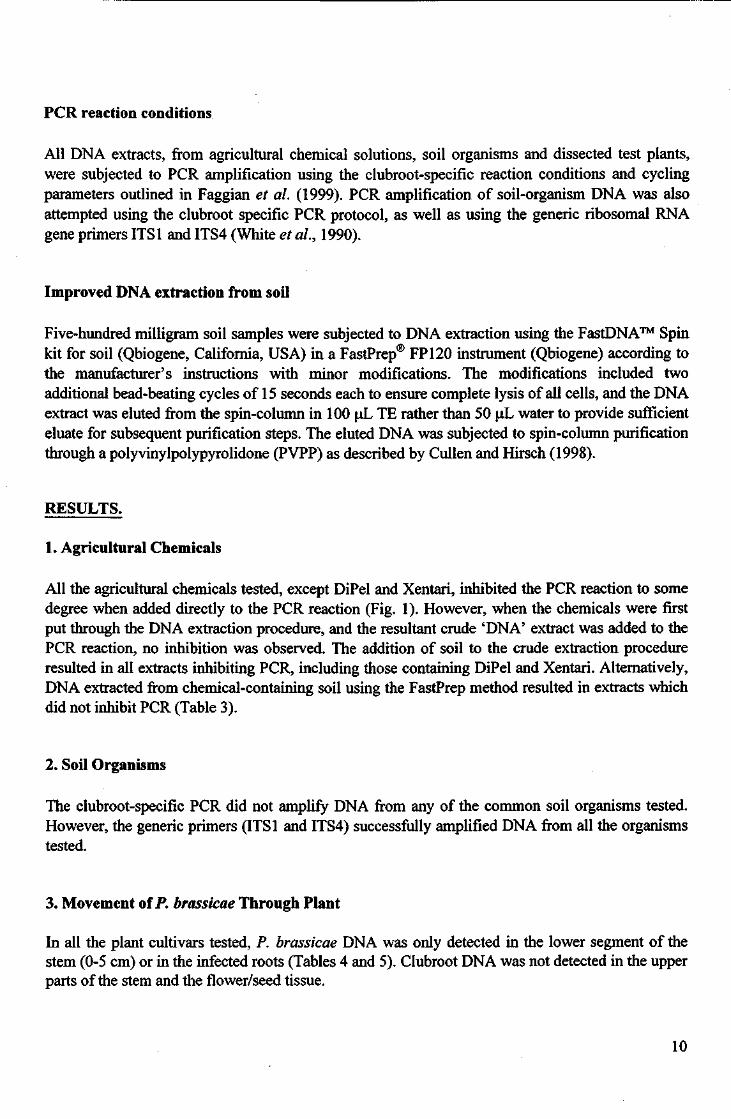
PCR reaction conditions
All DNA extracts, from agricultural chemical solutions, soil organisms and dissected test plants, were subjected to PCR amplification using the clubroot-specific reaction conditions and cycling parameters outlined in Faggian et al. (1999). PCR amplification of soil-organism DNA was also attempted using the clubroot specific PCR protocol, as well as using the generic ribosomal RNA gene primers ITS1 and ITS4 (White et al, 1990).
Improved DNA extraction from soil
Five-hundred milligram soil samples were subjected to DNA extraction using the FastDNA™ Spin kit for soil (Qbiogene, California, USA) in a FastPrep® FP120 instrument (Qbiogene) according to the manufacturer's instructions with minor modifications. The modifications included two additional bead-beating cycles of 15 seconds each to ensure complete lysis of all cells, and the DNA extract was eluted from the spin-column in 100 uL TE rather than 50 oL water to provide sufficient eluate for subsequent purification steps. The eluted DNA was subjected to spin-column purification through a polyvinylpolypyrolidone (PVPP) as described by Cullen and Hirsch (1998).
RESULTS.
1. Agricultural Chemicals
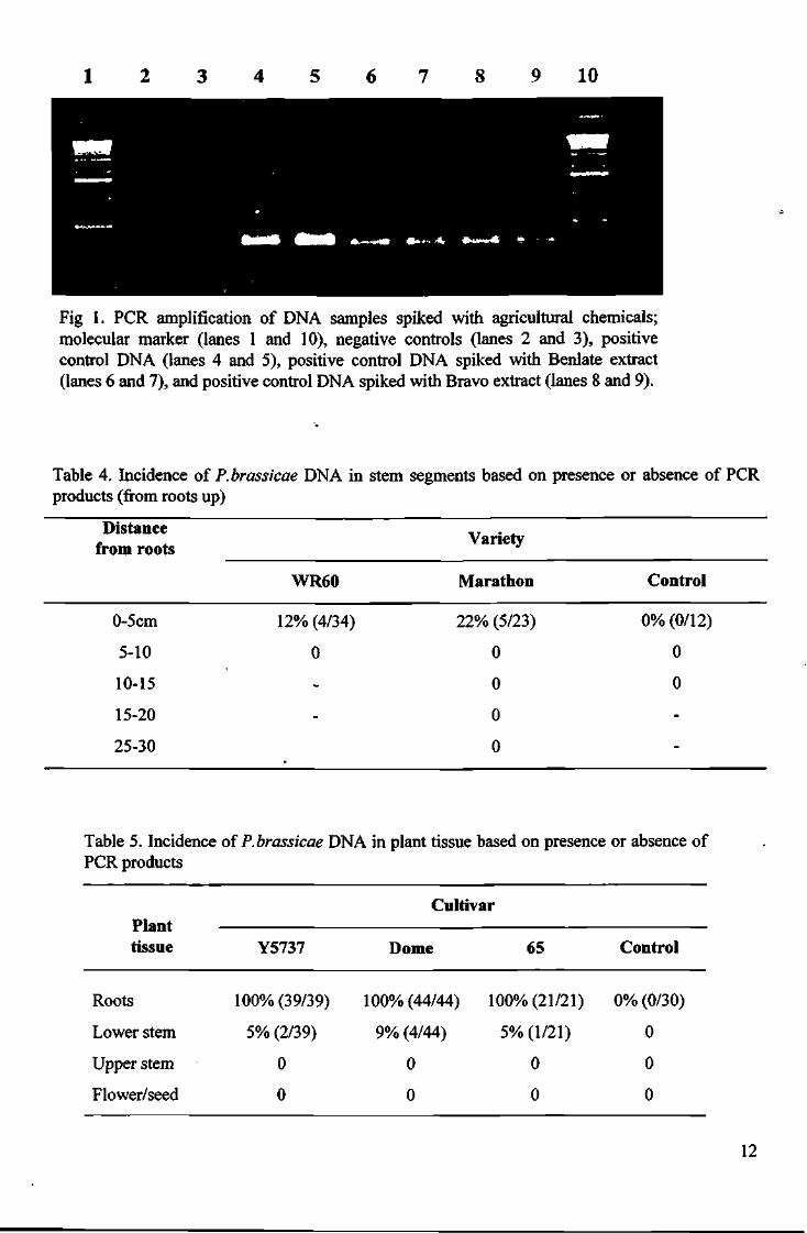
All the agricultural chemicals tested, except DiPel and Xentari, inhibited the PCR reaction to some degree when added directly to the PCR reaction (Fig. 1). However, when the chemicals were first put through the DNA extraction procedure, and the resultant crude 'DNA' extract was added to the PCR reaction, no inhibition was observed. The addition of soil to the crude extraction procedure resulted in all extracts inhibiting PCR, including those containing DiPel and Xentari. Alternatively, DNA extracted from chemical-containing soil using the FastPrep method resulted in extracts which did not inhibit PCR (Table 3).
2. Soil Organisms
The clubroot-specific PCR did not amplify DNA from any of the common soil organisms tested. However, the generic primers (ITS1 and ITS4) successfully amplified DNA from all the organisms tested.
3. Movement of P. brassicae Through Plant
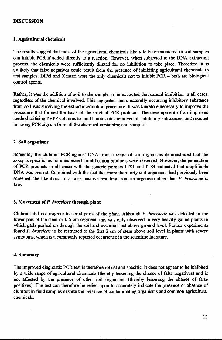
In all the plant cultivars tested, P. brassicae DNA was only detected in the lower segment of the stem (0-5 cm) or in the infected roots (Tables 4 and 5). Clubroot DNA was not detected in the upper parts of the stem and the flower/seed tissue.
10
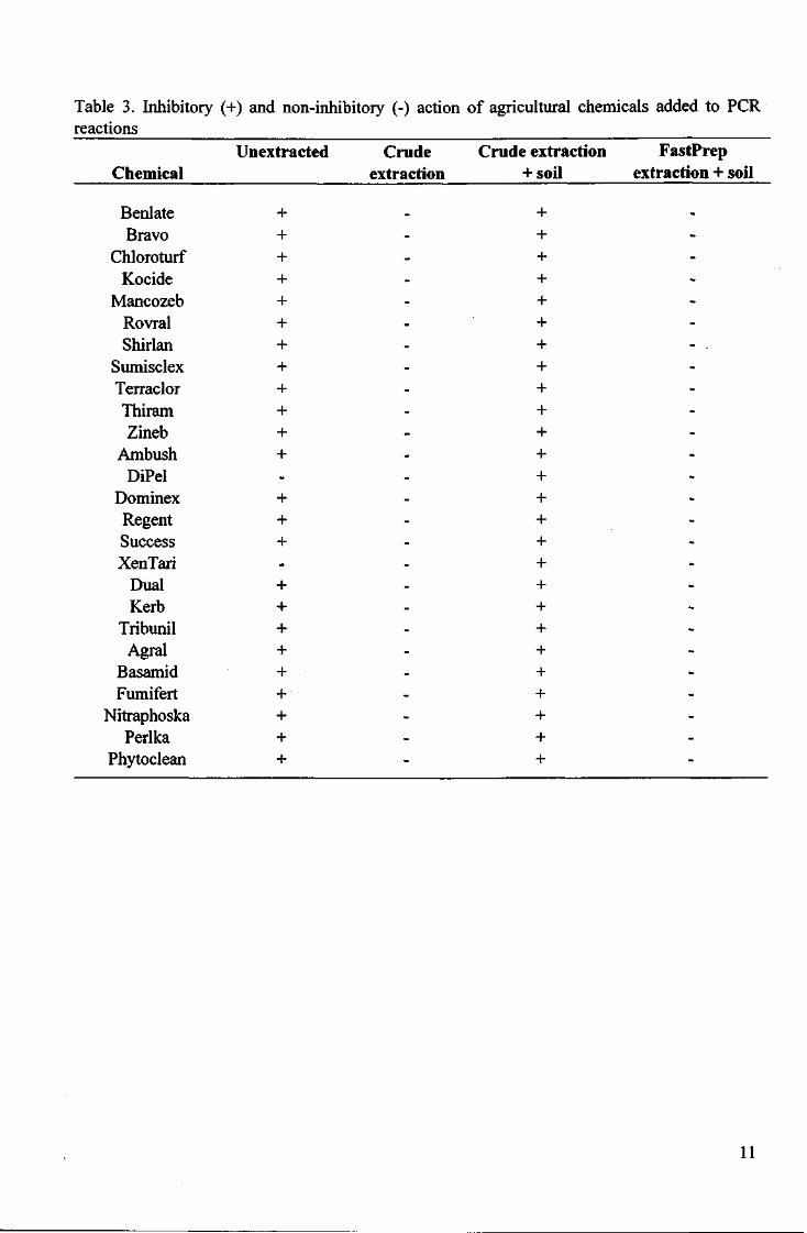
Table 3. Inhibitory (+) and non-inhibitory (-) action of agricultural chemicals added to PCR reactions
Unextracted Crude Crude extraction FastPrep Chemical extraction +soil extraction + soil
+ + + + + + + - . + + + + + + + + + + + + + + + + + + +
Benlate + Bravo +
Chloroturf + Kocide +
Mancozeb + Rovral + Shirlan +
Sumisclex + Terraclor + Thiram + Zineb +
Ambush + DiPel -
Dominex + Regent + Success + XenTari -
Dual + Kerb +
Tribunil + Agral +
Basamid + Fumifert +
Nitraphoska + Perlka +
Phytoclean +
11
1 2 3 4 5 6 7 8 9 10
Fig 1. PCR amplification of DNA samples spiked with agricultural chemicals; molecular marker (lanes 1 and 10), negative controls (lanes 2 and 3), positive control DNA (lanes 4 and 5), positive control DNA spiked with Benlate extract (lanes 6 and 7), and positive control DNA spiked with Bravo extract (lanes 8 and 9).
Table 4. Incidence of P. brassicae DNA in stem segments based on presence or absence of PCR products (from roots up)
Distance from roots
Variety
WR60 Marathon Control
0-5cm 12% (4/34) 22% (5/23) 0%(0/12)
5-10 0 0 0
10-15 - 0 0
15-20 - 0 -
25-30 0 -
Table 5. Incidence of P. brassicae DNA in plant tissue based on presence or absence of PCR products
Cultivar Plant
tissue Y5737 Dome 65 Control
Roots 100% (39/39) 100% (44/44) 100% (21/21) 0% (0/30)
Lower stem 5% (2/39) 9% (4/44) 5% (1/21) 0
Upper stem 0 0 0 0
Flower/seed 0 0 0 0
12
DISCUSSION
1. Agricultural chemicals
The results suggest that most of the agricultural chemicals likely to be encountered in soil samples can inhibit PCR if added directly to a reaction. However, when subjected to the DNA extraction process, the chemicals were sufficiently diluted for no inhibition to take place. Therefore, it is unlikely that false negatives could result from the presence of inhibiting agricultural chemicals in test samples. DiPel and Xentari were the only chemicals not to inhibit PCR - both are biological control agents.
Rather, it was the addition of soil to the sample to be extracted that caused inhibition in all cases, regardless of the chemical involved. This suggested that a naturally-occurring inhibitory substance from soil was surviving the extraction/dilution procedure. It was therefore necessary to improve the procedure that formed the basis of the original PCR protocol. The development of an improved method utilising PVPP columns to bind humic acids removed all inhibitory substances, and resulted in strong PCR signals from all the chemical-containing soil samples.
2. Soil organisms
Screening the clubroot PCR against DNA from a range of soil-organisms demonstrated that the assay is specific, as no unexpected amplification products were observed. However, the generation of PCR products in all cases with the generic primers ITS1 and ITS4 indicated that amplifiable DNA was present. Combined with the fact that more than forty soil organisms had previously been screened, the likelihood of a false positive resulting from an organism other than P. brassicae is low.
3. Movement of P. brassicae through plant
Clubroot did not migrate to aerial parts of the plant. Although P. brassicae was detected in the lower part of the stem or 0-5 cm segment, this was only observed in very heavily galled plants in which galls pushed up through the soil and occurred just above ground level. Further experiments found P. brassicae to be restricted to the first 2 cm of stem above soil level in plants with severe symptoms, which is a commonly reported occurrence in the scientific literature.
4. Summary
The improved diagnostic PCR test is therefore robust and specific. It does not appear to be inhibited by a wide range of agricultural chemicals (thereby lessening the chance of false negatives) and is not affected by the presence of other soil organisms (thereby lessening the chance of false positives). The test can therefore be relied upon to accurately indicate the presence or absence of clubroot in field samples despite the presence of contaminating organisms and common agricultural chemicals.
13
The results suggest that the clubroot organism, Plasmodiophora brassicae, does not migrate (actively or passively) to aerial parts of its host plant. This means that any PCR positives that result from seed are the result of contaminating clubroot inoculum on the seed surface, not from plasmodial stages of the organism within the seed.
During the course of this project a number of suspect seed lots have been tested for the presence of clubroot with the diagnostic test. All positives tests (i.e. where P. brassicae DNA was found) originated from tins or packets of seed which were already open and therefore subject to contamination before testing. When the same seed lots from unopened packets were tested, no positives were found. Therefore industry can have confidence in the diagnostic procedure when testing for the presence of clubroot on seed provided the seed has not had an opportunity to become contaminated during the sampling process.
14
Chapter 2: Effect of non-viable resting spores on PCR results.
INTRODUCTION
One of the drawbacks of the PCR as a tool for the detection of plant pathogens is that it does not differentiate between living and dead cells. Generally, PCR is only able to amplify DNA, which can be achieved using a tremendous variety of starting materials ranging from a single live cell or long-dead fragments retrieved from bones. Therefore, the presence of dead or non-viable clubroot inoculum will result in amplification products and could be interpreted as a positive test result. However, such a test outcome is of limited commercial value as it does not indicate disease potential. Where bacteria are the target organism, this problem can sometimes be circumvented by introducing an enrichment step - the target organism is cultured, or enriched, increasing its numbers to levels higher than the lower limit of detection of the assay. Alternatively, it is possible to develop a reverse-transcriptase PCR assay, which relies on the presence of messenger RNA - mRNA generally has a short lifespan, so its presence is indicative of a functioning cell, and therefore good evidence that the target is viable.
Neither of these approaches can be used for P. brassicae. Firstly, P. brassicae is an obligate biotroph and therefore cannot be cultured. Secondly, extracting PCR-amplifiable DNA from soil is problematic, but extracting intact RNA while still maintaining the sensitivity of the assay is virtually impossible.
Two novel approaches were used in an attempt to solve the problem: 1) the induction of resting spore germination followed by zoospore trapping and 2) an assessment of the persistence of nonviable resting spores in soil and their effect on PCR results.
MATERIALS AND METHODS
1. Preparation of resting spore suspensions
A resting spore suspension was prepared from freshly galled roots by macerating in 1:3 w/v sterile distilled water and filtering through cheesecloth according to the method of Casltebury et al. (1994). The spores were separated from contaminating bacteria and plant cell debris by successive centrifugation through water and LUDOX HS40, as per the above mentioned protocol. The concentration of the spore suspension was adjusted to 107 spore per mL after counting using a Neubauer haemocytometer and the viability of the spores was assessed using a fluorescent staining technique developed by Takahashi & Yamaguchi (1988).
2. Zoospore germination.
Broccoli (cv. Marathon) and Arabidopsis seeds were surface sterilised in 0.5% sodium hypochlorite for 5 minutes before being germinated on 0.1% water agar. The seedlings were transferred to separate 250mL flasks containing 50 mL Murashige and Skoog basal medium where they were incubated, with shaking, for two weeks at room temperature. After two weeks the plantlets were removed and the medium containing broccoli and Arabidopsis root exudates sterilised by
15
autoclaving. The root exudate solution was transferred to a sterile Falcon tube to which was added 1 mL of purified resting spore suspension. Resting spores were also added to control tubes containing only distilled water rather than root exudate. All the tubes were incubated in the dark for periods of 24 and 48 hours, after which times 10 uL aliquots were removed and examined microscopically for the presence of swimming primary zoospores and empty resting spores, or they were stained with the viability stain to test for the presence or absence of nuclear material. Treated spores (i.e. those suspended in root exudate solution) were compared with untreated spores (i.e. suspended in distilled water) to identify any increase in the natural rate of spore germination.
3. Persistence of P. brassicae DNA in non-sterile soil
Fresh resting spore suspensions were prepared as described above. The spore concentration was adjusted to 108 and 105 spores per mL and viability measured with the fluorescent staining technique of Takahashi & Yamaguchi (1988). The resting spores were then treated in a number of ways to render them non-viable: 1) autoclaving either once, twice or three times; 2) gamma irradiation at levels of 100 kGray; 3) immersion in 70% and 100% ethanol and 4) immersion in a range of detergent solutions including Tween and sodium dodecyl sulphate. The success of the treatments was assessed with the fluorescent staining technique.
Non-viable resting spores (70% ethanol-treated) were added to non-sterile soil to achieve final concentrations of 107 spores per gram and 104 spores per gram. Controls consisted of 100% viable resting spores inoculated into non-sterile soil at the same final concentrations. Three different soil types were used (originating from three Victorian sites - Knoxfield, Kalorama and Dingley) and all were incubated for 12 weeks in a glasshouse with regular, light misting to maintain soil moisture levels. Soil was sampled at three weekly intervals and subjected to a DNA extraction procedures and PCR amplification. Concurrent plant bioassays were run to ensure assessments of spore viability were accurate.
The experiment was repeated using artificially inoculated soil treated with the fumigant basamid at the labelled rate. Four kilograms of non-sterile soil was placed in a plastic bucket and artificially inoculated with viable resting spores to a final concentration of 107 spores per gram. The soil was then treated with basamid at the labelled rate, sealed tightly with plastic and left for 6 weeks. Soil samples were taken before treatment and at 3 weekly intervals after the initial 6 week incubation period.
RESULTS;
Zoospore germination.
Spore germination was not observed for either the spores treated with root exudate or those treated with distilled water. The experiments were repeated four weeks later, at which time it was possible to see some spore germination taking place, however, the rates did not vary significantly between treated and untreated. This approach was therefore abandoned.
16
Persistence of DNA in soil.
The only 100% effective method of rendering resting spores non-viable (assuming the fluorescent staining technique accurately represented viability) was to immerse them for 10 min in 70% ethanol. Autoclaving, detergents and gamma irradiation were all ineffective to varying degrees. Even after autoclaving spore suspensions three times, it was still possible to find a very small proportion of viable spores.
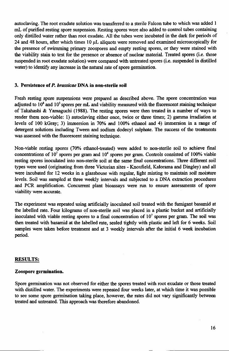
The PCR signal generated from soil samples containing viable resting spores (controls) remained relatively constant throughout the sampling period, for both inoculum concentrations (Table 6). On the other hand, the PCR signal generated from soil containing non-viable spores diminished over time. The PCR signal completely disappeared after 9 weeks in the lower concentration (104 spores per gram) and diminished gradually over the 12 week period for the higher concentration (107
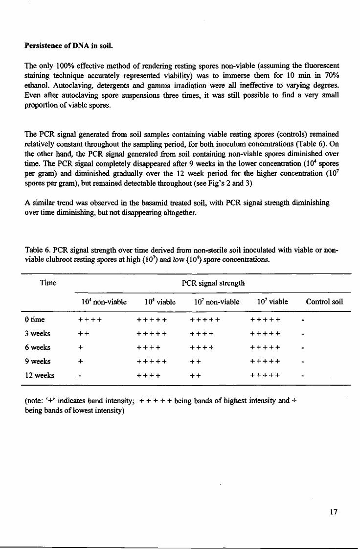
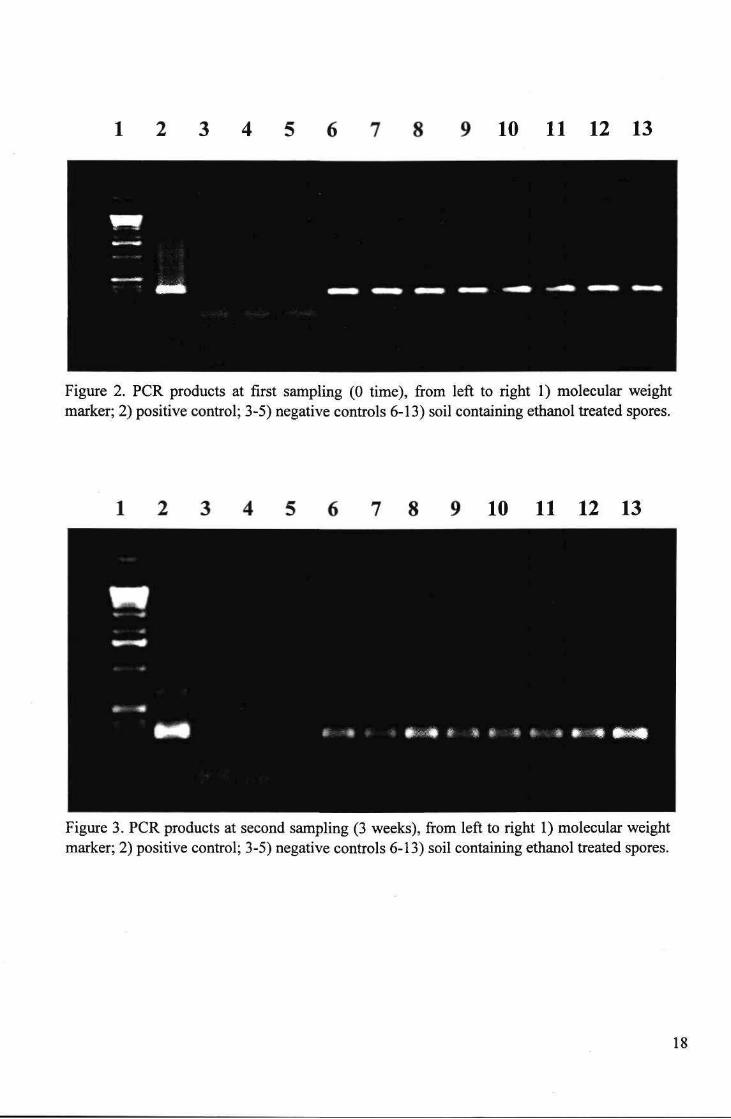
spores per gram), but remained detectable throughout (see Fig's 2 and 3)
A similar trend was observed in the basamid treated soil, with PCR signal strength diminishing over time diminishing, but not disappearing altogether.
Table 6. PCR signal strength over time derived from non-sterile soil inoculated with viable or nonviable clubroot resting spores at high (107) and low (104) spore concentrations.
Time PCR signal strength
104 non-viable 104 viable 107 non-viable 107 viable Control soil
Otime + + + + + + + + + + + + + + + + + + +
3 weeks + + + + + + + + + + + + + + + +
6 weeks + + + + + + + + + + + + + +
9 weeks + + + + + + + + + + + + +
12 weeks _ + + + + + + + + + + +
(note: '+' indicates band intensity; + + + + + being bands of highest intensity and + being bands of lowest intensity)
17
1 2 3 4 5 6 7 8 9 10 11 12 13
Figure 2. PCR products at first sampling (0 time), from left to right 1) molecular weight marker; 2) positive control; 3-5) negative controls 6-13) soil containing ethanol treated spores.
7 8 9 10 11 12 13
Figure 3. PCR products at second sampling (3 weeks), from left to right 1) molecular weight marker; 2) positive control; 3-5) negative controls 6-13) soil containing ethanol treated spores.
18
DISCUSSION
The first approach, inducing resting spore germination, had the aim of developing a method of trapping the resultant zoospores for PCR testing. This would ensure that a positive test could only be the result of viable clubroot resting spores. Research in Japan (Suzuki et al., 1992) partially identified a compound in root exudates that stimulated clubroot spore germination. However, in our experiments, no differences were observed between the germination rates of spores treated with the root exudates and those treated solely with distilled water. Higher rates of spore germination were observed over time, so it is possible that the spores needed to reach a particular stage of maturity before they are ready to germinate. Both these facts make this approach unsustainable because it is impossible to determine how mature spores are at any given time, and waiting for spores to mature would lengthen the testing time considerably. As spore germination could not be satisfactorily induced, zoospores could not trapped and this approach was abandoned.
The alternative approach was followed. This involved determining the time that non-viable resting spores persist in soil - that is, the time that non-viable clubroot inoculum is likely to contribute towards a PCR positive (false positive). The results (Table 6.) indicate that the DNA from P. brassicae resting spores will not persist for long in non-sterile soil - that is, the risk of obtaining a false PCR positive, or an over-estimate of clubroot spore density due to non-viable resting spores, is significantly reduced or eliminated after 9 weeks, depending on initial spore density. Lower levels of inoculum may disappear entirely within a 9 week time frame.
Relevance to Industry
The results indicate that the diagnostic assay can be used to measure a decrease in a clubroot population in response to a lethal treatment. This provides researchers with a valuable new tool to assist the development of clubroot management strategies. It will also enable strategic sampling of soil for clubroot testing. For instance, a soil sample could be tested before the application of a control treatment, followed by a 9 week lag period until the next sampling - if the treatment was successful, the clubroot population would either be undetectable after 9 weeks or would give a weaker PCR signal, indicating a reduction in soil inoculum. This scenario has already arisen. Several soil samples from a single site were sent for testing by the Queensland Department of Primary Industries. The samples tested positive for clubroot DNA and so the decision was made to fumigate the soil in question (with metham sodium). The same site was sampled 9 weeks later and this time tested negative for clubroot DNA, indicating that the fumigation had indeed been effective.
19
Chapter 3: Development of Quantitative assay.
INTRODUCTION
Real-time quantitative PCR is a relatively new technology which enables the monitoring, in real time, of the progress of a PCR amplification. The distinguishing feature of individual reactions is the point in time at which PCR products are first detected - the accumulation of PCR product is then plotted versus PCR cycle number. So, the more target sequence present at the start of the reaction, the sooner PCR products will be detected (i.e. the cycle number where product is first detected will be lower). Quantification of unknown samples can be achieved by plotting the amplification of a set of standards to creating a standard curve (straight line) from which to read unknown values (cycle numbers). The amount of PCR product can then be related to inoculum concentration, inoculum concentration to disease levels, disease levels to yield loss figures, and so on.
The technique is now widely used to rapidly detect and quantify soil-borne pathogens (Cullen et ah, 2002; Lees et al, 2002)
The ability to quantify clubroot inoculum is crucial as disease severity is related to inoculum concentration. Many clubroot researchers have studied this relationship, and although influenced by a number of factors (such as soil type, temperature, pH, etc.) is generally possible to develop a linear dose-response curve (MacFarlane, 1952; Karling, 1968; Webster & Dixon, 1991; Murakami et al., 2002). Previous research by NRE (Cross and Porter, unpublished data) supported these views by demonstrating a linear dose response curve in a glasshouse trial. Such data has made it possible to begin correlating TaqMan quantified soil inoculum levels and subsequent field disease levels.
MATERIALS AND METHODS
Assay design and production of standard curves
A new set of PCR primers, compatible with the TaqMan real-time quantitative PCR system, were designed using the Primer Express software package and sequence data and sequence alignments used to design the previous clubroot primers (Faggian et al., 1999). The oligonucleotide sequences of the new primers were CGCTGCATCCCATATCCAA (primer 1) and TCGGCTAGGATGGTTCGAAA (primer 2). A TaqMan probe was also designed with the Primer Express software package: FAM-CGCACGTCACCGGTTCACATGG-TAMRA.
The new TaqMan assay was used to produce a standard curve with DNA from a dilution series of resting spores in soil. Soil samples (from Knoxfield) were artificially inoculated with resting spores to achieve concentrations ranging from 102 — 107 spores per gram. DNA was extracted from 500 mg samples of the soil using the FastPrep and PVPP column methods outlined earlier. The samples were run through the TaqMan system and a standard curve was produced by the TaqMan software.
20
Predicting disease severity at trial sites nationally
Soil samples were collected from three trial sites in Victoria (Werribee, Boisedale and Launching place), two in Tasmania, and one in Queensland, New South Wales and Western Australia. DNA was extracted from all samples and then subjected to TaqMan analysis. The estimates of soil inoculum concentration were inferred from the standard curve developed above.
RESULTS
TaqMan amplification plots
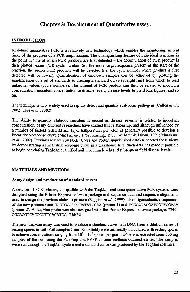
The amplification plot of the spore serial dilution in soil showed good reproducibility from 106
spores per gram down to 103 spores per gram (Fig 4). At 102 spores per gram, the replicate plots do not overlap, indicating the reliability of quantification at such low spore concentrations may not be good.
Fig. 4. Amplification plots of serial spore dilution in soil.
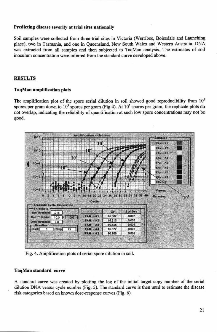
TaqMan standard curve
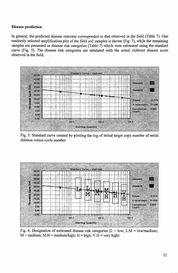
A standard curve was created by plotting the log of the initial target copy number of the serial dilution DNA versus cycle number (Fig. 5). The standard curve is then used to estimate the disease risk categories based on known dose-response curves (Fig. 6).
21
Disease prediction
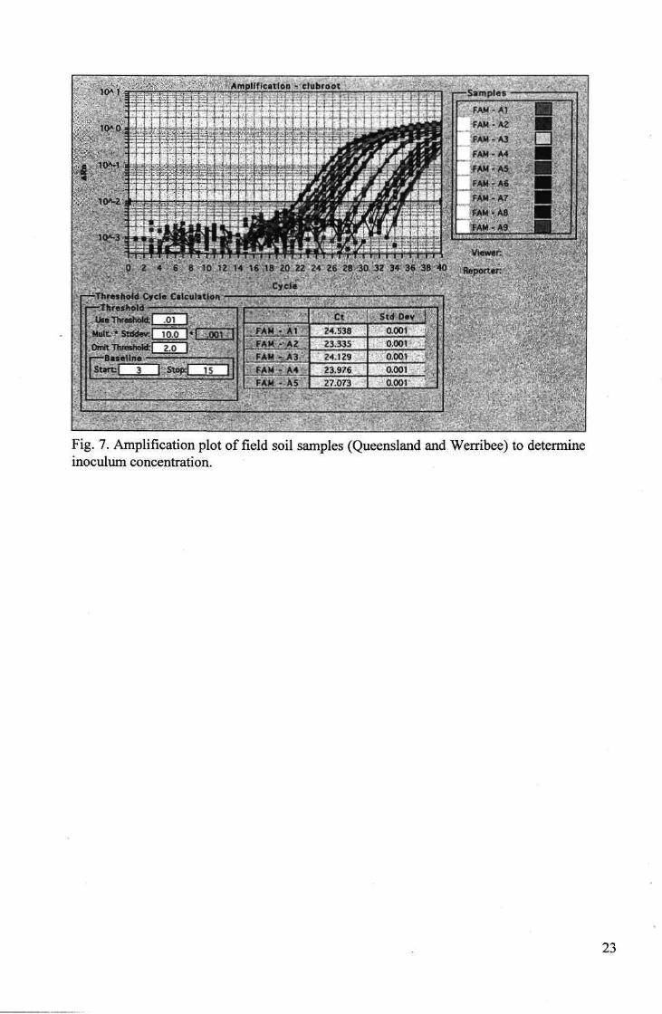
In general, the predicted disease outcome corresponded to that observed in the field (Table 7). One randomly selected amplification plot of the field soil samples is shown (Fig. 7), while the remaining samples are presented as disease risk categories (Table 7) which were estimated using the standard curve (Fig. 5). The disease risk categories are tabulated with the actual clubroot disease score observed in the field.
~TZT~—~—-. - . v.1-".,-.'--- :,.•;
Slope: -3,724
^ in tercept : 31.S86
Correlation ,0.993
Fig. 5. Standard curve created by plotting the log of initial target copy number of serial dilution versus cycle number
Fig. 6. Designation of estimated disease risk categories (L = low; L.M = low/medium; M = medium; M.H = medium/high; H = high; V.H = very high)
22
[77-Th r«* h»Jd Cye I
.: Use Threshoh
. Multvi* S t d ^ v : | _ r ^ 3 * ^ i y c i l J Qitnit Tlireslwkt | 2,0
Fig. 7. Amplification plot of field soil samples (Queensland and Werribee) to determine inoculum concentration.
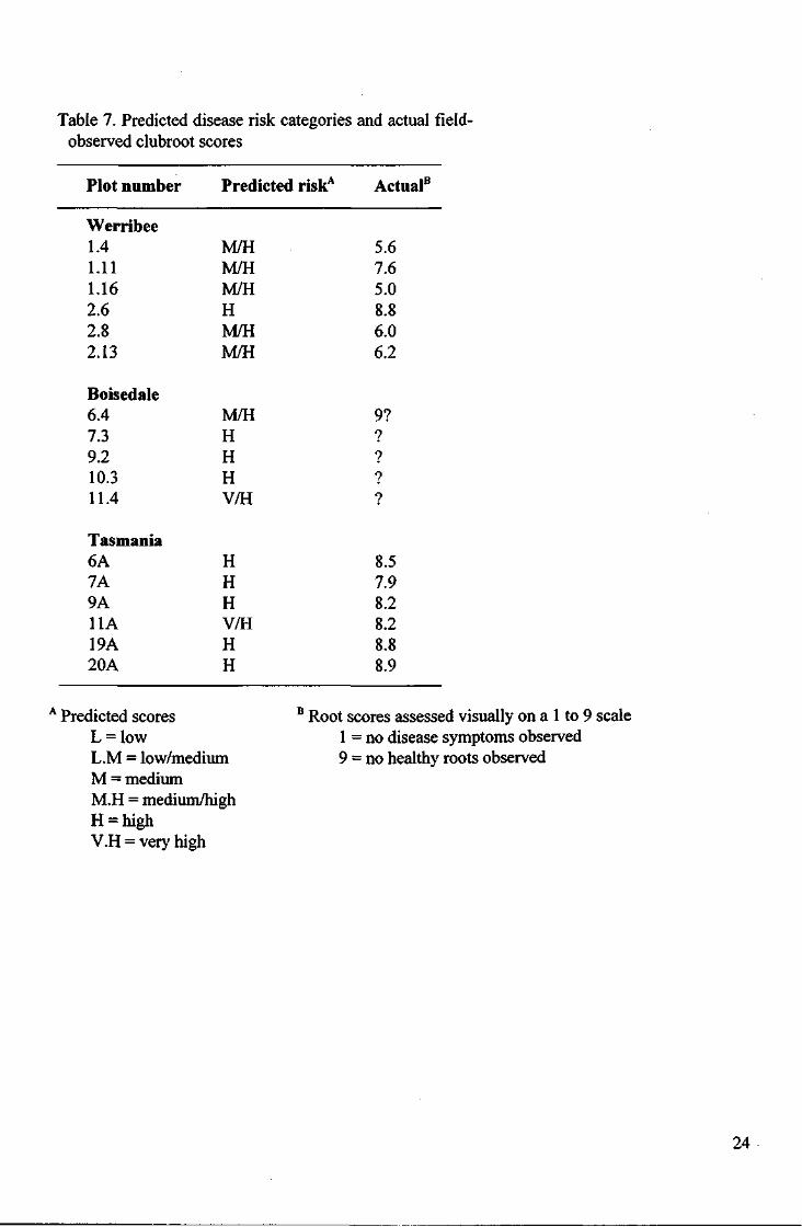
Table 7. Predicted disease risk categories and actual field-observed clubroot scores
Plot number Predicted riskA Actual8
Werribee 1.4 M/H 5.6 1.11 M/H 7.6 1.16 M/H 5.0 2.6 H 8.8 2.8 M/H 6.0 2.13 M/H 6.2
Boisedale 6.4 M/H 9? 7.3 H ? 9.2 H ? 10.3 H ?
11.4 V/H ?
Tasmania 6A H 8.5 7A H 7.9 9A H 8.2 11A V/H 8.2 19A H 8.8 20A H 8.9
A Predicted scores L = low L.M = low/medium M = medium M.H = medium/high H = high V.H = very high
B Root scores assessed visually on a 1 to 9 scale 1 = no disease symptoms observed 9 = no healthy roots observed
24
DISCUSSION
For the first time, a tool is available to the Australian brassica industry enabling the quantification of clubroot inoculum in soil. This capability, together with information on factors such as pH, soil moisture content, varietal resistance, and others, will allow researchers to accurately predict the severity of disease in crops before they are planted. Similarly, growers will be able to make informed decisions about potential disease management strategies, before planting.
The preliminary data presented here clearly demonstrates that real-time PCR can be used to accurately predict clubroot inoculum levels in the field. The predicted risk categories correlate well with the actual clubroot scores observed in the corresponding field sites. This is despite the fact that rudimentary dose-response data was used to assign the risk categories on the standard curve. To gain greater accuracy, a comparison of different soil types would need to be carried in order to highlight the error that can be expected when assigning risk categories. However, it may be preferable, and more practical for growers, to assign fewer risk categories, enabling a fail-safe integrated management option to be selected.
Further work will be conducted at a range of field sites in the final year of the national clubroot program (VG00044) to fully commercialise this quantitative technique.
25
Commercialisation Path
• A national tender process was initiated in the early stages of the project to identify prospective commercialisation partners. This was done in collaboration with RMIT University.
• Two international (Rhone Poulenc and Sigma) and three national firms responded with strong applications.
• Rhone Poulenc was selected as the respondent most well-placed to market the clubroot diagnostic test internationally, and also offered access to a substantial and experienced scientific network.
• Rhone Poulenc (which became Aventis Crop Science), through their Australasian subsidiary C-Qentec, carried out extensive market research which indicated that a single-pathogen test would not be commercially viable.
• However, C-Qentec remained keen to develop the clubroot technology as a model system on which to base other soil-borne pathogen tests.
• Potato was identified as the best crop vehicle for further development of the technology, particularly a multiplex test for numerous pathogens.
• A new HAL and potato industry-funded project was developed between NRE, C-Qentec and SARDI to develop a rapid, multiplex test for potato pathogens. C-Qentec committed $20,000 per year for three years and significant marketing expertise over a three year period.
• Newly developed HAL projects will have immediate benefits for the development of the clubroot assay.
• Clubroot diagnostic PCR test offered to Australian brassica growers on a commercial basis through Crop Health Services (NRE, Knoxfield). More than 50 samples processed in the first 6 months of the test being publicly available.
26
Recommendations
This project has developed the tools to enable growers to test their soil, water and planting material before planting. This will limit the spread of clubroot in recently infected areas and enable growers to select appropriate management strategies. Growers should use the diagnostic assay to form the basis of a testing regime for their farms and nurseries to ensure that the spread of clubroot is limited or stopped.
The commercial diagnostic test can rapidly predict the presence or absence of clubroot in soil, water and plant tissue,
However, for quantitative prediction of disease to be accurate, further field monitoring is required -up until now, researchers have not had the tools at their disposal to measure soil-borne clubroot inoculum. This project has delivered the necessary tools, and they will accelerate soil-borne pathogen research. It is important however that this situation be capitalised on, and the quantitative assay be commercialised. Extensive field monitoring will therefore be conducted in the final year of the national clubroot program. This data will be required to help validate that disease prediction is accurate in a range of soil types and under a range of climatic conditions.
The final benefit to growers is likely to come from the development of an inexpensive field test kit for clubroot. This work is currently underway in conjunction with Horticulture Research International (Wellesbourne, U.K.), a group with extensive experience in the development and use of on-farm pathogen test kits.
Currently, Australian vegetable brassica growers are the only ones with commercial access to a clubroot diagnostic procedure. This competitive advantage should be maintained and exploited for as long as possible.
27
Technology Transfer
Technology transfer activities were in part channelled through the National Clubroot team and HAL project VG97076 (Clubroot seminars at field days and grower meetings). Nonetheless, a number of activities were undertaken including:
• Summaries in national clubroot newsletter "Galls and All"
• Two radio interviews (with Kaaren Latham, 2UE rural radio. 2000/2001)
• Brassica grower meetings (hosted by IHD, including tour of facilities and run-through testing procedure. 2000)
• Seminars to Monsanto and Henderson seeds (2001)
• Presentation to Chisholm Institute of TAFE horticulture students (May, 2001)
• Japanese horticulture students (Nigata College. Oct, 2000)
• Presentation at Clubroot Industry day (held at IHD, attended by chemical, fertiliser and seed company reps. 1998)
Refereed Publications:
R. Faggian, S. R. Bulman, A. C. Lawrie and I. J. Porter. (1999) Specific PCR primers for the detection of Plasmodiophora brassicae in soil and water. Phytopathology 89 (5): 392-397.
Conference papers:
Faggian, R., Parsons, S., Porter, I. J. and Lawrie, A. C. (2001) Detection of clubroot in Australia: progress towards a reliable molecular diagnostic assay. 2nd Australasian Soilborne Diseases Symposium, Lome, Victoria.
Faggian, R., Porter I. J. and Lawrie, A. C. (2000) The use of molecular techniques to investigate the genetic variation of Australian clubroot isolates. Brassica 2000, 3rd ISHS International Symposium on Brassicas, HRI Wellesbourne, UK.
Faggian R., Donald E. C , Porter I. J. and Lawrie A. C. (1999) The use of molecular techniques to investigate the genetic variation of Australian clubroot isolates. 12th Australasian Plant Pathology Society Conference, Canberra, ACT.
Oral presentations:
Detection of clubroot in Australia: Progress towards a reliable molecular diagnostic assay. 2001. Second Australasian Soilborne Diseases Symposium, Lome, Victoria.
Current Status of molecular detection of clubroot in Australia. 2000. Clubroot Symposium, Brassica 2000 (3rd International Symposium on Brassicas), HRI Wellesbourne, England.
28
Epidemiology of recent clubroot outbreaks using PCR to trace sources of inoculum. 1999. 12th
Australasian Plant Pathology Society Conference, Canberra, ACT.
29
References:
Ausubel, F. M., Brent, R., Kingston, R. E., Moore, D. D., Seidman, J. G., Smith, J. A., and Struhl, K., eds. (1997). Current Protocols in Molecular Biology. Canada., John Wiley and Sons, Inc.
Castlebury, L. A., Maddox, J. V. and Glawe, D. A. (1994). A technique for the extraction and purification of viable Plasmodiophora brassicae resting spores from host root tissue. Mycologia 86: 458-460.
Cullen, D. W., and Hirsch, P. R. (1998). Simple and rapid method for direct extraction of microbial DNA from soil for PCR. Soil Biology and Biochemistry 30: 983-993.
Cullen, D. W., Lees, A. K., Toth, I. K., and Duncan, J. M. (2002). Detection of Colletotrichum coccodes from soil and potato tubers by conventional and quantitative real-time PCR. Plant Pathology 51: 281-292.
de Lomas, J. G., Sunzeri, F. J. and Busch, M. P. (1992). False-negative results by polymerase chain reaction due to contamination by glove powder. Transfusion 32: 83-85.
Dellaporta, S. L., Wood, J. and Hicks, J. B. (1983). A plant DNA minipreparation: version 2. Plant Molecular Biology Reporter 1(4): 19-21.
Faggian, R., Lawrie, A. C. and Porter, I. J. (1999). Polymerase chain reaction primers for the detection of Plasmodiophora brassicae in soil and water. Phytopathology 89(5): 392-397.
Karling, J. S. (1968). The Plasmodiophorales. New York, Hafner Publishing Company, Inc.
Lees, A. K., Cullen, D. W., Sullivan, L., and Nicholson, M. J. (2002). Development of conventional and quantitative real-time PCR assays for the detection and identification of Rhizoctonia solani AG-3 in potato and soil. Plant Pathology 51: 293-302.
MacFarlane, I. (1952). Factors affecting the survival of Plasmodiophora brassicae Wor. in the soil and its assessment by a host test. Annals of Appled Biology 39: 239-256.
Murakami, H., Tsushima, S., and Shishido, Y. (2002). Factors affecting the pattern of the dose response curve of clubroot disease caused by Plasmodiophora brassicae. Soil Science & Plant Nutrition. 48: 421-427.
St. Pierre, B. S., Neustock, P., Schramm, U., Wilhelm, D., Kirchner, H. and Bein, G. (1994). Seasonal breakdown of polymerase chain reaction. Lancet 343: 673.
Suzuki, K., Matsumiya, E., Ueno, Y. and Mizutani, J. (1992). Some properties of germination-stimulating factor from plants for resting spores of Plasmodiophora brassicae. Annals of the Phytopathology Society of Japan 58: 699-705.
Takahashi, K., and Yamaguchi, T. (1988). A method for assessing the pathogenic activity of resting spores of Plasmodiophora brassicae by fluorescence microscopy. Annals of the Phytopathology Society of Japan 54: 466-475.
30
Trevors, J. T. (1996). Nucleic acids in the Environment. Current Opinion in Biotechnology 7: 331-336.
Turco, R. F., and Sadowsky, M. J. (1995). The microflora of bioremediation. Bioremediation, science and applications. H. D. a. T. Skipper, R. F., Soil Science Society of America special publication 43: 87-103.
Webster, M. A., and Dixon, G. R. (1991a). Calcium, pH and inoculum concentration influencing colonization by Plasmodiophora brassicae. Mycological Research 95: 64-73.
White, T. J., Bruns, T., Lee, S. and Taylor, J. (1990). Amplification and direct sequencing of fungal ribosomal RNA genes for phylogenetics. PCR protocols: A guide to methods and applications. M. A. Innis, Gelfand, D. H., Sninsky, J. J. and White, T. J. London, Academic Press, Inc.: 315-322.
Wilson, I. G. (1997). Inhibition and facilitation of nucleic acid amplification. Applied and Environmental Microbiology 63: 3741-3751.
31