Embed Size (px)
Citation preview

HAL Id: hal-02733784https://hal.archives-ouvertes.fr/hal-02733784
Submitted on 22 Jan 2021
HAL is a multi-disciplinary open accessarchive for the deposit and dissemination of sci-entific research documents, whether they are pub-lished or not. The documents may come fromteaching and research institutions in France orabroad, or from public or private research centers.
L’archive ouverte pluridisciplinaire HAL, estdestinée au dépôt et à la diffusion de documentsscientifiques de niveau recherche, publiés ou non,émanant des établissements d’enseignement et derecherche français ou étrangers, des laboratoirespublics ou privés.
Distributed under a Creative Commons Attribution - NonCommercial| 4.0 InternationalLicense
A Proof-of-Concept Study on the Therapeutic Potentialof Au Nanoparticles Radiolabeled with the
Alpha-Emitter Actinium-225Evangelia-Alexandra Salvanou, Dimitris Stellas, Charalampos Tsoukalas,Barbara Mavroidi, Maria Paravatou-Petsotas, Nikolaos Kalogeropoulos,
Stavros Xanthopoulos, Franck Denat, Gautier Laurent, Rana Bazzi, et al.
To cite this version:Evangelia-Alexandra Salvanou, Dimitris Stellas, Charalampos Tsoukalas, Barbara Mavroidi, MariaParavatou-Petsotas, et al.. A Proof-of-Concept Study on the Therapeutic Potential of Au Nanoparti-cles Radiolabeled with the Alpha-Emitter Actinium-225. Pharmaceutics, MDPI, 2020, 12 (2), pp.188.10.3390/pharmaceutics12020188. hal-02733784

pharmaceutics
Article
A Proof-of-Concept Study on the TherapeuticPotential of Au Nanoparticles Radiolabeled with theAlpha-Emitter Actinium-225
Evangelia-Alexandra Salvanou 1, Dimitris Stellas 2, Charalampos Tsoukalas 1 ,Barbara Mavroidi 3, Maria Paravatou-Petsotas 1, Nikolaos Kalogeropoulos 4,Stavros Xanthopoulos 1, Franck Denat 5 , Gautier Laurent 6 , Rana Bazzi 6, Stephane Roux 6
and Penelope Bouziotis 1,*1 Institute of Nuclear & Radiological Sciences & Technology, Energy & Safety, National Center for Scientific
Research “Demokritos”, 15341 Athens, Greece; [email protected] (E.-A.S.);[email protected] (C.T.); [email protected] (M.P.-P.); [email protected] (S.X.)
2 Institute of Chemical Biology, National Hellenic Research Foundation, 11635 Athens, Greece; [email protected] Institute of Biosciences and Applications, National Center for Scientific Research “Demokritos”,
15341 Athens, Greece; [email protected] A’ Pathology Department, 401 General Military Hospital of Athens, 11525 Athens, Greece;
[email protected] ICMUB, UMR 6302 CNRS-UB, Université Bourgogne Franche-Comté, 21000 Dijon, France;
[email protected] Institut UTINAM, UMR 6213 CNRS-UBFC, Université Bourgogne Franche-Comté, 25030 Besançon, France;
[email protected] (G.L.); [email protected] (R.B.); [email protected] (S.R.)* Correspondence: [email protected]; Tel.: +30-21-0650-3687
Received: 1 December 2019; Accepted: 19 February 2020; Published: 21 February 2020
Abstract: Actinium-225 (225Ac) is receiving increased attention for its application in targetedradionuclide therapy, due to the short range of its emitted alpha particles in conjunction withtheir high linear energy transfer, which lead to the eradication of tumor cells while sparingneighboring healthy tissue. The objective of our study was the evaluation of a gold nanoparticleradiolabeled with 225Ac as an injectable radiopharmaceutical form of brachytherapy for local radiationtreatment of cancer. Au@TADOTAGA was radiolabeled with 225Ac at pH 5.6 (30 min at 70 C),and in vitro stability was evaluated. In vitro cytotoxicity was assessed in U-87 MG cancer cells,and in vivo biodistribution was performed by intravenous and intratumoral administration of[225Ac]225Ac-Au@TADOTAGA in U-87 MG tumor-bearing mice. A preliminary study to assesstherapeutic efficacy of the intratumorally-injected radio-nanomedicine was performed over a periodof 22 days, while the necrotic effect on tumors was evaluated by a histopathology study. We haveshown that [225Ac]225Ac-Au@TADOTAGA resulted in the retardation of tumor growth after itsintratumoral injection in U87MG tumor-bearing mice, even though very low activities were injectedper mouse. This gold nanoparticle radiopharmaceutical could be applied as an unconventionalbrachytherapy in injectable form for local radiation treatment of cancer.
Keywords: actinium-225; alpha emitters; gold nanoparticles; radiolabeling; brachytherapy; cancer therapy
1. Introduction
Radiation therapy, in conjunction with chemotherapy and surgery, is an effective cancer treatmentoption, especially for radiation-sensitive tumors. This method, which utilizes high-dose ionizingradiation to kill cancer cells and prevent progression and recurrence of the tumor, falls into one of three
Pharmaceutics 2020, 12, 188; doi:10.3390/pharmaceutics12020188 www.mdpi.com/journal/pharmaceutics

Pharmaceutics 2020, 12, 188 2 of 15
categories: external radiation, systemic radiation therapy and internal radiation. External radiationtherapy delivers high-energy X-rays or electron or proton beams to a tumor from outside the body,often under imaging guidance. Systemic radiation therapy delivers soluble radioactive substances,either by ingestion, catheter infusion, or intravenous administration of tumor-targeting carriers(such as antibodies or biocompatible materials) that carry selected radioisotopes. Internal radiationtherapy (also called brachytherapy) places radiation sources within or near the tumor using minimallyinvasive procedures. The skillful, precise and targeted characteristics of brachytherapy serve a numberof key advantages for the efficacious treatment of solid tumors such as a decrease in side effects,shortened treatment times and cost-effectiveness. Brachytherapy devices have given promising resultsin preclinical and clinical studies. However, they require a complicated implantation technique undergeneral anesthesia. Furthermore, seed migration may also occur after implantation, and seed removalcan be required [1].
Nanoparticles can be manufactured to directly deliver radiation dose to the tumor likebrachytherapy; thus, such a technology using nanoparticles is called nanobrachytherapy [2–4]. In thisperspective, radioactive nanoparticles could represent a promising alternative to current brachytherapymethods with outstanding results compared to conventional brachytherapy. A recent example ofthis approach is the work by Laprise-Pelletier et al., who report on a new brachytherapy procedureinvolving intratumoral injections of radioactive Au NPs as a form of brachytherapy for prostatecancer [5]. Herein, we propose a novel radiation nanomedicine that can be applied as unconventionalbrachytherapy in injectable form, after local intratumoral injection of gold nanoparticles labeled withan alpha (α-) emitter. This type of injectable system should sustain the advantages of brachytherapywhile making system administration easier, less invasive (injection instead of implantation) andpatient-tailored (splitting of doses into several parts). Furthermore, injectable radiopharmaceuticals donot require seed removal, thus decreasing the difficulty of handling and making them extremely useful.Their nanometer size may permit local diffusion from the injection site, thus further homogenizingradiation deposition within the tumor. Finally, gold nanoparticles are useful multifunctional carriersthat not only deliver radioisotopes but also provide imaging and therapy capabilities, since they can beused as contrast agents and can be utilized for photothermal therapy of cancer.
Alpha particles are highly cytotoxic agents, which deposit the whole of their energy within a few celldiameters (50–100µm). Actinium-225 (225Ac, t1/2 = 9.9 days) is a highly promisingα-emitter with a widerange of applications in radiotherapy of cancer mainly due to its long half-life that is well-matched for usein combination with targeting molecules, such as antibodies or nanoparticles. 225Ac-labeled moleculeshave attracted increasing attention the last few years because of their outstanding properties [6].Among these properties is the ability to induce significantly more double-strand breaks to a DNAmolecule than beta (β)-particles, meaning that they are more cytotoxic [7], do not depend upon hypoxiaor cell cycle considerations [8,9] and have a relatively low gamma (γ)-ray component in their decay,allowing for outpatient treatments and lower radiation doses to nuclear medicine personnel [10].As a result, the utilization of 225Ac-labeled molecules ensures high biological effectiveness withlow-dose radiation.
Under this scope, our aim in the current study was the development and evaluation of a goldnanoparticle radiolabeled with the α-emitter 225Ac via a DOTA-derivative chelator as an injectableradiopharmaceutical form of brachytherapy (BRT) seeds for local radiation treatment of cancer.The obtained radio-nanomedicine, mentioned henceforth as [225Ac]225Ac-Au@TADOTAGA, wasbiologically evaluated for its stability and in vitro toxicity in glioblastoma U-87 MG cells. Biodistributionin U-87 MG tumor-bearing SCID mice was studied at different time-points, after both intravenous andintratumoral injection of the radiotracer. Finally, therapeutic efficacy was also assessed by carrying outtumor regression studies in mice bearing subcutaneous U-87 MG tumors, after intratumoral injectionof [225Ac]225Ac-Au@TADOTAGA, over a period of 22 days.

Pharmaceutics 2020, 12, 188 3 of 15
2. Materials and Methods
Warning! The 225Ac isotope present serious health threats and requires special radioprotective precautionsduring handling to reduce the risk of harm. This research was conducted in a radiological facility which has allthe necessary infrastructure, expertise and license to safely conduct experiments with radioisotopes.
Materials used for the synthesis of gold nanoparticles were purchased from Sigma-Aldrich(Lyon, France) and CheMatec (Dijon, France). The buffer used for radiolabeling was prepared fromtrace-free reagents (Sigma-Aldrich, Munich, Germany). U-87 MG (human glioblastoma–astrocytoma)cell line was acquired from the cell bank of the Laboratory of Radiobiology, Institute of Nuclear& Radiological Sciences and Technology, Energy & Safety, NCSR “Demokritos” (Athens, Greece).Cells were free of mycoplasma contamination, as judged visually under microscope observationand by regular 4′,6-diamidine-2′-phenylindole dihydrochloride (DAPI) staining of the cell cultures.The media for the cultures were purchased from Biowest (Nuaillé, France) and the MTT reagent(3-(4,5-dimethylthiazol-2-yl)-2,5-diphenyl-tetrazolium bromide) was obtained from Applichem(Darmstadt, Germany). Optical density measurements in in vitro experiments were conducted using aSpark Multimode Microplate Reader (Tecan, Männedorf, Switzerland) and a LabSystems MultiskanRC Microplate Reader (Thermo Fisher Scientific, Waltham, MA, USA) for the non-radiolabeledand radiolabeled complex, respectively. Actinium-225 was purchased from ITG Garching (Garching,Germany). All other reagents and solvents used in these studies were obtained from commercial sourceswithout further purification. Radioactivity of the [225Ac]AcCl3 and the radiolabeled nanoparticleswas measured using a dose calibrator (Capintec, Ramsey, NJ, USA). Thin-layer chromatography(TLC) silica gel 60 sheets (5 × 10 cm) were purchased from Merck (Dermstadt, Germany) and alongwith a Radio-TLC Scanner (Scan-Ram, LabLogic, Sheffield, UK) were used in the determination ofradiolabeling yield/purity and in vitro stability studies. Purification of the radiolabeled nanoparticleswas performed with Amicon Ultra 2 mL centrifugal filters, 50.000 NMWL (Merck Millipore Ltd., Cork,Ireland). Water was deionized to 18 MΩ·cm using an easy-pure water filtration system (BarnsteadInternational, Dubuque, IA, USA). A gamma scintillation counter, Cobra II (Canberra, Packard,Downers Grove, IL, USA) was used to measure the radioactivity of each organ and blood samples inex vivo biodistribution studies.
Animals used for the biodistribution studies were obtained from the breeding facilities of theInstitute of Biosciences and Applications, NCSR “Demokritos”. Our experimental animal facilityis registered according to the Greek Presidential Decree 56/2013 (Reg. Number: EL 25 BIO 022), inaccordance to the European Directive 2010/63 which is harmonized with national legislation, on theprotection of animals used for scientific purposes. All applicable national guidelines for the care anduse of animals were followed. The study protocol was approved by the Department of Agricultureand Veterinary Service of the Prefecture of Athens (Protocol Number: 1607/11-04-2018).
2.1. Synthesis of Macrocycle-Coated Gold Nanoparticles
The syntheses were adapted from the single-phase protocol developed by Brust et al. [11].The reduction of the gold salt (HAuCl4·3H2O) by NaBH4 yields gold nanoparticles in presence ofligands whose adsorption on growing particles ensures the control of the size and the colloidal stability.
For a typical preparation of Au@TADOTAGA gold nanoparticles, 50 mg (1.22 × 10⁻⁴ mol)of HAuCl4·3H2O, dissolved in 20 mL of methanol, was placed in a 250 mL round-bottom flask.TADOTAGA (86 mg, 1.22 × 10⁻⁴ mol) in 10 mL of water was added to the gold salt solution understirring. The mixture turned from yellow to orange. After few minutes, an aqueous solution ofNaBH4 (48 mg, 12.7 × 10⁻⁴ mol in 3 mL of water) was added to the mixture under vigorous stirring atroom temperature (RT). The stirring was maintained for 1 h. Then, the mixture was dialyzed using a6000–8000 molecular weight cut-off membrane.

Pharmaceutics 2020, 12, 188 4 of 15
2.1.1. UV–Visible Spectroscopy
UV–visible absorption spectra of functionalized gold nanoparticle colloidal suspensions wererecorded at RT with a SPECORD 250 spectrophotometer (Analytic Jena AG, Jena, Germany) in the400–800 nm range. The spectral measurements were performed at different pH on a diluted colloid([Au] = 5 × 10⁻⁴ M) introduced in a standard quartz cuvette (10 mm path length).
2.1.2. Hydrodynamic Diameter and Zeta Potential Measurements
Direct determination of the hydrodynamic diameter and zeta potential of nanoparticleswas performed with a Zetasizer from Malvern Instruments (Malvern Panalytical, Orsav, France).The suspension was diluted to obtain a concentration in gold of 4 × 10⁻⁴ M in an aqueous solution (forhydrodynamic diameter measurements) and in NaCl (0.01 M) aqueous solution adjusted to the desiredpH (for zeta-potential measurements).
2.1.3. Transmission Electron Microscopy (TEM)
TEM was used to obtain detailed morphological information about the samples and was carriedout using a JEOL 2010 microscope (JEOL Europe SAS, Croissy-sur-Seine, France) operating at 200 kV.The samples for TEM were prepared by depositing a drop of a diluted aqueous suspension ofAu@TADOTAGA nanoparticles on a carbon grid (carbon film on Cu 300 square mesh, ultrathin coating,Delta Microscopies, Mauressac, France) and allowing the liquid to dry in air at RT.
2.2. Radiolabeling of Au@TADOTAGA Gold Nanoparticles with Actinium-225
Au@TADOTAGA gold nanoparticles (100 µL, Au: 5.07 nmol) were added to trace-free sodiumacetate buffer 0.2 M (pH 5.6). Then, 10 to 50 kBq of [225Ac]225AcCl3 were added, following whichthe mixture was slightly vortexed and consequently incubated at 70 C for 30 min. The reactionmixture was cooled to RT, and the percentage of 225Ac incorporated onto the NPs was determined byinstant thin-layer chromatography (ITLC) using silica gel impregnated glass fiber sheets and sodiumcitrate pH 4 as the mobile phase. In this chromatography system, the radiolabeled NPs remain at theorigin (Rf = 0.0), while unbound 225Ac migrates to the solvent front (Rf = 0.8–1.0). The radiolabeledsample was purified by centrifugation (8110 rcf, 15 min) with Amicon Ultra 2 mL centrifugal filters(50.000 NMWL). The % radiochemical purity of [225Ac]225Ac-Au@TADOTAGA was determined byITLC, as previously described. A control test was also performed, in the absence of [email protected] radioactivity on the ITLC-SG strips was visualized using a Radio-TLC Scanner.
2.3. In Vitro Stability Studies of [225Ac]225Ac-Au@TADOTAGA
In vitro stability studies were performed using purified [225Ac]225Ac-Au@TADOTAGA in thepresence of phosphate-buffered saline (PBS) of pH 7.4 and sodium acetate buffer of pH 5.6 at RT.For both stability tests, a sample of 10 µL of [225Ac]225Ac-Au@TADOTAGA was incubated with 90 µLof either PBS or sodium acetate at RT. Aliquots were taken from the mixtures up to 10 days after andanalyzed by ITLC as described above. Both experiments were performed in triplicate, from threeindependent radiolabeling procedures.
2.4. Cell Cultures
U-87 MG cells were grown in Dulbecco’s modified Eagle’s medium (DMEM) growth mediumof pH 7.4, supplemented with 10% FBS, 100 U/mL of penicillin, 2 mM glutamine, and 100 µg/mLof streptomycin. Cell cultures were maintained in flasks and were grown at 37 C in a humidifiedatmosphere of 5% CO2 in air. Subconfluent cells were detached using a 0.05% (w/v) trypsin-0.25% (w/v)ethylenediaminetetraacetic acid (EDTA) solution, and the subcultivation ratio was 1:2–1:5.

Pharmaceutics 2020, 12, 188 5 of 15
2.5. MTT Toxicity Assay
In vitro cytotoxicity of Au@TADOTAGA and [225Ac]225Ac-Au@TADOTAGA complex againstU-87 MG cells was determined by the MTT colorimetric assay. Briefly, cells were seeded in 96-wellplates (25 × 103 and 1 × 10⁴ cells/well for the 24 and 48 h incubation times, respectively) andgrown overnight at 37 C in a 5% CO2 incubator. Appropriate amounts of the Au@TADOTAGAand [225Ac]225Ac-Au@TADOTAGA complexes (stock concentration of 20 µg [Au]/mL and the sameconcentration of gold with an activity of 2 kBq/mL, in DMEM) were added to achieve the desired finalconcentrations (0.625, 1.25, 2.5, 5, 10 and 20 µg [Au]/mL for the non-radiolabeled complex and 0.0625,0.125, 0.25, 0.5, 1 and 2 kBq/mL for the radiolabeled complex). The medium was then removed andreplaced with 100 µL of MTT solution (1 mg/mL). After a 4 h incubation, the solution was aspirated,formazan crystals were solubilized in 100 µL of isopropanol and absorbance was recorded at 540 nm.The results were expressed as % cell viability = (mean optical density (OD) of treated cells/mean OD ofuntreated cells) × 100.
2.6. Ex Vivo Biodistribution Studies of [225Ac]225Ac-Au@TADOTAGA
For experimental tumor models, female SCID mice (8-week-old) were inoculated with 1 × 10⁷ U-87MG glioblastoma cells into the right front leg in 100 µL fetal bovine serum free medium. Biodistributionstudies were initiated approximately 12 days after cancer cell inoculation, when the tumors hadreached a volume of 200–400 mm3. Tumor-bearing mice were randomly separated into two groups(n = 3–5 mice/time-point/group). In the first group, [225Ac]225Ac-Au@TADOTAGA was administeredintravenously via the tail vein, while in the second the administration was conducted by intratumoralinjection (100 µL, ~1 kBq per mouse for both groups). At 2, 4, 24, 72 and 120 h post-injection (and at288 h post-injection for the second group), all mice were euthanized and their major organs and tissueswere removed, weighed and counted in an automatic γ-counter together with samples of muscles andurine. All measurements were corrected for background and radioactive decay. Tissue distributiondata were calculated as the percent injected dose per gram (% IA/g), using an appropriate standard.The stomach and intestines were not emptied before the measurements. The % IA in whole blood wasestimated assuming a whole blood volume of 6.5% of the total body weight.
2.7. Therapeutic Efficacy Studies
Therapeutic efficacy studies were performed in SCID mice bearing subcutaneous U-87 MGtumor xenografts when the tumor reached a volume of about 300 mm3 (about 12 days afterinoculation of glioma cells). Mice were randomly divided into two groups (n = 5 mice per group)and received three intratumoral injections of either normal saline (control group, 100 µL saline) or[225Ac]225Ac-Au@TADOTAGA (therapy group, 100 µL NPs/ 5 kBq) (days of injection designated asDay 1, Day 3 and Day 5). Tumor volume was monitored for 22 days using calipers, and was calculatedusing the formula (length ×width2)/2 [12,13]. The tumor growth index (TGI) for both animal groupswas calculated by dividing the tumor volume by the initial tumor volume on Day 1, before initiation ofintratumoral administration of [225Ac]225Ac-Au@TADOTAGA (in the therapy group). TGI was plottedvs. treatment time post-injection.
2.8. Histopathology Studies
After the therapeutic efficacy study the mice were euthanized and the tumors were surgicallyremoved. The tissues were then fixed in 10% formalin. Each tumor was further dissected intopieces approximately every 1 mm and embedded in paraffin. Using a microtome, each paraffinblock was dissected to generate 4 µm sections and each section was stained with hematoxylin andeosin. All the H&E slides per block, containing the total mass of each tumor, were evaluated tocalculate the total percentage of necrosis per sample. The pictures of each slide were created using anOlympus U-TVO.5XC-3 microscope, equipped with an Infinity1 Lumenera camera (Ontario, Canada,
Pharmaceutics 2020, 12, 188 6 of 15
magnification 100×). The digital analysis of the necrotic areas was performed using the FIJI/Image Jsoftware (version 2.0.0-rc-69/1.52p.).
2.9. Statistical Analysis
The data are presented as means± standard deviations (SD). For the cytotoxicity and biodistributionstudies, the data were compared using an unpaired t-test with a significance level of p < 0.05. All analyseswere performed using Microsoft Excel software. The p-values regarding the analysis of the necrosis werecalculated using the unpaired t-test with Welch’s correction using Prism 8 software version 8.1.1 (224).
3. Results
3.1. Synthesis and Characterization of Gold Nanoparticles
The reduction of the gold salt by sodium borohydride in presence of TADOTAGA chelators led tothe formation of ultrasmall Au@TADOTAGA gold nanoparticles. These nanoparticles are composed ofa gold core with a size ranging from 2 to 3 nm (according to the analysis of TEM micrographs (Figure 1a))and an organic shell which is composed of TADOTAGA chelators. DLS measurements indicate that thehydrodynamic diameter of Au@TADOTAGA nanoparticles which includes the gold core, the organicshell and the immobile solvatation layer is comprised between 5 and 9 nm (Figure 1d). Therefore, thesenanoparticles exhibit suited dimensions for renal clearance. The UV-visible spectra recorded at variouspH levels are perfectly superimposed in large pH range (between 3 and 11) (Figure 1b). This observationsuggests a high colloidal stability, in particular at the physiological pH (7.4). The colloidal stabilitycan be explained by the electrostatic repulsion between charged Au@TADOTAGA nanoparticles.The measurements of zeta potential from pH 2 to 11 clearly show that Au@TADOTAGA nanoparticlesare characterized by a negative zeta potential for pH>4 (Figure 1c). The evolution of the zetapotential with pH can be explained by the presence of ionizable groups in TADOTAGA (Figure 1e).This macrocyclic molecule is composed of four carboxylic acid (-COOH) groups and four ternary amine(NR1R2R3)) functions which are all protonated (-COOH and N+HR1R2R3) at low pH. Owing to thepositive charge of ammonium groups (N+HR1R2R3), the Au@TADOTAGA nanoparticles are positivelycharged at low pH: zeta potential is therefore positive for pH<2. When pH increases, protons areprogressively released from carboxylic acid and ammonium groups. Therefore, the deprotonationprovides non-charged amine functions and negatively charged carboxylate moieties. As a result, theAu@TADOTAGA nanoparticles are negatively charged for pH>4. The TADOTAGA chelators weredesigned for playing at least three important roles: (i) the presence of two sulfur atoms ensures thestrong immobilization onto the gold cores and therefore permits the control of particle growth duringthe synthesis; (ii) the hydrophilic character and the negative charges at physiological pH confer tothe gold nanoparticles a high colloidal stability in aqueous media; and (iii) the chelating propertiesof the DOTA-like macrocycle can be exploited for the immobilization of ions of interest for imagingand/or therapy.

Pharmaceutics 2020, 12, 188 7 of 15
Figure 1. (a) TEM micrograph of Au@TADOTAGA nanoparticles; (b) UV-visible spectra recordedat pH 3 (blue curve), 5 (red curve), 7 (green curve), 9 (purple curve) and 11 (cyan curve); (c) zetapotential of an aqueous suspension of Au@TADOTAGA nanoparticles as a function of pH; (d) schematicrepresentation of an Au@TADOTAGA nanoparticle (yellow: gold core; red: TADOTAGA molecules inthe organic shell; blue: the immobile solvatation layer); (e) chemical structure of TADOTAGA.
3.2. Radiolabeling of Au@TADOTAGA Gold Nanoparticles with Actinium-225
AuNPs bearing the TADOTAGA chelator were radiolabeled after 30 min incubation at 70 C.Radiochemical yield was assessed by ITLC and was found to be 86% ± 1.8%. Heating up to 2 h didnot improve the radiolabeling yield. Therefore, [225Ac]225Ac-Au@TADOTAGA NPs were subjected topurification by centrifugation with ultracentrifugation filters (recovery yield of radiolabeled NPs afterultracentrifugation: 68.5% ± 2.3%). TLC assessment showed radiochemical purity of the radiolabeledAuNPs of >93%. The sample was diluted with acetate buffer pH 5.6, for biodistribution experiments.
3.3. In Vitro Stability Studies
In order to assess the in vitro stability of [225Ac]225Ac-Au@TADOTAGA in various solutions, theradiolabeled sample was incubated with PBS and sodium acetate buffer pH 5.6 at RT, and stabilitywas monitored over 10 d. The results demonstrated satisfactory in vitro stability in both solutions(~78% stable [225Ac]225Ac-Au@TADOTAGA in PBS and ~84% in sodium acetate buffer, at 10 dayspost-incubation) as evaluated by TLC analysis.
Pharmaceutics 2020, 12, 188 8 of 15
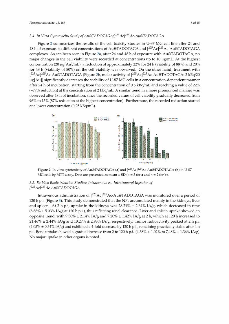
3.4. In Vitro Cytotoxicity Study of Au@TADOTAGA/[225Ac]225Ac-Au@TADOTAGA
Figure 2 summarizes the results of the cell toxicity studies in U-87 MG cell line after 24 and48 h of exposure to different concentrations of Au@TADOTAGA and [225Ac]225Ac-Au@TADOTAGAcomplexes. As can been seen in Figure 2a, after 24 and 48 h of exposure with Au@TADOTAGA, nomajor changes in the cell viability were recorded at concentrations up to 10 µg/mL. At the highestconcentration (20 µg[Au]/mL), a reduction of approximately 22% for 24 h (viability of 88%) and 20%for 48 h (viability of 80%) on the cell viability was observed. On the other hand, treatment with[225Ac]225Ac-Au@TADOTAGA (Figure 2b, molar activity of [225Ac]225Ac-Au@TADOTAGA: 2 kBq/20µg[Au]) significantly decreases the viability of U-87 MG cells in a concentration-dependent mannerafter 24 h of incubation, starting from the concentration of 0.5 kBq/mL and reaching a value of 22%(~77% reduction) at the concentration of 2 kBq/mL. A similar trend in a more pronounced manner wasobserved after 48 h of incubation, since the recorded values of cell viability gradually decreased from96% to 13% (87% reduction at the highest concentration). Furthermore, the recorded reduction startedat a lower concentration (0.25 kBq/mL).
Figure 2. In vitro cytotoxicity of Au@TADOTAGA (a) and [225Ac]225Ac-Au@TADOTAGA (b) in U-87MG cells by MTT assay. Data are presented as mean ± SD (n = 3 for a and n = 2 for b).
3.5. Ex Vivo Biodistribution Studies: Intravenous vs. Intratumoral Injection of[225Ac]225Ac-Au@TADOTAGA
Intravenous administration of [225Ac]225Ac-Au@TADOTAGA was monitored over a period of120 h p.i. (Figure 3). This study demonstrated that the NPs accumulated mainly in the kidneys, liverand spleen. At 2 h p.i, uptake in the kidneys was 28.21% ± 2.64% IA/g, which decreased in time(8.88% ± 5.03% IA/g at 120 h p.i.), thus reflecting renal clearance. Liver and spleen uptake showed anopposite trend, with 9.50% ± 2.14% IA/g and 7.20% ± 1.42% IA/g at 2 h, which at 120 h increased to21.46% ± 2.44% IA/g and 13.27% ± 2.93% IA/g, respectively. Tumor radioactivity peaked at 2 h p.i.(4.05% ± 0.34% IA/g) and exhibited a 4-fold decrease by 120 h p.i., remaining practically stable after 4 hp.i. Bone uptake showed a gradual increase from 2 to 120 h p.i. (4.38% ± 1.02% to 7.48% ± 1.36% IA/g).No major uptake in other organs is noted.

Pharmaceutics 2020, 12, 188 9 of 15
Figure 3. Ex vivo biodistribution of [225Ac]225Ac-Au@TADOTAGA after intravenous injection.Values represent the mean ± SD (n = 5 mice per group).
Biodistribution studies after intratumoral injection of [225Ac]225Ac-Au@TADOTAGA resulted inhigh tumor uptake at 2 h p.i. (60.67% ± 3.87% IA/g) which decreased over 228 h p.i. (5.21% ± 1.26%IA/g). Liver and spleen exhibited an opposite trend, showing ~3-fold and ~4-fold increases from 2 h to288 h p.i., respectively. Bone uptake was 2.95% ± 0.54% IA/g which increased to 3.48% ± 1.06% IA/g at288 h p.i. Radioactivity in all other organs was less than 2% IA/g from 4 h p.i., with the exception ofkidney uptake (Figure 4).
Figure 4. Ex vivo biodistribution of [225Ac]225Ac-Au@TADOTAGA after intratumoral injection. Valuesrepresent the mean ± SD (n = 5 mice per group).
3.6. Therapeutic Efficacy Study
The therapeutic efficacy of [225Ac]225Ac-Au@TADOTAGA was determined by estimation ofthe tumor growth index (TGI) of two groups of U-87 MG tumor-bearing SCID mice, up to22 days post-treatment by intratumoral injection of NPs (Group A) and normal saline (Group B)(Figure 5, Group A: blue line; Group B: red line). Mice in Group A received three injections of[225Ac]225Ac-Au@TADOTAGA (total activity: 15 kBq), while mice in Group B received three injectionsof normal saline, on Day 1 (day of treatment initiation), Day 3 and Day 5. TGI of Group A mice was~2.4-fold lower at 8 days p.i. and showed an upward trend, reaching ~3.9-fold lower at 22 days p.i.,when compared to the mice in Group B.

Pharmaceutics 2020, 12, 188 10 of 15
Figure 5. Effect of intratumoral injection of [225Ac]225Ac-Au@TADOTAGA (blue line) or normal saline(red line) on the tumor growth index (TGI) of U-87 MG tumor-bearing SCID mice. Values represent themean ± SD (n = 5 mice per group).
3.7. Histopathology Study
The total tumor and the subsequent necrotic areas were measured per slide (see Section 2). All theH&E slides per block, containing the total mass of each tumor, were evaluated to calculate the totalpercentage of necrosis per sample (Figure 6). The graph presents the average of the measurementsfrom all the samples per group. From the analysis of the necrotic areas from the treated and thecontrol animals, we reveal that the Au@TADOTAGA gold nanoparticles with Ac-225 caused increaseddamage to the tumors, resulting in increased tumor tissue necrosis. The tumor-adjacent tissues inboth the treated and the control animals revealed no significant necrosis, a fact which is related to thelocalization of the nanoparticles inside the tumors. Using a 100×magnification (Figure 7) we couldobserve the presence of the nanoparticles in the periphery of the necrotic lesions.
Figure 6. Percentage of the total necrotic areas per tumor. The 4 stars indicate the p value. p < 0.0001.

Pharmaceutics 2020, 12, 188 11 of 15
Figure 7. Representative images of H&E-stained slices of U-87 MG tumor tissue after the therapeuticefficacy study in mice, from the area of the necrotic lesion (100× magnification): (a) control mice,(b) treated mice. The arrows indicate the presence of nanoparticles in periphery of the necrotic lesionsof the treated tumors. The box shows a 5× digital zoom of the selected area. Scale bar = 40 µm.
4. Discussion
Actinium-225 is a highly promising alpha-emitter for therapeutic applications due to its favorablenuclear properties and high cytotoxicity, which is attributed to the generation of 4 high-energy α
particles during its decay. Nonetheless, the application of 225Ac remains challenging, as chelation viamost chelating agents is insufficient to stably retain its daughters, due to the α recoil effect which isobserved upon release of an α particle. While Thiele et al. recently reported on the development of aneighteen-membered macrocyclic ligand which rapidly and efficiently chelates 225Ac at RT, no mentionwas made with regard to how efficiently this chelator retains the daughter isotopes of 225Ac [14].DOTA is probably not the most suitable chelator for 225Ac and its daughters, however for the momentit remains the gold standard for the coordination of 225Ac [6]. This is reflected in numerous preclinicalstudies involving biomolecules radiolabeled with 225Ac via DOTA, which have shown promisingresults for the treatment of various cancers [15,16]. It is also evident in the clinic, where for the past10 years several clinical trials have been underway with 225Ac-DOTA constructs for the treatment of,e.g., prostate and gastroenteropancreatic cancers [17,18].
During the past decade, an increasing number of studies have been reported on tumor treatmentwith radiolabeled NPs, most of which have been performed with NPs radiolabeled with the β-emittingisotope Lutetium-177 (177Lu, half-life: t1/2 = 6.7 d) [12,19–24]. Regarding 225Ac, only a few studieshave examined NPs radiolabeled with this radionuclide for tumor treatment [25–27]. The presentwork was designed to investigate a gold nanoparticle radiolabeled with an α-emitter as an injectableradiopharmaceutical form of BRT for local radiation treatment of cancer. For this purpose, theaccumulation of [225Ac]225Ac-Au@TADOTAGA NPs in a U-87 MG tumor model, after both intravenousand intratumoral injection, was studied. Ex vivo data are provided up to 120 h post-injection, whiledata from one extra time-point are provided from the intratumorally-injected mice of the therapeuticefficacy study, after their euthanization at 288 h p.i. Our present work demonstrates the therapeuticpotential of [225Ac]225Ac-Au@TADOTAGA after their intratumoral injection in glioma xenografts.To our knowledge, this is the first long-term biodistribution study performed with 225Ac-labeledgold nanoparticles.

Pharmaceutics 2020, 12, 188 12 of 15
Au@TADOTAGA nanoparticles were efficiently labeled with 225Ac by heating at 70 C for 30 min,resulting in a radiochemical yield of 86% ± 1.8%. After purification by centrifugation, the radiochemicalpurity was ~93%. The radiolabeled NPs were stable up to ~80% in both PBS and acetate buffer pH 6.5at 10 days post-incubation.
In order to assess the cytotoxic properties of the new Au@TADOTAGA and[225Ac]225Ac-Au@TADOTAGA complexes, cell toxicity studies with the MTT assay were conductedwith the use of glioblastoma U-87 MG cell line. The experimental data revealed that Au@TADOTAGAshowed no remarkable cytotoxicity at 24 and 48 h after treatment. However, in the case of 24 htreatment with [225Ac]225Ac-Au@TADOTAGA, a dose-dependent cytotoxicity pattern was observedthat can only be attributed to the presence of 225Ac. A similar trend is also present at 48 h at aneven lower concentration (0.25 instead of 0.5 kBq/mL), additionally demonstrating a time-dependentmanner of [225Ac]225Ac-Au@TADOTAGA-induced toxicity in U-87 MG cells.
Ex vivo biodistribution studies of [225Ac]225Ac-Au@TADOTAGA were performed on U-87 MGtumor-bearing SCID mice, up to 120 h after intravenous injection, and 288 h after intratumoral injection.After their intravenous injection, [225Ac]225Ac-Au@TADOTAGA NPs were mainly located in thekidneys, liver and spleen. Initial high kidney uptake demonstrates route of clearance, as noted inthe literature for such gold NPs [28–30]. Tumor uptake peaked at 2 h p.i. and is due to the enhancedpermeability and retention (EPR) effect, which results in the accumulation of these NPs within thetumor because of the tumor’s leaky vasculature and poor lymphatic drainage. Thereafter, tumoruptake declined while liver and spleen uptake showed an upward trend. This may be attributedto two factors: (i) recirculation of the radiolabeled NPs in the mouse organism and consequentclearance from the body via the hepatobiliary route and (ii) slow and gradual release of the 225Acradiolabel from the chelator [15,31,32]. This is also evident in the decline in tumor-to-tissue ratios overtime (tumor:blood 3.66 ± 1.83 vs. 1.41 ± 1.24; tumor:liver 0.45 ± 0.14 vs. 0.05 ± 0.02; tumor:spleen0.58 ± 0.17 vs. 0.09 ± 0.07). The moderate liver uptake at early time-points (2 and 4 h p.i.) is unlikelyto be due to the phenomenon of opsonization, as this would lead to fast and pronounced uptake ofthe NPs by phagocytic-rich organs such as the liver and spleen [33,34]. Regarding tumor uptake, ourresults showed satisfactory tumor uptake via passive targeting, comparable to results presented in theliterature [35,36]. The observed bone uptake of [225Ac]225Ac-Au@TADOTAGA (4.38% ± 1.02% IA/gat 2 h p.i., increasing to 7.48% ± 1.36% IA/g at 120 h p.i.) may be attributed to partial release of theradiolabel over time [15].
After intratumoral injection, we observed a very high tumor uptake, which slowly decreasedover the duration of the study (2 to 288 h p.i.). On the contrary, radioactivity in the liver and spleensignificantly increased (liver uptake: 6.57% ± 0.75% IA/g and 19.37% ± 1.56% IA/g at 2 and 288 h p.i.,p = 0.012; spleen uptake: 3.80% ± 0.48% IA/g and 14.80% ± 1.30% IA/g at 2 and 288 h p.i., p = 0.0108).This behavior can be attributed to recirculation of the radiolabeled NPs, as well as to partial release of225Ac from the chelator, which was also demonstrated during our in vitro stability studies [15,31,32].Although [225Ac]225Ac-Au@TADOTAGA NPs do not bear a targeting vector, high tumor retentionwas provided after intratumoral injection. Recently, Yook et al. reported on the radiolabeling of bothnontargeted and targeted Au NPs with Lutetium-177 and showed that both radiolabeled NPs showedhigh tumor uptake after intratumoral injection, which slowly cleared over 48 h [12]. Similar results onactive and passive accumulation of gold nanomaterials have been reported [37,38].
While uptake of NPs by the RES organs is always an issue when these are systemically administered,previous results with Au@TADOTAGA radiolabeled with Indium-111 showed low to moderate liver andspleen uptake, which is in accordance to our results [28]. This study revealed a similar distribution of theradiolabeled NPs, however the NPs were monitored up to 72 h post intravenous injection. The presentwork provides in vivo data up to 120 h after intravenous injection and 288 h after intratumoral injectionof these NPs radiolabeled with 225Ac in U-87 MG tumor-bearing mice. A major difference betweenour study and the study by Laurent et al. is the high tumor uptake of [225Ac]225Ac-Au@TADOTAGA

Pharmaceutics 2020, 12, 188 13 of 15
after intratumoral administration when compared to their intravenous administration, which isexpected [28].
As proof-of-concept, a preliminary study to assess the therapeutic effect of intratumorally-injected[225Ac]225Ac-Au@TADOTAGA was performed over a period of 22 days. Our results suggest that theintratumoral route of administration was responsible for delaying tumor growth, even though theamount of radiotracer injected per mouse was low (~15 kBq over three i.t. injections). On the contrary,mice injected with normal saline exhibited more rapid tumor growth. Histopathology studies confirmour in vivo studies.
5. Conclusions
Gold nanoparticles radiolabeled with 225Ac via the macrocyclic chelator TADOTAGA resultedin the retardation of tumor growth after their intratumoral injection in U87MG tumor-bearing mice,even though very low activities were injected per mouse. To our knowledge, this is the first suchstudy reported in the literature. This gold nanoparticle radiopharmaceutical could be applied as anunconventional brachytherapy in injectable form for local radiation treatment of cancer. In orderto improve their properties, these AuNPs will be further derivatized with other ligands for 225Acchelation, in order to maximize their in vivo stability, which is particularly important for long-termin vivo studies with an α-emitter. Further preclinical evaluation will include dose-escalation studiesof [225Ac]225Ac-AuNPs in tumor-bearing mice, to determine whether or not this will lead to animprovement in the therapeutic effect. We aspire that this agent could also exploit the beneficialradiosensitizing effect of the gold core, enhancing therapeutic efficiency of brachytherapy.
Author Contributions: Conceptualization, C.T. and P.B.; formal analysis, D.S., M.P.-P. and N.K.; fundingacquisition, S.R.; investigation, E.-A.S., D.S., F.D., G.L. and R.B.; methodology, E.-A.S. and S.X.; projectadministration, P.B.; resources, S.X., F.D. and P.B.; supervision, P.B.; validation, D.S. and M.P.-P.; visualization, C.T.and B.M.; writing—original draft, E.-A.S., C.T. and P.B.; writing—review & editing, C.T., B.M., R.B., S.R. and P.B..All authors have read and agreed to the published version of the manuscript.
Funding: This research was funded by Cancéropôle Est and Région Bourgogne Franche-Comté (project e-NanoRX).
Acknowledgments: Acknowledgments are due to P. Petrou and S. Kakabakos, for assistance provided duringcytotoxicity experiments. F. Kapiris is also gratefully acknowledged for excellent technical assistance.
Conflicts of Interest: The authors declare no conflict of interest.
References
1. Older, R.A.; Synder, B.; Krupski, T.L.; Glembocki, D.J.; Gillenwater, J.Y. Radioactive implant migration inpatients treated for localized prostate cancer with interstitial brachytherapy. J. Urol. 2001, 165, 1590–1592.[CrossRef]
2. Biswal, B.M.; Yusoff, Z. Application of nanotechnology in cancer treatment. In Engineering Applications ofNanotechnology; Korada, V.S., Hamid, N.H., Eds.; Springer International Publishing: Cham, Switzerland,2017; pp. 269–311. [CrossRef]
3. Laprise-Pelletier, M.; Simão, T.; Fortin, M.A. Gold nanoparticles in radiotherapy and recent progress innanobrachytherapy. Adv. Healthc. Mater. 2018, 7. [CrossRef]
4. Ehlerding, E.B.; Cai, W. Smaller agents for larger therapeutic indices: Nanoscale brachytherapy with177Lu-labeled gold nanoparticles. J. Nucl. Med. 2016, 57, 834–835. [CrossRef]
5. Laprise-Pelletier, M.; Lagueux, J.; Côté, M.F.; LaGrange, T.; Fortin, M.A. Low-dose prostate cancerbrachytherapy with radioactive palladium–gold nanoparticles. Adv. Healthc. Mater. 2017, 6. [CrossRef]
6. Thiele, N.A.; Wilson, J.J. Actinium-225 for targeted α therapy: Coordination chemistry and current chelationapproaches. Cancer Biother. Radiopharm. 2018, 33, 336–348. [CrossRef]
7. Kampf, G. Induction of DNA double-strand breaks by ionizing radiation of different quality and theirrelevance for cell inactivation. Radiobiol. Radiother. 1988, 29, 631–658.
8. Hall, E.J.; Giaccia, A.J.; Technologies, I.O. Radiobiology for the Radiologist, 6th ed.; Lippincott Williams &Wilkins: Philadelphia, PA, USA, 2006.
Pharmaceutics 2020, 12, 188 14 of 15
9. Gadbois, D.M.; Crissman, H.A.; Nastasi, A.; Habbersett, R.; Wang, S.K.; Chen, D.; Lehnert, B.E. Alterations inthe progression of cells through the cell cycle after exposure to alpha particles or gamma rays. Radiat. Res.1996, 146, 414–424. [CrossRef]
10. Mulford, D.A.; Scheinberg, D.A.; Jurcic, J.G. The promise of targeted α-particle therapy. J. Nucl. Med. 2005,46, 199S–204S.
11. Brust, M.; Fink, J.; Bethell, D.; Schiffrin, D.J.; Kiely, C. Synthesis and reactions of functionalised goldnanoparticles. J. Chem. Soc. Chem. Commun. 1995, 1655–1656. [CrossRef]
12. Yook, S.; Cai, Z.; Lu, Y.; Winnik, M.A.; Pignol, J.P.; Reilly, R.M. Intratumorally injected 177Lu-labeled goldnanoparticles: Gold nanoseed brachytherapy with application for neoadjuvant treatment of locally advancedbreast cancer. J. Nucl. Med. 2016, 57, 936–942. [CrossRef]
13. Wang, Z.; Jacobson, O.; Tian, R.; Mease, R.C.; Kiesewetter, D.O.; Niu, G.; Pomper, M.G.; Chen, X.Radioligand therapy of prostate cancer with a long-lasting prostate-specific membrane antigen targetingagent 90Y-DOTA-EB-MCG. Bioconjug. Chem. 2018, 29, 2309–2315. [CrossRef] [PubMed]
14. Thiele, N.A.; Brown, V.; Kelly, J.M.; Amor-Coarasa, A.; Jermilova, U.; MacMillan, S.N.; Nikolopoulou, A.;Ponnala, S.; Ramogida, C.F.; Robertson, A.K.H.; et al. An eighteen-membered macrocyclic ligand foractinium-225 targeted alpha therapy. Angew. Chem. Int. Ed. 2017, 56, 14712–14717. [CrossRef] [PubMed]
15. Pruszynski, M.; D’Huyvetter, M.; Bruchertseifer, F.; Morgenstern, A.; Lahoutte, T. Evaluation of an Anti-HER2nanobody labeled with 225Ac for targeted α-particle therapy of cancer. Mol. Pharm. 2018, 15, 1457–1466.[CrossRef] [PubMed]
16. Graf, F.; Fahrer, J.; Maus, S.; Morgenstern, A.; Bruchertseifer, F.; Venkatachalam, S.; Fottner, C.; Weber, M.M.;Huelsenbeck, J.; Schreckenberger, M.; et al. DNA double strand breaks as predictor of efficacy of thealpha-particle emitter Ac-225 and the electron emitter Lu-177 for somatostatin receptor targeted radiotherapy.PLoS ONE 2014, 9. [CrossRef]
17. Kratochwil, C.; Bruchertseifer, F.; Giesel, F.L.; Weis, M.; Verburg, F.A.; Mottaghy, F.; Kopka, K.; Apostolidis, C.;Haberkorn, U.; Morgenstern, A. 225Ac-PSMA-617 for PSMA-targeted α-radiation therapy of metastaticcastration-resistant prostate cancer. J. Nucl. Med. 2016, 57, 1941–1944. [CrossRef]
18. Ballal, S.; Yadav, M.P.; Bal, C.; Sahoo, R.K.; Tripathi, M. Broadening horizons with 225Ac-DOTATATEtargeted alpha therapy for gastroenteropancreatic neuroendocrine tumour patients stable or refractory to177Lu-DOTATATE PRRT: First clinical experience on the efficacy and safety. Eur. J. Nucl. Med. Mol. Imaging2019. [CrossRef]
19. Salvanou, E.A.; Bouziotis, P.; Tsoukalas, C. Radiolabeled nanoparticles in nuclear oncology. ANR 2018, 1,38–55. [CrossRef]
20. Vilchis-Juárez, A.; Ferro-Flores, G.; Santos-Cuevas, C.; Morales-Avila, E.; Ocampo-García, B. Moleculartargeting radiotherapy with Cyclo-RGDfK (C) peptides conjugated to 177Lu-labeled gold nanoparticles intumor-bearing mice. J. Biomed. Nanotechnol. 2014, 10, 393–404. [CrossRef]
21. Jiménez-mancilla, N.; Ferro-flores, G.; Santos-Cuevas, C.; Ocampo-garcía, B.; Luna-gutiérrez, M.;Azorín-vega, E.; Isaac-olivé, K.; Camacho-lópez, M.; Torres-garcía, E. Multifunctional targeted therapysystem based on 99mTc/177Lu-labeled gold nanoparticles-Tat(49-57)-Lys3-bombesin internalized in nuclei ofprostate cancer cells. J. Label. Compd. Radiopharm. 2013, 56, 663–671. [CrossRef]
22. Cai, Z.; Yook, S.; Lu, Y.; Bergstrom, D.; Winnik, M.A.; Pignol, J.P.; Reilly, R.M. Local radiation treatment ofHER2-positive breast cancer using trastuzumab-modified gold nanoparticles labeled with 177Lu. Pharm. Res.2017, 34, 579–590. [CrossRef]
23. Yook, S.; Cai, Z.; Lu, Y.; Winnik, M.A.; Pignol, J.; Reilly, R.M. Radiation nanomedicine for EGFR-PositiveBreast Cancer:Panitumumab Modified Gold Nanoparticles Complexed to the β-Particle-Emitter, 177Lu.Mol. Pharm. 2015. [CrossRef]
24. Ferro-Flores, G.; Ocampo-García, B.; Santos-Cuevas, C.; María Ramírez, F.; Azorín-Vega, E.; Meléndez-Alafort, L.Theranostic radiopharmaceuticals based on gold nanoparticles labeled with 177lu and conjugated to peptides.Curr. Radiopharm. 2015, 8, 150–159. [CrossRef]
25. Cedrowska, E.; Pruszynski, M.; Majkowska-Pilip, A.; Meczynska-Wielgosz, S.; Bruchertseifer, F.;Morgenstern, A.; Bilewicz, A. Functionalized TiO2 nanoparticles labelled with 225Ac for targeted alpharadionuclide therapy. J. Nanoparticle Res. 2018, 20. [CrossRef]

Pharmaceutics 2020, 12, 188 15 of 15
26. Sempkowski, M.; Zhu, C.; Menzenski, M.Z.; Kevrekidis, I.G.; Bruchertseifer, F.; Morgenstern, A.; Sofou, S.Sticky patches on lipid nanoparticles enable the selective targeting and killing of untargetable cancer cells.Langmuir 2016, 32, 8329–8338. [CrossRef]
27. Mclaughlin, M.F.; Robertson, D.; Pevsner, P.H.; Wall, J.S. LnPO4 nanoparticles doped with Ac-225 andsequestered daughters for targeted alpha therapy. Cancer Biotherary Radiopharm. 2013, 29, 34–41. [CrossRef]
28. Laurent, G.; Bernhard, C.; Dufort, S.; Jiménez Sánchez, G.; Bazzi, R.; Boschetti, F.; Moreau, M.; Vu, T.H.;Collin, B.; Oudot, A.; et al. Minor changes in the macrocyclic ligands but major consequences on the efficiencyof gold nanoparticles designed for radiosensitization. Nanoscale 2016, 8, 12054–12065. [CrossRef]
29. Alric, C.; Miladi, I.; Kryza, D.; Taleb, J.; Lux, F.; Bazzi, R.; Billotey, C.; Janier, M.; Perriat, P.; Roux, S.; et al.The biodistribution of gold nanoparticles designed for renal clearance. Nanoscale 2013, 5, 5930–5939.[CrossRef]
30. Miladi, I.; Alric, C.; Dufort, S.; Mowat, P.; Dutour, A.; Mandon, C.; Laurent, G.; Bräuer-Krisch, E.; Herath, N.;Coll, J.; et al. The in vivo radiosensitizing effect of gold nanoparticles based MRI contrast agents. Small 2014,10, 1116–1124. [CrossRef]
31. Lee, J.; Chatterjee, D.K.; Lee, M.H.; Krishnan, S. Gold nanoparticles in breast cancer treatment: Promise andpotential pitfalls. Cancer Lett. 2014, 347, 46–53. [CrossRef]
32. Davis, I.A.; Glowienka, K.A.; Boll, R.A.; Deal, K.A.; Brechbiel, M.W.; Stabin, M.; Bochsler, P.N.; Mirzadeh, S.;Kennel, S.J. Comparison of 225actinium chelates: Tissue distribution and radiotoxicity. Nucl. Med. Biol. 1999,26, 581–589. [CrossRef]
33. Vonarbourg, A.; Passirani, C.; Saulnier, P.; Benoit, J.P. Parameters influencing the stealthiness of colloidaldrug delivery systems. Biomaterials 2006, 27, 4356–4373. [CrossRef]
34. Bertrand, N.; Leroux, J.C. The journey of a drug-carrier in the body: An anatomo-physiological perspective.J. Control. Release 2012, 161, 152–163. [CrossRef]
35. Bouziotis, P.; Stellas, D.; Thomas, E.; Truillet, C.; Tsoukalas, C.; Lux, F.; Tsotakos, T.; Xanthopoulos, S.;Paravatou-Petsotas, M.; Gaitanis, A.; et al. 68Ga-radiolabeled AGuIX nanoparticles as dual-modality imagingagents for PET/MRI-guided radiation therapy. Nanomedicine 2017, 12, 1561–1574. [CrossRef]
36. Xie, H.; Jim, Z.; Bao, A.; Goins, B.; Phillips, W.T. In vivo PET imaging and biodistribution of radiolabeledgold nanoshells in rats with tumor xenografts. Int. J. Pharm. 2010, 395, 324–330. [CrossRef]
37. Chattopadhyay, N.; Fonge, H.; Cai, Z.; Scollard, D.; Lechtman, E.; Done, S.J.; Pignol, J.-P.; Reilly, R.M. Role ofantibody-mediated tumor targeting and route of administration in nanoparticle tumor accumulation in vivo.Mol. Pharm. 2012, 9, 2168–2179. [CrossRef]
38. Huang, X.; Peng, X.; Wang, Y.; Wang, Y.; Shin, D.M.; El-Sayed, M.A.; Nie, S. A reexamination of active andpassive tumor targeting by using rod-shaped gold nanocrystals and covalently conjugated peptide ligands.ACS Nano 2010, 4, 5887–5896. [CrossRef]
© 2020 by the authors. Licensee MDPI, Basel, Switzerland. This article is an open accessarticle distributed under the terms and conditions of the Creative Commons Attribution(CC BY) license (http://creativecommons.org/licenses/by/4.0/).