Embed Size (px)
Citation preview

Co
CONSENSUS STATEMENT
A Practical Guide for the Diagnosis of Primary Enteric
Nervous System Disorders
�M.G. Schappi, yA. Staiano, zP.J. Milla, zV.V. Smith, §J.A. Dias, jjR. Heuschkel, �S. Husby,# �� yy
M.L. Mearin, A. Papadopoulou, F.M. Ruemmele,s, and jjjjS. Koletzko
§§Yvan Vandenplapyright 2013 by ESPGHAN and NASPGHAN. Unauthorized repro
analysis should be carried out in laboratories that have the necessary
expertise and access to their own validated reference values.
neuronal dysplasia (INand uncertain histomo
Received August 9, 2013.From the �Pediatric Center, Clinique des Grangettes, and Centre Medical
Universitaire, Geneva, Switzerland, the yDepartment of TranslationalMedical Science, Section of Pediatrics, University of Naples ‘‘FedericoII,’’ Naples, Italy, the zDepartments of Gastroenterology and Histo-pathology, UCL Institute of Child Health and Great Ormond StreetHospital, London, UK, the §Department of Paediatrics, Hospital S. Joao,Porto, Portugal, the jjDepartment of Paediatric Gastroenterology,Addenbrookes Hospital, Cambridge, UK, the �Hans Christian AndersenChildren’s Hospital, OUH, Odense, Denmark, the #Department ofPaediatrics, Leiden University Medical Center, Leiden, The Netherlands,the ��First Department of Paediatrics, University of Athens, Children’sHospital ‘‘Agia Sofia,’’ Athens, Greece, §§UZ Brussel, Vrije Universi-teit, Brussel, Brussels, Belgium, and the jjjjDr vonHaunersches Kinder-spital, Ludwig-Maximilians-University, Munich, Germany.
Address correspondence and reprint requests to Prof Annamaria Staiano,Department of Translational Medical Science, Section of Pediatrics,University of Naples ‘‘Federico II,’’ Naples, Italy (e-mail: [email protected]).
Drs Schappi, Staiano, and Koletzko participated equally in this study.Drs Milla and Smith served as guest authors as experts on enteric nervous
disorders.The authors report no conflicts of interest.Copyright # 2013 by European Society for Pediatric Gastroenterology,
Hepatology, and Nutrition and North American Society for PediatricGastroenterology, Hepatology, and Nutrition
DOI: 10.1097/MPG.0b013e3182a8bb50
JPGN � Volume 57, Number 5, November 2013
ABSTRACT
Objective: Primary gastrointestinal neuropathies are a heterogeneous group
of enteric nervous system (ENS) disorders that continue to cause difficulties
in diagnosis and histological interpretation. Recently, an international work-
ing group published guidelines for histological techniques and reporting,
along with a classification of gastrointestinal neuromuscular pathology. The
aim of this article was to review and summarize the key issues for pediatric
gastroenterologists on the diagnostic workup of congenital ENS disorders. In
addition, we provide further commentary on the continuing controversies in
the field.
Results: Although the diagnostic criteria for Hirschsprung disease are well
established, those for other forms of dysganglionosis remain ill-defined.
Appropriate tissue sampling, handling, and expert interpretation are crucial
to maximize diagnostic accuracy and reduce interobserver variability. The
absence of validated age-related normal values for neuronal density, along
with the lack of correlation between clinical and histological findings, result
in significant diagnostic uncertainties while diagnosing quantitative
aberrations such as hypoganglionosis or ultrashort Hirschsprung disease.
Intestinal neuronal dysplasia remains a histological description of unclear
significance.
Conclusions: The evaluation of cellular quantitative or qualitative
abnormalities of the ENS for clinical diagnosis remains complex. Such
Key Words: chronic intestinal pseudo-obstruction, gastrointestinal
neuropathic disease, Hirschsprung disease, intestinal neuronal dysplasia,
rectal suction biopsy
(JPGN 2013;57: 677–686)
I ntestinal dysmotility disorders can be classified by etiology intoprimary visceral myopathies; primary visceral neuropathies; and
secondary forms owing to toxic, metabolic, infectious, or othersystemic disorders affecting the smooth muscle, interstitial cells ofCajal (ICC), or the enteric or extrinsic nervous systems. Symptomsof intestinal dysmotility, such as dyspepsia, vomiting, abdominalpain, bloating, diarrhea, and constipation, are nonspecific. Thegastrointestinal (GI) transit may be too rapid or too slow or poorlycoordinated. The most severe manifestation of dysmotility ischronic intestinal pseudoobstruction (CIPO) with signs and symp-toms of bowel obstruction in the absence of a mechanical obstruc-tive lesion. In general, there is a poor correlation among symptoms,the underlying etiopathology, and histopathological findings. Forexample, distinct phenotypes such a megacystis, microcolon, andhypoperistalsis syndrome may be caused by neuropathy or myo-pathy with different genetic traits (1).
Recently, an international working group (IWG) formed ofpathologists, pediatric and adult gastroenterologists, and surgeonswith special interest in neurogastroenterology-classified neuromus-cular GI disorders based on the present evidence, the so-calledLondon classification (2). In a previous article, this group haddefined techniques for histopathological workup, interpretation,and reporting of intestinal neuromuscular disorders (3). A systema-tic review by the IWG revealed a lack of concordance in thequantitative assessment for elements of the enteric nervous system(ENS) (4).
The IWG acknowledged that most of the less well-under-stood entities have previously been described by their histomor-phology, with little or no correlation to symptoms or naturalhistory, and without comparison to any appropriate control group(5,6). Only recently have the age of the patient and the location ofbiopsies been recognized as crucial to the interpretation of histo-pathological findings (7,8). The clinical implications of manyhistopathological findings remain unclear, making it difficultfor clinicians to provide families with any concrete idea of naturalhistory of the disease.
The 3 recent articles (2–4) by the IWG have tried to collatepresent knowledge in this field. Despite these comprehensivepublications, the use of inaccurate terminology and vague diag-nostic criteria remain common (9). In the authors’ experience,inappropriate diagnoses such as hypoganglionosis and intestinal
duction of this article is prohibited.
D) are still being made based on symptomsrphological findings, thereby perpetuating
677

Co
diagnostic confusion and uncertainty. In addition, inappropriatelaboratory techniques and diagnostic criteria are still applied,leading to the prolonged misdiagnosis and potential of thesechildren.
The purpose of this article was to extract and summarizethe most important information from the IWG’s 3 articles,placing them into a clinical context that is useful for practicingpediatric gastroenterologists. This medical position paper focusesonly on congenital or developmental disorders of the ENSbecause these have proved particularly confusing. These dis-orders include more common diseases such as Hirschsprungdisease (HD), monogenetic disorders with a clear clinical phe-notype and well-defined histopathology, as well as ill-definedentities with no obvious etiology and little or no correlationamong clinical phenotype, outcome, and histopathology. Forclarity, we have not addressed acquired or secondary diseasesof the ENS, for example, secondary to inflammation, degener-ation of ganglion cells, or those characterized by abnormalities inglial or ICCs.
ABSENCE OF NEURONS (AGANGLIONOSIS)
HDHD (or synonym Hirschsprung neurocristopathy) is charac-
terized by the complete absence of enteric neurons in the myentericand submucosal plexuses along a variable portion of the distalintestine. HD affects approximately 1 in 5000 newborns with amale:female ratio of approximately 4:1. HD, both sporadic andfamilial, has a complex genetic, sex-modified inheritance pattern
Schappi et al
pyright 2013 by ESPGHAN and NASPGHAN. Un
and appears to be polygenic or multifactorial (10). Genetic factorshave been identified in approximately 50% of patients with HD
7th week
8th week
55th weekS2–S4
12th week
10/11th
week
9th week
6th week
A
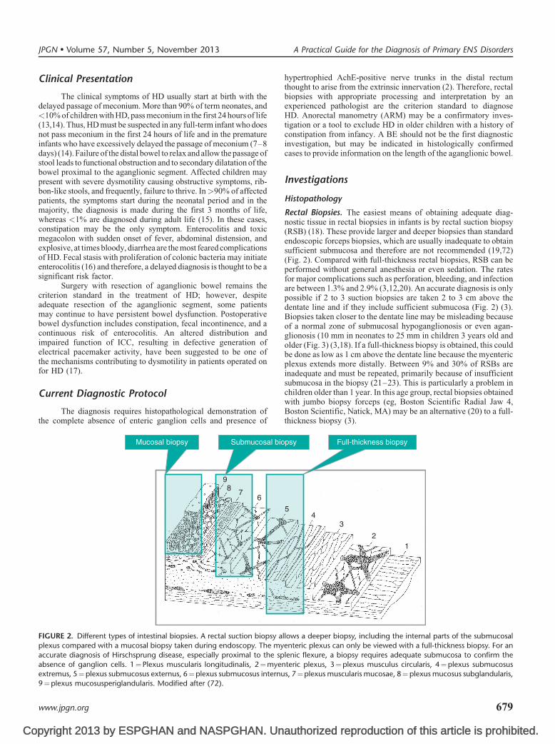
FIGURE 1. The intramural enteric nervous system develops from neural
During the 7th week of gestational age, the first enteric NCCs are visible in tdistally to reach the rectum at week 12 (A). The submucosal plexus develop
inner muscular layer into the submucosa. The extramural innervation cons
In addition, the distal colon (rectum, sigmoid, and a variable distance up to
In Hirschsprung disease (HD), the craniocaudal migration of NCCs is inrectosigmoid (B). The acetylcholine (Ach) output remains unmodulated, an
activity. The increased AchE activity is used as an additional diagnostic
distribution of the parasympathetic extramural input explains why AchE sta
in cases of total intestinal aganglionosis.
678
with a positive family history and in 15% to 20% of sporadic cases(10,11). In almost all of the sporadic cases, RET proto-oncogenehas been implicated, in part with other gene defects such as glialcell line–derived neurotrophic factor or the endothelin systems(10).
More than 10 genes mapping for transcription factors such asSOX10 and PHOX2B may be required for the HD phenotype to befully expressed. HD occurs as an isolated trait in approximately70% of cases (11). More frequent associated syndromes are Downsyndrome, Waardenburg syndrome, and Smith-Lemli-Opitz syn-drome (10). In a small number of cases (<5%), the aganglionosis isa manifestation of multiple endocrine neoplasia (MEN) 2A, anautosomal dominant inherited disorder caused by certain germlinemutations in the RET proto-oncogene with a high risk of medullarycarcinoma of the thyroid and pheochromocytoma (12). In thesecases, prophylactic thyroidectomy is recommended; however, rou-tine screening for RET or other mutations in nonsyndromic forms isnot yet practical and, because of the incomplete understanding ofthe complex genetic nature of the condition, not yet clinicallyindicated.
In the majority of cases (80%), the aganglionic segmentinvolves the rectum and the sigmoid colon only, whereas totalcolonic aganglionosis occurs in approximately 8% to 10% with analmost equal sex ratio. Rarely, aganglionosis extends into the smallbowel or encompasses the entire bowel (subtotal or total intestinalaganglionosis) (Fig. 1) (10).
In these patients, the diagnosis of HD is particularly challen-ging because both histology (with the lack of increased acetylcholinesterase staining) and barium enema (BE) (with the
JPGN � Volume 57, Number 5, November 2013
authorized reproduction of this article is prohibited.
absence of a visible change in caliber of the colon) maybe misleading.
6th week
7th week
8th week
th weekS2–S4
12th week
10/11th
week
9th week
B
crest cells (NCCs) predominantly arising from the vagal neural crest.
he upper gastrointestinal tract and jejunum, and subsequently migrates through migration of NCCs from the myenteric plexus, through the
ists of the sympathetic fibers from the ganglion mesentericum inferior.
the splenic flexure) receives parasympathetic input from sacral nerves.
complete over a variable segment of the distal hindgut mostly ind the high release of Ach induces increased acetycholinesterase (AchE)
tool in short-segment HD if insufficient submucosa is available. The
ining is normal in aganglionic bowel proximal of the splenic flexure and
www.jpgn.org
Co
Clinical Presentation
The clinical symptoms of HD usually start at birth with thedelayed passage of meconium. More than 90% of term neonates, and<10% of children with HD, pass meconium in the first 24 hours of life(13,14). Thus, HD must be suspected in any full-term infant who doesnot pass meconium in the first 24 hours of life and in the prematureinfants who have excessively delayed the passage of meconium (7–8days) (14). Failure of the distal bowel to relax and allow the passage ofstool leads to functional obstruction and to secondary dilatation of thebowel proximal to the aganglionic segment. Affected children maypresent with severe dysmotility causing obstructive symptoms, rib-bon-like stools, and frequently, failure to thrive. In>90% of affectedpatients, the symptoms start during the neonatal period and in themajority, the diagnosis is made during the first 3 months of life,whereas <1% are diagnosed during adult life (15). In these cases,constipation may be the only symptom. Enterocolitis and toxicmegacolon with sudden onset of fever, abdominal distension, andexplosive, at times bloody, diarrhea are the most feared complicationsof HD. Fecal stasis with proliferation of colonic bacteria may initiateenterocolitis (16) and therefore, a delayed diagnosis is thought to be asignificant risk factor.
Surgery with resection of aganglionic bowel remains thecriterion standard in the treatment of HD; however, despiteadequate resection of the aganglionic segment, some patientsmay continue to have persistent bowel dysfunction. Postoperativebowel dysfunction includes constipation, fecal incontinence, and acontinuous risk of enterocolitis. An altered distribution andimpaired function of ICC, resulting in defective generation ofelectrical pacemaker activity, have been suggested to be one ofthe mechanisms contributing to dysmotility in patients operated onfor HD (17).
Current Diagnostic Protocol
JPGN � Volume 57, Number 5, November 2013
pyright 2013 by ESPGHAN and NASPGHAN. Un
The diagnosis requires histopathological demonstration ofthe complete absence of enteric ganglion cells and presence of
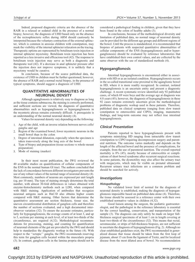
Mucosal biopsy Submucosal bio
678
9
FIGURE 2. Different types of intestinal biopsies. A rectal suction biopsy a
plexus compared with a mucosal biopsy taken during endoscopy. The my
accurate diagnosis of Hirschsprung disease, especially proximal to the sp
absence of ganglion cells. 1¼Plexus muscularis longitudinalis, 2¼myenextremus, 5¼plexus submucosus externus, 6¼plexus submucosus internu
9¼plexus mucosusperiglandularis. Modified after (72).
www.jpgn.org
hypertrophied AchE-positive nerve trunks in the distal rectumthought to arise from the extrinsic innervation (2). Therefore, rectalbiopsies with appropriate processing and interpretation by anexperienced pathologist are the criterion standard to diagnoseHD. Anorectal manometry (ARM) may be a confirmatory inves-tigation or a tool to exclude HD in older children with a history ofconstipation from infancy. A BE should not be the first diagnosticinvestigation, but may be indicated in histologically confirmedcases to provide information on the length of the aganglionic bowel.
Investigations
Histopathology
Rectal Biopsies. The easiest means of obtaining adequate diag-nostic tissue in rectal biopsies in infants is by rectal suction biopsy(RSB) (18). These provide larger and deeper biopsies than standardendoscopic forceps biopsies, which are usually inadequate to obtainsufficient submucosa and therefore are not recommended (19,72)(Fig. 2). Compared with full-thickness rectal biopsies, RSB can beperformed without general anesthesia or even sedation. The ratesfor major complications such as perforation, bleeding, and infectionare between 1.3% and 2.9% (3,12,20). An accurate diagnosis is onlypossible if 2 to 3 suction biopsies are taken 2 to 3 cm above thedentate line and if they include sufficient submucosa (Fig. 2) (3).Biopsies taken closer to the dentate line may be misleading becauseof a normal zone of submucosal hypoganglionosis or even agan-glionosis (10 mm in neonates to 25 mm in children 3 years old andolder (Fig. 3) (3,18). If a full-thickness biopsy is obtained, this couldbe done as low as 1 cm above the dentate line because the myentericplexus extends more distally. Between 9% and 30% of RSBs areinadequate and must be repeated, primarily because of insufficientsubmucosa in the biopsy (21–23). This is particularly a problem inchildren older than 1 year. In this age group, rectal biopsies obtainedwith jumbo biopsy forceps (eg, Boston Scientific Radial Jaw 4,
A Practical Guide for the Diagnosis of Primary ENS Disorders
authorized reproduction of this article is prohibited.
Boston Scientific, Natick, MA) may be an alternative (20) to a full-thickness biopsy (3).
psy Full-thickness biopsy
12
34
5
llows a deeper biopsy, including the internal parts of the submucosal
enteric plexus can only be viewed with a full-thickness biopsy. For an
lenic flexure, a biopsy requires adequate submucosa to confirm the
teric plexus, 3¼plexus musculus circularis, 4¼plexus submucosuss, 7¼plexus muscularis mucosae, 8¼plexus mucosus subglandularis,
679

Co
Dentate line
A B
Anal margin
Transition zone
Physiologicalaganglioniczone
Normalganglioniccolon
Anal margin
Dentate line
Pathologictransitionzone
Normalganglioniccolon
Pathologicalaganglioniczone
FIGURE 3. A, The normal rectum showing the physiological, hypoganglionic, or even aganglionic zone above the dentate line that transitions to
the normal, more proximal, ganglionic bowel. Biopsies taken too close to the dentate line may be hypoganglionic or aganglionic and hence
reported as false-positive for Hirschsprung disease. B, In Hirschsprung disease, the aganglionic zone extends from the dentate line proximally for avariable distance, before a transition zone leads to normal innervation. Modified after (18).
Schappi et al JPGN � Volume 57, Number 5, November 2013
In conclusion, correctly performed RSBs taken 2 to 3 cmabove the dentate line remain the preferred method in infants toobtain tissue for the diagnosis of HD (Fig. 3). The tissue mustcontain sufficient submucosa for the pathologist to confirm theabsence of ganglion cells with certainty.
Staining of Biopsies. Once adequate biopsies are obtained, it isstill controversial as to which histological methodology should bethe preferred approach to confirm or exclude the diagnosis of HD.Techniques include analysis of hematoxylin and eosin (H&E)–stained paraffin sections, snap-frozen sections stained for acetylcholinesterase enzyme histochemistry (AchE), and immunostainingfor neuronal markers (3). All of the techniques have high sensitivityand specificity in an experienced laboratory with reported accuracyrates as high as 99%, although the approach based entirely onhistochemistry of frozen sections may be difficult in generalpathology practices (5). In their consensus report, Knowles et al(3) concluded that insufficient data exist to firmly recommend oneapproach over the other, largely because of a lack of comparablestudies comparing AchE histochemistry with enzyme histochem-istry and immunostaining. American investigators favor the use ofH&E staining based on its availability in all centers, reliability, andlow cost. The IWG of the London classification recommends aninitial of 50 to 75 H&E-stained paraffin sections of each biopsy. Thepresence of a single unequivocal submucosal ganglion in RSB fromthe terminal 2 cm of rectum in H&E-stained paraffin sectionsexcludes the diagnosis of HD.
pyright 2013 by ESPGHAN and NASPGHAN. Un
Although it is possible to establish a diagnosis of HD with theuse of H&E-stained paraffin sections alone, combining H&E
680
staining with AchE enzyme histochemistry is still the most widelyused method, particularly in Europe and Asia (5,18). AchE stainingallows the identification of thickened nerves in the muscularismucosae in HD as early as 30 weeks’ gestation and hypertrophicnerve trunks in the lamina propria of the mucosa, which are morecommon after 3 months of age (18). AchE staining alone may givefalse normal results in total colonic aganglionosis or in biopsiestaken proximal to the splenic flexure because the activity of theextramural parasympathetic system from S2 to S4 decreases proxi-mally from the anal canal, becoming almost absent at the splenicflexure (24–26) (Fig. 1). In these cases, diagnosis should not rely onmucosal and muscularis staining alone. The combination of AchEand H&E staining on frozen sections is possible and increases thediagnostic accuracy because it improves the identification of sub-mucosal ganglion cells, as well as demonstrates the abnormal nervefiber distribution in the left colon (3).
Other enzyme histochemical techniques (all requiring frozensections), such as lactate dehydrogenases (LDHs), may be used toidentify neurons (27). The potential value of immunohistochemistryhas been evaluated in a number of studies. Calretinin is an immu-nohistochemical marker that may be a potential alternative to AchE(28–30). Calretinin is normally present in the perikarya and nerveprocesses of a subset of enteric ganglion cells. Immunoreactivity islost in the aganglionic segment of HD (3,18). A recent comparativestudy shows that calretinin is as sensitive and specific as AchE andthat the technique can be performed on paraffin sections (28);however, false-positive and false-negative results may occur withthis technique (29,31).
authorized reproduction of this article is prohibited.
In conclusion, at the present time, either H&E-stained par-affin-embedded sections or the analysis of frozen sections stained
www.jpgn.org

Co
contrast to HD, no nerve fibers with increased AChE activity havebeen observed in the lamina propria mucosa in suspected UHD (43).
TABLE 1. Sensitivity and specificity of different diagnostic procedures
for the diagnosis of HD
Parameter
Index test
Mean
sensitivity, %
(95% CI)
Mean
specificity, %
(95% CI)
Barium enema 70 (64–76) 83 (74–90)
Anorectal manometry 91 (85–95) 94 (89–97)
Rectal suction biopsy
(stained for AchE activity)
93 (88–95) 98 (95–99)
with H&E plus AchE enzyme histochemistry should be used as thestandard techniques to prove the absence of ganglion cells for thediagnosis of HD in rectal biopsies from children (3). AchE stainingis not appropriate in long-segment HD and in biopsies proximal tothe splenic flexure. If the success of the present studies on immu-nostaining is widely reproducible, it may be that H&E along withimmunostaining using markers such as calretinin will become the‘‘new’’ standard for diagnosis.
Intraoperative HistopathologySurgical resection of the aganglionic bowel and the hypo-
ganglionic ‘‘transitional zone,’’ which varies from a few milli-metres to several centimetres (32,33), is presently the onlytreatment for HD. At the time of resection, serial intraoperativefrozen seromuscular biopsies should be obtained to map the agan-glionic segment and to allow decisions to be made regarding thelevel of the proximal resection. Because the margin of transitionfrom the hypoganglionic zone to normal innervation is not clear-cut, the resection should be performed approximately 2 to 3 cmproximal to the first biopsy showing normal ganglion density. Toconfirm the intraoperative diagnosis, extemporaneous analyses ofstandard H&E-stained sections or LDH enzyme histochemistry aresufficient (3). There should be good communication between thesurgeon and the pathologist, and thicker tissue sections (14–16 mm)should be used to avoid false-positive results for aganglionosis.
According to the London classification, the entire circum-ference of the proximal resection margin should be carefullyevaluated to search for irregular distribution of ganglion cells inthe transitional zone. Transverse sections should be taken at inter-vals needed to map the interface between aganglionic and gangli-onic gut to within 1 cm. One suggested approach is to sample thedistal and proximal margins, the midpoint, and then more proximalmidpoints between the last sample and the proximal margin until thedistance is <1 cm. Another possibility is to obtain a longitudinalsection through the entire length of resection (3). A reduction ofICC shown by c-kit labeling has been suggested in the aganglionicand the transition zone and even proximally in the normallyinnervated bowel (17). It has been postulated that this deficiencyor disturbance of the networks of ICCs in the ganglionic bowel maycause the common dysmotility problems in children after surgeryfor HD; however, occurrence as a secondary phenomenon cannot beexcluded. In summary, there is insufficient evidence to use c-kitstaining for clinical decision making in the management of childrenwith HD (34).
In conclusion, intraoperative biopsy sampling with adequatecutting and staining of the tissue is crucial to define aganglionicsegments and should be available to children undergoing surgeryfor HD.
ARMARM assesses the rectoanal inhibitory reflex (RAIR), which
is absent in HD. Although the absence of the rectoanal inhibitoryreflex is specific for the diagnosis of HD, the role of ARM is stilldebated. ARM has the advantages of being a less-invasive methodwithout the exposure to ionizing radiation. The limitations includethe need for the patient to be in a normal physiologic and quiet stateto avoid possible artefacts (35,36). A recent comprehensive sys-tematic review by de Lorijn et al (35) compared the diagnosticaccuracy among RSB, ARM, and BE for the diagnosis of HD.Although RSB gave the highest mean sensitivity and specificity(93% and 98%, respectively), ARM showed similar values (91%
JPGN � Volume 57, Number 5, November 2013
pyright 2013 by ESPGHAN and NASPGHAN. Un
and 94%) (Table 1). Inconclusive results, however, are morecommon in ARM because of patient agitation (35). Because
www.jpgn.org
specificity is lower for ARM compared with RSB (36), ARMcannot reliably replace histology and biopsies.
ARM should not be used as a sole diagnostic tool for HD inneonates and infants; however; ARM is a useful screening test inolder children presenting with chronic constipation and furthersymptoms suggesting HD (empty rectal ampulla, nonresponsive-ness to standard therapy, early-onset constipation). If the rectoanalinhibitory reflex is absent, these patients should be referred for RSBto confirm the diagnosis of HD. If the rectoanal inhibitory reflex ispresent, HD could be reasonably excluded.
BEEspecially in young infants, a transitional zone is difficult to
demonstrate (37). The technique also fails in children with totalcolonic aganglionosis, of whom 75% have a normal-caliber colon(37). Furthermore, BE may not distinguish HD from other new-born’s pathologies, such as allergic colitis (38). A potential value ofBE may be in helping to determine the length of an aganglionicsegment. It may, however, not even be reliable in short-segmentHD; therefore, laparoscopic full-thickness or seromuscular biopsiesare required to reliably map the aganglionic segment in long-segment HD (39,40). If a BE is performed, this should be donewithout previous bowel preparation or recent digital rectal exam-ination (41). In comparison with ARM and RSB, BE has a lowsensitivity and specificity for the diagnosis of HD.
In conclusions, a BE should not be performed as an initialdiagnostic tool because it does not represent a valid alternative toRSB or ARM to exclude or diagnose HD, regardless of age;however, BE may have some use as an additional investigationin diagnosed cases to assess the length of the rectosigmoid agan-glionic segment before surgery.
Ultrashort HDUltrashort HD (UHD) is a controversial concept. Also
described as achalasia of the internal anal sphincter (42), UHDhas been reported with an incidence as high as 13.4% in a series ofchildren diagnosed as having HD (43). In a retrospective study,Ciamarra et al (42) reported 20 cases of children with UHD. All ofthese children had severe constipation, absence of RAIR at theARM, and normal rectal suction biopsy. Suggestive clinical symp-toms included earlier onset of symptoms, lack of fecal soiling,and no history of withholding behavior (42). Histopathologycannot distinguish UHD from the physiologically aganglionic orhypoganglionic segment of the terminal rectum (44), given that in
A Practical Guide for the Diagnosis of Primary ENS Disorders
authorized reproduction of this article is prohibited.
Modified after (35). AchE¼ acetycholinesterase; CI¼ confidence inter-val; HD¼Hirschsprung disease.
681
Co
Indeed, proposed diagnostic criteria are the absence of theRAIR in a relaxed or sedated child in the presence of a normalbiopsy; however, the diagnosis of UHD based only on the absenceof the rectosphincteric reflex during ARM should cautiously beinterpreted because the voluntary contraction of the external sphinc-ter, which is common in anxious children with constipation, maymask the visibility of the internal sphincter relaxation on the tracing.Therapeutic options are represented by botulinum toxin injection orinternal sphincter myectomy. Botulinum toxin injection has beenreported as a less invasive and efficacious tool (42). The response tobotulinum toxin injection may serve as both a diagnostic andtherapeutic tool (42). If a decrease in anal sphincter pressure afterthe injection does not improve symptoms, it is unlikely that amyectomy would be curative.
In conclusion, because of the scarce published data, theexistence of UHD in children must be further questioned; however,the absence of RAIR and a normal rectal biopsy, in the presence oftypical symptoms, should suggest a diagnosis of UHD.
QUANTITATIVE ABNORMALITIES OFNEURONAL DENSITY
Although aganglionosis is relatively easy to diagnose as longas the tissue contains submucosa, the staining is correctly performed,and sufficient sections are viewed, the diagnosis of quantitativeabnormalities such as hypoganglionosis or hyperganglionosis inthe myenteric plexus remains extremely difficult. A prerequisite is
Schappi et al
an u
py
this(3).
68
nderstanding of the normal neuronal density (4).Values for neuronal density vary depending on the following:
1. A
ge of the child, with an inverse relation between nerves cells/ganglia and age2. R
egion of the examined bowel, fewer myenteric neurons in thesmall bowel than in the colon3. D
egree of intestinal dilatation, especially when the specimen istaken transversely along the long axis of the bowel4. T
ype of biopsy and preparation (tissue sections vs whole-mount preparation)5. Mode of staining (marker)
In their most recent publication, the IWG reviewed the40 available studies on quantification of cellular components ofthe ENS in the normal human GI tract. The authors concluded thatthe lack of concordance between different investigators prevents theuse of any robust values of the normal range of neuronal density (4).Most commonly, numbers of neurons are expressed per unit length(eg, per 10 mm). The type of staining strongly determines the totalnumber, with almost 30-fold differences in values obtained withenzyme-histochemistry methods such as LDH, when comparedwith H&E staining. Application of antibodies that recognizeneuronal antigens such as HuC/D also obtains higher countscompared with H&E staining. Other factors that determine thequantitative assessment are section thickness, tissue size, theuneven circumferential distribution of ganglion cells and thereforethe number of sections evaluated, and a high documented inter-observer variation (32). To ensure the accurate estimates, particu-larly for hypoganglionosis, the average counts of at least 3, and upto 5, sections per staining at each level, of at least two-thirds of thecircumference, are required (4,32). Methodological recommen-dations for processing, staining, and counting for quantificationof neuronal elements of the gut are provided by the IWG and shouldhelp to standardize the diagnostic workup in the future (4). Withrespect to the ‘‘ectopic’’ ganglia, the IWG recommends reserving
right 2013 by ESPGHAN and NASPGHAN. Un
term for ganglion cells located within the muscularis propriaIn contrast, ganglion cells in the lamina propria should not be
2
considered a pathological finding in children, given that they havebeen found in the colon of healthy adults (3).
In conclusions, because of the methodological diversity andlimitation of published data, no normal range of neuronal densitycan be provided for the different age groups and different regions ofthe bowel. Until the process is standardized, it is recommended thatbiopsies of patients with suspected quantitative abnormalities ofcellular components of the ENS (hypoganglionosis and/or hyper-ganglionosis) should be evaluated by reference laboratories thathave established their own control values, and are collected by thesame observer with the use of standardized methods (4).
Hypoganglionosis
Intestinal hypoganglionosis is encountered either in associ-ation with HD or as an isolated condition. Hypoganglionosis occursin the so-called transitional zone proximal to the aganglionic bowelin HD, where it is more readily recognized. In contrast, isolatedhypoganglionosis is an uncertain entity and present a diagnosticchallenge. A recent systematic review identified only 92 publishedcases, of which 69 were boys and the average age at diagnosis was4.8 years (45); however, the diagnosis of hypoganglionosis in these92 cases remains extremely uncertain given the methodologicalproblems of diagnostic workup used in these patients. Therefore,published data on clinical symptoms (ranging from slow transitconstipation to enterocolitis and CIPO), imaging, manometricfindings, and long-term outcome may not reflect true intestinalhypoganglionosis.
Clinical Presentation
Patients reported to have hypoganglionosis present withsymptoms mimicking HD ranging from intractable slow transitconstipation to CIPO requiring decompression surgery and parent-eral nutrition. The outcome varies markedly and depends on thelength of the affected bowel and the presence of complications, forexample, from the use of parenteral nutrition. The overall reportedmortality is 8% (45), mostly because of enterocolitis or compli-cations of short bowel syndrome after recurrent surgical resections.In some patients, the dysmotility may also affect the urinary tractwith megacystis, which may be visible on prenatal ultrasound.Recurrent urinary tract infections are a common problem andshould be searched for actively.
Investigations
HistopathologyNo validated lower limit of normal for the diagnosis of
neuronal density is established, making the diagnosis of hypogan-glionosis impossible based on published data alone (4). Therefore,the tissue should be assessed in a reference center with their ownestablished normative values in children (4,32).
Good liaison among the surgeon, the pediatric gastroenter-ologist, and the pathologist in the reference laboratory is essentialfor the correct handling, conservation, and transportation of thesample (3). The diagnosis can only safely be made on larger full-thickness surgical specimens of at least 1 cm in length covering atleast two-thirds of the circumference (32). This means that rectal,seromuscular, or strips of full-thickness biopsies, are not sufficientto ascertain the diagnosis of hypoganglionosis (Fig. 2). Although noclear established guidelines exist, the IWG recommended in gener-alized disease that tissue should be taken from the first loop of
JPGN � Volume 57, Number 5, November 2013
authorized reproduction of this article is prohibited.
jejunum, about 15 cm from the ligament of Treitz, and for localizeddisease from the most dilated area of bowel. No recommendation
www.jpgn.org

Co
was made for children, but in our opinion, a full-thickness diag-nostic sample in congenital ENS disorders should only be taken ifdecompression surgery of the gut is needed. Neuronal cell countsmay be more accurate using immunohistochemistry for neuronalmarkers (eg, protein gene product 9.5, neuron-specific enolase,HuC/C) compared with H&E-stained slides (3); however, no studyso far has addressed whether immunohistochemistry is superior toconventional staining for diagnosing decreased neuronal density(3). The methods used depend on the experience of the referencelaboratory, but should take into consideration the published recom-mendations (3).
Conservative cutoff values for the diagnosis of hypoganglio-nosis in adults are suggested to be <1 ganglia per 10 mm with amean number of <2 neurons per ganglion (3). For infants, particu-larly neonates, the lower limit may differ greatly (3). Neuronalcounts in normal bowel in childhood have been published. Thereported mean neuronal densities, made both on transverse sections(TS) and longitudinal sections (LS) to avoid the above-mentionedbias, were for jejunum 3.6 to 3.7/mm for TS-LS, respectively; forileum 4.3/mm (TS, LS); and for colon 7 to 7.7/mm (TS-LS) (46).The literature shows discrepancies in the mean number of ganglioncells per centimetre, ranging from 5 to 149 (46–53). Because thisvariability exists in patients with normal innervation, it is clear thatrobust pathological abnormalities can be only determined onceappropriated controls are established.
RadiologyImaging and transit studies are helpful to rule out mechanical
obstruction, but they are unlikely to provide a definitive diagnosisof the underlying condition (54).
ManometryManometry is recommended in patients with intractable
chronic constipation who have failed medical management toexclude HD or in the absence of RAIR to prompt referral for rectalbiopsy (55). Anorectal, antroduodenal, and colonic manometry canshow a pattern of visceral neuropathy, with disturbed contractileactivity, yet cannot identify the specific underlying pathophysio-logy from the observed motor pattern alone.
In conclusion, given the numerous factors that may causesmall reductions in neuronal density and the difficulty in controllingfor them, the IWG advised that diagnosis of mild or moderatehypoganglionosis based on quantification should be avoided at thepresent time. Thus, hypoganglionosis is a histological diagnosis,which should only be made by an expert pathologist, preferably oncircumferential bowel sections of at least 1 cm in length. Findingsshould then include widely spaced myenteric ganglia that are smalland contain grossly reduced numbers of neurons (2,3).
Ganglioneuromatosis in MEN 2B
Transmural intestinal ganglioneuromatosis is a hallmark ofMEN type 2B and associated with a gain in function mutation of theRET gene (54). Medullary thyroid carcinoma (MTC) developsextremely early in life, mostly in infancy, and affects all patients,whereas adrenal pheochromocytomas only affect 40% to 50% ofpatients with MEN 2B.
Clinical Picture
In most patients, disorders of GI motility are the firstmanifestations of the disease, but the presentation is variable both
JPGN � Volume 57, Number 5, November 2013
pyright 2013 by ESPGHAN and NASPGHAN. Un
in severity and age of onset. Some patients present in early infancy,mimicking HD (56), and others develop the first GI symptoms in
www.jpgn.org
adult life (12). There is nearly always some colonic dysfunction andthe most usual presentations are flatulence with abdominal disten-sion, failure to thrive, chronic constipation, episodes of functionalobstruction or Hirschsprung-like symptoms, diarrhea, and vomiting(12). The abnormality of the enteric nervous system is present alongthe entire GI tract, and the hyperplastic neural tissue (ganglioneur-omata) may be seen by visual inspection of the mouth and lips, analcanal, or at GI endoscopy (57). They are associated with facial andmusculoskeletal abnormalities such as pescavum, pectus excava-tum, marfanoid features, and scoliosis (58). Occasionally, olderchildren will present with either medullary carcinoma of the thyroidor with hypertension associated with a pheochromocytoma.
Diagnosis
Diagnosis requires either biopsy of obvious neuromata orrectal suction or full-thickness biopsy to demonstrate the charac-teristic histopathological changes. If genetic testing confirms MEN2B, a prophylactic total thyroidectomy must be performed at thattime, irrespective of age. Clumps of malignant cells in situ withinthe thyroid gland or an overt tumor have been detected within thefirst 2 months of life (56). Catecholamine and metabolite measure-ments provide the only means of screening for development ofa pheochromocytoma.
Investigations
GeneticsThe autosomal dominant pattern is almost always secondary
to a specific activating mutation (Met918Thr in exon 16) in the RETproto-oncogene (59). This mutation often arises de novo and isassociated with the most aggressive clinical form of MTC inchildren. Genetic testing is recommended in MEN 2 and shouldalways form part of the management. An algorithm of a multistepprocess has recently been published (57).
Biopsy and HistopathologyAs for HD, a biopsy requires adequate submucosa to demon-
strate the characteristic findings of an increased density of nervefibres and possibly ganglion cells in the submucosal and myentericplexuses, with penetration of the hyperplastic nerve fibres intomucosa. The hyperplastic nerve fibers are accompanied by largeganglionic nodes containing numerous glial cells with an increasedquantity of neurons. The changes are readily seen in H&E-stainedsections. AchE activity is not increased in the muscularis mucosae.
In conclusion, MEN 2B should be considered in infantspresenting with HD-like symptoms or older children with featuresof the syndrome. Intestinal ganglioneuromatosis is a pathognomo-nic feature and can be identified on rectal suction or full-thicknessbiopsies. If genetic testing confirms the diagnosis, then thyroidec-tomy needs to be performed for early-onset MTC.
IND
The term neuronal intestinal dysplasia, first used by Nezelofet al (60) to describe hyperplasia of the myenteric plexus, renamedlater by Meier-Ruge as IND (or IND type B) (61), is now consideredas a morphologic phenotype affecting the submucosal plexus of theintestine, either in an isolated form or with known neuropathies suchas HD or neurofibromatosis. Rather frequent changes in histologicaldiagnostic criteria published during the last 4 decades have causednot only confusion but also scepticism about the existence of this
A Practical Guide for the Diagnosis of Primary ENS Disorders
authorized reproduction of this article is prohibited.
condition as a discrete entity in both children and adults (62–65).Despite >>250 published articles on the subject, neither the
683

Co
1.
2.
diagnostic criteria nor is the clinical significance of the findings areagreed on. The latest morphometric criteria are summarized asfollows: >8 neurons/ganglion (so-called giant ganglia) in >20% ofa minimum of 25 submucosal ganglia in patients older than 1 year(2). These criteria were developed with 15-mm-thick frozen sec-tions and enzyme histochemistry. The correlation with counts onparaffin-embedded sections stained by H&E or IHC is unclear (4).
Controversies
Some studies have challenged the dogma of giant ganglia as adiagnostic criterion: Lumb and Moore (66) have shown in an adultstudy the presence of up to 62% of ganglia with>7 neurons, makingthem a common feature. This clearly raises concerns that previousseries included an unrepresentative cohort. Coerdt et al (7) haveshown that in premature infants, the percentage of giant (>7 cells)ganglia is up to 32.7%, decreasing with increasing age: 21.5% in thefirst year and 16.5% in the 1- to 14-year-old group. Therefore,Koletzko et al (19) concluded that the criterion of hypercellularity isnot sufficient to define IND as an independent morphological entity,but rather describes a developmental variation. This is also sup-ported by the strong inverse correlation of ‘‘abnormal’’ morpho-logic features with age. Koletzko et al (19) found that biopsies fromyounger children were more often classified as compatible withIND or abnormal than those from older patients. In addition,interobserver agreement among the 3 experienced pathologistswas not significantly different from that by chance, whereas allof the cases of HD were identified with 100% agreement (19). High-density submucosal ganglia with increased numbers of neuronswere again found to be related to age in a blind assessment of rectalbiopsies because these were present in 73% (22/30) of specimenstaken in infants younger than 4 weeks, decreasing to 29% in olderpatients (67). A frequent history of prematurity has been noted inpatients whose biopsies were reported to have changes initiallyconsistent with IND but normalizing over time (68). Furtherevidence reinforces the relation of the morphologic findings ofIND with age and immaturity, rather than suggesting this as a novelpathology (47,69). The clinical significance also remains unclearbecause of the lack of correlation between clinical symptoms andhistological findings (19). Clinical improvement with conservativetreatment and spontaneous resolution is consistently reported(68–70), again arguing in favor of a developmental phenomenonor variation of normal rather than a pathological entity. A review ofthe literature supports this view and concludes that these appear-ances should not lead to major surgical intervention (67).
Morphologic features of IND have been reported in 2 siblingsfrom a consanguine Turkish family with congenital short bowelsyndrome and malrotation (71). This phenotype was describeddecades before and a monogenetic disorder had been suspected.Most recently, mutations in the Coxsackie- and adenovirus recep-tor–like membrane protein, which is essential for intestinal devel-opment and is important for junctional adhesion, have beendescribed to be responsible for the disease; however, only 2 of5 patients with Coxsackie- and adenovirus receptor–like membraneprotein mutations causing short bowel syndrome revealed findingsdescribed as IND, and it is not clear whether the morphologicalfeatures of IND disappeared with age; however, it cannot beexcluded that in some children, this morphology may be a hindfor a developmental disorder.
In conclusions, most of the evidence suggests that thehistological appearance of so-called IND is a normal variant relatedto age. Owing to the lack of sufficient normative data, IND remains
Schappi et al
pyright 2013 by ESPGHAN and NASPGHAN. Un
a histological description with poorly established clinical signifi-cance (3,65).
684
FUTURE DIRECTIONSPrimary enteric neuropathies are for the most part rare but
highly disabling diseases with a poor prognosis and a paucity ofavailable therapeutic options. Unfortunately, much remains unan-swered in understanding the pathogenesis of the majority of them.Nevertheless, this article presented an opportunity to better clarifythe diagnostic approach of these patients, underlying the subtledifferences among them, which makes difficult in most of the casesan accurate differential diagnosis. Further research needs to befocused on the development of comparable animal models as wellas on larger clinical studies to solve many of the controversies.Continued exploration of the functional and morphological con-sequences of targeted gene mutations in animal models has thepotential to increase the understanding of the causes of congenitaldisorders of GI motility. At the same time, it is essential tostandardize uniform methods to characterize specific defects intissue samples from patients with well-characterized GI motility. Itis hoped that the London classification will enhance our ability toclassify patients and provide better clinical and prognostic infor-mation. Improvement of this knowledge will promote the devel-opment of more targeted therapies to bridge the gap between basicscience and clinical observation.
JPGN � Volume 57, Number 5, November 2013
au
SUMMARY AND RECOMMENDATIONS
HD is a defined entity with clear diagnostic criteria.
a. Delayed passage of meconium and early-onset constipationare the closest clinical correlates.
b. Histology of adequate submucosal or full-thickness biopsiesremain the mainstay for diagnosis and must include anabsence of ganglion cells and, depending on biopsy site,hypertrophied nerve trunks.
c. Suction biopsies taken 2 to 3 cm above the dentate lineremain the preferred method.
d. Either H&E-stained paraffin-embedded sections or theanalysis of frozen sections stained with H&E plus AchEenzyme histochemistry should be used as standardtechniques for the diagnosis of HD in children.
e. Intraoperative biopsy sampling with adequate cutting andstaining of the tissue is crucial to define aganglionicsegments and surgical margins. This should be available toall of the children undergoing surgery for HD.
f. BE has no role in diagnosis but may be of value preoperativelyto help determine the length of aganglionosis.
g. ARM has a role in excluding the diagnosis but only in olderchildren with intractable constipation.
tho
d.
The absence of rectoanal inhibitory reflex and a normal
h. rectal biopsy, in the presence of typical symptoms, shouldsuggest a diagnosis of UHD.Quantitative or qualitative neuronal abnormalities are less welldefined and provide challenges for diagnosis.
a. Normative data of the ENS for the different age groups inpediatrics are urgently needed.
b. Radiological investigations and manometry may be helpfulin the diagnostic workup of congenital dysmotilitydisorders, but cannot provide a definitive diagnosis.
c. Ganglioneuromatosis as a sign of MEN 2B can beestablished on appropriate specimens of rectal biopsies.Clear diagnosis, including genetic testing, needs to be madeas early as possible to perform thyroidectomy before amedullary carcinoma develops.
rized reproduction of this article is prohibited.
Hypoganglionosis is an entity of uncertainty because thediagnosis is hampered by the lack of concordance for
www.jpgn.org

Copy
JPG
ww
normative data among different investigators. Full-thick-ness, circumferential sections of at least 1 cm in length arerequired and should be examined in reference laboratoriesthat have established their own normative age-related data.
N � Volume 57, Number 5, November 2013
rig
inteMed
w.jp
IND is a morphological description with no correlation to
e.clinical symptoms or outcome and therefore should not beused for clinical decision making.Acknowledgments: The authors thank Prof Dr A.M. Muller,Bonn, Germany, and Prof Dr H.J. Krammer, Mannheim, Germany,for drawing Figures 1 and 2, respectively, and Prof CharlesKnowles, Surgical Research Institute, Queen Mary University ofLondon; Prof Dr Niels Quist, Pediatric Surgeon, Odense, Denmark;and Prof Dr Udo Rolle, Pediatric Surgeon, University of Frankfurt,Germany, for critically reviewing the manuscript.
REFERENCES1. Gosemann J, Puri P. Megacystis microcolon intestinal hypoperistalsis
syndrome: systematic review of outcome. Pediatr Surg Int 2011;27:1041–6.
2. Knowles CH, De Giorgio R, Kapur RP, et al. The London Classificationof gastrointestinal neuromuscular pathology: report on behalf of theGastro 2009 International Working Group. Gut 2010;59:882–7.
3. Knowles CH, De Giorgio R, Kapur RP, et al. Gastrointestinal neuro-muscular pathology: guidelines for histological techniques and report-ing on behalf of the Gastro 2009 International Working Group. ActaNeuropathol 2009;118:271–301.
4. Knowles CH, Veress B, Kapur RP, et al. Quantitation of cellularcomponents of the enteric nervous system in the normal human gastro-intestinal tract–report on behalf of the Gastro 2009 International Work-ing Group. Neurogastroenterol Motil 2011;23:115–24.
5. Martucciello G, Pini Prato A, Puri P, et al. Controversies concerningdiagnostic guidelines for anomalies of the enteric nervous system: areport from the fourth International Symposium on Hirschsprung’sdisease and related neurocristopathies. J Pediatr Surg 2005;40:1527–31.
6. Meier-Ruge W, Longo-Bauer C. Morphometric determination of themethodological criteria for the diagnosis of intestinal neuronal dysplasia(IND B). Pathol Res Pract 1997;193:465–9.
7. Coerdt W, Michel J-S, Rippin G, et al. Quantitative morphometricanalysis of the submucous plexus in age-related control groups. Virch-ows Arch 2004;444:239–46.
8. Wester T, O’Briain DS, Puri P. Notable postnatal alterations in themyenteric plexus of normal human bowel. Gut 1999;44:666–74.
9. Montedonico S, Caceres P, Munoz N, et al. Histochemical staining forintestinal dysganglionosis: over 30 years experience with more than1500 biopsies. Pediatr Surg Int 2011;27:479–86.
10. Amiel J, Sproat-Emison E, Garcia-Barcelo M, et al. Hirschsprungdisease, associated syndromes and genetics: a review. J Med Genet2008;45:1–14.
11. Brooks A, Oostra B, Hofstra R. Studying the genetics of Hirschsprung’sdisease: unraveling an oligogenic disorder. Clin Genet 2005;67:6–14.
12. Cohen MS, Phay JE, Albinson C, et al. Gastrointestinal manifestationsof multiple endocrine neoplasia type 2. Ann Surg 2002;235:648–54discussion 54–5.
13. Clark DA. Times of first void and first stool in 500 newborns. Pediatrics1977;60:457–9.
14. Bekkali N, Hamers SL, Schipperus MR, et al. Duration of meconiumpassage in preterm and term infants. Arch Dis Child Fetal Neonatal Ed2008;93:F376–9.
15. Barnes PR, Lennard-Jones JE, Hawley PR, et al. Hirschsprung’s diseaseand idiopathic megacolon in adults and adolescents. Gut 1986;27:534–41.
16. Murphy F, Puri P. New insights into the pathogenesis of Hirschsprung’sassociated enterocolitis. Pediatr Surg Int 2005;21:773–9.
17. Rolle U, Piotrowska A, Nemeth L, et al. Altered distribution of
ht 2013 by ESPGHAN and NASPGHAN. Un
rstitial cells of Cajal in Hirschsprung?s disease. Arch Pathol Lab2002;126:928–33.
gn.org
18. Scudiere JR, Maitra A, Montgomery EA. Selected topics in the evalua-tion of pediatric gastrointestinal mucosal biopsies. Adv Anat Pathol2009;16:154–60.
19. Koletzko S, Jesch I, Faus-Kebetaler T, et al. Rectal biopsy for diagnosisof intestinal neuronal dysplasia in children: a prospective multicentrestudy on interobserver variation and clinical outcome. Gut 1999;44:853–61.
20. Hirsch B, Angelides A, Goods S, et al. Rectal biopsies obtained withjumbo biopsy forceps in the evaluation of Hirschsprung disease.J Pediatr Gastroenterol Nutr 2011;52:429–32.
21. Alizai NK, Batcup G, Dixon MF, et al. Rectal biopsy for Hirschsprung’sdisease: what is the optimum method? Pediatr Surg Int 1998;13:121–4.
22. Croffie JM, Davis MM, Faught PR, et al. At what age is a suction rectalbiopsy less likely to provide adequate tissue for identification of gang-lion cells? J Pediatr Gastroenterol Nutr 2007;44:198–202.
23. Hayes C, Kawatu D, Mangray S, et al. Rectal suction biopsy to excludethe diagnosis of Hirschsprung disease. J Pediatr Gastroenterol Nutr2012;55:268–71.
24. Barr LC, Booth J, Filipe MI, et al. Clinical evaluation of the histo-chemical diagnosis of Hirschsprung’s disease. Gut 1985;26:393–9.
25. Kurer MH, Lawson JO, Pambakian H. Suction biopsy in Hirschsprung’sdisease. Arch Dis Child 1986;61:83–4.
26. Moore SW, Johnson G. Acetylcholinesterase in Hirschsprung’s disease.Pediatr Surg Int 2005;21:255–63.
27. Meier-Ruge WA, Bruder E. Pathology of chronic constipation inpediatric and adult coloproctology. Pathobiology 2005;72:1–102.
28. Kapur R, Reed R, Finn L, et al. Calretinin immunohistochemistry versusacetylcholinesterase histochemistry in the evaluation of suction rectalbiopsies for Hirschsprung disease. Pediatr Dev Pathol 2009;12:6–15.
29. Guinard-Samuel V, Bonnard A, De Lagausie P, et al. Calretinin im-munohistochemistry: a simple and efficient tool to diagnose Hirsch-sprung disease. Mod Pathol 2009;22:1379–84.
30. Barshack I, Fridman E, Goldberg I, et al. The loss of calretininexpression indicates aganglionosis in Hirschsprung’s disease. J ClinPathol 2004;57:712–6.
31. Holland SK, Ramalingam P, Podolsky RH, et al. Calretinin immunos-taining as an adjunct in the diagnosis of Hirschsprung disease. AnnDiagn Pathol 2011;15:323–8.
32. Swaminathan M, Kapur RP. Counting myenteric ganglion cells inhistologic sections: an empirical approach. Hum Pathol 2010;41:1097–108.
33. Schulten D, Holschneider A, Meier-Ruge W. Proximal segment histol-ogy of resected bowel in Hirschsprung’s disease predicts postoperativebowel function. Eur J Pediatr Surg 2000;10:378–81.
34. Rolle U, Piaseczna-Piotrowska A, Puri P. Interstitial cells of Cajal in thenormal gut and in intestinal motility disorders of childhood. PediatrSurg Int 2007;23:1139–52.
35. de Lorijn F, Kremer LC, Reitsma JB, et al. Diagnostic tests in Hirsch-sprung disease: a systematic review. J Pediatr Gastroenterol Nutr2006;42:496–505.
36. Jarvi K, Koivusalo A, Rintala RJ, et al. Anorectalmanometry withreference to operative rectal biopsy for the diagnosis/exclusion ofHirschprung’s disease in children under 1 year of age. Int J ColorectalDis 2009;24:451–4.
37. Taxman TL, Yulish BS, Rothstein FC. How useful is the barium enemain the diagnosis of infantile Hirschsprung’s disease? Am J Dis Child1986;140:881–4.
38. Bloom DA, Buonomo C, Fishman SJ, et al. Allergic colitis: a mimic ofHirschsprung disease. Pediatr Radiol 1999;29:37–41.
39. Mazziotti MV, Langer JC. Laparoscopic full-thickness intestinal bio-psies in children. J Pediatr Gastroenterol Nutr 2001;33:54–7.
40. Carvalho JL, Soares-Oliveira M, Estevao-Costa J. Minimally invasivesurgery on full-thickness intestinal biopsies. J Pediatr GastroenterolNutr 2002;34:240.
41. Reid JR, Buonomo C, Moreira C, et al. The barium enema in con-stipation: comparison with rectal manometry and biopsy to excludeHirschsprung’s disease after the neonatal period. Pediatr Radiol2000;30:681–4.
42. Ciamarra P, Nurko S, Barksdale E, et al. Internal anal sphincter achalasia
A Practical Guide for the Diagnosis of Primary ENS Disorders
authorized reproduction of this article is prohibited.
in children: clinical characteristics and treatment with clostridiumbotulinum toxin. J Pediatr Gastroenterol Nutr 2003;37:315–9.
685
Co
43. Meier-Ruge WA, Bruder E, Holschneider AM, et al. Diagnosis andtherapy of ultrashort Hirschsprung’s disease. Eur J Pediatr Surg 2004;14:392–7.
44. Neilson IR, Yazbeck S. Ultrashort Hirschsprung’s disease: myth orreality. J Pediatr Surg 1990;25:1135–8.
45. Dingemann J, Puri P. Isolated hypoganglionosis: systematic review of arare intestinal innervation defect. Pediatr Surg Int 2010;26:1111–5.
46. Smith VV. Intestinal neuronal density in childhood: a baseline for theobjective assessment of hypo- and hyperganglionosis. Pediatr Pathol1993;13:225–37.
47. Aldridge RT, Campbell PE. Ganglion cell distribution in the normalrectum and anal canal. A basis for the diagnosis of Hirschsprung’sdisease by anorectal biopsy. J Pediatr Surg 1968;3:475–90.
48. Csendes A, Smok G, Braghetto I, et al. Histological studies ofAuerbach’s plexuses of the oesophagus, stomach, jejunum, and colonin patients with achalasia of the oesophagus: correlation with gastricacid secretion, presence of parietal cells and gastric emptying of solids.Gut 1992;33:150–4.
49. Krishnamurthy S, Heng Y, Schuffler MD. Chronic intestinal pseudo-obstruction in infants and children caused by diverse abnormalities ofthe myenteric plexus. Gastroenterology 1993;104:1398–408.
50. Scharli AF, Sossai R. Hypoganglionosis. Semin Pediatr Surg 1998;7:187–91.
51. Meier-Ruge WA, Brunner LA, Engert J, et al. A correlative morpho-metric and clinical investigation of hypoganglionosis of the colon inchildren. Eur J Pediatr Surg 1999;9:67–74.
52. Iwase H, Sadahiro S, Mukoyama S, et al. Morphology of myentericplexuses in the human large intestine: comparison between largeintestines with and without colonic diverticula. J Clin Gastroenterol2005;39:674–8.
53. Coerdt W, Michel J-S, Rippin G, et al. Quantitative morphometricanalysis of the submucous plexus in age-related control groups. Virch-ows Arch 2004;444:239–46.
54. Smith VV, Eng C, Milla PJ. Intestinal ganglioneuromatosis and multipleendocrine neoplasia type 2B: implications for treatment. Gut 1999;45:143–6.
55. Dinning P, Benninga M, Southwell B, et al. Paediatric and adult colonicmanometry: a tool to help unravel the pathophysiology of constipation.World J Gastroenterol 2010;16:5162–72.
56. Evans C, Nesbitt I, Walker J, et al. MEN 2B syndrome should be part of
Schappi et al
pyright 2013 by ESPGHAN and NASPGHAN. Un
2008;52:646–8.
686
57. Waguespack SG, Rich TA, Perrier ND, et al. Management of medullarythyroid carcinoma and MEN2 syndromes in childhood. Nature reviews.Endocrinology 2011;7:596–607.
58. Koletzko S, Schwarzer A. Other dysmotilies including chronic intestinalpseudo-obstruction syndrome. In: Kleinman R, Sanderson A, Goulet O,eds. Pediatric Gastrointestinal Disease. 5th ed Hamilton: BC DeckerInc; 2008:693–713.
59. Mulligan LM, Kwok JB, Healey CS, et al. Germ-line mutations of theRET proto-oncogene in multiple endocrine neoplasia type 2A. Nature1993;363:458–60.
60. Nezelof C, Guy-Grand D, Thomine E. [Megacolon with hyperplasia ofthe myentericplexua. An anatomo-clinical entity, apropos of 3 cases].Presse Med 1970;78:1501–6.
61. Meier-Ruge W. [Casuistic of colon disorder with symptoms of Hirsch-sprung’s disease (author’s transl)]. Verh Dtsch Ges Pathol 1971;55:506–10.
62. Berry CL. Intestinal neuronal dysplasia: does it exist or has it beeninvented? Virchows Arch A Pathol Anat Histopathol 1993;422:183–4.
63. Lumb PD, Moore L. Back to the drawing board. Intestinal neuronaldysplasia type B: not a histological entity yet. Virchows Arch1998;432:99–102.
64. Kapur RP. Neuronal dysplasia: a controversial pathological correlate ofintestinal pseudoobstruction. Am J Med Genet A 2003;122A:287–93.
65. de la Torre Mondragon L, Reyes-Mugica J. R.I.P. for IND B. PediatrDev Pathol 2006;9:425–6.
66. Lumb PD, Moore L. Are giant ganglia a reliable marker of intestinalneuronal dysplasia type B (IND B)? Virchows Arch 1998;432:103–6.
67. Cord-Udy CL, Smith VV, Ahmed S, et al. An evaluation of the role ofsuction rectal biopsy in the diagnosis of intestinal neuronal dysplasia. JPediatr Gastroenterol Nutr 1997;24:1–8.
68. Schofield DE, Yunis EJ. Intestinal neuronal dysplasia. J Pediatr Gastro-enterol Nutr 1991;12:182–9.
69. Rintala R, Rapola J, Louhimo I. Neuronal intestinal dysplasia. ProgPediatr Surg 1989;24:186–92.
70. Munakata K, Morita K, Okabe I, et al. Clinical and histologic studies ofneuronal intestinal dysplasia. J Pediatr Surg 1985;20:231–5.
71. Van Der Werf CS, Wabbersen TD, Hsiao NH, et al. CLMP is required forintestinal development, and loss-of-function mutations cause congenitalshort-bowel syndrome. Gastroenterology 2012;142:453–62e3.
72. Wedel T, Roblick U, Gleiss J, et al. Organization of the enteric nervoussystem in the human colon demonstrated by wholemount immunohis-
JPGN � Volume 57, Number 5, November 2013
tochemistry with special reference to the submucous plexus. Ann Anat
the working diagnosis of constipation of the newborn. Histopathologyauthorized reproduction of this article is prohibited.
1999;181:327–37.
www.jpgn.org


