Embed Size (px)
Citation preview

A Patient’s Guide to Shoulder Pain
Part 2
Evaluating the Patient
James T. Mazzara, M.D.
Shoulder and Elbow Surgery Sports Medicine Occupational Orthopedics

Patient Education Disclaimer
This presentation provides information to educate consumers on varioushealth topics. Its is NOT intended to provide instruction on medicaldiagnosis or treatment. The information contained in this presentation is compiled from a variety ofsources. It may not be complete or timely. It does not cover alldiseases, physical conditions, ailments or treatments. You should NOTrely on this information to determine a diagnosis or course oftreatment. The information should NOT be used in place of anindividual consultation, examination, visit or call with your physician or otherqualified health care provider. You should never disregard the advice of yourphysician or other qualified health care provider because of any informationyou read in this handout or on any websites you visit as a result of thispresentation. If you have any health care questions, please consult your physician or otherqualified health care provider promptly. Always consult your physician orother qualified health provider before you begin any new treatment, diet orfitness program.

My Background
• Manchester Orthopedic Surgery and Sports Medicine since 1991
• Board Certified• Hartford Hospital• Manchester Memorial Hospital• Shoulder and Elbow Surgery• Sports Medicine• Occupational Orthopedics

A Physicians Role
• A significant part of our role as physicians is to educate our patients about why they hurt and how we can help them get better.
• I hope this presentation helps someone get the relief they need from a painful shoulder problem.
• More information can be obtained on the internet from my website www.OrthoOnTheWeb.com or www.orthodoc.aaos.org/jtmazzara

Patient History
• Important things to know– Chronic symptoms or acute exacerbation– Stiffness, loss of motion– Weakness (when)
– Functional impairment– Catching, crepitus, grinding– Treatments and response

Shoulder Pain with Cuff Tears
• Rotator cuff pain– Constant ache – Varies with activity– Night pain– Wake up with position
change– May be severe– Constant or intermittent

Rotator Cuff Shoulder Pain
• Deep, dull, diffuse ache
C5
DeltoidAxillary nerve
The pain from rotator cuff pathology is often referred to the outer part of the arm. Sometimes as far as the elbow.

Non Rotator Cuff Shoulder Pain
• Pain to the back of shoulder upper back or neck
• Pain to top of shoulder– Think arthritis of the neck
• Pain beyond the elbow– Think pinched nerve in
the neck

Timing of Pain• Rest Pain (constant)
– Synovitis (Inflammation of the joint)
Calcification
–Calcific tendinitis or bursitis (constant and intense)

Timing of Pain
• Pain in mid range of motion– Arthritis - Damaged
joint surface– Inflamed irregular
joint surface– Inflamed tissues

Timing of Pain
• Pain at the end of the range of motion– Impingement pain– Bone spurs– Pinched and
stretched tissues around the shoulder joint
Frayed rotator cuff tendon

Physical Examination

Physical Examination
• Inspection– Symmetry– Atrophy– AC prominence– Biceps rupture
This patient cannot lift his arm due to a nerve injury not a rotator cuff tear.

Range of Motion
• Range of Motion– Active and passive– Forward elevation– ER @ side– ER in abduction– Internal rotation

Rotator Cuff Weakness• May be due splinting from
pain• Significant only if present
after a xylocaine block• Weakness after an injury
– Rotator cuff tear– Nerve injury
• Suprascapular• Axillary• Brachial plexus
– Cervical disk herniation

Strength Testing
• Assess elevation, abduction, ER, IR strength
• Compare to opposite side
• If weak, check reflexes

External Rotation Weakness
• Supraspinatus &/or infraspinatus• Small tears
– Minimal if any weakness noted• Large tears
– Weakness notable– May result in inability to maintain external
rotation• Fall off into internal rotation

Drop Arm Test
• Loss of control of the arm when gradually lowering it to the side
• Occurs in the range from 130o to 90o
• 100% positive predictive value if there is a tear
• 10% sensitivity – may be positive even if there is no tear

Massive Cuff Tear•Lag Sign
Walch, G, et.al, JBJS-B 1998

Massive Cuff Tear•Hornblower’s Sign
Walch, G, et.al, JBJS-B 1998

Internal Rotation Weakness• Belly Press Test
– Subscapularis weakness or tear
Negative Positive

Internal Rotation Weakness
• Lift Off Sign– Subscapularis weakness or tear
Positive Negative

Impingement SignsNeer Hawkins

Impingement Test
• Subacromial injection of 10cc 1% xylocaine (often with a corticosteroid)
• Rotator cuff tear– Pain relieved, weakness persists
• Impingement (tendinitis), bursitis– Pain relieved, strength improves
• Adhesive capsulitis / arthritis– Pain persists, motion unchanged

Impingement Test
• Subacromial injection of 1% xylocaine– At least 50% pain relief

AC Joint Exam
• Inspect for prominence• Palpate for tenderness• Provoke pain with
“cross-body adduction”• Relieve pain with 1cc
xylocaine injection• Consider arthritis and
osteolysis

Biceps Tendon Exam• Faces forward with arm IR 10O
• Tenderness over the intertubercular groove
• Palpate 7cm distal to acromion• Bicipital pain moves lateral as arm is
ER• Bicipital tenderness persists after SA
injection unless there is a cuff tear• Intraarticular or a bicipital groove
injection eliminates biceps pain

Speed’s Test
• Bicipital pain with resisted forward flexion, arm supinated, elbow @ 30O
– 90% sensitive– 14% specific– Positive predictive value
23%– Negative predictive value
83%

Yergason’s Test
• Bicipital pain with resisted supination, elbow @ 90O, arm @ side
– Neither sensitive nor specific
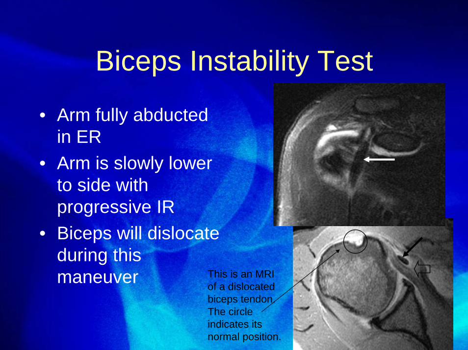
Biceps Instability Test
• Arm fully abducted in ER
• Arm is slowly lower to side with progressive IR
• Biceps will dislocate during this maneuver This is an MRI
of a dislocated biceps tendon. The circle indicates its normal position.

Best Tests for Diagnosing Cuff Tears
1. Weak supraspinatus testing– Arm in 90O forward elevation in scapular
plane2. Weakness in External Rotation
– Arm at the side3. Positive Impingement Sign

Best Tests for Diagnosing Cuff Tears
# Positive Tests Age ProbabilityAll 3 All 0.98
Any 2 >60 0.98Any 2 <60 0.64Any 1 >70 0.76Any 1 40-60 0.45Any 1 <40 0.12
0 All 0.05Murrell, AAOS Instr. Course Lect. March, 2004

Radiographs
• Acromial shape• Position of humeral
head• AC arthritis• Calcific tendinitis• Glenohumeral
arthritis• Destructive lesions

1 & 2: AP in Scapular Plane
• 2 Views: IR, ER• Calcium deposits• Greater tuberosities:
excrescences, cysts

1 & 2: AP in Scapular Plane
• 2 Views: IR, ER• Calcium deposits• Greater tuberosities:
excrescences, cysts
Moderate osteoarthritis
Severe osteoarthritis

3: Axillary View
• Evaluate GH joint & tuberosities
• Glenoid version• Joint space
narrowing • Os acromiale
– This is an anatomic variation best seen on this special view

4: Outlet View
• Evaluate subacromial space
• Acromial shape and thickness

5: 30O Caudal Tilt View
• AP view with a 30O caudal tilt
• Demonstrates anterior acromial projection
spur

Tendon Imaging
• MRI– 90% accurate in
diagnosing complete RC tears
– 70% accurate in diagnosing partialRC tears
– These data may vary. It depends on who is reading the MRI. This spur is pushing on the rotator
cuff causing “impingement”.

Best Studies for Diagnosing Cuff Tears
Full Thickness Tears AccuracyClinical Exam 0.4Ultrasound 0.7MRI 0.7Arthroscopy 0.9
Partial Thickness Tears <0.2 for all studies
Murrell, AAOS Instr. Course Lect. March, 2004

Overall Detection Accuracy
Full thickness Partial Thickness
Ultrasound 98% 68%MRI 100% 63%
JBJS, 86-A, April, 2004

Full thickness Partial ThicknessUltrasound 73% (retraction) 85% (length)
87% (width) 54% (width)
MRI 63% (retraction) 75% (length)85% (width) 75% (width)
JBJS, 86-A, April, 2004

End of part 2
• Part 3 reviews the treatment of rotator cuff problems
ThanksJames T. Mazzara, MD