Embed Size (px)
Citation preview

Zhang and Boutros BMC Bioinformatics 2013, 14:336http://www.biomedcentral.com/1471-2105/14/336
METHODOLOGY ARTICLE Open Access
A novel phenotypic dissimilarity method forimage-based high-throughput screensXian Zhang1,2* and Michael Boutros1*
Abstract
Background: Discovering functional relationships of genes through cell-based phenotyping has become animportant approach in functional genomics. High-throughput imaging offers the ability to quantitatively assesscomplex phenotypes after perturbation by RNA interference (RNAi). Such image-based high-throughput RNAiscreening studies have facilitated the discovery of novel components of gene networks and their interactions. Imagesgenerated by automated microscopy are typically analyzed by extracting quantitative features of individual cells,resulting in large multidimensional data sets. Robust and sensitive methods to interpret these data sets and to derivebiologically relevant information in a high-throughput and unbiased manner remain to be developed.
Results: Here we propose a new analysis method, PhenoDissim, which computes the phenotypic dissimilaritybetween cell populations via Support Vector Machine classification and cross validation. Applying this method to akinome RNAi screening data set, we demonstrate that the proposed method shows a good replicate reproducibility,separation of controls and clustering quality, and we are able to identify siRNA phenotypes and discover potentialfunctional links between genes.
Conclusions: PhenoDissim is a novel analysis method for image-based high-throughput screen, relying on twoparameters which can be automatically optimized without a priori knowledge. PhenoDissim is freely available as an Rpackage.
Keywords: Phenotypic dissimilarity, Image-based high-throughput screening, High-content screening, RNAi,Gene networks
BackgroundTo understand phenotypes and their regulations, it isimportant to identify key genetic components as wellas how they interact. Cell-based screening approacheshave been successfully used to monitor the effect of indi-vidual gene knockdowns or small molecule treatments,identify key regulators contributing to the assessed phe-notype and investigate their interactions [1,2]. Such high-throughput screening experiments can be divided intotwo categories: homogeneous intensity-based methods,such as reporter gene or cell viability assays, and image-based phenotyping approaches. Intensity-based methodsusually report the average of cell populations, leading
*Correspondence: [email protected]; [email protected] Cancer Research Center (DKFZ), Div. Signaling and FunctionalGenomics and Department of Cell and Molecular Biology, Medical FacultyMannheim, Im Neuenheimer Feld 580, D-69120 Heidelberg, Germany2Current address: Novartis Institutes for BioMedical Research, Basel,Switzerland
to scalar (or low dimensional) values per perturbation.Such screens have been designed, for example, to iden-tify novel signaling pathway components by associatingan intensity readout (e.g., luminescence or fluorescence)with a perturbation of a specific reporter gene activity[3-8]. In contrast, image-based methods mark cells withfluorescent dyes, and produce high-dimensional data setsbased on images of phenotypes on a single cell level andconsequently on cell populations [9-15]. Cellular phe-notyping by imaging offers many advantages includingflexible marker choices, subcellular resolution and abil-ity to address cell population heterogeneity (Figure 1A),but also pose new challenges such as lower throughput,more complex infrastructure, and in particular, challengesin data analysis [16].While the analysis of univariate readouts from intensity-
based screens has been greatly facilitated by the develop-ment of specific algorithms and analysis methods [17-20],how to effectively analyze image-based phenotypes is still
© 2013 Zhang and Boutros; licensee BioMed Central Ltd. This is an Open Access article distributed under the terms of theCreative Commons Attribution License (http://creativecommons.org/licenses/by/2.0), which permits unrestricted use,distribution, and reproduction in any medium, provided the original work is properly cited.

Zhang and Boutros BMC Bioinformatics 2013, 14:336 Page 2 of 9http://www.biomedcentral.com/1471-2105/14/336
RNAi treatment
A
Staining & imaging
Segmentation
Quantification
CB
Virtual pooling of objects
SVM classification
Cross validation
Phenotypic dissimilarity 0.88
SizeCell Intensity Texture
1 100 760 0 2 20
2 105 800 0 1 10
3 230 1250 0 2 15
124 900 0 3 30
siRNA 1 siRNA 2
Figure 1Workflow for image-based screening, image quantification and phenotypic dissimilarity measure with SVM classificationaccuracy. A) Cells are seeded into 384-well plates and treated with siRNA by reverse transfection. After incubation for 48 hours, cells are fixed,permeabilized and immunostained for DNA, tubulin and actin and imaged with an automated microscope. B) Cell images are processed withnucleus and cell segmentation using the R packages EBImage and imageHTS. Each cell is represented by a 46 image-based feature vector. Everytreatment generates a data matrix X[m,n], where m is the number of cells and n is the number of features. C) For each pair of RNAi treatments, SVMclassification is performed on the virtually pooled cell population based on cell features. Classification accuracy is estimated by cross validation, anddefined as the phenotypic dissimilarity between treatments.
being explored. In general, the analysis comprises twosteps: image quantification and phenotype-based analysisof gene networks. The image quantification step, whichincludes image pre-processing, cellular object segmenta-tion and feature extraction, is relatively well establishedwith several software tools offering automated, scalableand interactive pipelines [21,22]. This step generates amultidimensional data set containing cell feature infor-mation for typically 100–10000 cells per treatment and10–200 features measured per cell (Figure 1B). The sec-ond step, to derive functional relationships from thesecomplex datasets representing phenotypes, remains chal-lenging. While intuitively this is performed in any kindof genetic screens, e.g. in forward genetic screens inDrosophilamelanogaster orCaenorhabditis elegans, a sys-tematic implementation for quantitative cellular imagedata sets is still missing. One key question is how todefine quantitatives to represent the phenotype of a givenperturbation based on the multidimensional cell featuredata sets, before one can identify and potentially clusterphenotypes by similarity.Previous studies typically first applied a dimension
reduction or data transformation method, such as
principle component analysis [23], Kolmogorov-Smirnovstatistics [9], Support Vector Machine [12,24], or factoranalysis [10], and generate a single feature vector for eachperturbation treatment, i.e. a phenotypic profile. Then thedistance between feature vectors is computed based on adistance measure, such as Euclidean distance. Althoughthese approaches have been successfully applied in var-ious image-based analysis, they often require manuallycurated training data sets and/or multiple optimizationsteps. Thus, for a new image-based screen campaign,selecting and optimizing the appropriate method to per-form hit identification and clustering analysis remainschallenging.Here, we propose a novel method to measure pheno-
typic dissimilarity between cell populations in imagingscreens, based on cell classification and cross validation.We define the phenotypic dissimilarity between a pertur-bation and a control, or between two perturbations, asthe classification accuracy between the two correspond-ing cell populations. First, we virtually pool cells fromboth populations. Then, using Support Vector Machine(SVM) classification, the mixed cell population is classi-fied into two groups based on quantitative cell features.

Zhang and Boutros BMC Bioinformatics 2013, 14:336 Page 3 of 9http://www.biomedcentral.com/1471-2105/14/336
The classification accuracy can be estimated by cross vali-dation with the original cell labels, and defined as the phe-notypic dissimilarity. A higher accuracy indicates betterseparation between the cell populations, thus a larger phe-notypic dissimilarity. Evaluated on a kinome-wide RNAiscreen for cell morphology [12], the proposed phenotypicdissimilarity method (hereafter, PhenoDissim) was ableto identify RNAi perturbations causing distinct morphol-ogy phenotypes, such as siPLK1, siCOPB2 and siAKAP7.We then clustered the phenotypes based on theirpair-wise dissimilarity, and genes that clustered togetherwere functionally related.The PhenoDissim method is relatively straightfor-
ward to apply on different high-throughput screeningexperiments, as it has only two parameters for SVMclassification: cost and gamma, and parameter opti-mization can be automated. The method, as wellas the quality metrics for evaluation, is implementedin a freely available R/Bioconductor package phen-oDist (http://www.bioconductor.org/packages/release/bioc/html/phenoDist.html), a toolbox for data analysis inimage-based high-throughput screening.
ResultsWe used a previously generated image-based RNAiscreening data set as a benchmark for phenotypic dissim-ilarity analysis [12]. The genome-wide kinase screen wasconducted in duplicates using a cervix carcinoma cell line(HeLa). Cells were stained with cytoskeletal and nuclearmarkers (DNA, actin and tubulin) [12]. Plate layout islisted in Additional file 1: Table S4. We reanalyzed theimages with the R/Bioconductor package imageHTS, andmeasured 46 image-based features for every cell includinggeometric features, Haralick texture features and Zernikemoments (see Additional file 1: Table S1 for a list of allfeatures).
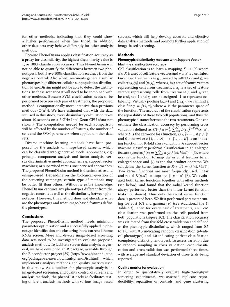
Phenotype identification with PhenoDissimOne major goal in image-based screens is to identify per-turbations that show significantly different phenotypeswhen compared to negative controls. Applying the Phen-oDissimmethod, we computed the phenotypic dissimilar-ity between each perturbation and the negative controls,which indicates how significant the phenotype is (seeMethods for details). The screening data set has one neg-ative control (siRLUC) and two positive controls (siUBCand siCLSPN), with four wells of each control per 384-wellplate. Figure 2A plots the distribution of the phenotypicdissimilarity of these control wells to negative controlwells. Since siRLUC is the negative control, these showa low phenotypic dissimilarity (0.64 ± 0.03). It is largerthan 0.5 due to noise and cell population variation withinsiRLUC wells. The positive controls siUBC and siCLSPNhave much higher phenotypic scores (0.92 ± 0.02 and
0.87 ± 0.02 respectively) and are well separated from thenegative control siRLUC (Z’ factor values 0.56 and 0.40respectively).Phenotypic dissimilarity of all perturbations in the
screen to the siRLUC control are plotted in Figure 2B withreplicate 1 on the X and replicate 2 on the Y axis. Thedata point and error bars represent the mean and stan-dard deviation of three independent calculations, and inmost cases the error bars are negligible. There is a goodcorrelation between biological replicates (Pearson corre-lation coefficient 0.75). Each control (siRLUC, siCLSPNand siUBC) is represented by 12 data points as there arethree plates and four wells for each control per plate. Datapoints of the same control cluster together, and nega-tive and positive controls are well separated, consistentwith the density plot in Figure 2A. There are a total of779 siRNA samples, with diverse phenotypic dissimilarityranging from 0.65 to 0.94.Three example perturbation with distinct phenotypes
are shown in Figure 2C (siPLK1, siCOPB2 and siAKAP7).PLK1 and COPB2 are essential genes which cause viabil-ity defects similar to UBC when depleted by siRNAs. Cellstreated with AKAP7 siRNAs display amorphology pheno-type whereby the cell shape is more round and actin signalis more evenly distributed over the whole cytoplasm. Thisphenotype is consistent with previous observations thatAKAP7, which encodes for A-kinase Anchoring Protein7, localizes to cortical actin cytoskeleton under the cellmembrane and when mutated, spreads to the cytoplasm[25,26].In total, 31 siRNA perturbations (averaging two repli-
cates of each gene) showing high phenotypic dissimilarityto siRLUC control (>0.85) indicate morphological phe-notypes. With the pair-wise phenotypic dissimilarity forthe 31 siRNA samples, we generated a network of pheno-types with nodes representing each phenotype and edgesfor phenotype dissimilarity between nodes as in Figure 3(only phenotypic dissimilarity smaller than 0.82 and con-nected nodes are shown), as well as representative cellimages. From network connectivity and visual inspection,we found three major groups of phenotypes. Genes high-lighted in green are essential genes, and cause viabilitydefect when knocked down. Within this group are genesPLK1 and COPB2, but also other genes such as PKM2and PMVK. Genes highlighted in blue cause cell shapedefect when depleted by siRNA. Cells are often elongatedwith thin stretches, suggesting defect in cell structuremaintenance. Genes highlighted in orange cause strongactin staining and also affect cell shape. Genes in grayshow intermediate phenotypes between the major groups.For example, siRAC1 treated cells show both a slightviability defect and an elongated shape. Further experi-ments to explain the underlying basis of these phenotypesare needed, however, in some cases previous functional

Zhang and Boutros BMC Bioinformatics 2013, 14:336 Page 4 of 9http://www.biomedcentral.com/1471-2105/14/336
Phenotypic dissim. (replicate 1)
Phen
otyp
ic d
issi
m. (
repl
icat
e 2)
PLK1
COPB2
AKAP7
siUBCsiCLSPNsiRLUC
sample siRNAs
siRNA hits
C
B
Phenotypic dissimilarity to siRLUC
siUBCsiCLSPNsiRLUC
06 07 08 09
05
1015
2025
Den
sity
A
DNA Actin Tubulin
siRLUCsiUBC siCLSPN
siPLK1 siAKAP7siCOPB2
05 06 07 08 09 10
0.5
0.6
0.7
0.8
0.9
1.0
Figure 2 Phenotype identification with PhenoDissim. A) Distributions of phenotypic dissimilarity of the controls, with siUBC (red), siCLSPN(purple) and siRLUC (blue). B) The correlation between two replicates. Replicate 1 of all treatments including samples and controls is plotted on theX axis and replicate 2 on the Y axis. siUBC treatments are in red, siCLSPN in purple and siRLUC in blue. All samples are in gray, with the strongestphenotypes in black and labeled with gene names. C) Cell images of the controls (siUBC, siCLSPN, siUBC) and three phenotype hits (siPLK1, siCOPB2,siAKAP7).
characterizations support the observed phenotypes andtheir mechanism. For example, MRC2 was previouslyshown to be responsible for the turn-over of collagen [27]and higher levels of collagen was associated with elon-gated cell shapes [28]. TESK2 was shown to be involvedin actin cytoskeletal organization [29]. It should also benoted that the samemorphology phenotype can be causedby unrelated mechanisms, nevertheless, grouping similarphenotypesmay help identify and understand functionallyrelated genes and their interactions.
Gene clustering analysis with PhenoDissimWe then clustered genes by pair-wise phenotypic dissim-ilarity of the whole screening set to identify genes thatperform potentially related functions. To this end, weaveraged the two replicates and generated a 779×779 phe-notypic dissimilarity matrix, with each row and each col-umn representing an siRNA treatment. Then hierarchicalclustering was performed based on the phenotypic dis-similarity matrix (shown as a dendrogram in Figure 4A).The clustering tree was cut into 20 clusters, and each
cluster analyzed for GO term enrichment (see Methodsfor details). The clusters are shown as colored bars, andthe height of each bar indicates how many enriched GOterms found in the corresponding cluster (Figure 4A).There are a total of 126 enriched GO terms identi-fied, with clusters vary in the number of enriched GOterms.Genes from the cluster with the highest number of
enriched GO terms (marked with an asterisk in Figure 4A)are shown in Figure 4B, where nodes represent genemembers and edges represent the gene-gene interactionidentified in the STRING database [29]. The weight ofeach edge is proportional to interaction confidence. 29 of32 genes were found to be connected in STRING. Par-ticularly, we noticed two functional groups, the mitogen-activated protein kinase (MAPK) signaling pathway andthe protein expressed in non-metastatic cells (NME) fam-ily. The MAPK pathway is involved in multiple cellu-lar functions including proliferation, differentiation andmigration [30]. Eight members of the MAPK pathwayare found in this cluster, MAP3K4, MAP3K5, MAP3K6,

Zhang and Boutros BMC Bioinformatics 2013, 14:336 Page 5 of 9http://www.biomedcentral.com/1471-2105/14/336
siCOPB2 siPMVK
siPIK3R4 siPRPSAP2
siRAC1
siMRC2
siMGC5601 siPINK1
siADRBK1siTESK2
siGKsiMAP3K3
PDGFRB
PMVK
PKM2
PLK1
RAC1
PIK3R4
COPB2 NTRK1
PRPSAP2
MPZL1
MRC2
ACAD10 GTF2H1
PINK1
TESK2
ADRBK1
GKABI1
CDKL5
MAP3K3
AKAP7
siMPZL1
siPDGFRB siGTF2H1
DNA-Actin-Tubulin
Figure 3 A network of identified phenotypes and their phenotypic dissimilarity. 31 siRNAs are identified as phenotype hits and calculated forpair-wise phenotypic dissimilarity. Each siRNA is represented by a node. siRNA pairs with phenotypic dissimilarity smaller than 0.82 are connectedwith an edge, with only connected nodes shown. Cell images for representative phenotypes are shown and labeled with the corresponding siRNAs.
MAP3K7, MAP4K1, MAP4K5, MAPK1, PRKACA. Thesegenes show phenotypic similarity among each other, andthe associated GO terms are enriched such as activa-tion of JUN kinase activity (GO:0007257, p value 0.002,odds ratio 10). The NME gene family was discovered asa metastasis suppressor [31], and was later shown to beinvolved in cell proliferation and differentiation [32,33] aswell. Five NME genes are present in this cluster and theassociated GO terms are enriched such as GTP biosyn-thetic process (GO:0006183, p value 1 × 10⁻⁵, odds ratio33). Grouping genes from the same pathway or familytogether validates the clustering analysis. Additionally, theMAPK pathway and the NME gene family are clusteredtogether, which may suggest a functional link. This issupported by the previous finding that overexpression ofNME represses MAPK phosphorylation, and thus inhibitscell migration and metastasis [34,35]. In summary, ouranalysis has shown that clustering based on the PhenoDis-sim method has the potential to identify gene functionalclusters.
DiscussionThe generation of phenotype-based perturbation net-works based on cellular phenotyping is becoming a pow-erful approach in systems biology, functional genomicsand drug discovery [36]. Several approaches have been
developed to quantify image-based readouts from image-based screening via segmentation and feature extraction[22,37,38]. However, translating multidimensional cellfeature data into phenotypic information remains elu-sive and hinders the further application of image-basedscreening. Previous studies have proposed multiple anal-ysis methods with different dimension reduction andstatistical learning algorithms [9,10,12,24], but thesemethods often rely on human experts to provide biologicalknowledge, such as for feature selection and training dataset annotation. These approaches also require the opti-mization of multiple parameters, which prevents an easyadaptation to other image-based screens with differentsetups.We have developed here a new phenotypic dissimilarity
measure, PhenoDissim, for image-based high-throughputscreening. The proposed method can identify phenotypesby computing the dissimilarity between samples and con-trols, and determine phenotype-based gene networks bycomputing the dissimilarity between samples. With theproposed method, we have identified distinct phenotypes,and functionally related genes by cluster analysis. Thismethod only requires the optimization of SVM classifi-cation parameters cost and gamma, without knowing celllines, fluorescent markers, treatment types or biologicalquestions.

Zhang and Boutros BMC Bioinformatics 2013, 14:336 Page 6 of 9http://www.biomedcentral.com/1471-2105/14/336
010
2030
No. of G
O term
s
A
B
PTPRG
NME4
NME6
NME1/2
NTRK3
PPP2CB
IKBKAP
MAP3K5
MAP4K5
MAPK1MAP3K4
IGF1R
ILKAPMAP4K1
MAP3K6MAP3K7
IHPK1ITPKA
PRKCD
PRKACANME5
ADRBK2
PKIA
IHPK2
IPMK
ICK
STK32C
PIP5K2C
Confidence
0.15 1.00
*
Figure 4 Phenotypic clustering with PhenoDissim. A) The phenotypic clustering tree is plotted as dendrogram. The hierarchical tree is cut into20 clusters, with each cluster analyzed for the number of enriched GO terms (indicated by the color and height of the bars). The cluster with themost enriched GO terms is marked by an asterisk. B) Genes from the marked cluster are shown with gene interactions retrieved from the STRINGinteraction database, where nodes represent genes in the cluster and edges represent interactions identified by the STRING database (edges areweighted based on the evidence score).
Comparing the performance of PhenoDissim with pre-vious methods is challenging due to the lack of goldstandards and different scales of screening data sets. Wehave designed quality metrics to assess replicate repro-ducibility and separation of controls, which provide eval-uation of the whole screen from different perspectives
(see Methods for details). As summarized in Table 1,the PhenoDissim method performs similarly or better onthe benchmark data set compared with previous meth-ods, in terms of replicate reproducibility, separation ofcontrols and gene clustering quality. It should be notedthat we have not extensively performed optimization
Table 1 Evaluation of analysis methods
Method Replicate Z’ factor GO enrichment
correlation siUBC siCLSPN
PCA 0.74 -0.74 -1.31 88
Factor analysis 0.39 -1.87 -0.12 92
KS statistic 0.66 0.07 0.21 51
SVM weight vector 0.07 -84.94 -3.49 101
SVM supervised 0.75 -0.29 -1.34 83
PhenoDissim* 0.75 ± 0.001 0.56 ± 0.01 0.40 ± 0.01 131 ± 18
*mean ± standard deviation for three independent runs.
Zhang and Boutros BMC Bioinformatics 2013, 14:336 Page 7 of 9http://www.biomedcentral.com/1471-2105/14/336
for other methods, indicating that they could showa higher performance when fine tuned. In additionother data sets may behave differently for other analysismethods.Because PhenoDissim applies classification accuracy as
a proxy for dissimilarity, the highest dissimilarity value is1, or 100% classification accuracy. Thus PhenoDissim willnot be able to quantify the differences between two phe-notypes if both have 100% classification accuracy from thenegative control. Also when treatments generate similarphenotypes but different cellular subpopulation distribu-tion, PhenoDissim might not be able to detect the distinc-tions. In these scenarios it will need to be combined withother methods. Because SVM classification needs to beperformed between each pair of treatments, the proposedmethod is computationally more intensive than previousmethods (O(n2)). We have estimated that with the dataset used in this study, every dissimilarity calculation takesabout 10 seconds on a 2 GHz Intel Xeon CPU (data notshown). The computation needed for each comparisonwill be affected by the number of features, the number ofcells and the SVM parameters when applied to other datasets.Diverse machine learning methods have been pro-
posed for the analysis of image-based screens, whichcan be classified into generative model approaches, e.g.principle component analysis and factor analysis, ver-sus discriminative model approaches, e.g. support vectormachines; or supervised versus unsupervised approaches.The proposed PhenoDissim method is discriminative andunsupervised. Depending on the biological question ofthe screening campaign, certain type of methods maybe better fit than others. Without a priori knowledge,PhenoDissim captures any phenotypes different from thenegative controls as well as the dissimilarity between phe-notypes. However, this method does not elucidate whatare the phenotypes and what image-based features definethe phenotypes.
ConclusionsThe proposed PhenoDissim method needs minimumparameter optimization and is successfully applied in phe-notype identification and clustering in the current kinomeRNAi screen. More and diverse image-based screeningdata sets need to be investigated to evaluate proposedanalysis methods. To facilitate screen data analysis in gen-eral, we have developed an R package, available throughthe Bioconductor project [39] (http://www.bioconductor.org/packages/release/bioc/html/phenoDist.html), whichimplements analysis methods and quality metrics usedin this study. As a toolbox for phenotypic analysis inimage-based screening, and quality control of screens andanalysis methods, the phenoDist package facilitates test-ing different analysis methods with various image-based
screens, which will help develop accurate and effectivedata analysis methods, and promote further application ofimage-based screening.
MethodsPhenotypic dissimilarity measure with Support VectorMachine classification accuracyCell classification is to learn a mapping X → Y , wherex ∈ X is a set of cell feature vectors and y ∈ Y is a cell label.Given two treatments (e.g., treated by siRNAs i and j), wecollect (xi,yi) and (xj,yj), where xi is a set of feature vectorsrepresenting cells from treatment i, xj is a set of featurevectors representing cells from treatment j, and yi canbe assigned 1 and yj can be assigned -1 to represent celllabeling. Virtually pooling (xi,yi) and (xj,yj), we can find aclassifier y = f (x,α), where α is the parameter space ofthe function. The accuracy of the classification representsthe separability of these two cell populations, and thus thephenotypic distance between the two treatments. One canestimate the classification accuracy by performing crossvalidation defined as CV (f ,α)= 1
N∑N
i=1 L(yi, f −K(i)(xi,α),where L is the zero-one loss function, L(y, y) = 1 if y �= y,and 0 otherwise; κ {1, . . . ,N} → {1, . . . ,K} is an index-ing function for K-fold cross validation. A support vectormachine classifier performs classification in an enlargedfeature space as f (x) = ∑N
i=1 αiyi〈h(x), h(xi)〉 + β0, whereh(x) is the function to map the original features to anenlarged space and 〈, 〉 is the dot product operator. Wecan define the kernel function as K(x, x′) = 〈h(x), h(x′)〉.Two kernel functions are most frequently used, linearand radial K(x, x′) = exp(−γ ‖ x − x′ ‖2). We evalu-ated both kernel functions together with other methods(see below), and found that the radial kernel functionalways performed better than the linear kernel function(data not shown). Thus only the radial kernel functiondata is presented here. We first performed parameter tun-ing for cost (C) and gamma (γ ) (see Additional file 1:Table S3). Then for every pair of treatments, an SVMclassification was performed on the cells pooled fromboth populations (Figure 1C). The classification accuracywas estimated from five-fold cross validation and definedas the phenotypic dissimilarity, which ranged from 0.5to 1.0, with 0.5 indicating random classification (identi-cal phenotypes) and 1.0 indicating perfect classification(completely distinct phenotypes). To assess variation dueto random sampling in cross validation, each classifi-cation and cross validation was performed three times,with average and standard deviation of three trials beingreported.
Quality metrics for evaluationIn order to quantitatively evaluate high-throughputscreening experiments, we assessed replicate repro-ducibility, separation of controls, and gene clustering

Zhang and Boutros BMC Bioinformatics 2013, 14:336 Page 8 of 9http://www.biomedcentral.com/1471-2105/14/336
quality. We applied PhenoDissim and previous methodsto the same screening data set, and the qualitymeasurements indicated the performance of different dataanalysis methods.Replicate reproducibility: In the data set, there were four
negative control wells (siRLUC) per plate. For each samplewell containing a perturbation, we computed the pheno-typic dissimilarity between the sample well and each ofthe four negative controls on the same plate. There werea total of 779 genes targeted in the human kinome librarywith two replicates for each gene. To measure repro-ducibility, we calculated the Pearson correlation coeffi-cient between replicate samples, of the sample phenotypicdissimilarity to each negative control, between replicatesamples.Separation of controls: There were two types of positive
controls (siUBC and siCLSPN) and one negative control(siRLUC) in the screening data set, with each control rep-resented by four wells per plate. For positive controls,phenotypic dissimilarity to negative control was calcu-lated the same way as the samples. For negative controls,we computed the phenotypic dissimilarity between eachnegative control well and the other three negative controlwells on the same plate, and averaged the three measure-ments. The performance of the phenotypic dissimilaritymethod can be indicated by the separation between neg-ative and positive controls, which can be measured bythe robust Z’ factor score, as Z′ = 1 − 3(MADpos +MADneg/abs(μ 1
2 pos− μ 1
2neg)), whereMAD is the median
absolute deviation, μ is the median and abs is the absolutevalue.Gene clustering quality: After averaging two replicates
of the same gene, we performed hierarchical clusteringof 779 genes, based on their pair-wise phenotypic dis-similarity matrix. For validation, the hierarchical tree wascut into 20 clusters (the number of clusters is deter-mined to maximize enriched GO terms), with genes ineach cluster analyzed for gene annotation enrichment.Since most genes were kinases, we used the biologicalprocess gene ontology annotation [40]. For each clus-ter, genes within the cluster were defined as the genesof interest and all genes in the library defined as thegene universe. Fisher’s exact test was performed andGO terms with p value smaller than 0.01 were iden-tified as enriched [41]. The total number of enrichedGO terms was used to evaluate the quality of theclustering.
Software implementationWe implemented the presented phenotypic dissimilaritymethod and quality control metrics in an R/Bioconductorpackage, named phenoDist (http://bioconductor.org/).The presented analysis was performed with R version 2.13and phenoDist version 1.0.0.
Additional file
Additional file 1: Supplementary methods.
Competing interestsThe authors declare that they have no competing interests.
Authors’ contributionsXZ and MB conceived the idea. XZ carried out the analysis. XZ and MB wrotethe manuscript. Both authors read and approved the final manuscript.
AcknowledgementsWe are grateful to Grainne Kerr, Thomas Sandmann, Greg Pau and WolfgangHuber for critical comments on the manuscript and inspiring discussions,Florian Fuchs for providing the benchmark data set. We acknowledge twoanonymous reviewers for their review and comments on the manuscript. XZwas supported by a DKFZ postdoctoral fellowship. This work was in partsupported by grants DFG SP1131 and DFG SFB873.
Received: 7 February 2013 Accepted: 13 November 2013Published: 21 November 2013
References1. Boutros M, Ahringer J: The art and design of genetic screens: RNA
interference. Nat Rev Genet 2008, 9(7):554–566.2. Feng Y, Mitchison TJ, Bender A, Young DW, Tallarico JA:Multi-parameter
phenotypic profiling: using cellular effects to characterizesmall-molecule compounds. Nat Rev Drug Discov 2009, 8(7):567–578.
3. Huang SM, Mishina YM, Liu S, Cheung A, Stegmeier F, Michaud GA,Charlat O, Wiellette E, Zhang Y, Wiessner S, Hild M, Shi X, Wilson CJ,Mickanin C, Myer V, Fazal A, Tomlinson R, Serluca F, Shao W, Cheng H,Shultz M, Rau C, Schirle M, Schlegl J, Ghidelli S, Fawell S, Lu C, Curtis D,Kirschner MW, Lengauer C, Finan PM, Tallarico JA, Bouwmeester T, PorterJA, Bauer A, Cong F: Tankyrase inhibition stabilizes axin andantagonizes Wnt signalling. Nature 2009, 461(7264):614–620.
4. Boutros M, Kiger AA, Armknecht S, Kerr K, Hild M, Koch B, Haas SA, Paro R,Perrimon N: Genome-wide RNAi analysis of growth and viability inDrosophila cells. Science 2004, 303(5659):832–835.
5. Muller P, Kuttenkeuler D, Gesellchen V, Zeidler MP, Boutros M:Identification of JAK/STAT signalling components by genome-wideRNA interference. Nature 2005, 436(7052):871–875.
6. Bartscherer K, Pelte N, Ingelfinger D, Boutros M: Secretion of Wntligands requires Evi, a conserved transmembrane protein. Cell 2006,125(3):523–533.
7. Whitehurst AW, Bodemann BO, Cardenas J, Ferguson D, Girard L, PeytonM, Minna JD, Michnoff C, Hao W, Roth MG, Xie XJ, White MA: Syntheticlethal screen identification of chemosensitizer loci in cancer cells.Nature 2007, 446(7137):815–819.
8. Berns K, Hijmans EM, Mullenders J, Brummelkamp TR, Velds A, HeimerikxM, Kerkhoven RM, Madiredjo M, Nijkamp W, Weigelt B, Agami R, Ge W,Cavet G, Linsley PS, Beijersbergen RL, Bernards R: A large-scale RNAiscreen in human cells identifies new components of the p53pathway. Nature 2004, 428(6981):431–437.
9. Perlman ZE, Slack MD, Feng Y, Mitchison TJ, Wu LF, Altschuler SJ:Multidimensional drug profiling by automated microscopy. Science2004, 306(5699):1194–1198.
10. Young DW, Bender A, Hoyt J, McWhinnie E, Chirn GW, Tao CY, Tallarico JA,Labow M, Jenkins JL, Mitchison TJ, Feng Y: Integrating high-contentscreening and ligand-target prediction to identify mechanism ofaction. Nat Chem Biol 2008, 4:59–68.
11. Loo LH, Lin HJ, Steininger RJ 3rd, Wang Y, Wu LF, Altschuler SJ: Anapproach for extensibly profiling the molecular states of cellularsubpopulations. Nat Methods 2009, 6(10):759–765.
12. Fuchs F, Pau G, Kranz D, Sklyar O, Budjan C, Steinbrink S, Horn T, Pedal A,Huber W, Boutros M: Clustering phenotype populations bygenome-wide RNAi andmultiparametric imaging.Mol Syst Biol 2010,6:370.
13. Neumann B, Walter T, Heriche JK, Bulkescher J, Erfle H, Conrad C, Rogers P,Poser I, Held M, Liebel U, Cetin C, Sieckmann F, Pau G, Kabbe R, WunscheA, Satagopam V, Schmitz MH, Chapuis C, Gerlich DW, Schneider R, Eils R,

Zhang and Boutros BMC Bioinformatics 2013, 14:336 Page 9 of 9http://www.biomedcentral.com/1471-2105/14/336
Huber W, Peters JM, Hyman AA, Durbin R, Pepperkok R, Ellenberg J:Phenotypic profiling of the human genome by time-lapsemicroscopy reveals cell division genes. Nature 2010,464(7289):721–727.
14. Snijder B, Sacher R, Ramo P, Damm EM, Liberali P, Pelkmans L: Populationcontext determines cell-to-cell variability in endocytosis and virusinfection. Nature 2009, 461(7263):520–523.
15. Laufer C, Fischer B, Billmann M, Huber W, Boutros M:Mapping geneticinteractions in human cancer cells with RNAi andmultiparametricphenotyping. Nat Methods 2013, 10(5):427–431.
16. Carpenter AE: Image-based chemical screening. Nat Chem Biol 2007,3(8):461–465.
17. Zhang JH, Chung TD, Oldenburg KR: A simple statistical parameter foruse in evaluation and validation of high throughput screeningassays. J Biomol Screen 1999, 4(2):67–73.
18. Boutros M, Bras LP, Huber W: Analysis of cell-based RNAi screens.Genome Biol 2006, 7(7):R66.
19. Birmingham A, Selfors LM, Forster T, Wrobel D, Kennedy CJ, Shanks E,Santoyo-Lopez J, Dunican DJ, Long A, Kelleher D, Smith Q, BeijersbergenRL, Ghazal P, Shamu CE: Statistical methods for analysis ofhigh-throughput RNA interference screens. Nat Methods 2009,6(8):569–575.
20. Pelz O, Gilsdorf M, Boutros M:web cellHTS2: a web-application for theanalysis of high-throughput screening data. BMC Bioinformatics 2010,11:185.
21. Carpenter AE, Jones TR, Lamprecht MR, Clarke C, Kang IH, Friman O,Guertin DA, Chang JH, Lindquist RA, Moffat J, Golland P, Sabatini DM:CellProfiler: image analysis software for identifying and quantifyingcell phenotypes. Genome Biol 2006, 7(10):R100.
22. Pau G, Fuchs F, Sklyar O, Boutros M, Huber W: EBImage–an R packagefor image processing with applications to cellular phenotypes.Bioinformatics 2010, 26(7):979–981.
23. Tanaka M, Bateman R, Rauh D, Vaisberg E, Ramachandani S, Zhang C,Hansen KC, Burlingame AL, Trautman JK, Shokat KM, Adams CL: Anunbiased cell morphology-based screen for new, biologically activesmall molecules. PLoS Biol 2005, 3(5):e128.
24. Loo LH, Wu LF, Altschuler SJ: Image-based multivariate profiling ofdrug responses from single cells. Nat Methods 2007, 4(5):445–453.
25. Fraser ID, Tavalin SJ, Lester LB, Langeberg LK, Westphal AM, Dean RA,Marrion NV, Scott JD: A novel lipid-anchored A-kinase AnchoringProtein facilitates cAMP-responsive membrane events. EMBO J 1998,17(8):2261–2272.
26. Li Y, Ndubuka C, Rubin CS: A kinase anchor protein 75 targetsregulatory (RII) subunits of cAMP-dependent protein kinase II to thecortical actin cytoskeleton in non-neuronal cells. J Biol Chem 1686,271(28):2–16869.
27. Behrendt N, Jensen ON, Engelholm LH, Mortz E, Mann M, Dano K: Aurokinase receptor-associated protein with specific collagenbinding properties. J Biol Chem 2000, 275(3):1993–2002.
28. Li F, Li B, Wang QM, Wang JH: Cell shape regulates collagen type Iexpression in human tendon fibroblasts. Cell Motil Cytoskeleton 2008,65(4):332–341.
29. Jensen LJ, Kuhn M, Stark M, Chaffron S, Creevey C, Muller J, Doerks T,Julien P, Roth A, Simonovic M, Bork P, von Mering C: STRING 8–a globalview on proteins and their functional interactions in 630 organisms.Nucleic Acids Res 2009, 37(Database issue):D412–D416.
30. Seger R, Krebs EG: The MAPK signaling cascade. FASEB J 1995,9(9):726–735.
31. Steeg PS, Bevilacqua G, Kopper L, Thorgeirsson UP, Talmadge JE, LiottaLA, Sobel ME: Evidence for a novel gene associated with low tumormetastatic potential. J Natl Cancer Inst 1988, 80(3):200–204.
32. Caligo MA, Cipollini G, Fiore L, Calvo S, Basolo F, Collecchi P, Ciardiello F,Pepe S, Petrini M, Bevilacqua G: NM23 gene expression correlates withcell growth rate and S-phase. Int J Cancer 1995, 60(6):837–842.
33. Venturelli D, Martinez R, Melotti P, Casella I, Peschle C, Cucco C,Spampinato G, Darzynkiewicz Z, Calabretta B: Overexpression ofDR-nm23, a protein encoded by a member of the nm23 gene family,inhibits granulocyte differentiation and induces apoptosis in32Dc13myeloid cells. Proc Natl Acad Sci USA 1995, 92(16):7435–7439.
34. Suzuki E, Ota T, Tsukuda K, Okita A, Matsuoka K, Murakami M, Doihara H,Shimizu N: nm23-H1 reduces in vitro cell migration and the liver
metastatic potential of colon cancer cells by regulating myosin lightchain phosphorylation. Int J Cancer 2004, 108(2):207–211.
35. Hartsough MT, Morrison DK, Salerno M, Palmieri D, Ouatas T, Mair M,Patrick J, Steeg PS: Nm23-H1metastasis suppressor phosphorylationof kinase suppressor of Ras via a histidine protein kinase pathway.J Biol Chem 2002, 277(35):32389–32399.
36. Conrad C, Gerlich DW: Automated microscopy for high-content RNAiscreening. J Cell Biol 2010, 188(4):453–461.
37. Carpenter AE: Extracting rich information from images.Methods MolBiol 2009, 486:193–211.
38. Gilbert DF, Meinhof T, Pepperkok R, Runz H: DetecTiff: a novel imageanalysis routine for high-content screening microscopy. J BiomolScreen 2009, 14(8):944–955.
39. Gentleman RC, Carey VJ, Bates DM, Bolstad B, Dettling M, Dudoit S, Ellis B,Gautier L, Ge Y, Gentry J, Hornik K, Hothorn T, Huber W, Iacus S, Irizarry R,Leisch F, Li C, Maechler M, Rossini AJ, Sawitzki G, Smith C, Smyth G,Tierney L, Yang JY, Zhang J: Bioconductor: open softwaredevelopment for computational biology and bioinformatics.Genome Biol 2004, 5(10):R80.
40. Ashburner M, Ball CA, Blake JA, Botstein D, Butler H, Cherry JM, Davis AP,Dolinski K, Dwight SS, Eppig JT, Harris MA, Hill DP, Issel-Tarver L, KasarskisA, Lewis S, Matese JC, Richardson JE, Ringwald M, Rubin GM, Sherlock G:Gene ontology: tool for the unification of biology. The GeneOntology Consortium. Nat Genet 2000, 25:25–29.
41. Falcon S, Gentleman R: Using GOstats to test gene lists for GO termassociation. Bioinformatics 2007, 23(2):257–258.
doi:10.1186/1471-2105-14-336Cite this article as: Zhang and Boutros: A novel phenotypic dissimilaritymethod for image-based high-throughput screens. BMC Bioinformatics2013 14:336.
Submit your next manuscript to BioMed Centraland take full advantage of:
• Convenient online submission
• Thorough peer review
• No space constraints or color figure charges
• Immediate publication on acceptance
• Inclusion in PubMed, CAS, Scopus and Google Scholar
• Research which is freely available for redistribution
Submit your manuscript at www.biomedcentral.com/submit