Embed Size (px)
Citation preview
HAL Id: insu-00804520https://hal-insu.archives-ouvertes.fr/insu-00804520
Submitted on 27 Mar 2013
HAL is a multi-disciplinary open accessarchive for the deposit and dissemination of sci-entific research documents, whether they are pub-lished or not. The documents may come fromteaching and research institutions in France orabroad, or from public or private research centers.
L’archive ouverte pluridisciplinaire HAL, estdestinée au dépôt et à la diffusion de documentsscientifiques de niveau recherche, publiés ou non,émanant des établissements d’enseignement et derecherche français ou étrangers, des laboratoirespublics ou privés.
A novel growth process of calcium carbonate crystals insilk fibroin hydrogel system
Yufei Ma, Qingling Feng, Xavier Bourrat
To cite this version:Yufei Ma, Qingling Feng, Xavier Bourrat. A novel growth process of calcium carbonate crystals insilk fibroin hydrogel system. Materials Science and Engineering: C, Elsevier, 2013, 33, pp.2413-2420.�10.1016/j.msec.2013.02.006�. �insu-00804520�
1
A Novel Growth Process of Calcium Carbonate Crystals in Silk
Fibroin Hydrogel System
Yufei Ma a, Qingling Feng
b,* and Xavier Bourrat
c
a State key laboratory of new ceramics and fine processing, Department of Materials Science and
Engineering, Tsinghua University, Beijing 100084, China
b Laboratory of Advanced Materials, Department of Materials Science and Engineering, Tsinghua
University, Beijing 100084, China
c CNRS, ISTO, 1A rue de la Ferollerie, F-45071 Orleans cedex2,France
*Corresponding author at: Department of Materials Science and Engineering, Tsinghua University, Haidian
District, Beijing 100084, China.
Fax: 86-10-62771160; Tel: 86-10-62782770.
E-mail: [email protected] (Qingling Feng).
2
ABSTRACT
We report an interesting finding of calcium carbonate (CaCO3) crystal growth in the silk fibroin (SF)
hydrogel with different concentrations by a simple ion diffusion method. The experimental results
indicate that the CaCO3 crystals obtained from silk fibroin gels with low and high concentrations are all
calcites with unusual morphologies. Time-dependent growth study was carried out to investigate the
crystallization process. It is believed that silk fibroin hydrogel plays an important role in the process of
crystallization. The possible formation mechanism of CaCO3 crystals is proposed. This study provides
a better explanation of the influence of silk fibroin concentration and its structure on CaCO3 crystals
growth.
Keywords: Biomimetic; Crystal growth; Polymers; silk fibroin hydrogel; Biomineralization
1. Introduction
Calcium carbonate is an important mineral due to its significance as a biomineral and its various
industrial applications [1-3]. As a kind of considerable biomineral, it receives much attention due to the
wide existence in nacre of mollusk shells, crustacean exoskeletons, and eggshells [3, 4]. Thereinto, one
of the most striking biomaterials, nacre has inspired researchers to investigate the formation
mechanisms for decades [5-7]. It is well-known that the formation of nacre layers is closely related to
the templating effect of organic matrix during CaCO3 crystallization. Nacre organic matrix is
comprised of three major components: acidic proteins, β-chitin and silk-fibroin-like protein [8, 9].
Among the organic matrices involved in nacre, acidic proteins are considered to play a pivotal role
during the formation of aragonite crystals in the nacre sheets. In recent years, most of the studies have
3
concentrated on the role of acidic proteins in nacre [10-12]. Zhang et al. [10] identified a novel acidic
protein from the nacreous layer of Pinctada fucata that could control CaCO3 crystal growth and induce
the needle-like aragonite crystals. Gong et al. [11] determined a nacre protein from pearl oyster and this
protein might initiate crystal nucleation and permit further calcium deposit formation with irregular
morphology. β-chitin acted as a structural scaffold and its effect often required the cooperation of other
organic matrix, such as acidic proteins [13]. Silk-fibroin-like protein, rich in glycine and alanine, is a
kind of insoluble protein with a secondary structure of antiparallel β-sheet. It has been found that silk
fibroin (Bombyx mori) is much more similar to silk-fibroin-like protein in nacre in amino acid sequence
and secondary structure [14] compared to the conventional synthesized acidic polymers [15-19]. Hence,
silk fibroin has been used as an additive in the solution to investigate its effect on CaCO3 crystal
growth in in-vitro study. Wang et al. [20] found CaCO3 formation steps greatly depended on the
presence of silk fibroin in mineralization process. A cryo-transmission electron microscopy (CTEM)
study of the structure of the Atrina shell nacreous organic matrix without dehydration suggested that
[9], at least prior to mineralization, the silk-fibroin-like protein existed as a hydrated gel phase.
Nudelman et al. [21] also identified a hydrated gel-like protein filling the space between two
interlamellar sheets prior to mineral formation. These results showed that mineral formation did not
occur in aqueous solution, but in a hydrated gel-like medium. However, the exact role of this hydrogel
in nacre growth is still so far limited.
In our research, we used a silk fibroin hydrogel system, which is beneficial to mimic the real
mineralization system of nacre. The motivation for this research is to explore how silk fibroin gel
mediates the crystal growth of calcium carbonate. In addition, by studying the crystallization process in
silk fibroin gel, the probable mechanism of CaCO3 crystal growth is proposed. Generally speaking, this
4
study provides clues to understand CaCO3 biomineralization process as it occurs in nacre and suggests
a pathway for the biomimetic fabrication of functional materials.
2. Experimental Section
2.1 Materials
Calcium chloride (AR) and sodium carbonate (AR) were bought from Chemical Agents Co. Ltd.,
Beijing, China. Bombyx mori silkworm silk was purchased from Yi Xian Raw Silk Factory in China.
All chemicals were used without further purification. Deionized water was used throughout the
experimental process.
2.2 Preparation of silk fibroin hydrogel
Degumming and dissolving process of Bombyx mori silk followed the established procedures [22].
About 6% (w/w) of aqueous regenerated silk fibroin was collected at room temperature and stored at
4 °C. Silk fibroin solutions with lower concentrations were prepared by diluting the 6% solution with
water. To obtain higher concentration solutions, the 6% silk fibroin solutions were dialyzed against
10% (w/v) PEG (10,000 g/mol) solution for at least 12 h at room temperature [23]. Finally, silk fibroin
solutions with different concentrations (w/w, 5%, 10%, 15% and 25%) were prepared. Silk fibroin
solutions with these four concentrations containing CaCl2 (20 mM) were prepared, then the hydrogels
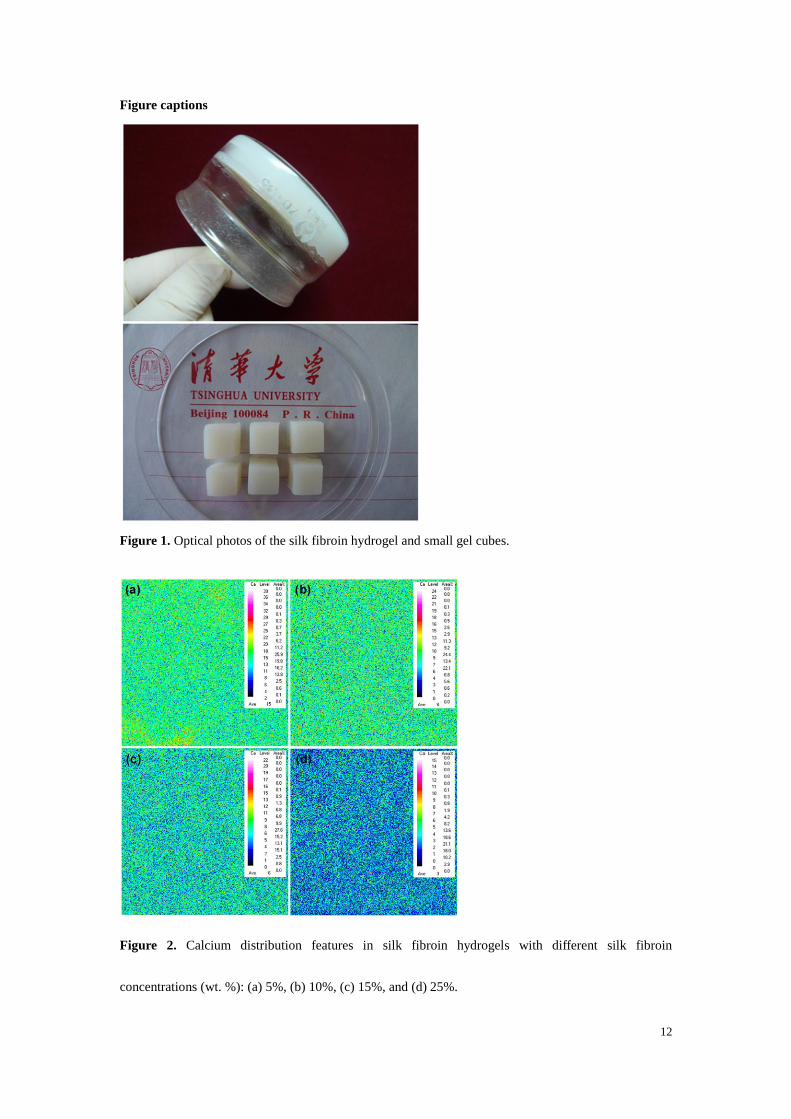
formed at 60 °C after 20 h. Silk fibroin gel was cut into small cubes (volume: ~1 cm3) as shown in
figure 1.
2.3 Crystallization of CaCO3 crystals in silk fibroin hydrogel
In-vitro crystallization experiments in the gels with the different silk fibroin concentrations were
carried out. The gel cubes were immersed in Na2CO3 solution (20 mM, 200 ml) and the mineralization
5
process lasted for 12 h at room temperature. In the control group, CaCO3 crystals grown in the same
condition without silk fibroin gels were collected by silicon substrates. For the time-dependent growth
study, CaCO3 crystallization experiment was carried out within 10% silk fibroin gel and stopped at the
designed time from 5 min to 12 h. When the mineralization process completed, the final products were
lyophilized for characterization.
2.4 Characterization
The cross-sections of the gel cubes with four silk fibroin concentrations were chosen to analyze
calcium distribution qualitatively by using electron probe microanalyzer (JXA-8230, JEOL). Crystal
morphology and crystallization process were imaged by LEO-1530 (LEO Company) scanning electron
microscope (SEM), fitted with a field-emission source operating at an accelerating voltage of 15 kV.
Phase analyses were performed by a Nicolet 6700 (ThermoFisher SCIENTIFIC) Fourier transform
infrared (FTIR) spectrometer using the KBr pellets method and a D8 ADVANCE (BRUKER Company)
X-ray diffractometer with Cu Kα radiation (40 kV, 40 mA). XRD patterns of silk fibroin/CaCO3
hybrids were collected at a 2θ range of 10-80°. Additionally, a RM 2000 laser microscopic Raman
spectrometer (Renishaw Company) with the Raman shifts ranging from 100 to 1200 cm-1
was also used
to confirm the polymorph. An argon ion laser at a wavelength of 514.5 nm was used.
3. Results and Discussion
3.1 Effect of silk fibroin hydrogel with different concentrations on calcium carbonate crystal growth
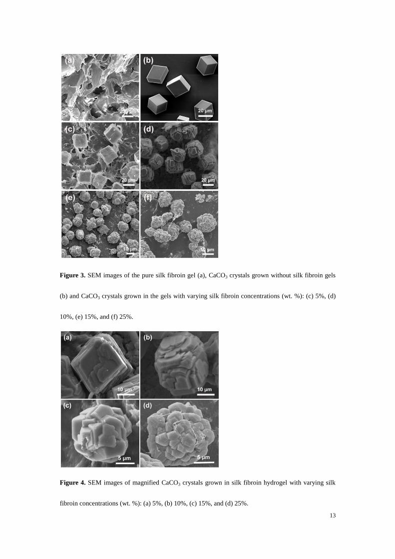
Calcium distribution of the four gels with different concentrations was investigated first and the
typical surface scanning images are shown in figure 2. Analysis area is 100 µm×100 µm. Warm color
represents high level of calcium element content and cool color is reversed. Test results show that the
6
detected calcium is homogeneously distributed in these samples. Additionally, calcium content
obviously decreased as silk fibroin concentration increased. High silk fibroin content leads to the low
solvent content in equal volume, accompanying with the same calcium ion concentration, which results
in the low calcium content.
In-vitro crystallization experiments in silk fibroin gels with varying concentrations and control group
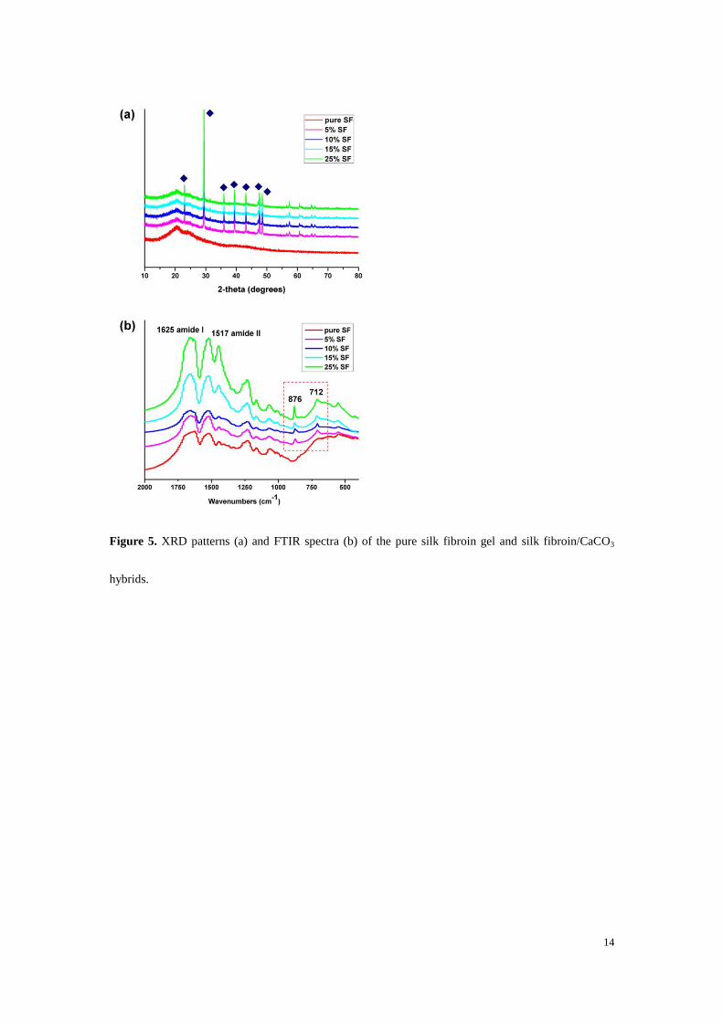
without silk fibroin gels were carried out at room temperature (~25 °C). Morphologies of the pure silk
fibroin gel and resulting CaCO3 crystals were characterized in figure 3a-f. A uniform network with
porous structure is revealed in pure silk fibroin gel (figure 3a). Figure 3b shows CaCO3 crystals
obtained from the control group without silk fibroin gels. The typical calcite crystals with the exposed
(104) crystalline faces are observed. In the experiment groups, the morphology of CaCO3 crystals is
homogeneous in each silk fibroin concentration (figure 3c-f), and it is well controlled in silk fibroin
hydrogel. To further investigate the details of morphology, CaCO3 crystals from the four experiment
groups are magnified, as shown in figure 4a-d. With the increase of silk fibroin concentration, CaCO3
crystals morphology transforms from regular rhombohedral appearance (figure 4a), step-like
morphology (figure 4b-c) to spherical aggregate (figure 4d). The typical (104) facets are clearly
observed on the crystal surface in figure 4a-d, suggesting that these crystals are all calcites. Meanwhile,
the obtained crystal sizes differ in the four kinds of gels. From 5% SF gel to 25% SF gel, the
corresponding crystal size is ~24 µm, ~ 21 µm, ~14 µm and ~16 µm respectively. According to the
difference in calcium content, the larger crystal size in low silk fibroin concentration gel can be
attributed to the higher calcium content. Additionally, it could be a result of fast growth rate of calcite
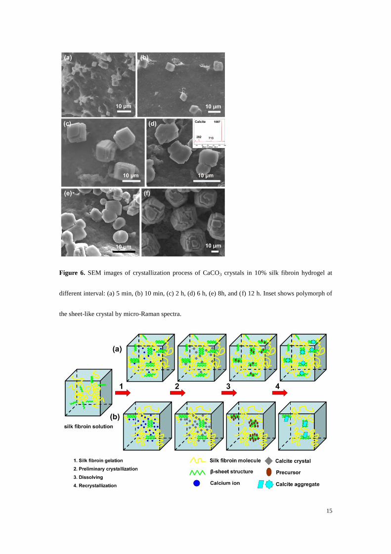
in the case of lower concentration of silk fibroin. Figure 5 shows XRD patterns and FTIR spectra of the
pure silk fibroin gel and silk fibroin/CaCO3 hybrids from the four crystallization experiments. The
7
typical XRD patterns in figure 5a clearly exhibits that no sharp peak is involved in the pure silk fibroin
gel, leaving only a broad peak at about 20°. The four silk fibroin/CaCO3 hybrids have a similar pattern.
The diffraction peaks at 2θ of 29.4°, 35.9° and 39.5° correspond to (104), (110) and (113)
crystallographic planes of calcite, respectively [24]. As can be seen in figure 5b, the peak in the ranges
of 1620-1625 cm-1
(amide I) and around 1516 cm-1
(amide II) are attributable to β-sheet structure of silk
fibroin [25]. The peaks at 876 and 712 cm-1
associated with ν2 (CO3 out-of-plane deformation) mode
and ν4 (OCO bending in-plane deformation) mode vibrations are distinctive features of calcite [26].
Thus, it can be concluded that the prepared CaCO3 products are calcite crystals and silk fibroin gel has
no effect on CaCO3 polymorph. Chen et al. [27] reported that silk fibroin as a soluble additive could
induce rice-grain-shaped aragonite crystals. As we know, secondary structure of silk fibroin can be
changed from random coil to β-sheet during the gel formation. Consequently, we presume that the
difference in silk fibroin conformation is responsible for the polymorph diversity.
3.2 Calcium carbonate crystallization process in silk fibroin hydrogel
To learn the detail of CaCO3 crystallization process in silk fibroin gel system, time-dependent
growth study was carried out. Observations using SEM show in detail the crystallization process of
calcium carbonate (figure 6). At the early stage, the typical rhombohedral crystals with uniform crystal
size of ~ 3 μm are observed (figure 6a). In succession, the initial crystals grow bigger and the
well-facetted calcite crystals are obtained (figure 6b). As reaction time goes on, the single calcite
crystal reveals multilayered morphology but the well-defined (104) facet is still observed. The calcite
crystal size reached to ~9 μm (figure 6c). Figure 6d-e show CaCO3 crystal growth at 6 and 8 h
respectively. Interestingly, the sheet-like crystals with smooth surface appear, suggesting the
dissolution process is involved in this process [20]. Micro-Raman spectra demonstrated these sheet-like
8
crystals are calcite (inset in figure 6d). It has three vibrational bands at 282 cm-1
, 713 cm-1
and 1087
cm-1
, which can be attributed to [libration/rotation, L] lattice vibrations of the calcite crystals, ν4
in-plane C–O bending mode vibration and ν1 symmetric C–O stretching mode vibration respectively
[28]. The size of this dissolved calcite reduced continually as the reaction time increased. Estroff et al.
[29] reported that the dissolution was observed on the gel-grown calcite crystals. As a result of
cross-linked macromolecules, the biological calcite increased solubility as compared to synthetic
calcite. Similarly, calcite with the dissolved morphology was also prepared with polyacrylamide
hydrogel [30]. Figure 6f shows the growth state of CaCO3 in silk fibroin gel for 12 h. Irregular
rhombohedral calcite with steps recrystallized under the control of silk fibroin gel and the final crystal
size was ~21 μm. Hence, calcite crystals growth in silk fibroin gel passed through the dissolution to
conduct mass transport and then recrystallization process. All observations indicated that calcite formed
at initial stage and no phase transition was existed in crystal growth process. Silk fibroin hydrogel
could regulate the morphology but not polymorph.
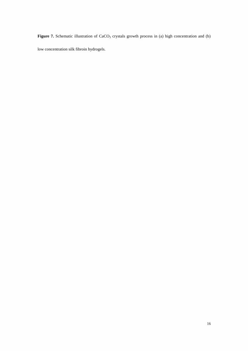
In combination with background information and our experimental results, the proposed growth
mechanism of calcium carbonate crystals in silk fibroin gels with high and low concentration is
illuminated (figure 7). Generally, the formation of CaCO3 crystals in silk fibroin hydrogel contains four
steps: (1) silk fibroin gelation, (2) preliminary crystallization, (3) dissolving, and (4) recrystallization.
Initially, the structure of silk fibroin in the solution with calcium chloride changed from random coil to
β-sheet, with some inter-chain physical cross-links to form the gel network. Calcium distributed
uniformly in both high and low concentration gels, and calcium content in high concentration gel was
lower than that in low concentration gel. When carbonate ions diffused into the gel and contacted with
calcium ions, calcium ions served as nucleation centers and then preliminary crystallization happened
9
to form calcite crystals. Next, the grown calcite dissolved to conduct mass transport, along with the
precursor formation. β-sheet of silk fibroin could cause the partial orientation of the protein chains [27].
The formed precursor tended to absorb on the highly oriented crystalline region of silk fibroin because
of the nucleation-dependent aggregation of β-sheets [31]. Finally, recrystallization took place on the
region of β-sheets due to the high localized concentration of sequestered Ca2+
. Growth of CaCO3
crystals was highly restrained in the gel network. This restrictive effect of macromolecule network in
high concentration gel was much stronger than that in low concentration gel. Thus, the spherical calcite
aggregates formed in high concentration gel (figure 7a), while the single irregular calcites appeared in
low concentration gel (figure 7b).
4. Conclusion
In this paper, we documented a sample system only containing silk fibroin hydrogel for a better
mimicking of calcium carbonate biomineralization process in nacre. SEM and FTIR spectra confirmed
the concentration of silk fibroin gel had influence on calcium carbonate morphology rather than
polymorph. CaCO3 crystals obtained from the low and high concentration gels were all calcites with
different morphologies. On the basis of the study on CaCO3 crystallization process in silk fibroin gel, a
potential growth mechanism of calcium carbonate was proposed. Preliminary crystallization, dissolving
and recrystallization were involved in crystal growth process. The difference in restrictive effect of
macromolecule network led to the obviously different CaCO3 morphologies in varying concentration
gels. Although detailed crystallographic data of CaCO3 were not available from the present study, the
results demonstrated significant aspects of calcium carbonate crystals grown in gel, which could further
provide clues for a deeper understanding of CaCO3 biomineralization process as it occurs in nature and
10
suggest a pathway for the biomimetic fabrication of functional materials.
Acknowledgments
The authors are grateful for the financial support from National Natural Science Foundation of China
(51072090, 51061130554) and Doctor Subject Foundation of the Ministry of Education of China
(20100002110074).
References
[1] M. Suzuki, H. Nagasawa, T. Kogure, Cryst. Growth Des. 6 (2006) 2004-2006.
[2] N. Gehrke, H. Colfen, N. Pinna, M. Antonietti, N. Nassif, Cryst. Growth Des. 5 (2005) 1317-1319.
[3] J.M. Ouyang, Prog. Chem. 17 (2005) 749-756.
[4] X.A. Zhang, J.F. Wang, W.J. Wu, C.L. Liu, J. Inorg. Mater. 21 (2006) 257-266.
[5] S. Weiner, L. Addadi, J. Mater. Chem. 7 (1997) 689-702.
[6] L. Addadi, D. Joester, F. Nudelman, S. Weiner, Chem-Eur. J. 12 (2006) 981-987.
[7] J.H.E. Cartwright, A.G. Checa, J. R. Soc. Interface 4 (2007) 491-504.
[8] G. Falini, S. Albeck, S. Weiner, L. Addadi, Science 271 (1996) 67-69.
[9] Y. Levi-Kalisman, G. Falini, L. Addadi, S. Weiner, J. Struct. Biol. 135 (2001) 8-17.
[10] C. Zhang, S. Li, Z. Ma, L. Xie, R. Zhang, Mar. Biotechnol. 8 (2006) 624-633.
[11] N. Gong, Z. Ma, Q. Li, Q. Li, Z. Yan, L. Xie, R. Zhang, Mar. Biotechnol. 10 (2008) 457-465.
[12] Y.F. Ma, Y.H. Gao, Q.L. Feng, J. Cryst. Growth 312 (2010) 3165-3170.
[13] G. Falini, S. Fermani, A. Ripamonti, J. Inorg. Biochem. 91 (2002) 475-480.
[14] S. Sudo, T. Fujikawa, T. Nagakura, T. Ohkubo, K. Sakaguchi, M. Tanaka, K. Nakashima, T.
Takahashi, Nature 387 (1997) 563-564.
[15] L. He, R. Xue, R. Song, J. Solid State Chem. 182 (2009) 1082-1087.
[16] N. Wada, S. Suda, K. Kanamura, T. Umegaki, J. Colloid Interf. Sci. 279 (2004) 167-174.
[17] S.R. Payne, M. Heppenstall-Butler, M.F. Butler, Cryst. Growth Des. 7 (2007) 1262-1276.
[18] A. Kotachi, T. Miura, H. Imai, Cryst. Growth Des. 6 (2006) 1636-1641.
[19] A. Sugawara, A. Oichi, H. Suzuki, Y. Shigesato, T. Kogure, T. Kato, J. Polym. Sci. Pol. Chem. 44
(2006) 5153-5160.
[20] T. Wang, R. Che, W. Li, R. Mi, Z. Shao, Cryst. Growth Des. 11 (2011) 2164-2171.
[21] F. Nudelman, E. Shimoni, E. Klein, M. Rousseau, X. Bourrat, E. Lopez, L. Addadi, S. Weiner, J.
Struct. Biol. 162 (2008) 290-300.
[22] Y.H. Yang, Z.Z. Shao, X. Chen, P. Zhou, Biomacromolecules 5 (2004) 773-779.
[23] U.J. Kim, J.Y. Park, C.M. Li, H.J. Jin, R. Valluzzi, D.L. Kaplan, Biomacromolecules 5 (2004)
11
786-792.
[24] R. Fried, Y. Mastai, J. Cryst. Growth 338 (2012) 147-151.
[25] A. Matsumoto, J. Chen, A.L. Collette, U.-J. Kim, G.H. Altman, P. Cebe, D.L. Kaplan, J. Phys.
Chem. B 110 (2006) 21630-21638.
[26] F.A. Andersen, L. Brecevic, Acta Chem. Scand. 45 (1991) 1018-1024.
[27] C. Cheng, Z. Shao, F. Vollrath, Adv. Funct. Mater. 18 (2008) 2172-2179.
[28] S.R. Stock, A. Veis, X. Xiao, J.D. Almer, J.R. Dorvee, J. Struct. Biol. 180 (2012) 280-289.
[29] L.A. Estroff, L. Addadi, S. Weiner, A.D. Hamilton, Org. Biomol. Chem. 2 (2004) 137-141.
[30] O. Grassmann, P. Lobmann, Chem-Eur. J. 9 (2003) 1310-1316.
[31] G.Y. Li, P. Zhou, Z.Z. Shao, X. Xie, X. Chen, H.H. Wang, L.J. Chunyu, T.Y. Yu, Eur. J. Biochem.
268 (2001) 6600-6606.
12
Figure captions
Figure 1. Optical photos of the silk fibroin hydrogel and small gel cubes.
Figure 2. Calcium distribution features in silk fibroin hydrogels with different silk fibroin
concentrations (wt. %): (a) 5%, (b) 10%, (c) 15%, and (d) 25%.
13
Figure 3. SEM images of the pure silk fibroin gel (a), CaCO3 crystals grown without silk fibroin gels
(b) and CaCO3 crystals grown in the gels with varying silk fibroin concentrations (wt. %): (c) 5%, (d)
10%, (e) 15%, and (f) 25%.
Figure 4. SEM images of magnified CaCO3 crystals grown in silk fibroin hydrogel with varying silk
fibroin concentrations (wt. %): (a) 5%, (b) 10%, (c) 15%, and (d) 25%.
14
Figure 5. XRD patterns (a) and FTIR spectra (b) of the pure silk fibroin gel and silk fibroin/CaCO3
hybrids.
15
Figure 6. SEM images of crystallization process of CaCO3 crystals in 10% silk fibroin hydrogel at
different interval: (a) 5 min, (b) 10 min, (c) 2 h, (d) 6 h, (e) 8h, and (f) 12 h. Inset shows polymorph of
the sheet-like crystal by micro-Raman spectra.
16
Figure 7. Schematic illustration of CaCO3 crystals growth process in (a) high concentration and (b)
low concentration silk fibroin hydrogels.


![Filler and Coating Pigments for Papermakers€¦ · includes SC, MFC, WFU, and WFC) are calcium carbonate (pre-cipitated calcium carbonate [PCC] and ground calcium carbonate [GCC]),](https://img.dokumen.tips/doc/110x75/5eae9d439cc5d419877523e0/filler-and-coating-pigments-for-papermakers-includes-sc-mfc-wfu-and-wfc-are.jpg)