Embed Size (px)
Citation preview

HAL Id: hal-00174330https://hal.archives-ouvertes.fr/hal-00174330
Submitted on 24 Sep 2007
HAL is a multi-disciplinary open accessarchive for the deposit and dissemination of sci-entific research documents, whether they are pub-lished or not. The documents may come fromteaching and research institutions in France orabroad, or from public or private research centers.
L’archive ouverte pluridisciplinaire HAL, estdestinée au dépôt et à la diffusion de documentsscientifiques de niveau recherche, publiés ou non,émanant des établissements d’enseignement et derecherche français ou étrangers, des laboratoirespublics ou privés.
A Nonlinear Bayesian Filtering Framework for ECGDenoising
Reza Sameni, Mohammad Shamsollahi, Christian Jutten, Gari Clifford
To cite this version:Reza Sameni, Mohammad Shamsollahi, Christian Jutten, Gari Clifford. A Nonlinear Bayesian Fil-tering Framework for ECG Denoising. IEEE Transactions on Biomedical Engineering, Institute ofElectrical and Electronics Engineers, 2007, 54 (12), pp.2172-85. �10.1109/TBME.2007.897817�. �hal-00174330�

This article has been accepted for publication in a future issue of this journal, but has not been fully edited. Content may change prior to final publication.
IEEE TRANSACTIONS ON BIOMEDICAL ENGINEERING, VOL. XX, NO. YY 1
A Nonlinear Bayesian FilteringFramework for ECG Denoising
Reza Sameni*, Student Member, IEEE, Mohammad B. Shamsollahi, Member, IEEE,Christian Jutten, Senior Member, IEEE, and Gari D. Clifford, Senior Member, IEEE
Abstract— In this paper a nonlinear Bayesian filtering frame-work is proposed for the filtering of single channel noisy ECGrecordings. The necessary dynamic models of the ECG are basedon a modified nonlinear dynamic model, previously suggested forthe generation of a highly realistic synthetic ECG. A modifiedversion of this model is used in several Bayesian filters, includingthe Extended Kalman Filter, Extended Kalman Smoother, andUnscented Kalman Filter. An automatic parameter selectionmethod is also introduced, to facilitate the adaptation of themodel parameters to a vast variety of ECGs. This approach isevaluated on several normal ECGs, by artificially adding whiteand colored Gaussian noises to visually inspected clean ECGrecordings, and studying the SNR and morphology of the filteroutputs. The results of the study demonstrate superior resultscompared with conventional ECG denoising approaches such asband-pass filtering, adaptive filtering, and wavelet denoising, overa wide range of ECG SNRs. The method is also successfullyevaluated on real non-stationary muscle artifact. This methodmay therefore serve as an effective framework for the model-based filtering of noisy ECG recordings.
Index Terms— ECG denoising, Kalman filtering, Model-basedfiltering, Nonlinear Bayesian filtering, Adaptive filtering
I. INTRODUCTION
THE extraction of high resolution cardiac signals froma noisy electrocardiogram (ECG) remains an important
problem for the biomedical engineering community. Despiteof the rich literature in this field, there are still many clinicalapplications that lack reliable signal processing tools to extractthe weak ECG components contaminated with backgroundnoise and permit the measurement of subtle features in theECG. The numerous non-cardiac ECG contaminants, such
Manuscript received February 7, 2006; revised June 7, 2006 and December12, 2006. Asterisk indicates corresponding author.
*R. Sameni is with the Biomedical Signal and Image Processing Lab-oratory (BiSIPL), School of Electrical Engineering, Sharif University ofTechnology, Tehran, Iran and also with the Laboratory of Grenoble Im-age Parole Signal Automatique (GIPSA), INPG, Grenoble, France (e-mail:r [email protected]).
M. B. Shamsollahi is with the Biomedical Signal and Image ProcessingLaboratory (BiSIPL), School of Electrical Engineering, Sharif University ofTechnology, Tehran, Iran (e-mail: [email protected]).
C. Jutten is with the Laboratory of Grenoble Image Parole Signal Automa-tique (GIPSA), INPG, Grenoble, France (e-mail: [email protected]).
G. D. Clifford is with the Harvard-MIT Division of Health Sciences &Technology, Massachusetts Institute of Technology, Cambridge, MA 02142,USA (e-mail: [email protected]).
Copyright (c) 2007 IEEE. Personal use of this material is permitted.However, permission to use this material for any other purposes must beobtained from the IEEE by sending an email to [email protected].
as electromyographic (EMG) noise, overlap with the car-diac components in the frequency domain, particularly inthe 0.01Hz to 100Hz range. Band-pass filtering is thereforeinadequate to suppress such contaminants [1], [2].
Ensemble Averaging (EA) is another common approach forthe extraction of small cardiac components from the noisecontaminated ECG. However, as EA requires the averagingof many beats, the subtle but important inter-beat variationsin the cardiac cycle are lost in the averaging procedure [3].As an improvement over EA, classical Adaptive Filter (AF)architectures have also been used for the noise cancellationof ECGs containing baseline wander, power line interference,EMG noise, and motion artifacts [4], [5].
For stationary signals, the Wiener Filter (WF) is the optimallinear filtering technique in the Minimum Mean Square Error(MMSE) sense, applied either in a causal sense in the time-domain, or as the non-causal WF applied in the frequencydomain. However, the WF is not expected to (and does not)give good results for a noisy ECG, due to the non-stationarynature of the cardiac signal. In some related works, filteringapproaches have been proposed based on time-frequency [6],[3], and time-scale [7], [8] WFs. The intuition behind theuse of the time-frequency or wavelet transforms in theseapplications is to apply the WF in two domains, to facilitatethe tracking of ECG non-stationarities.
Wavelet Denoising (WD) is now a common practice fordenoising of signals having multi-resolution characteristicssuch as the cardiac signal in the ECG. Donoho and Johnstone[9] proposed a soft thresholding method for the so-calledshrinkage of the noise components in the wavelet domain.Their approach together with some ad hoc variants of it,have since been used for many applications, including high-resolution ECG denoising [7], [10], [11]. In these cases, themodel of the ECG is essentially based on the frequency contentof the ECG and to some degree, the localization of the ECGpeaks in time.
Statistical techniques such as Principal Component Analysis(PCA) [12], Independent Component Analysis (ICA) [13],[14], and Neural Networks (NNs) [15] have also been used toextract a statistical-based model of the signal and noise, allow-ing the removal of in-band noise by discarding the dimensionscorresponding to noise. Although these are powerful in-bandnoise filtering schemes, the model is fairly arbitrary and,unless the basis functions are trained on a global set of beat-types, can be extremely sensitive to small changes in either
0000–0000/00$00.00 c© 2007 IEEE

This article has been accepted for publication in a future issue of this journal, but has not been fully edited. Content may change prior to final publication.
2 IEEE TRANSACTIONS ON BIOMEDICAL ENGINEERING, VOL. XX, NO. YY
the signal or the noise. Nonlinear projective filtering [16], isanother similar method of forming an ad hoc statistical modelof the ECG and although potentially extremely powerful, isextremely computationally intensive and highly sensitive tooutliers and changes in data dimensionality through changes inbackground noise. These methods can be generally consideredas members of the AF family, in which some model of thecardiac signal is constructed (either explicitly or implicitly),and used as a reference signal to constrain the filter toimprove the signal-noise separation performance. In this paper,we demonstrate that by using a realistic model to describethe quasi-periodic behavior of the ECG, the idea of model-based filtering may be further extended to a general Bayesianfiltering framework for online ECG denoising.
The framework presented in this paper is based on adynamic model, previously developed for the generation ofsynthetic PQRST complexes with their relationship to thebeat-to-beat RR-interval timing [17], [18]. Considering thesimplicity and flexibility of this model it is reasonable toassume that it can describe the dynamics of a broad classof normal and abnormal ECGs. Moreover, as shown in [19],[20], the applications of this model are not limited to ECGsand may be extended to other quasi-periodic signals. In recentworks [21], [22] the authors employed this model to developan Extended Kalman Filter (EKF) for noisy ECG filtering. Inthis paper, this synthetic ECG model is further modified andused in conjunction with several nonlinear Bayesian filteringapproaches such as the EKF, Extended Kalman Smoother(EKS), and Unscented Kalman Filter (UKF). Furthermore,the model parameter selection is automated in order to adaptthe method to different ECG channels. In order to evaluatethe proposed method, different portions of white and coloredGaussian noises have been added to visually inspected cleanECGs recorded from various ECG lead configurations withdiffering morphologies. The Signal-to-Noise Ratios (SNRs)of the filter outputs have then been compared with severalconventional ECG denoising schemes. An example of the fil-tering performance, in presence of real non-stationary muscleartifact (MA) is also presented. The results demonstrate thatthe proposed filters can accurately track the ECG signal in verylow SNR conditions, where the cardiac signal is almost lost inbackground noise. This method is believed to serve as a novelframework for the model-based extraction of high-resolutionECG signals from noisy measurements. In particular, sincethe presented method provides an accurate separation ofnonlinear and non-stationary signal and in-band noise, it ishoped that this method will be suitable for applications suchas the extraction of ECG late potentials from high-resolutionECGs [23], or the noninvasive extraction of fetal ECG fromthe signals recorded from an array of electrodes placed onthe maternal abdomen [24], where due to the low SNR ofthese signals, conventional filtering approaches do not givesatisfactory results.
The paper is organized as follows. In sections II andIII, the required theoretical background and the previouslydeveloped synthetic ECG generator are described. The detailsof the proposed tracking and filtering methods are presentedin section IV. In section V some implementation issues and a
description of standard ECG denoising methods that have beenused as a benchmark for the proposed methods are presented.The results of the different filtering methods are presented anddiscussed in section VI, and the final section is devoted tosome concluding remarks concerning the presented approach.
II. REVIEW OF THE BAYESIAN FILTERING THEORY
A classical problem in estimation theory is the estimationof the hidden states, that are observable through a set ofmeasurements, of a system with an underlying dynamic model. The well-known Kalman filter (KF) is one such method andunder certain general constraints, it can be proved to be theoptimal filter in the MMSE sense [25]. The conventional KFassumes a linear model for the system dynamics and observa-tion equations. In practice however, most systems are nonlinearin nature and in order to extend the idea of conventionalKF to such systems, several variants of the original KF havebeen developed. In this section, the theoretical foundations ofsome of these extensions are briefly reviewed to facilitate thepresentation of the proposed methods.
A. The Extended Kalman Filter
The Extended Kalman Filter (EKF) is an extension of thestandard KF to nonlinear systems. Consider a discrete-timenonlinear system with the unobserved underlying state vectorxk and observation vector yk at time instant k. A dynamicmodel of this system may be represented as follows:
{xk+1 = f(xk, wk, k)yk = g(xk, vk, k) (1)
where f(·) is the state evolution function and g(·) representsthe relationship between the state vector and the observations.The process and measurement noise vectors are wk andvk respectively, with associated covariance matrices Qk =E{wkwT
k } and Rk = E{vkvTk }. The initial estimate of the
state vector is also assumed to be known and is given by:x0 = E{x0}, with P0 = E{(x0 − x0)(x0 − x0)T }.
In order to use the KF formalism for this system, it isnecessary to derive a linear approximation of (1) near adesired reference point (xk, wk, vk) [26], [27]. This leads tothe following linear approximate model:{
xk+1 ≈ f(xk, wk, k) + Ak(xk − xk) + Fk(wk − wk)yk ≈ g(xk, vk, k) + Ck(xk − xk) + Gk(vk − vk) (2)
where
Ak = ∂f(x, wk, k)∂x
∣∣∣x=xk
Fk = ∂f(xk, w, k)∂w
∣∣∣w=wk
Ck = ∂g(x, vk, k)∂x
∣∣∣x=xk
Gk = ∂g(xk, v, k)∂v
∣∣∣v=vk
(3)
Moreover, to simplify the matrix notations, the Fk and Gk ma-trices are usually absorbed into the noise covariance matricesas follows:
FkQkFTk → Qk , GkRkGT
k → Rk

This article has been accepted for publication in a future issue of this journal, but has not been fully edited. Content may change prior to final publication.
SAMENI et al.: A NONLINEAR BAYESIAN FILTERING FRAMEWORK FOR ECG DENOISING 3
With these notations, the EKF algorithm may be summarizedas follows:
x−k+1 = f(x+k , w, k)
∣∣∣w=wk
, rk = yk − g(x−k , v, k)∣∣∣v=vk
Kk = P−k CT
k [CkP−k CT
k + Rk]−1, x+k = x−k + Kkrk
P−k+1 = AkP+
k ATk + Qk, P+
k = P−k − KkCkP−
k
(4)
where by definition rk is the innovation signal, wk = E{wk},vk = E{vk}, x−k = E{xk|yk−1, ..., y1} is the a priori estimateof the state vector in the kth stage using the observations y1 toyk−1, and x+
k = E{xk|yk, ..., y1} is the a posteriori estimateof this state vector after using the kth observation yk. P−
k andP+
k are defined in the same manner to be the a priori anda posteriori estimates of the state vector covariance matricesbefore and after using the kth observation, respectively.
As it can be seen in (4), the key idea of the EKF is tolinearize the nonlinear system model in the vicinity of theprevious estimated point, and to recursively calculate the filtergain Kk, and the state covariance matrices P−
k and P+k from
the linearized equations, while the KF time propagation isperformed via the original nonlinear equation [26].
B. The Extended Kalman Smoother
As with the Kalman Smoother, the Extended KalmanSmoother (EKS) uses the information of future observationsto give better estimates of the current state. Due to thisnon-causal nature, the EKS is expected to have a betterperformance compared with the EKF. The EKS algorithmbasically consists of a forward EKF stage followed by a back-ward recursive smoothing stage. Depending on the smoothingstrategy, smoothing algorithms are usually classified into fixedlag or fixed interval smoothers [28]. In this paper the fixedinterval EKS is used, since the filtering procedure is carriedout offline on the entirety of each ECG signal. For real-time applications of the proposed EKS methods, the fixed lagsmoother is usually more appropriate.
C. The Unscented Kalman Filter
For highly nonlinear systems, the linear estimate of thenonlinear model does not provide a good approximation of themodel, and hence the EKF will not track the desired signalaround sharp turning points (such as for the ECG). In recentyears there has been great interest towards the extensions of theKF to highly nonlinear systems [29]. The Unscented KalmanFilter (UKF) is a filter based on the Unscented Transform(UT), a method for the estimation of the first and secondorder statistics of the outputs of highly nonlinear systems withGaussian inputs [26]. In fact, for the UKF the linearizationof the system model is no longer necessary since the priorestimate of the state covariance matrix, which is required forthe Kalman gain calculations in (4), is directly estimated usingthe UT. The theory of the UKF and its implementation issueshave already been discussed in the literature and the readeris referred to [26] for a detailed mathematical description.Note that the UKF is numerically sensitive and the covariancematrices estimated by the UT may become semi-definite andtherefore much effort has been made to achieve numerically
TABLE I
PARAMETERS OF THE SYNTHETIC ECG MODEL IN (5)
Index(i) P Q R S Tθi(rads.) −π/3 −π/12 0 π/12 π/2ai 1.2 −5.0 30 −7.5 0.75bi 0.25 0.1 0.1 0.1 0.4
stable versions of this algorithm. The UKF algorithm usedin this paper is based on the ReBEL Matlab R© library, previ-ously developed for nonlinear Bayesian filtering [30] and isoptimized to prevent the estimated covariance matrices frombecoming semi-definite.
III. A SYNTHETIC ECG GENERATOR
The dynamic equations used as the state model for theBayesian filter variants in this paper are modifications of thesynthetic ECG generator proposed by McSharry et al. [17].This model has a variable number of free parameters that makeit adaptable to many normal and abnormal ECGs. The dynamicmodel consists of a set of nonlinear dynamic state equationsin the Cartesian coordinates:⎧⎪⎪⎪⎨
⎪⎪⎪⎩
x = ρx − ωyy = ρy + ωx
z = −∑
i∈{P,Q,R,S,T}aiΔθiexp(−Δθ2
i
2b2i
) − (z − z0)(5)
where x, y, and z are the state variables, ρ = 1−√
x2 + y2,Δθi = (θ − θi)mod(2π), θ = atan2(y, x) is the fourquadrant arctangent of the elements of x and y, with −π ≤atan2(y, x) ≤ π, and ω is the angular velocity of the trajectoryas it moves around the limit cycle in the x− y plane. The ai,bi, and θi terms in (5) correspond to the amplitude, width,and center parameters of the Gaussian terms of this equation.Some typical values of these parameters taken from [17] arelisted in Table I. In this model, the baseline wander of theECG is modeled with the parameter z0 that is assumed to bea relatively low amplitude sinusoidal component coupled withthe respiratory frequency. As it is seen in (5), each of the P, Q,R, S, and T waves of the ECG waveform are modeled with aGaussian function and are located at specific angular positionsθi. In fact, the three dimensional trajectory generated by (5),consists of a circular limit cycle in the x − y plane that ispushed up and down as it approaches each of the θi. The zcoordinate of this three dimensional trajectory, when plottedversus time gives the synthetic ECG. By introducing somerandom variations into the parameters of Table I, it is possibleto generate quasi-periodic signals that appear to be realisticECG. Clifford et al. [31] have recently developed methodsfor the estimation of the values of the model parametersfor realistic ECGs based on nonlinear optimization of theparameters of (5) for a given ECG dataset. A rather similarapproach is later used for the initial estimation of the proposedKF model parameters.
IV. METHODS
The KF theory and the previously developed dynamicalECG model were reviewed in preceding sections. With this

This article has been accepted for publication in a future issue of this journal, but has not been fully edited. Content may change prior to final publication.
4 IEEE TRANSACTIONS ON BIOMEDICAL ENGINEERING, VOL. XX, NO. YY
general overview, in this paper it is intended to use thesynthetic dynamical ECG model within a KF framework. Inorder to do so, the dynamic equations of (5) need to bemodified for the problem of interest.
A. Modification of the Dynamic ECG Model
The dynamic equations proposed by McSharry et al. [17]are in Cartesian coordinates. As previously reported in [22],as a first modification, these equations can be transferred intopolar coordinates. Moreover assuming the z state variable in(5) to be in millivolts, bi’s and θi’s in radians, and time inseconds, it is clear that the ai’s are in mV/(rads.× s). So inorder to simplify the dimensions and later relate the modelparameters with real ECG recordings, the ai terms in (5) willbe replaced with:
ai =αiω
b2i
i ∈ {P, Q, R, S, T},
where the αi are the peak amplitudes of the Gaussian functionsused for modeling each of the ECG components, in millivolts.This definition may be verified from (5), by neglecting thebaseline wander term (z − z0) and integrating the z equationwith respect to t. With these changes, the new form of thedynamic equations in polar coordinates is as follows:⎧⎪⎪⎪⎨
⎪⎪⎪⎩
r = r(1 − r)θ = ω
z = −∑
i∈{P,Q,R,S,T}
αiω
b2i
Δθiexp(−Δθ2i
2b2i
) − (z − z0)(6)
where r and θ are respectively the radial and angular statevariables in polar coordinates. These new set of equationshave some benefits compared with the original equationsproposed in [17]. First of all, the polar form is much simplerand its interpretation is straightforward. Accordingly, the firstequation in (6) represents the radial behavior of the generatedtrajectory, and converges to the limit cycle of r = 1 forany initial value of r ≥ 1. However, the second and thirdequations of (6) are independent from r, making the firstdifferential equation redundant. Therefore, this first equationmay be excluded as it does not affect the synthetic ECG (the zstate variable). Another benefit of this representation is that thephase parameter θ, is an explicit state-variable that indicatesthe angular location of the P, Q, R, S and T waves (Table I).This point is further used in the implementation of the filter.For the problem of interest, (6) may be further simplified bydiscarding the baseline wander term (z− z0). In this case, thesimplified dynamic model of (6) in its discrete form, with theassumption of a small sampling period of δ is as follows:⎧⎨
⎩θk+1 = (θk + ωδ)mod(2π)
zk+1 = −∑
i
δαiω
b2i
Δθiexp(−Δθ2i
2b2i
) + zk + η (7)
where Δθi = (θk − θi)mod(2π), η is a random additive noisethat models the inaccuracies of the dynamic model (includingthe baseline wander), and the summation of i is taken overthe number of Gaussian functions used for modeling the shapeof the desired ECG channel. In fact, due to the flexibility of
Gaussian mixtures, it is believed that by using a sufficientnumber of Gaussian functions, they can be fitted to signalsrecorded from different ECG leads. However, in order toillustrate the general filtering framework, in this paper we onlyuse five Gaussians to model the ECG channels containing theP, Q, R, S, and T waves.
Here forth, θk and zk are assumed as the state variables,and ω, αi, θi, bi and η are assumed as i.i.d Gaussian randomvariables considered to be process noises. Following the no-tation of (1), the system state and process noise vectors aredefined as follows:
xk = [θk, zk]T ,
wk = [αP , ...αT , bP , ..., bT , θP , ..., θT , ω, η]T ,(8)
and the process noise covariance matrix is given asQk = E{wkwT
k }.
B. Linearization of the Nonlinear Dynamic ECG Model
In order to set up an EKF model based on the nonlinearsynthetic model of (7), it is necessary to have a linearizedversion of the model. Consequently, the state-equation of (7)requires linearization using (2) and (3). By defining:{
θk+1 = F1(θk, ω, k)zk+1 = F2(θk, zk, ω, αi, θi, bi, η, k), (9)
the following equations represent the linearized model withrespect to the state variables θk and zk:
∂F1
∂zk= 0
∂F1
∂θk=
∂F2
∂zk= 1
∂F2
∂θk= −
∑i∈{P,Q,R,S,T}
δαiω
b2i
[1 − Δθ2i
b2i
]exp(−Δθ2i
2b2i
)(10)
Similarly, the linearization of (9) with respect to the processnoise components yields:
∂F1
∂ω= δ
∂F2
∂η= 1 i ∈ {P, Q, R, S, T}
∂F1
∂αi=
∂F1
∂bi=
∂F1
∂θi=
∂F1
∂η= 0
∂F2
∂αi= −δ
ωΔθi
b2i
exp(−Δθ2i
2b2i
)
∂F2
∂bi= 2δ
αiωΔθi
b3i
[1 − Δθ2i
2b2i
]exp(−Δθ2i
2b2i
)
∂F2
∂θi= δ
αiω
b2i
[1 − Δθ2i
b2i
]exp(−Δθ2i
2b2i
)
∂F2
∂ω= −
∑i
δαiΔθi
b2i
exp(−Δθ2i
2b2i
)
(11)
C. Observation Equations
The noisy ECG recordings are assumed to be observationsfor the KF. The relationship between the states and obser-vations of the KF depends on the location of the electrodesand the origin of the measurement noise. For example, motionartifacts, environmental noise or bioelectrical artifacts such asEMG or electrogastric noise, may be assumed as the mea-surement noises. While the measurement noise can generally

This article has been accepted for publication in a future issue of this journal, but has not been fully edited. Content may change prior to final publication.
SAMENI et al.: A NONLINEAR BAYESIAN FILTERING FRAMEWORK FOR ECG DENOISING 5
contaminate the ECG in a nonlinear form, the results of thispaper are based on the assumption of additive Gaussian noise.
In addition to the noisy ECG observations, the phase θ mayalso be added as a second observation. In fact, by studyingthe values of Table I, it is noticed that the R-peak is alwaysassumed to be located at θ = 0 and the ECG contents lyingbetween two consecutive R-peaks are assumed to have a phasebetween 0 and 2π (or −π and π). So by simply detecting theR-peaks an additional observation is achieved. While the R-wave detection is a rather simple and routine procedure, onemay benefit from more sophisticated and robust approachesfor very low SNR applications [32]. This additional phaseinformation will also help to synchronize the dynamical KFtrajectories with the reference noisy signals, without the needfor manual synchronization. This RR-interval phase warpingtechnique may be assumed as a generalization of the externalreference, previously used for the synchronization of AFs forevent-related signals [33], [5].
Hence the phase observations φk and the noisy ECG mea-surements sk may be related to the state vector as follows:[
φk
sk
]=
[1 00 1
].
[θk
zk
]+
[uk
vk
](12)
where Rk = E{[uk, vk]T [uk, vk]} is the observation noisecovariance matrix.
In the context of estimation theory, the variance of theobservation noise in (12) represents the degree of reliabilityof a single observation. In other words, when a rather precisemeasurement of the states of a system is valid the diagonalentries of Rk are small, and the KF gain is adapted so as torely on that specific measurement. While for the epochs wherethe measurements are too noisy or there are no measurementsavailable, the Rk entries are large and the KF tends to followits internal dynamics rather than tracking the observations [21].Recall that for the phase state variable, θk has a periodic valuethat starts from θ = 0 at the R-peak and ends at θ = 2π withthe next R-peak. Although the only valid phase observation isobtained from the R-peak locations, it is possible to linearlyassign a phase value between 0 and 2π to the intermediatesamples, as illustrated in Fig. 1. The later presented results areall based on this linear phase assignment. However, to indicatethe increased uncertainty in the phases assigned to the inter-mediate samples, the first diagonal entry of Rk correspondingto the time varying variance of the measurement phase noise,may be increased. Another alternative and rather sophisticated,approach for the estimation of the intermediate phase values
440 480 520 560 600 640
−3
0
3
6
Sample Index
Am
plitu
de (
mV
)
Phase (radians)
PhaseECG
R−peak R−peak
0
2π
π
Fig. 1. An illustration of the phase assignment approach
is to directly detect the location of the P, Q, S, and T wavesfrom the original signal. However, the previous approach ispreferred since R-peak detection is far more reliable in highnoise scenarios.
D. Estimation of the Model Parameters
Prior to the implementation of the filter, it is necessaryto select the values of the process and measurement noisecovariance matrices. Generally, by using m Gaussian kernelsin (7), the process noise vector defined in (8) has 3m + 2entries (here 17), leading to a (3m + 2) × (3m + 2) processnoise covariance matrix of Qk. But if the noise sources areassumed to be uncorrelated with each other, a reasonableapproximation adopted here, then the matrix is simplified tobe diagonal. The measurement noise covariance matrix Rk issimilarly considered to be diagonal.
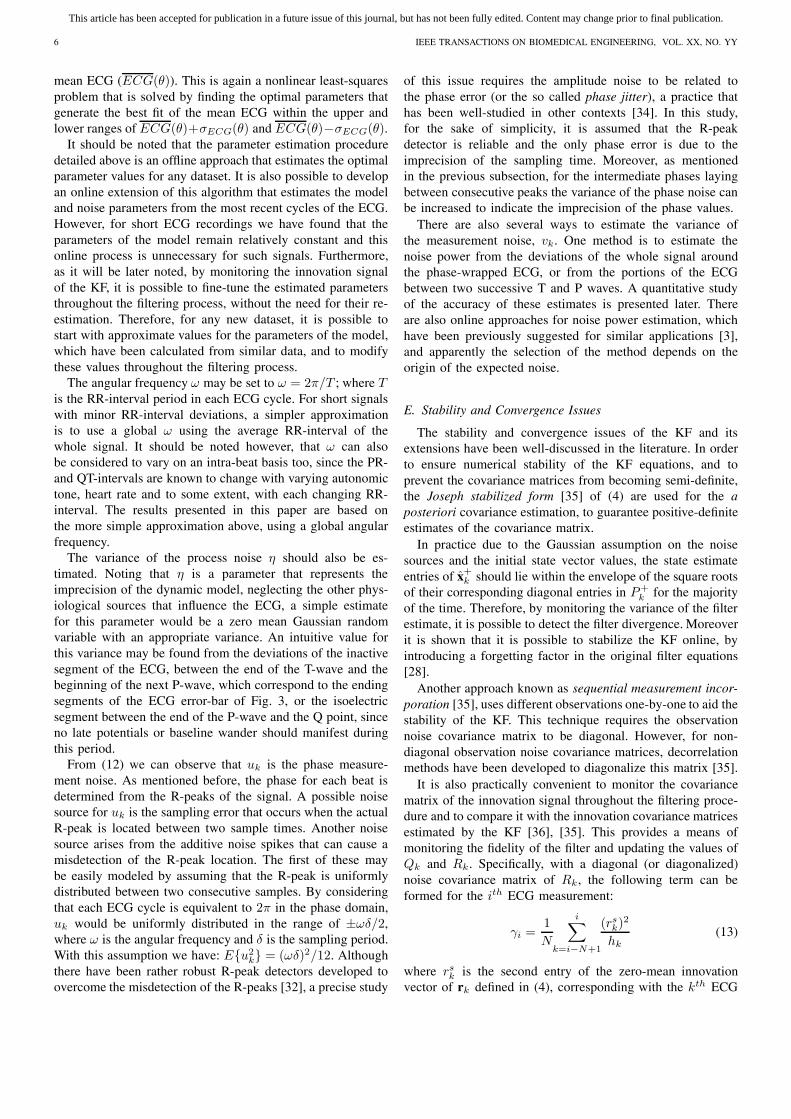
In order to automate the parameter selection procedure forany given ECG, the parameters should be estimated from thesignal itself. For this, as described in the previous subsection,any noisy ECG may be transformed to a three dimensionalrepresentation by plotting the noisy ECG versus the periodicphases that are assigned to each sample in polar coordinateson the unit circle (r = 1). A typical phase-wrapped ECG withadditive noise may be seen in Fig. 2. It is now possible toestimate the dynamic model parameters for the given ECG.For this, the mean and variance of the phase-wrapped ECGis calculated for all phases between 0 and 2π. This gives theaverage of the ECG waveform. A typical signal produced bythis approach is depicted in Fig. 3. The error bar in this figurecorresponds with the standard deviation (SD) of different ECGcycles around the mean ECG. Next, the problem is to find theoptimal parameters of (7) that can best fit the mean ECG.In this stage, many optimization methods may be used. Forexample as suggested in [31], by using a nonlinear least-squares approach, the best estimate of these parameters in theMMSE sense can be found. A practical means of solving thisnonlinear least-squares problem is the lsqnonlin function ofMatlab R© that was used to estimate the initial parameters forthe results presented later in this paper.
The next step is to find an estimate for the covariance valuesof Qk. This may be done by using the error values as depictedin Fig. 3. In fact, in this step we are attempting to calculatethe magnitude of the deviation of the parameters of the fiveGaussian functions in (5) around the estimated mean, thatbest model the acceptable deviations of the ECG around the
−1−0.5
00.5
1
−1 −0.5
0 0.5
1 −2
0
2
4
X (mV)
Y (mV)
Z (
mV
)
Fig. 2. Several cycles of the ECGphase-wrapped in the state space
0
−1
0
1
2
3
Phase (radians)
Am
plitu
de (
mV
)
SDMean
π−π
Fig. 3. An average and standarddeviation-bar of 30 ECG cycles of anoisy ECG
This article has been accepted for publication in a future issue of this journal, but has not been fully edited. Content may change prior to final publication.
6 IEEE TRANSACTIONS ON BIOMEDICAL ENGINEERING, VOL. XX, NO. YY
mean ECG (ECG(θ)). This is again a nonlinear least-squaresproblem that is solved by finding the optimal parameters thatgenerate the best fit of the mean ECG within the upper andlower ranges of ECG(θ)+σECG(θ) and ECG(θ)−σECG(θ).
It should be noted that the parameter estimation proceduredetailed above is an offline approach that estimates the optimalparameter values for any dataset. It is also possible to developan online extension of this algorithm that estimates the modeland noise parameters from the most recent cycles of the ECG.However, for short ECG recordings we have found that theparameters of the model remain relatively constant and thisonline process is unnecessary for such signals. Furthermore,as it will be later noted, by monitoring the innovation signalof the KF, it is possible to fine-tune the estimated parametersthroughout the filtering process, without the need for their re-estimation. Therefore, for any new dataset, it is possible tostart with approximate values for the parameters of the model,which have been calculated from similar data, and to modifythese values throughout the filtering process.
The angular frequency ω may be set to ω = 2π/T ; where Tis the RR-interval period in each ECG cycle. For short signalswith minor RR-interval deviations, a simpler approximationis to use a global ω using the average RR-interval of thewhole signal. It should be noted however, that ω can alsobe considered to vary on an intra-beat basis too, since the PR-and QT-intervals are known to change with varying autonomictone, heart rate and to some extent, with each changing RR-interval. The results presented in this paper are based onthe more simple approximation above, using a global angularfrequency.
The variance of the process noise η should also be es-timated. Noting that η is a parameter that represents theimprecision of the dynamic model, neglecting the other phys-iological sources that influence the ECG, a simple estimatefor this parameter would be a zero mean Gaussian randomvariable with an appropriate variance. An intuitive value forthis variance may be found from the deviations of the inactivesegment of the ECG, between the end of the T-wave and thebeginning of the next P-wave, which correspond to the endingsegments of the ECG error-bar of Fig. 3, or the isoelectricsegment between the end of the P-wave and the Q point, sinceno late potentials or baseline wander should manifest duringthis period.
From (12) we can observe that uk is the phase measure-ment noise. As mentioned before, the phase for each beat isdetermined from the R-peaks of the signal. A possible noisesource for uk is the sampling error that occurs when the actualR-peak is located between two sample times. Another noisesource arises from the additive noise spikes that can cause amisdetection of the R-peak location. The first of these maybe easily modeled by assuming that the R-peak is uniformlydistributed between two consecutive samples. By consideringthat each ECG cycle is equivalent to 2π in the phase domain,uk would be uniformly distributed in the range of ±ωδ/2,where ω is the angular frequency and δ is the sampling period.With this assumption we have: E{u2
k} = (ωδ)2/12. Althoughthere have been rather robust R-peak detectors developed toovercome the misdetection of the R-peaks [32], a precise study
of this issue requires the amplitude noise to be related tothe phase error (or the so called phase jitter), a practice thathas been well-studied in other contexts [34]. In this study,for the sake of simplicity, it is assumed that the R-peakdetector is reliable and the only phase error is due to theimprecision of the sampling time. Moreover, as mentionedin the previous subsection, for the intermediate phases layingbetween consecutive peaks the variance of the phase noise canbe increased to indicate the imprecision of the phase values.
There are also several ways to estimate the variance ofthe measurement noise, vk. One method is to estimate thenoise power from the deviations of the whole signal aroundthe phase-wrapped ECG, or from the portions of the ECGbetween two successive T and P waves. A quantitative studyof the accuracy of these estimates is presented later. Thereare also online approaches for noise power estimation, whichhave been previously suggested for similar applications [3],and apparently the selection of the method depends on theorigin of the expected noise.
E. Stability and Convergence Issues
The stability and convergence issues of the KF and itsextensions have been well-discussed in the literature. In orderto ensure numerical stability of the KF equations, and toprevent the covariance matrices from becoming semi-definite,the Joseph stabilized form [35] of (4) are used for the aposteriori covariance estimation, to guarantee positive-definiteestimates of the covariance matrix.
In practice due to the Gaussian assumption on the noisesources and the initial state vector values, the state estimateentries of x+
k should lie within the envelope of the square rootsof their corresponding diagonal entries in P+
k for the majorityof the time. Therefore, by monitoring the variance of the filterestimate, it is possible to detect the filter divergence. Moreoverit is shown that it is possible to stabilize the KF online, byintroducing a forgetting factor in the original filter equations[28].
Another approach known as sequential measurement incor-poration [35], uses different observations one-by-one to aid thestability of the KF. This technique requires the observationnoise covariance matrix to be diagonal. However, for non-diagonal observation noise covariance matrices, decorrelationmethods have been developed to diagonalize this matrix [35].
It is also practically convenient to monitor the covariancematrix of the innovation signal throughout the filtering proce-dure and to compare it with the innovation covariance matricesestimated by the KF [36], [35]. This provides a means ofmonitoring the fidelity of the filter and updating the values ofQk and Rk. Specifically, with a diagonal (or diagonalized)noise covariance matrix of Rk, the following term can beformed for the ith ECG measurement:
γi =1N
i∑k=i−N+1
(rsk)2
hk(13)
where rsk is the second entry of the zero-mean innovation
vector of rk defined in (4), corresponding with the kth ECG

This article has been accepted for publication in a future issue of this journal, but has not been fully edited. Content may change prior to final publication.
SAMENI et al.: A NONLINEAR BAYESIAN FILTERING FRAMEWORK FOR ECG DENOISING 7
measurement, N is the length of the averaging window, andhk is the KF estimated variance of rs
k given by:
hk = E{(rsk)2} = cT
k P−k ck + σ2
vk(14)
where ck is the second row of the Ck matrix defined in (3),and σ2
vk= E{v2
k} is the second diagonal entry of Rk. Anidentical term can be defined for the phase observation φk.
In fact, γi is an average of the variances of the N recentECG innovations, normalized by their KF estimated varianceshk. Therefore, as long as the KF is performing correctly,γi ≈ 1. Values of γi much greater than unity indicate that theinnovation signal variance is being underestimated by the KF,while values close to zero indicate that the innovation signalvariance is being overestimated. Therefore, by monitoring γi
it is possible to adaptively modify the KF noise parameters(such as Qk and Rk), to ensure the filter stability and toachieve a better filtering performance. For example, by usingthe M most recent samples of the innovation signal, σ2
vkcan
be adaptively modified as follows:
σ2vk
= λσ2vk−1
+ (1 − λ)1M
k−1∑j=k−M
(rsj )
2 (15)
where 0 < λ < 1 is the adaptation coefficient 1. In [37], asimilar means of online modification of the Rk and Qk entrieshave been presented.
For the UKF, the algorithm presented in [26] has threeparameters, α, β, and κ. These control the stability of the filterand enable the algorithm to be fine-tuned for systems withdifferent degrees of nonlinearity and non-Gaussian inputs. Theparameter α is an indication of the spread of the state variablesaround their mean and is selected to be a small positive valuein the range of 10−4 ≤ α ≤ 1. The parameter β is used toincorporate prior knowledge about the distribution of the statevector, with β = 2 being optimal for Gaussian distributions[26]. The parameter κ is a secondary ad hoc scaling parameterthat is selected in accordance with the size of the state vectorand the higher order statistics of the noise distributions [38],with κ = 0 being the optimal selection for a state vector of sizetwo. A mathematical study of the effect of these parameterson the UKF accuracy compared with the EKF, can be foundin [38], [39].
F. Practical Filtering Schema
Before presenting the experiments and results, the scopeof the proposed filtering scheme needs to be further clari-fied. Following the discussions of this section, by using theBayesian framework we are attempting to utilize a prioriinformation about the underlying dynamics of ECG signals toextract the ECG components from background noise. Hence,compared with conventional filtering schemes that performrather ‘blindly’, Bayesian filters are naturally expected to givesuperior results as long as we provide them with valid a
1For M = 1 (single-step update), (15) reduces to the autoregressive modelsuggested in [37], and for M > 1 (15) represents a moving average filterwith the λ parameter changing the slope of the filter’s response. For ECGsignals having sharp changes, the moving average model was found to bemore robust to the peak changes.
priori information concerning the signal and noise dynamics.This point becomes important when considering that abnormalECGs can have high inter-beat variations in their wave timingsor morphology, meaning that the underlying dynamics of thesignals are not valid in pathological beats.
In the presented approach, due to the phase wrapping ofthe RR-interval to 2π, normal inter-beat variations of the RR-interval (between 10% to 20%), or consistent RR-intervalabnormalities such as Bradycardia or Tachycardia do notconsiderably affect the filter performance. However, for abnor-malities that only appear in some of the ECG cycles, the phaseerror of the model can lead to large errors in the Gaussianfunction locations. A similar case can occur when the R-peakis misdetected. In particular, for morphological abnormalitiesthat appear in some of the ECG cycles, such as the PrematureVentricular Contraction (PVC) [40], the filtering performanceis not expected to be satisfactory for low input SNRs, sinceneither the model nor the measurements are reliable for thefilter. For such occasional morphologic changes, even temporaladaptation of the filter parameters is not helpful, as the filterdoes not have sufficient time to adapt itself. However thebenefit of the Gaussian mixture representation is that the effectof each Gaussian term vanishes very quickly (in less than theECG period), meaning that the errors are not propagated to thefollowing ECG cycles2. Moreover by monitoring the state esti-mates’ covariance matrices and the variations of the innovationsignals, it is possible to detect such unexpected abnormalities.Of course, it should be considered that the accurate denoisingof abnormal ECGs with high morphologic changes remains anopen problem even for conventional filtering methods.
Finally we note that the later presented results have beenimplemented offline. However the recursive KF equations areoriginally designed for online applications and even for theEKS, considering the quasi-periodic nature of the ECG, fixedlag smoothers with only one or two cycles of ECG lag can beused.
V. EXPERIMENTS
A. The Dataset
The MIT-BIH Normal Sinus Rhythm Database [41], [42],was used to study the performance of the proposed methods.This database was recorded at a sampling rate of 128Hz from18 subjects with no significant arrhythmias. From this database190 low-noise segments of 30 seconds without considerableartifacts were visually selected for the implementation of theproposed filters. These segments were taken from differentsubjects, recorded from the standard VI , VII , and VIII ECGleads. The heart rate of these ECG segments varied from55 BPM to 90 BMP (74.1±12.0 BMP on average), withRR-interval deviations between 5% to 25% (16.7±6.2% onaverage).
Moreover, to show the filtering performance in the presenceof non-stationary noise, real muscle artifact (MA) was takenfrom the MIT-BIH Noise Stress Test Database [43], [44]. The
2A formal justification of this statement requires mathematical proof ofstability and convergence of the EKF, EKS, and UKF for the proposed model,and is beyond the scope of the current paper.

This article has been accepted for publication in a future issue of this journal, but has not been fully edited. Content may change prior to final publication.
8 IEEE TRANSACTIONS ON BIOMEDICAL ENGINEERING, VOL. XX, NO. YY
MA originally had a sampling rate of 360Hz and thereforethey were anti-alias resampled to 128Hz in order to matchthe sampling rate of the ECG. Note that the resampling didnot considerably change the shape of the MA noise, sincethe frequency content of the MA noise was concentrated infrequencies below 64Hz.
B. Noise Generation
Mathematically, white noise is defined to have a flat spectraldensity function over all frequencies. However, real noisesources have non-flat spectral densities that decrease in powerat higher frequencies, making the spectrum colored and thenoise samples correlated in time. There are different ways ofgenerating colored noise [45], and realistic ECG artifacts [46].For the current study, we will model the noise color by a singleparameter representing the slope of a spectral density functionthat decreases monotonically with frequency:
S(f) ∝ 1fβ
, (16)
where f is the frequency and β is a measure of noise color.White noise (β = 0), pink noise (β = 1) or flicker noise,and brown noise (β = 2) or the random walk process, arethree of the most commonly referenced noises. The realizationof colored noises with spectral densities described by (16),generally require nonlinear frequency domain filtering of whitenoise 3. For random processes, the expected value of thesquared magnitudes of their frequency transforms, or namelythe periodogram, is known to be an estimate of the spectraldensity function of the original samples [47]. Therefore, inorder to generate colored noise following (16), samples ofwhite noise can be generated and transferred into the frequencydomain using the Discrete Fourier Transform (DFT). Byaltering the frequency components of the DFT according to(16), and transferring the reshaped DFT back to the time-domain, typical samples of colored noise are realized. Notethat this approach of frequency domain filtering causes tran-sient behavior in the generated noise time series that shouldbe discarded from the samples.
C. Implementation
Having derived the state equations (7), the observationequations (12), the linearized state equations of the ECGdynamic model (10), (11), and the model parameters, theimplementation of the EKF, EKS and the UKF are nowpossible. The procedure for calculating the parameters of themodel and the noise covariance matrix entries were explainedin section IV. Using the explained methods, the mean ECGwaveform was extracted for each ECG segment and theparameters of the five Gaussian kernels were calculated usingthe nonlinear least-squares method for each ECG segment.Due to the variety of the ECG leads and the wide range ofstudied SNR, the covariance matrices were calculated usinga simple approach using the peak values of the Gaussianfunctions. The parameter selection approach is summarized
3Except for special cases such as β = 2 that can also be achieved throughlinear time-domain filtering of white noise.
TABLE II
PARAMETERS OF THE PROCESS AND OBSERVATION NOISES
θi(rads.) Gaussian kernel center ±0.05παi(mV ) Gaussian kernel peak ±10% of the peak amplitudebi(rads) Gaussian kernel width ±0.05πη(mV ) 1% of maximum ECG peakω(rads./sec) Mean beat-to-beat angular frequency (ω) ± SDuk(rads.) 0.00 ± (ωδ)/
√12
vk(rads.) Ranges over different SNRs
TABLE III
WD PARAMETER COMBINATIONS TESTED OVER THE DATABASE
Parameter ValuesMother wavelet Daubechies 1...8, Coiflets 1...5, Symlets 1...8Shrinkage rule SURE, Heuristic SURE, Universal, MinimaxThresholding strategy Hard, SoftRescaling approach No scaling, Single level, Multiple levelDecomposition level 1...10
in Table II. These parameters can be further customized forspecific ECG leads and special ranges of the input SNR.
In IV-E, the control parameters of the UKF algorithm wereintroduced. Throughout the simulations, these parameters wereset to β = 2 and κ = 0, and are the optimal selections fora system having two state variables with a Gaussian inputnoise. The parameter α = 1 was also empirically derived asa compromise between performance and stability in differentSNRs for the studied database.
D. Benchmark Methods
In order to have a comparison between the performanceof the proposed methods and conventional ECG denoisingschemes, wavelet denoising (WD), adaptive filtering (AF),and conventional finite impulse response (FIR) filtering werealso tested on the database. Implementations of these standardmethods are now described.
Conventional WD schemes are characterized by differentparameters that allow the algorithms to be customized fordifferent mixtures of signal and noise sources. The type of themother wavelet, shrinkage rule, hard- versus soft-thresholding,noise level rescaling approach, and number of decompositionlevels are among the different parameters of common WDalgorithms [48]. There are of course, some general rulesconcerning the selection of these parameters. For example,the mother wavelet is usually selected from families thatsomehow resemble the shape of the desired signal, or therescaling approach and shrinkage rules are selected accordingto the nature (white versus colored) and variance of the noise.However, the literature on the applications of WD for ECG israther broad and diverse, making it difficult to judge what maybe the best combination of these parameters. Consequently,a rather exhaustive search was carried out on the differentcombinations of the above mentioned parameters to find thebest WD scheme for the database being studied. The differentparameters that were tested on this database are listed inTable III. Among the different tested combinations, the Stein’sUnbiased Risk Estimate (SURE) shrinkage rule, together witha single level rescaling and a soft thresholding strategy alwaysgave superior results. Among the tested mother wavelets,

This article has been accepted for publication in a future issue of this journal, but has not been fully edited. Content may change prior to final publication.
SAMENI et al.: A NONLINEAR BAYESIAN FILTERING FRAMEWORK FOR ECG DENOISING 9
Coiflets2, Coiflets3, Symlets4, and Symlets5 gave superiorresults. The best decomposition level ranged from 5 to 8, andshowed little significant difference within this range. Theseresults were achieved by the comparison of the SNR curves forinput SNR ranging from 35dB to -5dB and averaged over 100Monte Carlo runs with different random noise input vectors.The following reported results are based on the Coiflets3mother wavelet with 6 levels of decomposition.
The adaptive filtering approach suggested in [5], was thenext filtering approach implemented on the dataset. Theoriginal results presented in [5] have been reported on asynthetic ECG, formed by taking a single QRS complexfrom an ECG and concatenating the same waveform severaltimes to generate a deterministic ECG with no timing ormorphology variation from beat to beat. An impulse signal,time-synchronized with the R-wave, was also used as thereference channel to enable single channel filtering of theECG. Although their reported results are rather impressingfor this simulated ECG, the method is not expected to giveidentical results on real ECGs. This is mainly due to the non-stationary behavior of the ECG that causes the ECG shapeand RR-duration to change from beat-to-beat. However thisAF scheme was also implemented on the dataset as anotherbenchmark. In the original work [5], the number of the AFweights (L) is selected to be equal to the number of samplesof the deterministic QRS complexes. For operating on realECGs with variable RR-intervals we set L to the maximumsample period between the RR-waves of the input ECG. Aconvergence rate of μ = 0.1, led to rapid adaptation and stablefilter outputs for all the SNR range of this study (-5dB – 30dB).
The last filtering approach applied to the dataset was atypical FIR filter, consisting of two cascaded highpass andlowpass filters, with an overall pass-band of 0.4Hz–40Hz, apass-band ripple of 1dB, and a stop-band attenuation of about60dB. The main frequency components of typical ECGs liewithin this frequency range [49], and the selection of a wideror narrower bandwidth would be a compromise between theattenuation of in-band ECG and noise components.
VI. RESULTS AND DISCUSSIONS
In order to investigate the performance of the differentmethods, artificial white and colored Gaussian noise withdifferent variances were generated and added to the ECGsegments, and the noisy signals were presented to the proposedfilters. To ensure the consistency of the results, the wholeprocedure was repeated over the 190 ECG segments; eachtime using a different set of random noise at the input. Thefilter output SNR calculation was averaged over the whole 190results. The SNRs were generally calculated over the secondhalf of the filtered segments, to ensure that the transient effectsof the filters would not influence the SNR calculations.
In Fig. 4, typical results of the FIR, AF, WD, EKF,EKS, and the UKF are presented for an input SNR of 6dB.Visually comparing these results, it can be seen that theproposed methods have admirably tracked the original signalin a rather low input SNR scenario. The EKS demonstratesthe smoothest result, while the UKF outperforms the EKF,
(a)
0 0.5 1 1.5 2−200
0
200
400
time (sec.)
Am
plitu
de (
mV
)
Original
(b)
0 0.5 1 1.5 2−200
0
200
400
time (sec.)
Am
plitu
de (
mV
)
Noisy
(c)
0 0.5 1 1.5 2−200
0
200
400
time (sec.)
Am
plitu
de (
mV
)
EKF
(d)
0 0.5 1 1.5 2−200
0
200
400
time (sec.)
Am
plitu
de (
mV
)
UKF
(e)
0 0.5 1 1.5 2−200
0
200
400
time (sec.)
Am
plitu
de (
mV
)
EKS
(f)
0 0.5 1 1.5 2−200
0
200
400
time (sec.)
Am
plitu
de (
mV
)
WD
(g)
0 0.5 1 1.5 2−200
0
200
400
time (sec.)
Am
plitu
de (
mV
)
AF
(h)
0 0.5 1 1.5 2−200
0
200
400
time (sec.)
Am
plitu
de (
mV
)
FIR
Fig. 4. Typical filtering results for an input signal of 6dB: (a) Original, (b)Noisy, (c) EKF, (d) UKF, (e) EKS, (f) WD, (g) AF, (h) FIR.
particularly around the sharp turning points of the signal. Infact, the main difference between the EKF and UKF resultsare in the QRS complex of the ECG, where the EKF performsslightly less well, since it tends to follow the noisy signalrather than the system dynamics. The reason for this may beseen by observing equations (2) to (4). According to (3), atlow sampling rates and in the rapidly changing regions of theECG, the approximated matrix Ak has large entries due to thedifferentiation, that in turn causes an increase in the valuesof the P−
k+1 matrix. This means that the KF tends to relyless on the dynamic model. This assumption was validated bylimiting the maximum and minimum values of the derivativescalculated from (3). This change led to results that demonstrateincreased flexibility around the QRS complex of the ECG andis reflective of the fact that derivative-free filters such as theUKF are more robust to severe nonlinearities of the input timeseries. This also suggests that the EKF and EKS can providea better performance for signals having higher sampling rates.
Among the conventional filtering approaches, the WD out-performs the AF and the FIR; but contains some large ripplesthat do not correspond to the true ECG.
For a quantitative comparison, the mean and SD of theSNR improvements4 versus different input SNRs, achievedover 190 ECG segments are plotted in Fig. 5. The results of the
4The SNR improvement is defined as the output SNR of the filter (indecibels) minus the input SNR (in decibels). Negative SNR improvementsapparently indicate a degradation of the input SNR caused by the filteringprocedure.

This article has been accepted for publication in a future issue of this journal, but has not been fully edited. Content may change prior to final publication.
10 IEEE TRANSACTIONS ON BIOMEDICAL ENGINEERING, VOL. XX, NO. YY
−5051015202530−25
−20
−15
−10
−5
0
5
10
15
Input SNR (dB)
SN
R Im
prov
emen
t (dB
)
EKFEKSUKFEKF2EKS2UKF2WDFIRAF
−50510152025300
1
2
3
4
5
Input SNR (dB)
SN
R Im
prov
emen
t SD
(dB
)
Fig. 5. The mean (top) and Standard Deviation (bottom) of the filter outputSNR improvements versus different input SNRs. In these curves EKF, UKF,and EKS correspond to the results without σ2
vkadaptation, and EKF2, UKF2,
and EKS2 refer to the ones with σ2vk
adaptation. Refer to text for furtherdetails.
SNR improvements calculated over the ST-segment, which isextremely sensitive to noise and of great clinical significance,can also be seen in Fig. 6. In the presented results of Figs.5 and 6, two approaches have been used for the EKF, UKF,and the EKS; the first without the online adaptation of σ2
vk,
and the second with adaptation. For the first case σ2vk
wasfixed to the a priori known variance of the additive noise, andfor the second case, this initial value was adaptively modifiedby the filter. For the latter, the adaptation window length wasselected to be M = 13 and is approximately equivalent to a100ms window for the sampling rate of 128Hz. This windowlength is wider than the normal QRS complex, and thereforeshould be sufficient to prevent sharp variations in σ2
vk. The
adaptation coefficient in (15) was set to λ = 0.6, a valuethat was empirically found to provide a compromise betweenadaptation time and stability of σ2
vk. Figs. 5 and 6 illustrate
how the results achieved for a constant σ2vk
(i.e. without itsonline adaptation), are almost linearly related to the inputSNR, and are generally better than the adaptively changing σ2
vk
results. In fact for input SNRs below 18dB, the EKF, UKF,and the EKS degrade the input SNR when σ2
vkis adapted;
but for SNRs below approximately 10dB, the results with andwithout adaptation are asymptotically the same. In either casethe EKS demonstrates the best average performance, and theUKF performs marginally better than the EKF. Among theconventional filtering methods the WD outperforms both theFIR filter and the AF, both of which are inferior to the other
−5051015202530−25
−20
−15
−10
−5
0
5
10
15
Input SNR (dB)
SN
R Im
prov
emen
t (dB
)
EKFEKSUKFEKF2EKS2UKF2WDFIRAF
−50510152025300
1
2
3
4
5
Input SNR (dB)
SN
R Im
prov
emen
t SD
(dB
)
Fig. 6. The mean (top) and Standard Deviation (bottom) of the filter outputSNR improvements over the ST-segment of the ECG, versus different inputSNRs. In these curves EKF, UKF, and EKS correspond to the results withoutσ2
vkadaptation, and EKF2, UKF2, and EKS2 refer to the ones with σ2
vkadaptation. Refer to text for further details.
techniques. Furthermore, for input SNRs above 18dB WDoutperforms the proposed methods with σ2
vkadaptation, but
still underperforms the constant σ2vk
results.The reason for the asymptotic behavior of the results with
and without σ2vk
adaptation can be explained by revisiting(14), where we see that the variance of the innovation signalestimated by the KF consists of two parts; the uncertaintyof the model parameters (the first term), and the uncertaintyof the observations (σ2
vk). In high input SNR scenarios, the
first term dominates the second. Therefore, when adaptivelychanging σ2
vkin high input SNRs, we are in fact miscounting
the model uncertainties as measurement errors, leading to theoverestimation of the measurement noise. Conversely, in lowSNR scenarios σ2
vkdominates the first term, and the online
adaptation is performed correctly. In different applicationswith specific ranges of input SNR, this information can helpto make online corrections to the estimated noise parameters.These results also suggest that for stationary noise processes,it is preferable to keep the filter noise parameters constant, oralternatively, increase the innovation variance averaging lengthof M .
From the SD plots in Figs. 5 and 6, it is seen that WDhas the least deviation over the different ECG segments, andamong the proposed methods the UKF has the least deviation;meaning that the UKF’s results are more robust to datasetvariations.
In IV-D it was noted that the initial values of the mea-

This article has been accepted for publication in a future issue of this journal, but has not been fully edited. Content may change prior to final publication.
SAMENI et al.: A NONLINEAR BAYESIAN FILTERING FRAMEWORK FOR ECG DENOISING 11
−5 0 5 10 15 20 25 30−5
0
5
10
15
20
25
30
True SNR (dB)
Est
imat
ed S
NR
(dB
)
From isoelectric segmentFrom whole ECG cycle
Fig. 7. Estimated signal SNR versus true SNR, from the isoelectric linesegment of the ECG between the T-waves and the P-waves (solid), and fromthe whole ECG cycle (dashed).
−50
10
20
30
00.2
0.40.6
0.81
−10
−5
0
5
10
15
Input SNR (dB)Spectral Color (β)
SN
R Im
prov
emen
t (dB
)
Fig. 8. Effect of noise color on the EKS results. (top) without σ2vk
adaptation,(bottom) with σ2
vkadaptation. Both curves merge for input SNRs below 10dB.
surement noise variance may be estimated from the SD ofthe whole phased-wrapped ECG, or just using the SD of theisoelectric segment of the ECGs between two consecutiveT and P waves. To show the accuracy of these estimates,the SNRs estimated from both methods were compared withthe true SNR of the signal in presence of additive whitenoise. The mean and SD of these estimates versus the trueinput SNR, calculated over the whole database are depictedin Fig. 7 . As can be seen from this figure, both methodshave underestimated the true SNR (especially in high inputSNRs); but the SNR estimated from the isoelectric segmentis very close to the true values for input SNRs below 20dB.In practice, as we have to estimate the measurement noisevariance from the noisy signals, this information may be usedas a correction curve for finding accurate estimates of the noisevariance.
The result of the noise color study is depicted in Fig. 8 forthe SNR improvement of the EKS as a function of the inputSNR and the input noise color β. This result was achievedusing the average of the 190 ECG segments, both withand without the online adaptation of the measurement noisevariance σ2
vk. As can be seen in Fig. 8, the EKS performance
decreases almost linearly as the noise color ranges from white(β = 0) to pink (β = 1), while the slope of decrease is largerfor lower input SNRs. As with the previous results, it is seenthat the constant σ2
vkresults (top curve) outperforms the results
with σ2vk
adaptation (bottom curve), and both curves merge ininput SNRs below approximately 10dB.
The study of the noise color effect was not extended beyond
pink noise, since as the noise becomes more colored inspectrum (β increases), the time-domain samples of the noisewill have longer-term correlations. This means that for a validstatistical study of the noise effect longer ECG signals arerequired. Moreover, colored noise as defined in (16) is notgenerally guaranteed to be a Wide Sense Stationary (WSS)process, meaning that the colored noise samples do not havethe same variance5. When adding such noise to the ECG, eachsample receives a different amount of noise; making the overallSNR criteria rather meaningless.
It should be further noted that although the monotonic shapeof the SNR surfaces of Fig. 8 proves the consistent behavior ofthe filtering approaches in different noise colors, the Bayesianframework is originally based on the assumption of whitenoise sources. In fact, for colored noise with a known spectralbehavior, the systematic approach is to use parametric spectralestimation methods to model the colored noise as the outputof a system driven by white noise. Subsequently, the state-space model of this system can be augmented with the originalsystem model. In this way, the dynamics of the noise sourcesare also considered in the filtering procedure.
A more practical example is now given, using real non-stationary MA. For this study normalized values of real MAs,as explained in section V, were added to the ECG signals.Due to the non-stationarity of the MAs, the variance of themeasurement noise varies in time; so using a constant Rk
for the whole signal is no longer an optimal choice. Herewe can update the Rk matrix by monitoring the variance ofthe filters’ innovation signals using (13) and (15). For thisstudy, the monitoring and adaptation window lengths wererespectively selected to be N = 26 and M = 13, values thatare approximately equivalent to 200ms and 100ms windowsrespectively for a sampling rate of 128Hz. The adaptationcoefficient in (15), was again set to λ = 0.6. The parameterσ2
vkwas initially set to a value close to the variance of
the beginning segments of the non-stationary noise, and thealgorithm was allowed to modify this value according to thevariance of the different noisy segments. The results of thisstudy for different input SNRs, are presented in Table IV andcompared with the case where σ2
vkis not changed. As it can
be seen, the variance adaptation generally improves the filterperformance. The noisy input signals and the results of theEKS with and without σ2
vkadaptation are depicted in Fig. 9
for an input SNR of 6.0dB. The calculated γi terms with andwithout the variance adaptation are also depicted in the lasttwo pannels. As noticed in this figure, when the measurementnoise variance is kept constant, as the noise power increasesat t=3s, γi increases, indicating that the measurement noise isbeing underestimated. By allowing the measurement noise tobe adaptively updated, γi remains around unity. It should benoted that in practice, σ2
vkshould not be manipulated based
on very short windows of innovation signal estimates, and aslong as γi is in the range of 0.5–2.0, the modification of σ2
vk
is not required.
5Specifically for the random walk process (β = 2), the variance of thenoise samples increases linearly with time [47].

This article has been accepted for publication in a future issue of this journal, but has not been fully edited. Content may change prior to final publication.
12 IEEE TRANSACTIONS ON BIOMEDICAL ENGINEERING, VOL. XX, NO. YY
TABLE IV
SNR RESULTS WITH REAL MUSCLE ARTIFACTS
SNR(dB) without σ2vk
adaptation with σ2vk
adaptationInput 6.0 12.0 18.0 6.0 12.0 18.0EKF 10.0 14.1 18.8 14.5 17.8 20.1UKF 9.5 13.8 18.7 13.8 16.5 18.2EKS 12.0 15.5 19.5 15.8 18.5 20.1WD 6.9 12.9 18.9 – – –FIR 5.9 9.7 11.7 – – –AF 5.0 5.4 5.5 – – –
(a)
−200
0
200
400
Am
plitu
de (
mV
)
(b)
−200
0
200
400
Am
plitu
de (
mV
)
(c)
−200
0
200
400
Am
pitu
de (
mV
)
(d)
0
100
200
300
γi
(e)
0 1 2 3 4 5 6 7 80
1
2
3
time (Sec.)
γi
Fig. 9. Muscle artifact removal: (a) Noisy ECG with SNR=6dB (b) EKSresult without measurement noise variance adaptation (c) EKS result usingmeasurement noise variance adaptation (d) γi without adaptation (e) γi withadaptation.
VII. CONCLUSIONS
In this paper, a mathematical framework was proposedfor the model-based Bayesian filtering of single channelnoisy ECG recordings. Within this framework several sub-optimal filtering schemes were developed and the results werecompared with conventional filtering methods. The resultsdemonstrate that the proposed approach can serve as a novelframework for achieving high-resolution ECG, which is thestate of the art in applications such as the detection of latepotential ECG signals [23], or the noninvasive extraction offetal cardiac signals from abdominal sensors [24].
The filtering schemes were based on a modified versionof a previously proposed dynamic ECG model. However, thegenerality and modularity of the proposed methods allow forthe improvement of the selected dynamic model. As it wasseen, the architecture of the Bayesian filtering methods is suchthat the filters can work with a rather coarse dynamic model
as long as the covariance matrices of the system noise vectorsare well estimated. Nevertheless, it is also possible to improvethe results by using more than five Gaussian functions to beable to model a broader range of normal and abnormal ECGs.Moreover, the derivative-free characteristics of the UKF alsoremoves the need for an analytical form of the system dynamicmodel and the statistics of the signals are directly estimatedfrom a finite number of samples, rather than the linearizedmodels required in the EKF or the EKS. This suggests thatother morphological models of the ECG can be used instead ofmathematical dynamic models. In other words, the nonlineardynamical model of the ECG may be replaced with anysynthetic ECG generator that is capable of generating normalor abnormal ECGs.
The presented results were based on the assumption ofadditive Gaussian noise sources. With recent developments inBayesian filtering approaches such as the Particle Filter (PF),other noise distributions may also be considered [29].
Another extension of the proposed method would be to usethe recently developed 3-dimensional models of the vector-cardiogram (VCG) [46], to develop a multi-channel extensionof the hereby proposed methods. In fact, every ECG channelcould be assumed as the projection of the VCG in the directionof the recording electrodes. Hence, multiple recordings fromdifferent electrodes could be used as additional observationsfor the filtering procedure and therefore further improve theperformance of the filters.
As discussed in section IV-F, in future works the problemsrelated with abnormal ECGs should also be addressed in thefiltering models. The extension of these methods to manyof the common ECG abnormalities is rather straightforward,since the model parameters may be simply recalculated andused in the filter model. However, for some heart defects suchas the PVC, where the abnormal wave only appears in certaincycles of the ECG, some revisions are necessary in the filteringprocess to be able to simultaneously filter the normal andabnormal segments of the ECG. An intuitive approach to thisproblem would be to pre-calculate the filter model parametersfor different abnormalities and switch between these values bypredicting the existence of an abnormal beat, using a standardECG classifier or directly using the derived parameters of themodel to make a classification itself, as suggested in [31].
Another related issue is the study of the appropriateness ofthe filtering procedure. In fact, while the Bayesian filteringframework can serve as a powerful tool for the removal ofnoise from the ECG, one should always be aware of the over-filtering of the signals that can lead to the removal of clinicallyimportant information from the ECG. However, results of thisstudy suggest that by selecting a flexible dynamic model forthe ECG and an adaptive update of the model parameters, itshould be possible to prevent such issues. In future works,problems concerning the convergence-time, stability, estima-tion bias, and preciseness of the filter results can also bestudied within a mathematical framework and by using theCramer Rao lower estimation error bounds [25].
While the results of this paper were achieved with artificiallygenerated noise, without any specific assumption on the noiseorigin, the generality of the approach allows the model to

This article has been accepted for publication in a future issue of this journal, but has not been fully edited. Content may change prior to final publication.
SAMENI et al.: A NONLINEAR BAYESIAN FILTERING FRAMEWORK FOR ECG DENOISING 13
be customized for specific applications. In fact, as it wasmentioned in previous sections, for situations where the natureof the contaminating noises are more well known, the noisemodel may be selected according to the particular situation.For example, any spectral information about the noise sources,such as the baseline wander caused by the respiratory system,may be transferred into state-space form by using conventionalspectral factorization methods, and be augmented with thedynamic model of the system.
Although not addressed in this paper, the proposed methodscould also be linked with blind source separation (BSS)techniques. Even though the idea behind conventional BSStechniques are somewhat different to KFs, it has been shownin previous works that it is possible to combine state-spacemodels with BSS, to simultaneously benefit from the dynam-ical filtering abilities of KFs and the spatial filtering abilitiesof BSS [50]. In other words, the proposed framework togetherwith recent BSS developments may be used to construct a jointspatio-temporal dynamic filter for noisy ECG recordings. Thisframework will possibly be of interest in applications such asfetal ECG extraction, where conventional filtering schemes andBSS have (separately) failed to extract the complete fetal ECGwaveform due to the spatial, temporal, and spectral overlap ofthe fetal ECG with contaminating noises (such as the maternalECG). Specifically, if an efficient 3-dimensional extension ofthe proposed methods is constructed, it may be further possibleto identify the maternal ECG signal that is projected ontothe abdominal electrodes, and to remove it from the fetalrecordings.
ACKNOWLEDGMENT
This work was partially supported by Iran Telecommunica-tion Research Center (ITRC), the French Embassy in Iran (PAIGundishapur), and by the Center for International Researchand Collaboration (ISMO) in Iran. Dr. Clifford was supportedby the U.S. National Institute of Biomedical Imaging andBioengineering under Grant Number R01 EB001659, and theInformation & Communications University, Daejeon, SouthKorea, under Grant Number 6914565.
REFERENCES
[1] I. I. Christov and I. K. Daskalov, “Filtering of electromyogram artifactsfrom the electrocardiogram,” Medical Engineering and Physics, vol. 21,pp. 731–736, 1999.
[2] A. Gotchev, N. Nikolaev, and K. Egiazarian, “Improving the transformdomain ECG denoising performance by applying interbeat and intra-beat decorrelating transforms,” in Proc. of the 2001 IEEE InternationalSymposium on Circuits and Systems; IEEE Circuits and Systems Society2001, Sydney, Australia, 2001, pp. 17–20.
[3] P. Lander and E. J. Berbari, “Time frequency plane Wiener filtering ofthe high resolution ECG: development and applications,” IEEE Trans.Biomed. Eng., vol. 44, pp. 256–265, 1997.
[4] N. V. Thakor and Y. S. Zhu, “Application of adaptive filtering to ECGanalysis: noise cancellation and arrhythmia detection,” IEEE Trans.Biomed. Eng., vol. 38, pp. 785–794, 1991.
[5] P. Laguna, R. Jane, O. Meste, P. W. Poon, P. Caminal, H. Rix, and N. V.Thakor, “Adaptive filter for event-related bioelectric signals using animpulse correlated reference input,” IEEE Trans. Biomed. Eng., vol. 39,pp. 1032–1044, 1992.
[6] P. Lander and E. J. Berbari, “Time frequency plane Wiener filtering ofthe high resolution ECG: background and time frequency representa-tions,” IEEE Trans. Biomed. Eng., vol. 44, pp. 247–255, 1997.
[7] H. A. Kestler, M. Haschka, W. Kratz, F. Schwenker, G. Palm, V. Hom-bach, and M. Hoher, “Denoising of High-Resolution ECG-Signals byCombining the Discrete Wavelet Transform with the Wiener Filter,” inProceedings IEEE Conference on Computers in Cardiology, 1998, pp.233–236.
[8] N. Nikolaev and A. Gotchev, “ECG signal denoising using waveletdomain Wiener filtering,” in Proc. European Signal Processing Conf.EUSIPCO-2000, Tampere, Finland, September 2000, pp. 51–54.
[9] D. L. Donoho, “De-noising by soft-thresholding,” IEEE Trans. Inform.Theory, vol. 41, pp. 613–627, 1995.
[10] M. Popescu, P. Cristea, and A. Bezerianos, “High Resolution ECG Fil-tering Using Adaptive Bayesian Wavelet Shrinkage,” in Proc. Computersin Cardiology 1998 Conference, Cleveland, Ohio, USA, September 13-16 1998, pp. 401–404.
[11] P. M. Agante and J. P. M. de Sa, “ECG Noise Filtering Using Waveletswith Soft-thresholding Methods,” in Proc. Computers in Cardiology’99,1999, pp. 535–542.
[12] G. Moody and R. Mark, “QRS morphology representation and noiseestimation using the Karhunen-loeve transform,” Computers in Cardiol-ogy, vol. 16, pp. 269–272, 1989.
[13] A. Barros, A. Mansour, and N. Ohnishi, “Removing artifacts from ECGsignals using independent components analysis,” Neurocomputing, pp.173–186, 1998.
[14] T. He, G. D. Clifford, and L. Tarassenko, “Application of ICA inremoving artefacts from the ECG,” Neural Processing Letters, 2006,in Press.
[15] G. D. Clifford and L. Tarassenko, “One-pass training of optimal archi-tecture auto-associative neural network for detecting ectopic beats,” IEEElectronic Letters, vol. 37, no. 18, pp. 1126–1127, Aug 2001.
[16] T. Schreiber and D. T. Kaplan, “Nonlinear noise reduction for electro-cardiograms,” Chaos, vol. 6, no. 1, pp. 87–92, 1996.
[17] P. E. McSharry, G. D. Clifford, L. Tarassenko, and L. A. Smith, “ADynamic Model for Generating Synthetic Electrocardiogram Signals,”IEEE Trans. Biomed. Eng., vol. 50, pp. 289–294, mar 2003.
[18] P. E. McSharry and G. D. Clifford, ECGSYN - A realistic ECGwaveform generator. [Online]. Available: http://www.physionet.org/physiotools/ecgsyn/
[19] G. D. Clifford and P. E. McSharry, “A realistic coupled nonlinearartificial ECG, BP, and respiratory signal generator for assessing noiseperformance of biomedical signal processing algorithms,” Proc of SPIEInternational Symposium on Fluctuations and Noise, vol. 5467, no. 34,pp. 290–301, 2004.
[20] G. Clifford, “A novel framework for signal representation and sourceseparation,” Journal of Biological Systems, vol. 14, no. 2, pp. 169–183,June 2006.
[21] R. Sameni, M. B. Shamsollahi, and C. Jutten, “Filtering Electrocardio-gram Signals Using the Extended Kalman Filter,” in Proceedings ofthe 27th Annual International Conference of the IEEE Engineering inMedicine and Biology Society (EMBS), Shanghai, China, September 1-42005, pp. 5639–5642.
[22] R. Sameni, M. B. Shamsollahi, C. Jutten, and M. Babaie-Zadeh, “Fil-tering Noisy ECG Signals Using the Extended Kalman Filter Based ona Modified Dynamic ECG Model,” in Proceedings of the 32nd AnnualInternational Conference on Computers in Cardiology, Lyon, France,September 25-28 2005, pp. 1017–1020.
[23] J. Bronzino, Ed., The Biomedical Engineering Handbook, 2nd ed. CRCPress, Inc., 2000.
[24] F. Vrins, V. Vigneron, C. Jutten, and M. Verleysen, “Abdominal Elec-trodes Analysis by Statistical Processing for Fetal ElectrocardiogramExtraction,” in Proc. 2nd IASTED Int. Conf. on Biomedical Engineering(BioMed 2004), Innsbruck , Austria, February 16-18 2004, pp. 244–249.
[25] S. M. Kay, Fundamentals of Statistical Signal Processing: EstimationTheory. Prentice Hall PTR, 1993.
[26] S. Haykin, Ed., Kalman Filtering and Neural Networks. John Wiley& Sons Inc., 2001.
[27] M. Nørgaard, The Kalman Filter Toolbox. [Online]. Available:http://www.iau.dtu.dk/research/control/kalmtool.html
[28] A. Gelb, Ed., Applied Optimal Estimation. MIT Press, 1974.[29] Z. Chen, “Bayesian Filtering: From Kalman Filters to particle Filters,
and beyond,” adaptive Syst. Lab., McMaster Univ., Hamilton, ON,Canada. [Online]. Available: http://soma.crl.mcmaster.ca/∼zhechen/download/ieee bayesian.ps
[30] E. Wan et al., ReBEL: Recursive Bayesian Estimation Library. [Online].Available: http://choosh.ece.ogi.edu/rebel/
[31] G. D. Clifford, A. Shoeb, P. E. McSharry, and B. A. Janz, “Model-basedfiltering, compression and classification of the ECG,” InternationalJournal of Bioelectromagnetism, vol. 7, no. 1, pp. 158–161, 2005.

This article has been accepted for publication in a future issue of this journal, but has not been fully edited. Content may change prior to final publication.
14 IEEE TRANSACTIONS ON BIOMEDICAL ENGINEERING, VOL. XX, NO. YY
[32] B-U Kohler, C. Hennig, and R. Orglmeister, “The principles of softwareQRS detection. Review and comparing algorithms for detecting thisimportant ECG waveform,” IEEE Eng. Med. Biol. Mag., vol. 21, pp.42–57, Jan/Feb 2002.
[33] S. Olmos, L. Sornmo, and P. Laguna, “Block adaptive filters withdeterministic reference inputs for event related signals; BLMS andBRLS,” IEEE Trans. Signal Processing, vol. 50, pp. 1102–1112, 2002.
[34] Y. Surrel, “Additive noise effect in digital phase detection,” AppliedOptics, vol. 36, pp. 271–276, 1997.
[35] M. S. Grewal, L. R. Weill, and A. P. Andrews, Eds., Global PositioningSystems, Inertial Navigation, and Integration. John Wiley & Sons Inc.,2001.
[36] M. A. Maasoumnia, “Estimation Theory and Optimal Filtering,” 2003,[Lecture Notes], Sharif University of Technology, Tehran, Iran.
[37] M. P. Tarvainen, S. D. Georgiadis, P. O. Ranta-aho, and P. A. Kar-jalainen, “Time-varying analysis of heart rate variability signals witha Kalman smoother algorithm,” Physiol. Meas., vol. 27, pp. 225–239,Mar. 2006.
[38] S. J. Julier, J. K. Uhlmann, and H. F. Durrant-Whyte, “A New Approachfor Filtering Nonlinear Systems,” in Proceedings of the AmeriacanControl Conference, vol. 3, 21-23 June 1995, pp. 1628–1632.
[39] S. Julier, J. Uhlmann, and H. Durrant-Whyte, “A new method forthe nonlinear transformation of means and covariances in filters andestimators,” IEEE Trans. Automat. Contr., vol. 45, pp. 477–482, Mar.2000.
[40] J. A. Malmivuo and R. Plonsey, Eds., Bioelectromagnetism, Principlesand Applications of Bioelectric and Biomagnetic Fields. OxfordUniversity Press, 1995. [Online]. Available: http://butler.cc.tut.fi/∼malmivuo/bem/bembook
[41] A. L. Goldberger, L. A. N. Amaral, L. Glass, J. M. Hausdorff, P. C.Ivanov, R. G. Mark, J. E. Mietus, G. B. Moody, C.-K. Peng, and H. E.Stanley, “PhysioBank, PhysioToolkit, and PhysioNet: Components of anew research resource for complex physiologic signals,” Circulation,vol. 101, no. 23, pp. e215–e220, 2000 (June 13), circulation ElectronicPages: http://circ.ahajournals.org/cgi/content/full/101/23/e215.
[42] The MIT-BIH Normal Sinus Rhythm Database. [Online]. Available:http://www.physionet.org/physiobank/database/nsrdb/
[43] G. Moody, W. Muldrow, and R. Mark, “A noise stress test for arrhythmiadetectors,” in Computers in Cardiology, 1984, pp. 381–384.
[44] The MIT-BIH Noise Stress Test Database. [Online]. Available:http://www.physionet.org/physiobank/database/nstdb/
[45] S. M. Kay, “Efficient Generation of Colored Noise,” Proc. IEEE, vol. 69,pp. 480–481, Apr. 1981.
[46] R. Sameni, G. D. Clifford, C. Jutten, and M. B. Shamsollahi,“Multichannel ECG and Noise Modeling: Application to Maternaland Fetal ECG Signals,” EURASIP Journal on Advances in SignalProcessing, vol. 2007, pp. Article ID 43 407, 14 pages, 2007,doi:10.1155/2007/43407. [Online]. Available: http://ecg.sharif.edu
[47] A. Papoulis, Probability, random variables, and stochastic processes,3rd ed. McGraw-Hill, 1991.
[48] M. Misiti, Y. Misiti, G. Oppenheim, and J.-M. Poggi, Matlab R© WaveletToolbox User’s guide version 3.
[49] J. J. Bailey, A. S. Berson, and A. J. Garson, “Recommendations forstandardization and specifications in automated electrocardiography:bandwidth and digital signal processing.” Circulation, vol. 81, pp. 730–739, 1990.
[50] A. Cichocki and S. Amari, Eds., Adaptive Blind Signal and ImageProcessing. John Wiley & Sons Inc., 2003.
Reza Sameni was born in Shiraz, Iran, in 1977.He received a B.Sc. in electronics engineering fromShiraz University, Iran, and an M.Sc. in bioelectricalengineering from Sharif University of Technology,Iran, in 2000 and 2003, respectively. He is cur-rently a joint Ph.D. student of electrical engineeringin Sharif University of Technology and the Insti-tut National Polytechnique de Grenoble (INPG),France. His research interests include statisticalsignal processing and time-frequency analysis ofbiomedical recordings, and he is working on the
modeling, filtering, and analysis of fetal cardiac signals in his Ph.D. thesis.He has also worked in industry on the design and implementation of digitalelectronics and software defined radio systems.
Mohammad B. Shamsollahi was born in Qom,Iran, in 1965. He received the B.Sc. degree in Elec-trical Engineering from Tehran University, Tehran,Iran, in 1988, and the M.Sc. degree in ElectricalEngineering, Telecommunications, from Sharif Uni-versity of Technology, Tehran, Iran, in 1991. Hereceived the Ph.D. degree in Electrical Engineering,Biomedical Signal Processing, from the Universityof Rennes 1, Rennes, France, in 1997. Currently,he is an Associate Professor with the Department ofElectrical Engineering, Sharif University of Technol-
ogy, Tehran, Iran. His research interests include biomedical signal processing,brain computer interface, time-scale and time-frequency signal processing.
Christian Jutten received the PhD degree in 1981and the Docteur es Sciences degree in 1987 fromthe Institut National Polytechnique of Grenoble(France). He taught as associate professor in theElectrical Engineering Department from 1982 to1989. He was a visiting professor in the Swiss Fed-eral Polytechnic Institute in Lausanne in 1989, be-fore becoming a full professor in University JosephFourier of Grenoble, more precisely in the sciencesand technologies department: Polytech’Grenoble. Heis currently the associate director of the Grenoble
images, speech, signal and control laboratory (GIPSA, 300 peoples) and headof the Department Images-Signal (DIS) of this laboratory. For 25 years, hisresearch interests are blind source separation, independent component analysisand learning in neural networks, including theoretical aspects (separability,source separation in nonlinear mixtures), applications in signal processing(biomedical, seismic, speech) and data analysis. He is author or co-authorof more than 40 papers in international journals, 16 invited papers and 100communications in international conferences. He has been associate editorof IEEE Trans. on Circuits and Systems (1994-95), and co-organizer of the1st International Conference on Blind Signal Separation and IndependentComponent Analysis (Aussois, France, January 1999). He is a reviewerof many international journals (IEEE Trans. on Signal Processing, IEEESignal Processing Letters, IEEE Trans. on Neural Networks, Signal Process-ing, Neural Computation, Neurocomputing, etc.) and conferences in signalprocessing and neural networks (ICASSP, ISCASS, EUSIPCO, IJCNN, ICA,ESANN, IWANN, etc.).
Gari D. Clifford received a B.Sc. in physics andelectronics from Exeter University, U.K., an M.Sc. inmathematics and theoretical physics from Southamp-ton University, U.K, and a Ph.D. in neural networksand biomedical engineering from Oxford University,U.K., in 1992, 1995, and 2003, respectively. He hasworked in industry on the design and productionof several Cand FDA-approved medical devices.Dr. Clifford is currently a research scientist in theHarvard-MIT Division of Health Sciences where heis the engineering manager of an R01 NIH-funded
research program, ”Integrating Data, Models, and Reasoning in Critical Care,”and a major contributor to the well-known PhysioNet Research Resource.He has taught at Oxford, MIT, and Harvard and is currently an instructor inbiomedical engineering at MIT. Dr. Clifford, a senior member of the IEEE, hasauthored and coauthored more than 40 publications in the field of biomedicalengineering, including a recent book on ECG analysis. Dr. Clifford is onthe editorial boards of BioMedical Engineering OnLine and the Journal ofBiological Systems. His research interests include multidimensional biomed-ical signal processing, linear and nonlinear time series analysis, relationaldatabase mining, decision support, and mathematical modeling of the ECGand the cardiovascular system.